fluid, electrolyte, and acid-base balance 15 fluid... · fluid, electrolyte, and acid-base balance...
TRANSCRIPT
© 2017 Ebneshahidi
Fluid, Electrolyte, and Acid-Base Balance
Dr. Ali Ebneshahidi
© 2017 Ebneshahidi
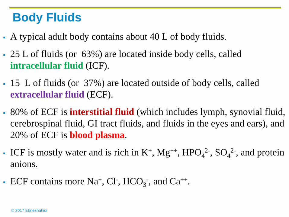
Body Fluids
A typical adult body contains about 40 L of body fluids.
25 L of fluids (or 63%) are located inside body cells, called
intracellular fluid (ICF).
15 L of fluids (or 37%) are located outside of body cells, called
extracellular fluid (ECF).
80% of ECF is interstitial fluid (which includes lymph, synovial fluid,
cerebrospinal fluid, GI tract fluids, and fluids in the eyes and ears), and
20% of ECF is blood plasma.
ICF is mostly water and is rich in K+, Mg++, HPO42-, SO4
2-, and protein
anions.
ECF contains more Na+, Cl-, HCO3-, and Ca++.

© 2017 Ebneshahidi

© 2017 Ebneshahidi

© 2017 Ebneshahidi
Concentrations of substances dissolved in ICF and ECF
are constantly different because the cell membrane is
selectively permeable, which maintains a relatively
unchanged distribution of substances in different body
fluids.
Fluid balance refers to the proper levels of water and
electrolytes being in the various body compartments
according to their needs.
Osmotic pressure (created by the dissolved electrolytes
in body fluids) and hydrostatic pressure (created by the
water in body fluids) are the main forces behind any
molecular movement between body compartments.
© 2017 Ebneshahidi
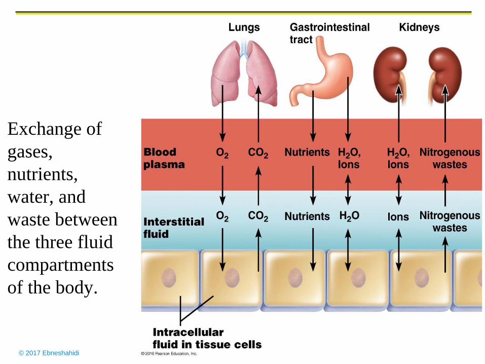
Exchange of
gases,
nutrients,
water, and
waste between
the three fluid
compartments
of the body.
© 2017 Ebneshahidi
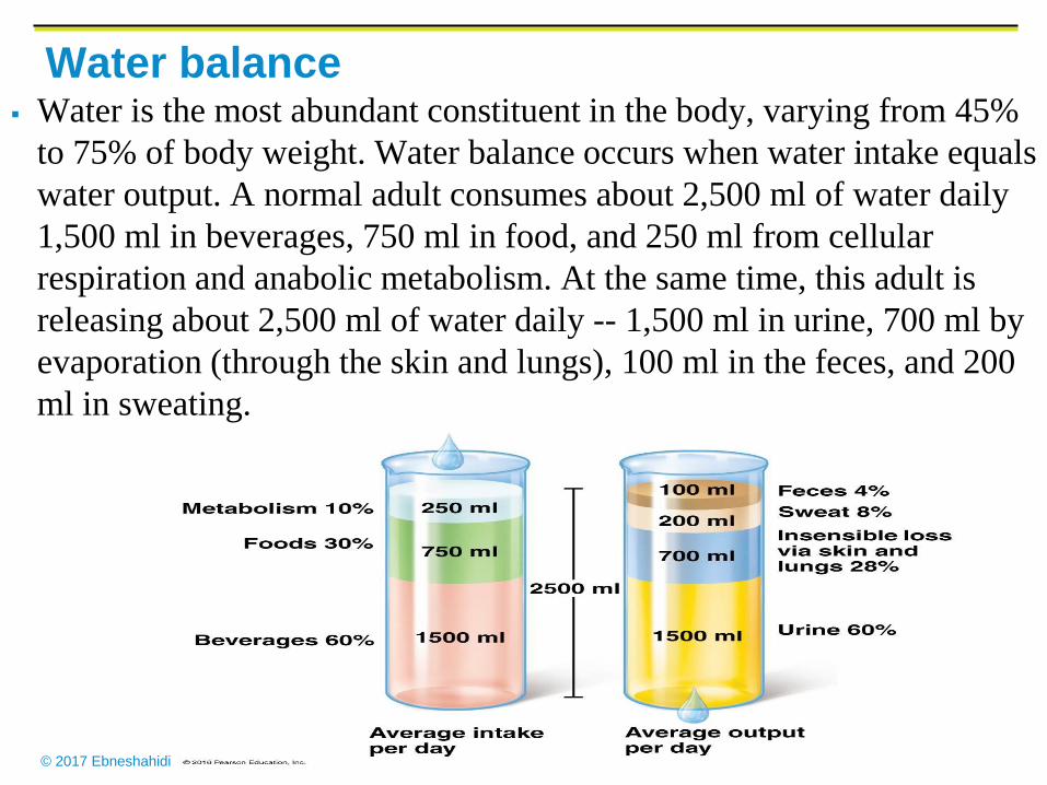
Water balance
Water is the most abundant constituent in the body, varying from 45%
to 75% of body weight. Water balance occurs when water intake equals
water output. A normal adult consumes about 2,500 ml of water daily
1,500 ml in beverages, 750 ml in food, and 250 ml from cellular
respiration and anabolic metabolism. At the same time, this adult is
releasing about 2,500 ml of water daily -- 1,500 ml in urine, 700 ml by
evaporation (through the skin and lungs), 100 ml in the feces, and 200
ml in sweating.
© 2017 Ebneshahidi
Regulation of Water intake
1. The body loses as little as 1% of its water.
2. An increase in osmotic pressure of extracellular fluid due to
water loss stimulates osmoreceptors in the thirst center
(hypothalamus).
3. Activity in the hypothalamus causes the person to be thirsty
and to seek H2O.
Drinking and the resulting distension of the stomach by water
stimulants nerve impulses that inhibit the thirst center.
water is absorbed through the wall of the stomach, small
intestine, and large intestine.
The osmotic pressure of extracellular fluid returns to normal.
© 2017 Ebneshahidi
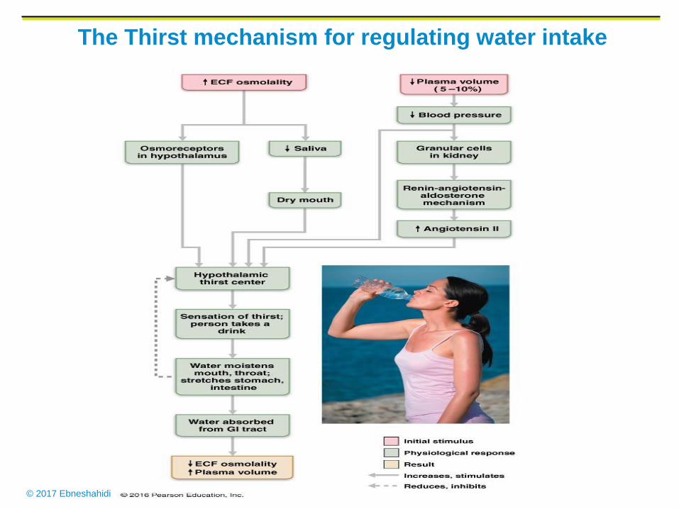
The Thirst mechanism for regulating water intake
© 2017 Ebneshahidi
Events in regulation of water output
I. Dehydration:
1. Extracellular fluid becomes osmotically more concentrated.
2. Osmoreceptors in the hypothalamus are stimulated by the increase in
the osmotic pressure of body fluids.
3.The Hypothalamus signals the posterior pituitary gland to release
ADH into the blood.
4. Blood carries ADH to the kidneys.
5. ADH causes the distal convoluted tubules & collecting ducts to
increase water reabsorption.
6. urine output decreases, and further water loss is minimized.

© 2017 Ebneshahidi
Dehydration

© 2017 Ebneshahidi
ADH
© 2017 Ebneshahidi
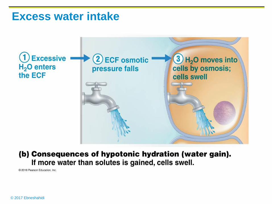
Excess water intake
1. Extracellular fluid becomes osmotically less concentrated.
2. This change stimulates osmoreceptors in the
hypothalamus.
3.The posterior Pituitary gland decrease ADH release.
4. Renal tubules decrease water reabsorption.
5. Urine output, increases and excess water is excreted.
© 2017 Ebneshahidi
Excess water intake
© 2017 Ebneshahidi
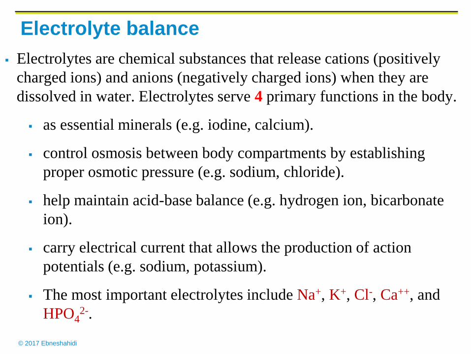
Electrolyte balance
Electrolytes are chemical substances that release cations (positively
charged ions) and anions (negatively charged ions) when they are
dissolved in water. Electrolytes serve 4 primary functions in the body.
as essential minerals (e.g. iodine, calcium).
control osmosis between body compartments by establishing
proper osmotic pressure (e.g. sodium, chloride).
help maintain acid-base balance (e.g. hydrogen ion, bicarbonate
ion).
carry electrical current that allows the production of action
potentials (e.g. sodium, potassium).
The most important electrolytes include Na+, K+, Cl-, Ca++, and
HPO42-.
© 2017 Ebneshahidi
Na+ is the most abundant extracellular cation;
involved in nerve impulse transmission, muscle
contraction, and creation of osmotic pressure.
Cl- is a major extracellular anion; involved in
regulating osmotic pressure between body
compartments, forming HCI in stomach, and
involved in the “chloride shift” process in blood.
K+ is the most abundant cation in ICF; involved in
maintaining fluid volume, nerve impulse
transmission, muscle contraction, and regulating
pH.
© 2017 Ebneshahidi
Ca++ is the most abundant ion in the body, located mainly in
ECF; a major structural component of bones and teeth;
functions in blood clotting, neurotransmitter release, muscle
tone, and excitability of nervous and muscle tissues.
HPO42- is an important intracellular anion; another major structural
component of bones and teeth; required for synthesis of nucleic
acids and ATP, and for buffering reactions.
Level of electrolytes are mainly regulated by hormones:
Aldosterone (from adrenal cortex) causes an increase in sodium
reabsorption and potassium secretion at the kidney tubules.
Parathyroid hormone (PTH) from the parathyroid glands and
Calcitonin (CT) from the thyroid gland regulate calcium
balance.
© 2017 Ebneshahidi
Regulation of electrolyte Intake & output
Electrolyte intake:
Electrolytes are usually obtained in sufficient
quantities in response to hunger and thirst mechanism.
In a severe electrolyte deficiency, a person may
experience a salt craving.
Electrolyte output:
Electrolytes are lost through perspiration, feces and
urine. The greatest electrolyte loss occurs as a result of
kidney functions.
Quantities lost vary with temp. and exercise.
© 2017 Ebneshahidi
Electrolyte Balance
1. Concentrations of Na, K and calcium ions in the body fluid
are very important.
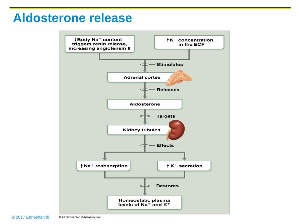
2.The regulation of Na+ and K+ ions involve the secretion of
Aldosterone from adrenal glands.
1. K+ ion concentration increases.
2. Adrenal cortex is signaled.
3. Aldosterone is secreted.
4. Renal tubules increase reabsorption of Na+ ion and
increase secretion of K+ ions (excretes K ions).
5. Na+ ions are conserved and K+ ions are excreted.
© 2017 Ebneshahidi
Aldosterone release
© 2017 Ebneshahidi
3. Calcitonin from the thyroid gland and
parathyroid hormone from the parathyroid glands
regulate Ca+ ion concentration.
- Parathyroid hormone increases activity in bone-
resorbing cells (osteocytes & osteoclasts) which
increase the conc. of both Ca+ and phosphate ions
in extracellular fluids. This hormone also causes
increase absorption of Ca+ and increase excretion
of phosphate, from the kidney.

© 2017 Ebneshahidi
Acid-base balance
Acids are electrolytes that
release hydrogen ions (H+)
when they are dissolved in
water.
Bases are electrolytes are
release hydroxide ions (OH-)
when they are dissolved in
water.
Acid-base balance is
primarily regulated by the
concentration of H+ (or the
pH level) in body fluids,
especially ECF.
© 2017 Ebneshahidi
Acid-base balance
Normal pH range of ECF is from 7.35 to 7.45.
Most H+ comes from metabolism -- glycolysis, oxidation of fatty
acids and amino acids, and hydrolysis of proteins.
Homeostasis of pH in body fluids is regulated by acid-base buffer
systems (primary control), respiratory centers in brain stem, and by
kidney tubule secretion of H+.
Acid-base buffer systems are chemical reactions that consist of a
weak acid and a weak base, to prevent rapid, drastic changes in
body fluid pH. one of the most carefully regulated conc. in the
body is that of H+ ion.
© 2017 Ebneshahidi
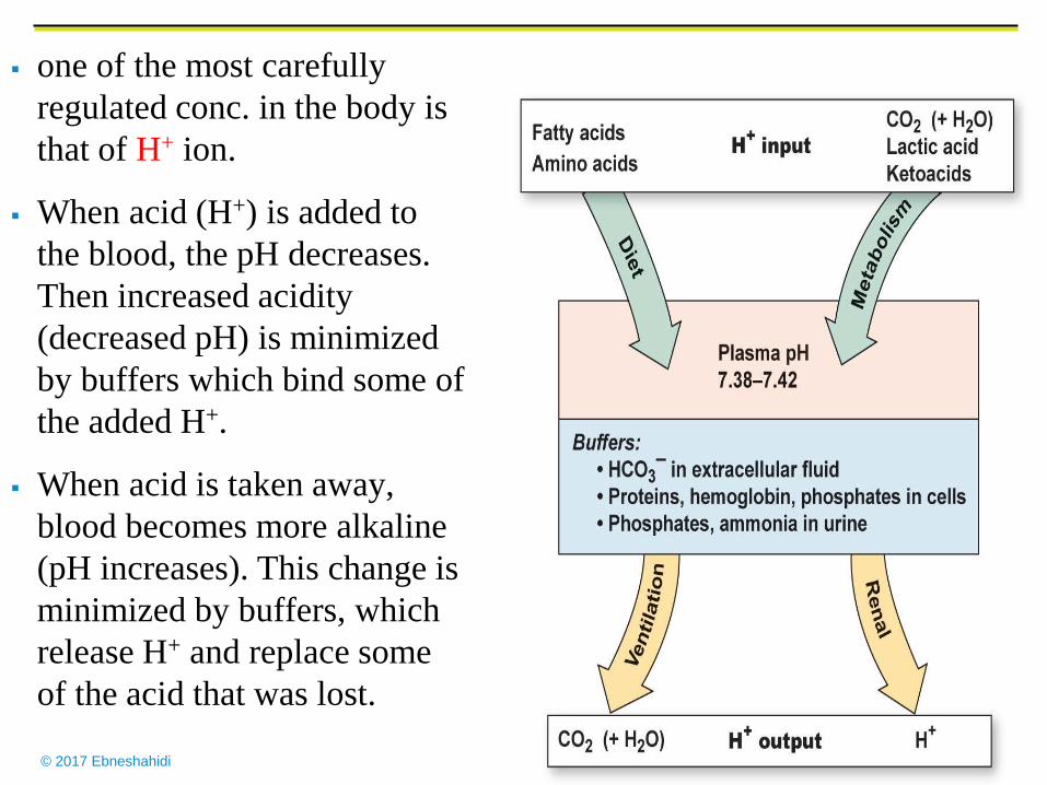
one of the most carefully
regulated conc. in the body is
that of H+ ion.
When acid (H+) is added to
the blood, the pH decreases.
Then increased acidity
(decreased pH) is minimized
by buffers which bind some of
the added H+.
When acid is taken away,
blood becomes more alkaline
(pH increases). This change is
minimized by buffers, which
release H+ and replace some
of the acid that was lost.
© 2017 Ebneshahidi
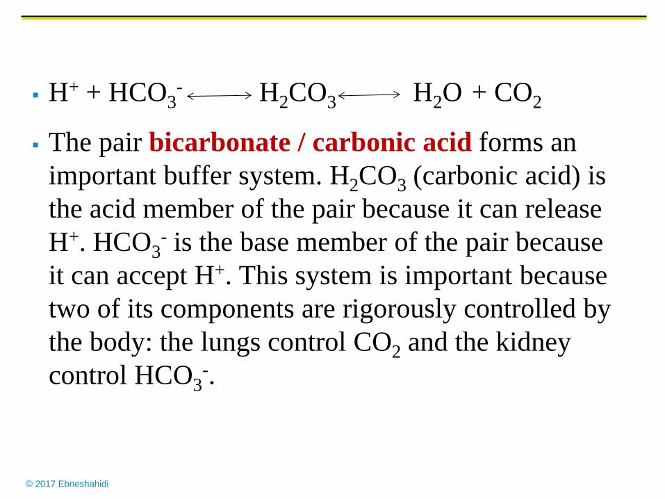
H+ + HCO3- H2CO3 H2O + CO2
The pair bicarbonate / carbonic acid forms an
important buffer system. H2CO3 (carbonic acid) is
the acid member of the pair because it can release
H+. HCO3- is the base member of the pair because
it can accept H+. This system is important because
two of its components are rigorously controlled by
the body: the lungs control CO2 and the kidney
control HCO3-.
© 2017 Ebneshahidi
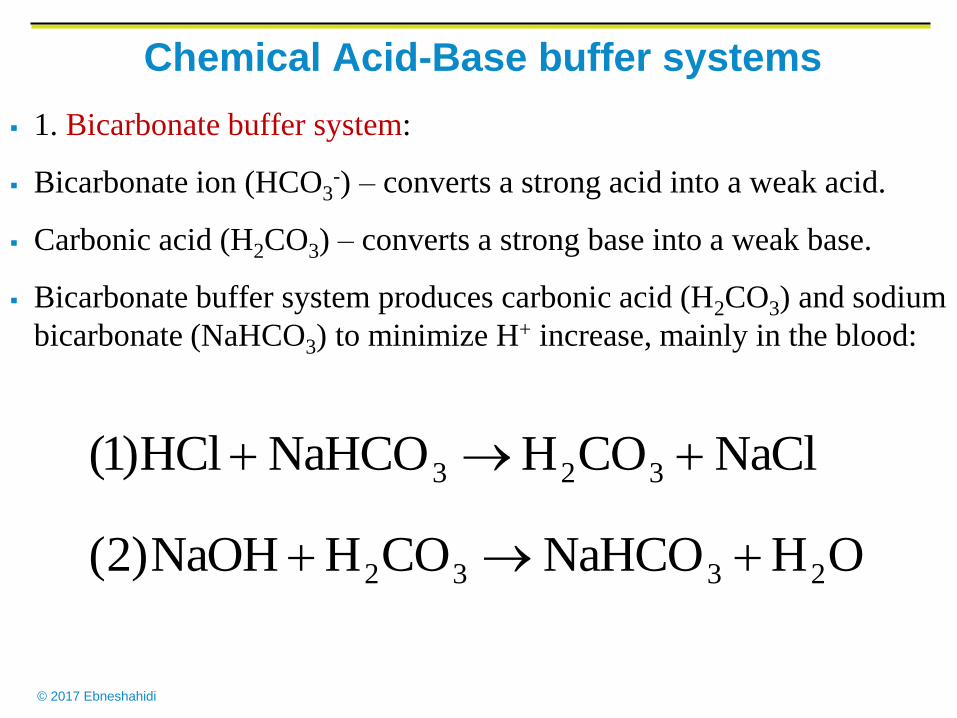
Chemical Acid-Base buffer systems
1. Bicarbonate buffer system:
Bicarbonate ion (HCO3-) – converts a strong acid into a weak acid.
Carbonic acid (H2CO3) – converts a strong base into a weak base.
Bicarbonate buffer system produces carbonic acid (H2CO3) and sodium
bicarbonate (NaHCO3) to minimize H+ increase, mainly in the blood:
OHNaHCOCOHNaOH)2(
NaClCOHNaHCOHCl)1(
2332
323
© 2017 Ebneshahidi
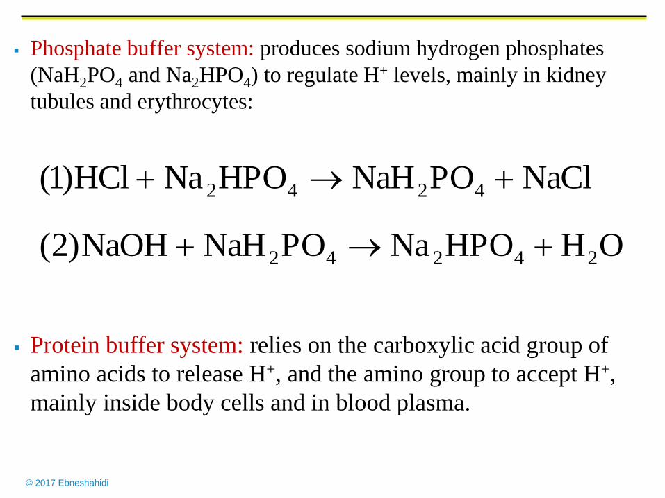
Phosphate buffer system: produces sodium hydrogen phosphates
(NaH2PO4 and Na2HPO4) to regulate H+ levels, mainly in kidney
tubules and erythrocytes:
Protein buffer system: relies on the carboxylic acid group of
amino acids to release H+, and the amino group to accept H+,
mainly inside body cells and in blood plasma.
OHHPONaPONaHNaOH)2(
NaClPONaHHPONaHCl)1(
24242
4242
© 2017 Ebneshahidi
Respiratory centers in the pons and medulla oblongata regulate the rate
and depth of breathing, which controls the amount of carbon dioxide
gas (CO2) remained in the blood and body fluid -- e.g. slower berating
rate an increase in blood CO2 level an increase in carbonic
acid (H2CO3) in blood more H+ is released into body fluids
pH of blood and body fluids drops.
Nephrons react to the pH of body fluids and regulate the secretion of
H+ into urine -- e.g. a diet high in proteins causes more H+ to be
produced in body fluids (which lowers body fluid pH), as a result the
nephrons will secrete more H+ into the urine.
© 2017 Ebneshahidi
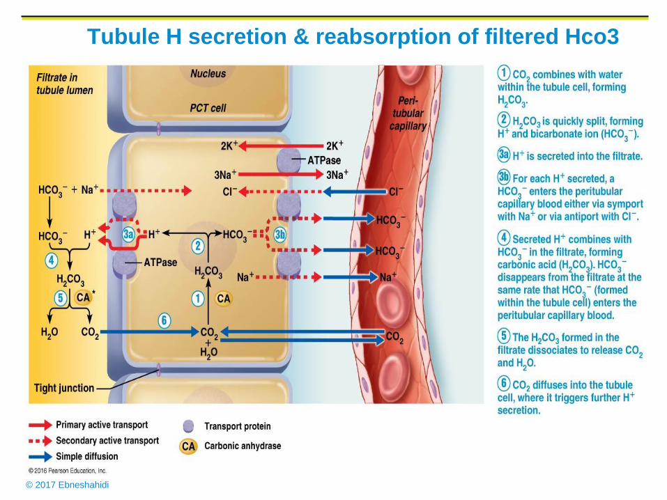
Tubule H secretion & reabsorption of filtered Hco3
© 2017 Ebneshahidi
Compensation Compensation is a series of physiological responses that react to acid-
base imbalances, by returning blood pH to the normal range (7.35 –
7.45).
Respiratory acidosis: (due to deficiency of CO2 expiration) and
respiratory alkalosis (due to abnormally high CO2 expiration) are
primary disorders of CO2 pressure in the lungs. These may be
compensated by renal mechanisms where nephrons will secrete more
H+ to correct acidosis and secrete less H+ to correct alkalosis.
It is due to increased CO2 retention (due to hypoventilation), which
can result in the accumulation of carbonic acid and thus a fall in
blood pH to below normal.
Metabolic Acidosis: increased production of acids such as lactic
acid, fatty acids, and ketone bodies, or loss of blood bicarbonate
(such as by diarrhea), resulting in a fall in blood pH to below normal.
© 2017 Ebneshahidi
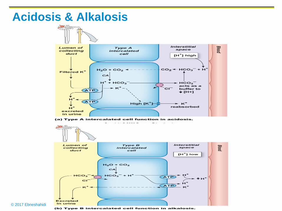
Acidosis & Alkalosis
© 2017 Ebneshahidi
Respiratory Alkalosis:
A rise in blood pH due loss of CO2 and carbonic
acid (through hyperventilation).
Metabolic Alkalosis:
A rise in blood pH produced by loss of acids (such
as excessive vomiting) or by excessive
accumulation of bicarbonate base.
© 2017 Ebneshahidi
Respiratory Excretion of CO2
The respiratory center is located in the brain stem.
It helps control pH by regulating the rate and depth
of breathing.
Increasing CO2 and H+ ions conc. stimulate chemo
receptors associated with the respiratory center;
breathing rate and depth increase, and CO2 conc.
decreases.
If the CO2 and H+ ion concentrations are low, the
respiratory center inhibits breathing.
© 2017 Ebneshahidi
Renal excretion of H+
Nephrons secrete hydrogen ions to regulate pH.
phosphate buffer hydrogen ions in urine.
Ammonia produced by renal cells help transport H+ to the outside of
the body:
chemical buffer system (Bicarbonate buffer system, phosphate buffer,
and protein buffer system) act rapidly and are the first line of defense
against pH shift.
physiological buffer (respiratory mechanism CO2 excretion), renal
mechanism (H+ excretion) act slowly and are the 2nd line of defense
against pH shift.
43 NHNHH
© 2017 Ebneshahidi
Source of H+
a. Aerobic respiration of glucose
produces CO2 , which reacts with water to from carbonic acid.
carbonic acid dissociates to release H+ and bicarbonate ions.
b. Anaerobic respiration of glucose produce lactic acid.
c. Incomplete oxidation of fatty acids releases acidic ketone
bodies.
d. Oxidation of sulfur-containing amino acids produce sulfuric
acid.
e. Hydrolysis of phosphoproteins and nucleic acids gives rise to
phosphoric acid.

© 2017 Ebneshahidi
Factors Associated with Edema
1. Low plasma protein concentration: cause is liver disease, kidney
disease, loss of protein in urine, lack of protein in diet due to
starvation.
Effect: plasma osmotic pressure decreases, less fluid enters venular end
of capillaries by osmosis.
2. Obstruction of lymph vessels: causes are surgical removal of
portions of lymphatic pathways and parasitic infections.
Effect: back pressure in lymph vessels, interferes with movement of
fluid from interstitial spaces into lymph capillaries.
3. Increased venous pressure: venous obstruction or faulty valves.
Effect: back pressure in veins increases capillary filtration and
interferes with return of fluid from interstitial spaces into venular end
of capillaries.
© 2017 Ebneshahidi
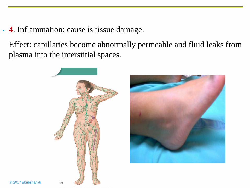
4. Inflammation: cause is tissue damage.
Effect: capillaries become abnormally permeable and fluid leaks from
plasma into the interstitial spaces.