florence f. roussotte, ph.d. hhs public access xue hua, ph...
TRANSCRIPT
The C677T variant in MTHFR modulates associations between brain integrity, mood, and cognitive functioning in old age
Florence F. Roussotte, Ph.D.1,3, Xue Hua, Ph.D.3,4, Katherine L. Narr, Ph.D.1, Gary W. Small, M.D.2, Paul M. Thompson, Ph. D.1,2,3,4,5, and the Alzheimer's Disease Neuroimaging Initiative1 Department of Neurology, Semel Institute, David Geffen School of Medicine, University of California, Los Angeles, Los Angeles, California 90095, USA
2 Department of Psychiatry, Semel Institute, David Geffen School of Medicine, University of California, Los Angeles, Los Angeles, California 90095, USA
3 Imaging Genetics Center, Keck School of Medicine, University of Southern California, Los Angeles, California 90033, USA
4 Department of Neurology, Keck School of Medicine, University of Southern California, Los Angeles, California 90033, USA
5 Departments of Psychiatry, Radiology, Engineering, Pediatrics, and Ophthalmology, Keck School of Medicine, University of Southern California, Los Angeles, California 90033, USA.
Abstract
Introduction—The C677T functional variant in the methylene-tetrahydrofolate reductase
(MTHFR) gene leads to reduced enzymatic activity and elevated blood levels of homocysteine.
Hyperhomocysteinemia has been linked with higher rates of cardiovascular diseases, cognitive
decline, and late-life depression.
Methods and Materials—Here, 3D magnetic resonance imaging data was analyzed from 738
individuals (age: 75.5 ± 6.8 years; 438 men/300 women) including 173 Alzheimer's patients, 359
subjects with mild cognitive impairment, and 206 healthy older adults, scanned as part of the
Alzheimer's Disease Neuroimaging Initiative (ADNI).
Results—We found that this variant associates with localized brain atrophy, after controlling for
age, sex, and dementia status, in brain regions implicated in both intellectual and emotional
functioning, notably the medial orbitofrontal cortices. The medial orbitofrontal cortex is involved
in the cognitive modulation of emotional processes, and localized atrophy in this region was
previously linked with both cognitive impairment and depressive symptoms. Here, we report that
Corresponding Author: Florence F. Roussotte, Department of Neurology, 635 Charles E. Young Drive South, Neuroscience Research Building (NRB) Suite 225, Los Angeles, CA 90095, USA, [email protected].
Publisher's Disclaimer: This is a PDF file of an unedited manuscript that has been accepted for publication. As a service to our customers we are providing this early version of the manuscript. The manuscript will undergo copyediting, typesetting, and review of the resulting proof before it is published in its final citable form. Please note that during the production process errors may be discovered which could affect the content, and all legal disclaimers that apply to the journal pertain.
FINANCIAL DISCLOSURESAll authors report no biomedical financial interests or potential conflicts of interest.
HHS Public AccessAuthor manuscriptBiol Psychiatry Cogn Neurosci Neuroimaging. Author manuscript; available in PMC 2018 April 01.
Published in final edited form as:Biol Psychiatry Cogn Neurosci Neuroimaging. 2017 April ; 2(3): 280–288. doi:10.1016/j.bpsc.2016.09.005.
Author M
anuscriptA
uthor Manuscript
Author M
anuscriptA
uthor Manuscript

increased plasma homocysteine mediates the association between MTHFR genotype and lower
medial orbitofrontal volumes, and that these volumes mediate the association between cognitive
decline and depressed mood in this elderly cohort. We additionally show that vitamin B12
deficiency interacts with the C677T variant in the etiology of hyperhomocysteinemia.
Conclusion—This study sheds light on important relationships between vascular risk factors,
age-related cognitive decline, and late-life depression, and represents a significant advance in our
understanding of clinically relevant associations relating to MTHFR genotype.
Keywords
MTHFR; homocysteine; brain atrophy; age-related cognitive decline; late-life depression; MRI
INTRODUCTION
Hyperhomocysteinemia, a metabolic anomaly involving elevated levels of the amino acid
homocysteine in the blood, is associated with higher rates of numerous age-related
disorders, such as cardiovascular diseases (CVDs) (1, 2) including vascular dementia (3, 4);
cognitive decline (5-9); and depressed mood (10-12). Elevated plasma homocysteine levels
may stem from the use of certain therapeutic drugs, elevated alcohol ingestion, intestinal
malabsorption, or from impaired metabolism due to genetic alterations in metabolic
enzymes, including methyltetrahydrofolate reductase (MTHFR), most commonly when
combined with insufficient dietary intake of B vitamins. Notably, homocysteine is recycled
to methionine using vitamin B12 as a cofactor, and MTHFR – the rate-limiting enzyme in the
methyl cycle – catalyzes the conversion of 5,10-methylenetetrahydrofolate to 5-
methyltetrahydrofolate, a cosubstrate for homocysteine remethylation by methionine
synthase. These relationships are illustrated in Figure 1.
We recently reported that older adults with higher homocysteine levels had more pronounced
regional brain atrophy (13) and thinner cortical gray matter (14) on MRI. We also found that
the C677T variant in MTHFR was associated with smaller regional brain volumes in two
independent elderly cohorts with mild cognitive impairment (MCI) (15). Increased
susceptibility for CVDs (16), which are strongly linked to both cognitive decline and
depressive symptoms in old age (17), are also associated with the C677T variant.
Here, we first determined if our previously reported associations between the T “risk” allele
and more pronounced brain atrophy extended beyond individuals with MCI to both dementia
patients and healthy older adults. We further sought to model some of the mechanisms
underlying relationships between brain integrity, clinical outcomes, and the genetic and
environmental modulators of homocysteine metabolism. To this end, we first examined
whether the effects of this MTHFR polymorphism on medial orbitofrontal volumes were
mediated by its impact on plasma homocysteine. We also determined whether vitamin B12
deficiency influenced the strength of the relationship between this variant and homocysteine
levels. We then found that lower cognitive performance and reduced medial orbitofrontal
volumes were significant predictors of depressed mood and tested the hypothesis that
compromised integrity in this brain region involved in the cognitive control of emotion may
partially explain the relationship between cognitive impairment and depressive symptoms.
Roussotte et al. Page 2
Biol Psychiatry Cogn Neurosci Neuroimaging. Author manuscript; available in PMC 2018 April 01.
Author M
anuscriptA
uthor Manuscript
Author M
anuscriptA
uthor Manuscript

METHODS AND MATERIALS
Subjects
We analyzed a large sample of elderly individuals from the Alzheimer's Disease
Neuroimaging Initiative (ADNI). The study was conducted according to the Good Clinical
Practice guidelines, the Declaration of Helsinki, and U.S. 21 CFR Part 50 (Protection of
Human Subjects), and Part 56 (Institutional Review Boards). Written informed consent was
obtained from all participants before protocol-specific procedures were performed. All
ADNI data are publicly available (at http://adni.loni.usc.edu). To avoid the known effects of
population stratification on genetic analysis (18), we only included non-Hispanic Caucasian
subjects identified by self-report and confirmed by multi-dimensional scaling (MDS)
analysis (19). The ADNI cohort included multiple diagnostic groups: patients with
Alzheimer's disease (“AD”), subjects with MCI, and healthy elderly (cognitively normal,
“CON”) participants. All subjects were administered the Mini Mental Status Examination
(MMSE (20)) and the 15-item version of the Geriatric Depression Scale (GDS-15 (21)). Our
final analysis comprised 738 individuals (average age ± s.d. = 75.53 ± 6.78 years; 438
men/300 women) including 173 AD, 359 MCI, and 206 healthy older adults. All participants
received pre-mortem clinical diagnoses, as described in detail in ADNI's General Procedures
Manual: http://adni.loni.usc.edu/wp-content/uploads/2010/09/
ADNI_GeneralProceduresManual.pdf). Table 1 illustrates demographic, cognitive, and
mood data for all participants, stratified by genotype and sub-stratified by diagnostic groups.
Genotyping and Allele Frequency
The ADNI sample was genotyped using the Illumina 610-Quad BeadChip (San Diego, CA,
USA). The only polymorphism examined in this study was the C677T functional variant in
the methylene-tetrahydrofolate reductase (MTHFR) gene, at the rs1801133 locus. Allele
frequency was computed from genotype frequency. The distributions of allele frequencies by
diagnostic groups were evaluated by χ2 tests with a 0.05 significance level, using 3 × 2 and
2 × 2 contingency tables in SPSS 23.0.
Neuroimaging
Whole-Brain Analyses: Tensor Based Morphometry (TBM)—The C677T
polymorphism was analyzed for association with regional brain volumes in ADNI
participants, as detailed below. Subjects were scanned with a standardized MRI protocol
developed for this cohort (22, 23). Briefly, high-resolution structural brain MRI scans were
acquired at 58 sites across North America, using 1.5 Tesla MRI scanners. A sagittal 3D MP-
RAGE sequence was used, optimized for consistency across sites (23) (TR/TE = 2400/1000
ms; flip angle = 8°; FOV = 24 cm; final reconstructed voxel resolution = 0.9375 × 0.9375 ×
1.2 mm3). Image corrections were applied using a processing pipeline at the Mayo Clinic,
consisting of: 1) a procedure termed GradWarp to correct geometric distortion due to
gradient non-linearity (24), 2) a “B1-correction”, to adjust for image intensity
inhomogeneity due to B1 non-uniformity using calibration scans (23), 3) “N3” bias field
correction, for reducing residual intensity inhomogeneity (25), and 4) geometrical scaling,
according to a phantom scan acquired for each subject (23), to adjust for scanner- and
session-specific calibration errors. To adjust for global differences in brain positioning and
Roussotte et al. Page 3
Biol Psychiatry Cogn Neurosci Neuroimaging. Author manuscript; available in PMC 2018 April 01.
Author M
anuscriptA
uthor Manuscript
Author M
anuscriptA
uthor Manuscript
scale, all subjects’ scans were linearly registered to the stereotaxic space defined by the
International Consortium for Brain Mapping (ICBM-53) (26), using a 9-parameter (9P)
transformation (three translations, three rotations, three scales) (27). We used standard
trilinear interpolation and resampled the resulting aligned scans to have 1mm isotropic
voxels.
We then created a minimal deformation target (MDT), which serves as an unbiased average
template image for automated image registration, and to reduce statistical bias. The MDT
was created from the MRI scans of 40 randomly selected healthy elderly subjects, as
detailed elsewhere (28, 29). To quantify 3D patterns of volumetric tissue variations, all
individual T1-weighted images were non-linearly aligned to the template with an inverse-
consistent 3D elastic warping technique using a mutual information cost function (30).
Consequently, for each subject, a separate Jacobian matrix field was derived from the
gradients of the deformation field that aligned that individual brain to the MDT template.
The determinant of the local Jacobian matrix was derived from the forward deformation
field to characterize local volume differences. Color-coded Jacobian determinants were used
to illustrate regions of volume expansion, i.e. those with det J(r) > 1, or contraction, i.e., det
J(r) < 1 (31-34) relative to the group template. As all images were registered to the same
study-specific template, these Jacobian maps shared common anatomical coordinates,
defined by the normal template. Individual Jacobian maps were retained for further
statistical analyses.
To model effects of the C677T functional variant in MTHFR on local brain volumes, we
used univariate linear regression to associate the number of minor T alleles (0,1, or 2) with
the Jacobian values (describing the amount of brain tissue deficit or excess relative to the
standard template) at each voxel in the brain, after controlling for age, sex, and diagnosis. To
minimize Type I errors, we used a searchlight method for false discovery rate (FDR)
correction (35), which controls the false discovery rate in all reported statistical maps. We
implemented this method to correct the maps of statistical associations between the image
phenotype (morphometry) and genotype at the rs1801133 locus (number of T alleles). All
maps shown were thresholded at the appropriate corrected p-value, after performing
searchlight FDR (q=0.05), to show only regions of significance that passed the multiple
comparisons correction.
Region of Interest (ROI) Analyses: Medial Orbitofrontal Volumes—Five MRI core
analysis laboratories have provided feature extraction and numeric summaries from the high
quality ADNI MRI data, which are publicly available in the ADNI data archive (http://
adni.loni.usc.edu). One of these core analysis laboratories, the Center for Imaging of
Neurodegenerative Diseases at UCSF (co-investigator: Norbert Schuff), has provided
volumetric segmentation using the FreeSurfer image analyses suite, which is documented
and freely available for download online (http://surfer.nmr.mgh.harvard.edu/). Version 4.3 is
used for ADNI's cross-sectional data. The input for ADNI FreeSurfer is a T1 weighted
image in NiFTI format, which has been preprocessed (gradient warping, scaling, B1
correction, and N3 inhomogeneity correction) by the Mayo Clinic preprocessing stream as
describe above.
Roussotte et al. Page 4
Biol Psychiatry Cogn Neurosci Neuroimaging. Author manuscript; available in PMC 2018 April 01.
Author M
anuscriptA
uthor Manuscript
Author M
anuscriptA
uthor Manuscript
Briefly, Freesurfer processing includes motion correction, removal of non-brain tissue using
a hybrid watershed/surface deformation procedure (36), automated Talairach transformation,
segmentation of the subcortical white matter and deep gray matter (37, 38), intensity
normalization (25), tessellation of the gray matter/white matter boundary, automated
topology correction (39, 40), and surface deformation following intensity gradients to
optimally place the gray/white and gray/cerebrospinal fluid borders at the location where the
greatest shift in intensity defines the transition to the other tissue class (41-43), followed by
rigorous QC procedures that allow for the exclusion of failed segmentations, due to poor
image quality, registration issues, or processing errors. We downloaded the numeric
summaries for “medial orbital frontal” volumes, and retained the data pertaining to subjects
with frontal segmentations that satisfied all QC requirements. We obtained the volumes of
the medial orbitofrontal cortices (in mm3) for 640 of our participants.
Blood-based markers
In the ADNI public database, plasma levels of homocysteine were available for 732 of our
participants, 634 of whom also had usable medial orbitofrontal volumes. The database also
contained plasma vitamin B12 concentrations for 680 of our subjects (including 675 with
available homocysteine levels and 587 with both homocysteine and medial orbitofrontal
volume data). Homocysteine and vitamin B12 levels (in pg/mL) were extracted from blood
samples collected via standard venipuncture protocols. Vitamin B12 deficiency was defined
as plasma levels < 250 pg/mL.
Statistical analyses
To ensure consistency, we used an additive model of minor T allele effects – the number of
T allele carried by each participant was coded as 0, 1, or 2 – in all analyses aimed at testing
association between the C677T variant and another variable. However, since medial
orbitofrontal volumes seemed to be affected in a recessive manner, we additionally ran every
regression model using a recessive model of minor allele effects (i.e., comparing C allele
carriers with T homozygotes). These analyses produced similar results, which are presented
in the Supplemental Information section (Tables S2-S4).
We used general linear models to examine the predictors of medial orbitofrontal volumes,
plasma homocysteine concentrations, and GDS-15 scores. Shapiro-Wilk tests showed that
medial orbitofrontal volumes were normally distributed (p=0.601), but homocysteine levels
(p<0.001) and GDS-15 scores (p<0.001) were not. We therefore used standardized scores in
all regression models including plasma homocysteine or mood scores as the dependent
variable. Age and sex were included as covariates in all analyses. We also controlled for
diagnosis, except when MMSE was used as a factor in the model, since MMSE scores are
one of the major diagnostic criteria for dementia (as described in ADNI's General
Procedures Manual: http://adni.loni.usc.edu/wp-content/uploads/2010/09/
ADNI_GeneralProceduresManual.pdf). These statistical analyses were conducted in SPSS
23.0.
Simple mediation analyses were conducted using Andrew Hayes's PROCESS Procedure
(v2.15) for SPSS (http://www.processmacro.org/download.html). We obtained path
Roussotte et al. Page 5
Biol Psychiatry Cogn Neurosci Neuroimaging. Author manuscript; available in PMC 2018 April 01.
Author M
anuscriptA
uthor Manuscript
Author M
anuscriptA
uthor Manuscript

coefficients (a, b, c, and c’) representing the linear regression coefficients for each path in
the mediation model. We standardized all variables to facilitate the interpretation of path
coefficients, now bounded by −1 and 1 across all measures. The a-path represents the
association between the predictor and mediator variables. The b-path denotes the
relationship between the mediator and outcome variables, while also controlling for the
predictor variable. The c’-path (also called “direct effect”) and the c-path (also known as
“total effect”) represent the associations between the predictor and outcome variables
including and excluding the mediator variable, respectively. If the difference between c and
c’ is statistically significant, then there is a significant mediation effect. It was previously
shown that a*b = c-c’; therefore, we tested the significance of a*b (also known as “indirect
effect”) using bootstrapped confidence intervals (44). Bootstrapping creates thousands of
simulated datasets using resampling with replacement and runs the analysis once in each
simulated sample (45). 95% of the generated statistics fall between two values, and if that
confidence interval (CI) for a*b does not include 0, then a significant (p<0.05) mediation has
occurred. Percent mediation [PM] is a measure of effect size interpreted as the percent of the
total effect (c) accounted for by the indirect effect (a*b); that is, PM = (a*b)/c (44).
RESULTS
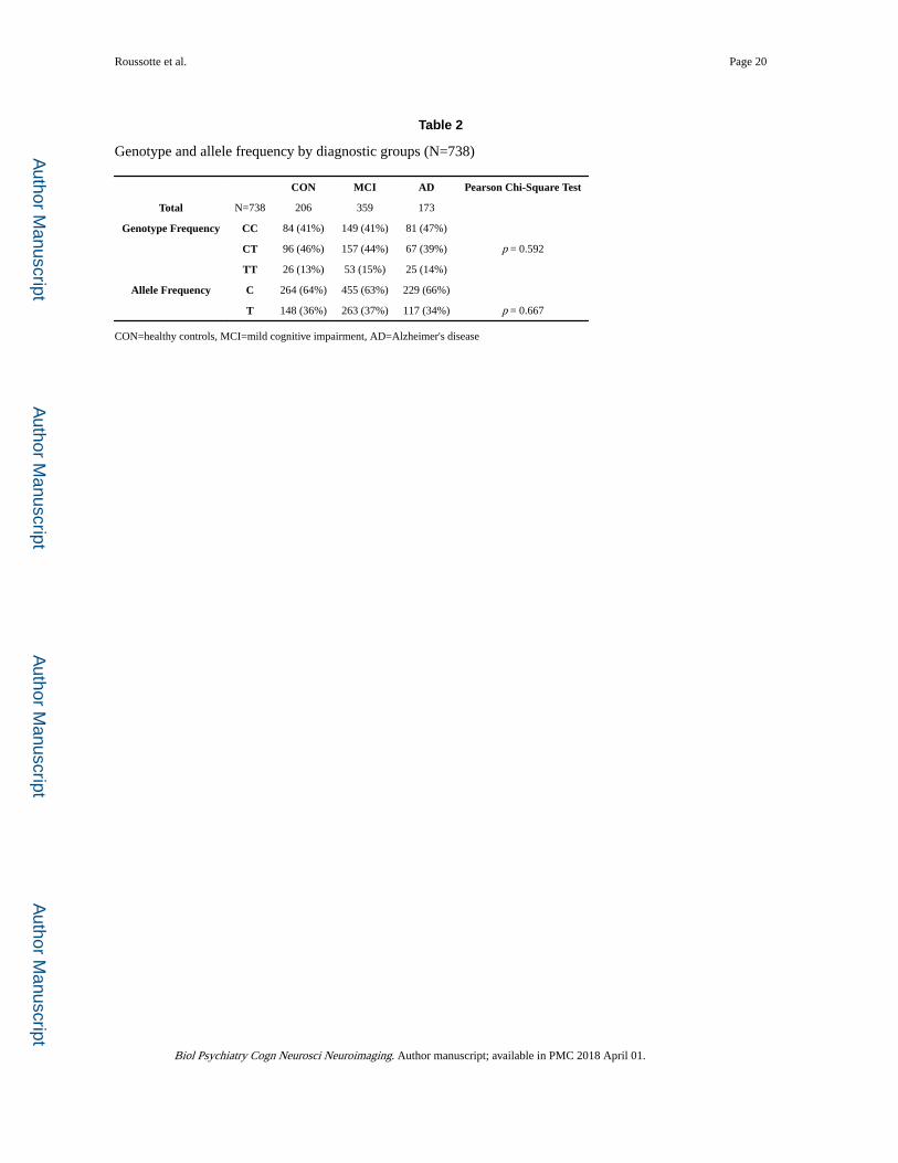
Consistent with the prevailing view that the MTHFR C677T polymorphism may not be a
risk factor for Alzheimer's disease (AD), but appears to be associated with various types of
age-related disorders (46), the distributions of genotype (p=0.592) and allele frequency
(p=0.667) for rs1801133 did not significantly differ across the 3 diagnostic groups in the
ADNI cohort (Table 2). Nonetheless, this functional variant in MTHFR predicted
differences in regional brain volumes in our large elderly sample, after controlling for age,
sex, and diagnosis. As depicted in Figure 2, smaller volumes in the frontal (including the
bilateral cingulate gyri, middle frontal gyri, as well as lateral and medial orbitofrontal
cortices), parietal (notably the inferior parietal lobule), and temporal lobes (especially the
superior temporal gyrus), as well as in the thalamus, were statistically related to carrying the
minor T allele at the rs1801133 locus. Strong genotype group differences were also observed
in periventricular regions. Each copy of the “risk” allele was associated with a 2-4 %
reduction in local brain volumes(Figure 2).
Among the regions showing a significant association with MTHFR genotype in our whole-
brain analyses, the medial orbitofrontal cortex was particularly noteworthy because of its
involvement, not just in mood states and intellectual functioning, but specifically in the
cognitive modulation of emotional processes. This brain area was therefore selected as the
region of interest in our investigations of the relationships between brain integrity, clinical
outcomes, and homocysteine metabolism. Our ROI analyses, which used Freesurfer volumes
in a subset of the same elderly sample (Table S1), confirmed that the C677T variant was
associated with reduced medial orbitofrontal volumes (p=0.033, F-ratio=3.428) after
controlling for age, sex, and diagnosis (Table S2). As expected, the C677T variant was also
associated with significant elevations in plasma homocysteine levels (p<0.001, F-
ratio=10.375) after controlling for age, sex, and dementia status (Table S2). Mediation
analyses further revealed a significant indirect effect of the number of T alleles on medial
orbitofrontal volumes through plasma homocysteine levels, a*b=−0.015, CI [−0.039,
Roussotte et al. Page 6
Biol Psychiatry Cogn Neurosci Neuroimaging. Author manuscript; available in PMC 2018 April 01.
Author M
anuscriptA
uthor Manuscript
Author M
anuscriptA
uthor Manuscript
−0.003]. The mediator accounted for about one quarter of the total effect, PM=0.253 (Figure 3).
We then tested the hypothesis that the relationship between genotype and plasma
homocysteine concentrations may differ between individuals with inadequate circulatory
levels of vitamin B12 (i.e., who are deficient in a major cofactor used by methionine
synthase to re-methylate homocysteine into methionine) and non-deficient subjects, by
introducing vitamin B12 deficiency status as a covariate in addition to age, sex, and
diagnosis in the regression models. The C677T variant showed even stronger associations
with elevations in plasma homocysteine levels (p<0.001, F-ratio=12.143), vitamin B12
deficiency was an independent predictor of homocysteine concentrations (p=0.021, F-
ratio=5.348), and the genotype by deficiency status interaction term was significant
(p=0.011, F-ratio=4.529), supporting our hypothesis (Table S3). Within-group analyses
confirmed that, in vitamin B12 deficient individuals, carrying the allele conferring reduced
enzymatic activity was more strongly associated with increased plasma homocysteine
concentrations (p=0.008, F-ratio=5.122) than in non-deficient subjects (p=0.055, F-
ratio=2.911), after controlling for age, sex, and dementia status (Table S3).
We subsequently examined predictors of GDS-15 scores, across the whole sample. We
found no significant main effects of the C677T variant (p=0.256, F-ratio=1.368),
homocysteine levels (p=0.823, F-ratio=0.050), or vitamin B12 deficiency status (p=0.961, F-
ratio=0.002) on mood scores. However, cognitive decline, assessed with the MMSE
(p<0.001, F-ratio=12.808), and medial orbitofrontal atrophy (p=0.005, F-ratio=7.840) were
associated with greater depressive symptoms (i.e., higher GDS-15 scores), after controlling
for age and sex (Table S4). Further analyses revealed a significant mediation of MMSE-
GDS associations by medial orbitofrontal volumes, a*b=−0.012, CI [−0.027, −0.003]. The
mediator accounted for about 8% of the total effect, PM=0.075 (Figure 4). These results are
summarized in Figure S1.
DISCUSSION
The C677T functional variant in MTHFR is a risk factor for hyperhomocysteinemia, and has
been associated with higher rates of various age-related disorders (46). This study expands
on our earlier report of a link between the C677T variant and regional brain atrophy in two
independent elderly cohorts with mild cognitive impairment (15), by providing evidence for
these associations across the spectrum of normal cognitive aging, MCI, and AD. For the first
time in this report, we address the mechanisms through which genetic variation in MTHFR alters brain integrity, and affects mood-cognition associations in the elderly. These novel
findings bring together prior work reporting that both the C677T variant (15) and plasma
homocysteine levels (13) are significant predictors of reduced regional brain volumes in
older adults. These results also suggest the importance of adequate vitamin B12 intake,
especially in carriers of the thermolabile variant, consistent with a prior report highlighting
the significance of this vitamin in relation to homocysteine-induced regional brain atrophy
(47).
Roussotte et al. Page 7
Biol Psychiatry Cogn Neurosci Neuroimaging. Author manuscript; available in PMC 2018 April 01.
Author M
anuscriptA
uthor Manuscript
Author M
anuscriptA
uthor Manuscript

Whole-brain TBM analyses revealed associations between the C677T variant and reduced
volumes in several brain regions involved in intellectual functioning (e.g., the middle frontal
gyrus and inferior parietal lobule), and in the regulation of emotional and cognitive aspects
of goal-directed behavior (e.g., the superior temporal gyrus, cingulate gyrus, and
orbitofrontal cortex). In particular, the medial orbitofrontal cortex is a functionally complex
structure with extensive projections to and from primary sensory cortices, different
prefrontal regions and other association areas, limbic structures, and the medial temporal
lobe. It is implicated in high-level aspects of cognition and mediates important aspects of
emotional behavior. Notably, converging evidence from multiple lines of study suggests its
involvement in the cognitive modulation of the affective and reward value of stimuli and
emotion-related states (48).
Medial orbitofrontal atrophy occurs early in the course of dementia (49, 50) and structural
abnormalities in this region have been associated with depressive symptoms in both middle-
aged (51) and geriatric subjects (52-54). In fact, the largest ever and most recent worldwide
meta-analysis of cortical thickness reductions in depressed patients relative to controls
reported the largest effect sizes in medial orbitofrontal cortices (55). Cognitive decline in the
elderly is frequently accompanied by depressed mood (56, 57), and neurodegeneration
appears to play an important role in the pathogenesis of depression associated with cognitive
complaints (58). Here, we examined predictors of mood scores and found that the only two
variables significantly associated with depressive symptoms were medial orbitofrontal
atrophy and cognitive impairment. We also uncovered a significant mediation of these
mood-cognition associations by medial orbitofrontal volumes, thereby integrating findings
from multiple prior studies.
Our TBM analyses also showed significant associations between the C677T variant and
reduced volumes in periventricular regions. A decrease in the size of periventricular
structures allows the ventricles to expand; thus, this finding appears consistent with prior
reports of an association between plasma homocysteine and ventricular enlargement in older
adults (59, 60). However, this result should be interpreted with caution. Movement artifacts
are sometimes more evident at tissue interfaces where changes in signal intensity are most
pronounced – such as along the brain-CSF border – and head motion during MRI acquisition
can also tend to reduce gray matter volume and thickness estimates (61). Moreover,
participant motion is increased in elderly and clinical populations (62) and most image
processing methods combined with the exclusion of low quality scans based on visual
inspection are not always sufficient to fully account for motion as a confounding variable
(61). Therefore, despite the rigorous motion correction and QC procedures implemented in
this study, we cannot exclude the possibility that the periventricular volume differences we
observed may be due in part to greater head motion.
Carriers of the T allele have higher plasma homocysteine concentrations, as evidenced by
multiple genome-wide association studies (63-67). Homocysteine is prothrombotic and
proatherogenic, resulting in damage to vessel walls. It is also toxic to neurons via multiple
mechanisms, including inflammation and pro-oxidation, direct DNA damage, and glutamate
excitotoxicity (68). Elevated homocysteine levels have been associated with brain atrophy in
the elderly, which may be due to the cerebrovascular as well as the direct neurotoxic effects
Roussotte et al. Page 8
Biol Psychiatry Cogn Neurosci Neuroimaging. Author manuscript; available in PMC 2018 April 01.
Author M
anuscriptA
uthor Manuscript
Author M
anuscriptA
uthor Manuscript

of this amino acid. Notably, we showed that higher plasma homocysteine levels predicted
regional brain volume deficits in older adults (13), and the present study suggests that our
previously reported associations between the C677T variant and more pronounced brain
atrophy in MCI (15) also apply to dementia patients and healthy older adults. Here, we
additionally show that the effect of this polymorphism on medial orbitofrontal volumes is
mediated by increased plasma homocysteine levels, which further elucidates a possible
causal pathway between MTHFR genotype and brain tissue loss in the elderly.
Our results also suggest that vitamin B12 deficiency interacts with the C677T variant in the
etiology of hyperhomocysteinemia and associated disorders. While it may be possible to
offset the pathogenic effects of certain variants via dietary supplementation, our findings
imply that the same interventions may be ineffective in individuals who do not carry these
variants. This provides a potential explanation for the discrepancies reported in studies
evaluating the efficacy of supplementation with B vitamins (69). The metabolism of
homocysteine is complex; thus, stratifying participants by genetic and physiological risk
factors for hyperhomocysteinemia – or a combined risk index based on genetic, imaging,
and peripheral blood markers – may allow for more sensitive and focused future clinical
trials of degenerative brain diseases and age-related disorders. Furthermore, as high intake of
B vitamins can have detrimental effects in some individuals (70), this report also highlights
the importance of personalized medicine in determining appropriate levels of B vitamins
intake, and underscores the need for novel approaches to reducing plasma homocysteine
concentrations (e.g., by enhancing the conversion of this amino acid to cysteine in the liver
or by supporting its urinary excretion (71)).
Since the Alzheimer's Disease Neuroimaging Initiative was established in 2004, over 500
studies of the ADNI dataset have been published, and have resulted in numerous major
accomplishments (72). Notably, following the identification of novel genetic risk factors for
age-related disorders, many studies have focused on associations between these risk variants
and brain measures. These include our prior reports of the effects of Alzheimer's risk
variants in APOE and CLU on ventricular expansion rate (73) and obesity-related
polymorphism in FTO on regional brain atrophy (74). An important insight from this line of
study was the understanding that genetic risk factors affect the trajectory of brain aging even
in cognitively normal individuals. Another area of focus in research using ADNI data has
been the elucidation of relationships between blood metabolites and various imaging,
genetic, and clinical correlates. Examples include our prior studies of associations between
plasma leptin and brain volumes (75), and between serum cholesterol, a cholesterol-related
gene, and white matter microstructure (76). This line of study has led to a better
understanding of the link between different biomarkers associated with aging and
neurodegenerative disorders.
The present report goes one step further and proposes a mechanistic model of the
relationships among three clusters of information - homocysteine metabolism (MTHFR genotype, vitamin B12 deficiency, and plasma homocysteine concentrations), regional brain
volumes, and clinical symptoms (MMSE and GDS-15 scores). We found that the association
between the C677T variant and reduced volumes of medial orbitofrontal cortices was
mediated by increased plasma homocysteine levels and that the link between cognitive
Roussotte et al. Page 9
Biol Psychiatry Cogn Neurosci Neuroimaging. Author manuscript; available in PMC 2018 April 01.
Author M
anuscriptA
uthor Manuscript
Author M
anuscriptA
uthor Manuscript

impairment and depressive symptoms was mediated by decreased medial orbitofrontal
volumes, suggesting that this functional variant may affect the relationship between
cognitive decline and depressed mood in older adults, perhaps through its effect on regional
atrophy in brain regions involved in the cognitive modulation of emotional processes.
Future studies will be needed to provide a validation of these models in different elderly
cohorts. It will also be important for future investigations to address how this genetic variant
interacts with other polymorphisms (especially variants that also confer a predisposition to
hyperhomocysteinemia, such as those in the ZNF366 and PTPRD genes (64)) and other
environmental factors (including vitamin deficiencies as well as alcohol consumption and
therapeutic drug use) to affect the trajectory of brain aging, and, indirectly intellectual and
emotional functioning in older adults. Nonetheless, by modeling some of the mechanisms
through which the C677T variant affects regional brain volumes, and how these changes
relate to cognitive impairment and depressive symptoms across the spectrum of healthy
aging, MCI, and AD; this study represents an important advance in our understanding of
clinically relevant associations relating to this widely studied polymorphism in MTHFR.
Supplementary Material
Refer to Web version on PubMed Central for supplementary material.
ACKNOWLEDGEMENTS
F.F.R. was supported, in part, by a Turken Research Award from the Sam and Ida Turken Charitable Foundation, and by a training grant from the National Institute of Neurological Disorders and Stroke of the National Institutes of Health (T32NS048004). This work was additionally supported by National Institute of Health grants (R01 MH097268, R01 AG040060) to P.M.T. Data collection and sharing for this project was funded by the Alzheimer's Disease Neuroimaging Initiative (ADNI) (National Institutes of Health Grant U01 AG024904). ADNI is funded by the National Institute on Aging, the National Institute of Biomedical Imaging and Bioengineering, and through generous contributions from the following: Alzheimer's Association; Alzheimer's Drug Discovery Foundation; BioClinica, Inc.; Biogen Idec Inc.; Bristol-Myers Squibb Company; Eisai Inc.; Elan Pharmaceuticals, Inc.; Eli Lilly and Company; F. Hoffmann-La Roche Ltd and its affiliated company Genentech, Inc.; GE Healthcare; Innogenetics, N.V.; IXICO Ltd.; Janssen Alzheimer Immunotherapy Research & Development, LLC.; Johnson & Johnson Pharmaceutical Research & Development LLC.; Medpace, Inc.; Merck & Co., Inc.; Meso Scale Diagnostics, LLC.; NeuroRx Research; Novartis Pharmaceuticals Corporation; Pfizer Inc.; Piramal Imaging; Servier; Synarc Inc.; and Takeda Pharmaceutical Company. The Canadian Institutes of Health Research is providing funds to support ADNI clinical sites in Canada. Private sector contributions are facilitated by the Foundation for the National Institutes of Health (www.fnih.org). The grantee organization is the Northern California Institute for Research and Education, and the study is coordinated by the Alzheimer's Disease Cooperative Study at the University of California, San Diego. ADNI data are disseminated by the Laboratory for Neuro Imaging at the University of Southern California.
Data used in preparation of this article were obtained from the Alzheimer's Disease Neuroimaging Initiative (ADNI) database (adni.loni.usc.edu). As such, the investigators within the ADNI contributed to the design and implementation of ADNI and/or provided data but did not participate in analysis or writing of this report. A complete listing of ADNI investigators can be found at: adni.loni.usc.edu/wp-content/uploads/how_to_apply/ADNI_Acknowledgement_List.pdf
References
1. Cattaneo M. Hyperhomocysteinemia, atherosclerosis and thrombosis. Thrombosis and haemostasis. 1999; 81:165–176. [PubMed: 10063987]
2. Zhou J, Austin RC. Contributions of hyperhomocysteinemia to atherosclerosis: Causal relationship and potential mechanisms. Biofactors. 2009; 35:120–129. [PubMed: 19449439]
3. McIlroy SP, Dynan KB, Lawson JT, Patterson CC, Passmore AP. Moderately elevated plasma homocysteine, methylenetetrahydrofolate reductase genotype, and risk for stroke, vascular
Roussotte et al. Page 10
Biol Psychiatry Cogn Neurosci Neuroimaging. Author manuscript; available in PMC 2018 April 01.
Author M
anuscriptA
uthor Manuscript
Author M
anuscriptA
uthor Manuscript
dementia, and Alzheimer disease in Northern Ireland. Stroke; a journal of cerebral circulation. 2002; 33:2351–2356.
4. Hainsworth AH, Yeo NE, Weekman EM, Wilcock DM. Homocysteine, hyperhomocysteinemia and vascular contributions to cognitive impairment and dementia (VCID). Biochimica et biophysica acta. 2016; 1862:1008–1017. [PubMed: 26689889]
5. Budge MM, de Jager C, Hogervorst E, Smith AD, Oxford Project To Investigate M, Ageing. Total plasma homocysteine, age, systolic blood pressure, and cognitive performance in older people. Journal of the American Geriatrics Society. 2002; 50:2014–2018. [PubMed: 12473014]
6. Riggs KM, Spiro A 3rd, Tucker K, Rush D. Relations of vitamin B-12, vitamin B-6, folate, and homocysteine to cognitive performance in the Normative Aging Study. The American journal of clinical nutrition. 1996; 63:306–314. [PubMed: 8602585]
7. Lehmann M, Gottfries CG, Regland B. Identification of cognitive impairment in the elderly: homocysteine is an early marker. Dementia and geriatric cognitive disorders. 1999; 10:12–20. [PubMed: 9844033]
8. Duthie SJ, Whalley LJ, Collins AR, Leaper S, Berger K, Deary IJ. Homocysteine, B vitamin status, and cognitive function in the elderly. The American journal of clinical nutrition. 2002; 75:908–913. [PubMed: 11976166]
9. Agrawal A, Ilango K, Singh PK, Karmakar D, Singh GP, Kumari R, et al. Age dependent levels of plasma homocysteine and cognitive performance. Behav Brain Res. 2015; 283:139–144. [PubMed: 25601573]
10. Kim JM, Stewart R, Kim SW, Yang SJ, Shin IS, Yoon JS. Predictive value of folate, vitamin B12 and homocysteine levels in late-life depression. The British journal of psychiatry : the journal of mental science. 2008; 192:268–274. [PubMed: 18378986]
11. Bhatia P, Singh N. Homocysteine excess: delineating the possible mechanism of neurotoxicity and depression. Fundam Clin Pharmacol. 2015
12. Almeida OP, McCaul K, Hankey GJ, Norman P, Jamrozik K, Flicker L. Homocysteine and depression in later life. Arch Gen Psychiatry. 2008; 65:1286–1294. [PubMed: 18981340]
13. Rajagopalan P, Hua X, Toga AW, Jack CR Jr. Weiner MW, Thompson PM. Homocysteine effects on brain volumes mapped in 732 elderly individuals. Neuroreport. 2011; 22:391–395. [PubMed: 21512418]
14. Madsen SK, Rajagopalan P, Joshi SH, Toga AW, Thompson PM, Alzheimer's Disease Neuroimaging I. Higher homocysteine associated with thinner cortical gray matter in 803 participants from the Alzheimer's Disease Neuroimaging Initiative. Neurobiology of aging. 2015; 36(Suppl 1):S203–210. [PubMed: 25444607]
15. Rajagopalan P, Jahanshad N, Stein JL, Hua X, Madsen SK, Kohannim O, et al. Common folate gene variant, MTHFR C677T, is associated with brain structure in two independent cohorts of people with mild cognitive impairment. NeuroImage: Clinical. 2012; 1:179–187. [PubMed: 24179750]
16. Sheweita SA, Baghdadi H, Allam AR. Role of genetic changes in the progression of cardiovascular diseases. International journal of biomedical science : IJBS. 2011; 7:238–248. [PubMed: 23675242]
17. da Costa AL, Santos Varela J, Mazetti O, Restelatto L, F. CA, Godinho C, et al. Comparison of the Mini Mental State Examination and depressive symptoms between high cardiovascular risk and healthy community elderly groups. Dementia & Neuropsychologia. 2008; 2:294–299.
18. Lander ES, Schork NJ. Genetic dissection of complex traits. Science. 1994; 265:2037–2048. [PubMed: 8091226]
19. Stein JL, Hua X, Morra JH, Lee S, Hibar DP, Ho AJ, et al. Genome-wide analysis reveals novel genes influencing temporal lobe structure with relevance to neurodegeneration in Alzheimer's disease. Neuroimage. 2010; 51:542–554. [PubMed: 20197096]
20. Folstein MF, Folstein SE, McHugh PR. “Mini-mental state”. A practical method for grading the cognitive state of patients for the clinician. Journal of psychiatric research. 1975; 12:189–198. [PubMed: 1202204]
Roussotte et al. Page 11
Biol Psychiatry Cogn Neurosci Neuroimaging. Author manuscript; available in PMC 2018 April 01.
Author M
anuscriptA
uthor Manuscript
Author M
anuscriptA
uthor Manuscript

21. Almeida OP, Almeida SA. Short versions of the geriatric depression scale: a study of their validity for the diagnosis of a major depressive episode according to ICD-10 and DSM-IV. International journal of geriatric psychiatry. 1999; 14:858–865. [PubMed: 10521885]
22. Leow AD, Klunder AD, Jack CR Jr. Toga AW, Dale AM, Bernstein MA, et al. Longitudinal stability of MRI for mapping brain change using tensor-based morphometry. Neuroimage. 2006; 31:627–640. [PubMed: 16480900]
23. Jack CR Jr. Bernstein MA, Fox NC, Thompson P, Alexander G, Harvey D, et al. The Alzheimer's Disease Neuroimaging Initiative (ADNI): MRI methods. J Magn Reson Imaging. 2008; 27:685–691. [PubMed: 18302232]
24. Jovicich J, Czanner S, Greve D, Haley E, van der Kouwe A, Gollub R, et al. Reliability in multi-site structural MRI studies: effects of gradient non-linearity correction on phantom and human data. Neuroimage. 2006; 30:436–443. [PubMed: 16300968]
25. Sled JG, Zijdenbos AP, Evans AC. A nonparametric method for automatic correction of intensity nonuniformity in MRI data. IEEE Trans Med Imaging. 1998; 17:87–97. [PubMed: 9617910]
26. Mazziotta J, Toga A, Evans A, Fox P, Lancaster J, Zilles K, et al. A probabilistic atlas and reference system for the human brain: International Consortium for Brain Mapping (ICBM). Philos Trans R Soc Lond B Biol Sci. 2001; 356:1293–1322. [PubMed: 11545704]
27. Collins DL, Neelin P, Peters TM, Evans AC. Automatic 3D intersubject registration of MR volumetric data in standardized Talairach space. J Comput Assist Tomogr. 1994; 18:192–205. [PubMed: 8126267]
28. Hua X, Leow AD, Lee S, Klunder AD, Toga AW, Lepore N, et al. 3D characterization of brain atrophy in Alzheimer's disease and mild cognitive impairment using tensor-based morphometry. Neuroimage. 2008; 41:19–34. [PubMed: 18378167]
29. Hua X, Leow AD, Parikshak N, Lee S, Chiang MC, Toga AW, et al. Tensor-based morphometry as a neuroimaging biomarker for Alzheimer's disease: an MRI study of 676 AD, MCI, and normal subjects. Neuroimage. 2008; 43:458–469. [PubMed: 18691658]
30. Leow A, Huang SC, Geng A, Becker J, Davis S, Toga A, et al. Inverse consistent mapping in 3D deformable image registration: its construction and statistical properties. Inf Process Med Imaging. 2005; 19:493–503. [PubMed: 17354720]
31. Freeborough PA, Fox NC. Modeling brain deformations in Alzheimer disease by fluid registration of serial 3D MR images. J Comput Assist Tomogr. 1998; 22:838–843. [PubMed: 9754126]
32. Thompson PM, Giedd JN, Woods RP, MacDonald D, Evans AC, Toga AW. Growth patterns in the developing brain detected by using continuum mechanical tensor maps. Nature. 2000; 404:190–193. [PubMed: 10724172]
33. Chung MK, Worsley KJ, Paus T, Cherif C, Collins DL, Giedd JN, et al. A unified statistical approach to deformation-based morphometry. Neuroimage. 2001; 14:595–606. [PubMed: 11506533]
34. Riddle WR, Li R, Fitzpatrick JM, DonLevy SC, Dawant BM, Price RR. Characterizing changes in MR images with color-coded Jacobians. Magn Reson Imaging. 2004; 22:769–777. [PubMed: 15234445]
35. Langers DR, Jansen JF, Backes WH. Enhanced signal detection in neuroimaging by means of regional control of the global false discovery rate. Neuroimage. 2007; 38:43–56. [PubMed: 17825583]
36. Segonne F, Dale AM, Busa E, Glessner M, Salat D, Hahn HK, et al. A hybrid approach to the skull stripping problem in MRI. Neuroimage. 2004; 22:1060–1075. [PubMed: 15219578]
37. Fischl B, Salat DH, Busa E, Albert M, Dieterich M, Haselgrove C, et al. Whole brain segmentation: automated labeling of neuroanatomical structures in the human brain. Neuron. 2002; 33:341–355. [PubMed: 11832223]
38. Fischl B, Salat DH, van der Kouwe AJ, Makris N, Segonne F, Quinn BT, et al. Sequence-independent segmentation of magnetic resonance images. Neuroimage. 2004; 23(Suppl 1):S69–84. [PubMed: 15501102]
39. Fischl B, Liu A, Dale AM. Automated manifold surgery: constructing geometrically accurate and topologically correct models of the human cerebral cortex. IEEE Trans Med Imaging. 2001; 20:70–80. [PubMed: 11293693]
Roussotte et al. Page 12
Biol Psychiatry Cogn Neurosci Neuroimaging. Author manuscript; available in PMC 2018 April 01.
Author M
anuscriptA
uthor Manuscript
Author M
anuscriptA
uthor Manuscript

40. Segonne F, Pacheco J, Fischl B. Geometrically accurate topology-correction of cortical surfaces using nonseparating loops. IEEE Trans Med Imaging. 2007; 26:518–529. [PubMed: 17427739]
41. Dale AM, Fischl B, Sereno MI. Cortical surface-based analysis. I. Segmentation and surface reconstruction. Neuroimage. 1999; 9:179–194. [PubMed: 9931268]
42. Dale AM, Sereno MI. Improved Localizadon of Cortical Activity by Combining EEG and MEG with MRI Cortical Surface Reconstruction: A Linear Approach. J Cogn Neurosci. 1993; 5:162–176. [PubMed: 23972151]
43. Fischl B, Dale AM. Measuring the thickness of the human cerebral cortex from magnetic resonance images. Proceedings of the National Academy of Sciences of the United States of America. 2000; 97:11050–11055. [PubMed: 10984517]
44. Hayes, AF. Introduction to Mediation, Moderation, and Conditional Process Analysis: A Regression-Based Approach. Guilford Press; 2013.
45. Hesterberg, T., Monaghan, S., Moore, DS., Clipson, A., Epstein, R. Bootstrap Methods and Permutation Tests. W. H. Freeman; New York: 2003.
46. Liew SC, Gupta ED. Methylenetetrahydrofolate reductase (MTHFR) C677T polymorphism: epidemiology, metabolism and the associated diseases. Eur J Med Genet. 2015; 58:1–10. [PubMed: 25449138]
47. Douaud G, Refsum H, de Jager CA, Jacoby R, Nichols TE, Smith SM, et al. Preventing Alzheimer's disease-related gray matter atrophy by B-vitamin treatment. Proceedings of the National Academy of Sciences of the United States of America. 2013; 110:9523–9528. [PubMed: 23690582]
48. Rolls ET, Grabenhorst F. The orbitofrontal cortex and beyond: from affect to decision-making. Progress in neurobiology. 2008; 86:216–244. [PubMed: 18824074]
49. Prestia A, Baglieri A, Pievani M, Bonetti M, Rasser PE, Thompson PM, et al. The in vivo topography of cortical changes in healthy aging and prodromal Alzheimer's disease. Suppl Clin Neurophysiol. 2013; 62:67–80. [PubMed: 24053032]
50. Pini L, Pievani M, Bocchetta M, Altomare D, Bosco P, Cavedo E, et al. Brain atrophy in Alzheimer's Disease and aging. Ageing research reviews. 2016
51. Bremner JD, Vythilingam M, Vermetten E, Nazeer A, Adil J, Khan S, et al. Reduced volume of orbitofrontal cortex in major depression. Biological psychiatry. 2002; 51:273–279. [PubMed: 11958777]
52. Lee SH, Payne ME, Steffens DC, McQuoid DR, Lai TJ, Provenzale JM, et al. Subcortical lesion severity and orbitofrontal cortex volume in geriatric depression. Biological psychiatry. 2003; 54:529–533. [PubMed: 12946881]
53. Taylor WD, Macfall JR, Payne ME, McQuoid DR, Steffens DC, Provenzale JM, et al. Orbitofrontal cortex volume in late life depression: influence of hyperintense lesions and genetic polymorphisms. Psychol Med. 2007; 37:1763–1773. [PubMed: 17335636]
54. Ribeiz SR, Duran F, Oliveira MC, Bezerra D, Castro CC, Steffens DC, et al. Structural brain changes as biomarkers and outcome predictors in patients with late-life depression: a cross-sectional and prospective study. PloS one. 2013; 8:e80049. [PubMed: 24244606]
55. Schmaal L, Hibar DP, Samann PG, Hall GB, Baune BT, Jahanshad N, et al. Cortical abnormalities in adults and adolescents with major depression based on brain scans from 20 cohorts worldwide in the ENIGMA Major Depressive Disorder Working Group. Molecular psychiatry. 2016
56. Wang S, Blazer DG. Depression and cognition in the elderly. Annu Rev Clin Psychol. 2015; 11:331–360. [PubMed: 25581234]
57. Morimoto SS, Kanellopoulos D, Manning KJ, Alexopoulos GS. Diagnosis and treatment of depression and cognitive impairment in late life. Annals of the New York Academy of Sciences. 2015; 1345:36–46. [PubMed: 25655026]
58. Kim HK, Nunes PV, Oliveira KC, Young LT, Lafer B. Neuropathological relationship between major depression and dementia: A hypothetical model and review. Prog Neuropsychopharmacol Biol Psychiatry. 2016; 67:51–57. [PubMed: 26780170]
59. Jochemsen HM, Kloppenborg RP, de Groot LC, Kampman E, Mali WP, van der Graaf Y, et al. Homocysteine, progression of ventricular enlargement, and cognitive decline: the Second
Roussotte et al. Page 13
Biol Psychiatry Cogn Neurosci Neuroimaging. Author manuscript; available in PMC 2018 April 01.
Author M
anuscriptA
uthor Manuscript
Author M
anuscriptA
uthor Manuscript

Manifestations of ARTerial disease-Magnetic Resonance study. Alzheimer's & dementia : the journal of the Alzheimer's Association. 2013; 9:302–309.
60. Feng L, Isaac V, Sim S, Ng TP, Krishnan KR, Chee MW. Associations between elevated homocysteine, cognitive impairment, and reduced white matter volume in healthy old adults. Am J Geriatr Psychiatry. 2013; 21:164–172. [PubMed: 23343490]
61. Reuter M, Tisdall MD, Qureshi A, Buckner RL, van der Kouwe AJ, Fischl B. Head motion during MRI acquisition reduces gray matter volume and thickness estimates. Neuroimage. 2015; 107:107–115. [PubMed: 25498430]
62. Pardoe HR, Kucharsky Hiess R, Kuzniecky R. Motion and morphometry in clinical and nonclinical populations. Neuroimage. 2016; 135:177–185. [PubMed: 27153982]
63. Hazra A, Kraft P, Lazarus R, Chen C, Chanock SJ, Jacques P, et al. Genome-wide significant predictors of metabolites in the one-carbon metabolism pathway. Hum Mol Genet. 2009; 18:4677–4687. [PubMed: 19744961]
64. Malarstig A, Buil A, Souto JC, Clarke R, Blanco-Vaca F, Fontcuberta J, et al. Identification of ZNF366 and PTPRD as novel determinants of plasma homocysteine in a family-based genome-wide association study. Blood. 2009; 114:1417–1422. [PubMed: 19525478]
65. Pare G, Chasman DI, Parker AN, Zee RR, Malarstig A, Seedorf U, et al. Novel associations of CPS1, MUT, NOX4, and DPEP1 with plasma homocysteine in a healthy population: a genome-wide evaluation of 13 974 participants in the Women's Genome Health Study. Circ Cardiovasc Genet. 2009; 2:142–150. [PubMed: 20031578]
66. Tanaka T, Scheet P, Giusti B, Bandinelli S, Piras MG, Usala G, et al. Genome-wide association study of vitamin B6, vitamin B12, folate, and homocysteine blood concentrations. Am J Hum Genet. 2009; 84:477–482. [PubMed: 19303062]
67. Lange LA, Croteau-Chonka DC, Marvelle AF, Qin L, Gaulton KJ, Kuzawa CW, et al. Genome-wide association study of homocysteine levels in Filipinos provides evidence for CPS1 in women and a stronger MTHFR effect in young adults. Hum Mol Genet. 2010; 19:2050–2058. [PubMed: 20154341]
68. Sachdev PS. Homocysteine and brain atrophy. Prog Neuropsychopharmacol Biol Psychiatry. 2005; 29:1152–1161. [PubMed: 16102882]
69. Morris MS. The role of B vitamins in preventing and treating cognitive impairment and decline. Advances in nutrition. 2012; 3:801–812. [PubMed: 23153734]
70. Shorter KR, Felder MR, Vrana PB. Consequences of dietary methyl donor supplements: Is more always better? Progress in biophysics and molecular biology. 2015
71. Loscalzo J. Homocysteine trials--clear outcomes for complex reasons. The New England journal of medicine. 2006; 354:1629–1632. [PubMed: 16531615]
72. Weiner MW, Veitch DP, Aisen PS, Beckett LA, Cairns NJ, Cedarbaum J, et al. 2014 Update of the Alzheimer's Disease Neuroimaging Initiative: A review of papers published since its inception. Alzheimer's & dementia : the journal of the Alzheimer's Association. 2015; 11:e1–120.
73. Roussotte FF, Gutman BA, Madsen SK, Colby JB, Thompson PM, Alzheimer's Disease Neuroimaging I. Combined effects of Alzheimer risk variants in the CLU and ApoE genes on ventricular expansion patterns in the elderly. The Journal of neuroscience : the official journal of the Society for Neuroscience. 2014; 34:6537–6545. [PubMed: 24806679]
74. Ho AJ, Stein JL, Hua X, Lee S, Hibar DP, Leow AD, et al. A commonly carried allele of the obesity-related FTO gene is associated with reduced brain volume in the healthy elderly. Proceedings of the National Academy of Sciences of the United States of America. 2010; 107:8404–8409. [PubMed: 20404173]
75. Rajagopalan P, Toga AW, Jack CR, Weiner MW, Thompson PM, Alzheimer's Disease Neuroimaging I. Fat-mass-related hormone, plasma leptin, predicts brain volumes in the elderly. Neuroreport. 2013; 24:58–62. [PubMed: 23238164]
76. Warstadt NM, Dennis EL, Jahanshad N, Kohannim O, Nir TM, McMahon KL, et al. Serum cholesterol and variant in cholesterol-related gene CETP predict white matter microstructure. Neurobiology of aging. 2014; 35:2504–2513. [PubMed: 24997672]
Roussotte et al. Page 14
Biol Psychiatry Cogn Neurosci Neuroimaging. Author manuscript; available in PMC 2018 April 01.
Author M
anuscriptA
uthor Manuscript
Author M
anuscriptA
uthor Manuscript

Figure 1. Simplified illustration of the one-carbon cycle
Roussotte et al. Page 15
Biol Psychiatry Cogn Neurosci Neuroimaging. Author manuscript; available in PMC 2018 April 01.
Author M
anuscriptA
uthor Manuscript
Author M
anuscriptA
uthor Manuscript
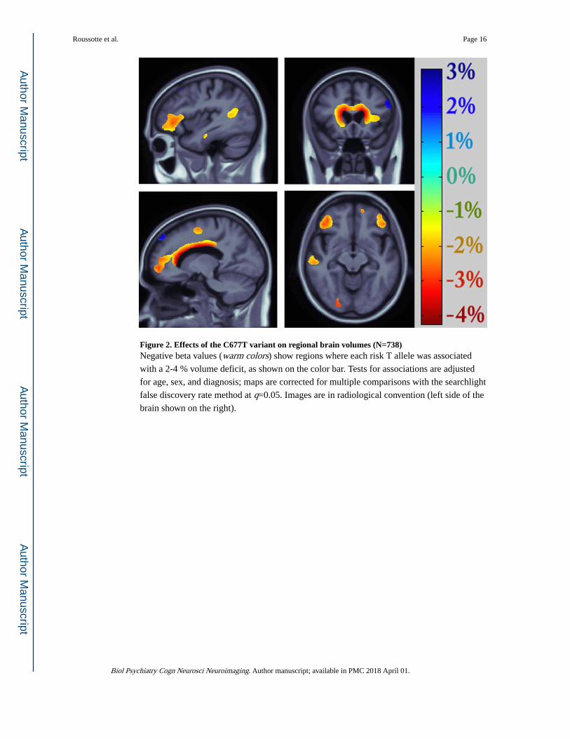
Figure 2. Effects of the C677T variant on regional brain volumes (N=738)Negative beta values (warm colors) show regions where each risk T allele was associated
with a 2-4 % volume deficit, as shown on the color bar. Tests for associations are adjusted
for age, sex, and diagnosis; maps are corrected for multiple comparisons with the searchlight
false discovery rate method at q=0.05. Images are in radiological convention (left side of the
brain shown on the right).
Roussotte et al. Page 16
Biol Psychiatry Cogn Neurosci Neuroimaging. Author manuscript; available in PMC 2018 April 01.
Author M
anuscriptA
uthor Manuscript
Author M
anuscriptA
uthor Manuscript

Figure 3. Mediation of the association between genotype and medial orbitofrontal volumes by plasma homocysteine levels (N=634)Path coefficients for simple mediation analysis using the PROCESS Procedure for SPSS.
The a-path represents the association between genotype and plasma homocysteine levels.
The b-path denotes the relationship between plasma homocysteine levels and medial
orbitofrontal volumes, while also controlling for genotype. The c’-path and the c-path
represent the associations between genotype and medial orbitofrontal volumes with and
without plasma homocysteine levels included as a mediator, respectively. *p<0.05,
**p<0.01, ***p<0.001.
Roussotte et al. Page 17
Biol Psychiatry Cogn Neurosci Neuroimaging. Author manuscript; available in PMC 2018 April 01.
Author M
anuscriptA
uthor Manuscript
Author M
anuscriptA
uthor Manuscript

Figure 4. Mediation of the association between MMSE performance and GDS-15 scores by medial orbitofrontal volumes (N=640)Path coefficients for simple mediation analysis using the PROCESS Procedure for SPSS.
The a-path represents the association between MMSE scores and medial orbitofrontal
volumes. The b-path denotes the relationship between medial orbitofrontal volumes and
GDS scores, while also controlling for MMSE performance. The c’-path and the c-path
represent the associations between MMSE and GDS scores with and without medial orbital
cortical volumes included as a mediator, respectively. *p<0.05, **p<0.01, ***p<0.001.
Roussotte et al. Page 18
Biol Psychiatry Cogn Neurosci Neuroimaging. Author manuscript; available in PMC 2018 April 01.
Author M
anuscriptA
uthor Manuscript
Author M
anuscriptA
uthor Manuscript
Author M
anuscriptA
uthor Manuscript
Author M
anuscriptA
uthor Manuscript
Roussotte et al. Page 19
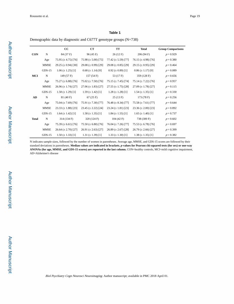
Table 1
Demographic data by diagnostic and C677T genotype groups (N=738)
CC CT TT Total Group Comparisons
CON N 84 (37 F) 96 (45 F) 26 (12 F) 206 (94 F) p = 0.929
Age 75.95 (± 4.75) [76] 75.98 (± 5.00) [75] 77.42 (± 5.59) [77] 76.15 (± 4.98) [76] p = 0.380
MMSE 29.25 (± 0.94) [30] 29.08 (± 0.99) [29] 29.08 (± 0.85) [29] 29.15 (± 0.95) [29] p = 0.464
GDS-15 1.06 (± 1.25) [1] 0.68 (± 1.14) [0] 0.92 (± 0.89) [1] 0.86 (± 1.17) [0] p = 0.089
MCI N 149 (57 F) 157 (54 F) 53 (17 F) 359 (128 F) p = 0.656
Age 75.27 (± 6.88) [76] 75.02 (± 7.50) [76] 75.15 (± 7.45) [74] 75.14 (± 7.22) [76] p = 0.957
MMSE 26.96 (± 1.74) [27] 27.06 (± 1.83) [27] 27.55 (± 1.75) [28] 27.09 (± 1.78) [27] p = 0.115
GDS-15 1.58 (± 1.29) [1] 1.59 (± 1.42) [1] 1.28 (± 1.28) [1] 1.54 (± 1.35) [1] p = 0.330
AD N 81 (40 F) 67 (25 F) 25 (13 F) 173 (78 F) p = 0.256
Age 75.04 (± 7.69) [76] 75.91 (± 7.30) [77] 76.48 (± 8.34) [77] 75.58 (± 7.61) [77] p = 0.644
MMSE 23.33 (± 1.88) [23] 23.45 (± 2.22) [24] 23.24 (± 1.81) [23] 23.36 (± 2.00) [23] p = 0.892
GDS-15 1.64 (± 1.42) [1] 1.58 (± 1.35) [1] 1.84 (± 1.55) [1] 1.65 (± 1.40) [1] p = 0.737
Total N 314 (134 F) 320 (124 F) 104 (42 F) 738 (300 F) p = 0.602
Age 75.39 (± 6.61) [76] 75.50 (± 6.80) [76] 76.04 (± 7.26) [77] 75.53 (± 6.78) [76] p = 0.697
MMSE 26.64 (± 2.70) [27] 26.91 (± 2.63) [27] 26.89 (± 2.67) [28] 26.79 (± 2.66) [27] p = 0.399
GDS-15 1.50 (± 1.33) [1] 1.31 (± 1.39) [1] 1.33 (± 1.30) [1] 1.38 (± 1.35) [1] p = 0.382
N indicates sample sizes, followed by the number of women in parentheses. Average age, MMSE, and GDS-15 scores are followed by their standard deviations in parentheses. Median values are indicated in brackets. p-values for Pearson chi-squared tests (for sex) or one-way ANOVAs (for age, MMSE, and GDS-15 scores) are reported in the last column. CON=healthy controls, MCI=mild cognitive impairment, AD=Alzheimer's disease
Biol Psychiatry Cogn Neurosci Neuroimaging. Author manuscript; available in PMC 2018 April 01.
Author M
anuscriptA
uthor Manuscript
Author M
anuscriptA
uthor Manuscript
Roussotte et al. Page 20
Table 2
Genotype and allele frequency by diagnostic groups (N=738)
CON MCI AD Pearson Chi-Square Test
Total N=738 206 359 173
Genotype Frequency CC 84 (41%) 149 (41%) 81 (47%)
CT 96 (46%) 157 (44%) 67 (39%) p = 0.592
TT 26 (13%) 53 (15%) 25 (14%)
Allele Frequency C 264 (64%) 455 (63%) 229 (66%)
T 148 (36%) 263 (37%) 117 (34%) p = 0.667
CON=healthy controls, MCI=mild cognitive impairment, AD=Alzheimer's disease
Biol Psychiatry Cogn Neurosci Neuroimaging. Author manuscript; available in PMC 2018 April 01.