flaviviruses in mosquitoes from southern portugal de mestrado_sónia... · west nile virus has been...
TRANSCRIPT

Universidade Nova de Lisboa
FLAVIVIRUSES IN MOSQUITOES FROM SOUTHERN
PORTUGAL, 2009-2010
Sónia Cristina Fernandes Da Costa
DISSERTAÇÃO PARA A OBTENÇÃO DO GRAU DE MESTRE EM
PARASITOLOGIA MÉDICA
NOVEMBRO, 2011
Universidade Nova de Lisboa
Instituto de Higiene e Medicina Tropical
FLAVIVIRUSES IN MOSQUITOES FROM SOUTHERN
PORTUGAL, 2009-2010
Sónia Cristina Fernandes Da Costa
Dissertação apresentada para cumprimento dos requisitos necessários à obtenção do grau de
Mestre em Parasitologia Médica realizada sob a orientação científica de:
Orientador: Professor Doutor Paulo Almeida
Co-Orientador: Professor Doutor Ricardo Parreira

iii
I dedicate this thesis to my family, especially
to João and Daniela who have given up
so much for me to achieve my goals.

iv
ACKNOWLEDGEMENTS
Firstly and foremost, I want to thank everyone that directly or indirectly contributed to
the making of this thesis, especially my dear colleague, Dr. Ferdinando Freitas, who
made this work possible.
I wish to thank my supervisors, Professor Dr. Paulo Almeida and Professor Dr. Ricardo
Parreira for the continuous support, encouragement, and friendship. I thank them for the
invaluable teachings; unending patience, good advice and critique that helped me
develop my scientific knowledge, in both Medical Entomology and Virology.
To Dr. Ferdinando Freitas, I want to thank for his great support, encouragement and
friendship. Foremost I am grateful for his teachings and mentoring in conducting
research in the Virology and Entomology laboratories, and for allowing me personal
freedom to learn through the trials and tribulations presented throughout the course of
this research. I thank him for performing tasks essential for this project to go forward,
such as field mosquito collection and specimen identification. I am thankful for the
time, dedication and expertise devoted to this project; his enthusiasm was contagious
and the experience gained while working with him has strengthen my determination in
pursuing a career in research.
I want to give thanks to all the members of staff of the Medical Parasitology and
Microbiology Unit of the Institute of Hygiene and Tropical Medicine who have been
extremely helpful from the very beginning.
To Professor Carla Sousa, Professor Teresa Novo and Dr. Bruno Gomes I want to thank
for performing tasks, such as fieldwork and specimen identification. To Dr. Cristina
Branco and Dr. Sandra Castro for their support and encouragement. To Dr. José Luís
Vicente for his good advice and willingness to help. To Dr. Vera Benavente for the
companionship, encouragement and good moments. To my colleagues Dr. Gonçalo
Seixas, Dr. Sofia Branco, Dr. Manuel Roque and Dr. Eliane Arez for the incentives,
companionship and good moments shared.
I want to especially thank my family. My daughter Daniela for keeping me on my toes
and giving me strength to continue. To my partner for his unconditional support, love,

v
patience, for believing in me, and for always being there throughout the highs and lows.
To my mom and dad for their patience, support and help with Daniela. To my brothers
Hélio Costa and Mário Costa for always being there when I needed them the most. To
my older brother I want to thank especially for being the greatest role model and the
rock we expect an older brother to be. To Sónia Luz for always showing support,
friendship, encouragement, companionship and for the good moments shared. To my
cousin Ariana Cruz for her friendship, support, encouragement and help with Daniela,
and for so much more that I will never find the right words to thank her enough.
vi
ABSTRACT
Flaviviruses are viruses belonging to the Flaviviridae family, genus Flavivirus. They
comprise a large group of widely spread and genetically diverse arthropod-borne viruses
including human and animal pathogens that can potentially cause large-scale epidemics
and high mortality and morbidity. In the past few years, flaviviruses have largely
expanded their geographical distribution and host range. West Nile virus has been
continuously detected throughout Europe lately and has been isolated from mosquitoes
in Southern Portugal, where human and animal cases have been reported.
The main aim of this work was to search for flaviviruses in mosquitoes collected from
two areas in Southern Portugal where West Nile virus and other flaviviruses have
previously been detected.
Mosquito surveys were carried out in 24 locations in the wetlands of the Faro and
Setúbal districts, by CDC-CO2 light-traps and indoors resting collections. Pools
containing approximately 50 mosquitoes were screened for flaviviruses by heminested
RT-PCR, directed at the amplification of a small fragment of the viral NS5 gene, using
degenerated flavivirus-specific primers.
A total of 36273 mosquitoes were collected during 2009 and 2010 from April through
October, from the following species: Anopheles algeriensis, An.atroparvus, Aedes
berlandi, Ae. caspius, Ae. detritus, Coquillettidia richiardii, Culex laticinctus, Cx.
pipiens, Cx. theileri, Cx. univittatus, Culiseta annulata, Cs. longiareolata, Cs.
subochrea, and Uranotaenia unguiculata. Most abundant species were Ae. caspius Cx.
theileri and Cx. pipiens, respectively. However, mosquito densities varied according to
collection method and sampling area. A fourfold increase in mosquito density was
registered in 2010 compared to 2009. A total of 745 pools were analysed of which 31%
tested positive for flaviviral sequences.
The species with higher positivity rates were An. algeriensis with Minimum infection
rate (MIR) of 56/1000 in the Algarve 2009, Cs. annulata MIR =22/1000 in the Algarve
2010, Cx.theileri and Cx.pipiens in Setúbal 2010, MIR =20/1000. An. atroparvus, Ae.
caspius, Ae. detritus and Cx. univittatus also yielded positive pools. Overall, positivity
was higher in the Algarve.
Viral sequences obtained from positive pools showed homology with insect-specific
flavivirus (ISF) sequences deposited in free access public databases. Phylogenetic
analysis reflected the genetic variability of flaviviruses and revealed the relatedness of
our sequences with other known flaviviruses, especially the insect-specific.
In view of previous WNV isolations and assessing from the four-fold increase in
mosquito density, the increasing temperatures, the recent cases throughout Europe and
the unknown and unpredictable pattern of flaviviruses outbreaks, continuous
epidemiological surveillance programmes are quickly becoming indispensable tools for
Public Health.
vii
RESUMO
Os flavivírus são vírus pertencentes à família Flaviviridae, género Flavivirus. Estes
formam um grande grupo caraterizado pela sua ampla distribuição e diversidade
genética. Os flavivírus são, na sua maioria, transmitidos por artrópodes vectores
incluíndo agentes patogénicos para humanos e animais que podem potencialmente
provocar grandes epidemias e causar elevadas taxas de mortalidade e morbidade. Nos
últimos anos, tem-se registado uma grande expansão a nível da distribuição geográfica
dos flavivírus e diversidade dos seus hospedeiros. O vírus do Nilo Ocidental tem sido
continuamente detectado em toda a Europa recentemente, e também isolado de
mosquitos colhidos no Sul de Portugal, onde já foram registados casos humanos e
animais.
O principal objectivo deste trabalho é o rastreio de flavivírus em mosquitos colhidos em
duas regiões do Sul de Portugal, onde os mesmos foram anteriormente detectados.
As colheitas de mosquitos foram realizadas em 24 locais em zonas húmidas nos
districtos de Faro e Setúbal, através de armadilhas luminosas tipo CDC com CO2 e
aspiradores mecânicos manuais para colheita de mosquitos em repouso em abrigos de animais.
Os mosquitos colhidos foram agrupados por lotes contendo aproximadamente 50
espécimens cada, e rastreados para a presença de flavivírus por heminested RT-PCR,
direccionado à amplificação de um pequeno fragmento do gene NS5 usando
oligonucleótidos degenerados específicos para flavivírus.
Entre Abril e Outubro de 2009 e 2010 foram colhidos no total 36273 mosquitos
pertencentes às seguintes espécies: Anopheles algeriensis, An.atroparvus, Aedes
berlandi, Ae.caspius, Ae.detritus, Coquillettidia richiardii, Culex laticinctus,
Cx.pipiens, Cx.theileri, Cx.univittatus, Culiseta annulata, Cs.longiareolata,
Cs.subochrea, e Uranotaenia unguiculata. As espécies mais abundantes foram
Ae.caspius, Cx.theileri e Cx.pipiens, respectivamente. Contudo, as densidades de
mosquitos foram variáveis de acordo com o método de colheita e área de amostragem.
As densidades de mosquitos colhidos em 2010 foram quatro vezes superior às
registadas no ano anterior. No total foram analisados 745 lotes dos quais 31% testaram
positivos para a presença de sequências de flavivírus.
As espécies que apresentaram taxas de positividade mais elevadas foram: An.algeriensis
com uma Taxa Mínima de Infecção (TMI) de 56/1000 no Algarve em 2009,
Cs.annulata TMI =22/1000 no Algarve em 2010, Cx.theileri e Cx.pipiens em Setúbal
em 2010, TMI =20/1000. An. atroparvus, Ae. caspius, Ae. detritus e Cx. univittatus
também produziram lotes positives. No geral, a positividade foi maior no Algarve.
Análise das sequências virais obtidas revelou homologia das nossas sequências virais
com sequências de referência de flavivírus específicos de mosquitos depositadas em
bases de dados de acesso livre. A análise filogenética reflectiu a variabilidade genética
dos flavivírus e revelou a relação genética das nossas sequências com as de outros
flavivírus, especialmente os específicos de insectos.
viii
Tendo em consideração os anteriores isolamentos do vírus do Nilo Ocidental, o
aumento acentuado nas densidades de mosquitos, o aumento de temperaturas que se tem
vindo a registar, os casos recentes de transmissão de flavivírus por toda a Europa e o
padrão desconhecido e imprevisível dos surtos destes vírus, os programas contínuos de
vigilância epidemiológica têm-se revelado uma ferramenta indispensável para a Saúde
Pública.
FLAVIVIRUSES IN MOSQUITOES FROM SOUTHERN PORTUGAL,
2009-2010
Sónia Cristina Fernandes Da Costa
Keywords: West Nile virus, flaviviruses, arboviruses, Algarve, flavivirus detection,
insect-specific flaviviruses.
Palavras Chave: Vírus do Nilo Ocidental, flavivírus, arbovírus, Algarve, detecção de
flavivírus, flavivírus específicos de insectos.

ix
TABLE OF CONTENTS
ACKNOWLEDGEMENTS ........................................................................................................iv
ABSTRACT ................................................................................................................................vi
RESUMO .................................................................................................................................. vii
TABLE OF CONTENTS ............................................................................................................ix
LIST OF ABBREVIATIONS ...................................................................................................... 1
1. Introduction ................................................................................................................... 5
1.1. Arboviruses ........................................................................................................................... 5
1.1.2. Medical importance of mosquitoes (Diptera: Culicidae) ............................................ 8
1.2. The Flaviviridae family ...................................................................................................... 10
1.3. The importance of Flaviviruses ........................................................................................... 11
1.3.1. Flavivirus: genome structure and morphology of the viral particle .......................... 11
1.3.2. Replication cycle ...................................................................................................... 14
1.3.3. Flavivirus classification: phylogeny ......................................................................... 16
1.4. Medically Important Pathogenic Flaviviruses ..................................................................... 20
1.4.1. Dengue and Yellow Fever ........................................................................................ 21
1.4.2. West Nile Virus ........................................................................................................ 25
1.5. Insect-specific flaviviruses .................................................................................................. 31
1.6. Europe: current situation ..................................................................................................... 40
1.7. Portugal: the imminent risk ................................................................................................. 46
2. Objectives ................................................................................................................... 52
2.1 Specific objectives ............................................................................................................... 52
3. Materials and methods ................................................................................................ 54
3.1. Mosquito surveys ................................................................................................................ 54
3.1.1. Characterisation of the study areas ........................................................................... 55
3.1.2. Mosquito sampling ................................................................................................... 55

x
3.1.3. Specimen identification ............................................................................................ 56
3.1.4. Mosquito density and seasonal dynamics study ........................................................ 56
3.1.5. Statistical analysis .................................................................................................... 56
3.2. Viral screening .................................................................................................................... 57
3.2.1. Mechanical maceration of mosquitoes...................................................................... 57
3.2.2. RNA extraction ........................................................................................................ 57
3.2.3. cDNA synthesis ........................................................................................................ 58
3.2.4. Preliminary tests ....................................................................................................... 59
3.2.5. Amplification of viral sequences by PCR ................................................................. 60
3.2.6. Purification and sequencing of PCR products .......................................................... 62
3.2.7. Phylogenetic analysis of viral sequences .................................................................. 63
3.2.8 Extraction controls .................................................................................................... 64
4. Results ......................................................................................................................... 66
4.1. Mosquito collections ........................................................................................................... 66
4.2. Mosquito seasonal dynamics in the Algarve, 2009-2010 .................................................... 68
4.3. Mosquito relative species distribution in the Algarve, 2009-2010....................................... 73
4.4. Mosquito screening for flaviviral sequences ....................................................................... 78
4.4.1. RNA extracted from mosquito homogenates ............................................................ 79
4.4.2. Amplification reactions preliminary tests ................................................................. 79
4.5. Phylogenetic analysis of flaviviral sequences obtained ....................................................... 86
5. Discussion and conclusions ........................................................................................ 90
5.1. Mosquito abundance and seasonal dynamics............................................................... 90
5.2. Mosquito screening for flaviviral sequences and respective phylogeny ...................... 98
5.3. Final conclusions ....................................................................................................... 105
6. Bibliography ............................................................................................................. 108
7. List of figures ............................................................................................................ 123
8. List of tables .............................................................................................................. 126

Flaviviruses in mosquitoes from Southern Portugal, 2009-2010
1
LIST OF ABBREVIATIONS
µl Micro litre
AeFv Aedes flavivirus
bp Base pair
BSA Bovine serum albumin
C Capsid
CDC Centres for Disease Control and Prevention
cDNA Complementary deoxyribonucleic acid
CFAV Cell-fusing agent virus
CO2 Carbon dioxide
CPE Cytopathic effect
CSA Cell-silent agent
CSAV Cell silent agent virus
CxFv Culex flavivirus
DENV Dengue virus
DEPC Diethylpyrocarbonate
DF Degrees of freedom
DHF Dengue haemorrhagic fever
DNA Deoxyribonucleic acid
DSS Dengue shock syndrome
E Envelope
ER Endoplasmic reticulum
g Grams
IR Indoor resting
Flaviviruses in mosquitoes from Southern Portugal, 2009-2010
2
ISF Insect-specific flavivirus
JEV Japanese encephalitis virus
KDFV Kyasanur Forest Disease virus
KRV Kamiti River virus
LAMV Lammi virus
LIV Louping ill virus
M Membrane
MBV Mosquito-borne viruses
MIR Minimum infection rate
ml Millilitre
MVEV Murray Valley encephalitis virus
NAKV Nakiwogo virus
NCR Non-coding region
NKV No-known vector
nm Nano metre
NOUV Nounané virus
NS Non-structural
nt Nucleotide
ºC Degrees Celsius
OHFV Omsk haemorrhagic fever virus
ORF Open reading frame
PBS Phosphate buffered saline
PCR Polymerase Chain Reaction
pmol Pico moles
POWV Powassan virus
Flaviviruses in mosquitoes from Southern Portugal, 2009-2010
3
prM Precursor M protein
QBV Quang Binh virus
RBV Rio Bravo virus
RFV Royal Farm virus
RNA Ribonucleic acid
spp Species
TABV Tamana bat virus
TBEV Tick-borne encephalitis virus
TBV Tick-borne viruses
TGN Trans-Golgi network
USA United States of America
UTR Untranslated region
UV Ultraviolet
WHO World Health Organisation
WNV West Nile virus
YFV Yellow fever virus

Flaviviruses in mosquitoes from Southern Portugal, 2009-2010
4
1.
INTRODUCTION

Flaviviruses in mosquitoes from Southern Portugal, 2009-2010
5
1. Introduction
1.1. Arboviruses
The term arbovirus originated in the 1940’s as the result of the abbreviations made to
describe the viruses transmitted by arthropods (arthropod-borne viruses) (Kuno and
Chang, 2005). Arboviruses are recognised as an extremely diverse group that harbours
many medically important viruses (table 1), which can cause serious disease such as
yellow fever, dengue and several encephalitis (Pabbaraju et al., 2009). They are
included mainly into three viral families: Flaviviridae, Togaviridae and Bunyaviridae
(Pabbaraju et al., 2009).
Table 1: Medically important arboviruses belonging to families Flaviviridae, Togaviridae and
Bunyaviridae (adapted from Gubler, 2002).
Virus Human disease Reservoir host Arthropod vector Geographic
distribution
Flaviviridae
Yellow fever virus
(YFV)
Yellow fever –
haemorrhagic fever
Primates,
humans
Mosquito Africa, America
Dengue virus 1-4
(DENV)
Dengue
haemorrhagic fever/
shock syndrome
Humans,
primates
Mosquito Africa, America,
Asia
Japanese encephalitis
virus
(JEV)
Encephalitis Birds Mosquito Asia
Saint Louis
encephalitis virus
(SLEV)
Encephalitis Birds Mosquito North America
West Nile virus
(WNV)
FAR syndrome,
encephalitis
Birds Mosquito Worldwide
Murray Valley
encephalitis virus
(MVEV)
Encephalitis Birds
Mosquito Australia
Tick-borne
encephalitis virus
(TBEV)
Encephalitis Small mammals,
rodents, birds
Ticks Europe, Asia
Togaviridae Chikungunya virus FAR syndrome Primates,
humans
Mosquito Africa, Asia,
Europe
Ross River virus FAR syndrome Marsupials Mosquito Australasia
Sindbis virus Fever/Rash Birds Mosquito Africa, Asia,
Australia, Europe
O’Nyong nyong Fever Unknown Mosquito Africa
Equine encephalitis
viruses (EEV,WEV)
encephalitis Passerine birds Mosquito America
Bunyaviridae
Bunyamwera Fever Rodents Mosquito Global
California
encephalitis virus
Encephalitis Mammals Mosquito North America
La Cross virus Encephalitis Mammals
Mosquito North America
Tahyna virus Fever, respiratory
disease, encephalitis
Mammals Mosquito Asia, Europe
FAR – fever/arthralgia/rash
Flaviviruses in mosquitoes from Southern Portugal, 2009-2010
6
These viruses are, thus, transmitted by arthropod vectors via a biological process, which
can occur vertically or horizontally (Kuno and Chang, 2005; Weaver and Reisen, 2010).
For viruses to be biologically transmitted they must replicate in the arthropod vector
prior to transmission (Kuno and Chang, 2005; Goddard, 2008; Weaver and Reisen,
2010). In vertical transmission, the virus is passed on from the female to both male and
female offsprings by trans-ovarial or trans-stadial transmission (Kuno and Chang, 2005;
Weaver and Reisen, 2010). In turn, horizontal transmission can occur venereally (where
the virus is passed on from infected males directly onto females, when mating), or
orally. This is the most typical arboviral transmission mode, which involves the
infection of a susceptible arthropod vector after ingestion of viruses during feeding or,
from maternal origin. Viruses are subsequently disseminated within the arthropod,
replicate in the salivary glands, ensuring that transmission might occur during the
following bloodmeal, through injection of contaminated saliva in a susceptible host
(Kuno and Chang, 2005; Weaver and Riesen, 2010).
Furthermore, not all infected arthropods are capable of pathogen transmission. For it to
occur it must be competent for transmission, that is, it must be susceptible to infection
by the pathogen, allowing the above mentioned replication and dissemination thus
becoming infective, and able to transmit the pathogen via an infective bite when blood-
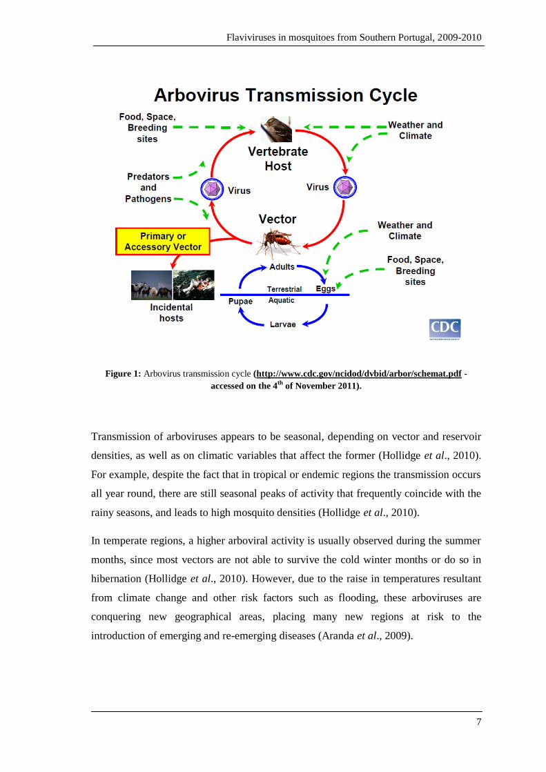
feeding (Goddard, 2008; Weaver and Reisen, 2010). Arboviral transmission can only
take place where the three principal elements are present: the virus, the vector and
vertebrate hosts (figure 1).
Flaviviruses in mosquitoes from Southern Portugal, 2009-2010
7
Figure 1: Arbovirus transmission cycle (http://www.cdc.gov/ncidod/dvbid/arbor/schemat.pdf -
accessed on the 4th
of November 2011).
Transmission of arboviruses appears to be seasonal, depending on vector and reservoir
densities, as well as on climatic variables that affect the former (Hollidge et al., 2010).
For example, despite the fact that in tropical or endemic regions the transmission occurs
all year round, there are still seasonal peaks of activity that frequently coincide with the
rainy seasons, and leads to high mosquito densities (Hollidge et al., 2010).
In temperate regions, a higher arboviral activity is usually observed during the summer
months, since most vectors are not able to survive the cold winter months or do so in
hibernation (Hollidge et al., 2010). However, due to the raise in temperatures resultant
from climate change and other risk factors such as flooding, these arboviruses are
conquering new geographical areas, placing many new regions at risk to the
introduction of emerging and re-emerging diseases (Aranda et al., 2009).
Flaviviruses in mosquitoes from Southern Portugal, 2009-2010
8
1.1.2. Medical importance of mosquitoes (Diptera: Culicidae)
The Culicidae family is very important from a human and veterinary medical
perspective since it harbours a large number of species, including some of the most
important hematophagous arthropods capable of transmitting infectious agents (Eiras,
2004; Eldridge, 2005). This family comprises over 3,500 mosquito species and
subspecies, belonging to two medical important subfamilies: the Anophelinae and
Culicinae. (Eldridge, 2005). Mosquitoes included in these subfamilies are capable of
transmitting arboviruses such as the dengue and yellow fever viruses as is the case of
Aedes aegypti and Ae. albopictus; moreover, Culex species mosquitoes that can transmit
West Nile virus, Saint Louis encephalitis and Rift Valley fever viruses are also part of
the Culicinae subfamily (Manson-Bahr and Bell, 1987; Eldridge, 2005). Apart from
viruses, mosquitoes can also transmit nematode worms and protozoa (Rutledge, 2008).
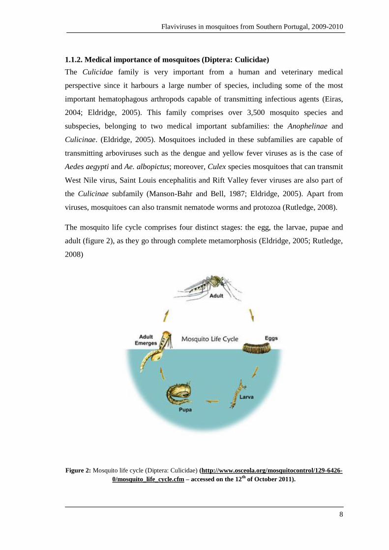
The mosquito life cycle comprises four distinct stages: the egg, the larvae, pupae and
adult (figure 2), as they go through complete metamorphosis (Eldridge, 2005; Rutledge,
2008)
Figure 2: Mosquito life cycle (Diptera: Culicidae) (http://www.osceola.org/mosquitocontrol/129-6426-
0/mosquito_life_cycle.cfm – accessed on the 12th
of October 2011).

Flaviviruses in mosquitoes from Southern Portugal, 2009-2010
9
The immature stages of mosquitoes, egg, larvae and pupae, are aquatic while the adult
stage is the only that is terrestrial (Rutledge, 2008). Mosquito biology is highly
dependent on climatic conditions, particularly temperature variations, and in warmer
temperatures, the life cycle may take 10 days or less to complete (Eldridge, 2005). Eggs
may be deposited in water surface or moist ground, in groups or individually, and
hatching occurs either in a day or so or when flooding occurs (Eldridge, 2005; Rutledge,
2008). A larva must undergo four molts to become a pupa; though their time of
development has no direct relation to water temperatures (Eldridge, 2005). Adult
emergence usually happens 1-3 days after pupa formation (Eldridge, 2005; Rutledge,
2008). Mosquito distribution is determined by the climatic conditions, hence their
permanent existence in tropical, warm and humid climates, temperate countries and in
cooler climate countries (e.g. northern Europe) (Rutledge, 2008). Therefore, changes in
these climatic conditions may forcibly change their bioecology.
Mosquito vectors of flaviviruses, including arboviruses, are autochthonous in many
European countries, including Portugal, where due to beneficial climatic and ecological
conditions there is production of very high-density populations in some areas, especially
in the Mediterranean Basin (Almeida et al., 2008). This is the case of Aedes and Culex
species mosquitoes, in wetlands, estuarine regions and manmade and natural farming
lands all across Europe. Many mosquitoes established in Portugal include potential
vectors of WNV (e.g. Aedes caspius, Cx.theileri, Cx. pipiens and Cx. univitattus),
Sindbis and Rift Valley fever viruses (e.g. Cx.theileri and Aedes caspius), Chikungunya
and Tahyna viruses (Aedes caspius), and it is only a matter of time and viral pathogen
seasonality, until arboviral activity is detected (Jupp et al., 1972; McIntosh et al., 1980;
Jupp et al., 1985; Turell et al., 1996; Hubalek and Halouzka, 1999; Lundstrom, 1999;
Vazeille et al., 2008).

Flaviviruses in mosquitoes from Southern Portugal, 2009-2010
10
1.2. The Flaviviridae family
Flaviviruses are viruses belonging to the Flaviviridae family, genus Flavivirus. The
Flaviviridae is a large, widely spread and genetically diverse family of viral agents, that
includes human and animal pathogens that can potentially cause large-scale epidemics
and tens of thousands of deaths annually (Cook et al., 2003; Mukhopadhyay et al.,
2005). This family comprises four genera:
The Pestivirus genus (derives from the Latin word “pestis” which means
“plague”) that includes four viruses, namely border disease virus, bovine viral
diarrhoea viruses 1 and 2, and classical swine fever virus (Lindenbach et al.,
2007);
The Hepacivirus genus (derives from the Greek words “hepar” and “hepatos”,
which mean “liver”) the sole member of which is the Hepatitis C virus
(Lindenbach et al., 2007);
The Flavivirus genus (derives from the Latin word “flavus”, that means
“yellow”) which is the largest genus and contains more than 70 RNA viruses
(Mukhopadhyay et al., 2005; Cook and Holmes, 2006; Lindenbach et al., 2007;
Cook et al., 2009);
The Pegivirus genus (“pe” from the word persistent and “g” from GB or G)
which includes the GB viruses (variants A, B, C and D) and Hepatitis G virus
(HGV) (Lindenbach et al., 2007; Stapleton et al., 2011).
The worldwide spread flaviviruses comprises over seventy recognised RNA viruses
including many that are responsible for epidemics and high mortality rates among
humans (Cook et al., 2006; Cook et al., 2009; Junglen et al., 2009; Huhtamo et al.,
2009; Monini et al., 2010). At least 30 members of the genus Flavivirus are regarded as
medically important since they can cause serious human disease, including
haemorrhagic fever and encephalitis (Sanchéz-Seco et al., 2005; Hoshino et al., 2009).
Nonetheless, the clinical condition of the infected individual is not always life
threatening since it may present itself as a mild febrile illness or be completely
asymptomatic, as most infections are (Sanchéz-Seco et al., 2005; Hoshino et al., 2009).
Approximately 30% of flaviviruses are not known to have vertebrate hosts and are,
Flaviviruses in mosquitoes from Southern Portugal, 2009-2010
11
therefore, considered to be insect-specific (Morales-Betoulle et al., 2008; Blitvich et al.,
2009). Furthermore, there are also some members of the genus Flavivirus for which no
vector is known, designated by no-known vector (NKV) viruses (Cook et al., 2006;
Blitvich et al., 2009).
1.3. The importance of Flaviviruses
Viruses belonging to genus Flavivirus represent some of the most important emerging
or re-emerging pathogenic agents that cause disease in humans (Solomon and Mallewa,
2001). The expanding distribution of these viruses is directly related to the spread and
extension of vector distribution within an environment that provides beneficial
conditions for vector maintenance and establishment, as well as the presence of
vertebrate hosts (Weaver and Reisen, 2010).
The major factors that contribute to geographical dispersal of arboviral diseases include
human activity, genetic and environmental changes. These include (Petersen and
Marfin, 2005; Gould and Solomon, 2008; Weaver and Reisen, 2010):
The ability of the RNA viruses to undergo rapid genetic alterations that allow
them to adapt more easily to virtually any hosts, vertebrate or invertebrate, under
changing climate conditions;
Population growth and urbanisation;
Increased travel and commercial transportation around the world;
The receptivity of an area to viral emergence;
Invasion of vector natural habitats;
Lack of vaccination and/or effective vector control programmes in endemic
areas possibly due to economic or political issues.
1.3.1. Flavivirus: genome structure and morphology of the viral particle
The flavivirus genome, of approximately 11 kilobases in length, is a single stranded,
positive polarity RNA molecule that encodes three structural (capsid [C], membrane

Flaviviruses in mosquitoes from Southern Portugal, 2009-2010
12
[M] and envelope [E]) and seven non-structural proteins (NS1, NS2a, NS2b, NS3,
NS4a, NS4b and NS5), as shown on figure 3 (Sanchéz-Seco et al., 2005; Harris et al.,
2006; Hoshino et al., 2009).
Figure 3: Flavivirus genome structure and functions of viral proteins
(Fernandez-Garcia et al., 2009).
Flavivirus genomes encode a large polyprotein and contain a single open reading frame
(ORF) flanked by two non-coding regions (NCR) - the NCR of 5’ end is approximately
100 nucleotides (nt) in length, whereas the NCR at the 3’ end is between 400 and 700
nucleotides long (Lindenbach et al., 2007). Viruses within this genus are small (around
50 nm), spherical particles that contain an electron dense core (approximately 30 nm)
surrounded by a lipid envelope formed from membranes (derived from the endoplasmic
reticulum) of host cells (Barrett, 2001; Lindenbach et al., 2007). The virions present a

Flaviviruses in mosquitoes from Southern Portugal, 2009-2010
13
complex structure that contains a nucleocapsid, which is hexahedral/icosahedral in
symmetry as shown in figure 4 (Carter et al., 2008).
Figure 4: The flavivirus virion (Petersen et al., 2001).
Flavivirus virions contain two structural proteins at its surface: the E protein (the
envelope glycoprotein that surrounds the nucleocapsid) and the M protein (Barrett,
2001; Lindenbach et al., 2007). In addition, surrounding the single stranded viral RNA
genome is a small capsid protein that has a basic charge in order to interact with the
genome (Barrett, 2001).
The envelope glycoprotein E is the major antigenic determinant for the production of
neutralizing antibodies that relate to a protective immune response (McMinn, 1997).
Furthermore, it mediates receptor binding and membrane fusion, through its cellular
receptor binding sites and fusion peptides (Lindenbach et al., 2007).
These virions can exhibit two different forms, the mature and the immature virions
(figure 5); the mature virions are extracellular and contain the M (membrane) protein,
whereas the immature ones are intracellular and contain precursor M protein (prM) that
is cleaved by proteolysis to produce the M protein (Barrett, 2001).

Flaviviruses in mosquitoes from Southern Portugal, 2009-2010
14
Figure 5: Flavivirus particle structure. A- Envelope proteins of immature and mature virions;
B- Immature dengue virus type-2 virion; C- Mature dengue virus type-2 virion
(adapted from Lindenbach et al., 2007).
1.3.2. Replication cycle
The molecular biology of flavivirus has been actively studied, as the Flaviviridae family
comprises many viruses with major medical importance. A schematic representation of
the viral replication cycle (Lindenbach and Rice, 2003) is shown in figure 6.
Flavivirus virions adhere to the surface of the host cell and enter it by means of
receptor-mediated endocytosis via attachment to high affinity cellular receptors specific
for viral envelope proteins (unknown for most viruses) (Mukhopadhyay et al., 2005;
Harris et al., 2006; Lindenbach et al., 2007). The low pH of the endosomal vesicles
triggers the particles to undergo conformational changes and induces viral fusion with
host cell membranes and virus disassembly, causing the uncoating and release of the
virus nucleocapsid into the cytoplasm (Lindenbach and Rice, 2003; Mukhopadhyay et
al., 2005; Harris et al., 2006; Lindenbach et al., 2007). It is thought that viral genomes
are available for translation immediately after membrane fusion. Thus, once the viral
genome is released into the cytoplasm, the RNA molecule is translated into a single

Flaviviruses in mosquitoes from Southern Portugal, 2009-2010
15
polyprotein that is cleaved by viral and host proteases (Mukhopadhyay et al., 2005;
Lindenbach et al., 2007).
Receptor binding and endocytosis
Acid catalysed fusion and uncoating
Translation and polyprotein processing
Viral genome replication
Virus assembly
Virus maturation
Exocytosis
Figure 6: Flavivirus replication cycle (adapted from Fernandez-Garcia et al., 2009).
The translation process yields proteins that play an important role in the replication of
the viral genome and the formation of new virus particles (Lindenbach and Rice, 2003).
Genome replication takes place on intracellular membranes, more specifically on
cytoplasm replication complexes associated with perinuclear membranes (Lindenbach
and Rice, 2003; Lindenbach et al., 2007). Virions are thought to assemble by budding
into an intracellular membrane compartment, in the lumen of the endoplasmic reticulum
(ER), resulting in the formation of immature non-infectious viral particles containing E
and prM proteins, nucleocapsid and lipid membrane thus making them unable to induce
host-cell fusion (Mukhopadhyay et al., 2005; Lindenbach et al., 2007). These immature
non-infectious viral particles are transported through the trans-Golgi network (TGN)
where cleavage of the prM protein occurs, by the host protease furin, thus creating
Flaviviruses in mosquitoes from Southern Portugal, 2009-2010
16
mature infectious virions (Mukhopadhyay et al., 2005). Finally, the mature virions are
ready to be released from the host cell by exocytosis (Mukhopadhyay et al., 2005).
1.3.3. Flavivirus classification: phylogeny
The genus Flavivirus is unique in the Flaviviridae family since, contrarily to genus
Hepacivirus and Pestivirus, its members present the ability to infect and replicate in
vertebrate and invertebrate host cells and display genetic, epidemiological and
ecological characteristics that are distinct from the other two genera (Gould et al.,
2003). Even though flaviviruses are known to be related, based on phylogenetic and
antigenic analysis, the members of this genus characteristically present high genetic
divergence thus emphasizing that these correlations are not simple, nor are they always
clear (Kuno et al., 1998; Gaunt et al., 2001; Gould et al., 2003; Cook and Holmes,
2006). The evolutionary process may have contributed greatly to the genetic diversity
and divergence among these viruses, through factors such as the gradual adaptation to
new hosts, new geographical areas and genetic alterations, which might still be
occurring these days (Cook and Holmes, 2006).
The genus Flavivirus is considered to be an invaluable model for the evolutionary
investigation of vector-borne diseases and their modes of transmission, since it has been
confirmed by many publications that the viral transmission mode is strongly related to
virus phylogeny (Gaunt et al., 2001; Cook and Holmes, 2006). In order to understand
the origin, and spread patterns of emerging and re-emerging diseases, it is essential to
gather valuable knowledge by the analysis of the evolutionary history of flaviviruses
(Kuno et al., 1998; Gaunt et al., 2001). Genetic characteristics were typically
investigated based on antigenic cross reactivity, haemagglutination and complement
fixation tests (Gaunt et al., 2001). More recently, investigators have implemented the
use of other tools such as molecular sequencing, and phylogenetic data analysis, in
order to understand and solve taxonomic issues, thus allowing viruses to be correctly
assigned positions within their respective genus (Gaunt et al., 2001).
Phylogenetic analysis of flaviviruses commonly separates the genus into three different
groups, namely the mosquito-borne viruses, the tick-borne viruses and the no-known
Flaviviruses in mosquitoes from Southern Portugal, 2009-2010
17
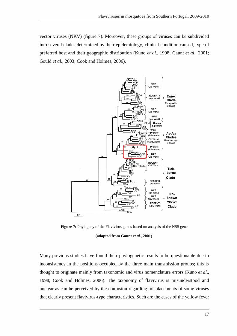
vector viruses (NKV) (figure 7). Moreover, these groups of viruses can be subdivided
into several clades determined by their epidemiology, clinical condition caused, type of
preferred host and their geographic distribution (Kuno et al., 1998; Gaunt et al., 2001;
Gould et al., 2003; Cook and Holmes, 2006).
Figure 7: Phylogeny of the Flavivirus genus based on analysis of the NS5 gene
(adapted from Gaunt et al., 2001).
Many previous studies have found their phylogenetic results to be questionable due to
inconsistency in the positions occupied by the three main transmission groups; this is
thought to originate mainly from taxonomic and virus nomenclature errors (Kuno et al.,
1998; Cook and Holmes, 2006). The taxonomy of flavivirus is misunderstood and
unclear as can be perceived by the confusion regarding misplacements of some viruses
that clearly present flavivirus-type characteristics. Such are the cases of the yellow fever

Flaviviruses in mosquitoes from Southern Portugal, 2009-2010
18
virus and many others that are not incorporated into any of the serological complexes
that flaviviruses are divided into (Kuno et al., 1998). For instance, in the study carried
out by Kuno et al (1998), phylogenetic analysis of sequences obtained from the E and
NS5 genes yielded conflicting results. Despite the similarities in the overall division and
positioning of viruses, there were some exceptions, as was the case for some bat-related
viruses that were included in the mosquito-transmitted clade, grouped together with
yellow fever and Sepik viruses (figure 7, red box) (Kuno et al., 1998; Gaunt et al.,
2001).
To the contrary, there are also viruses that do not display clear relationships to other
members and are placed within this genus (Kuno et al., 1998). Tamana bat virus and
cell-fusing agent are good examples of “candidate” viruses occupying uncertain
positions within the genus Flavivirus (Gould et al., 2003). Despite many attempts, the
doubts and uncertainty surrounding the flaviviruses phylogenetic relationships still
linger on (Cook and Holmes, 2006).
Flavivirus classification through molecular genetics has been undertaken using different
methods and targeting different viral genes (Gould et al., 2003; Cook and Holmes,
2006). Viral sequences obtained from the envelope (E) gene, NS3, NS4, and NS5 have
been used for phylogenetic inference. However, recent work has been mainly directed at
the analysis of the most conserved region of the flavivirus genome (the NS5 coding
sequence), as well as their complete sequence (Cook and Holmes, 2006).
The analysis of sequences gathered from distinct genes indicates an important
separation between the mosquito-borne viruses from those transmitted by ticks (Cook
and Holmes, 2006). According to the study carried out by Kuno et al (1998), the non-
vectored and vector-borne virus groups were the first to have emerged, assuming cell-
fusing agent virus (CFAV) as sharing a common ancestor with which other members of
the genus Flavivirus. Following these events, the vector-borne group was split into tick-
borne and mosquito-borne clades (Kuno et al., 1998; Cook and Holmes, 2006). Based
on phylogenetic analysis of the envelope gene, it is hypothesized that CFAV may have
diverged from flavivirus prior to the separation between mosquito and tick-borne
groups, thus forming a basal lineage (Sang et al., 2003).
Flaviviruses in mosquitoes from Southern Portugal, 2009-2010
19
Furthermore, other associations have resulted from this type of analysis, as is the case of
the mosquito-borne clade that was subdivided into a further two groups according to the
mosquito vector species, their vertebrate hosts and the nature of the disease caused
(Kuno et al., 1998; Gaunt et al., 2001). As shown on figure 7, the mosquito-borne
viruses were clearly separated into two clades: the viruses isolated from Aedes species
and those from Culex spp. (Gaunt et al., 2001). Besides, when other characteristics such
as the sort of disease caused were included in the analysis, the generated tree revealed a
solid association between the Culex clade and the neurotropic viruses causing
encephalitic disease; and between the Aedes clade and the non-neurotropic viruses that
tend to produce hemorrhagic disease (Gaunt et al., 2001; Gould et al., 2003). In
addition, correlations were established based on host preference, linking Culex species
mosquitoes with birds, and Aedes mosquitoes with primates (Gaunt et al, 2001; Gould
et al., 2003).
Although no-known vector (NKV) viruses have not been thoroughly examined, the
results from many studies tend to support the hypothesis that they have evolved away
from the vector-borne group of viruses (Kuno et al., 1998; Gaunt et al., 2001; Gould et
al., 2003; Cook and Holmes, 2006). Sequence analysis of the NS5 gene produces a
phylogenetic tree that separates the NKV group into three, the rodent and bat clades and
a “basal lineage” composed of an individual virus – the APOI virus that is linked to
rodents and therefore would later be added to the rodent-borne clade (Kuno et al., 1998;
Gaunt et al., 2001). However, as mentioned before, some members of this group were
included into the mosquito-borne clade, which suggests that during the flavivirus
evolution, and following the divergence of the NKV viruses from the vector-borne
viruses, there was a consequent loss of vector-borne transmission (Gould et al., 2003;
Cook and Holmes, 2006). Phylogenetic analysis was not conclusive as to which group
was the most divergent (Gould et al., 2003). Contrarily, more recently it was concluded
that the NKV viruses group is indeed the most divergent of the three main flavivirus
groups (Cook and Holmes, 2006).
The tick-borne group of viruses is composed of two distinct clades (figure 8), of which
one includes flaviviruses that infect seabirds and the other contains the mammalian tick-
borne viruses (Kuno et al., 1998; Gaunt et al., 2001; Gould et al., 2003; Cook and

Flaviviruses in mosquitoes from Southern Portugal, 2009-2010
20
Holmes, 2006). The latter is said to be linked to rodent hosts and Ixodes spp. ticks in
woodland areas (Gaunt et al., 2001; Gould et al., 2003). With the exception of Omsk
haemorrhagic fever virus (OHFV) and Kyasanur Forest disease virus (KFDV) that tend
to cause haemorrhagic disease, infections by flaviviruses within this group primarily
produces encephalitic disease (Gaunt et al., 2001).
Figure 8: Phlylogenetic tree of tick-borne flaviviruses based on the analysis of NS3 gene
(Mansfield et al., 2009).
1.4. Medically Important Pathogenic Flaviviruses
Included into the Flavivirus genus are some of the most virulent and medically
important viruses known to medical science including, amongst others, the Dengue
viruses (DENV- 1, DENV- 2, DENV- 3 and DENV- 4), Yellow Fever virus (YFV) and
West Nile virus (WNV) (Harris et al., 2006; Junglen et al. 2009; Calzolari et al., 2010).
The distribution of these viruses has been increasing geographically and, over the years,
new host populations have been targeted (Junglen et al., 2009).
Flaviviruses in mosquitoes from Southern Portugal, 2009-2010
21
Hypothetically, in case of continued Ae. albopictus geographic expansion (especially
northwards) and Aedes aegypti reestablishment, combined with changes to ecological
conditions as a consequence of climate change, there may be a risk of eventual
pathogenic flaviviruses transmission in Europe (Reiter, 2010). This may be the case of
DENV and YFV since the historical records of these diseases in the continent confirm
that the conditions existent are suitable for their transmission (Reiter, 2010).
1.4.1. Dengue and Yellow Fever
1.4.1.1. Epidemiology
Viruses of the genus Flavivirus are the causative agents of both dengue and yellow
fever, which are associated with Aedes spp mosquitoes and cause haemorrhagic disease
in humans and simians (Endy et al., 2010; Reiter, 2010). These viruses are considered
the most important arboviral pathogens that, for a long time, have been causing disease
around the world (Endy et al., 2010; Weaver et al., 2010). Infection by DENV and YFV
can be manifested as a mild or potentially fatal disease, and it can take a mere seven to
ten days of disease before death occurs (Gould and Solomon, 2008; Endy et al., 2010;
Weaver and Reisen, 2010).
The main factors that contributed to the great geographical dispersal of Aedes aegypti
and Aedes albopictus, the principal vectors of dengue and yellow fever, include the
commercial transportation of goods and people during the slave trade, particularly by
ship (Endy et al., 2010). This resulted in the evolution and adaptation of these vectors to
urban environments and, consequently, in epidemics affecting particularly port cities
(Reiter, 2010; Weaver and Reisen, 2010). Northern cities such as Dublin (Ireland) and
Boston (North America) were unexpectedly hit by these epidemics (Reiter, 2010).
The risk of contracting dengue threatens more than 2.5 billion individuals, in tropical
and subtropical countries, causing 50-100 million new infections, 500,000
hospitalisations, and 24,000 reported deaths, per year (Samuel and Tyagi, 2006; Gould
and Solomon, 2008; Weaver and Reisen, 2010). The World Health Organization
(WHO) confirmed that a hundred countries around the globe are endemic for dengue
disease (figure 9) (Endy et al., 2010).

Flaviviruses in mosquitoes from Southern Portugal, 2009-2010
22
Figure 9: Dengue risk areas, 2010 (http://www.wpro.who.int/health_topics/dengue/ – accessed on
the 5th
July 2011).
Dengue is an emerging disease that has shown dramatic geographic and incidence
increase in the past few decades, and has caused large epidemics in the Americas in
2007 (890,000 cases) and in Greece in 1927-28 (over one million cases) (Endy et al.,
2010; Reiter, 2010). Recently, an outbreak of dengue was reported in Cape Verde,
affecting thousands of people though not causing as many fatalities (DREF, 2009).
According to WHO, the yellow fever virus is responsible for approximately 200,000
clinical cases of haemorrhagic fever resulting in around 30,000 deaths per year,
occurring mainly in South America and Sub-Saharan Africa (figure 10) (Petersen and
Marfin, 2005; Gould and Solomon, 2008).

Flaviviruses in mosquitoes from Southern Portugal, 2009-2010
23
Figure 10: Geographic distribution of yellow fever.
(http://www.cdc.gov/mmwr/preview/mmwrhtml/rr5907a1.htm – accessed on the 7th
of July 2011).
Reports state that the true incidence of this haemorrhagic disease is unknown, and the
real figure is thought to be far higher (Petersen and Marfin, 2005). Despite the number
of people affected, this virus is the only flavivirus for which a vaccine is available
internationally, and nevertheless, it may be of too high a cost for endemic countries to
create a standard vaccination programme against the disease (Reiter, 2010).
1.4.1.2. Transmission cycle
Dengue and yellow fever viruses are both transmitted by Aedes spp mosquitoes that are
characterised as anthropophilic1 and live in close association with domestic and
peridomestic environments (Reiter, 2010). Originally, dengue and yellow fever shared
an enzootic/sylvatic cycle that consisted in the transmission of viruses between sylvatic
Aedes mosquitoes and primates (Endy et al., 2010; Reiter, 2010). Nowadays, dengue is
mainly transmitted via the direct/urban transmission cycle between mosquitoes and
humans (figure 11), perhaps because of the adaptation of Aedes aegypti to urban
environments (Endy et al., 2010; Reiter, 2010).
1 Feed on humans.
Flaviviruses in mosquitoes from Southern Portugal, 2009-2010
24
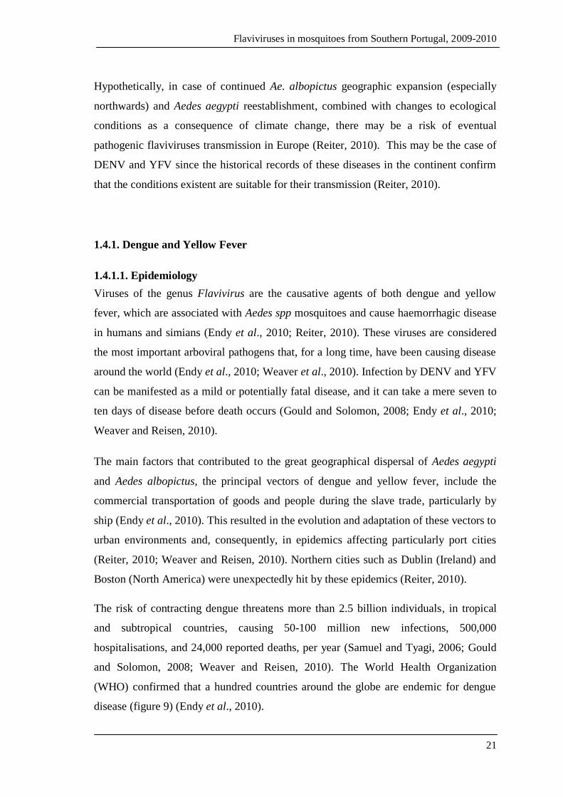
Figure 11: Dengue and yellow fever transmission cycle.
(http://www.stanford.edu/group/parasites/ParaSites2008/Nkem_Cristina%20Valdoinos/ugonabon_
valdovinosc_dengueproposal.htm – accessed on the 6th
July 2011).
Yellow fever can be transmitted via three different cycles (figure 12). In the sylvatic
cycle YFV is transmitted between primates and forest-dwelling mosquitoes, humans
become infected when in contact with the sylvatic cycle through forest invasion and the
virus can then be transmitted via the urban cycle between infected humans and Ae.
aegypti (CDC, 2010b). The intermediate cycle which occurs only in Africa, is
maintained between primates/humans and Aedes spp mosquitoes which bite humans
who live or work in the forest periphery (CDC, 2010b).
Figure 12: Yellow fever transmission cycles.
(http://www.cdc.gov/mmwr/preview/mmwrhtml/rr5907a1.htm - accessed on the 24th
November
2011)
Flaviviruses in mosquitoes from Southern Portugal, 2009-2010
25
1.4.2. West Nile Virus
1.4.2.1. Epidemiology
West Nile virus (WNV) is a mosquito-transmitted flavivirus that belongs to the
Japanese encephalitis antigenic complex (Hubalek et al., 1999; Petersen and Roehrig,
2001). This virus was isolated in 1937 for the first time in the West Nile Province in
Uganda, from the blood of a woman presenting with classical systemic febrile illness
(Hubalek et al., 1999; Monini et al., 2010). The virus was later recognized and isolated
from mosquitoes, birds and humans in the 1950’s, in Egypt, and also in France (1962-
63). During the 1970s-1990s WNV caused isolated outbreaks in countries such as South
Africa (1974), India (1980-81) and Romania (1996) (Hubalek et al., 1999; Zeller and
Schuffenecker, 2004; Monini et al., 2010). Since its first isolation, WNV has been
considered one of the most widely spread flaviviruses thus becoming an increasing
public health and veterinarian problem all over the globe (Zeller and Schuffenecker,
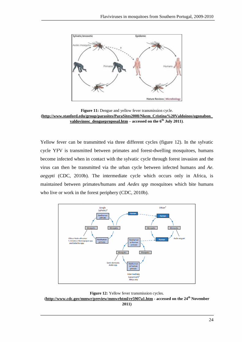
2004; Monini et al., 2010). The virus already affects several European countries,
predominantly in the Mediterranean region, whereas in the Americas its transmission
has been recorded from Canada to Argentina, after its recent introduction in the United
States (figure 13) (Zeller and Schuffenecker, 2004; Monini et al., 2010).
Figure 13: West Nile distribution in North America, as of 15th of November 2011.
(http://www.cdc.gov/ncidod/dvbid/westnile/Mapsactivity/surv&control11MapsAnybyState.htm –
accessed on the 22nd
November 2011).
Flaviviruses in mosquitoes from Southern Portugal, 2009-2010
26
The virus is thought to have entered the United States through viraemic migratory birds
or imported domestic birds, however the exact means of entry are unclear (Epstein and
Defilippo, 2001; Solomon and Mallewa, 2001). WNV is the causative agent of West
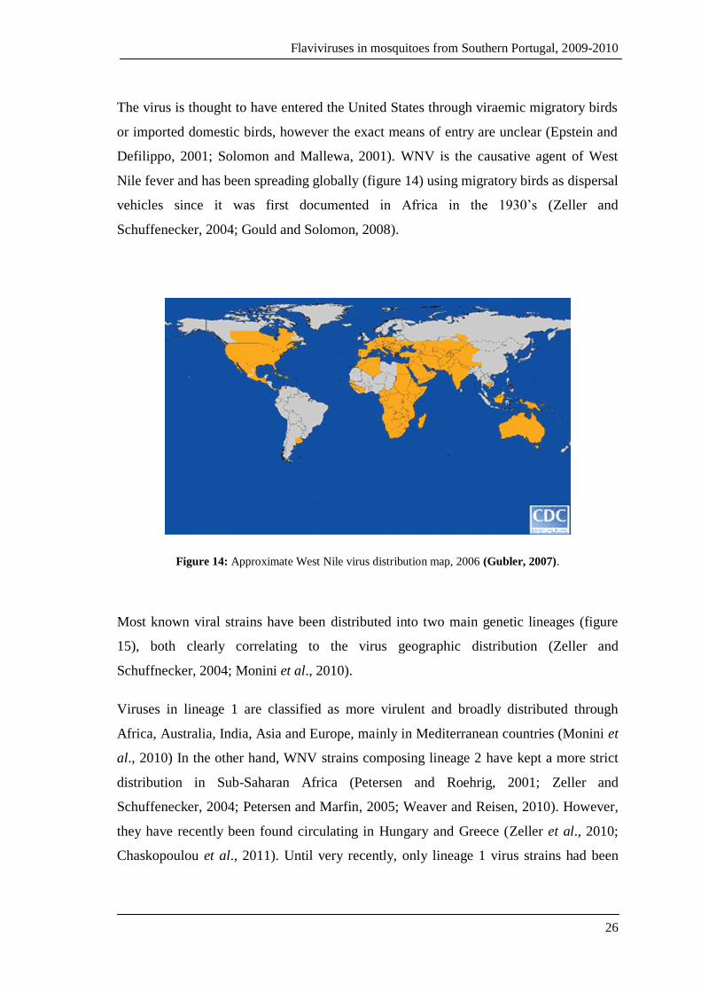
Nile fever and has been spreading globally (figure 14) using migratory birds as dispersal
vehicles since it was first documented in Africa in the 1930’s (Zeller and
Schuffenecker, 2004; Gould and Solomon, 2008).
Figure 14: Approximate West Nile virus distribution map, 2006 (Gubler, 2007).
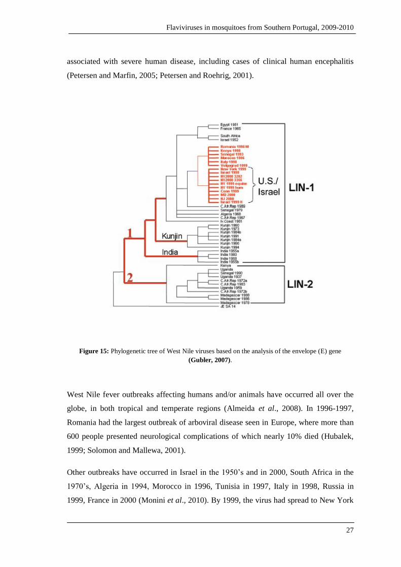
Most known viral strains have been distributed into two main genetic lineages (figure
15), both clearly correlating to the virus geographic distribution (Zeller and
Schuffnecker, 2004; Monini et al., 2010).
Viruses in lineage 1 are classified as more virulent and broadly distributed through
Africa, Australia, India, Asia and Europe, mainly in Mediterranean countries (Monini et
al., 2010) In the other hand, WNV strains composing lineage 2 have kept a more strict
distribution in Sub-Saharan Africa (Petersen and Roehrig, 2001; Zeller and
Schuffenecker, 2004; Petersen and Marfin, 2005; Weaver and Reisen, 2010). However,
they have recently been found circulating in Hungary and Greece (Zeller et al., 2010;
Chaskopoulou et al., 2011). Until very recently, only lineage 1 virus strains had been
Flaviviruses in mosquitoes from Southern Portugal, 2009-2010
27
associated with severe human disease, including cases of clinical human encephalitis
(Petersen and Marfin, 2005; Petersen and Roehrig, 2001).
Figure 15: Phylogenetic tree of West Nile viruses based on the analysis of the envelope (E) gene
(Gubler, 2007).
West Nile fever outbreaks affecting humans and/or animals have occurred all over the
globe, in both tropical and temperate regions (Almeida et al., 2008). In 1996-1997,
Romania had the largest outbreak of arboviral disease seen in Europe, where more than
600 people presented neurological complications of which nearly 10% died (Hubalek,
1999; Solomon and Mallewa, 2001).
Other outbreaks have occurred in Israel in the 1950’s and in 2000, South Africa in the
1970’s, Algeria in 1994, Morocco in 1996, Tunisia in 1997, Italy in 1998, Russia in
1999, France in 2000 (Monini et al., 2010). By 1999, the virus had spread to New York

Flaviviruses in mosquitoes from Southern Portugal, 2009-2010
28
causing 60 clinical cases of encephalitis leading to 7 human deaths with a dramatically
higher equine and bird mortality rate (figure 16) (Hubalek, 1999; Zeller and
Schuffenecker, 2004; Gould and Solomon, 2008).
Figure 16: West Nile virus epidemics between 1937 and 2006 (Gubler, 2007).
Red stars represent epidemics associated with severe neurological disease in both humans and animals.
More recently, nine cases of WNV infection were reported in Italy in 2010, six of which
were symptomatic (Barzon et al., 2011). Three of the patients presenting symptoms
were confirmed to have developed neurological disease (Barzon et al., 2011). In Greece
35 deaths from 262 human cases were reported in that same year (Chaskopoulou et al.,
2011). In 2011, WNV activity was detected in sentinel chickens between May and July,
and by August 37 cases had been registered, of which 31 evolved into neurological
disease (Danis et al., 2011).
Flaviviruses in mosquitoes from Southern Portugal, 2009-2010
29
1.4.2.2. Transmission cycle
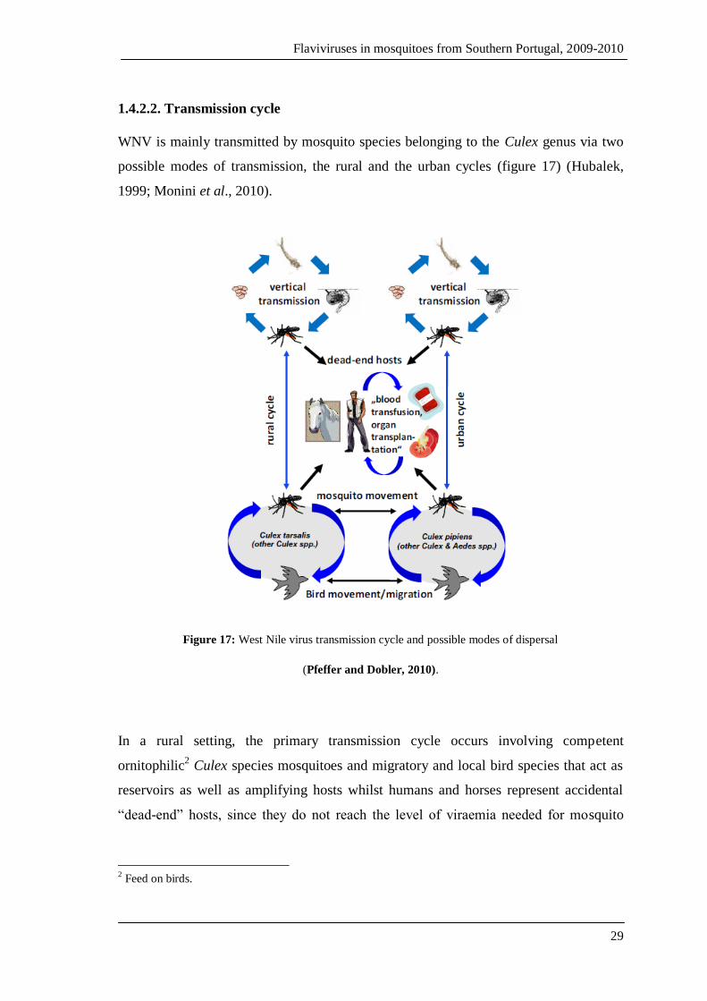
WNV is mainly transmitted by mosquito species belonging to the Culex genus via two
possible modes of transmission, the rural and the urban cycles (figure 17) (Hubalek,
1999; Monini et al., 2010).
Figure 17: West Nile virus transmission cycle and possible modes of dispersal
(Pfeffer and Dobler, 2010).
In a rural setting, the primary transmission cycle occurs involving competent
ornitophilic2 Culex species mosquitoes and migratory and local bird species that act as
reservoirs as well as amplifying hosts whilst humans and horses represent accidental
“dead-end” hosts, since they do not reach the level of viraemia needed for mosquito
2 Feed on birds.

Flaviviruses in mosquitoes from Southern Portugal, 2009-2010
30
infection (Solomon and Mallewa, 2001; Petersen and Marfin, 2005; Monini et al., 2010;
Weaver and Reisen, 2010).
The urban cycle occurs in a similar way, however it does involve different vector and
bird species; for example, in this transmission mode, the principal vectors are Culex
pipiens and Cx. molestus seen as both species feed on synanthropic3, domestic birds and
humans (Hubalek, 1999).
Moreover, the main WNV vectors vary according to their geographic distribution, for
example, the predominant vector species in Europe are Culex pipiens, Cx. molestus and
Coquillettidia richiardii (Hubalek, 1999). In Africa and The Middle East, the most
important vector is Culex univittatus, whereas in Asia, Culex quinquefasciatus, Cx.
tritaeniorhyncus and Cx. vishnui are the main vectors (Hubalek, 1999).
Although the rural cycle is of utmost importance for WNV transmission, this flavivirus
can also be transmitted via organ transplants, blood transfusions and via infectious
maternal milk (Zeller and Schuffenecker, 2004; Gould and Solomon, 2008).
Additionally, reports show that transovarial transmission occurs in some species,
namely Cx. univittatus, Cx. tritaeniorhyncus, Aedes aegypti and Ae. albopictus
(Hubalek,1999).
1.4.2.3. Clinical features
West Nile disease was at first considered a minor health concern, as the majority of
infections were mild or completely asymptomatic; however, after the major outbreaks
that occurred in Europe and especially in North America from 1999, WN disease
became a public health priority (Zeller and Schuffenecker, 2004; Gould and Solomon,
2008).
After an incubation period of 3 to 14 days, between 15-20% of humans present with a
mild febrile illness accompanied by flu-like symptoms, a rapid onset of moderate to
high fever, headache, malaise, myalgia, anorexia, nausea, backache and retro-orbital
3 Ecologically associated with humans.
Flaviviruses in mosquitoes from Southern Portugal, 2009-2010
31
pain (Hubalek, 1999; Zeller and Schuffenecker, 2004; Gould and Solomon, 2008).
Other disease manifestations include lymphadenopathy, conjunctivitis, maculopapular
or roseolar rash, which is normally observed in 50% of patients (Hubalek, 1999; Gould
and Solomon, 2008). Around 1% of all patients tend to develop neurological signs such
as acute aseptic meningitis, encephalitis or myelitis4 (Hubalek, 1999; Zeller and
Schuffenecker, 2004). Additionally, severe infections may also provoke hepato- and
splenomegaly, hepatitis, pancreatitis and myocarditis (Zeller and Schuffenecker, 2004;
Gould and Solomon, 2008). A mortality rate of 5 to 10% usually results from all
patients presenting with neurological symptoms (Gould and Solomon, 2008).
During the outbreaks in North America, Romania, Russia and Israel mortality rates of
5% to 14% among neurologically affected patients were registered, as well as other
symptoms such as serious motor complications, skin rash and lymphadenopathy
(Petersen and Roehrig, 2001). In the Romania outbreak alone, 17 fatalities resulted from
352 neurological affected patients, from whom 44% presented with
meningoencephalitis, a further 40% had meningitis and the remaining 16% suffered
from encephalitis (Zeller and Schuffenecker, 2004). In accordance with most studies,
fatalities are more likely to arise among patients over 50 years of age (Zeller and
Schuffenecker, 2004; Gould and Solomon, 2008).
1.5. Insect-specific flaviviruses
This group of flaviviruses have had many designations throughout the years, from “non-
classical” to “mosquito-only” flaviviruses. However, as the latter designation is
somehow conflicting since insect-specific flaviviruses have been detected in field
caught phlebotomine sand flies and ticks it completely invalidates the continued use of
that designation, hence in this study it is referred to as insect-specific flaviviruses.
As previously mentioned, most viruses within the genus Flavivirus are arthropod-borne,
and only a few have no-known vectors (Crabtree et al., 2003; Cook et al., 2003; Sang et
al., 2003; Mukhopadhyay et al., 2005). However, the increasing surveillance and
investigations into these pathogens have lead to a better understanding of their genetics,
4 Inflammation of the spinal cord.
Flaviviruses in mosquitoes from Southern Portugal, 2009-2010
32
classification and phylogenetic relationships (Sang et al., 2003). It is now common
knowledge that pathogenic flaviviruses do not exhaust the Flaviviridae family, as it also
harbours another type of viruses that are, so far, not known to be medically important
(Blitvitch et al., 2009). On the contrary, they are thought to be insect-specific since they
replicate in mosquitoes but have not yet been detected in a vertebrate host. They are,
thus, designated by insect-specific flaviviruses (ISFs) (Blitvitch et al., 2009). ISFs are a
heterogeneous group of highly diverse and widely geographically dispersed viruses.
Even though most ISFs have only been described and classified over the past decade, it
is certain that they have existed for much longer. For example, the first one was found
many years ago and was referred to as “an agent in the Ae. aegypti cell line that causes
fusion of Ae. albopictus cells” by Stollar and Thomas (1975), and was, as a result,
named cell-fusing agent virus (CFAV). This insect-specific flavivirus causes
distinguishable massive syncytia formation, an effect that had only been observed with
JEV, WNV and DENV (Stollar and Thomas, 1975; Igarashi et al., 1976). However, it
was stated that syncytium formation was not observed immediately after infection,
instead, it became evident after 60 hours of cell infection and, after 72 hours more than
90% of the cells in culture would be fused having formed large syncytia (Stollar and
Thomas, 1975). Infection of mammalian cells with CFAV resulted in no cytopathic
effect (CPE), including cell fusion. Indeed, the virus does not seem to replicate at all,
thus confirming that it is insect-specific (Stollar and Thomas, 1975; Sang et al., 2003).
In addition, replication in Ae. aegypti cells is not cytopathic (Stollar and Thomas, 1975).
Further analysis revealed that CFAV did not cross react with other known flaviviruses
(Igarashi et al., 1976). CFAV was described as similar to togaviruses in size and
morphology, whereas other characteristics indicated strong similitude to flaviviruses
(Igarashi et al., 1976). However, despite all evidence and tests, the classification of
CFAV remains unresolved.
The isolation of a new flavivirus described as CFA-like from mosquitoes collected in
Kenya’s Central Province, during a rainy season in 1999 was reported and later
genetically and phenotypically characterised (Sang et al., 2003).

Flaviviruses in mosquitoes from Southern Portugal, 2009-2010
33
This virus, named Kamiti River virus (KRV), was isolated from Ae. macintoshi
immature mosquitoes and was temporarily classified as a flavivirus (Crabtree et al.,
2003). The fact that KRV was isolated from adult male and female mosquitoes that
were collected, in nature, in their immature forms, is indicative of the maintenance of
this virus via transovarial transmission between generations. Nonetheless, there also
remains the possibility that the virus may have been acquired by larval ingestion
(Crabtree et al., 2003).
Like CFAV, only replicated in mosquito cells and presented no antigenic cross-reaction
with other arboviruses (Sang et al., 2003). However, contrarily to CFAV, there was no
cell fusion observed in culture, despite the morphologic and genomic similarities
(Crabtree et al., 2003). The genomic organisation of KRV was found to be similar to
that of other flaviviruses, though its nucleotide sequence was considerably longer
(11,375 nt) (Crabtree et al., 2003). Two viral strains were isolated and their RNA
sequences compared which revealed they were virtually identical, thus suggesting they
represent the same virus; however, when comparison was extended to other flaviviruses
a low sequence identity was observed (Crabtree et al., 2003; Sang et al., 2003). KRV
showed maximum identity to CFAV based on nucleotide and amino acid sequence
analysis of the NS3 and NS5 genes (Crabtree et al., 2003; Sang et al., 2003). Both KRV
isolates produced cytopathic effect5 (CPE) in Ae. albopictus cells (C6/36) in culture.
However, the same was not observed with Vero cells (monkey kidney cell line) or in
baby hamster kidney cells (BHK-21), as expected (Crabtree et al., 2003; Sang et al.,
2003).
Some years later, the discovery of 40 CFA-like viral sequences from adult mosquitoes
collected in Puerto Rico after the rainy season of 2002 was reported (Cook et al., 2006).
These were obtained from male and female mosquitoes of different species, namely Ae.
aegypti, Ae. albopictus and Culex sp. The sequences analysed were found to represent
different strains of CFAV and, therefore have been referred to as CFAV Culebra stains
(Cook et al., 2006).
5 Structural cell changes, such as cell degeneration or detachment, caused by viral infection.
Flaviviruses in mosquitoes from Southern Portugal, 2009-2010
34
In addition to CFAV and KRV, other insect-specific flaviviruses have been detected and
isolated. However, the new isolate seems to be associated only to Culex sp. mosquitoes,
especially Cx. pipiens, hence their name Culex flavivirus (CxFV) (Hoshino et al., 2007).
The genome of this new flavivirus is 10,834 nucleotides long, also flanked by two
untranslated (UTR) regions, as described for other flaviviruses (Hoshino et al., 2007).
Infection of mosquito C6/36 cells in culture resulted in moderate CPE visible 4 days
post-infection. In addition, no viral replication was detected in mammalian cell lines
(Hoshino et al., 2007). Phylogenetically, CxFV showed greater similarities with CFAV
and KRV than with other mosquito-transmitted flaviviruses (Hoshino et al., 2007).
Alike the CFA-like viruses and KRV, CxFV have been detected in healthy wild-caught
mosquitoes, thus suggesting once again that vertical transmission occurs in the wild,
and despite causing moderate CPE in culture, it does not seem to negatively affect their
hosts. (Hoshino et al., 2007).
A virus similar to CxFV found in Japan was identified in Guatemala and was later
named CxFV Izabal 2006; it was isolated from Culex quinquefasciatus collected from
urban and rural areas between March and October 2006 (Morales-Betoulle et al., 2008).
Analysis of the envelope and NS5 gene sequences were the basis for characterisation of
the detected viral strains which, after phylogenetic assessment were classified as strains
of the CxFV species following a result of 89% nucleotide homology with the Japanese
virus (Morales-Betoulle et al., 2008). The isolates were cultured into C6/36 and Vero
cells, with no CPE observed for either (Morales-Betoulle et al., 2008).
In addition to the diversity of this heterogeneous group of flaviviruses previously
identified, Crabtree et al (2009) have also found a new insect-only flavivirus from Cx.
tritaeniorhyncus captured in Vietnam in August 2002. These virus, denominated Quang
Binh virus (QBV) was described as genetically different from pre-existing flaviviruses
and though its complete genome had similar size (10,865 nt) and genomic organisation
(Crabtree et al., 2009). Sequence analysis revealed that Quang Binh virus had over 60%
identity with Japanese CxFV and, when in culture, it induced plaque formation and CPE
only in C6/36 cells. (Crabtree et al., 2009).

Flaviviruses in mosquitoes from Southern Portugal, 2009-2010
35
Meanwhile, a new ISF of Asian origin was discovered and designated Aedes flavivirus
(AEFV) (Hoshino et al., 2009). Contrarily to most pre-existing flaviviruses, AEFV was
isolated from Aedes and not Culex mosquitoes, namely Ae. albopictus and Ae.
flavopictus collected from Japan and Indonesia (Hoshino et al., 2009). However, other
flavivirus-like sequences were also obtained (Hoshino et al., 2009). Infection of C6/36
produced moderate CPE after 4 days, whereas, as expected for ISFs, mammalian cells
showed no such effects (Hoshino et al., 2009). AEFV was therefore classified a
flavivirus with similar replication and translation mechanisms; furthermore, it was
stated that it showed high virus-host adaptation to its invertebrate host, an Aedes
mosquito (Hoshino et al., 2009).
AEFV’s genome mirrors that of flaviviruses and its polyprotein is thought to be divided
into 3 structural (C, prM and E) and seven non-structural proteins (NS1, NS2A, NS2B,
NS3, NS4A, NS4B and NS5) (Hoshino et al., 2009). AEFV was classified as a new
species of insect flavivirus, particularly similar to KRV (Hoshino et al., 2009).
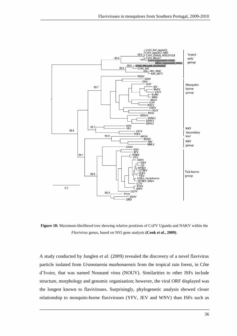
Insect-only flaviviruses previously characterised have been isolated from Aedes and
Culex mosquitoes; however, a study carried out by Cook and others (2009) reports the
first isolations of a ISF from a Mansonia species mosquito and an African strain of
CxFV. Both new strains, namely CxFV Uganda and Nakiwogo virus (NAKV) were
obtained from mosquitoes collected in the same trap (CDC-type, with CO2) on the 24th
of February 2008 in Entebbe, Uganda (Cook et al., 2009). The NAKV and CxFV
Uganda strain were isolated from Mansonia africana nigerrima and Cx.
quinquefasciatus, respectively (Cook et al., 2009). The genomes of both viruses were
found to be 10,092 nt long for CxFV, and 10,122 nt long for NAKV (Cook et al., 2009).
These viruses were morphologically identical to flaviviruses, and phylogenetic analysis
revealed that CxFV Uganda to be more closely related to the Mexico strain, while
NAKV was inserted into the ISF clade, but as a sister group (figure 18) (Cook et al.,
2009).
Infection of C6/36 cells with NAKV resulted in moderate CPE with the formation of
large syncytia, similarly to what was described for CFA, whereas CxFV induced
structural changes, causing them to turn triangular from their normal circular shapes,
and reduced density (Cook et al., 2009).
Flaviviruses in mosquitoes from Southern Portugal, 2009-2010
36
Figure 18: Maximum-likelihood tree showing relative positions of CxFV Uganda and NAKV within the
Flavivirus genus, based on NS5 gene analysis (Cook et al., 2009).
A study conducted by Junglen et al. (2009) revealed the discovery of a novel flavivirus
particle isolated from Uranotaenia mashonaensis from the tropical rain forest, in Côte
d’Ivoire, that was named Nounané virus (NOUV). Similarities to other ISFs include
structure, morphology and genomic organisation; however, the viral ORF displayed was
the longest known to flaviviruses. Surprisingly, phylogenetic analysis showed closer
relationship to mosquito-borne flaviviruses (YFV, JEV and WNV) than ISFs such as

Flaviviruses in mosquitoes from Southern Portugal, 2009-2010
37
CFAV and KRV (Junglen et al., 2009). As observed for ISFs, viral replication was
observed only in insect-cell culture (Junglen et al., 2009).
Further away, in North America many other flavivirus-like sequences have been found.
For example, as a result of a surveillance programme in Alberta (Canada), some
flaviviruses were detected, the majority were isolated from Culex tarsalis mosquitoes
while a lower number was obtained from non-Culex mosquitoes; these sequences
showed higher similarities to KRV (Pabbaraju et al., 2009). In Iowa, again as part of the
surveillance programme, mosquitoes were collected between May and October 2007, of
which Cx. pipiens and Cx. tarsalis pools turned out positive for flaviviruses (Blitvich et
al., 2009). Analysis of the sequences obtained revealed that the isolates had 98%
homology with CxFV isolated in Japan but also shows close relation to the Texas and
Mexico strains (Huhtamo et al., 2009). It was characterised as having a 10,089 long
genomic sequence and production of moderate CPE and cell clumping were observed in
culture with insect cell lines only (Huhtamo et al., 2009).
Once again, it was due to arboviral surveillance programmes that flavivirus strains have
been found and isolated from mosquitoes collected from January to December 2007, in
the Yucatan Peninsula of Mexico (Farfan-Ale et al., 2009). Several of the analysed
pools tested positive for flaviviruses, the majority of them showing identical sequences
to that of CxFV (Farfan-Ale et al., 2009). A new strain of CxFV closely related to the
Japanese strain (figure 19) was isolated from Cx. quinquefasciatus, and as some pools
consisted only of male specimens, this suggested, again, vertical transmission in nature
(Farfan-Ale et al., 2009). However, contrarily to previously isolated strains of CxFV,
this virus did not produce CPE on C6/36 cells (Farfan-Ale et al., 2009).
It is noteworthy that CxFV presented a high prevalence, as it was found in 145 of 146
pools from which flavivirus RNA was detected (Farfan-Ale et al., 2009; Kim et al.,
2009).
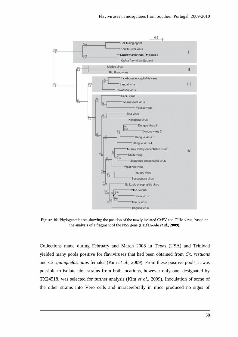
Flaviviruses in mosquitoes from Southern Portugal, 2009-2010
38
Figure 19: Phylogenetic tree showing the position of the newly isolated CxFV and T’Ho virus, based on
the analysis of a fragment of the NS5 gene (Farfan-Ale et al., 2009).
Collections made during February and March 2008 in Texas (USA) and Trinidad
yielded many pools positive for flaviviruses that had been obtained from Cx. restuans
and Cx. quinquefasciatus females (Kim et al., 2009). From these positive pools, it was
possible to isolate nine strains from both locations, however only one, designated by
TX24518, was selected for further analysis (Kim et al., 2009). Inoculation of some of
the other strains into Vero cells and intracerebrally in mice produced no signs of
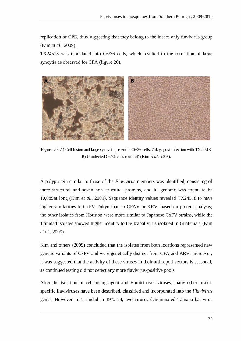
Flaviviruses in mosquitoes from Southern Portugal, 2009-2010
39
replication or CPE, thus suggesting that they belong to the insect-only flavivirus group
(Kim et al., 2009).
TX24518 was inoculated into C6/36 cells, which resulted in the formation of large
syncytia as observed for CFA (figure 20).
Figure 20: A) Cell fusion and large syncytia present in C6/36 cells, 7 days post-infection with TX24518;
B) Uninfected C6/36 cells (control) (Kim et al., 2009).
A polyprotein similar to those of the Flavivirus members was identified, consisting of
three structural and seven non-structural proteins, and its genome was found to be
10,089nt long (Kim et al., 2009). Sequence identity values revealed TX24518 to have
higher similarities to CxFV-Tokyo than to CFAV or KRV, based on protein analysis;
the other isolates from Houston were more similar to Japanese CxFV strains, while the
Trinidad isolates showed higher identity to the Izabal virus isolated in Guatemala (Kim
et al., 2009).
Kim and others (2009) concluded that the isolates from both locations represented new
genetic variants of CxFV and were genetically distinct from CFA and KRV; moreover,
it was suggested that the activity of these viruses in their arthropod vectors is seasonal,
as continued testing did not detect any more flavivirus-positive pools.
After the isolation of cell-fusing agent and Kamiti river viruses, many other insect-
specific flaviviruses have been described, classified and incorporated into the Flavivirus
genus. However, in Trinidad in 1972-74, two viruses denominated Tamana bat virus
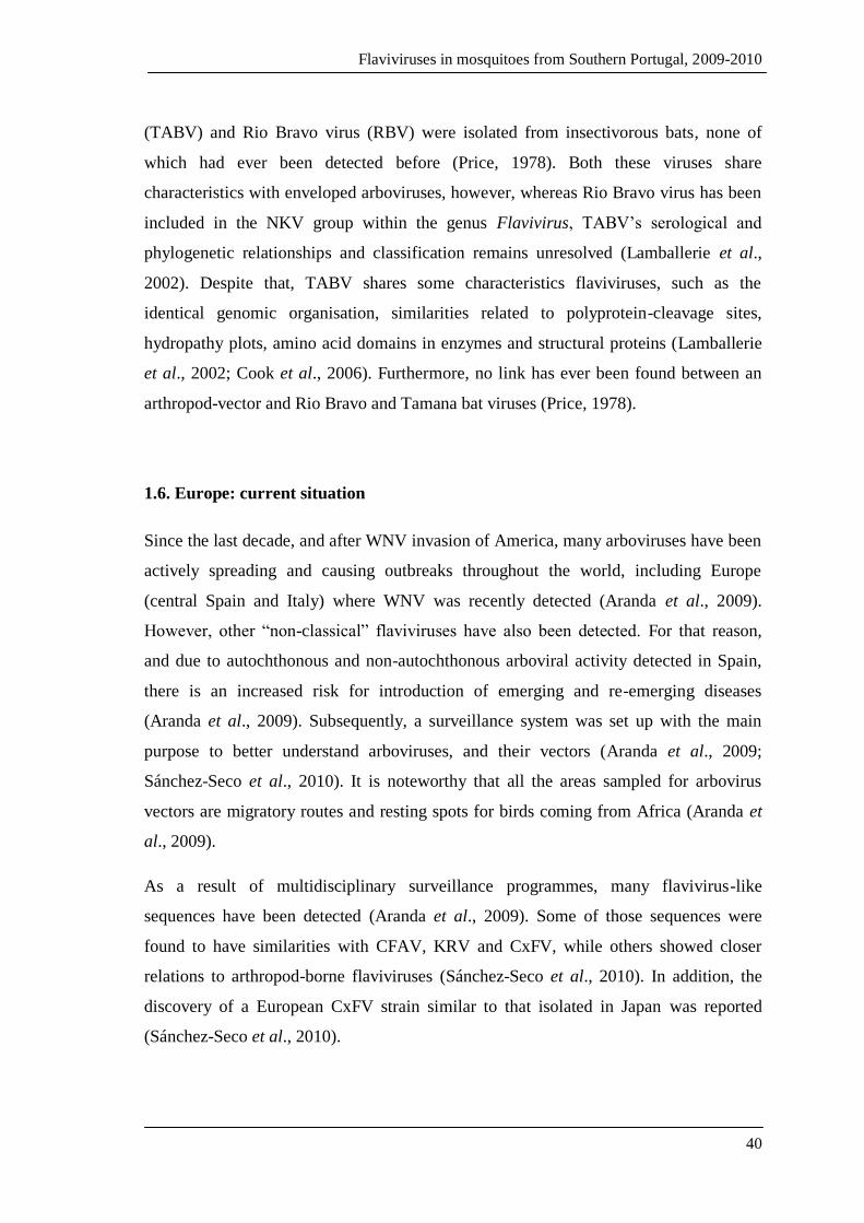
Flaviviruses in mosquitoes from Southern Portugal, 2009-2010
40
(TABV) and Rio Bravo virus (RBV) were isolated from insectivorous bats, none of
which had ever been detected before (Price, 1978). Both these viruses share
characteristics with enveloped arboviruses, however, whereas Rio Bravo virus has been
included in the NKV group within the genus Flavivirus, TABV’s serological and
phylogenetic relationships and classification remains unresolved (Lamballerie et al.,
2002). Despite that, TABV shares some characteristics flaviviruses, such as the
identical genomic organisation, similarities related to polyprotein-cleavage sites,
hydropathy plots, amino acid domains in enzymes and structural proteins (Lamballerie
et al., 2002; Cook et al., 2006). Furthermore, no link has ever been found between an
arthropod-vector and Rio Bravo and Tamana bat viruses (Price, 1978).
1.6. Europe: current situation
Since the last decade, and after WNV invasion of America, many arboviruses have been
actively spreading and causing outbreaks throughout the world, including Europe
(central Spain and Italy) where WNV was recently detected (Aranda et al., 2009).
However, other “non-classical” flaviviruses have also been detected. For that reason,
and due to autochthonous and non-autochthonous arboviral activity detected in Spain,
there is an increased risk for introduction of emerging and re-emerging diseases
(Aranda et al., 2009). Subsequently, a surveillance system was set up with the main
purpose to better understand arboviruses, and their vectors (Aranda et al., 2009;
Sánchez-Seco et al., 2010). It is noteworthy that all the areas sampled for arbovirus
vectors are migratory routes and resting spots for birds coming from Africa (Aranda et
al., 2009).
As a result of multidisciplinary surveillance programmes, many flavivirus-like
sequences have been detected (Aranda et al., 2009). Some of those sequences were
found to have similarities with CFAV, KRV and CxFV, while others showed closer
relations to arthropod-borne flaviviruses (Sánchez-Seco et al., 2010). In addition, the
discovery of a European CxFV strain similar to that isolated in Japan was reported
(Sánchez-Seco et al., 2010).
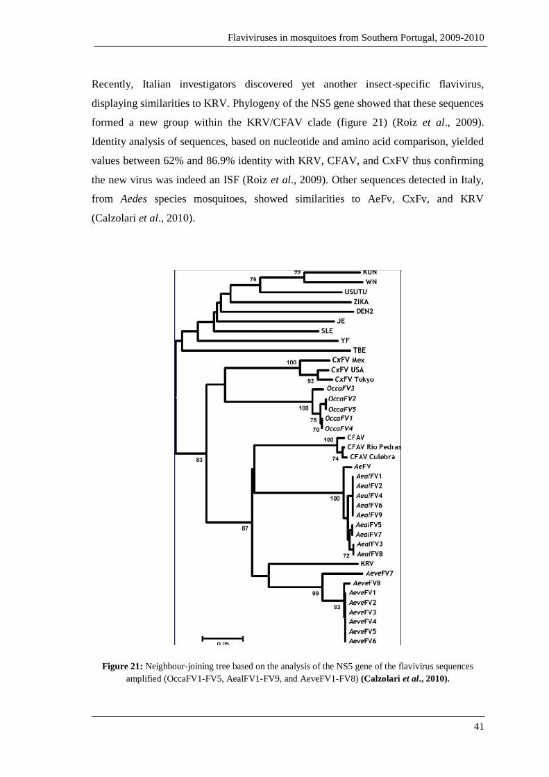
Flaviviruses in mosquitoes from Southern Portugal, 2009-2010
41
Recently, Italian investigators discovered yet another insect-specific flavivirus,
displaying similarities to KRV. Phylogeny of the NS5 gene showed that these sequences
formed a new group within the KRV/CFAV clade (figure 21) (Roiz et al., 2009).
Identity analysis of sequences, based on nucleotide and amino acid comparison, yielded
values between 62% and 86.9% identity with KRV, CFAV, and CxFV thus confirming
the new virus was indeed an ISF (Roiz et al., 2009). Other sequences detected in Italy,
from Aedes species mosquitoes, showed similarities to AeFv, CxFv, and KRV
(Calzolari et al., 2010).
Figure 21: Neighbour-joining tree based on the analysis of the NS5 gene of the flavivirus sequences
amplified (OccaFV1-FV5, AealFV1-FV9, and AeveFV1-FV8) (Calzolari et al., 2010).
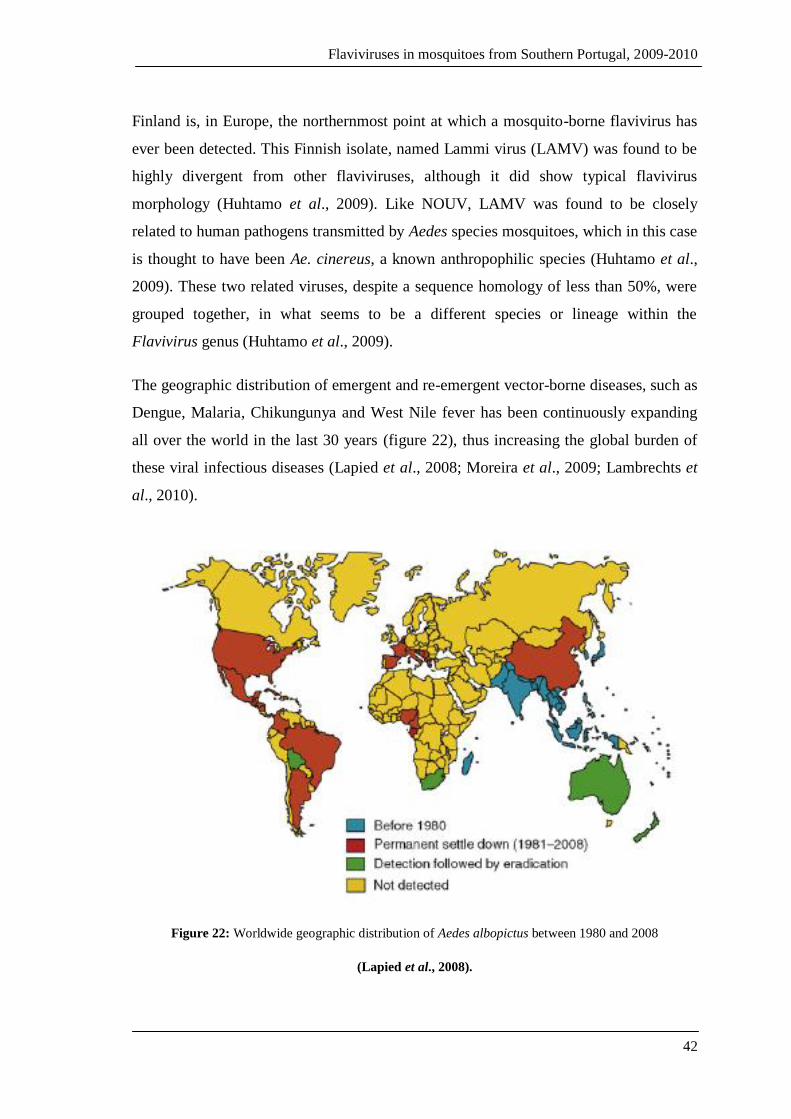
Flaviviruses in mosquitoes from Southern Portugal, 2009-2010
42
Finland is, in Europe, the northernmost point at which a mosquito-borne flavivirus has
ever been detected. This Finnish isolate, named Lammi virus (LAMV) was found to be
highly divergent from other flaviviruses, although it did show typical flavivirus
morphology (Huhtamo et al., 2009). Like NOUV, LAMV was found to be closely
related to human pathogens transmitted by Aedes species mosquitoes, which in this case
is thought to have been Ae. cinereus, a known anthropophilic species (Huhtamo et al.,
2009). These two related viruses, despite a sequence homology of less than 50%, were
grouped together, in what seems to be a different species or lineage within the
Flavivirus genus (Huhtamo et al., 2009).
The geographic distribution of emergent and re-emergent vector-borne diseases, such as
Dengue, Malaria, Chikungunya and West Nile fever has been continuously expanding
all over the world in the last 30 years (figure 22), thus increasing the global burden of
these viral infectious diseases (Lapied et al., 2008; Moreira et al., 2009; Lambrechts et
al., 2010).
Figure 22: Worldwide geographic distribution of Aedes albopictus between 1980 and 2008
(Lapied et al., 2008).
Flaviviruses in mosquitoes from Southern Portugal, 2009-2010
43
As previously described, many factors have contributed to that range expansion; they
include the increasing human movement, demographic growth, ecologic and climatic
alterations probably resulting from global warming effects, as well as economic
determinants that may restrict vaccination and vector control programmes (Lapied et al.,
2008; Moreira et al., 2009). However, viral evolution has promoted increased virulence
and allowed viruses to better adapt to a wider variety of vectors, as is the case for West
Nile and Chikungunya viruses (Lapied et al., 2008). Additionally, the distribution of
these potentially fatal diseases is usually parallel and highly dependent on the presence
and range distribution of their vectors, especially Ae. aegypti and Ae. albopictus
(Lambrechts et al., 2010).
The past three decades have seen Ae. albopictus spread to all continents from its origins
in Asia; and the major cause for concern is that this mosquito species is known to be
experimentally susceptible to the infection of at least 22 arboviruses (Lapied et al.,
2008). Moreover, the presence and establishment of Ae. aegypti in certain regions is
also an important factor in the increasing risk of arboviral disease transmission in
Europe and neighbouring countries.
Aedes aegypti is the main and most competent vector for the transmission of dengue
viruses, but Ae. albopictus is also capable of transmitting the viruses even though it is
considered a secondary vector mainly because it is considered less competent and
presents different host preferences (Lambrechts et al., 2010). Moreover, each species is
associated with a different degree of disease severity, being Ae. aegypti associated with
the more serious forms of dengue such as dengue haemorrhagic fever whereas Ae.
albopictus is usually linked to the classical dengue fever (Lambrechts et al., 2010).
Aedes albopictus has become established in many European countries, especially those
situated around the Mediterranean Sea, and including Albania, Greece, Serbia and
Montenegro, Croatia, France, Italy, Spain, but also in the Netherlands, and in
Switzerland (figure 23) (Lambrechts et al., 2010).
The establishment of Ae. albopictus in Europe was first reported in Albania in 1979,
and has since spread to Italy where, in 1991, a breeding population was found followed
by the subsequent invasion across the country; and in 1997, the vector finally reached

Flaviviruses in mosquitoes from Southern Portugal, 2009-2010
44
Rome where colonies were found throughout the entire city (Aranda et al., 2006; Lapied
et al., 2008).
In 1999, populations of Ae. albopictus were discovered in tyre dumps in France,
followed by their appearance in Belgium in 2000; after that and more recently countries
such as Switzerland, Greece, Serbia and Montenegro have reported the establishment of
this mosquito species (Aranda et al., 2006; Lapied et al., 2008). However, other regions
have been invaded by populations of Ae. albopictus and their range expansion is still
ongoing (figure 23) (Aranda et al., 2006).
Figure 23: Distribution of Aedes albopictus in Europe, as of 2009 (adapted from Reiter, 2010).

Flaviviruses in mosquitoes from Southern Portugal, 2009-2010
45
As shown above, Ae. albopictus has been found all over Europe, with particular
emphasis in Italy where it has been established for over a decade, and has also been
recorded since 2005 in France, all down the length of the Italian border (Lapied et al.,
2008). For that reason, it is not surprising that major outbreaks have occurred with this
mosquito as the potential vector incriminated for the 2007 Chikungunya outbreak,
which resulted in the infection of 205 individuals (Rezza et al., 2007; Lapied et al.,
2008). Additionally, in August 2004, following complaints of the presence of “black
flies” in a residential area in the capital city of Catalonia, the search for which started by
sampling breeding sites that lead to the finding of an adult male and many immature
forms to be collected (Aranda et al., 2006). From the immature forms, another male
emerged and after identification, both were confirmed as being Ae. albopictus (Aranda
et al., 2006). Consequently, many other searches for these mosquitoes resulted in the
discovery of other adults, males and females, an also immature forms; thus suggesting
that this species was already established in the country (Aranda et al., 2006). Following
these events, further investigations took place leading to the conclusion that these
mosquitoes had been settled for at least two years (Aranda et al., 2006).
West Nile and Usutu virus activity has been reported in Europe, and recently a highly
lethal (for humans) WN strain was circulating in Northern Greece in 2010 and more
cases have been reported during this year in other parts of the country (Chaskopoulou et
al., 2011; Vázquez et al., 2011a). WNV was also detected in Russia and Spain affecting
humans and horses (Zeller et al., 2010; Vázquez et al., 2011a). Usutu virus was detected
in Italy in the summer of 2009 and caused neurological involvement in humans, among
other symptoms; and although it has been actively circulating in Spain, no cases have
been detected so far (Vázquez et al., 2011a). It has been postulated that WNV and
Usutu virus are circulating endemically around the Mediterranean, and in some areas
co-circulation of these two flaviviruses has been reported (Vázquez et al., 2011a).

Flaviviruses in mosquitoes from Southern Portugal, 2009-2010
46
1.7. Portugal: the imminent risk
A potentially dangerous tropical disease vector is now present and well established in
the Iberian Peninsula, in Spain (Aranda et al., 2006). Spain and Portugal present
favourable ecological and climatic conditions for the development of certain arthropods
that may act as disease vectors, especially those of tropical origins brought by travelling
or transportation of goods (Aranda et al., 2006). The main means of dispersal of Aedes
albopictus are known to be the transportation of immature forms in tyres and, more
recently, it was found that the importation of the Lucky Bamboo plant (Dracaena spp.)
was also to blame (Aranda et al., 2006). However, it has been suggested that the
transportation of adult mosquitoes in cars or lorries may have played an essential part in
their geographical spread (Aranda et al., 2006). Furthermore, the Iberian Peninsula is
directly under the flight path of migratory birds coming from Africa (figure 24), this
alone presents a relatively high risk of the introduction of arboviruses, since there is a
vast fauna of ornitophilic mosquitoes established in Portugal and Spain, such as Culex
species mosquitoes. Usually, warmer temperatures and rainy seasons determine
mosquito density peaks; however, climate changes due to global warming could bring
storms, flash floods, heat waves and droughts, as has been predicted to occur more
frequently and intensely in Portugal and other countries with temperate climates in
Europe (Epstein, 2000; Santos et al., 2002; Santos and Miranda, 2006). These extreme
climatic events would greatly benefit high population densities due to the reduction of
mosquito predators in nature, the hatching of pre-existing desiccated eggs (floods),
creation of new breeding sites and larval habitats (Epstein, 2000; Santos et al., 2002).
Conditions in Portugal favour the breeding of large populations of anthropophilic
mosquitoes as well as, for example, Culex species mosquitoes that may act as bridge
vectors of West Nile. The Culex pipiens complex is composed of two forms, the
“pipiens” and “molestus” forms; hybrids of these two species have been described as
opportunistic feeders that, for that reason, are considered epidemiologically important
as they may act as bridge vectors of viruses such as WNV (Gomes et al., 2009).
Moreover, since the only member of the complex present in our collections was the
ornitophilic “pipiens” form that reaches the highest densities in estuarine regions, some
of which harbour migratory birds from Africa, their presence in conjunction with
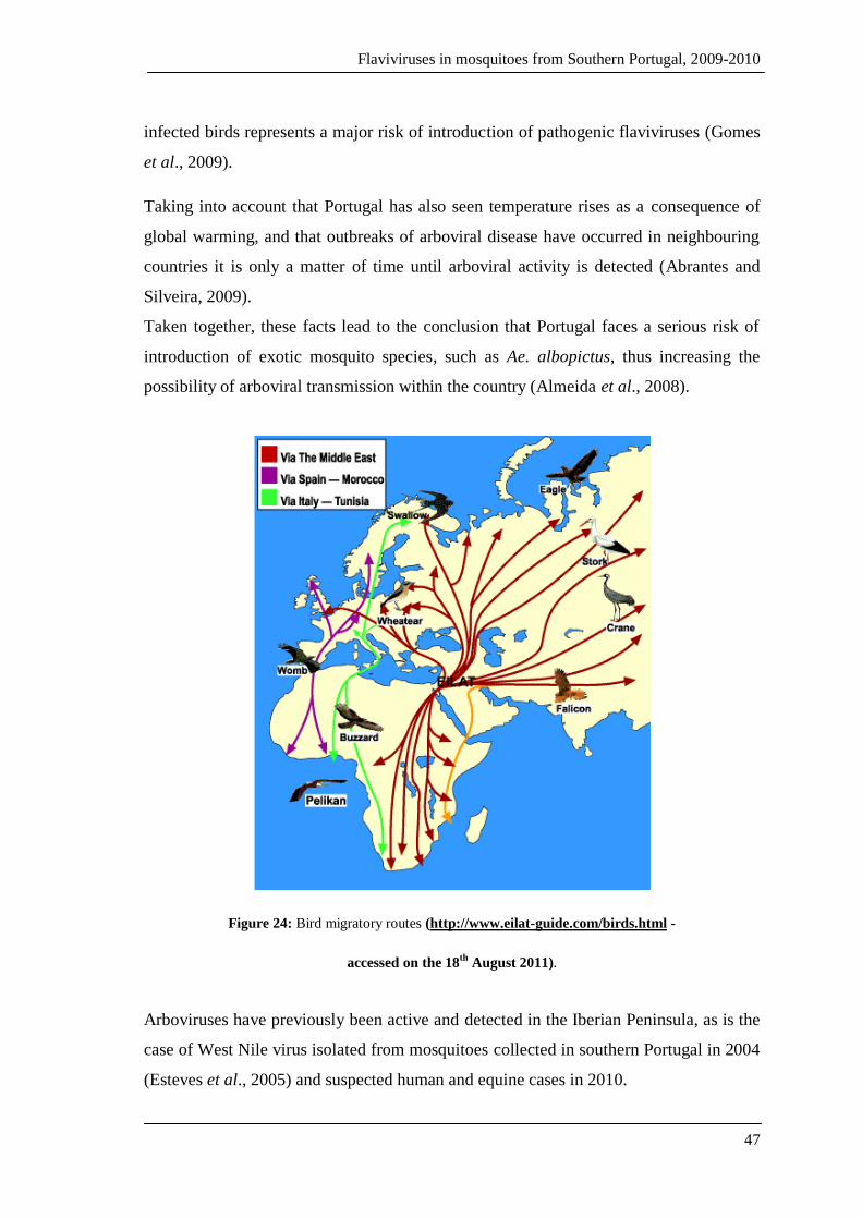
Flaviviruses in mosquitoes from Southern Portugal, 2009-2010
47
infected birds represents a major risk of introduction of pathogenic flaviviruses (Gomes
et al., 2009).
Taking into account that Portugal has also seen temperature rises as a consequence of
global warming, and that outbreaks of arboviral disease have occurred in neighbouring
countries it is only a matter of time until arboviral activity is detected (Abrantes and
Silveira, 2009).
Taken together, these facts lead to the conclusion that Portugal faces a serious risk of
introduction of exotic mosquito species, such as Ae. albopictus, thus increasing the
possibility of arboviral transmission within the country (Almeida et al., 2008).
Figure 24: Bird migratory routes (http://www.eilat-guide.com/birds.html -
accessed on the 18th
August 2011).
Arboviruses have previously been active and detected in the Iberian Peninsula, as is the
case of West Nile virus isolated from mosquitoes collected in southern Portugal in 2004
(Esteves et al., 2005) and suspected human and equine cases in 2010.
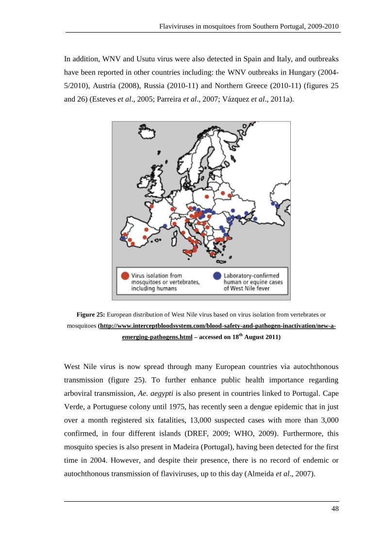
Flaviviruses in mosquitoes from Southern Portugal, 2009-2010
48
In addition, WNV and Usutu virus were also detected in Spain and Italy, and outbreaks
have been reported in other countries including: the WNV outbreaks in Hungary (2004-
5/2010), Austria (2008), Russia (2010-11) and Northern Greece (2010-11) (figures 25
and 26) (Esteves et al., 2005; Parreira et al., 2007; Vázquez et al., 2011a).
Figure 25: European distribution of West Nile virus based on virus isolation from vertebrates or
mosquitoes (http://www.interceptbloodsystem.com/blood-safety-and-pathogen-inactivation/new-a-
emerging-pathogens.html – accessed on 18th
August 2011)
West Nile virus is now spread through many European countries via autochthonous
transmission (figure 25). To further enhance public health importance regarding
arboviral transmission, Ae. aegypti is also present in countries linked to Portugal. Cape
Verde, a Portuguese colony until 1975, has recently seen a dengue epidemic that in just
over a month registered six fatalities, 13,000 suspected cases with more than 3,000
confirmed, in four different islands (DREF, 2009; WHO, 2009). Furthermore, this
mosquito species is also present in Madeira (Portugal), having been detected for the first
time in 2004. However, and despite their presence, there is no record of endemic or
autochthonous transmission of flaviviruses, up to this day (Almeida et al., 2007).

Flaviviruses in mosquitoes from Southern Portugal, 2009-2010
49
Figure 26: West Nile virus transmission in European countries.
(http://ecdc.europa.eu/en/activities/diseaseprogrammes/emerging_and_vector_borne_diseases/Page
s/West_Niles_fever_Risk_Maps.aspx – accessed on 4th
of November 2011).
Besides, WNV was first detected in Portugal in the 1970’s but was first isolated in 1971
from An. maculipennis s.l. and, more recently, in 2004 from Cx. univittatus and Cx.
pipens s.l., which are usually part of the normal mosquito fauna within the country, as
shown on figures 27 and 28 (Almeida et al., 2010). West Nile virus detected in 2004
affected two Irish tourists who developed influenza-like symptoms after returning home
from a holiday in the Algarve region; with one of the tourists ending up developing
central nervous system infection (encephalitis) (Connell et al., 2004). The permanent
presence of Ae. albopictus in Europe and Ae. aegypti in Madeira and Cape Verde and
other potential vectors such as Culex spp., the continuing but seasonal outbreaks of
arboviral disease within neighbouring countries, together with the autochthonous and
non-autochthonous transmission of arboviruses pose a major public health risk for
Portugal that calls for regular entomological and epidemiological surveillance in order
to predict and, if possible, avoid major epidemics of deadly arboviral diseases.
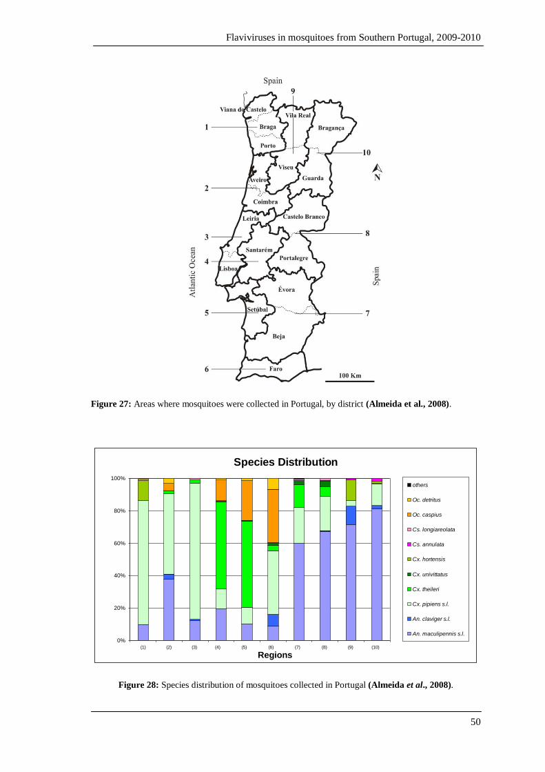
Flaviviruses in mosquitoes from Southern Portugal, 2009-2010
50
Figure 27: Areas where mosquitoes were collected in Portugal, by district (Almeida et al., 2008).
Species Distribution
0%
20%
40%
60%
80%
100%
(1) (2) (3) (4) (5) (6) (7) (8) (9) (10)
Regions
others
Oc. detritus
Oc. caspius
Cs. longiareolata
Cs. annulata
Cx. hortensis
Cx. univittatus
Cx. theileri
Cx. pipiens s.l.
An. claviger s.l.
An. maculipennis s.l.
Figure 28: Species distribution of mosquitoes collected in Portugal (Almeida et al., 2008).
Flaviviruses in mosquitoes from Southern Portugal, 2009-2010
51
2.
OBJECTIVES

Flaviviruses in mosquitoes from Southern Portugal, 2009-2010
52
2. Objectives
The main purpose of this work was to search for flaviviruses in mosquitoes of two
distinct areas of continental Portugal where West Nile virus and other flaviviruses have
previously been found.
2.1 Specific objectives
To study mosquito densities and seasonal dynamics of mosquitoes collected
in wetlands of the Algarve, during two consecutive years.
To screen collected mosquitoes, both from Setúbal and The Algarve, for
flavivirus sequences.
To study the phylogeny of potentially detected flaviviral sequences.
Flaviviruses in mosquitoes from Southern Portugal, 2009-2010
53
3.
MATERIALS AND METHODS
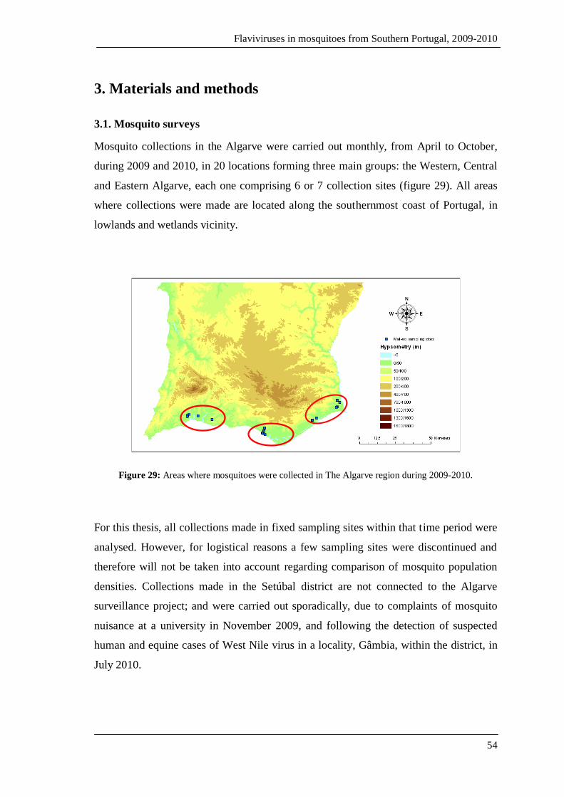
Flaviviruses in mosquitoes from Southern Portugal, 2009-2010
54
3. Materials and methods
3.1. Mosquito surveys
Mosquito collections in the Algarve were carried out monthly, from April to October,
during 2009 and 2010, in 20 locations forming three main groups: the Western, Central
and Eastern Algarve, each one comprising 6 or 7 collection sites (figure 29). All areas
where collections were made are located along the southernmost coast of Portugal, in
lowlands and wetlands vicinity.
Figure 29: Areas where mosquitoes were collected in The Algarve region during 2009-2010.
For this thesis, all collections made in fixed sampling sites within that time period were
analysed. However, for logistical reasons a few sampling sites were discontinued and
therefore will not be taken into account regarding comparison of mosquito population
densities. Collections made in the Setúbal district are not connected to the Algarve
surveillance project; and were carried out sporadically, due to complaints of mosquito
nuisance at a university in November 2009, and following the detection of suspected
human and equine cases of West Nile virus in a locality, Gâmbia, within the district, in
July 2010.
Flaviviruses in mosquitoes from Southern Portugal, 2009-2010
55
3.1.1. Characterisation of the study areas
An initial prospective study was carried out in order to select the most appropriate
sampling sites, in Faro district (Algarve). Collections were also made in the district of
Setúbal (City centre, Comporta, the Estate of Gâmbia); however, no prospective study
was carried out. These two regions, where mosquitoes were collected, present different
ecological and geographical characteristics. Nevertheless, both districts include
wetlands characterised by fresh and salt-water marshes, saltpans, dunes, extensive
beaches and small islands (Osório et al., 2010). In these areas rice cultivation and reed
plantations are abundant, but large areas of cork oak (Quercus suber L.) and pine forest
(Pinus pinaster A., Pinus pinea L.), as well as bird sanctuaries that harbour some
species of migratory birds, amongst others are also present (Almeida et al., 2010).
These areas display the typical Mediterranean climate (according to Koppen
Classification System, group C- temperate/mesothermal climates) with a humid sub-
tropical climate with a dry warm summer and a mild winter. Overall, these areas
comprise beneficial conditions for the establishment and maintenance of mosquito
breeding sites.
3.1.2. Mosquito sampling
Mosquitoes were collected using CDC light traps baited with dry ice (as a source of
CO2 used as an olfactory stimulus) for a period of at least 12 hours (in order to include
sunset and sunrise) thus covering mosquito activity peaks. CDC light traps were placed
mainly outside animal shelters or in their close proximity. In addition, indoor resting
(IR) mosquitoes were sampled, in every location, with the use of battery operated
handheld mechanical aspirators, inside those same locations.
Field-collected mosquitoes were kept in refrigerated boxes for transportation to the
Medical Entomology Laboratory at the Institute of Hygiene and Tropical Medicine, in
Lisbon, where they were frozen at -80ºC. Some characteristics such as the site and time
of collections, number of collectors, the presence or absence of potential hosts, both at
and in the vicinity of the collection sites, as well as the duration of each IR collection,
were registered for every location.
Flaviviruses in mosquitoes from Southern Portugal, 2009-2010
56
3.1.3. Specimen identification
Collected specimens were morphologically identified to the species level by trained
staff, according to Ribeiro & Ramos (1999) identification keys. Mosquitoes were then
pooled into groups of approximately 50 specimens based on their species, sex,
gonotrophic stage (for females) and collection site, were assigned numbers, placed into
1.5ml flat cap tubes, and stored at -80ºC until further processing.
3.1.4. Mosquito density and seasonal dynamics study
Mosquito densities were determined by group, month and year according to collection
method, IR (indoor resting) or CDC light traps baited with CO2.
Mosquito monthly densities per group within the Algarve region were calculated as the
arithmetic mean of collection yield obtained for each individual collection. Densities are
presented as the number of mosquitoes captured per trap-night for CDC collections, or
as the number of mosquitoes collected per collector/hour for IR collections. For all
collections made in each geographical group, the mean density and the standard error
were estimated.
3.1.5. Statistical analysis
Statistical analysis was carried out using the statistical package SPSS version 19.0 for
Windows (SPSS Inc., Chicago, USA).
Kolmogorov-Smirnov and Shapiro-Wilk tests were used to analyse data for normality,
while Levene’s test was used to test for homogeneity of variance. Due to the lack of
normality of the data, the huge standard deviations and the lack of homogeneity of
variance, non-parametric tests were used to analyse mosquito densities (Siegel and
Castellan, 1988). Mean densities in the different regions where compared for each year
with the Kruskal-Wallis test for independent samples, and mean monthly densities were
tested with Wilcoxon Signed Ranks test for related samples, thus comparing mean
densities for all surveyed regions corresponding to the same month in subsequent years.
Flaviviruses in mosquitoes from Southern Portugal, 2009-2010
57
3.2. Viral screening
3.2.1. Mechanical maceration of mosquitoes
Mosquito pool maceration was performed following the protocol previously described
by Huang et al (2001), with modifications. Briefly, for each mosquito pool to be
processed a 15ml Falcon tube was prepared with 5 glass beads (previously washed with
hydrochloric acid and sterilised by autoclaving), 0.25g of alumina and 1.5ml of
phosphate–buffered saline solution supplemented with 4% Bovine Serum Albumin
(BSA). These pools were homogenised by vortexing for about one minute each, and
were then centrifuged at low speed (1,800 g), for 10 minutes at 4ºC. Following
centrifugation, the supernatant was transferred into a 1.5ml flat-cap tube, which was
centrifuged for a second time, at higher speed (10,400 g) for clarification. Clarified
supernatant (150µl) were transferred into a 2 ml flat-cap tube and kept at -80ºC until
RNA extraction. The remaining volume was distributed into two aliquots of
approximately equal volume for long-term storage at -80ºC.
Alumina has two distinct functions in maceration, one is due to its sand-like texture
which aids the mechanical maceration of mosquitoes; secondly it works as a matrix for
the adsorption of positively charged molecules, therefore facilitating their elimination.
BSA is a serum albumin protein that is used as a protein stabiliser as well as to prevent
proteins from adhering to tube walls. It was used at 4% since this concentration is
known to increase Polymerase Chain Reaction (PCR) sensitivity (Huang et al., 2001).
3.2.2. RNA extraction
Total RNA was extracted from mosquito homogenates using the Instant Virus RNA
Extraction Kit (Analytikjena, Germany) following the manufacturer’s instructions,
starting with the 150µl of clarified supernatant previously stored at -80ºC.
In summary, mosquito homogenates were defrosted at room temperature and added to
450µl of lysis solution (RL) in an extraction tube. This procedure took place under a
laminar flow cabinet of Bio Safety Level 3. This mixture was homogenised by
vortexing and then submitted to 15-minute incubation, at room temperature, after which

Flaviviruses in mosquitoes from Southern Portugal, 2009-2010
58
a binding solution (RBS) was added. This was homogenised, and 650µl was applied
onto a spin filter, where the RNA was retained, and quickly centrifuged at
approximately 16,000 g, for one minute. This step was repeated for the residual volume
of the RNA-containing mixture. After two washes with the washing solutions provided
(HS and LS), the extracted RNA was eluted in 60µl of RNase-free water, added 30µl at
a time, followed by incubation at room temperature (2 minute) and centrifugation (1
minute). The eluate containing the extracted RNA was split into three different aliquots
with volumes of 5µl, 25µl and 30µl, respectively. The 5µl aliquot was kept for reverse
transcription (RT) while the others were stored at -80ºC.
3.2.3. cDNA synthesis
Extracted RNA molecules were used to synthesise cDNA using the RevertAid™
H
Minus First Strand cDNA Synthesis Kit (Fermentas, Lithuania), according to the
manufacturer’s instructions. In sum, 5µl of extracted RNA was added to a reaction
mixture consisting of 1µl of random hexamer primers (mixture of oligonucleotides
representing all possible hexamer sequences) and 6µl of diethylpyrocarbonate (DEPC)-
treated water. This mixture was incubated in a water bath at 70ºC for 5 minutes (for
RNA denaturation) and then placed in ice for an additional 5 minutes. A second reaction
mix containing 4µl of reaction buffer, 1µl of Ribolock™ RNase Inhibitor, 2µl of dNTP
Mix (10mM) and 1µl of reverse transcriptase enzyme (RevertAid™ H Minus M-MuLV
Reverse Transcriptase, in a concentration of 200U/µl) was prepared and later added to
the first one in a laminar flow cabinet. It is important to emphasize that all the reagents
and samples were kept on ice at all times. Negative controls were included in each set of
RT reactions in order to assess for genomic DNA contaminations.
The RT reactions took place in a Thermal Cycler (iCycler, BioRad, USA), in
accordance with the programme shown in table 2.
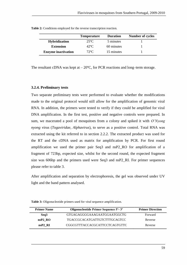
Flaviviruses in mosquitoes from Southern Portugal, 2009-2010
59
Table 2: Conditions employed for the reverse transcription reaction.
Temperature Duration Number of cycles
Hybridization 25ºC 5 minutes 1
Extension 42ºC 60 minutes 1
Enzyme inactivation 72ºC 15 minutes 1
The resultant cDNA was kept at – 20ºC, for PCR reactions and long–term storage.
3.2.4. Preliminary tests
Two separate preliminary tests were performed to evaluate whether the modifications
made to the original protocol would still allow for the amplification of genomic viral
RNA. In addition, the primers were tested to verify if they could be amplified for viral
DNA amplification. In the first test, positive and negative controls were prepared. In
sum, we macerated a pool of mosquitoes from a colony and spiked it with O’Nyong
nyong virus (Togaviridae, Alphavirus), to serve as a positive control. Total RNA was
extracted using the kit referred to in section 2.2.2. The extracted product was used for
the RT and the cDNA used as matrix for amplification by PCR. For first round
amplification we used the primer pair Seq3 and nsP2_RO for amplification of a
fragment of 723bp, expected size, whilst for the second round, the expected fragment
size was 606bp and the primers used were Seq3 and nsP2_RI. For primer sequences
please refer to table 3.
After amplification and separation by electrophoresis, the gel was observed under UV
light and the band pattern analysed.
Table 3: Oligonucleotide primers used for viral sequence amplification.
Primer Name Oligonucleotide Primer Sequence 5’- 3’ Primer Direction
Seq3 GTGAGAGGGGAAAGAATGGAATGGCTG Forward
nsP2_RO TGACCGCACATGATTGTCTTTGCAGTCC Reverse
nsP2_RI CGGCGTTTACCACGCATTCCTCAGTGTTC Reverse

Flaviviruses in mosquitoes from Southern Portugal, 2009-2010
60
For the second test, West Nile and Dengue viruses obtained from our Virology Unit
were used. The RNA extraction, and RT-PCR reactions were carried out as described
previously. For the PCR reactions, the primers used were Flavi1RY and Flavi2RY, for
the first round, and primer pair Flavi2RY and Flavi3RY for the second round
amplification reaction (please refer to the following section). Once again, the amplified
product was separated by electrophoresis, the gel was observed under UV light and the
band pattern analysed.
3.2.5. Amplification of viral sequences by PCR
After synthesis of cDNA from RNA extracted from mosquito macerates, amplification
of viral sequences by PCR was carried out using the primers designed to target the
highest conservation region of the flavivirus genome, the NS5 gene. However, due to
high genetic diversity within the Flavivirus, the primers were degenerated in order to
allow the detection/amplification of a larger number of flavivirus sequences. These
primers were designed using, as a starting point, the primer sequences as published by
Moureau et al (2007) and Sánchez-Seco et al (2005), having been modified based on
the analysis of a multiple alignment sequences including pathogenic arboviruses and
insect-specific flaviviruses (R. Parreira, personal communication). The degenerate
primers used for amplification of flaviviruses sequences were as shown on table 4.
Table 4: Primers used for amplification of flavivirus sequences.
Primer name Primer sequences 5’-3’ Primer direction Temperature
FLAVI 1 TGY RTN TAY AAC AYV ATG GG Forward 58.6 ºC
FLAVI 2 GTG TCC CAN CCD GCD RYR TC Reverse 69.6 ºC
FLAVI 3 ATH TGG TWY ATG TGG YTN GG Forward 59.6 ºC
M= A+C; W= A+T; R= A+G; K= G+T; D= G+A+T; N= A+C+G+T.
In general, nested–PCR reactions use two sets of primers. The so-called inner primers
target DNA sequence located within the region limited by another set of primers – the
outer primers. In the first PCR reaction, the cDNA yielded by RT was used as the
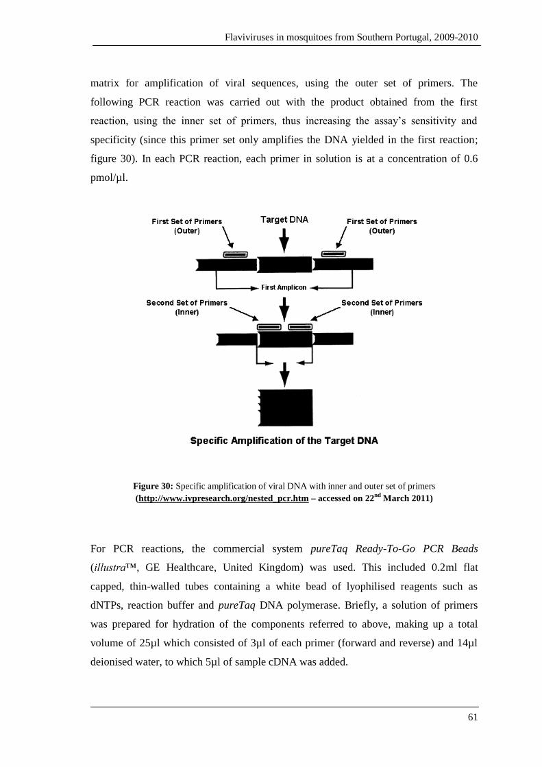
Flaviviruses in mosquitoes from Southern Portugal, 2009-2010
61
matrix for amplification of viral sequences, using the outer set of primers. The
following PCR reaction was carried out with the product obtained from the first
reaction, using the inner set of primers, thus increasing the assay’s sensitivity and
specificity (since this primer set only amplifies the DNA yielded in the first reaction;
figure 30). In each PCR reaction, each primer in solution is at a concentration of 0.6
pmol/µl.
Figure 30: Specific amplification of viral DNA with inner and outer set of primers
(http://www.ivpresearch.org/nested_pcr.htm – accessed on 22nd
March 2011)
For PCR reactions, the commercial system pureTaq Ready-To-Go PCR Beads
(illustra™, GE Healthcare, United Kingdom) was used. This included 0.2ml flat
capped, thin-walled tubes containing a white bead of lyophilised reagents such as
dNTPs, reaction buffer and pureTaq DNA polymerase. Briefly, a solution of primers
was prepared for hydration of the components referred to above, making up a total
volume of 25µl which consisted of 3µl of each primer (forward and reverse) and 14µl
deionised water, to which 5µl of sample cDNA was added.

Flaviviruses in mosquitoes from Southern Portugal, 2009-2010
62
For flavivirus-positive samples, for DNA purification purposes, viral sequence
amplification was carried out using the same kit, but making up a total volume of 50µl.
In this case, each primer in solution was at a concentration of 0.4 pmol/µl.
The PCR reaction mixtures were prepared in a laminar flow cabinet, and all the reagents
were kept on ice, at all times. After cDNA addition, the PCR tubes were placed on a
thermal cycler (iCycler, Bio-Rad, USA) for amplification under the conditions
presented on table 5.
Table 5: PCR conditions for viral sequence amplification.
Temperature Duration Number of cycles
Initial denaturation 95ºC 2 minutes 1
Denaturation 94ºC 45 seconds
35 Annealing 50ºC 45 seconds
Extension 72ºC 30 seconds
Final extension 72ºC 5 minutes 1
Amplified PCR products were separated by electrophoresis in agarose gel at 1.5%
stained with 1µg/ml of ethidium bromide, with 0.5x TAE buffer solution. A 100bp
molecular weight marker (100bp GeneRuler™ Express DNA Ladder) was used as a
reference. Finally, the gel was observed under UV light using the Gel Doc XR System
(Bio-Rad, USA).
3.2.6. Purification and sequencing of PCR products
The amplicons obtained from the amplified DNA fragments were used as basis for
analysis of the nucleotide sequences for the NS5 region of the flavivirus genome. The
amplified products were purified using a commercial kit (Zymoclean™ Gel DNA
Recovery Kit, Zymo Research, USA) according to the manufacturer’s instructions.

Flaviviruses in mosquitoes from Southern Portugal, 2009-2010
63
Briefly, after separation in agarose gel (1.5%), the DNA fragment was excised and
dissolved in a buffered solution (Agarose Dissolving Buffer) at approximately 55ºC, for
5 to 10 minutes. The dissolved mixture was transferred onto a column (Zymo-Spin™
Column) where the DNA was fixed, followed by two washes with Wash buffer that
allows for the removal of non-specifically bound molecules. The DNA was eluted in
10µl of 10mM Tris-HCl, pH 8.0. In order to assess the yield of the extraction and
purification processes, the purified product (1µl) was analysed by electrophoresis in
agarose gel (1.5%).
STAB VIDA biotechnology company (Portugal) sequenced the purified products using
one of the primers utilised for viral DNA amplification. The primers used were either
the reverse primer (FLAVI 2) for products obtained from the first and second rounds
reactions simultaneously, since this primer is common to both reactions; or the forward
primer used for products yielded in second round amplification reactions only.
3.2.7. Phylogenetic analysis of viral sequences
In order to analyse the sequences obtained from the amplified products, these were
aligned with reference flavivirus sequences available in free access public databases,
such as GenBank, where our reference sequences were obtained.
BLAST (Basic Local Alignment Search Tool) was used for basic alignment and
comparison of our sequences with those of flavivirus reference. The editing and
multiple alignments of sequences were performed with the aid of bioinformatics
software BioEdit Sequence Alignment Editor version 7.0.5.3 and MAFFT version 6,
respectively. Once aligned, these sequences were used to generate phylogenetic trees
obtained by the neighbour-joining method from a matrix of genetic evolutionary
distances calculated using Kimura’s two parameter estimate (Tamura et al., 2007), with
the use of Mega version 5. Bootstrap analysis, based on 1000 replicates of the original
sequence data, was carried out in order to evaluate the consistency of tree topology.
The groups that displayed bootstrap values equal or greater than 70% were considered
consistent form a phylogenetic perspective.

Flaviviruses in mosquitoes from Southern Portugal, 2009-2010
64
3.2.8 Extraction controls
In order to evaluate for contaminations, randomly selected samples were used as
extraction controls; firstly, to each extracted RNA sample (5 µl), 1µl of RNase
(Ribonuclease A, USB Corporation, USA) was added at a concentration of 0.1µg/µl.
The mixture was left to incubate at room temperature for two hours. The treated
samples underwent a similar RT-PCR protocol as that described in 3.2.3 and 3.2.5. PCR
products of RNase-treated samples were charged into a 1.5% agarose gel containing
ethidium bromide alongside the untreated samples for separation by electrophoresis.
Flaviviruses in mosquitoes from Southern Portugal, 2009-2010
65
4.
RESULTS
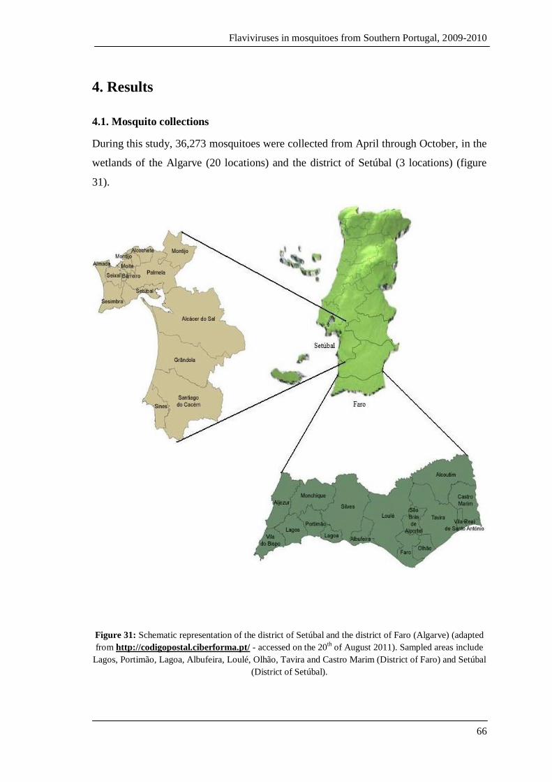
Flaviviruses in mosquitoes from Southern Portugal, 2009-2010
66
4. Results
4.1. Mosquito collections
During this study, 36,273 mosquitoes were collected from April through October, in the
wetlands of the Algarve (20 locations) and the district of Setúbal (3 locations) (figure
31).
Figure 31: Schematic representation of the district of Setúbal and the district of Faro (Algarve) (adapted
from http://codigopostal.ciberforma.pt/ - accessed on the 20th of August 2011). Sampled areas include
Lagos, Portimão, Lagoa, Albufeira, Loulé, Olhão, Tavira and Castro Marim (District of Faro) and Setúbal
(District of Setúbal).

Flaviviruses in mosquitoes from Southern Portugal, 2009-2010
67
Mosquito specimens collected belonged to six genera and fifteen different species,
namely Anopheles algeriensis, An.atroparvus, Aedes berlandi, Ae. caspius, Ae.detritus,
Ae. mariae, Coquillettidia richiardii, Culex laticinctus, Cx.pipiens, Cx.theileri,
Cx.univittatus, Culiseta annulata, Cs.longiareolata, Cs.subochrea, and Uranotaenia
unguiculata.
In Setúbal 2009, a total of 135 mosquito specimens were captured; however, only two
species were collected, both belonging to the same genus. The most abundant species
was Ae. caspius with 132 specimens collected against 3 specimens of Ae. detritus (table
6).
Table 6: Summary of collections made in the district of Setúbal.
Collected mosquito
species
Total mosquitoes captured Total Percentage
2009 2010
Aedes caspius 132 565 697 36.45%
Aedes detritus 3 1 4 0.21%
Anopheles atroparvus 0 114 114 5.96%
Culex pipiens 0 106 106 5.54%
Culex theileri 0 876 876 45.82%
Culex univittatus 0 115 115 6.01%
Sub-total 135 1,777 1,912 100.00%
Mosquito collections in the Algarve region yielded the 35,967 mosquitoes of 15
different species (table 7). The three predominant species captured were Ae. caspius,
Cx. pipiens and Cx. theileri, in that order. Anopheles atroparvus was also present in
significant numbers corresponding to approximately 6% of all collections.
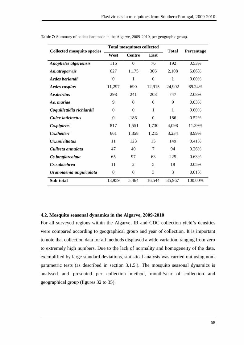
Flaviviruses in mosquitoes from Southern Portugal, 2009-2010
68
Table 7: Summary of collections made in the Algarve, 2009-2010, per geographic group.
Collected mosquito species Total mosquitoes collected
Total Percentage West Centre East
Anopheles algeriensis 116 0 76 192 0.53%
An.atroparvus 627 1,175 306 2,108 5.86%
Aedes berlandi 0 1 0 1 0.00%
Aedes caspius 11,297 690 12,915 24,902 69.24%
Ae.detritus 298 241 208 747 2.08%
Ae. mariae 9 0 0 9 0.03%
Coquillettidia richiardii 0 0 1 1 0.00%
Culex laticinctus 0 186 0 186 0.52%
Cx.pipiens 817 1,551 1,730 4,098 11.39%
Cx.theileri 661 1,358 1,215 3,234 8.99%
Cx.univittatus 11 123 15 149 0.41%
Culiseta annulata 47 40 7 94 0.26%
Cs.longiareolata 65 97 63 225 0.63%
Cs.subochrea 11 2 5 18 0.05%
Uranotaenia unguiculata 0 0 3 3 0.01%
Sub-total 13,959 5,464 16,544 35,967 100.00%
4.2. Mosquito seasonal dynamics in the Algarve, 2009-2010
For all surveyed regions within the Algarve, IR and CDC collection yield’s densities
were compared according to geographical group and year of collection. It is important
to note that collection data for all methods displayed a wide variation, ranging from zero
to extremely high numbers. Due to the lack of normality and homogeneity of the data,
exemplified by large standard deviations, statistical analysis was carried out using non-
parametric tests (as described in section 3.1.5.). The mosquito seasonal dynamics is
analysed and presented per collection method, month/year of collection and
geographical group (figures 32 to 35).
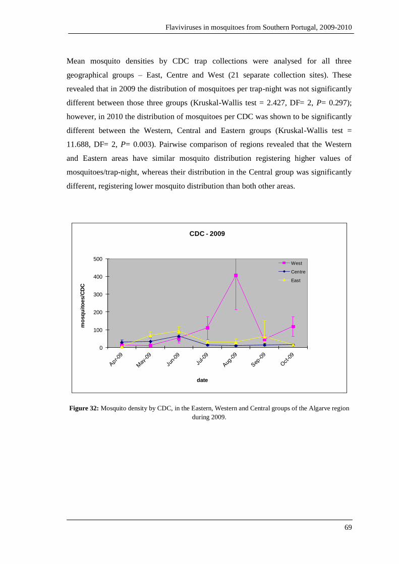
Flaviviruses in mosquitoes from Southern Portugal, 2009-2010
69
Mean mosquito densities by CDC trap collections were analysed for all three
geographical groups – East, Centre and West (21 separate collection sites). These
revealed that in 2009 the distribution of mosquitoes per trap-night was not significantly
different between those three groups (Kruskal-Wallis test = 2.427, DF= 2, P= 0.297);
however, in 2010 the distribution of mosquitoes per CDC was shown to be significantly
different between the Western, Central and Eastern groups (Kruskal-Wallis test =
11.688, DF= 2, P= 0.003). Pairwise comparison of regions revealed that the Western
and Eastern areas have similar mosquito distribution registering higher values of
mosquitoes/trap-night, whereas their distribution in the Central group was significantly
different, registering lower mosquito distribution than both other areas.
0
100
200
300
400
500
mo
sq
uit
oes/C
DC
date
CDC - 2009
West
Centre
East
Figure 32: Mosquito density by CDC, in the Eastern, Western and Central groups of the Algarve region
during 2009.

Flaviviruses in mosquitoes from Southern Portugal, 2009-2010
70
0
500
1000
1500
2000
2500m
osq
uit
oes/C
DC
date
CDC - 2010
West
Centre
East
Figure 33: Mosquito density by CDC, in the Eastern, Western and Central groups of the Algarve region
during 2010. As opposed to figure 31, the y-axis goes up to 2500 mosquitoes/CDC.
Mean mosquito densities by IR collections varied according to month and area of
collection yielding peak densities between June and September, whereas the lowest
densities were recorded in April and October.
0
500
1000
mo
sq
uit
oes/p
ers
on
/ho
ur
date
IR - 2009
West
Centre
East
Figure 34: Mosquito density by IR, in the Eastern, Western and Central groups of the Algarve region
during 2009.
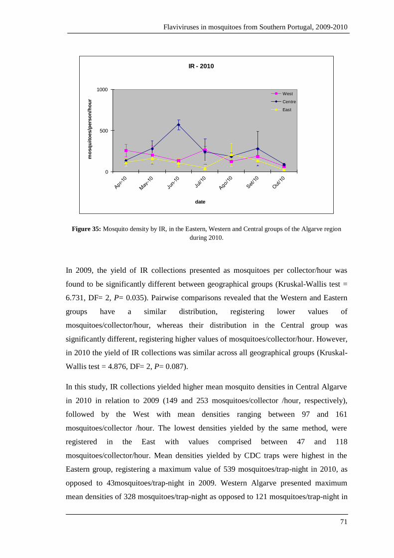
Flaviviruses in mosquitoes from Southern Portugal, 2009-2010
71
0
500
1000m
osq
uit
oes/p
ers
on
/ho
ur
date
IR - 2010
West
Centre
East
Figure 35: Mosquito density by IR, in the Eastern, Western and Central groups of the Algarve region
during 2010.
In 2009, the yield of IR collections presented as mosquitoes per collector/hour was
found to be significantly different between geographical groups (Kruskal-Wallis test =
6.731, DF= 2, P= 0.035). Pairwise comparisons revealed that the Western and Eastern
groups have a similar distribution, registering lower values of
mosquitoes/collector/hour, whereas their distribution in the Central group was
significantly different, registering higher values of mosquitoes/collector/hour. However,
in 2010 the yield of IR collections was similar across all geographical groups (Kruskal-
Wallis test = 4.876, DF= 2, P= 0.087).
In this study, IR collections yielded higher mean mosquito densities in Central Algarve
in 2010 in relation to 2009 (149 and 253 mosquitoes/collector /hour, respectively),
followed by the West with mean densities ranging between 97 and 161
mosquitoes/collector /hour. The lowest densities yielded by the same method, were
registered in the East with values comprised between 47 and 118
mosquitoes/collector/hour. Mean densities yielded by CDC traps were highest in the
Eastern group, registering a maximum value of 539 mosquitoes/trap-night in 2010, as
opposed to 43mosquitoes/trap-night in 2009. Western Algarve presented maximum
mean densities of 328 mosquitoes/trap-night as opposed to 121 mosquitoes/trap-night in
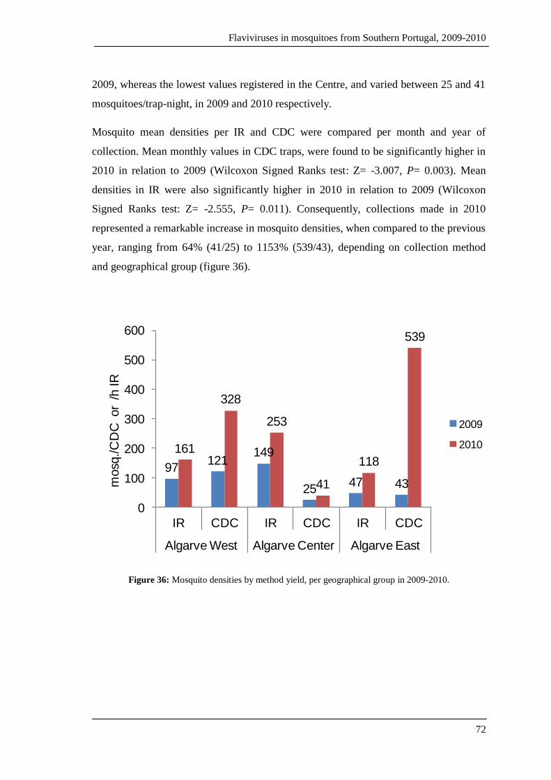
Flaviviruses in mosquitoes from Southern Portugal, 2009-2010
72
2009, whereas the lowest values registered in the Centre, and varied between 25 and 41
mosquitoes/trap-night, in 2009 and 2010 respectively.
Mosquito mean densities per IR and CDC were compared per month and year of
collection. Mean monthly values in CDC traps, were found to be significantly higher in
2010 in relation to 2009 (Wilcoxon Signed Ranks test: Z= -3.007, P= 0.003). Mean
densities in IR were also significantly higher in 2010 in relation to 2009 (Wilcoxon
Signed Ranks test: Z= -2.555, P= 0.011). Consequently, collections made in 2010
represented a remarkable increase in mosquito densities, when compared to the previous
year, ranging from 64% (41/25) to 1153% (539/43), depending on collection method
and geographical group (figure 36).
97121
149
2547 43
161
328
253
41
118
539
0
100
200
300
400
500
600
IR CDC IR CDC IR CDC
Algarve West Algarve Center Algarve East
mosq
./C
DC
or /h IR
2009
2010
Figure 36: Mosquito densities by method yield, per geographical group in 2009-2010.
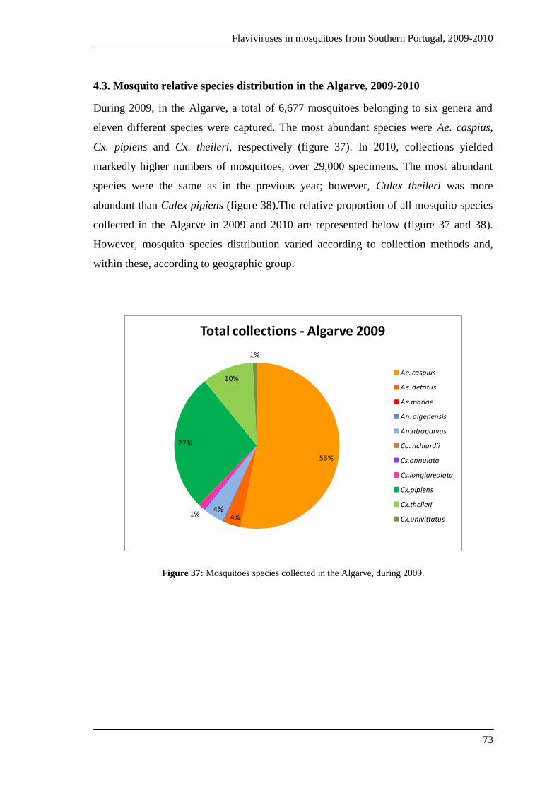
Flaviviruses in mosquitoes from Southern Portugal, 2009-2010
73
4.3. Mosquito relative species distribution in the Algarve, 2009-2010
During 2009, in the Algarve, a total of 6,677 mosquitoes belonging to six genera and
eleven different species were captured. The most abundant species were Ae. caspius,
Cx. pipiens and Cx. theileri, respectively (figure 37). In 2010, collections yielded
markedly higher numbers of mosquitoes, over 29,000 specimens. The most abundant
species were the same as in the previous year; however, Culex theileri was more
abundant than Culex pipiens (figure 38).The relative proportion of all mosquito species
collected in the Algarve in 2009 and 2010 are represented below (figure 37 and 38).
However, mosquito species distribution varied according to collection methods and,
within these, according to geographic group.
53%
4%4%
1%
27%
10%
1%
Total collections - Algarve 2009
Ae. caspius
Ae. detritus
Ae.mariae
An. algeriensis
An.atroparvus
Co. richiardii
Cs.annulata
Cs.longiareolata
Cx.pipiens
Cx.theileri
Cx.univittatus
Figure 37: Mosquitoes species collected in the Algarve, during 2009.
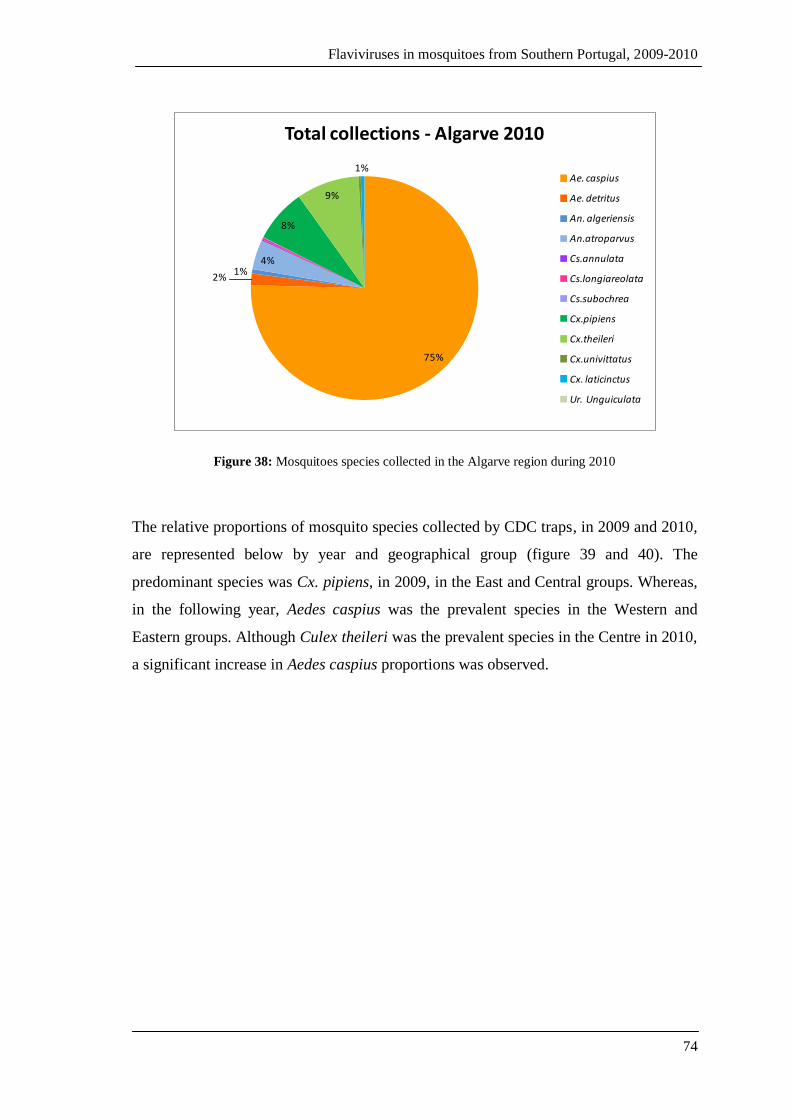
Flaviviruses in mosquitoes from Southern Portugal, 2009-2010
74
75%
2%1%
4%
8%
9%
1%
Total collections - Algarve 2010
Ae. caspius
Ae. detritus
An. algeriensis
An.atroparvus
Cs.annulata
Cs.longiareolata
Cs.subochrea
Cx.pipiens
Cx.theileri
Cx.univittatus
Cx. laticinctus
Ur. Unguiculata
Figure 38: Mosquitoes species collected in the Algarve region during 2010
The relative proportions of mosquito species collected by CDC traps, in 2009 and 2010,
are represented below by year and geographical group (figure 39 and 40). The
predominant species was Cx. pipiens, in 2009, in the East and Central groups. Whereas,
in the following year, Aedes caspius was the prevalent species in the Western and
Eastern groups. Although Culex theileri was the prevalent species in the Centre in 2010,
a significant increase in Aedes caspius proportions was observed.

Flaviviruses in mosquitoes from Southern Portugal, 2009-2010
75
1%
56%17%
1%
2%21%
2%
Total mosquitoes captured per Region/species/CDC-2009
An.maculipennis
Cx.pipiens
Cx.theileri
Cx.univittatus
Cs.annulata
Cs.longiareolata
Ae.caspius
Ae.detritusEast
1%
56%24%
5%
1%
3% 5%
5%
Centre
3%
5% 5%
1%
83%
3%
West
Figure 39: Relative proportion of mosquito species captured by CDC traps in 2009.
4%7%
88%
1%
Total mosquitoes captured per Region/ species/CDC-2010
An.maculipennis
Cx.pipiens
Cx.theileri
Cx.univittatus
Cs.annulata
Cs.longiareolata
Ae.caspius
Ae.detritus
East
1%
26%
38%3%
1%
2%
23%
6%
Centre
6%
5%
87%
2%
West
Figure 40: Relative proportion of mosquito species collected by CDC traps in 2010.
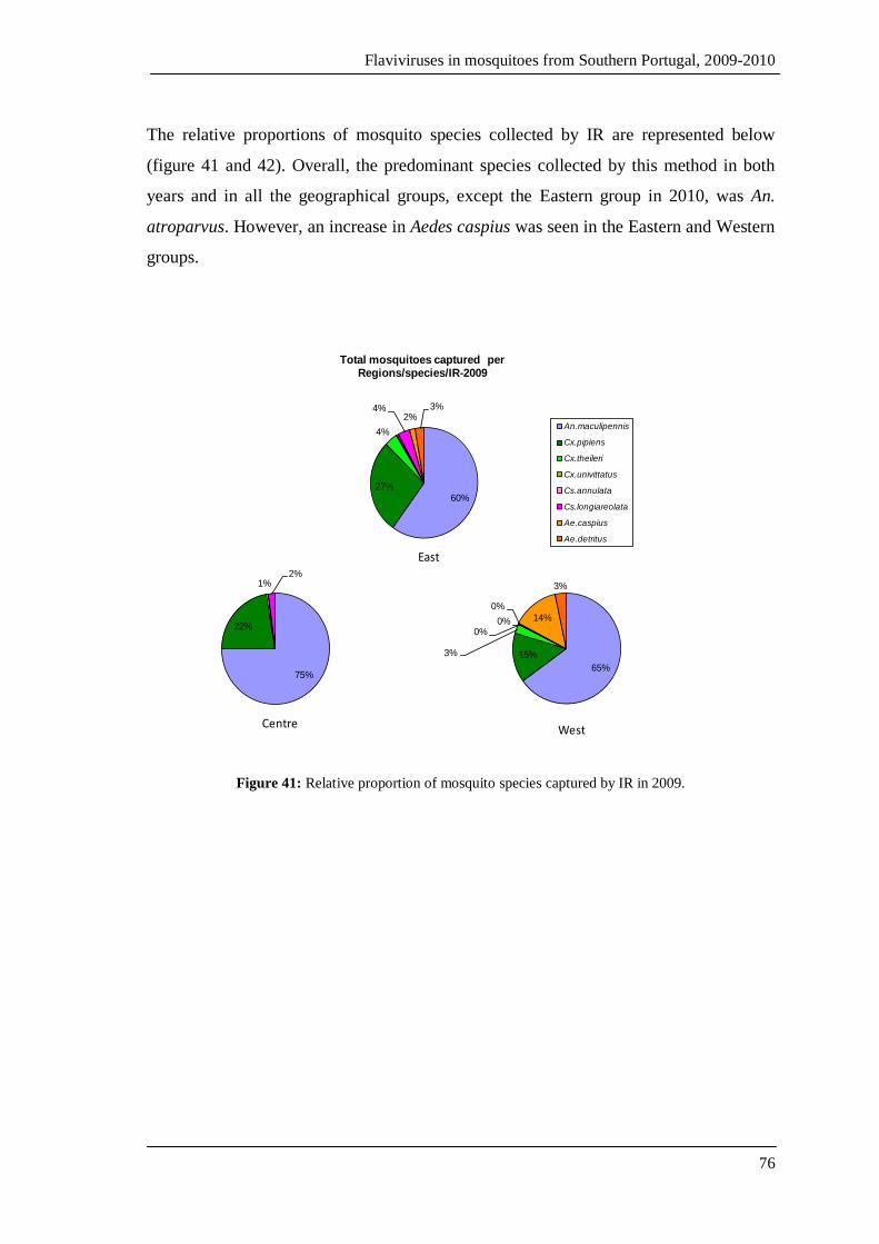
Flaviviruses in mosquitoes from Southern Portugal, 2009-2010
76
The relative proportions of mosquito species collected by IR are represented below
(figure 41 and 42). Overall, the predominant species collected by this method in both
years and in all the geographical groups, except the Eastern group in 2010, was An.
atroparvus. However, an increase in Aedes caspius was seen in the Eastern and Western
groups.
60%
27%
4%
4%2%
3%
Total mosquitoes captured per Regions/species/IR-2009
An.maculipennis
Cx.pipiens
Cx.theileri
Cx.univittatus
Cs.annulata
Cs.longiareolata
Ae.caspius
Ae.detritus
75%
22%
1%2%
Centre
65%
15%3%
0%0%
0%
14%
3%
West
East
Figure 41: Relative proportion of mosquito species captured by IR in 2009.

Flaviviruses in mosquitoes from Southern Portugal, 2009-2010
77
27%
20%
3%1%
46%
3%
Total mosquitoes captured per Region/species/IR -2010
An.maculipennis
Cx.pipiens
Cx.theileri
Cx.univittatus
Cs.annulata
Cs.longiareolata
Ae.caspius
Ae.detritus
77%
16%
4%
1%
1%
1%
Centre
53%
15%1%
1%
27%
3%
West
East
Figure 42: Relative proportion of mosquito species captured by IR in 2010.
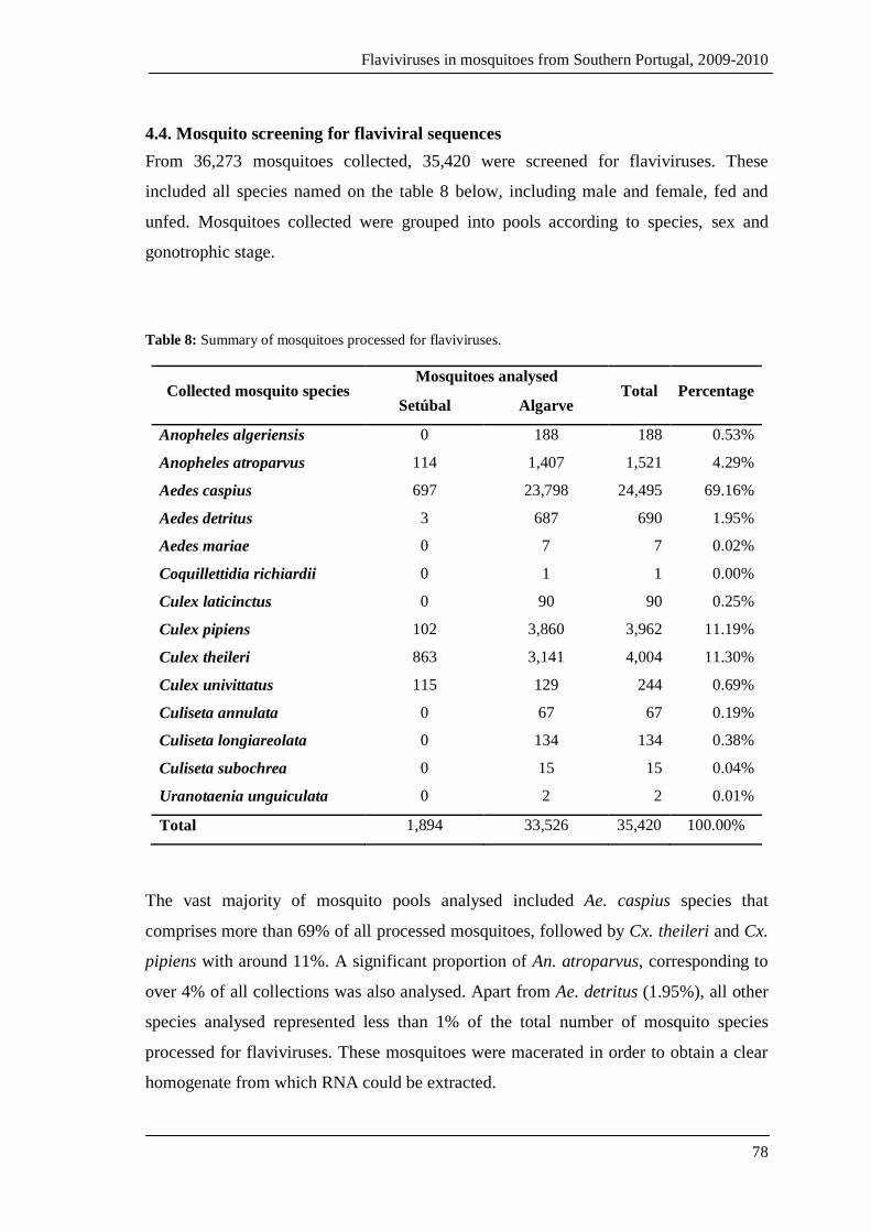
Flaviviruses in mosquitoes from Southern Portugal, 2009-2010
78
4.4. Mosquito screening for flaviviral sequences
From 36,273 mosquitoes collected, 35,420 were screened for flaviviruses. These
included all species named on the table 8 below, including male and female, fed and
unfed. Mosquitoes collected were grouped into pools according to species, sex and
gonotrophic stage.
Table 8: Summary of mosquitoes processed for flaviviruses.
Collected mosquito species Mosquitoes analysed
Total Percentage Setúbal Algarve
Anopheles algeriensis 0 188 188 0.53%
Anopheles atroparvus 114 1,407 1,521 4.29%
Aedes caspius 697 23,798 24,495 69.16%
Aedes detritus 3 687 690 1.95%
Aedes mariae 0 7 7 0.02%
Coquillettidia richiardii 0 1 1 0.00%
Culex laticinctus 0 90 90 0.25%
Culex pipiens 102 3,860 3,962 11.19%
Culex theileri 863 3,141 4,004 11.30%
Culex univittatus 115 129 244 0.69%
Culiseta annulata 0 67 67 0.19%
Culiseta longiareolata 0 134 134 0.38%
Culiseta subochrea 0 15 15 0.04%
Uranotaenia unguiculata 0 2 2 0.01%
Total 1,894 33,526 35,420 100.00%
The vast majority of mosquito pools analysed included Ae. caspius species that
comprises more than 69% of all processed mosquitoes, followed by Cx. theileri and Cx.
pipiens with around 11%. A significant proportion of An. atroparvus, corresponding to
over 4% of all collections was also analysed. Apart from Ae. detritus (1.95%), all other
species analysed represented less than 1% of the total number of mosquito species
processed for flaviviruses. These mosquitoes were macerated in order to obtain a clear
homogenate from which RNA could be extracted.

Flaviviruses in mosquitoes from Southern Portugal, 2009-2010
79
4.4.1. RNA extracted from mosquito homogenates
The clarified mosquito homogenates obtained from the maceration process were used
for the extraction of total RNA. During this process, unspecific binding of, for example,
soluble macromolecular cellular components, salts and metabolites to the extraction
column where RNA is fixed usually occurs and that can potentially cause inhibition of
subsequent RT-PCR reactions. Even though this procedure is carried out to eliminate
unspecific bound molecules, some potential contaminants may not be removed.
Therefore, in order to ensure that no contaminants are present in the samples and the
extracted RNA was viral and, consequently, the positive pools obtained were a result of
viral DNA amplification extraction controls were carried out. Due to cost restrictions,
extraction controls were performed only for a few, randomly selected samples. These
RNA extracts were first treated with Ribonuclease A before undergoing a retro-
transcription reaction in order to assess whether all RNA had been digested, and
consequently if any cDNA would be synthesized. The RT reaction product was used as
matrix for amplification and the PCR products were subject to electrophoresis. After
separation in agarose gel with ethidium bromide, it was possible to observe that none of
the RNAse-treated samples produced any bands, thus confirming that the there were no
genomic DNA contaminants in our samples suggesting that there was viral RNA only.
4.4.2. Amplification reactions preliminary tests
The first preliminary test was carried out in order to verify whether there was any step
in the protocol that would, somehow, interfere with our ability to detect viral RNA in
the analysed samples. For this, two types of experiments were performed.
In the first, a mosquito macerate including mosquitoes maintained in a colony at IHMT
was spiked with O’Nyong nyong virus GULU strain (Togaviridae, Alphavirus) genomic
RNA. Total RNA was then extracted from the clarified macerate, and submitted to RT-
PCR, using specific primers. After amplification and separation by electrophoresis, the
gel was observed under UV light. Analysis of the band pattern obtained revealed the
expected size bands indicating that the detection of O’Nyong nyong virus RNA had
been successful.
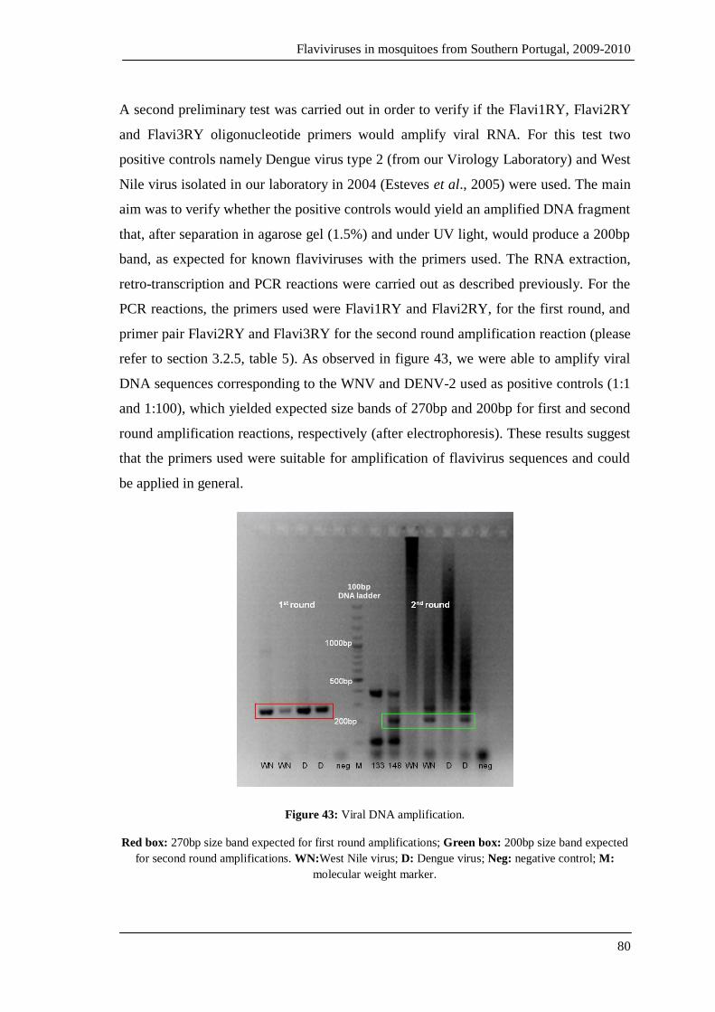
Flaviviruses in mosquitoes from Southern Portugal, 2009-2010
80
A second preliminary test was carried out in order to verify if the Flavi1RY, Flavi2RY
and Flavi3RY oligonucleotide primers would amplify viral RNA. For this test two
positive controls namely Dengue virus type 2 (from our Virology Laboratory) and West
Nile virus isolated in our laboratory in 2004 (Esteves et al., 2005) were used. The main
aim was to verify whether the positive controls would yield an amplified DNA fragment
that, after separation in agarose gel (1.5%) and under UV light, would produce a 200bp
band, as expected for known flaviviruses with the primers used. The RNA extraction,
retro-transcription and PCR reactions were carried out as described previously. For the
PCR reactions, the primers used were Flavi1RY and Flavi2RY, for the first round, and
primer pair Flavi2RY and Flavi3RY for the second round amplification reaction (please
refer to section 3.2.5, table 5). As observed in figure 43, we were able to amplify viral
DNA sequences corresponding to the WNV and DENV-2 used as positive controls (1:1
and 1:100), which yielded expected size bands of 270bp and 200bp for first and second
round amplification reactions, respectively (after electrophoresis). These results suggest
that the primers used were suitable for amplification of flavivirus sequences and could
be applied in general.
100bp DNA ladder
Figure 43: Viral DNA amplification.
Red box: 270bp size band expected for first round amplifications; Green box: 200bp size band expected
for second round amplifications. WN:West Nile virus; D: Dengue virus; Neg: negative control; M:
molecular weight marker.
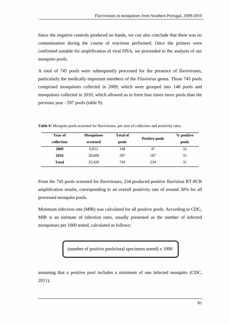
Flaviviruses in mosquitoes from Southern Portugal, 2009-2010
81
Since the negative controls produced no bands, we can also conclude that there was no
contamination during the course of reactions performed. Once the primers were
confirmed suitable for amplification of viral DNA, we proceeded to the analysis of our
mosquito pools.
A total of 745 pools were subsequently processed for the presence of flaviviruses,
particularly the medically important members of the Flavivirus genus. Those 745 pools
comprised mosquitoes collected in 2009, which were grouped into 148 pools and
mosquitoes collected in 2010, which allowed us to form four times more pools than the
previous year - 597 pools (table 9).
Table 9: Mosquito pools screened for flaviviruses, per year of collection and positivity rates.
Year of
collection
Mosquitoes
screened
Total of
pools Positive pools
% positive
pools
2009 6,812 148 47 32
2010 28,608 597 187 31
Total 35,420 745 234 31
From the 745 pools screened for flaviviruses, 234 produced positive flavivirus RT-PCR
amplification results, corresponding to an overall positivity rate of around 30% for all
processed mosquito pools.
Minimum infection rate (MIR) was calculated for all positive pools. According to CDC,
MIR is an estimate of infection rates, usually presented as the number of infected
mosquitoes per 1000 tested, calculated as follows:
(number of positive pools/total specimens tested) x 1000
assuming that a positive pool includes a minimum of one infected mosquito (CDC,
2011).

Flaviviruses in mosquitoes from Southern Portugal, 2009-2010
82
MIR was calculated for positive pools of mosquitoes captured in both the Setúbal and
Faro districts, during 2009-2010, and the results are shown on the graphs 44 and 45
below.
In the Algarve region, in 2009, the lowest MIR values were recorded for the following
species: Cx. pipiens (MIR= 2.24/1000), Ae. caspius (MIR= 3.66/1000) and Ae. detritus
(MIR= 8.97/1000). The highest values were registered for An. algeriensis with a MIR of
more than 50 infected mosquitoes per 1000 tested, followed by Cx. univittatus and An.
atroparvus, both showing MIR values of around 20 infected mosquitoes in a thousand.
0.00 10.00 20.00 30.00 40.00 50.00 60.00
An.algeriensis
An.atroparvus
Ae.caspius
Ae.detritus
Cx.pipiens
Cx.theileri
Cx.univittatus
Cs.annulata
MIR
Mo
squ
ito
sp
eci
es
Algarve 2010
Algarve 2009
Figure 44: Minimum infection rates per species, in the Algarve 2009-2010.
In 2010, the highest MIR values were obtained for Culiseta annulata (22.22 mosquitoes
infected/1000 tested) and Cx. theileri with approximately 12.6 infected mosquitoes in a
thousand. The lowest registered MIR values belonged to Ae. caspius (MIR= 1.23/1000),
Cx. pipiens (MIR= 1.44/1000) and finally An. atroparvus (MIR= 1.68/1000). Anopheles
algeriensis and Ae. detritus showed similar MIR values of around 6 infected mosquitoes
per a thousand tested.
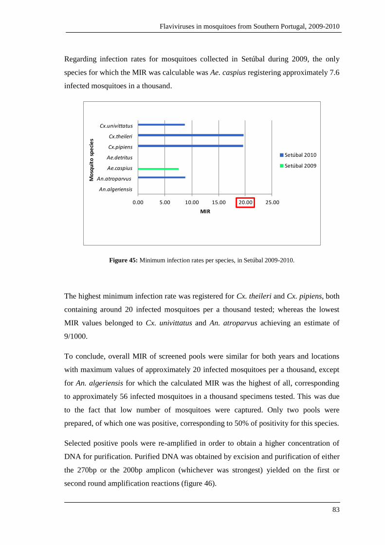
Flaviviruses in mosquitoes from Southern Portugal, 2009-2010
83
Regarding infection rates for mosquitoes collected in Setúbal during 2009, the only
species for which the MIR was calculable was Ae. caspius registering approximately 7.6
infected mosquitoes in a thousand.
0.00 5.00 10.00 15.00 20.00 25.00
An.algeriensis
An.atroparvus
Ae.caspius
Ae.detritus
Cx.pipiens
Cx.theileri
Cx.univittatus
MIR
Mo
squ
ito
sp
eci
es
Setúbal 2010
Setúbal 2009
Figure 45: Minimum infection rates per species, in Setúbal 2009-2010.
The highest minimum infection rate was registered for Cx. theileri and Cx. pipiens, both
containing around 20 infected mosquitoes per a thousand tested; whereas the lowest
MIR values belonged to Cx. univittatus and An. atroparvus achieving an estimate of
9/1000.
To conclude, overall MIR of screened pools were similar for both years and locations
with maximum values of approximately 20 infected mosquitoes per a thousand, except
for An. algeriensis for which the calculated MIR was the highest of all, corresponding
to approximately 56 infected mosquitoes in a thousand specimens tested. This was due
to the fact that low number of mosquitoes were captured. Only two pools were
prepared, of which one was positive, corresponding to 50% of positivity for this species.
Selected positive pools were re-amplified in order to obtain a higher concentration of
DNA for purification. Purified DNA was obtained by excision and purification of either
the 270bp or the 200bp amplicon (whichever was strongest) yielded on the first or
second round amplification reactions (figure 46).

Flaviviruses in mosquitoes from Southern Portugal, 2009-2010
84
However, after purification, not all positive samples contained enough pure DNA to
produce a band when 1µl of the purified product was charged into an agarose gel (1.5%)
to evaluate yield of the purification reaction. Consequently, those samples were not
used for further study, while some of the samples that yielded positive results
consistently were sent for sequencing.
506 507 509 514 515 518 519 523 548 549 551 552 554 555 559 572 368 487 451 M
200bp 270bp
2nd round – Flavi 2+ Flavi 3
100bp DNA ladder
500bp
1000bp
Figure 46: PCR amplification of positive pools for DNA purification. M: Molecular weight marker; the
numbers represent the flavivirus-positive pools.
Out of the 234 pools that tested positive for flaviviruses, only 32 NS5 amplicons were
sequenced. Among those, there were DNA fragments obtained from pools of Ae.
caspius, Ae. detritus, An. atroparvus, Cx. pipiens, Cx. theileri and Cx.univittatus.
However, after sequence analysis, the majority of sequences were excluded, as they did
not present enough quality to be included in phylogenetic inference studies. In total, 11
out of 32 sequences were used for further analysis; these belonged to 3 different species,
Ae. caspius (n=5), Cx. pipiens (n=1) and Cx. theileri (n=5).
Sequence analysis was based on the comparison of a short fragment (270bp or 200bp)
of the viral sequence amplified from the highly conserved region of the flavivirus
genome, the NS5 gene, with reference sequences deposited in public databases.
PCR positive pools

Flaviviruses in mosquitoes from Southern Portugal, 2009-2010
85
BLAST was used to perform similarity searches against the flavivirus reference
sequences. Sequence analysis via BLAST (high similarity search) revealed that all our
sequences showed significant similarity with insect-specific flavivirus genome
sequences deposited in databases. The results of this search are shown on the table 10
below.
Table 10: Flavivirus sequences identity results.
Pool
number
Mosquito
species
Related sequences (NS5 gene) Query
coverage
Maximum
identity
188
207
219
350
357
Aedes caspius Mosquito flavivirus isolate OccaFV5
Mosquito flavivirus isolate FE-Ac3
Mosquito flavivirus isolate FE-Ac2
Mosquito flavivirus isolate FE-Ac1
Mosquito flavivirus isolate FE-Cp
Culicinae flavivirus PoMoFlav_A95
Mosquito flavivirus isolate OccaFV1
Mosquito flavivirus isolate OccaFV4
Culicinae flavivirus PoMoFlav_A163
Culicinae flavivirus PoMoFlav_A155
Culicinae flavivirus PoMoFlav_A153
Culicinae flavivirus PoMoFlav_A136
Culicinae flavivirus PoMoFlav_A128
Culicinae flavivirus PoMoFlav_A126
Flavivirus Phlebotomine/76/Arrabida/07
Culicinae flavivirus PoMoFlav_A103
Culicinae flavivirus PoMoFlav_A91
Mosquito flavivirus isolate OccaFV3
78-90%
78-90%
78-90%
78-90%
78-90%
78-90%
78-90%
78-90%
78-90%
78-90%
78-90%
78-90%
78-90%
78-90%
78-90%
78-90%
78-90%
78-90%
93-98%
93-98%
93-98%
93-98%
93-98%
93-98%
93-98%
93-98%
93-98%
93-98%
93-98%
93-98%
93-98%
93-98%
93-98%
93-98%
93-98%
93-98%
387 Culex pipiens Cx. theileri flavivirus PoMoFlav
Wang Thong virus
83%
81%
96%
90%
871 Culex theileri Cx. theileri flavivirus PoMoFlav
Wang Thong virus
80%
78%
95%
89%
869
200
154
383
Culex theileri Cx. theileri flavivirus
PoMoFlav_A131
80-81% 96-97%

Flaviviruses in mosquitoes from Southern Portugal, 2009-2010
86
4.5. Phylogenetic analysis of flaviviral sequences obtained
Classification of flaviviruses is an issue yet to be resolved, which has been complicated
by the inclusion of “non-classical” flaviviruses such as the heterogeneous group that
make up ISFs as members of the genus.
Following the production of multiple alignments of flavivirus sequences, they were
used to generate phylogenetic trees in order to ascertain the relatedness of the viral
sequences obtained in the course of this work, and those of numerous other flaviviruses.
Moreover, it is noteworthy that these sequences were compared and analysed based on
pairwise nucleotide sequence identity of a short and highly conserved fragment of the
flavivirus genome, which does not allow many conclusions to be drawn from tree
topology alone. In addition, in this tree, some members of the Flavivirus genus such as
the YFV, Apoi virus, Sepik virus and Kamiti River virus, among others, were excluded
since they added entropy to the tree thus diminishing tree robustness.
All sequences obtained in this study from pools of Ae.caspius, Cx. theileri and Cx.
pipiens were included into the ISF group (figure 47). These sequences formed a sister
group separate from the medically important flaviviruses such as TBEV, WNV and
DENV.
The sequences detected in Ae. caspius grouped together and, not only did they show
close relation to “Culicinae flaviviruses” sequences detected in Portugal (direct
submission, accession numbers displayed on the tree) but they were also closely related
to “Mosquito flaviviruses” (Calzolari et al., 2010a; Calzolari et al., 2010b) detected in
Italy. In both occasions, the sequences were obtained from a group of various species
including Ae. caspius, Cx. pipiens, Cx. theileri and Cx. univittatus.
Our sequences also showed similarities to sequences from “Mosquito flavivirus”
derived from either Culex or Aedes species mosquitoes, whereas a more distant relation
to the group composed of Culex flavivirus (CxFv), Aedes flavivirus (AeFv) and CFAV
was revealed.
The sequences detected from Culex species mosquitoes were incorporated in a large
group that includes Culex theileri flavivirus sequences, CFAV and AeFv (figure 47).

Flaviviruses in mosquitoes from Southern Portugal, 2009-2010
87
EU716417| Culicinae flavivirus (Culex pipiens)
EU716415| Culicinae flavivirus (Culex univittatus)
EU716418| Culicinae flavivirus (Culex univittatus)
EU716419| Culicinae flavivirus (Culex theileri)
EU716421| Culicinae flavivirus (Culex pipiens)
EU716422| Culicinae flavivirus (Ochlerotatus caspius)
EU716424| Culicinae flavivirus (Ochlerotatus detritus)
EU716423| Culicinae flavivirus (Ochlerotatus caspius)
EU716416| Culicinae flavivirus (Ochlerotatus caspius)
219+Flav2RY - Ae.caspius
350+Flav2RY - Ae.caspius
207+Flav2RY - Ae.caspius
188+Flav2RY - Ae.caspius
357+Flav2RY - Ae.caspius
GQ476993| Mosquito flavivirus (OccaFV2)
GQ476992| Mosquito flavivirus (OccaFV5)
GQ476995| Mosquito flavivirus (OccaFV3)
HQ441844| Mosquito flavivirus (FE-Ac2)
HQ441845| Mosquito flavivirus (FE-Ac3)
HQ441842| Mosquito flavivirus (FE-Cp)
GQ476994| Mosquito flavivirus (OccaFV1)
HQ441843| Mosquito flavivirus (FE-Ac1)
GQ476991| Mosquito flavivirus (OccaFV4)
AB377213| Culex flavivirus (Japan)
NC 008604| Culex flavivirus (Tokyo)
FJ663034| Culex flavivirus (Iowa07)
FJ502995| Culex flavivirus (Houston)
EU879060| Culex flavivirus (Mexico07)
GQ165808| Culex flavivirus (Uganda08)
NC 001564| Cell fusing agent virus
GQ165810| Cell fusing agent virus (Puerto Rico)
AB488408| Aedes flavivirus (Japan)
AY457040| Wang Thong virus (Cx.fuscocephala)
EU716420| Culex theileri flavivirus PoMoFlav A131 (Portugal)
387+Flav2RY - Cx.pipiens
393+FLAV3RY - Cx.theileri
871+FLAV3RY - Cx.theileri
869+FLAV3RY - Cx.theileri
154+FLAV3RY - Cx.theileri
200+FLAV3RY - Cx.theileri
AF013393| Omsk hemorrhagic fever virus
Y07863| Louping ill virus
U39292|Tick-borne encephalitis virus
U27495|Tick-borne encephalitis virus
NC 003687| Powassanvirus
AF013398| Royal Farm virus
AF013399| Russian Spring-Summer encephalitis virus
AF013396| Rio Bravo virus
NC 004119| Montana myotis leukoencephalitis virus
NC 003635| Modoc virus
AF013412| Usutu virus
GU047874| Kunjin virus
AF013415| Zika virus
AF013407| Stratford virus
AF161266| Murray Valley encephalitis virus
M12294|West Nile virus
NC 001437| Japanese encephalitis virus
AF013397| Rocio virus
AF013416| Saint Louis encephalitis virus
AF013411| Uganda S virus
EU159426| Nounane virus
FJ711167| Nounane virus
AY618993| Dengue virus type 4
M87512| Dengue virus type 1
DQ675533| Dengue virus type 3
M19197|Dengue virus type 2
99
100
93
100
73
86
98
73
82
77
95
95
70
79
87
94
92
82
92
71
93
100
0.05
ISFs
Tick-borne
No-known vector
Mosquito-borne
Portugal
Italy
Figure 47: Neighbour-joining tree based on the analysis of a short fragment of the NS5 gene. Bootstrap
analysis was based on a thousand replicates of original sequence data. Groups presenting bootstrap values
below 70 are not shown. Sequences in bold correspond to those detected in this study. Accession numbers
of sequences used are displayed on the tree.
Flaviviruses in mosquitoes from Southern Portugal, 2009-2010
88
The sequences detected from Cx. pipiens and Cx. theileri were incorporated close to one
another within the ISF group, as expected. They show higher sequence identity to a
Culex theileri flavivirus sequence (95-97%) from Portugal and to Wang Thong virus
(89-90%) than to the CFA/AeFv group, which was likely since those sequences where
all obtained from Culex species mosquitoes, as opposed to CFA/AeFv, which derive
from Aedes species.
Surprisingly, overall the sequences derived from Culex species mosquitoes show a
weaker relation to CxFv strains found in Japan, North America, Mexico and Uganda
than to the CFA/AeFv group, even though they derive from different mosquito species.
Taking the BLAST results into account, the relative position of our sequences in the
phylogenetic tree does, in the vast majority, coincide with the homology of sequences
shown on table 10 presented above.
Similarity search results for Ae. caspius sequences are in accordance with the relative
position they occupy in the phylogenetic tree. With regards to Culex pipiens and Culex
theileri sequences, similarity searches indicate homology Culex theileri flavivirus
sequences and with Wang Thong virus (Cx.fuscocephala), to which the close relation is
clearly shown by the position they possess within the tree.
In regards to tree topology, the consistency of relative position and composition of the
three main groups is well supported by bootstrap values (based on 1000 replicates of the
original sequence data) of 70% for the mosquito-borne group, 77% for the tick-borne
and 82% for the no-known vector group.
The insect-specific flavivirus group is mainly separated into two groups, one of which
presents a bootstrap value of 100%; this group includes the five sequences obtained
from Aedes caspius, the “Culicinae flavivirus” and “Mosquito flavivirus” reference
sequences obtained from Genbank. However, the group including “Culex flavivirus”,
CFAV, AeFv and the obtained sequences detected in Culex species mosquitoes is not a
phylogenetically consistent group, since it presents a bootstrap value of 57%.
Flaviviruses in mosquitoes from Southern Portugal, 2009-2010
89
5.
DISCUSSION AND CONCLUSIONS

Flaviviruses in mosquitoes from Southern Portugal, 2009-2010
90
5. Discussion and conclusions
5.1. Mosquito abundance and seasonal dynamics
Collections made for this study, both in the Algarve and Setúbal regions of Southern
Portugal, yielded specimens belonging to fifteen species from the forty (38%) taxa
previously described to exist in Continental Portugal (Ribeiro et al., 1988). This is in
agreement with previous studies regarding species distribution in Portugal, with 38%
and 40% of the forty known species found by Almeida et al (2008) and Osório et al
(2010), respectively.
In the Algarve, both in 2009 and 2010, the most abundant species were Aedes caspius,
Culex pipiens and Cx. theileri, which have been considered to be amongst the
commonest, most abundant and widespread mosquitoes caught in Portugal (Almeida et
al., 2008; Osório et al., 2010). Although collections made in Setúbal were sporadic, a
total of 1,777 mosquitoes were sampled, five different species belonging to three genera
were identified, and Cx. theileri was the prevalent one representing approximately 45%
(n=876) of all specimens collected, followed by Ae. caspius with around 36% (n=697).
All other species, namely An. atroparvus, Cx. pipiens and Cx. univittatus, display an
almost equal proportion within the total mosquito collections. The presence and high
prevalence of both Aedes caspius and Culex theileri over the other species is an
expected finding in southern, coastal and estuarine regions such as the districts of
Setúbal and Faro (Almeida et al., 2008; Osório et al., 2010). From this point onwards,
the mosquitoes collected in Setúbal will not be discussed because the collections, as
referred to above, were made sporadically, and thus, no conclusions can be drawn
regarding mosquito seasonal dynamics.
In the Algarve, regardless of collection method, the abundance of all species captured
varied according to the geographical group. More specimens were collected in the
Western and Eastern groups than in the Centre. The highest number of mosquitoes was
collected in Eastern Algarve with 46% (n=16,544), followed by the Western group with
36% (n=13,959) while the Central group yielded only 15% (n=5,464). In both years,
Aedes caspius was the most abundant species in the Eastern and Western geographical
groups, while in the Centre the predominant species was Culex pipiens closely followed
Flaviviruses in mosquitoes from Southern Portugal, 2009-2010
91
by Culex theileri. In addition, it was in the Central group that higher numbers of
Anopheles atroparvus were collected.
Although the most abundant mosquito species in the Algarve were Ae.caspius (69%),
Cx. pipiens (11%) and Cx. theileri (9%), an enormous gap of more than 50% separating
the relative abundance of Ae. caspius from that of the other two species was observed,
clearly disclosing an unmistakable predominance over all other mosquitoes.
Mosquito collections made in 2010 corresponded roughly to a four-fold increase in
mosquito densities when compared to 2009. Differences in mean mosquito densities
were registered by both methods.
Mosquito abundance and seasonal dynamics are partially determined by climatic and
ecological conditions, with warmer temperatures and rainy seasons usually defining the
peak mosquito densities. In this study, overall mosquito densities were higher in warmer
months whereas the lowest were registered in April and October, as these presented
cooler temperatures from 16 to 21ºC in 2009 and approximately 18ºC in 2010 (figure
48). Accordingly, the highest densities in collections using CDC traps were observed in
August (usually the warmest summer month) 2009 and 2010, with the highest mean
temperatures registered (25.6ºC and 26.8ºC, respectively), and no precipitation. In IR
collections the peak densities were observed between June and September where
temperatures varied from 23ºC to 25.6ºC, with low precipitation (1.7 to 2.5mm).
Regarding mosquito seasonal dynamics by CDC light trap collections, the highest
densities were observed in August, reaching 400 mosquitoes/trap-night in the Western
group in 2009. Despite registering the highest densities, there was also high variability.
The Central and Eastern groups reached higher densities in June, with values not
exceeding 100 mosquitoes/trap-night. In 2010, the peak month was maintained from the
previous year. In the Eastern group, mosquito densities were highest in August, with
over 1800 mosquitoes captured per trap-night although, once again, a high variability
was observed as we can see from figure 33 (results section), there are wide error bars
and y-axis values reach 2500, as compared to 500 in the 2009 graph (figure 32). In
addition, for that same group, densities between 490 and 580 mosquitoes/trap-night
were registered in June and September 2010. The Central group registered a reduction
Flaviviruses in mosquitoes from Southern Portugal, 2009-2010
92
in mosquito densities comparing to the previous year, especially between April and
July, since the rest of the year seems to maintain the same low densities. The Western
group had a peak mosquito density of nearly a 1000 mosquitoes/trap-night in
September.
The IR collection data from 2009 revealed a peak density registered in August with
values not surpassing 200 mosquitoes/collector/hour, for the Western group. The
highest mosquito density was recorded in the Centre of the Algarve where
approximately 500 mosquitoes/collector/hour were captured (as represented in figure
34, results section). In contrast, the Eastern group showed consistently low-density
values, having been slightly higher in May 2009 but not surpassing 135
mosquitoes/collector/hour. In 2010, the peak mosquito density was registered in June
where around 570 mosquitoes per collector/hour were collected, in the Centre of the
Algarve. However, IR collections made in July and September in the Central group
yielded considerable densities between 240 and 270 mosquitoes/collector/hour. In the
West, the highest densities were detected in April and July with values of no more than
250 mosquitoes/collector/hour; whereas the lowest densities were observed in October.
In the East, August was the month when highest mosquito densities were recorded;
however, significant densities were obtained in May 2010. The lowest density in this
area was also registered in October that had a mean temperature of 18.8ºC, a drop from
the 23.4 ºC recorded for September, which may explain the lower density yields.
Collectively, mosquito densities were usually lower in April and October when lower
temperatures were recorded, although precipitation values were higher than on all other
months before and when collections were made. That trend was once again clear in the
CDC collections in both years, except for the Western group. It was also clear that, for
IR collections peak densities were usually observed between June and September,
whereas for CDC collections the peak densities were recorded mainly in August.
In conclusion, mosquito density values recorded for CDC and IR collections contrasted
consistently between collection years. Mean mosquito densities by CDC traps were
similar for all three geographical groups in 2009, whereas in 2010 the Western and
Eastern groups were similar (higher) but different from the Central group (lower).
Whereas mean mosquito densities by IR were different amongst geographical groups in
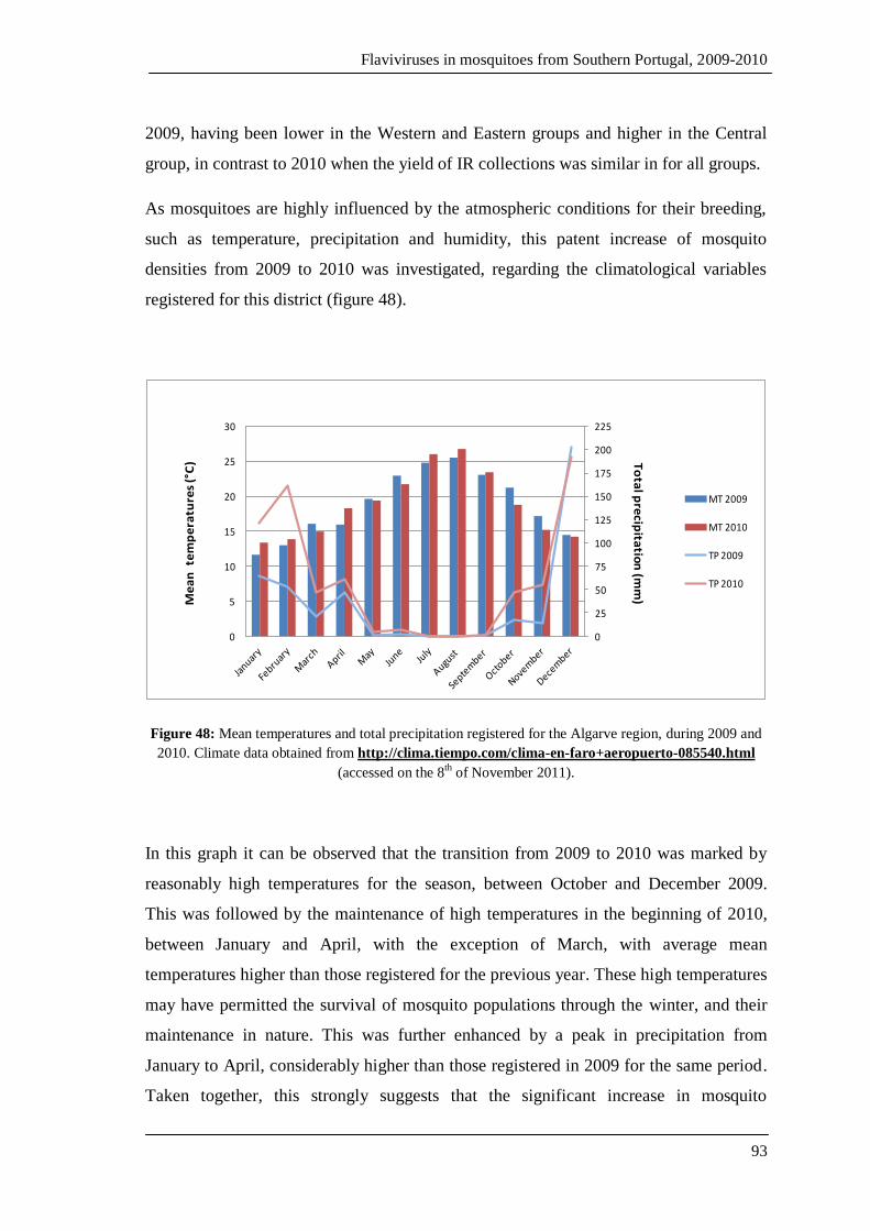
Flaviviruses in mosquitoes from Southern Portugal, 2009-2010
93
2009, having been lower in the Western and Eastern groups and higher in the Central
group, in contrast to 2010 when the yield of IR collections was similar in for all groups.
As mosquitoes are highly influenced by the atmospheric conditions for their breeding,
such as temperature, precipitation and humidity, this patent increase of mosquito
densities from 2009 to 2010 was investigated, regarding the climatological variables
registered for this district (figure 48).
0
25
50
75
100
125
150
175
200
225
0
5
10
15
20
25
30
MT 2009
MT 2010
TP 2009
TP 2010
Me
an
tem
pe
ratu
res
(°C
) To
tal p
recip
itatio
n (m
m)
Figure 48: Mean temperatures and total precipitation registered for the Algarve region, during 2009 and
2010. Climate data obtained from http://clima.tiempo.com/clima-en-faro+aeropuerto-085540.html
(accessed on the 8th of November 2011).
In this graph it can be observed that the transition from 2009 to 2010 was marked by
reasonably high temperatures for the season, between October and December 2009.
This was followed by the maintenance of high temperatures in the beginning of 2010,
between January and April, with the exception of March, with average mean
temperatures higher than those registered for the previous year. These high temperatures
may have permitted the survival of mosquito populations through the winter, and their
maintenance in nature. This was further enhanced by a peak in precipitation from
January to April, considerably higher than those registered in 2009 for the same period.
Taken together, this strongly suggests that the significant increase in mosquito

Flaviviruses in mosquitoes from Southern Portugal, 2009-2010
94
populations from 2009 to 2010 could be due to the combined effects of higher
temperatures at the end of 2009 and beginning of 2010, and the increase in total
precipitation before the beginning of spring, thus providing optimal conditions for the
survival and reproduction of mosquitoes in 2010. In fact, Calheiros and Casimiro (2006)
have already stated that climate change in the Algarve may result in an increase of
mosquito population density, particularly of Anopheles atroparvus and Culex pipiens
during cold months, thus causing an increase in the vectorial capacity and,
consequently, the risk of transmission of vector-borne diseases especially West Nile
fever.
As well as mean densities, the relative proportions of all mosquito species collected also
varied according to collection method, geographical group and month/year of
collection. This is a direct consequence of the fact that different collection methods
target different types of mosquito behaviour. For example, while CDC traps aim at
active host-seeking mosquitoes, whether indoors or outdoors, the main targets of IR
collections are mosquitoes resting indoors after feeding, that is, endophilic mosquitoes.
In CDC trap collections, in 2009, Cx. pipiens was the predominant species representing
56% of all mosquitoes collected, followed by Cx. theileri 17-24% and Ae. caspius 21-
25%, in Eastern and Central groups, respectively, as represented in the figures 39 in the
results section. However, the Western group was dissimilar to the other two, with Ae.
caspius as the most abundant species 83% and all other species taking a share of 5% or
less each.
In 2009, Ae. caspius represented more than 80% of all CDC collections in the West, and
that was maintained in the following year. A remarkable increase in Ae. caspius relative
proportion was registered in the Eastern group where a rise of 67% was recorded, from
21% in 2009 to 88% in 2010 (as shown on figure 40, in the results section). This trend
was also observed in CDC collections in the Central group, where an increase of
Ae.caspius relative proportion of nearly 20% from 2009 to 2010 was registered. In
addition, in this same area, the most abundant species was Cx. theileri with 38%, which
also registered a significant increase of 14% from the previous year, closely followed by
Cx. pipiens (26%) and Ae. caspius (23%).
Flaviviruses in mosquitoes from Southern Portugal, 2009-2010
95
In IR collections, the predominant species in both years was An. atroparvus, the only
member of the An. maculipennis complex captured, in all three groups, ranging between
27 and 77% (as represented in the figures 41 and 42 in the results section). The second
most abundant species, collected by this method, was Cx. pipiens with over 20% of all
mosquitoes collected, except in the Western group where it represents only 15%, in the
following year. In 2010, the most abundant species was An. atroparvus in the Central
and Western groups, while in the East, Ae. caspius took over as the predominant
species, with a 46%, followed by An. atroparvus and Cx. pipiens with 27% and 20%,
respectively. The prevalence of Anopheles atroparvus in IR collections, in most
geographical groups in both years, is a reflection of their animal feeding behaviour and
endophily (Cambournac and Hill, 1938; Sousa, 2001). The relative proportion for this
species by IR collections suffered a significant reduction from 60% (2009) to 27%
(2010) in the Eastern group, which may reflect fewer IR collections made (47 in 2009 to
36 in 2010), and possibly even a lesser availability of vertebrate hosts as was confirmed
in at least two collection sites.
Moreover, the relative proportions of Ae. caspius collected by IR in the Western group
nearly doubled from 2009 to 2010 (from 14% to 27% ). The values for the Central area
suffered only minor alterations; this species appeared in 2010 and was not recorded in
the previous year, however such slight changes would be predictable in nature, and
could be simply due to seasonality.
In sum, in 2009, the predominant species in IR collections, in all geographical groups,
were An. atroparvus and Cx. pipiens, respectively. This was maintained in 2010 only in
the Centre, as in the Western and Eastern groups the predominant species were An.
atroparvus and Ae. caspius. Regarding CDC collections, an absolute predominance of
Ae. caspius was registered for the West in both years, and in Eastern Algarve in 2010.
In the East and Centre 2009, the most abundant species was Cx. pipiens. The Central
Algarve (2010) was the only area where a predominance of Cx. theileri was registered.
The considerable higher numbers of Ae. caspius captured by CDC traps, in contrast to
IR collections, clearly reflects their predominant exophilic behaviour characteristic of
this species, as was also described by Almeida et al (2008, 2010).

Flaviviruses in mosquitoes from Southern Portugal, 2009-2010
96
Aedes caspius is mainly found in coastal and wetland areas, reaching the highest
densities in estuarine regions in the districts of Faro and Setúbal (Shaffner et al., 2001),
which can be explained by their halophilic6 nature. However, it also breeds in rice fields
that are vastly spread over both districts, especially in, and around, the Comporta area in
the district of Setúbal (Almeida et al., 2010). This mosquito species is described as
opportunistic and highly anthropophilic, often found in high numbers in outdoors and
human baited collections (Almeida et al., 2008; Osório et al., 2008). Aedes caspius is
considered an aggressive mosquito species commonly incriminated of nuisance biting
(Shaffner et al., 2001; Beker and Zgomba, 2007) in many densely populated areas,
which is the case of the districts of Setúbal and Faro. For these reasons, its public health
importance cannot be disregarded since its human biting rate can reach up to 300
bites/person/hour in the Comporta region (Sousa, 2008). Furthermore, it is also a
competent vector for WNV (Hubalek and Halouzka, 1999), Chikungunya virus
(Vazeille et al., 2008), Tahyna virus (Lundstrom, 1999), and Rift Valley fever virus
(Turell et al., 1996).
Culex theileri has been found to breed, among others, in rice farming areas (Novo,
2008), reaching high densities when climatic conditions are propitious (Almeida et al.,
2010). It is considered one of the most predominant, and widespread, mosquitoes
present in Portugal, especially in the southern regions (Almeida et al., 2008). This
mosquito feeds mainly in mammals, humans included, and is capable of transmitting
West Nile virus, Sindbis viruses and Rift Valley fever (Jupp et al., 1972; McIntosh et
al., 1980; Jupp, 1985). Its public health impact is reflected by the aggressive behaviour,
reflected by the human biting rate of approximately 400 bites per person per hour (in the
Comporta area, Setúbal), making it an incommodity agent responsible for nuisance
biting complaints (Sousa, 2008).
Culex pipiens was one of the most abundant and widespread species captured in
estuarine areas in both regions. This mosquito species is known as the main vector of
WNV in Europe and exists in Portugal in both the “pipiens” and “molestus” forms, with
high interbreeding which heightens their importance as bridge vectors between birds,
6 Requires a salty environment for breeding and development.
Flaviviruses in mosquitoes from Southern Portugal, 2009-2010
97
humans and other hosts due to their ornitophilic and anthropophilic behaviour (Gomes
et al., 2009),.
Anopheles atroparvus was found in both the Setúbal and Faro districts. This mosquito is
mainly found in animal shelters due to its zoophilic behaviour (Cambournac and Hill,
1938; Sousa, 2001). This species has been previously found infected with WNV in
Portugal (Filipe, 1972), hence its public health relevance.
Anopheles algeriensis was also collected in the Algarve, in agreement with other studies
(Ramos et al., 1982; Osório et al., 2010); however, none was found in Setúbal, perhaps
due to the limited number of collections as this species has previously been found in
that region (Osório et al., 2010).
Culex univittatus was, in agreement with Almeida and others (2008) present in the
Southern Portugal in low densities. This species has previously been found infected
with WNV (Esteves et al., 2005), and although it was collected in smaller numbers, its
ecology, geographical distribution and ability of vertically transmit WNV (Miller et al.,
2000) may constitute a potential cause for concern from a public health viewpoint.
Aedes detritus were collected in estuarine regions, though in low numbers when
compared to other species. However, in accordance with past studies, in itself this
values would not be an unexpected finding since Ae. detritus is also usually present in
wetlands and coastal areas (Ribeiro et al., 1988; Almeida et al., 2008). Although its
medical importance has not yet been clearly confirmed, it has, at least, been found
susceptible to infection by the Chikungunya virus (Vazeille et al., 2008).
Culiseta annulata and Cs.longiareolata were only collected in the Algarve region,
though in similarly low numbers.
Culiseta annulata was previously found mainly in inland regions (Almeida et al., 2008)
and, perhaps, the low numbers collected in this study could be because coastal and
estuarine areas, where collections were made, may not present the ideal conditions for
their breeding and development.
Cs. longiareolata has been considered one of the most widespread mosquitoes in
Portugal (Almeida et al., 2008).
Flaviviruses in mosquitoes from Southern Portugal, 2009-2010
98
Other species of mosquitoes such as Ae. berlandi, Ae. mariae, Coquillettidia richiardii,
Cx. laticinctus, Cs.subochrea, and Uranotaenia unguiculata were found in much lower
numbers. Most of these have no known medical importance, except for Co. richiardii
that, although very poorly represented in Portugal, it is a vector of WNV and
bunyaviruses, such as Calovo and Batai viruses (Hubalek and Halouzka, 1999;
Lundstrom, 1999; Higgs et al., 2004). This mosquito species has been found to share
habitats with migratory birds from Africa, which may be infected with arboviruses, they
feed on humans as well as birds, and thus may act as bridge vectors enhancing the
possibility of transmission of any of the above mentioned viruses to humans (Service,
1971).
Importantly, the spread of flaviviruses via arthropod vectors and/or their natural
reservoirs, is influenced by changing climatic patterns due to global warming, thus
expanding their distribution northwards as temperatures become higher. A study by
Epstein and Defilippo (2001) provided evidence that a correlation exists between WNV
outbreaks and drought. According to these investigators, the large outbreaks that
occurred in Romania (1996), USA (1999) and Russia (1999) followed short or long
periods of drought. Therefore, since an increase in the frequency and intensity of
periods of drought has been predicted to occur in Portugal because of climate change
(Calheiros and Casimiro, 2006), the probability of WNV outbreaks eventually occurring
in the country would also increase, if these changes were verified.
5.2. Mosquito screening for flaviviral sequences and respective phylogeny
The screening for flaviviral sequences was performed using, as starting point, the
clarified homogenates obtained from mosquito pool maceration, from which total RNA
was extracted. It is important to take into account that the products of total RNA
extraction are most certainly not pure. Indeed, apart from viral RNA (if present), other
RNA molecules from mosquitoes (at least) and other organisms (fungi, bacteria and
ectoparasites infecting the mosquitoes themselves) are most probably present in these
extracts. Furthermore, the primers used for viral sequence amplification were
degenerated in order to allow the amplification of a larger number of flaviviral NS5
sequences, and although this strategy may work in our favour, it may also be considered

Flaviviruses in mosquitoes from Southern Portugal, 2009-2010
99
risky. These risks translate in the form of amplification products that result from retro-
transcription of non-targeted RNA molecules from total RNA extracts. As a result, we
could be amplifying a variety of RNA molecules from different organisms and not only
those specifically targeted by the primers used. As a result, the possibility remains that
the obtained amplification products were produced from RNA contaminants and not
from viral RNA. Therefore, to rule that out, extraction controls were performed (as
described in section 3.2.8). Since these extraction controls produced negative results (as
expected), we can conclude that, under the conditions used, these RNA contaminants
present in the analysed samples were not having a significant impact in the obtained
amplification results.
In the past few years, studies conducted by Crochu et al (2004), Roiz et al (2009) and
Vázquez et al (2011b) have reported the integration of viral RNA sequences like that of
insect-specific viruses, genetically similar to that of CSAV, and other particles into the
mosquitoes’ genomes. As speculated for Ae. albopictus, sequence integration might
possibly occur following infection by a flavivirus, resulting in a novel mechanism by
which genetic diversity in eukaryotic cells may be produced (Crochu et al., 2004; Roiz
et al., 2009). However, though it is known that integration events occur, the respective
mechanism has not yet been determined. In addition, it is not known whether the
genetic diversity potentially created in eukaryotic cells would be beneficial or
detrimental and what effects it could generate on viral transmission and in vector
susceptibility and competence. Taken together, these findings also pose two other
potential problems to our analysis. If the prepared RNA extracts would be contaminated
with either mosquito genomic DNA (integrated viral sequences), or RNA molecules
expressed from these putative DNA forms, then amplification results might translate the
amplification of these “secondary contaminants”, and not from viral RNA contained
within virions, thus producing false positive results. This is a major pitfall of this type of
viral detection method since there are no guarantees that the positive results are not due
to integration events. Therefore, the only way to clearly prove that positive
amplification pools resulted exclusively from the presence of virion RNA is to perform
viral isolation in cell cultures followed by search of flavivirus virions by electron
microscopy and their complete sequence analysis. In their absence, one cannot conclude
Flaviviruses in mosquitoes from Southern Portugal, 2009-2010
100
that positive pools resulted solely from viral RNA since these procedures were not
systematically carried out for this study.
Whatever the nature of the obtained amplification results, the sequences obtained
resulted from the specific amplification of the target region of the NS5 gene of the
flavivirus genome, as previously suggested by preliminary control experiments (section
4.4.2.). However, their small size, ranging between 165bp and 170bp, limits the
accuracy of subsequent phylogenetic analysis considerably (bootstrap certainty is
compromised). Since our PCR products resulted in the amplification of several
molecules apart from the targeted one (which can be observed by the appearance of
multiple bands for the same sample, after gel electrophoresis), DNA had to be purified
from the agarose gel, after separation of the amplified molecules. This method is usually
associated with the loss of a fraction of the purified product, in contrast to DNA
purification directly from the PCR product, which would only be applicable if the
purification products were resultant from a single targeted molecule.
The use of oligonucleotide primers that amplify, in average, a fragment of
approximately 200bp of the flavivirus NS5 gene is quite common among research
groups. For example, Scaramozzino et al (2001) and Pabbaraju et al (2009) analysed
250bp sized amplimers, Calzolari et al (2010) reported NS5 sequences ranging between
200bp and 262bp, whereas Sánchez-Seco et al (2005) obtained even shorter sequences
of 143bp in length. However, as such small fragments do not allow for accurate
phylogenetic analyses, some research groups have designed and adapted primers in
order to amplify larger fragments for better and more precise phylogenetic
characterisation of sequence sets. Some of these groups aimed to obtain partial
sequences of large sizes of, for example, 1000bp (Kuno et al., 1998), 1075bp (Ayers et
al., 2006), 800bp (Maher-Sturgess et al., 2008), 720bp (Morales-Betoulle et al., 2008)
and 1019bp (Vázquez et al., 2011b). Though these groups used different primer sets, the
methodology used was usually similar with only a few variations.
The flaviviruses are known to be globally widespread viruses, with the exception of
Antarctica (Weaver, 2006). Since the beginning of the 21st century, the circulation of a
variety of pathogenic and insect-specific flavivirus sequences from different mosquito
species (as described in sections 1.5 and 1.6) has been discovered all over the world,

Flaviviruses in mosquitoes from Southern Portugal, 2009-2010
101
including Europe. Insect-specific flaviviruses recently detected in Portugal (directly
deposited in the sequence database), Italy and Spain are included in the phylogenetic
tree presented below (figure 49). The majority of the sequences obtained were detected
in Aedes mosquitoes (mainly Ae. caspius) and in Culex species mosquitoes.
Similarly to what occurs in phylogenetic analysis of the genus Flavivirus in agreement
with Kuno et al (1998) and Gaunt et al (2001) (as described in section 1.3.3, figure 7),
all these sequences grouped together mainly according to vector species. Consistent
with the separation of viruses by vector species, most sequences derived from Culex
mosquitoes grouped together at the top half of the tree, where the sequences we detected
in Culex species mosquitoes clustered together with the Spanish “Mediterranean Culex
Flavivirus” sequences.
The “Mediterranean Culex Flavivirus” group includes sequences detected in Spain
mainly in Cx. theileri and Cx. pipiens (Vázquez et al, 2011b); this is in agreement with
our findings, where a higher prevalence of positive pools composed of Cx. theileri over
those of Cx. pipiens was verified. Consequently, more sequences were detected in the
former (n=5) than in the latter species (n=1). Although a “Culex theileri flavivirus”
sequence from Portugal (EU716420) is included in the analysis, all our sequences
derived from Culex spp. show closer relation to their Spanish counterparts. The entire
group of sequences derived from Culex mosquitoes is well supported by a 96%
bootstrap value; this is in accordance with the positioning of the Culex group of
sequences in the tree shown in figure 47 (section 4.5).
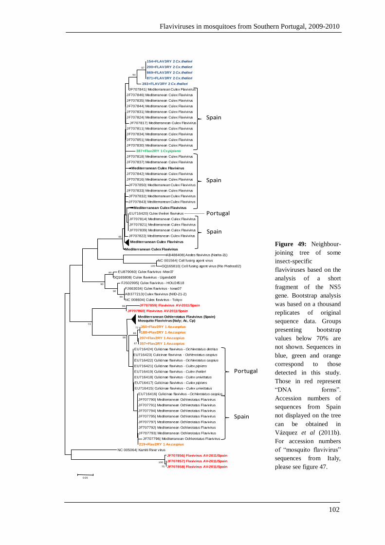
Flaviviruses in mosquitoes from Southern Portugal, 2009-2010
102
154+FLAV3RY 2 Cx.theileri
200+FLAV3RY 2 Cx.theileri
869+FLAV3RY 2 Cx.theileri
871+FLAV3RY 2 Cx.theileri
393+FLAV3RY 2 Cx.theileri
JF707841| Mediterranean Culex Flavivirus
JF707846| Mediterranean Culex Flavivirus
JF707835| Mediterranean Culex Flavivirus
JF707844| Mediterranean Culex Flavivirus
JF707831| Mediterranean Culex Flavivirus
JF707824| Mediterranean Culex Flavivirus
JF707817| Mediterranean Culex Flavivirus
JF707811| Mediterranean Culex Flavivirus
JF707834| Mediterranean Culex Flavivirus
JF707851| Mediterranean Culex Flavivirus
JF707830| Mediterranean Culex Flavivirus
387+Flav2RY 1 Cx.pipiens
JF707818| Mediterranean Culex Flavivirus
JF707837| Mediterranean Culex Flavivirus
Mediterranean Culex Flavivirus
JF707842| Mediterranean Culex Flavivirus
JF707816| Mediterranean Culex Flavivirus
JF707850| Mediterranean Culex Flavivirus
JF707833| Mediterranean Culex Flavivirus
JF707832| Mediterranean Culex Flavivirus
JF707843| Mediterranean Culex Flavivirus
Mediterranean Culex Flavivirus
EU716420| Culex theileri flavivirus -------------------
JF707814| Mediterranean Culex Flavivirus
JF707821| Mediterranean Culex Flavivirus
JF707839| Mediterranean Culex Flavivirus
JF707822| Mediterranean Culex Flavivirus
Mediterranean Culex Flavivirus
Mediterranean Culex Flavivirus
AB488408| Aedes flavivirus (Narita-21)
NC 001564| Cell fusing agent virus
GQ165810| Cell fusing agent virus (Rio Piedras02)
EU879060| Culex flavivirus -Mex07
GQ165808| Culex flavivirus - Uganda08
FJ502995| Culex flavivirus - HOU24518
FJ663034| Culex flavivirus - Iowa07
AB377213| Culex flavivirus (NIID-21-2)
NC 008604| Culex flavivirus - Tokyo
JF707859| Flavivirus AV-2011/Spain
JF707860| Flavivirus AV-2011/Spain
Mediterranean Ochlerotatus Flavivirus (Spain)Mosquito Flavivirus (Italy; Ac, Cp)
350+Flav2RY 1 Ae.caspius
188+Flav2RY 1 Ae.caspius
207+Flav2RY 1 Ae.caspius
357+Flav2RY 1 Ae.caspius
EU716424| Culicinae flavivirus - Ochlerotatus detritus
EU716423| Culicinae flavivirus - Ochlerotatus caspius
EU716422| Culicinae flavivirus - Ochlerotatus caspius
EU716421| Culicinae flavivirus - Culex pipiens
EU716419| Culicinae flavivirus - Culex theileri
EU716418| Culicinae flavivirus - Culex univittatus
EU716417| Culicinae flavivirus - Culex pipiens
EU716415| Culicinae flavivirus - Culex univittatus
EU716416| Culicinae flavivirus - Ochlerotatus caspius
JF707790| Mediterranean Ochlerotatus Flavivirus
JF707791| Mediterranean Ochlerotatus Flavivirus
JF707794| Mediterranean Ochlerotatus Flavivirus
JF707795| Mediterranean Ochlerotatus Flavivirus
JF707797| Mediterranean Ochlerotatus Flavivirus
JF707792| Mediterranean Ochlerotatus Flavivirus
JF707793| Mediterranean Ochlerotatus Flavivirus
JF707796| Mediterranean Ochlerotatus Flavivirus
219+Flav2RY 1 Ae.caspius
NC 005064| Kamiti River virus
JF707856| Flavivirus AV-2011/Spain
JF707857| Flavivirus AV-2011/Spain
JF707858| Flavivirus AV-2011/Spain75
100
100
99
90
85
90
72
67
86
98
73
90
97
94
96
0.05
Spain
Spain
Spain
Portugal
Portugal
Spain
Figure 49: Neighbour-
joining tree of some
insect-specific
flaviviruses based on the
analysis of a short
fragment of the NS5
gene. Bootstrap analysis
was based on a thousand
replicates of original
sequence data. Groups
presenting bootstrap
values below 70% are
not shown. Sequences in
blue, green and orange
correspond to those
detected in this study.
Those in red represent
“DNA forms”.
Accession numbers of
sequences from Spain
not displayed on the tree
can be obtained in
Vázquez et al (2011b).
For accession numbers
of “mosquito flavivirus”
sequences from Italy,
please see figure 47.
Flaviviruses in mosquitoes from Southern Portugal, 2009-2010
103
In contrast, the sequences detected in Ae. caspius including the Spanish, the Italian and
those obtained in the course of this work, formed a large group at the bottom half of the
tree, which is well supported by solid statistical certainty. However, within the
“Mosquito Flavivirus” group (Italy) there is a single sequence derived from Cx. pipiens.
Apart from this isolated case within the group, all other RNA viruses in this cluster
were detected in Aedes caspius. The viral sequences detected in this study (from Ae.
caspius) show a stronger relation to the group including “Culicinae flavivirus” from
Portugal and “Mediterranean Ochlerotatus Flavivirus” from Spain than to that
composed of Spanish and Italian sequences from the same mosquito species. This could
be due to the relative geographical proximity of collection sites, since sampled sites are
located in southern Spain (Huelva) and southern Portugal (Faro district, Algarve), and
mosquito dispersal between these two regions is feasible.
Notably, the group of Culicinae flaviviruses from Portugal also includes sequences
derived from Culex species mosquitoes (5 out of 9); however, these also clustered with
the other “Culicinae flavivirus” sequences detected in Ae.caspius.
It is noteworthy that some groups of viral sequences have derived from different vector
species, as is the case of the “Mosquito flavivirus” group from Italy and “Culicinae
flavivirus” group from Portugal. The fact that a certain type/species of virus infects
more than one vector/host species and that some mosquito-borne viruses have been
isolated from ticks in the wild and some tick-borne viruses have been detected in
mosquitoes in the wild, invalidates the theory that viruses evolve progressively with
their hosts. In addition, since some viruses share the same host/vector species the
simultaneous circulation of different viruses may occur (Sánchez-Seco et al., 2005) in
the same geographical areas, thus making the viruses difficult to identify.
The classification of flaviviruses presents many difficulties that partly stem from the
widespread distribution of these viruses as well as with the variety of vectors species
and vertebrate hosts associated with their biological transmission (Kuno et al., 1998).
Thus, the distribution of flaviviruses usually overlaps that of their arthropod vectors and
is accompanied by rapid genetic alterations, which facilitate host range expansion.
Importantly, under optimal climate conditions RNA viruses are capable of undergoing
rapid genetic changes through mutations, which contributes to the genetic variability

Flaviviruses in mosquitoes from Southern Portugal, 2009-2010
104
and facilitates the adaptation of those viruses to a high diversity of hosts, thus
promoting their host range expansion in distinct geographical areas. In addition,
according to an in vitro study conducted by Kuno (2007), the diversity of host range
strongly correlates with the standard classification of flaviviruses (Karabatsos, 1985) as
well as with the segregation of viral sequences in flavivirus phylogeny (Kuno et al.,
1998), in agreement with the positioning of flaviviruses sequences obtained in this
study. Another factor that influences host-specificity is the composition of the viral
genome. Greenbaum et al (2008) discuss that a trend exists in the expression of
flaviviruses sequences composition (CpG expression) and the host species, that is, that
flaviviruses suppress CpG because their hosts also suppress it. However, Cook and
Holmes (2006) and Schubert and Putonti (2010) disagree and affirm that according to
sequence composition, a correlation does not exist between codon usage and vector
species, thus there is no co-variation of codon usage within a vector group.
Other factors may play important roles in the emergence or re-emergence of viruses
worldwide. For example, DENV has been remerging in new tropical and subtropical
areas globally, most probably due to human activity that contributes to the expansion of
their vectors, Ae. aegypti and Ae. albopictus, into new areas; whereas WNV has been
circulating and become established in new geographical areas due to natural factors
such as bird migration, amongst others (Mackenzie et al., 2004). From its African origin
where it was first reported in 1937 WNV has since been extending its distribution, the
virus caused a large-scale epidemic in the USA, where it entered in 1999 and quickly
spread throughout the country, though the means of entry are still unclear (Epstein and
Defilippo, 2001). Following repeated isolations of WNV throughout Europe, it has been
suggested that the virus has become endemic in many European countries, and both
lineage 1(widespread) and 2 (previously found only in Africa) strains have been
detected (ECDC, 2011). The most likely means of dispersal of WNV is by migratory
birds as Europe sits right below the main migration routes (as shown on figure 24,
section 1.7). This is also the principal means of dispersal of WNV through Asia via the
Africa - Middle East route. Therefore, potentially infected migratory birds can spread
flaviviruses when resting and settling in new geographical areas in search of warmer
temperatures, thus infecting local populations of ornitophilic mosquitoes capable of
acting as bridge vectors and potentially causing “spill-over” to humans and animals.

Flaviviruses in mosquitoes from Southern Portugal, 2009-2010
105
Since co-circulation (Vázquez et al., 2011a) and co-infection (Kihara et al., 2007;
Kanthong et al., 2010) of flaviviruses have previously been reported, it is important to
investigate the influence of co-infection of flaviviruses on the capacity and competence
of vectorial transmission, whether the co-infection consists of antigenically distinct
flaviviruses, such as MBV/MBV or MBV/ISF. Especially since infection of mosquitoes
by insect-specific flavivirus does not seem to have a negative effect on their fitness, and
infection with one virus reduces the probability of infection and transmission with a
second antigenically related virus, as recently evidenced by Pesko and Moores (2009).
Therefore, it would be interesting to see if the infection by an insect-specific flavivirus
has any effects (positive or negative) on subsequent infection by arboviruses and on
their virulence.
5.3. Final conclusions
In this study, we were able to detect viral sequences in a significant proportion of the
screened mosquito pools (30%). Our results strongly suggest that these sequences
relate to insect-specific flaviviruses. As evidenced by the high minimum infection rates
(MIR) registered ( 22/1000), there was a large proportion of mosquitoes exhibiting
these sequences, thus potentially infected with these viruses.
A remarkable increase in mosquito densities was registered in the Algarve, from 2009
to 2010, and although no sequences of human pathogenic flaviviruses were detected,
several insect-specific flavivirus sequences were found in considerably high
percentages. The future importance of these is still to be known as these viruses are
widely dispersed and may integrate mosquito genomes. Furthermore, Aedes albopictus
and Aedes aegypti were not detected, despite the fact that the former has been
established in Europe for a few years, and the latter is settled in the Madeira
Archipelago (Portugal). However, there are other mosquito species that due to their
presence, high abundance and ability to transmit flaviviruses such as West Nile virus,
may be a cause for concern to Public Health. This importance is enhanced by the
vector’s choice of vertebrate hosts since Portugal harbours many migratory bird species
from Africa, and a diversity of ornitophilic mosquitoes that may act as bridge vectors,

Flaviviruses in mosquitoes from Southern Portugal, 2009-2010
106
some of which have been reported to share a common habitat, thus increasing the
probability of exotic arboviral transmission to animals and humans.
Portugal is a country that gathers many favourable climatic and ecological conditions
for the establishment of invasive species such as Aedes albopictus, and consequently the
introduction and transmission of arboviruses. Mosquito surveys are essential for
monitoring of mosquito species distribution, abundance and seasonal dynamics that
gives glues as to what potential vectors are present and what viruses are circulating in
association with those vectors, thus allowing for the risk assessment of arboviral
transmission. Epidemiological surveys have, thus, become indispensable tools for the
prediction, risk assessment and management of arboviral outbreaks, and all the above-
mentioned factors emphasize the need for continuous surveillance programmes in order
to ensure Public Health, especially in Southern Mediterranean countries where
pathogenic flaviviruses circulation has increased in recent years causing many fatalities.
In conclusion, the screening for viral sequences revealed the presence and high
prevalence of these, in mosquito pools. Further analysis of a limited number of positive
pools demonstrated that the detected sequences did not correspond to arboviruses (NS5
amplifiable by the employed method), but to a group of flaviviruses generally
designated by insect-specific flaviviruses (ISF). While their biology remains unclear,
they are dispersed over a wide geographical area, and their prevalence appears to be
high in natural mosquito populations of different species. Moreover, as a direct result of
this work, R. Parreira has isolated an insect-specific flavivirus, from our mosquito
homogenates, in cell culture (R. Parreira, personal communication).

Flaviviruses in mosquitoes from Southern Portugal, 2009-2010
107
6.
BIBLIOGRAPHY
Flaviviruses in mosquitoes from Southern Portugal, 2009-2010
108
6. Bibliography
Almeida, A.P.G, Freitas, F.B., Novo, M.T., Sousa, C.A., Rodrigues, J.C., Alves, R.,
Esteves, A. 2010. Mosquito surveys and West Nile virus screening in two different
areas of Southern Portugal, 2004–2007. Vector-Borne and Zoonotic Diseases, 10 (7).
Almeida, A.P.G, Galão, R.P., Novo, M.T., Sousa, C.A., Parreira, R., Rodrigues, J.C.,
Pinto, J.C., Carvalho, L. 2005. Update on the distribution of some mosquito (Diptera:
Culicidae) species in Portugal. European Mosquito Bulletin, 19, 20-25.
Almeida, A.P.G, Galão, R.P., Sousa, C.A., Novo, M.T., Parreira, R., Pinto, J., Piedade,
J., Esteves, A. 2008. Potential mosquito vectors of arboviruses in Portugal: species,
distribution, abundance and West Nile infection. Transactions of the Royal Society of
Tropical Medicine and Hygiene, 102, 823-832.
Almeida, A.P.G., Gonçalves, Y.M., Novo, M.T., Sousa, C.A., Melim, M., Grácio,
A.J.S. 2007. Vector monitoring of Aedes aegypti in the Autonomous Region of
Madeira, Portugal. Eurosurveillance [Online], 12. Available from:
http://www.eurosurveillance.org/ViewArticle.aspx?ArticleId=3311 [Accessed on the 10th of
August 2011].
Aranda, C., Eritja, R., Roiz, D. 2006. First record and establishment of the mosquito
Aedes albopictus in Spain. Medical and Veterinary Entomology, 20, 150-152.
Aranda, C., Sánchez-Seco, M., Cáceres, F., Escosa, R., Gálvez, J.C., Masià, M.,
Marquéz, E., Ruíz, Alba, A., S., Busquets, N., Vázquez, A., Castellà, J., Tenorio, A.
2009. Detection and monitoring of mosquito flaviviruses in Spain between 2001 and
2005. Vector-borne and Zoonotic diseases, 9 (2).
Ayers, M., Adachi, D., Johnson, G., Andonova, M., Drebot, M., Tellier, R. 2006. A
single tube RT-PCR assay for the detection of mosquito-borne flaviviruses.
J.Virol.Methods, 135, 235-239.
Barrett, A.D.T. 2001. Flaviviruses. In Encyclopedia of Life Sciences, Nature
Publishing Group.
Flaviviruses in mosquitoes from Southern Portugal, 2009-2010
109
Barzon, L., Pacenti, M., Cusinato, R., Cattai, M., Franchin, E., Pagni, S., Martello,T.,
Bressan, S., Squarzon, L., Cattelan, A.M., Pellizzer, G., Scotton, P., Beltrame, A.,
Gobbi, F., Bisoffi, F., Russo, F., Palù, G. 2011. Human cases of West Nile virus
infection in north-eastern Italy, 15 June to 15 November 2010. Eurosurveillance
[Online],16(33).Available:http://www.eurosurveillance.org/ViewArticle.aspx?ArticleId=1994
9 [Accessed on the 7th
of October 2011].
Becker, N., Zgomba, M.2007. Mosquito control in Europe. In Emerging pets and
vector-borne diseases in Europe. Ecology and control of vector-borne diseases.
Wageningen Academic Publishers, The Netherlands.
Blitvich, B.J., Lin, M., Dorman, K.S., Soto, V., Hovav, E., Tucker, B.J., Staley, M.,
Platt, K.B., Bartholomay, L.C. 2009. Journal of Medical Entomology, 46(4), 934-941.
Calheiros, J.M, Casimiro, E., Almeida, A.P.G., Alves-Pires, C., Collares Pereira, M.,
Cardoso, M.F., Santos-Silva, M., Afonso, O., Sousa, R. 2006. 6 - Saúde Humana e
Implicações para o Turismo. Pp. 233-270. In F.D. Santos & Miranda, P., Eds,
Alterações Climáticas em Portugal: Cenários, Impactos e Medidas de Adaptação.
Projecto SIAM II, Gradiva, Lisboa, Portugal.
Calzolari, M., Bonilauri, P., Bellini, R., Albieri, A., Defilippo, F., Maioli, G., Galletti,
G., Gelati, A., Barbieri, I., Tamba, M., Lelli, D., Carra, E., Cordioli, P., Angelini, P.,
Dottori, M. 2010b. Evidence of simultaneous circulation of West Nile and Usutu viruses
in mosquitoes sampled in Emilia-Romagna region (Italy) in 2009. PLoS One, 5(12).
Calzolari, M., Bonilauri, P., Bellini, R., Caimi, M., Defilippo, F., Maioli, G., Albieri,
A., Medici, A., Veronesi, R., Pilani, R., Gelati, A., Angelini, P., Parco, V., Fabbi, M.,
Barbieri, I., Lelli, D., Lavazza, A., Cordioli, P., Dottori, M. 2010a. Arboviral survey of
mosquitoes in two Northern Italian regions in 2007 and 2008. Vector-borne and
Zoonotic diseases, 00.
Cambournac, F.J.C., Hill, R.B. 1938. The biology of Anopheles maculipennis var.
atroparvus in Portugal. Transactions of the Third International Congress of Tropical
Medicine and Malaria, 2, 178-184.
Flaviviruses in mosquitoes from Southern Portugal, 2009-2010
110
Carter, G.R., Wise, D.J., Flores, E.F. 2008. Flaviviridae In A concise review of
veterinarian virology, Ithaca NY, International Veterinarian Information Service.
CDC, 2010a. West Nile virus activity – United States, 2009. MMWR Morbidity and
Mortality Weekly Report [Online], 59. Available from:
http://www.cdc.gov/ncphi/disss/nndss/casedef/arboviral_current.htm. [Accessed on the 8th
of
July 2011].
CDC, 2010b. Yellow fever vaccine. Morbidity and Mortality Weekly Report [Online],
59. Available from: http://www.cdc.gov/mmwr/preview/mmwrhtml/rr5907a1.htm [Accessed
on the 24th
of November 2011].
CDC, 2011 – Available from http://www.cdc.gov/ncidod/dvbid/westnile/software.htm -
accessed on the 22nd
of August 2011 [Accessed on the 18th of September 2011].
Charrel, R.N., Moureau, G., Temmam, S., Izri, A., Marty, P., Parola, P., Da Rosa,
A.T., Tesh, R.B., de Lamballerie, X. 2009. Massilia virus, a novel Phlebovirus
(Bunyaviridae) isolated from sandflies in the Mediterranean. Vector-Borne and
Zoonotic Diseases, 9(5).
Chaskopoulou, A., Dovas, C.I., Chaintoutis, S.C, Bouzalas, I., Ara, G.,
Papanastassopoulou, M. 2011. Evidence of enzootic circulation of West Nile virus (Nea
Santa-Greece-2010, lineage 2) Greece, May to July 2011. Eurosurveillance [Online], 16
(31). Available from:http://www.eurosurveillance.org/ViewArticle.aspx?ArticleId=19933
[Accessed on the 8th
of August 2011].
Connell, J., McKeown, P., Garvey, P., Cotter, S., Conway, A., O’Flanagan, D.,
O’Herlihy, B.P., Morgan, D., Nicoll, A., Lloyd, G. 2004.Two linked cases of West Nile
virus (WNV) acquired by Irish tourists in the Algarve, Portugal. Eurosurveillance
[Online], 8. Available on:http://www.eurosurveillance.org/ViewArticle.aspx?ArticleId=2517
[Accessed on 10th
August 2011].
Cook, S., Bennett, S.N., Holmes, E.C., De Chesse, R., Moureau, G., de Lamballerie, X.
2006. Isolation of a new strain of the flavivirus cell fusing agent virus in a natural
mosquito population from Puerto Rico. Journal of General Virology, 87, 735-748.
Flaviviruses in mosquitoes from Southern Portugal, 2009-2010
111
Cook, S., Holmes, E.C. 2006. A multigene analysis of phylogenetic relationships
among the flaviviruses (Family: Flaviviridae) and the evolution of vector transmission.
Arch Virol, 151, 309-325.
Cook, S., Moureau, G., Harbach, R.E., Mukwaya, L., Goodger, K., Ssenfuka, F., Gould,
E., Holmes, E.C., de Lamballerie, X. 2009. Isolation of a novel strain of Culex
flavivirus (Flaviviridae) from a natural mosquito population in Uganda. Journal of
General Virology, 90, 2669-2678.
Crabtree, M.B., Nga, P.T, Miller, B.R. 2009. Isolation and characterization of a new
mosquito flavivirus, Quang Binh virus, from Vietnam. Arch Virol, 154, 857-860.
Crabtree, M.B., Sang, R.C., Stollar, V., Dunster, L.M., Miller, B.R. 2003. Genetic
characterization of the newly described insect flavivirus, Kamiti River virus. Arch Virol,
148, 1095-1118.
Crochu, S., Cook, S., Attoui, H., Charrel, R.N., De Chesse, R., Belhouchet, M.,
Lemasson, J., de Micco, P., de Lamballerie, X. 2004. Sequences of flavivirus’related
RNA viruses persist in DNA form integrated in the genome of Aedes spp. mosquitoes.
Journal of General Virology, 85, 1971-1980.
Danis, K., Papa, A., Papanikolaou, E., Dougas, G., Terzaki, I., Baka, A., Vrioni, G.,
Kapsimali, V., Tsakris, A., Kansouzidou, A., Tsiodras, S., Vakalis, N., Bonovas, S.,
Kremastinou, J. 2011. Ongoing outbreak of West Nile virus infection in humans,
Greece, July to August 2011. Eurosurveillance [Online], 16 (34). Available from:
http://www.eurosurveillance.org/ViewArticle.aspx?ArticleId=19951 [Accessed on the 8th
of
August 2011].
DREF, 2009. Cape Verde: Dengue epidemic. Available from:
http://www.ifrc.org/docs/appeals/09/MDRCV001do.pdf [Accessed on the 8th of July 2011].
ECDC, 2011. Review of the epidemiological situation of West Nile virus infection in
the European Union. Rapid Risk Assessment [Online]. Available from:
http://ecdc.europa.eu/en/publications/Publications/110920_TER_Rapid%20risk%20assessment
_WNF.pdf [Accessed on the 20th
of October 2011].
Flaviviruses in mosquitoes from Southern Portugal, 2009-2010
112
Eiras, A.E. 2004. Culicidae, In Parasitologia Humana (11th
edition), Atheneu
Publishers.
Eldridge, B.F. 2005. Mosquitoes, the Culicidae. In Biology of Disease Vectors (2nd
edition), London, Elsevier Academic Press 2005: 167-185.
Endy, T.P., Weaver, S.C., Hanley, K.A. 2010. Frontiers in dengue virus research,
Dengue virus: Past, present and future, Portland, Caister Academic Press.
Epstein, P. 2000. Is global warming harmful to Health? Scientific American, August,
36-43.
Epstein, P.R., Defilippo, C. 2001. West Nile virus and drought. Global Change &
Human Health, 2(2).
Esteves, A., Almeida, A.P.G., Galão, R.P., Parreira, R., Piedade, J., Rodrigues, J.C.,
Sousa, C.A., Novo, M.T. 2005. West Nile Virus in Southern Portugal, 2004. Vector-
Borne and Zoonotic Diseases, 5 (4).
Farfan-Ale, J., Lorono-Pino, M.A., Garcia-Rejon, J.E., Hovav, E., Powers, A.M., Lin,
M., Dorman, K.S., Platt, K.B., Bartholomay, L.C., Soto, V., Beaty, B.J., Lanciotti, R.S.,
Blitvich, B.J. 2009. Detection of RNA from a novel West Nile-like virus and high
prevalence of an insect-specific flavivirus in mosquitoes in the Yucatan Peninsula of
Mexico. Am J Trop Med Hyg, 80(1), 85-95.
Fernandez-Garcia, M., Mazzon, M., Jacobs, M., Amara, A. 2009. Pathogenesis of
flavivirus infections: using and abusing the host cell. Cell Host and Microbe, 5.
Filipe, A.R., 1972. Isolation in Portugal of West Nile virus from Anopheles
maculipennis mosquitoes. Acta Virol. 16, 361.
Freitas, F.B., Novo, M.T., Esteves, A., Almeida, A.P.G. Species Composition and WNV
Screening of Mosquitoes from Lagoons in a wetland area of the Algarve, Portugal.
Submitted
Flaviviruses in mosquitoes from Southern Portugal, 2009-2010
113
Gaunt, M.W., Sall, A.A., de Lamballerie, X., Falconar, A.K.I., Dzhivanian, T.I., Gould,
E.A. 2001. Phylogenetic relationships of flaviviruses correlate with their epidemiology,
disease association and biogeography. Journal of General Virology, 82, 1867-1876.
Goddard, J. 2008. Infectious diseases and arthropods, Mississippi, Humana Press,
Springer Publishers.
Gomes, B., Sousa, C.A., Novo, M.T., Freitas, F.B., Alves, R., Côrte-Real, A.R.,
Salgueiro, P., Donnelly, M.J., Almeida, A.P.G., Pinto, J. 2009. Asymmetric
introgression between sympatric molestus and pipiens forms of Culex pipiens (Diptera:
Culicidae) in the Comporta region, Portugal. Evolutionary Biology, 9 (262).
Gould, E.A, de Lamballerie, X., de A Zanotto, P.M., Holmes, E.C. 2003. Origins,
evolution, and vector/host coadaptations within the genus Flavivirus. Advances in Virus
Research, 59.
Gould, E.A., Higgs, S. 2009. Impact of climate change and other factors on emerging
arbovirus diseases. Trans R Soc Trop Med Hyg, 103(2), 109-121.
Gould, E.A., Solomon, T. 2008. Pathogenic flaviviruses. The Lancet, 371, 500-509.
Greenbaum, B.D., Levine, A.J., Bhanot, G., Rabadan, R. 2008. Patterns of evolution
and host gene mimicry in influenza and other RNA viruses. PloS Pathog., 4.
Gubler, D.J. 2002. The global emergence/resurgence of arboviral diseases as public
health problems. Arch Med Res; 33, 330-342.
Gubler, D.J. 2007. The continuing spread of West Nile virus in the Western
Hemisphere. Clinical Infectious Diseases, 45, 1039-1046.
Harris, E., Holden, K.L., Edgil, D., Polacek, C., Clyde, K. 2006. New treatment
strategies for dengue and other flaviviral diseases. Wiley, Chichester, Novartis
Foundation Symposium, 23-47.
Hollidge, B.S., González-Sacarano, F., Soldan, S.S. 2010. Arboviral encephalitides:
transmission, emergence and pathogenesis. J Neuroimmune Pharmacol, 5, 428-442.
Flaviviruses in mosquitoes from Southern Portugal, 2009-2010
114
Hoshino, K., Isawa, H., Tsuda, Y., Sawabe, K., Kobayashi, M. 2009. Isolation and
characterization of a new insect flavivirus from Aedes albopictus and Aedes flavopictus
mosquitoes in Japan. Virology, 391, 119-129.
Hoshino, K., Isawa, H., Tsuda, Y., Yano, K., Sasaki, T., Yuda, M., Takasaki, T.,
Kobayashi, M., Sawabe, K. 2007. Genetic characterization of a new insect flavivirus
isolated from Culex pipiens mosquito in Japan. Virology, 359, 405-414.
Huang, C., Slater, B., Campbell, W., Howard, J., White, D. 2001. Detection of
arboviral RNA directly from mosquito homogenates by reverse-transcription –
polymerase chain reaction. Journal of Virological Methods, 94, 121-128.
Hubalek, Z., Halouzka, J., 1999. West Nile Fever - a reemerging mosquito-borne viral
disease in Europe. Emerging Infectious Diseases, 5, 643-650.
Huhtamo, E., Putkuri, N., Kurkela, S., Manni, T., Vaheri, A., Valapahti, O., Uzcátegui,
N.Y. 2009. Characterization of a novel flavivirus from mosquitoes in Northern Europe
that is related to mosquito-borne flaviviruses of the Tropics. Journal of Virology,
83(18), 9532-9540.
Igarashi, A., Harrap, K.A., Casals, J., Stollar, V. 1976. Morphological, biochemical,
and serological studies on a viral agent (CFA) which replicates in and causes fusion of
Aedes albopictus (Singh) cells. Virology, 74, 174-187.
Johnson, N., Wakeley, P.R., Mansfield, K.L., McCracken, F., Haxton, B., Phipps, L.P.,
Fooks, A.R. 2010. Assessment of a novel real-time pan-flavivirus RT-polymerase chain
reaction. Vector-borne and Zoonotic diseases, 10 (7).
Junglen, S., Kopp, A., Kurth, A., Pauli, G., Ellerbrok, H., Leendertz, F.H. 2009.A new
flavivirus and a new vector: characterization of a novel flavivirus isolated from
Uranotaenia mosquitoes from a Tropical Rainforest. Journal of Virology, 83(9), 4462-
4468.
Jupp, P.G., McIntosh, B.M., Dickinson, D.B., 1972. Quantitative experiments on the
vector capability of Culex (Culex) theileri Theobald with West Nile and Sindbis viruses.
J. Med. Entomol., 9, 393-395.

Flaviviruses in mosquitoes from Southern Portugal, 2009-2010
115
Jupp,P.G. 1985. Culex theileri and Sindbis virus; salivary glands infection in relation to
transmission. J Am Mosq Control Assoc, 1, 374-376.
Kanthong, N., Khemnu, N., Pattanakitsakul, S.N., Malasit, P., Flegel, T.W. 2010.
Persistent triple-virus co-infections in mosquito cells. BMC Microbiol, 20, 10-14.
Karabatsos, N. 1985. International catalogue of arboviruses (3rd
ed.). American Society
of Tropical Medicine and Hygiene, San Antonio, Texas.
Kihara, Y., Satho, T., Eshita, Y., Sakai, K., Kotaki, A., Takasaki, T., Rongsriyam, Y.,
Komalamisra, N., Srisawat, R., Lapcharoen, P., Sumroiphon, S., Iwanaga, S., Ushijima,
H., Endoh, D.,Miyata, T., Sakata, A., Kashige, N., Miake, F., Fukushi, S., Saijo, M.,
Kurane, I., Morikawa, S., Mizutani, T. 2007. Rapid determination of viral RNA
sequences in mosquitoes collected in the field. Journal of Virological Methods, 146,
372-374.
Kim, D.Y., Guzman, H., Bueno Jr, R., Dennett, J.A., Auguste, A.J., Carrington, C.V.F.,
Popov, V.L., Weaver, S.C., Beasley, D.W.C., Tesh, R.B. 2009. Characterization of
Culex Flavivirus (Flaviviridae) strains isolated from mosquitoes in the United States
and Trinidad. Virology, 386, 154-159.
Kuno, G. 2007. Host range specificity of flaviviruses: correlation with in vitro
replication. Journal of Medical Entomology, 44(1), 93-101.
Kuno, G., Chang, G.J. 2005. Biological transmission of arboviruses: re-examination of
and new insights into components, mechanisms and unique traits as well as their
evolutionary trends. Clinical Microbiology Reviews, 18(4), 608-637.
Kuno, G., Gwong-Jen, J.C., Tsuchiya, K.R., Karabatsos, N., Cropp, C.B. 1998.
Phylogeny of the genus Flavivirus. Journal of Virology, 72, 73-83.
Lambrechts, L., Scott, T.W., Gubler, D.J. 2010. Consequences of the expanding global
distribution of Aedes albopictus for dengue virus transmission. Neglected Tropical
Diseases, 4(5).

Flaviviruses in mosquitoes from Southern Portugal, 2009-2010
116
Lapied, B., Pennetier, C., Apaire-Marchais, V., Licznar, P., Corbel, V. 2008.
Innovative applications for insect viruses: towards insecticide sensitization. Trends in
Biotechnology, 27(4).
Lindenbach, B. D., Thiel, H., Rice, C.M. 2007. Flaviviridae: the viruses and their
replication In Fields Virology (5th
Ed.), Philadelphia, Lippincott-Raven Publishers.
Lindenbach, B.D., Rice, C.M. 2003. Molecular biology of flaviviruses. Advances in
Virus Research, 59.
Lundstrom, J.O. 1999. Mosquito-borne viruses in Western Europe. J.Vect.Ecol., 24, 1-
39.
Mackenzie, J.S., Gubler, D.J., Petersen, L.R. 2004. Emerging flaviviruses: the spread
and resurgence of Japanese encephalitis, West Nile and dengue viruses. Nature
Medicine, 10, 98-109.
Maher-Sturgess, S.L., Forrester, N.L., Wayper, P.J., Gould, E.A., Hall, R.A., Barnard,
R.T., Gibbs, M.J. 2008. Universal primers that amplify RNA from all three flavivirus
subgroups. Virology Journal, 5 (16).
Mansfield, K.L., Johnson, N., Phipps, L.P., Stephenson, J.R., Fooks, A.R., Solomon, T.
2009. Tick-borne encephalitis virus – a review of an emerging zoonosis. Journal of
General Virology, 90, 1781-1794.
McIntosh, B.M., Jupp, P.G., dos Santos, I., Barnard, B.J., 1980. Vector studies on Rift
Valley Fever virus in South Africa. S. Afr. Med. J. 58, 127-132.
McMinn, P.C. 1997. The molecular basis of virulence of the encephalitogenic
flaviviruses. Journal of General Virology, 78, 2711-2722.
Miller, B.R., Nasci, R.S., Godsey, M.S., Savage, H.M., Lutwama, J.J.,Lanciotti, R.S.,
Peters, C.J., 2000. First field evidence for natural vertical transmission of West Nile
virus in Culex univittatus complex mosquitoes from Rift Valley province, Kenya. Am.J.
Trop. Med. Hyg., 62, 240-246.
Flaviviruses in mosquitoes from Southern Portugal, 2009-2010
117
Monini, M., Falcone, E., Busani, L., Romi, R., Ruggeri, F.M. 2010. West Nile virus:
characteristics of an African virus adapting to the third millennium world. The Open
Virology Journal, 4, 42-51.
Morales-Betoulle, M.E., Pineda, M.L.M., Sosa, S.M., Panella, N., López, M.R.,
Cordón-Rosales, C., Komar, N., Powers, A., Johnson, B.W. 2008. Culex flavivirus
isolates from mosquitoes in Guatemala. Journal of Medical Entomology, 45, 1187-
1190.
Moreira, L.A., Iturbe-Ormaetxe, I., Jeffrey, J.A., Lu, G., Pyke, A.T., Hedges, L.M.,
Rocha, B.C., Hall-Medelin, S., Day, A., Riegler, M., Hugo, L.E., Johnson, K.N., Kay,
B.H., McGraw, E.A., van den Hurk, A.F., Ryan, P.A., O’Neill, S.L. 2009. A Wolbachia
symbiont in Aedes aegypti limits infection with dengue, chikungunya, and Plamodium.
Cell, 139, 1268-1278.
Moureau, G., Ninove, L., Izri, A., Cook, S., De Lamballerie, X., Charrel, R.N. 2010.
Flavivirus RNA in phlebotomine sandflies. Vector-Borne and Zoonotic Diseases, 10(2).
Moureau, G., Temmam, S., Gonzalez, J.P., Charrel, R.N., Grard, G., De Lamballerie,
X. 2007. A Real-Time RT-PCR method for the universal detection and identification of
flaviviruses. Vector-Borne and Zoonotic Diseases, 7(4).
Mukhopadhyay, S., Kuhn, R.J., Rossmann, M.G. 2005. A structural perspective of the
Flavivirus life cycle. Nature Reviews Microbiology, 3.
Novo, M.T. Contributo para o estudo bioecológico de Culex (Culex) theileri Theobald,
1903 e Ochlerotatus (Ochlerotatus) caspius (Pallas, 1771) (Diptera: Culicidae) na área
da Comporta, Alcácer do Sal. Perspectivas para o seu controlo. Ph.D. Thesis,
Universidade Nova de Lisboa, Portugal, 2008.
Osório, H.C., Amaro, F., Zé-Zé, L., Moita, S., Labuda, M., Alves, M.J. 2008. Species
composition and dynamics of adult mosquitoes of southern Portugal. European
Mosquito Bulletin, 25, 12-23.
Flaviviruses in mosquitoes from Southern Portugal, 2009-2010
118
Osório, H.C., Amaro, F., Zé-Zé, L., Pardal, S., Mendes, L., Ventim, R., Ramos, J.A.,
Nunes, S., Revive workgroup, Alves, M.J. 2010. Mosquito species distribution in
mainland Portugal 2005-2008. European Mosquito Bulletin, 28, 187-193.
Pabbaraju, K., Ho, K.C.Y., Wong, S., Fox, J.D., Kaplen, B., Tyler, S., Drebot, M.,
Tilley, P.A.G. 2009. Surveillance of mosquito-borne viruses in Alberta using reverse
transcription polymerase chain reaction with generic primers. Journal of Medical
Entomology, 46, 640-648.
Parreira, R., Severino, P., Freitas, F., Piedade, J., Almeida, A.P.G., Esteves, A. 2007.
Two distinct introductions of the West Nile Virus in Portugal disclosed by phylogenetic
analysis of genomic sequences. Vector-Borne and Zoonotic Diseases, 7(3).
Pesko, K., Mores, C.N. 2009. Effect on sequential exposure on infection and
dissemination rates for West Nile and St. Louis encephalitis viruses in Culex
quinquefasciatus. Vector-Borne and Zoonotic Diseases, 9, 281-286.
Petersen, L.R., Marfin, A.A. 2005. Shifting epidemiology of Flaviviridae. J Travel
Med, 12: 3-11.
Petersen, L.R., Roerhig, J.T. 2001. West Nile virus: A reemerging global pathogen.
Emerging Infectious Diseases, 7(4).
Pffefer, M., Dobler, G. 2010. Emergence of zoonotic arboviruses by animal trade and
migration. Parasites & Vectors, 3(35).
Reiter, P. 2010. Yellow fever and dengue: a threat to Europe? Eurosurveillance
[Online], 15. Available: http://www.eurosurveillance.org/ViewArticle.aspx?ArticleId=19509
[Accessed on 3rd
July 2011].
Rezza, G., Nicoletti, L., Angelini, R., Romi, R., Finarelli, A.C., Panning, M., Cordioli,
P., Fortuna, C., Boros, S., Magurano, F., Silvi, G., Angelini, P., Dottori, M., Ciufolini,
M.G., Majori, G.C., Cassone, A. 2007. Infection with chikungunya virus in Italy: an
outbreak in a temperate region. Lancet, 370, 1840-1846.

Flaviviruses in mosquitoes from Southern Portugal, 2009-2010
119
Ribeiro, H., Pires, C., Capela, R. 1988. An annotated checklist of the mosquitoes of
continental Portugal (Diptera,Culicidae). Actas do III Congresso Ibérico de
Entomologia, 233-253.
Roiz, D., Vázquez, A., Sánchez-Seco., M.P:, Tenorio, A., Rizolli, A. 2009. Detection of
novel insect flavivirus sequences integrated in Aedes albopictus (Diptera: Culicidae) in
Northern Italy. Virology Journal, 6(93).
Rutledge, C. R. 2008. Mosquitoes (Diptera:Culicidae) In Encyclopedia of Entomology
(2nd
Ed.), Florida, Springer Publishers.
Saitou, N., Nei, M. 1987. The neighbor-joining method: a new method for
reconstructing phylogenetic trees. Mol Biol Evol, 4(4), 406-425.
Samuel, P.P., Tyagi, B.K. 2006. Diagnostis methods for detection & isolation of
dengue viruses from vector mosquitoes. Indian J Med Res, 123, 615-628.
Sánchez-Seco, M., Rosario, D., Domingo, C., Hernández, L., Valdéz, K., Guzmán,
M.G, Tenorio, A. 2005. Generic RT-nested-PCR for detection of flaviviruses using
degenerated primers and internal control followed by sequencing for specific
identification. Journal of Virological Methods, 126, 101-109.
Sánchez-Seco, M., Vázquez, A., Collao, X., Hernández, L., Aranda, C., Ruiz, S.,
Escosa, R., Marqués, E., Bustillo, M., Molero, F., Tenorio, A. 2010. Surveillance of
arboviruses in Spanish wetlands: detection of new flavi- and phleboviruses. Vector-
borne and Zoonotic diseases, 10 (2).
Sang, R.C., Gichogo, A., Gachoya, J., Dunster, M.D., Ofula, V., Hunt, A.R., Crabtree,
M.B., Miller, B.R., Dunster, L.M. 2003. Isolation of a new flavivirus related to Cell
fusing agent virus (CFAV) from field-collected flood-water Aedes mosquitoes sampled
from a dambo in central Kenya. Arch Virol, 148, 1085-1093.
Santos, F.D., Forbes, K. & Moita, R. 2002. Climate change in Portugal. Scenarios,
impacts, and adaptation measures. SIAM Project, Gradiva, Lisbon, Portugal.
Flaviviruses in mosquitoes from Southern Portugal, 2009-2010
120
Scaramozzino, N., Crance, J.-M., Jouan, A., DeBriel, D.A., Stoll, F., Garin, D., 2001.
Comparison of Flavivirus universal primer pairs and development of a rapid, highly
sensitive heminested reverse transcription-PCR assay for detection of Flaviviruses
targeted to a conserved region of the NS5 gene sequences. J. Clin. Microbiol. 39, 1922-
1927.
Schaffner, F., Angel, G., Geoffroy, B., Hervy, J.P., Rhaiem, A., Brunhes, J. 2001. The
mosquitoes of Europe. CD-ROM. IRD editions, Paris, France.
Schubert, A.M., Putonti, C. 2010. Evolution of the sequence composition of
Flaviviruses. Infection, Genetics and Evolution, 10, 129-136.
Service, M.W. 1971. Feeding behaviour and host preferences of British mosquitoes.
Bulletin of Entomological Research, 60, 653-661.
Siegel, S., Castellan Jr, N.J. 1988. Nonparametric statistics for the behavioural
sciences, 2nd ed. McGraw-Hill, New York, USA.
Solomon, T., Mallewa, M. 2001. Dengue and other emerging flaviviruses. Journal of
Infection, 42, 104-115.
Sousa, C.A. Malaria vectorial capacity and competence of Anopheles atroparvus Van
Thiel, 1927 (Diptera: Culicidae): implications for the potential re-emergence of malaria
in Portugal. Ph.D. Thesis, Universidade Nova de Lisboa, Portugal.2008:187.
Sousa, C.A., Novo, M.T., Santos, C., Diniz, M.C., Parreira, R.,Pinto, L., Almeida,
A.P.G., Ramos, H.C., 2001. Mosquitoes in Comporta, Portugal. Some Bioecological
Characteristics. Third International Congress of Vector Ecology; 16-21 September
2001, Barcelona, Spain [Abstract Book]: pp39.
Stollar, V., Thomas, V.L. 1975. An agent in the Aedes aegypti cell line (Pelleg) which
causes fusion of Aedes albopictus cells. Virology, 64, 367-377.
Tamura, K., Dudley, J., Nei, M., Kumar, S. 2007. MEGA4: Molecular Evolutionary
Genetics Analysis (MEGA) Software version 4.0. Mol.Biol.Evol., 24(8), 1596-1599.

Flaviviruses in mosquitoes from Southern Portugal, 2009-2010
121
Turell, M.J., Presley, S.M., Gad, A.M., Cope, S.E., Dohm, D.J., Morrill,J.C., Arthur,
R.R., 1996. Vector competence of Egyptian mosquitoes for Rift Valley fever virus. Am.
J. Trop. Med. Hyg., 54, 136-139.
Vazeille, M., Jeannin, C., Martin, E., Schaffner, F., Failloux, A.B., 2008. Chikungunya:
a risk for Mediterranean countries? Acta Trop., 105, 200-202.
Vázquez, A., Ruiz, S., Herrero, L., Moreno, J., Molero, F., Magallanes, A., Sánchez-
Seco, M.P., Figuerola, J., Tenorio, A. 2011a. Short report: West Nile and Usutu viruses
in Spain, 2008-2009. American Journal of Tropical Medicine and Hygiene, 85(1), 178-
181.
Vázquez, A., Sánchez-Seco, M., Palacios, G., Molero, F., Reyes, N., Ruiz, S., Aranda,
C., Marqués, E., Escosa, R., Moreno, J., Figuerola, J., Tenorio, A. 2011b. Novel
flavivirus detected in different species of mosquitoes in Spain. Vector-Borne and
Zoonotic Diseases [October 24, Ahead of print].
Waldentrom, J., Lundkvist, A., Falk, K.I., Garpmo, U., Sjostedt, A., Mejlon, H.,
Fransson, T., Haemig, P.D., Olsen, B. 2007. Migrating birds and Tick-borne
Encephalitis virus. Emerging Infectious Diseases, 13.
Weaver, S.C. 2006. Evolutionary influences in arboviral diseases. Curr Top Microbio
Immunol; 299: 285-314.
Weaver, S.C., Barrett, A.D.T. 2004. Transmission cycles, host range, evolution and
emergence of arboviral disease. Nature Reviews, 2.
Weaver, S.C., Reisen, W.K. 2010. Present and future arboviral threats. Antiviral
Research, 85, 328-345.
WHO, 2009. Dengue fever, Cape Verde. Weekly Epidemiological Record [Online], 84,
469-476. Available from: http://www.who.int/wer/2009/wer8445.pdf [Accessed on the 17th
August 2011].
Zeller, H., Lenglet, A., Van Bortel, W. 2010. West Nile virus: the need to strengthen
preparedness in Europe. Eurosurveillance [Online], 15(34). Available:
Flaviviruses in mosquitoes from Southern Portugal, 2009-2010
122
http://www.eurosurveillance.org/ViewArticle.aspx?ArticleId=19647 [Accessed on the 10th
of
October 2011].
Zeller, H.G., Schuffenecker, I. 2004. West Nile virus: an overview of its spread in
Europe and the Mediterranean Basin in contrast to its spread in the Americas. Eur J
Clin Microbiol Infect Dis, 23, 147-156.
Internet links:
http://clima.tiempo.com/clima-en-faro+aeropuerto-085540.html
http://www.cdc.gov/mmwr/preview/mmwrhtml/rr5907a1.htm
http://www.cdc.gov/ncidod/dvbid/arbor/schemat.pdf
http://www.cdc.gov/ncidod/dvbid/westnile/Mapsactivity/surv&control11MapsAnybySt
ate.htm
http://www.eilat-guide.com/birds.html
http://www.interceptbloodsystem.com/blood-safety-and-pathogen-inactivation/new-a-
emerging-pathogens.html
http://www.ivpresearch.org/nested_pcr.htm
http://www.osceola.org/mosquitocontrol/129-6426-0/mosquito_life_cycle.cfm
http://www.stanford.edu/group/parasites/ParaSites2008/Nkem_Cristina%20Valdoinos/u
gonabon_valdovinosc_dengueproposal.htm
Flaviviruses in mosquitoes from Southern Portugal, 2009-2010
123
7. List of figures
Figure 1: Arbovirus transmission cycle ........................................................................................ 7
Figure 2: Mosquito life cycle (Diptera: Culicidae). ...................................................................... 8
Figure 3: Flavivirus genome structure and functions of viral proteins ....................................... 12
Figure 4: The flavivirus virion. .................................................................................................. 13
Figure 5: Flavivirus particle structure. ....................................................................................... 14
Figure 6: Flavivirus replication cycle ......................................................................................... 15
Figure 7: Phylogeny of the Flavivirus genus based on analysis of the NS5 gene ....................... 17
Figure 8: Phlylogenetic tree of tick-borne flaviviruses based on the analysis of NS3 gene ........ 20
Figure 9: Dengue risk areas, 2010 .............................................................................................. 22
Figure 10: Geographic distribution of yellow fever.................................................................... 23
Figure 11: Dengue and yellow fever transmission cycle.. .......................................................... 24
Figure 12: Yellow fever transmission cycles. ............................................................................. 24
Figure 13: West Nile distribution in North America, as of 15th of November 2011. .................. 25
Figure 14: Approximate West Nile virus distribution map, 2006. .............................................. 26
Figure 15: Phylogenetic tree of West Nile viruses based on the analysis of the envelope gene. 27
Figure 16: West Nile virus epidemics between 1937 and 2006 .................................................. 28
Figure 17: West Nile virus transmission cycle and possible modes of dispersal ........................ 29
Figure 18: Maximum-likelihood tree showing relative positions of CxFV Uganda and NAKV
within the Flavivirus genus, based on NS5 gene analysis. ......................................................... 36
Figure 19: Phylogenetic tree showing the position of the newly isolated CxFV and T’Ho virus,
based on the analysis of a fragment of the NS5 gene. ................................................................ 38
Figure 20: A) Cell fusion and large syncytia present in C6/36 cells, 7 days post-infection with
TX24518; B) Uninfected C6/36 cells (control) .......................................................................... 39
Figure 21: Neighbour-joining tree based on the analysis of the NS5 gene of the flavivirus
sequences amplified ................................................................................................................... 41
Figure 22: Worldwide geographic distribution of Aedes albopictus between 1980 and 2008 .... 42
Figure 23: Distribution of Aedes albopictus in Europe, as of 2009 ............................................ 44
Figure 24: Bird migratory routes ................................................................................................ 47
Flaviviruses in mosquitoes from Southern Portugal, 2009-2010
124
Figure 25: European distribution of West Nile virus based on virus isolation from vertebrates or
mosquitoes ................................................................................................................................. 48
Figure 26: West Nile virus transmission in European countries. ................................................ 49
Figure 27: Areas where mosquitoes were collected in Portugal, by district ............................... 50
Figure 28: Species distribution of mosquitoes collected in Portugal .......................................... 50
Figure 29: Areas where mosquitoes were collected in The Algarve region during 2009-2010. . 54
Figure 30: Specific amplification of viral DNA with inner and outer set of primers .................. 61
Figure 31: Schematic representation of the district of Setúbal and the district of Faro. .............. 66
Figure 32: Mosquito density by CDC, in the Eastern, Western and Central groups of the Algarve
region during 2009. .................................................................................................................... 69
Figure 33: Mosquito density by CDC, in the Eastern, Western and Central groups of the Algarve
region during 2010. .................................................................................................................... 70
Figure 34: Mosquito density by IR, in the Eastern, Western and Central groups of the Algarve
region during 2009. .................................................................................................................... 70
Figure 35: Mosquito density by IR, in the Eastern, Western and Central groups of the Algarve
region during 2010. .................................................................................................................... 71
Figure 36: Mosquito densities by method yield, per geographical group in 2009-2010. ............ 72
Figure 37: Mosquitoes species collected in the Algarve, during 2009. ....................................... 73
Figure 38: Mosquitoes species collected in the Algarve region during 2010.............................. 74
Figure 39: Relative proportion of mosquito species captured by CDC traps in 2009. ................ 75
Figure 40: Relative proportion of mosquito species collected by CDC traps in 2010. ............... 75
Figure 41: Relative proportion of mosquito species captured by IR in 2009. ............................. 76
Figure 42: Relative proportion of mosquito species captured by IR in 2010. ............................. 77
Figure 43: Viral DNA amplification. ......................................................................................... 80
Figure 44: Minimum infection rates per species, in the Algarve 2009-2010. ............................. 82
Figure 45: Minimum infection rates per species, in Setúbal 2009-2010. .................................... 83
Figure 46: PCR amplification of positive pools for DNA purification. ...................................... 84
Figure 47: Neighbour-joining tree based on the analysis of a short fragment of the NS5 gene. . 87
Figure 48: Mean temperatures and total precipitation registered for the Algarve region, during
2009 and 2010. ........................................................................................................................... 93
Flaviviruses in mosquitoes from Southern Portugal, 2009-2010
125
Figure 49: Neighbour-joining tree of some insect-specific flaviviruses based on the analysis of a
short fragment of the NS5 gene.. .............................................................................................. 102

Flaviviruses in mosquitoes from Southern Portugal, 2009-2010
126
8. List of tables
Table 1: Medically important arboviruses belonging to families Flaviviridae, Togaviridae and
Bunyaviridae ................................................................................................................................ 5
Table 2: Conditions employed for the reverse transcription reaction. ........................................ 59
Table 3: Oligonucleotide primers used for viral sequence amplification. ................................... 59
Table 4: Primers used for amplification of flavivirus sequences. ............................................... 60
Table 5: PCR conditions for viral sequence amplification. ........................................................ 62
Table 6: Summary of collections made in the district of Setúbal. .............................................. 67
Table 7: Summary of collections made in the Algarve, 2009-2010, per geographic group. ....... 68
Table 8: Summary of mosquitoes processed for flaviviruses. .................................................... 78
Table 9: Mosquito pools screened for flaviviruses, per year of collection and positivity rates. .. 81
Table 10: Flavivirus sequences identity results. ......................................................................... 85