flavan-3-ols are an effective chemical defense against
TRANSCRIPT

Flavan-3-ols Are an Effective Chemical Defense againstRust Infection1[OPEN]
Chhana Ullah,a Sybille B. Unsicker,a Christin Fellenberg,b C. Peter Constabel,b Axel Schmidt,a
Jonathan Gershenzon,a and Almuth Hammerbachera,c,2
aDepartment of Biochemistry, Max Planck Institute for Chemical Ecology, 07745 Jena, GermanybDepartment of Biology and Centre for Forest Biology, University of Victoria, Victoria, British Columbia V8W3N5, CanadacDepartment of Microbiology and Plant Pathology, Forestry and Agricultural Biotechnology Institute,University of Pretoria, Pretoria 0028, South Africa
ORCID IDs: 0000-0002-8898-669X (C.U.); 0000-0002-7627-7597 (C.P.C.); 0000-0002-1812-1551 (J.G.); 0000-0002-0262-2634 (A.H.).
Phenolic secondary metabolites are often thought to protect plants against attack by microbes, but their role in defense againstpathogen infection in woody plants has not been investigated comprehensively. We studied the biosynthesis, occurrence, andantifungal activity of flavan-3-ols in black poplar (Populus nigra), which include both monomers, such as catechin, and oligomers,known as proanthocyanidins (PAs). We identified and biochemically characterized three leucoanthocyanidin reductases andtwo anthocyanidin reductases from P. nigra involved in catalyzing the last steps of flavan-3-ol biosynthesis, leading to theformation of catechin [2,3-trans-(+)-flavan-3-ol] and epicatechin [2,3-cis-(2)-flavan-3-ol], respectively. Poplar trees that wereinoculated with the biotrophic rust fungus (Melampsora larici-populina) accumulated higher amounts of catechin and PAs thanuninfected trees. The de novo-synthesized catechin and PAs in the rust-infected poplar leaves accumulated significantly at thesite of fungal infection in the lower epidermis. In planta concentrations of these compounds strongly inhibited rust sporegermination and reduced hyphal growth. Poplar genotypes with constitutively higher levels of catechin and PAs as wellas hybrid aspen (Populus tremula 3 Populus alba) overexpressing the MYB134 transcription factor were more resistant torust infection. Silencing PnMYB134, on the other hand, decreased flavan-3-ol biosynthesis and increased susceptibility to rustinfection. Taken together, our data indicate that catechin and PAs are effective antifungal defenses in poplar against foliarrust infection.
Plant phenolics are a diversified group of secondarymetabolites that serve as structural components of plantcells, coloring agents of flowers and fruits, protectionagainst biotic and abiotic stresses, and important agents inhumanmedicine (Treutter, 2005; Pereira et al., 2009;Dixonet al., 2013). Most of these compounds are derived fromthe aromatic amino acid Phe via the phenylpropanoidpathways. The major subclasses of Phe-derived phenoliccompounds include the chalcones, flavones, flavonols,isoflavones, anthocyanidins, proanthocyanidins (PAs),stilbenes, coumarins, furanocoumarins, hydroxycinnamic
acids, monolignols, and lignans (Bennett andWallsgrove,1994; Kutchan et al., 2015).
PAs (also known as condensed tannins) are phenolicsthat are major end products of the flavonoid biosyn-thetic pathway in the tissues of many terrestrial plantspecies. The building blocks of oligomeric or polymericPAs are commonly known as flavan-3-ols, which havethe characteristic C6-C3-C6 flavonoid backbone (Dixonet al., 2005). Flavan-3-ols differ structurally from otherflavonoids by having a nearly saturated C ring withan additional hydroxyl group on the 3-position, whichas a chiral center gives rise to cis- and trans-forms ofthe basic PA-forming units, 2,3-trans-(+)-catechin or2,3-cis-(2)-epicatechin. The structural diversity of PAsis increased further by the hydroxylation patterns ofthe B-ring (monohydroxylation, dihydroxylation, ortrihydroxylation) and the degree of polymerization(up to more than 100 flavan-3-ol units; Ferreira andSlade, 2002; Hammerbacher et al., 2014).
The biosynthesis of flavan-3-ols and PAs has beenstudied in many plant species (Xie et al., 2004; Bogset al., 2005; Paolocci et al., 2007; Pang et al., 2013). Thelast steps of monomer biosynthesis are catalyzed by twodistinct enzymes. For the biosynthesis of 2,3-trans-(6)-flavan-3-ols (e.g. catechin), leucoanthocyanidins arereduced directly to the corresponding flavan-3-ol
1 This work was supported by the Jena School for Microbial Com-munication (CUL2014) and the Max Planck Society (GER).
2 Address correspondence to [email protected] author responsible for distribution of materials integral to the
findings presented in this article in accordance with the policy de-scribed in the Instructions for Authors (www.plantphysiol.org) is:Almuth Hammerbacher ([email protected]).
C.U., S.B.U., J.G., and A.H. designed the research; C.U. performedthe experiments and analyzed the data, assisted by A.H.; A.S. pro-duced the transgenic black poplar lines; C.P.C. and C.F. produced thetransgenic hybrid aspen lines and conducted the inoculation experi-ment with the rust fungusM. aecidiodes; C.U., J.G., and A.H. wrote thearticle; all authors read and approved the article.
[OPEN] Articles can be viewed without a subscription.www.plantphysiol.org/cgi/doi/10.1104/pp.17.00842
1560 Plant Physiology�, December 2017, Vol. 175, pp. 1560–1578, www.plantphysiol.org � 2017 American Society of Plant Biologists. All Rights Reserved.

(Tanner et al., 2003; Pang et al., 2013) by leucoan-thocyanidin reductase (LAR). For the biosynthesis ofthe 2,3-cis-type compounds (e.g. epicatechin), leucoantho-cyanidins are converted to anthocyanidins by anthocya-nidin synthase (ANS) and then reduced by anthocyanidinreductase (ANR) tomake the corresponding 2,3-cis-flavan-3-ol (Xie et al., 2004). Recently, LAR also was shown toconvert 4b-(S-cysteinyl)-epicatechin to free epicatechinin Medicago truncatula and so plays an important role inregulating the length of PA polymers (Liu et al., 2016).Both LAR and ANR are NADPH/NADH-dependentisoflavone-like reductases belonging to the reductase-epimerase-dehydrogenase superfamily.Both monomeric flavan-3-ols and PAs have been
shown to contribute to plant defense against microbialpathogens, insects, and mammalian herbivores as wellas abiotic stresses (for review, see Dixon et al., 2005).Especially in woody plant species, catechin and PAsaccumulate upon pathogen infection and are thought torepresent antimicrobial defenses (Barry et al., 2002;Mirandaet al., 2007; Danielsson et al., 2011; Hammerbacher et al.,2014; Nemesio-Gorriz et al., 2016). In support of this hy-pothesis, pretreatment of roots with catechin induced sys-temic resistance in shoots against the bacterial pathogenPseudomonas syringae (Prithiviraj et al., 2007). Catechin alsohas been shown to quench bacterial quorum sensing andbiofilm formation by inhibiting the quorum sensing-regulated gene expression involved in the productionof virulence factors (Vandeputte et al., 2010). Further-more, catechin and epicatechin have been reported toinhibit the appressorial melanization of the necrotrophicfungus Colletotrichum kahawae causing coffee-berry dis-ease (Chen et al., 2006). However, strong evidence for adefensive function, including in vitro and in plantastudies, are lacking for most systems. In addition, thespatial and temporal dynamics ofmonomericflavan-3-oland PA accumulation in planta upon challenge withfungal pathogens are not well understood.Poplars (Populus spp.) are widely distributed tree
species in the northern hemisphere and are consideredgoodmodel organisms for woody plant research due tothe availability of the complete genome of Populus tri-chocarpa (black cottonwood) and established platformsfor genetic, molecular, and biochemical research (Tuskanet al., 2006; Jansson and Douglas, 2007). Poplar speciesalso are economically important plantation trees due totheir fast growth, use in bioenergy, pulping, and plywoodproduction (Stanturf et al., 2001), and role in phytor-emediation of contaminated soil as well as municipallandfills (Schnoor et al., 1995; Robinson et al., 2000).However, under natural conditions, poplars are chal-lenged by a plethora ofmicrobial pathogens (Newcombeet al., 2001).Poplar rust fungi (Melampsora spp.) are the most
devastating foliar fungal pathogens of poplar and causesubstantial losses in biomass production (Newcombeet al., 2001; Duplessis et al., 2009). The two most de-structive species of rust fungi infecting poplar planta-tions in North America and Eurasia are Melampsoramedusae and Melampsora larici-populina, respectively
(Newcombe, 1996). Melampsora spp. are obligate bio-trophic fungi belonging to the phylum Basidiomycota(Pucciniomycotina, Pucciniomycetes, Pucciniales,Melampsoraceae). Their heteroecious life cycle is verycomplex, such that two completely different hosts(poplar and larch [Larix spp.]) and five different sporetypes (uredinia, telia, basidia, pycnia, and aecia) arerequired for completion of the life cycle. The asexualstage of these fungi occurs during early spring andsummer on poplar and is characterized by the forma-tion of yellow pustules on the leaves (uredinia) thatproduce copious amounts of asexual urediniosporeswith which the fungus can infect new poplar hosts (forreview, see Hacquard et al., 2011). With the availabilityof the complete genomes of both poplar (Tuskan et al.,2006) and the rust fungus (Duplessis et al., 2011), thissystem has become an important resource for studyingplant-pathogen interactions.
Poplars synthesize and accumulate several classes ofphenolic metabolites, including salicinoids (phenolic gly-cosides), anthocyanins, PAs, and low molecular weightphenolic acids and their esters, in leaves, stems, and roots(Pearl and Darling, 1971; Palo, 1984; Tsai et al., 2006;Boeckler et al., 2011). Salicinoids and PAs are generally themost abundant secondary metabolites and together canmake up to 30% to 35% of leaf dry weight (Lindroth andHwang, 1996; Tsai et al., 2006). Although very little isknown about the biosynthesis of salicinoids, advanceshave been made on research into PA biosynthesisin poplar. Three candidate genes encoding LARs andtwo genes encoding ANRs were discovered in theP. trichocarpa genome (Tsai et al., 2006), and a subset ofthese genes have been genetically characterized inChinese white poplar (Populus tomentosa; Yuan et al.,2012; Wang et al., 2013). Furthermore, the biosynthesisand accumulation of PAs have been shown to be tran-scriptionally regulated either positively or negatively byMYB transcription factors that have been characterizedin poplar (Mellway et al., 2009; Yoshida et al., 2015;Wang et al., 2017). However, biochemical characteriza-tion of poplar LAR or ANR proteins catalyzing the twodifferent branches of flavan-3-ol biosynthesis, and regu-latory studies of the corresponding genes, have not beenattempted so far in black poplar (Populus nigra).
Although PAs are constitutively present in poplarspecies, their biosynthesis can be up-regulated byinsect herbivory and mechanical wounding (Peters andConstabel, 2002; Mellway et al., 2009). The role of PAsin defense against leaf-chewing insects is controversial,and these substances have been frequently shown to beineffective (Hemming and Lindroth, 1995; Ayres et al.,1997; Boeckler et al., 2014). On the other hand, PAsmight be an effective defense against pathogen infection.In hybrid poplar (P. trichocarpa3 Populus deltoides), genesof the flavonoid pathway are activated transcriptionallyupon infectionwith the rust fungusM.medusae, leading tothe accumulation of PAs (Miranda et al., 2007). Over-expression of a PA biosynthetic gene, PtrLAR3, in Chinesewhite poplar increased resistance against Marssoninabrunnea causing leaf spot (Yuan et al., 2012). However,
Plant Physiol. Vol. 175, 2017 1561
Catechin and Proanthocyanidins Are Antifungal Defenses in Poplar

further evidence is required to substantiate the function offlavan-3-ols in poplar defense against pathogens.
In this study, we provide evidence that flavan-3-olsare effective chemical defenses in poplar against foliarrust fungus infection. Catechin and PAs, as well as thetranscript abundances of the three LAR and two ANRgenes involved in their biosynthesis, increased signifi-cantly upon rust infection in black poplar leaves. Wealso show that these compounds accumulate at the siteof infection. In vitro assays using artificial mediumamended with physiologically relevant concentrationsof catechin and PAs revealed that these compounds aredirectly toxic to M. larici-populina. In planta infectionassays using genetically manipulated poplar as well asnatural black poplar genotypes with different levels offlavan-3-ols further supported the role of catechin andPAs in the defense of black poplar against pathogenattack.
RESULTS
Catechin and PAs Accumulate in the Leaves of BlackPoplar after Rust Fungus Infection
To determine if there is any change in the contents ofphenolic compounds in black poplar leaves upon fun-gal infection, a controlled infection experiment wasconducted on young clonal saplings (genotype NP1)using the rust fungus M. larici-populina. Poplar plantswere inoculated thoroughly by spraying with an aque-ous suspension of rust spores, and in parallel, controlplants were treated with water. Six fully expanded ma-ture leaves from each rust-infected or control plant weresampled and pooled together (Fig. 1A). The leaf extractswere analyzed byHPLC coupled to diode array detectionorfluorescence detection (HPLC-DAD/FLD) or byHPLCcoupled to tandem mass spectrometry (LC-MS/MS).
Catechin accumulated in significantly higher amounts(approximately 2.5- to 3-fold) in rust-infected leaves incomparison with uninfected control plants from 3 to21 d post inoculation (dpi; ANOVA, P , 0.001; Fig.1B). The isomeric epicatechin also accumulated ingreater amounts in rust-infected leaves comparedwithcontrol leaves at 7 dpi (ANOVA, P, 0.001; SupplementalFig. S1), but the concentration was much lower than thatof catechin. The concentrations of flavan-3-ol dimers, mainlyPA-B1 [2,3-cis-(2)-epicatechin-(4b→8)-2,3-trans-(+)-catechin], increased significantly 2.5- to 3-fold afterrust infection (ANOVA, P, 0.001; Fig. 1C).We detectedand quantified PA oligomers containing flavan-3-olmonomeric size units up to 8 and found that these alsoincreased significantly to a similar degree after rust in-fection (ANOVA, P , 0.001; Fig. 1D; Supplemental Fig.S5). Furthermore, we analyzed the cell wall-bound PAsfrom the residues after methanol and acetone extractionsusing the acid butanol method (Porter et al., 1985). Inter-estingly, the insoluble PAs also accumulated in higheramounts in rust-infected poplar leaves than in the healthyleaves over the course of infection (ANOVA, P , 0.001;Fig. 1E). Reductive hydrolysis of soluble PAs into their
respective monomeric units revealed that epicatechinand catechin levels increased after rust infection (ANOVA,P , 0.001; Fig. 1F).
We quantified other major phenolics in black poplarby HPLC-DAD to determine whether these com-pounds also accumulate upon rust infection. Theconcentration of the flavonoid quercetrin increasedsignificantly after rust infection, whereas rutin (querce-tin-3-O-glucoside-rhamnoside) decreased significantly(ANOVA, P , 0.001; Supplemental Fig. S1). Black pop-lars are well known to synthesize high amounts ofsalicinoids (Boeckler et al., 2013), which we quantifiedusing HPLC-DAD. Salicin, the simplest salicinoid,accumulated in greater amounts in P. nigra leaves afterrust infection at 21 dpi compared with uninfectedcontrol leaves (ANOVA, P = 0.017; Supplemental Fig.S1). However, concentrations of the other salicinoids,salicortin, homaloside D, and tremulacin, decreased inthe rust-infected leaves in comparison with the leavesof corresponding control plants (ANOVA, P # 0.005for all salicinoids; Supplemental Fig. S1).
To determine the degree of colonization of the fungusin black poplar leaves, we quantified the mRNA levelsof the rust actin gene and normalized this quantity tothe mRNA levels of poplar ubiquitin (PtUBQ). Theabundance of rust fungus in the leaves was low until3 dpi and then increased exponentially at 7 dpi, coin-ciding with the appearance of visible symptoms (rusturedia) on the lower surfaces of infected leaves (ANOVA,P , 0.001; Fig. 1G).
Three LAR and Two ANR Enzymes Catalyze the Last Stepsof Flavan-3-ol Biosynthesis in Black Poplar
To better understand the biosynthesis of flavan-3-olsin black poplar, we utilized the genome of P. trichocarpato identify genes involved in the last steps of the path-way that are specific to flavan-3-ol formation. As iden-tified previously by Tsai et al. (2006), we found threeLAR and two ANR candidate protein sequences fromP. trichocarpa in GenBank and the Populus trichocarpaversion 3.0 database (Phytozome 11; https://phytozome.jgi.doe.gov/pz/portal.html) using BLAST searches tar-geting proteome data. The coding sequences of putativePtLAR andPtANR geneswere retrieved fromPhytozome,and open reading frames were identified and ampli-fied from P. nigra cDNA using primers designed fromP. trichocarpamRNA sequences. All three LAR and thetwo ANR genes are located on separate chromosomesin the P. trichocarpa genome. To investigate the biochem-ical functions of PnLARs and PnANRs, the genes werecloned and heterologously expressed inEscherichia coli. Todetermine LAR enzyme activity, each PnLAR and anapple (Malus domestica) dihydroflavonol reductase (DFR)gene were coexpressed in E. coli cells, and the catalyticactivities of the proteins were determined by incubatingcrude protein extracts with the substrate taxifolin (adihydroflavonol) and NADPH. The DFR protein con-verted taxifolin to leucocyanidin, whichwas then used by
1562 Plant Physiol. Vol. 175, 2017
Ullah et al.

all three PnLAR enzymes to make the final product cat-echin (Fig. 2B). To characterize ANR enzymes, heterolo-gously expressed PnANR was mixed with an expressedPetunia hybrida ANS and incubated with the substratecatechin and known cofactors. The P. hybrida ANS
converted catechin to cyanidin, which was then usedas a substrate by both PnANR enzymes to form epi-catechin (Fig. 2C).
To determine the evolutionary relationships of theLAR and ANR enzymes from black poplar and other
Figure 1. Catechin and PAs accumulate in rust-infected black poplar leaves as antimicrobial defenses. A, Experimental design forcontrolled inoculationwith rust fungus using 50 young black poplar trees (left). At each time point, five plants from each treatmentwere harvested, with each replicate consisting of the same six leaves from a single plant as depicted (right). B and C, Catechin(flavan-3-ol monomer; B) and PA dimers (C) were measured by LC-MS/MS. D, PA oligomers with up to eight monomeric unitswere measured by HPLC-FLD. E, Cell wall-bound PAs were measured from the residue remaining after the extraction of solublecatechin and PAs using the butanol-HClmethod. The amounts of PA oligomers and cell wall-bound PAs are expressed as catechinand procyanidin-B1 equivalents, respectively. Data were analyzed by two-way ANOVA (factors were as follows: tr = treatment,t = time post inoculation, and tr 3 t = interaction effect). Corresponding P values are indicated in the graphs. F, Composition offlavan-3-ol monomeric units after hydrolytic cleavage of PAs. Metabolite data (catechin and epicatechin) were analyzed sepa-rately by two-way ANOVA (tr, P, 0.01; t, P, 0.01; and tr3 t, P, 0.01 for both metabolites). G, Colonization of rust fungus inpoplar leaves at different times after inoculation. The relative growth of the rust fungus was determined with qRT-PCR by nor-malizing poplar UBQ gene expression to quantify the relative growth of the fungus. Data were analyzed by one-way ANOVAfollowed by Tukey’s posthoc test, and different letters denote statistically significant differences at 95% confidence. Data pre-sented in all graphs are means 6 SE (n = 5). ctrl, Control; DW, dry weight; rust, rust infected.
Plant Physiol. Vol. 175, 2017 1563
Catechin and Proanthocyanidins Are Antifungal Defenses in Poplar

plant species, a maximum-likelihood tree was con-structed (Fig. 3). The closely related protein sequencePtrDFR (an ortholog of apple DFR)was included in thetree. The consensus tree was noticeably bifurcatedwith two clades: one for all the LARs and the other forall the ANRs. The DFR was more closely related to theANRs. Within each LAR and ANR cluster, enzymesfrom gymnosperms and angiosperms clustered sepa-rately. The PnLAR3 characterized in this study wasclosely related to TcLAR (Theobroma cacao), with 57%identity at the amino acid level, while PnLAR1 andPnLAR2 clustered with LAR proteins from a range ofplant species. LAR1 and LAR2 shared 84% similarityin their sequences at the amino acid level and mayhave resulted from a recent duplication. Within the
ANR cluster, both PnANR1 and PnANR2 clusteredtogether and showed the greatest similarities withMtANR1 (Medicago truncatula) and LcANR1 (Lotuscorniculatus). PnANR1 and PnANR2 share 62% and64% similarity with Arabidopsis (Arabidopsis thaliana)AtANR (AtBAN), respectively, on the basis of theirdeduced amino acid sequences.
To gain a broader understanding of flavan-3-ol bio-synthesis in black poplar at the organ level, we ana-lyzed the constitutive levels of monomeric flavan-3-olsand PAs in leaf laminae, petioles, stems, and roots of6-month-old black poplar saplings (Supplemental Fig.S2). Concentrations of catechin, PA dimers, and poly-merswere lower in the leaf laminae than in the stems androots. Leaf petioles also contained significantly higher
Figure 2. Heterologous expression and biochemical characterization of enzymes involved in the last steps of flavan-3-ol bio-synthesis in black poplar. A, Biosynthetic route to monomeric flavan-3-ols and PAs. B, Catalytic activities of LARs. A construct foreach LAR gene was coexpressedwithMdDFR in the BL-21 strain of E. coli. The crude protein extracts were assayedwith taxifolin(a dihydroflavonol) as a substrate. The apple DFR converted taxifolin to leucocyanidin, which was subsequently converted tocatechin by the poplar LAR proteins, asmeasured by LC-MS. C, Catalytic activities of ANRs. EachANR construct was expressed inBL-21, and the crude extract of the expressed protein was used for the enzymatic assay. The crude extract of a P. hybridaANS alsowas added to each assay, and catechin was added as a substrate (shown with a dashed-line arrow in A). The ANS convertedcatechin to cyanidin, which was then used as a substrate for the poplar ANRs to produce epicatechin. The numbers on top of thechromatograms correspond to the compounds shown in brackets in A.
1564 Plant Physiol. Vol. 175, 2017
Ullah et al.

levels of catechin and PAs than the leaf laminae(Supplemental Fig. S2). The concentration of epicatechinwas very low in all parts of the plant (Supplemental Fig.S2). Steady-state transcript levels of the three PnLAR andtwo PnANR genes also were measured in differenttissues. Transcript levels of PnLARs were 2- to 3-foldhigher in fully expanded mature leaves and stems incomparison with expanding young leaf laminae androots (Supplemental Fig. S3). While higher levels ofPnANR1 transcript were found in all leaf laminae thanin other tissues (Supplemental Fig. S3), the PnANR2was expressed at higher levels in fully expanded ma-ture leaves and older stems (Supplemental Fig. S3).
Increases in Catechin and PA in Rust-Infected Black PoplarLeaves Are Transcriptionally Regulated
To determine if the accumulation of catechin and PAsduring rust infection also is transcriptionally regulatedin a wild P. nigra genotype, we measured the relativetranscript abundances of the PnLAR and PnANRbiosynthetic genes characterized in this study as wellas PnMYB134, an R2R3 domain transcription factorknown to regulate PA biosynthesis in poplar (Mellwayet al., 2009), by quantitative real-time (qRT)-PCR from thesame samples used to measure phenolics. The gene ex-pression data revealed that transcription of the threePnLAR genes, the two PnANR genes, and the MYB134gene was activated after rust fungus infection (ANOVA,P , 0.001 for all genes; Fig. 4). PnLAR and PnANR tran-scripts increased 3- to 4-fold in the rust-infected leaves at7 dpi compared with the corresponding control plants(Fig. 4, A–E). Interestingly, the transcription factorPnMYB134 responded quickly after 6 h, with the highest
expression at 7 dpi, which is a faster response than forthe genes encoding the enzymes of flavan-3-ol biosyn-thesis (Fig. 4F).
Poplar Genotypes with Constitutively Higher Levels ofCatechin and PAs Are More Resistant to RustFungus Infection
To elucidate the defensive roles of catechin and PAsin planta during poplar-rust interactions, we testeddifferent poplar genotypes for their susceptibility toM. larici-populina infection during June to October 2014.From this preliminary screening, five genotypes rangingfrom highly susceptible to moderately resistant werethen selected for controlled inoculation under naturalenvironmental conditions in summer (June to August)2015 (Supplemental Table S1).
The constitutive levels of catechin were at least 2 to3 times higher in the moderately resistant genotypes(Dorn, Kew, and Bla) in comparison with the highlysusceptible genotypes (NP1 and Leip; ANOVA, P ,0.001; Fig. 5A). All genotypes showed increased levelsof catechin in leaves after rust infection (ANOVA, P =0.002; Fig. 5A). The levels of PAs in leaves also weresignificantly different among the genotypes (ANOVA,P , 0.001) and followed the same trend observed forcatechin concentration, with moderately resistant geno-types containing higher levels than sensitive genotypesbefore rust infection and an increase in concentrationin response to rust infection (Fig. 5, B and C). Cellwall-bound insoluble PAs also accumulated to higherlevels in resistant clones compared with the suscep-tible clones (ANOVA, P , 0.001; Fig. 5D). The minorflavan-3-ols, epicatechin and gallocatechin, did not
Figure 3. Evolutionary relationship of LARand ANR genes of black poplar and otherplant species. The corresponding proteinsequences were aligned with MAFFTusingthe L-INS-I method. The maximum likeli-hood tree was constructed using PhyML-3.1 employing the amino acid substitutionmodel LG (Le and Gascuel, 2008). Nonpara-metric bootstrap analysis was performed with1,000 iterations, and values next to eachnode indicate the branch support percent-ages (values greater than 70 are included).The scale bar indicates amino acid substi-tutions per site. The tree was rooted to themidpoint. The peptide sequence alignmentis provided in Supplemental Figure S14.Accession numbers of all sequences, in-cluding species names, are given at theend of “Materials and Methods.”
Plant Physiol. Vol. 175, 2017 1565
Catechin and Proanthocyanidins Are Antifungal Defenses in Poplar

change significantly after rust infection (SupplementalFig. S6). While higher basal levels of epicatechin werefound in the susceptible NP1 and Leip genotypes(Supplemental Fig. S6), more constitutive gallocatechinswere found in the resistant Dorn, Kew, and Bla geno-types (Supplemental Fig. S6). After acid hydrolysis ofPAs, approximately 20% to 30% more gallocatechin andepigallocatechin were recovered in samples taken fromthe moderately resistant genotypes (Supplemental Fig.S7). Total amounts of flavan-3-ol monomers increasedapproximately 25% in all genotypes after rust infection8 dpi (Supplemental Fig. S7). To confirm the resistance ofthe poplar genotypes to rust fungus infection in thisstudy, we quantified the relative growth of the fungusin rust-infected samples from all five genotypes. Thehighest rust colonization was found in the genotypeNP1, whereas the lowest was found in the Kew and Blagenotypes (ANOVA, P , 0.001; Fig. 5E). Transcriptlevels of two LARs were around 2-fold lower in thesusceptible genotype NP1 than in the resistant geno-types, with a significant induction after rust infection.The level of ANR transcripts also was lower in theNP1 and Dorn genotypes than in all other genotypes(Supplemental Fig. S7).
To compare the detrimental effects of rust infectionin different poplar genotypes, we allowed a subset ofplants of the same age from each genotype to growunder natural conditions for a complete season in 2015.Susceptible, low-flavan-3-ol plants were heavily infec-ted and defoliated by rust damage during mid summerto early autumn (July to September), but plants of the
high-flavan-3-ol, moderately resistant genotypes werehealthy until November. After seasonal leaf drop inwinter, we measured biomass gain in all genotypes.The low-flavan-3-ol genotypes gained 20% to 30% lessbiomass than the resistant genotypes containing highflavan-3-ols (ANOVA, P , 0.001; Fig. 5F). Our resultsindicated that rust infection can be detrimental to bio-mass gain over a full growing season, but high flavan-3-ol levels presumably mediate resistance and preventany decrease in biomass.
Catechin and PAs Are Toxic to M. larici-populina in Vitro
To investigate the direct antifungal activities of rust-induced levels of catechin and PAs, an in vitro bio-assay was developed for the biotrophic rust fungusM. larici-populina (Supplemental Fig. S9). The sporegermination and hyphal growth in medium contain-ing the test compounds were monitored with aninverted light microscope. The spores started to ger-minate after 4 to 5 h of incubation in control medium,but germination was strongly inhibited in mediumsupplemented with catechin or PAs (Fig. 6, A and B).After 24 h, germinated spores on control slides werehighly branched, whereas mycelial branching wasinhibited on the catechin- or PA-supplemented slides(Fig. 6, C and D). The germination percentage in con-trol medium was 89.3%, while catechin and PA sup-plementation significantly reduced germination to21.1% and 13.3%, respectively (ANOVA, P , 0.001;
Figure 4. Transcript accumula-tion of flavan-3-ol biosyntheticgenes and a transcription factorregulating PA biosynthesis inP. nigra after rust infection. Thegene expression of three PnLARs(A–C) and two PnANRs (D and E)from P. nigra NP1 that were bio-chemically characterized in thisstudy was measured by qRT-PCR.Gene expression was normalizedto PnUBQ. Data were analyzedby two-way ANOVA (factors wereas follows: tr = treatment, t = timepost inoculation, and tr 3 t = in-teraction effect). Correspondingmetabolite data are depicted inFigure 1. Data represented ingraphs are means6 SE (n = 5), andeach biological replicate con-sisted of three technical replicates.
1566 Plant Physiol. Vol. 175, 2017
Ullah et al.

Fig. 6E). The average hyphal length on the controlslides was 60 mm, while the lengths in the catechinand PA treatments were 17 to 20 mm, respectively(Fig. 6F). Decreased spore germination, reduced hy-phal length, and decreased branching on catechin-and PA-supplemented slides indicate that flavan-3-olsare directly toxic to the poplar rust fungus. We alsotested different concentrations of catechin and PAson rust spore germination in vitro. Both catechin andPAs inhibited spore germination at 0.25 mg mL21,but significant inhibition was observed only from0.5 mg mL21 (Supplemental Fig. S10). Furthermore,we tested the antifungal activities of epicatechin,salicin, naringenin, and quercetin against M. larici-populina. Naringenin inhibited spore germination,
but the other compounds did not show any effect on rustspore germination in vitro (Supplemental Fig. S10).
Overexpression of the MYB134 Transcription Factor inHybrid Aspen Increased Flavan-3-ol Levels and ReducedRust Susceptibility
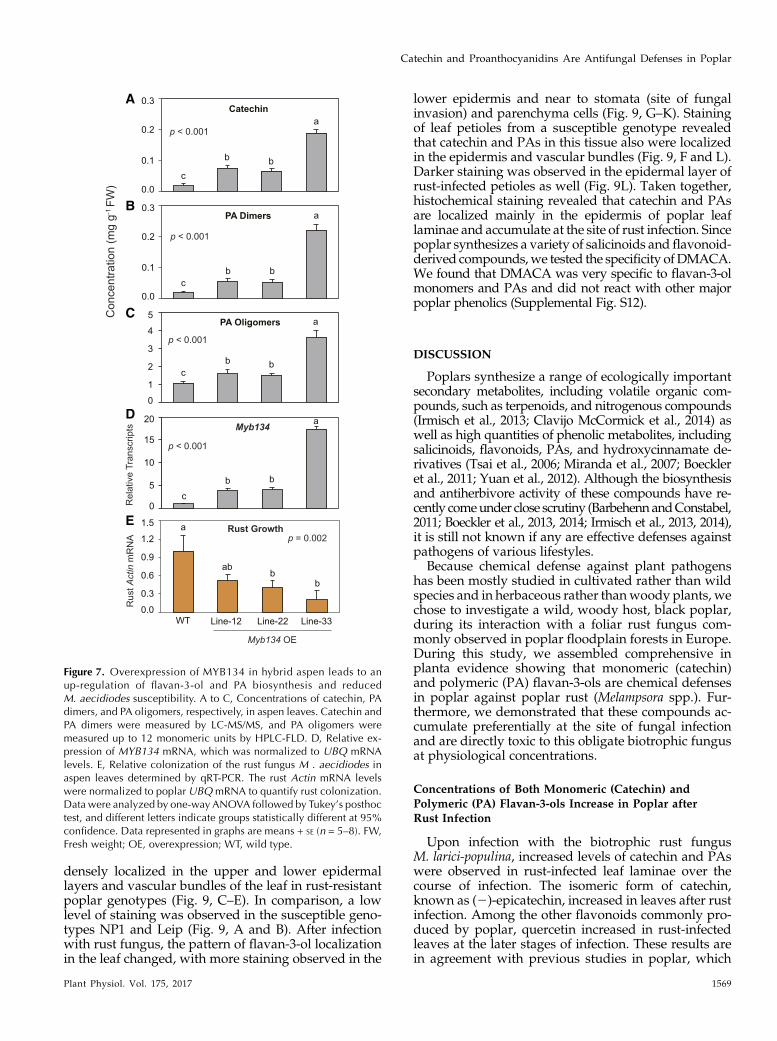
The poplar MYB134 gene encodes an R2R3 MYBtranscription factor and acts as a positive regulator ofPAs in poplar. MYB134 overexpression in transgenicPopulus spp. leads to a strong accumulation of PAs andcatechin, but anthocyanins and other flavonoids areminimally affected (Mellway et al., 2009). To directlytest the effects of this overaccumulation of flavan-3-olsand PAs onMelampsora spp. resistance, we propagated
Figure 5. Poplar genotypes moderately resistant to rust fungus contain constitutively higher amounts of catechin and PAs. Aand B, Catechin (flavan-3-ol monomer; A) and PA dimers (B) were measured by LC-MS/MS. C, Flavan-3-ol oligomers weremeasured up to 10 monomeric units by HPLC-FLD. D, Cell wall-bound PAs were measured from the residue remaining afterthe extraction of soluble catechin and PAs using the butanol-HCl method. The amounts of PA oligomers and cell wall-bound PAs are expressed as catechin and procyanidin-B1 equivalents, respectively. Data were analyzed by two-wayANOVA (factors were as follows: g = genotype, tr = treatment, and g 3 tr = interaction effect). Corresponding P values areindicated in the graphs. E, Fungal growth in different poplar genotypes 8 dpi. The growth of the fungus was determined byqRT-PCR. M. larici-populina actin gene expression was normalized to poplar UBQ gene expression to quantify the colo-nization of the fungus in poplar leaves. F, Biomass of poplar genotypes in one growing season under natural environmentalconditions. The shoot biomass was determined in autumn (November 2015), when all leaves had dropped at the end of thegrowing season. In the susceptible genotypes, defoliation was earlier due to severe rust infection. Data in E and F wereanalyzed by one-way ANOVA followed by Tukey’s posthoc test, with different letters indicating statistically significantdifferences at 95% confidence. Data represented in the graphs are means 6 SE (n = 5). DW, Dry weight.
Plant Physiol. Vol. 175, 2017 1567
Catechin and Proanthocyanidins Are Antifungal Defenses in Poplar

three previously characterized hybrid aspen (Populustremula 3 Populus alba) MYB134-overexpressing linesand inoculated these with Melampsora aecidiodes, aclosely related poplar rust that infects this hybrid. Asexpected, concentrations of catechin, PA dimers, andPA oligomers were enhanced and up to 3- to 8-foldhigher in the MYB134-overexpressing lines (ANOVA,P, 0.01; Fig. 7, A–C), andMYB134 expressionwas 4- to16-fold higher compared with the wild type (ANOVA,P, 0.01; Fig. 7D). To determine rust colonization in thetransgenic lines and controls, we quantifiedMelampsoraspp. growth using qRT-PCR. Growth of the rust fungusM. aecidiodes was reduced significantly in the MYB134-overexpressing lines in comparison with the wild type(ANOVA, P = 0.002; Fig. 7E) and roughly inverselyproportional to catechin and PA concentrations.
RNA Interference-Mediated Knockdown of MYB134Transcription Factor in Black Poplar Down-RegulatesCatechin and PA Biosynthesis and Leads to IncreasedRust Susceptibility
To complement these findings in black poplar, a na-tive host of the rust fungus M. larici-populina, we down-regulated the MYB134 gene in this species (genotypeNP1) by RNA interference (RNAi) to reduce the bio-synthesis of flavan-3-ols. We obtained two independenttransgenic lineswith lower flavan-3-olmonomer and PAlevels. Transcript abundances of MYB134 in the MYB-RNAi lines were significantly lower than in the controls(ANOVA, P = 0.006; Fig. 8D). The concentrations of
catechin, PA dimers, and oligomers were approxi-mately 40% to 50% lower in the RNAi lines in com-parison with the vector control and wild-type plants(ANOVA, P , 0.001; Fig. 8, A–C). The colonization ofinfected leaves by the rust fungus was up to 50%higher in the two PnMYB134-RNAi lines comparedwith the vector control andwild-type plants (ANOVA,P = 0.003; Fig. 8E). Epicatechin and naringenin concen-trations also were significantly lower in the PnMYB134-silenced lines (ANOVA,P, 0.001), but quercetin did notchange (P = 0.92). The concentrations of the salicinoidssuch as salicin (P , 0.001), salicortin (P = 0.02), andtremulacin (P = 0.015) increased slightly in the transgeniclines, but homaloside D (P = 0.38) did not change sig-nificantly (Supplemental Fig. S11).
Localization of Catechin and PAs at the Site of RustInfection in Poplar Leaves
During compatible interactions in poplar hosts, rustspores germinate within 6 to 12 h after inoculation andestablish an intercellular mycelial network within 2 to4 d without any visible symptoms (Hacquard et al.,2011). To better understand the role of catechin andPAs against rust infection, we studied their localiza-tion in poplar leaves. Tissue-specific localization ofPAs was shown in many plant species by stainingwith 4-dimethylaminocinnamaldehyde (DMACA),whichproduces a blue color (Kao et al., 2002; Abeynayakeet al., 2011; Jun et al., 2015). Histochemical stainingwith DMACA revealed that catechin and PAs were
Figure 6. Catechin and PAs show direct inhibitoryeffects on spore germination and hyphal growth ofthe biotrophic rust fungus in vitro. A and B, Germi-nationof rust urediniospores onglass slides at 9 hpostinoculation (hpi). A, Spore germination shown incontrol medium. B, Spore germination shown inmedium supplemented with catechin. C and D, Pat-terns of mycelial branching at 18 hpi in control me-dium (C) and medium supplemented with catechin(D). Bars in A to D = 20 mm. E, Urediniospore ger-mination percentage determined at 9 hpi. F, Hyphallengths of germinated urediniospores at 12 hpi. Datawere analyzed by one-way ANOVA followed byTukey’s posthoc test, and different letters indicatetreatment groups statistically different at 95% confi-dence.Data represented in the graphs aremeans6 SE
(n = 8), where each replicate is a mean of threetechnical replicates.
1568 Plant Physiol. Vol. 175, 2017
Ullah et al.
densely localized in the upper and lower epidermallayers and vascular bundles of the leaf in rust-resistantpoplar genotypes (Fig. 9, C–E). In comparison, a lowlevel of staining was observed in the susceptible geno-types NP1 and Leip (Fig. 9, A and B). After infectionwith rust fungus, the pattern of flavan-3-ol localizationin the leaf changed, with more staining observed in the
lower epidermis and near to stomata (site of fungalinvasion) and parenchyma cells (Fig. 9, G–K). Stainingof leaf petioles from a susceptible genotype revealedthat catechin and PAs in this tissue also were localizedin the epidermis and vascular bundles (Fig. 9, F and L).Darker staining was observed in the epidermal layer ofrust-infected petioles as well (Fig. 9L). Taken together,histochemical staining revealed that catechin and PAsare localized mainly in the epidermis of poplar leaflaminae and accumulate at the site of rust infection. Sincepoplar synthesizes a variety of salicinoids and flavonoid-derived compounds,we tested the specificity ofDMACA.We found that DMACA was very specific to flavan-3-olmonomers and PAs and did not react with other majorpoplar phenolics (Supplemental Fig. S12).
DISCUSSION
Poplars synthesize a range of ecologically importantsecondary metabolites, including volatile organic com-pounds, such as terpenoids, and nitrogenous compounds(Irmisch et al., 2013; Clavijo McCormick et al., 2014) aswell as high quantities of phenolic metabolites, includingsalicinoids, flavonoids, PAs, and hydroxycinnamate de-rivatives (Tsai et al., 2006; Miranda et al., 2007; Boeckleret al., 2011; Yuan et al., 2012). Although the biosynthesisand antiherbivore activity of these compounds have re-cently comeunder close scrutiny (BarbehennandConstabel,2011; Boeckler et al., 2013, 2014; Irmisch et al., 2013, 2014),it is still not known if any are effective defenses againstpathogens of various lifestyles.
Because chemical defense against plant pathogenshas been mostly studied in cultivated rather than wildspecies and in herbaceous rather thanwoody plants, wechose to investigate a wild, woody host, black poplar,during its interaction with a foliar rust fungus com-monly observed in poplar floodplain forests in Europe.During this study, we assembled comprehensive inplanta evidence showing that monomeric (catechin)and polymeric (PA) flavan-3-ols are chemical defensesin poplar against poplar rust (Melampsora spp.). Fur-thermore, we demonstrated that these compounds ac-cumulate preferentially at the site of fungal infectionand are directly toxic to this obligate biotrophic fungusat physiological concentrations.
Concentrations of Both Monomeric (Catechin) andPolymeric (PA) Flavan-3-ols Increase in Poplar afterRust Infection
Upon infection with the biotrophic rust fungusM. larici-populina, increased levels of catechin and PAswere observed in rust-infected leaf laminae over thecourse of infection. The isomeric form of catechin,known as (2)-epicatechin, increased in leaves after rustinfection. Among the other flavonoids commonly pro-duced by poplar, quercetin increased in rust-infectedleaves at the later stages of infection. These results arein agreement with previous studies in poplar, which
Figure 7. Overexpression of MYB134 in hybrid aspen leads to anup-regulation of flavan-3-ol and PA biosynthesis and reducedM. aecidiodes susceptibility. A to C, Concentrations of catechin, PAdimers, and PA oligomers, respectively, in aspen leaves. Catechin andPA dimers were measured by LC-MS/MS, and PA oligomers weremeasured up to 12 monomeric units by HPLC-FLD. D, Relative ex-pression of MYB134 mRNA, which was normalized to UBQ mRNAlevels. E, Relative colonization of the rust fungus M . aecidiodes inaspen leaves determined by qRT-PCR. The rust Actin mRNA levelswere normalized to poplar UBQmRNA to quantify rust colonization.Data were analyzed by one-way ANOVA followed by Tukey’s posthoctest, and different letters indicate groups statistically different at 95%confidence. Data represented in graphs are means + SE (n = 5–8). FW,Fresh weight; OE, overexpression; WT, wild type.
Plant Physiol. Vol. 175, 2017 1569
Catechin and Proanthocyanidins Are Antifungal Defenses in Poplar

showed that several flavonoid biosynthetic genes aretranscriptionally activated in poplar after rust colo-nization, especially during the sporulation phase(Miranda et al., 2007; Azaiez et al., 2009). Studies alsodemonstrated that the biosynthesis of flavan-3-olsand PAs increased after infection by fungal endo-phytes in poplar (Pfabel et al., 2012) as well as afterinfection by pathogenic fungi in other plant species,such as bilberry (Vaccinium myrtillus; Koskimäkiet al., 2009) and Fagus crenata (Yamaji and Ichihara,2012). Increased accumulation of flavan-3-ols also hasbeen recorded in Norway spruce (Picea abies) duringinfection by necrotrophic fungi (Danielsson et al.,2011; Hammerbacher et al., 2014). Therefore, a rangeof plants, including poplar, respond to pathogen at-tack by accumulating both monomeric flavan-3-olsand PAs.
However, not all phenolics increase after pathogeninfection. Salicinoids, an abundant class of phenolics inpoplar leaves that are known to defend against herbi-vores (Boeckler et al., 2011), decreased after rust infec-tion, except for salicin, which was induced slightly atthe later stages of infection. Thus, salicinoids are notlikely to be deployed by poplar for pathogen defense.They might decline because of their metabolism by thefungus as a potential food source; the sugar moiety, inparticular, could be cleaved and assimilated by thepathogen (Hammerbacher et al., 2013). A more likelyexplanation, however, is that lower levels of salicinoidsin rust-infected leaves result from elevated flavan-3-olbiosynthesis, which was shown previously to reduce sal-icinoid biosynthesis. Up-regulation of PA biosynthesis byoverexpressing the transcription factor MYB134 led tolower salicinoid content in hybrid poplar (Mellway et al.,2009; Kosonen et al., 2012; Boeckler et al., 2014). In theabsence of fungal infection or other biotic stresses, thereare typically no dramatic differences in flavan-3-ol orsalicinoid concentrations between young expanding andfully expanded mature leaves (Supplemental Figs. S2 andS4; Massad et al., 2014), but this generalization does notapply after herbivore or pathogen attack. In line withprevious studies, our phenolicmeasurements suggest thatthere is a tradeoff between flavan-3-ol (catechin and PAs)versus salicinoid biosynthesis in poplar leaves (Boeckleret al., 2014), implying a tradeoff between antipathogenand antiherbivore defense.
Transcripts of Flavan-3-ol Biosynthetic Genes Increase inResponse to Fungal Infection
The biosynthesis of flavan-3-ols has been well char-acterized in many plant species, both genetically andbiochemically. Two distinct enzymes, LAR and ANR,are involved in catalyzing the last steps of the pathwayto flavan-3-ol monomers in PA-producing plants (Bogset al., 2005; Pang et al., 2013; Liao et al., 2015). Genesencoding LAR and ANR can occur as single genes, forexample in Arabidopsis (Xie et al., 2004), or as multi-gene families, for example in grapevine (Vitis vinifera;
Figure 8. Down-regulation of flavan-3-ol and PA biosynthesis in blackpoplar (NP1) by silencing the MYB134 transcription factor results inan increased susceptibility to rust infection (M. larici-populina). A andB, Concentrations of catechin and PA dimers, respectively, in poplarleaves measured by LC-MS/MS. C, PA oligomers were measured up to8 monomeric units by HPLC-FLD. D, Relative expression of MYB134mRNA, which was normalized to UBQ mRNA levels. E, Relativecolonization of rust fungus in poplar leaves determined by qRT-PCR.The rust ActinmRNA levels were normalized to poplarUBQmRNA toquantify relative rust colonization. Data in A to D were analyzed bytwo-way ANOVA (factors were as follows: L = poplar lines, tr =treatment [control and rust], and L 3 tr = interaction effect) followedby Tukey’s posthoc test, and different letters indicate groups statisti-cally different at 95% confidence. Data in E were analyzed by one-way ANOVA followed by Tukey’s posthoc test, and different lettersindicate lines statistically different at 95% confidence. Data repre-sented in graphs are means + SE (n = 4–5). DW, Dry weight; pCambia,vector control; WT, wild type.
1570 Plant Physiol. Vol. 175, 2017
Ullah et al.

Bogs et al., 2005) and tea (Camellia sinensis; Pang et al.,2013). Analysis of the P. trichocarpa genome revealedthree loci encoding LAR proteins and two loci encodingANRproteins. This ismore than the two PtLAR and onePtANR (Yuan et al., 2012; Wang et al., 2013) that werereported previously and genetically characterized inP. trichocarpa. We confirmed the enzymatic activity ofthe proteins encoded by all loci by heterologous ex-pression and in vitro enzyme assays and showed thatthey are likely involved in the catalysis of the last stepsof flavan-3-ol biosynthesis in native black poplar. Our
phylogenetic analysis shows that ANRs and LARs aretwo distinct classes of enzymes and that DFR is morerelated to ANRs than LARs. Similar evolutionary rela-tionships for ANR and LAR proteins were shown byother authors (Pang et al., 2013; Wang et al., 2013).
Transcript levels of all three PnLAR and two PnANRgenes increased in rust-infected black poplar leavesover the course of infection. Previous microarray dataalso demonstrated that some of the genes of this path-way are transcriptionally induced in hybrid poplar af-ter infection with M. medusae (Miranda et al., 2007). As
Figure 9. Localization of flavan-3-ols and PAs in poplar leaves with or without rust infection. Sections (20 mm thickness) weremade from the first fully expanded mature leaf (leaf 5) and were stained with DMACA. A to E, Cross sections (leaf lamina) of thegenotypes NP1, Leip, Dorn, Kew, and Bla, respectively. F, Cross section of an NP1 petiole. G to K, Cross sections (leaf lamina) ofNP1, Leip, Dorn, Kew, and Bla genotypes, respectively, after rust infection. L, Petiole cross section of NP1 infected with rustfungus 5 dpi. The genotypes NP1 and Leip were found to be very susceptible to the rust fungus, while the genotypes Dorn, Kew,and Bla were found to be moderately resistant. Triangles indicate fungal penetration and colonization sites. c, Cortex; e, epi-dermis; le, lower epidermis; pa, palisade parenchyma; s, stomata; sp, spongy parenchyma; ue, upper epidermis; vb, vascularbundles. Bars = 100 mm (K, for all leaf laminae) and 200 mm (F and L).
Plant Physiol. Vol. 175, 2017 1571
Catechin and Proanthocyanidins Are Antifungal Defenses in Poplar

reported previously for hybrid poplar (Mellway et al.,2009), the transcription factor PnMYB134, a positiveregulator of PA biosynthesis, also was transcriptionallyinduced and responded quickly after rust inoculationin our study. Our transcript and metabolite analysessuggest that both LAR andANRbranches of flavan-3-olbiosynthesis are transcriptionally activated upon rustinfection. Monomeric catechin synthesized from theLAR branch is freely available and accumulated inblack poplar, while free ANR-dependent epicatechinwas observed only at very low concentrations. The re-covery of epicatechin after hydrolysis of PAs indicatesthat epicatechin might contribute to the extension of PAchains. Similar mechanisms also were observed in grapeand Norway spruce (Bogs et al., 2005; Hammerbacheret al., 2014).
High Levels of Catechin and PAs Are Associated withResistance against Rust Fungus Infection
Various constitutive and induced plant phenoliccompounds are thought to contribute to defense againstmicrobial pathogens (Osbourn, 1996; Lattanzio et al.,2006), but not all phenolics have this effect (Henriquezet al., 2012; Zhang et al., 2015). In order to determine ifflavan-3-ols have this function in planta, we screenedfive poplar genotypes for resistance against rust infec-tion and quantified their phenolic contents. Interest-ingly, genotypes moderately resistant to rust infectionhad constitutively higher amounts of catechin and PAs intheir leaves than susceptible genotypes, and substantiallyhigher induced levels of these flavan-3-ols were foundafter artificial inoculation with rust. Similar results havebeen shown in other woody plant species. For example,crude extract from coffee (Coffea arabica) cultivars resistantto coffee rust (Hemileia vastatrix), which contained higheramounts of PAs in comparison with the extracts of sus-ceptible cultivars, was found more effective in inhibitingH. vastatrixuredospore germination (deColmenares et al.,1998). In addition, higher levels of constitutive and in-duced (+)-catechin were found in a rust-resistant willow(Salix myrsinifolia) clone compared with the levels in sus-ceptible clones (Hakulinen et al., 1999). Recently, Wanget al. (2017) showed that P. tomentosa increased its PAlevels under elevated temperature as well as after infec-tion by the necrotrophic fungus Dothiorella gregaria.
To further explore the roles of catechin and PAs asantifungal defenses in poplar, we conducted an infec-tion experiment using M. aecidiodes in hybrid aspenoverexpressing the MYB134 transcription factor. Pre-viously, this gene was characterized and shown to be apositive regulator of PA biosynthesis in hybrid poplarand shown to be inducible by biotic and abiotic stresses(Mellway et al., 2009). Rust susceptibility was reducedsignificantly in MYB134-overexpressing lines accumu-lating higher levels of flavan-3-ols than the wild-typeplants. To investigate the role of catechin and PAs inEuropean black poplar and the rust system, we silencedthe PnMYB134 transcription factor by RNAi in P. nigra
NP1.Weobserved a 40% to 60% reduction ofmonomericflavan-3-ols and PAs in black poplar after silencingPnMYB134. Silenced lines were more susceptible to therust fungus M. larici-populina in whole-plant infectiontrials (Fig. 8E), confirming the antifungal activity ofthese compounds in planta. Overexpression of MYB134in a hybrid poplar (P. tremula 3 Populus tremuloides)caused a significant reduction in salicinoid concen-tration (Mellway et al., 2009; Boeckler et al., 2014), butsuch a tradeoff was not observed in the MYB134-silenced P. nigra lines accumulating reduced levels offlavan-3-ol and PAs. Silencing offlavan-3-ol biosynthesisis metabolically less costly for poplar than constitutiveoverexpression or accumulation of flavan-3-ols underpathogen attack. In agreement with our results, over-expression of a PA biosynthetic gene in P. tomentosaresulted in increased resistance against necrotrophicfungi (Yuan et al., 2012; Wang et al., 2017). A negativeassociation between fungal endophyte communities andthe levels of condensed tannin also was shown in NorthAmerican poplar species (Whitham et al., 2006). How-ever, infection by necrotrophic fungi was higher inPopulus angustifolia (Busby et al., 2013), which isknown to accumulate high amounts of condensedtannins (Whitham et al., 2006). These conflicting re-sults suggest that poplar-rust interactions are verycomplex and that other factors, such as pathogen vir-ulence and nonphenolic defenses, including surfaceimmunity and effector-triggered immunity, mightcontribute to different outcomes of the infection pro-cess. Further investigation is necessary using geno-types containing high PAs under natural conditionsand also different rust fungus strains.
The Site and Magnitude of Flavan-3-ol AccumulationAre Consistent with a Defensive Role againstFungal Pathogens
The accumulation of flavan-3-ol monomers and PAsoften is limited to specific tissue types and develop-mental stages of plant organs. For example, in whiteclover (Trifolium repens), flavan-3-olmonomers and PAsare localized in the epidermal layers of floral organs(Abeynayake et al., 2011), while in Arabidopsis, PAsaccumulate mainly in the seed coat, especially in theendothelial cells (Debeaujon et al., 2003). The biosyn-thesis and spatial distribution of these compounds inspecific tissues or organs might have ecological signif-icance. Our histochemical staining with DMACArevealed that flavan-3-ols are localized mainly in theleaf epidermis and vascular tissues (Fig. 9). Moderatelyresistant genotypes that contained higher levels of cat-echin and PAs had a more restricted localization ofthese compounds in the epidermal layers comparedwith the susceptible genotypes. After rust infection,high amounts of flavan-3-ols also were observed inthe parenchyma cells. The epidermal localization offlavan-3-ols could provide a defensive barrier to earlyfungal colonization of the leaf. Dark PA staining also
1572 Plant Physiol. Vol. 175, 2017
Ullah et al.

was observed in the lower surface of the aspen leaves(Kao et al., 2002) and in hybrid poplar stems infestedby the galling aphid Phloeomyzus passerinii (Dardeauet al., 2014).The effectiveness of an epidermal flavan-3-ol barrier
depends on the inhibitory effect of these compoundson rust development. Using a novel in vitro bioassaytechnique, we showed that physiologically relevantconcentrations of both catechin and PAs stronglyinhibited rust spore germination and reduced hyphalgrowth. However, epicatechin did not show antifun-gal activity even at a 10 times higher concentrationthan found in poplar leaves (Supplemental Fig. S9),although this compound is an extender unit in PAchains (Fig. 1F). Therefore, our data clearly show thatcatechin and PAs are active antifungal metabolites inpoplar and might serve as an effective chemical de-fense at the surface or in other tissues of the plant.In conclusion, black poplar, a perennial woody spe-
cies of Europe, Asia, and northwestern Africa, wasshown to synthesize monomeric and polymeric flavan-3-ols as a phenolic defense against the rust fungusM. larici-populina in concentrations demonstrated tohave antifungal activity in vitro at sites on the epider-mis and in vascular tissue, where they form a barrier tofungal invasion. The rust-resistant poplar genotypesused in this study constitutively accumulate moreflavan-3-ols than susceptible genotypes. Transgenicblack poplar trees with reduced levels of catechin andPAs were more susceptible. Future work is needed toinvestigate such topics as the mode of action of flavan-3-ols on fungi, how infection triggers flavan-3-ol ac-cumulation, and if other poplar metabolites act indefense against fungal infection.
MATERIALS AND METHODS
Plant Materials
Black poplar (Populus nigra, genotype NP1) was propagated from stemcuttings and grown in the greenhouse (22°C day temperature and 19°C nighttemperature, 60% relative humidity, 16-h/8-h light/dark cycle) in 2-L potshaving a 1:1 mixture of sand and soil (Klasmann potting substrate; Klasmann-Deilmann). Other poplar genotypes were supplied by the Northwest GermanForest Research Station in Hannoversch Münden in 2014 as stem cuttings.Plants were regenerated under greenhouse conditions and subsequently mul-tiplied in large quantities. The transgenic black poplar plants used in this studywere amplified by micropropagation as described by Irmisch et al. (2013) andthen multiplied by stem cuttings. Transgenic hybrid aspen (Populus tremula 3Populus alba INRA 717-1-B4) was grown and maintained as described byMellway et al. (2009). Plants with a height of approximately 80 to 100 cm wereused for inoculation with fungi. Some plants from each genotype were grownoutside the greenhouse to allow natural infection byMelampsora larici-populina.The disease resistance and susceptibility levels were scored based on thenumber of uredinia on the abaxial leaf surface as well as by qRT-PCR (a list ofgenotypes with resistance levels is given in Supplemental Table S1).
Fungal Pathogens and Culture Maintenance
Virulent M. larici-populina was collected from a natural population of blackpoplar located in the floodplain forest of an island in the Oder River nearKüstrin-Kietz, Germany. The funguswasmultiplied from a single uredium on asusceptible poplar genotype (NP1). The infected plants were covered withpolyethylene bags in order to collect spores without allowing any condensation
of water inside the bags, which might lead to spore germination. The ure-diniospores were collected from infected poplar leaves using fine brushes andplaced in 2-mL microcentrifuge tubes. To dry spores, the tube was inserted in aclosed beaker containing dry silica gel for 2 to 3 d with the cap open. The sporeswere then stored at 220°C until further use. This spore preservation techniqueavoided continuous in planta culturing of this obligate biotrophic fungus.Uredospores of Melampsora aecidiodes were collected from a local P. alba tree.
Inoculation of Poplars with M. larici-populina orM. aecidiodes
Freshly harvested or frozenurediniospores ofM. larici-populinawere used forinoculation experiments. Young black poplar trees (approximately 80 cm heightand 15–20 leaves) grown in the greenhouse were transferred to a climatechamber (22°C day temperature and 19°C night temperature, 70% relativehumidity, 16-h/8-h light/dark cycle) 7 d before inoculation. For the kineticinfection experiment, 50 individual black poplar trees of approximately equalsize were chosen. Each young potted tree was placed in a separate receptacle(18 cm diameter) for watering independently. Half of the plants (n = 25) wereinoculated by thoroughly spraying M. larici-populina spore suspension (6105
spores mL21) onto the abaxial leaf surfaces. Control plants were sprayed withwater (n = 25). Immediately after spraying, each plant was covered with apolyethylene terephthalate bag (Bratschlauch) to maintain high humidity andkept in the dark to facilitate spore germination. After 18 h, the bagswere openedfrom the top to ensure proper aeration. Five time points were chosen for sam-pling based on the lifestyle of the fungus (Hacquard et al., 2011). Samples weretaken at 6 hpi and 3, 7, 14, and 21 dpi. At each time point, five individual plantswere sampled from rust-infected and water-sprayed treatments. Six leaves atthe same position (leaf 5-10) on each plant were harvested, and midribs wereremoved and pooled together to obtain one biological sample and immediatelyflash frozen under liquid N2. Unless stated otherwise, similar inoculation andsampling techniques were followed for the other rust infection experiments, butonly one time point (8 dpi) was used for harvesting leaves. Inoculation of hybridaspen with M. aecidiodes was carried out in the Glover Greenhouse at theUniversity of Victoria using a similar setup.
In Vitro Bioassays with M. larici-populina on Glass Slides
Antifungal activities of catechin and PAs against the biotrophic rust funguswere evaluated onglass slides. The germinationmedium consisted of 1.1%plantagar (Duchefa Biochemie) and 10 mM KCl in water. The medium was sterilizedby autoclaving before adding catechin or PAs (1.5 mg mL21). The media wereincubated in a water bath at 65°C for 1 h to ensure that the phenolic compoundswere dissolved completely. Autoclavedmediumwithout compounds was usedas a control. Approximately 300 mL of liquid medium was pipetted carefullyonto a clean glass slide and allowed to solidify. After 5 to 10 min, 20 mL offreshly prepared spore suspension (6104mL21) in 10mMKClwas pipetted ontothe glass slides and spread carefully onto the solidified medium with a plasticinoculation loop. Ten glass slides were prepared for each treatment, and each ofthemwas kept in a sterile petri dish (9 cmdiameter) withmoist blotting paper tomaintain the high humidity required for spore germination. The petri disheswere incubated in a dark cabinet, and the spore germination was monitoredevery 1 h using an inverted light microscope (Axiovert 200; Carl Zeiss Mi-croscopy) coupled with a camera (AxioVision). The urediniospore germinationrate was determined at 9 hpi, and hyphal length was measured at 12 hpi. Threemicroscopic fields were photographed randomly and considered as technicalreplicates. The percentage of germinated spores was calculated based on thenumber of spores germinated divided by the total number of spores observedper microscopic field. Hyphal lengths of the germinated spores in each micro-scopic field were measured by ImageJ software (https://imagej.nih.gov/ij/index.html). For an illustration of the technique, see Supplemental Figure S8.
Extraction of Phenolic Compounds from Poplar
For the extraction of phenolic compounds, poplar tissues (leaf laminae,petiole, stem, and root) were ground to fine powder under liquid N2. The stemsamples, which contained both bark and wood, were ground with the help of avibrating mill (Pulverisette 0; Fritsch), while the leaf materials were groundmanually using mortar and pestle. The ground samples were lyophilized usingan Alpha 1-4 LD Plus freeze dryer (Martin Christ) at 0.001 mbar pressure and276°C temperature for 2 d. Approximately 10 mg of freeze-dried tissue was
Plant Physiol. Vol. 175, 2017 1573
Catechin and Proanthocyanidins Are Antifungal Defenses in Poplar

weighed using an XP26 Microbalance (Mettler-Toledo) into 2-mL micro-centrifuge tubes. Phenolics were extracted with 1 mL of extraction buffer,which contained 10 mg of apigenin-7-glucoside (Carl Roth) and 0.4 mgphenyl b-D-glucopyranoside (Sigma-Aldrich) as internal standards per1 mL of methanol (analytical grade). One milliliter of extraction buffer wasadded to each microcentrifuge tube, vortexed vigorously, and incubatedfor 30 min at 20°C with shaking at 2,000 rpm. The extracts were centrifugedat 13,000 rpm at 4°C for 5 min, and approximately 900 mL of supernatantwas transferred to a new microcentrifuge tube. Salicinoids were analyzeddirectly with HPLC-DAD. Samples were diluted 20-fold before analyzingflavan-3-ols by LC-MS/MS.
For the extraction of oligomeric or polymeric PAs, approximately 50 mg offreeze-dried plant tissue was extracted with analytical grade methanol fol-lowing the above extraction protocol, but additionally, the insoluble materialwas reextracted with 1 mL of 70% acetone. Both supernatants were combinedand dried under a stream of nitrogen. The dried samples were redissolved in1mL of solvent (1:1, methanol:acetonitrile) andwere analyzed byHPLC-FLD.Cell wall-bound PAs were measured from the residue remaining after theextraction of soluble catechin and PAs using the acid butanol method (Porteret al., 1985), which was modified by Boeckler et al. (2013). Amounts of cellwall-bound PAs were determined using a procyanidin-B1 calibration curve(Extrasynthese).
Identification of Phenolics by Liquid Chromatography-MassSpectrometry with Electrospray Ionization
The phenolic compounds identified from poplar leaf extracts as well as fromLAR and ANR enzyme assays were separated on a reverse-phase NucleodurSphinx RP18ec column with dimensions of 2503 4.6 mm and a particle size of5 mm (Macherey-Nagel) using an Agilent 1100 series HPLC device (AgilentTechnologies) with a solvent system of 0.2% aqueous formic acid (A) and ac-etonitrile (B) at a flow rate of 1 mL min21. The column temperature wasmaintained at 25°C. The proportion of B was increased from 14% to 58% in alinear gradient of 22 min. After the column was washed for 3 min with 100%B, it was reequilibrated to the initial eluent composition for 5 min prior to thenext analysis. Flow coming from the column was diverted in a ratio of 4:1before entering the mass spectrometer electrospray chamber. Compounddetection and quantification were accomplished with an Esquire 6000 elec-trospray ionization (ESI) ion-trap mass spectrometer (Bruker Daltronics).Phenolic compounds were analyzed in negative mode scanning a mass-to-charge ratio (m/z) between 100 and 1,600 with a skimmer voltage of 60 V, acapillary exit voltage of 2121 V, and a capillary voltage of 4,000 V. Nitrogenwas used as a drying gas (11 mL L21, 330°C), and the nebulizer gas pressurewas 35 p.s.i. Compounds were identified by mass spectra and by directcomparison with commercial standards as described previously (Boeckleret al., 2013; Hammerbacher et al., 2014). Bruker Daltronics Quant Analysissoftware version 3.4 was used for data processing and compound quantifi-cation using a standard smoothing width of 3 and Peak Detection Algorithmversion 2.
Quantification of Flavan-3-ol Monomers and Dimersby LC-ESI-MS/MS
Chromatography was performed on an Agilent 1200 HPLC system. An API3200 tandem mass spectrometer (Applied Biosystems) equipped with a tur-bospray ion source was operated in negative ionization mode. Separation wasachieved on a Zorbax Eclipse XDB-C18 column (503 4.6 mm, 1.8 mm; Agilent).Formic acid (0.05%) in water and acetonitrile were employed as mobile phasesA and B, respectively. The elution profile was as follows: 0 to 1min, 100%A; 1 to7 min, 0% to 65% B; 7 to 8 min, 65% to 100% B; 8 to 9 min, 100% B; and 9 to10min, 100%A. The total mobile phase flow ratewas 1.1mLmin21. The columntemperature was maintained at 25°C. The instrument parameters wereoptimized by infusion experiments with pure standards as described byHammerbacher et al. (2014). The ion spray voltage was maintained at24,500 V. The turbo gas temperature was set at 700°C. Nebulizing gas wasset at 70 p.s.i., curtain gas at 25 p.s.i., heating gas at 60 p.s.i., and collisiongas at 10 p.s.i. Multiple reaction monitoring was used to monitor analyteparent ion→product ion as follows: m/z 288.9→109.1 (collision energy [CE],234 V; declustering potential [DP], 230 V) for catechin; m/z 289→109 (CE,234 V; DP, 230 V) for epicatechin; m/z 304.8→125 (CE, 228 V; DP, 230 V)for gallocatechin; m/z 576.9→289.1 (CE, 230 V; DP, 250 V) for PA B1; and
m/z 430.8→ 268 (CE, 246 V; DP, 280 V) for apigenin 7-glucoside. Both Q1and Q3 quadrupoles were maintained at unit resolution. Data acquisitionand processing were performed using Analyst 1.5 software (Applied Bio-systems). Linearity in ionization efficiencies was verified by analyzing di-lution series of a standard. Flavan-3-ol concentrations were determinedrelative to the calibration curve for apigenin 7-glucoside as an internalstandard and multiplied by the corresponding response factor.
Quantification of PAs by HPLC-FLD
PAswere separatedonaLiChrospherediol columnwithdimensions of 25034mm and a particle size of 5mm (Merck Chemicals) using anAgilent 1100 seriesHPLC device employing a modified method described previously by Kelmet al. (2006) andHammerbacher et al. (2014). Briefly, the total mobile phase flowrate for chromatographic separation was 1.2 mL min21. The column tempera-ture was maintained at 30°C. Compounds were separated using acetonitrile:acetic acid (98:2) andmethanol:water:acetic acid (95:3:2) asmobile phases A andB, respectively, with the following elution profile: 0 to 35min, 0% to 40% B in A;35 to 40 min, 40% B; 40 to 45 min, 40% to 0% B; and 45.1 to 50 min, 0% B. Eluentwas monitored by FLD with excitation at 276 nm and emission at 316 nm. PAoligomer and polymer concentrations were determined relative to the calibra-tion curve for catechin.
Quantification of Other Flavonoids and Salicinoidsby HPLC-DAD
To quantify flavonoids other than flavan-3-ols and salicinoids, an Agilent1100 Series HPLC System with diode array detector (Agilent Technologies)was used. The compoundswere separated by aNucleodur Sphinx RP columnwith dimensions of 2503 4.6 mm and a particle size 5 mm (Macherey-Nagel).The solvent system for the mobile phase was Milli-Q water (Millipore) andacetonitrile, but otherwise, the chromatographic conditions were the same asdescribed above for LC-ESI-MS of poplar phenolics. Data were exported bythe software Data Trans at different wavelengths for the various phenolics asdescribed previously by Boeckler et al. (2013). For absolute quantification,analyte peak areas were divided by the peak area of the internal standardphenyl b-D-glucopyranoside and multiplied by the corresponding responsefactors.
Reductive Cleavage of PAs
Reductive cleavage of PAs was carried out as described previously byHammerbacher et al. (2014). Briefly, the same samples that were used for PAanalysis were diluted 50-fold with methanol. The reaction was performed inHPLC glass vials containing 780 mL of diluted extract, 20 mL of trifluoroaceticacid, and 100 mL of sodium cyanoborohydrate (0.5 g mL21 methanol). Reactionmixtures were heated to 65°C for 15 min before adding an additional 20 mL oftrifluoroacetic acid. Vials were sealed tightly and incubated overnight at65°C. The next morning, the reaction was dried completely under a stream ofnitrogen, resuspended in 800 mL of methanol, and centrifuged for 5 min at11,000 rpm at 4°C, and 780 mL of supernatant was transferred to a new vial.The samples were analyzed by LC-ESI-MS/MS as described for flavan-3-olmonomers and dimers.
RNA Isolation and cDNA Synthesis
Total RNA from leaf and stem tissue was extracted using the Invitrap SpinPlant RNA Mini Kit (Stratec Biomedical) following the protocols of the manu-facturer, except that an additional DNase treatment was included (RNase-FreeDNase Set; Qiagen). The first washing step was conducted with 300 mL of washbuffer R1. After that, DNase (30 Kunitz units in 80 mL volume; 10 mL of RNase-free water and 70 mL of buffer RDD) was added onto the column and incubatedat room temperature for 15 min. The column was washed with an additional300 mL of wash buffer R1 before continuing with the manufacturer’s protocol.The quantity and quality of the RNA were checked by spectrophotometry(Thermo Scientific NanoDrop 2000). Reverse transcription of 1 mg of RNA intocDNA was achieved by using SuperScript II reverse transcriptase (Invitrogen)and 50 pmol of oligo(dT)12-18 primer (Invitrogen) in a reaction volume of 20 mL.The cDNA was diluted 5-fold with sterile water, and quality was checked bysemiquantitative reverse transcription-PCR using PtUBQ primer pairs (primersequences are given in Supplemental Table S2).
1574 Plant Physiol. Vol. 175, 2017
Ullah et al.

Identification, Cloning, and Sequencing of PnLAR andPnANR Genes from Black Poplar
The genome of Populus trichocarpa (Tuskan et al., 2006) was utilized to findcandidate LAR and ANR genes for P. nigra, as these two species are closelyrelated. The LAR and ANR protein sequences from apple (Malus domestica),grape (Vitis vinifera), and tea (Camellia sinensis) were used to identify LAR andANR candidates using BLASTp searches in the National Center for Bio-technology Information and Phytozome version 11 databases. The codingsequences were obtained from Phytozome version 11, and complete openreading frames were identified using the SeqBuilder software of theDNASTAR Lasergene 12 package (DNASTAR). Consistent with Tsai et al.(2006), three PtLAR and two PtANR genes were identified in the P. trichocarpagenome. Gateway (Invitrogen)-compatible primers were designed for can-didate sequences by using the 59 and 39 ends of putative LAR and ANR genesfrom P. trichocarpa (primer sequences are provided in Supplemental TableS2). Genes encoding LAR and ANR were PCR amplified with Gateway-compatible primers from the cDNA of black poplar using Phusion High-Fidelity DNA Polymerase (New England Biolabs). The PCR products werepurified with the QIAquick PCR purification kit (Qiagen). Gateway entryclones were made by using BP Clonase II and pDONR207 (Invitrogen) fol-lowing the manufacturer’s protocol. The pDONR207 constructs harboringPnLAR and PnANR genes were sequenced using 10 pmol of pDON primers(primer sequences are provided in Supplemental Table S2) and the BigDyeTerminator version 3.1 Cycle Sequencing Kit (Thermo Fisher Scientific) on anABI Prism R 3100 sequencing system (Applied Biosystems). Sequences fromeach construct were assembled and translated into protein sequences usingDNASTAR Lasergene 12 software.
Heterologous Expression of PnLAR and PnANR Genes inEscherichia coli
Three putativePnLAR and two PnANR entry cloneswere subclonedwith LRClonase II (Invitrogen) according to the manufacturer’s instructions into theGateway-compatible expression vector pDEST15 (Invitrogen), which contains aGST tag on theN terminus of the expressed protein. All constructs were verifiedby sequencing with gene-specific primers. A dihydroflavonol reductase gene(MdDFR) from apple, which transforms dihydroflavonols to leucoanthocya-nidins (Fischer et al., 2003), also was cloned using the above protocols into theGateway-compatible expression vector pH9GW, a modified pET28a(+) vector(Novagen), which carries a sequence encoding a 9-His tag at the N terminus ofthe expressed protein (O’Maille et al., 2004). For PnLAR expression, chemicallycompetent E. coli BL21 [DE3] (Invitrogen) was cotransformed with the PnLARandMdDFR expression constructs. For protein expression, single colonies wereinoculated into 5 mL of Luria-Bertani broth with 100 mg mL–1 ampicillin and50 mg mL–1 kanamycin for positive selection and grown overnight at 37°C. ForPnANR expression, BL21 cells were transformed with PnANR expressionclones with 100 mg mL–1 ampicillin for positive selection and grown overnightat 37°C. The 5-mL starter cultures were used to inoculate 50 mL of overnightexpression medium supplemented with their respective antibiotics and grownat 18°C with continuous shaking (220 rpm) for 2 d. The bacterial cells wereharvested by centrifugation. The crude proteins were extracted according toHammerbacher et al. (2014) and used for enzyme assays or stored at 220°C.
In Vitro Enzyme Assays for Functional Characterization ofLARs and ANRs
Since the substrates leucocyanidin and cyanidin were not available for LARand ANR assays, respectively, enzyme assays were conducted using two-stepreactions. For LAR characterization, taxifolin was used as a substrate with thecoexpressed (PnLAR + MdDFR) crude protein extract. The reactions wereperformed in a 100-mL reaction volume containing 70 mL of crude protein ex-tract, 10 mL of NADPH (20 mM), and 20 mL of taxifolin (50 mM) for 40 min atroom temperature. The reactions were stopped by adding 100 mL of methanol.The mixtures were centrifuged at 11,000 rpm for 4 min, and supernatant wasanalyzed by an LC-ESI-ion-trap mass spectrometer. For ANR characterization,anANS gene from Petunia hybridawas cloned into pDEST15 and heterologouslyexpressed in E. coli, and crude protein was extracted using the protocols de-scribed above. The enzyme assay was performed with 200 mL of crude proteinextracts (100 mL each of the ANR and ANS) and 10 mL of catechin (10 mM) assubstrate. The reaction mixture also contained 20 mL of NADPH (20 mM), 10 mL
of oxoglutarate (10 mM), 1 mL of enzyme bovine catalase (40 units mL21; Sigma-Aldrich), 40 mL of potassium phosphate buffer (0.2 M; pH 6), 10 mL of ascorbate(50 mM), and 1 mL of FeSO4 (1 mM). The reaction mixture was incubated at 28°Covernight. The reaction was stopped by adding another 100mL ofmethanol andcentrifuged for 4 min at 11,000 rpm, and the supernatant was analyzed by theLC-ESI-ion-trap-MS.
qRT-PCR
Toquantify the expressionof target genes, a segmentof approximately 150bpwas amplified using gene-specific primers. All primers were designed usingPrimer3 Web version 4.0.0 (http://bioinfo.ut.ee/primer3/). The efficiency ofeach primer pair was tested before qPCR. Primers with efficiencies below 95%were discarded. The reactions were performed in a 20-mL volume containing10 mL of Brilliant III Ultra-Fast SYBR Green QPCR Master Mix (Agilent Tech-nologies), 10 pmol of forward and 10 pmol of reverse primer, and 2 mL of di-luted cDNA (approximately 100 ng). The qRT-PCR was performed using theCFX Connect Real-Time PCR Detection System (Bio-Rad) using a two-stepamplification protocol (cycling parameters: 3 min at 95°C followed by 40 cy-cles of 10 s at 95°C and 30 s at 55°C). A nontemplate water sample was used as areaction control. Transcript abundance was normalized to the abundance of thePtUBQ (Irmisch et al., 2013) and was calculated from five biological replicates,with each biological sample being analyzed from three technical replicates.Primer sequences for all genes used in this study are given in SupplementalTable S2.
Histochemical Staining and Microscopy
Fresh plant specimens were embedded into Tissue Freezing Medium (Jung,Leica Biosystems) and left for 1 h at 220°C. Sections (10–20 mm) were madeusing a CM1850 cryotome (Leica Biosystems). Then, two to three sections weretransferred to a clean glass slide and stained for 10 min with approximately50 mL of freshly prepared 1% DMACA solution (1% [v/v] DMACA in absoluteethanol containing 5 N [1:1, v/v] HCl). After 10 min of staining, the excessDMACA solution was wiped off carefully, and one drop of 70% (v/v) glycerolwas added to the sections. A clean coverslip was put carefully on the sectionsand observed using an inverted light microscope (Axiovert 200; Carl ZeissMicroscopy), and photographs were taken with a camera (AxioVision).
Phylogenetic Analysis
Full-length amino acid sequences of LARs and ANRs were retrievedfrom public databases (GenBank/National Center for Biotechnology In-formation/EMBL and Phytozome version 11). A poplar DFR protein se-quence (ortholog of apple DFR) was included. Protein sequences werealigned using the multiple sequence alignment program MAFFT version 7(Katoh and Standley, 2013) by employing the highly accurate methodL-INS-I (sequence alignment is provided in Supplemental Fig. S13). Thealigned sequences were then verified and edited using Mesquite 3.04(http://mesquiteproject.org). The maximum likelihood tree was con-structed using the software package PhyML-3.0 (Guindon et al., 2010). Theamino acid substitution model was LG (Le and Gascuel, 2008). A BIONJdistance-based tree was the starting tree by default and then improved bytree topology search by combining Nearest Neighbor Interchange andSubtree Pruning and Regrafting. Bootstrapping was estimated with 1,000replicates. The tree was viewed using Figtree (http://tree.bio.ed.ac.uk/software/figtree/) and rooted at the midpoint. The tree readability wasimproved using Adobe Illustrator CS5. The accession numbers of allpeptide sequences are given at the end of “Materials and Methods.”
Vector Construction and Plant Transformation
The binary vector was constructed following themethod described by Levéeet al. (2009). The transformation of P. nigra NP1 by an RNAi construct wasachieved following a protocol optimized for Populus canescens (Meilan and Ma,2006). To target PnMYB134 mRNA, a fragment between positions 512 and696 of the coding sequence was selected. Transgenic RNAi poplar plants wereamplified by micropropagation as described by Behnke et al. (2007). To verifythe level of transgenicity, qRT-PCR analysis was done on wild-type, vectorcontrol (pCambia), and RNAi (lines 2 and 9) plants. Primers for RNAi andqRT are given in Supplemental Table S2. MYB134-overexpressing lines of
Plant Physiol. Vol. 175, 2017 1575
Catechin and Proanthocyanidins Are Antifungal Defenses in Poplar
P. tremula 3 P. alba (clone INRA 717-1B4) were available in the Constabel lab-oratory, propagated onWoody Plant Medium, and acclimated in a mist chamberbefore being moved to the greenhouse.
Statistical Analysis
All data were analyzed by using the statistical package R (version R 3.4.0).Before analysis, normality and homogeneity of variances were verified usingShapiro-Wilk and Levene’s tests, respectively. Whenever necessary, data weresquare root or log transformed to meet the assumptions for parametric testing.Data were analyzed either by one-way or two-way ANOVA, depending on theoverall number of factors in each experiment. Tukey’s posthoc test was per-formed to compare differences among different groups.
Accession Numbers
Sequences used in this article can be found in the GenBank/EMBL/Phytozome libraries under the following accession numbers: MdLAR1(DQ139836;Malus domestica), MdLAR2 (DQ139837;Malus domestica), PcLAR1(DQ251190; Pyrus communis), PcLAR2 (DQ251191; Pyrus communis), FxaLAR1(DQ834906; Fragaria 3 ananassa), VvLAR1 (BN000698; Vitis vinifera), VvLAR2(3I6IA; Vitis vinifera), CsLAR (GU992401; Camellia sinensis), MtLAR (CAI56327.1;Medicago truncatula), LcLAR1 (DQ49103; Lotus corniculatus), LcLAR2 (DQ49105;Lotus corniculatus), PaLAR1 (KC589001; Picea abies), PaLAR2 (KC589002; Piceaabies), PaLAR3 (KC589003; Picea abies), TcLAR (ADD51358; Theobroma cacao),GbANR (AAU95082; Ginkgo biloba), LcANR (ABC71337; Lotus corniculatus),TcANR (ADD51354; Theobroma cacao), FxaANR (ABG76842; Fragaria3 ananassa),MtANR (AAN77735.1; Medicago truncatula), MdANR (AAZ79363; Malus domes-tica), CsANR1 (GU992402; Camellia sinensis), CsANR2 (GU992400; Camelliasinensis), PnLAR1 (KY751539; Populus nigra), PnLAR2 (KY751540; Populus nigra),PnLAR3 (KY751541; Populus nigra), PnANR1 (KY751542; Populus nigra), PnANR2(KY751543; Populus nigra), and PtDFR1 (Potri.005G229500; Populus trichocarpa).
Supplemental Data
The following supplemental materials are available.
Supplemental Figure S1. Changes in poplar flavonoids and salicinoidsduring rust infection.
Supplemental Figure S2. Constitutive levels of flavan-3-ols and PAs indifferent tissues of 6-month-old black poplar (NP1) saplings.
Supplemental Figure S3. Transcript abundances of LAR and ANR genescatalyzing the last steps of flavan-3-ol biosynthesis in black poplar (NP1)saplings.
Supplemental Figure S4. Constitutive levels of salicinoids in different tis-sues of black poplar.
Supplemental Figure S5. Degrees of polymerization in poplar leaves andstems.
Supplemental Figure S6. Changes in the flavan-3-ol monomers epicate-chin and gallocatechin in leaves of different poplar genotypes after 8 d ofinfection with rust fungus.
Supplemental Figure S7. Composition of flavan-3-ol monomers afterhydrolysis of PAs in different poplar genotypes with or without rustinfection.
Supplemental Figure S8. Relative abundances of LAR and ANR mRNAtranscripts in water-treated control and rust-infected leaves of poplargenotypes 8 dpi.
Supplemental Figure S9. Illustration of in vitro bioassay withM. larici-populinaon glass slides.
Supplemental Figure S10. Effects of different phenolic secondary metab-olites on M. larici-populina spore germination in vitro.
Supplemental Figure S11. Silencing PnMYB134-regulating flavan-3-olsand PA biosynthesis in black poplar partially influenced other phenolicmetabolites.
Supplemental Figure S12. Specificity of DMACA staining for differentpoplar phenolic metabolites.
Supplemental Figure S13. Alignment of the protein sequences.
Supplemental Table S1. Polar genotypes used for the rust (M. larici-populina)inoculation experiment under natural conditions.
Supplemental Table S2. List of primers used in this study.
ACKNOWLEDGMENTS
We thank Dr. Michael Reichelt for help with chemical analyses and BettinaRaguschke for assistance in the laboratory; Charlotte Lauder for work duringher internship; all members of the greenhouse team, especially Andreas Weber,for growing hundreds of poplar plants used for this study; Richard Hamelinand Nicolas Feau for the M. aecidiodes spores; Jake Ketterer for help with plantcare; the Northwest German Forest Research Station for providing stem cut-tings of different poplar genotypes; and anonymous reviewers for their valu-able comments and suggestions.
Received June 21, 2017; accepted October 23, 2017; published October 25, 2017.
LITERATURE CITED
Abeynayake SW, Panter S, Mouradov A, Spangenberg G (2011) A high-resolution method for the localization of proanthocyanidins in planttissues. Plant Methods 7: 13
Ayres MP, Clausen TP, MacLean SF, Redman AM, Reichardt PB (1997)Diversity of structure and anti-herbivore activity in condensed tannins.Ecology 78: 1696–1712
Azaiez A, Boyle B, Levée V, Séguin A (2009) Transcriptome profiling inhybrid poplar following interactions with Melampsora rust fungi. MolPlant Microbe Interact 22: 190–200
Barbehenn RV, Constabel CP (2011) Tannins in plant-herbivore interac-tions. Phytochemistry 72: 1551–1565
Barry KM, Davies NW, Mohammed CL (2002) Effect of season and dif-ferent fungi on phenolics in response to xylem wounding and inocula-tion in Eucalyptus nitens. For Pathol 32: 163–178
Behnke K, Ehlting B, Teuber M, Bauerfeind M, Louis S, Hänsch R, PolleA, Bohlmann J, Schnitzler JP (2007) Transgenic, non-isoprene emittingpoplars don’t like it hot. Plant J 51: 485–499
Bennett RN, Wallsgrove RM (1994) Secondary metabolites in plant defencemechanisms. New Phytol 127: 617–633
Boeckler GA, Gershenzon J, Unsicker SB (2011) Phenolic glycosides of theSalicaceae and their role as anti-herbivore defenses. Phytochemistry 72:1497–1509
Boeckler GA, Gershenzon J, Unsicker SB (2013) Gypsy moth caterpillarfeeding has only a marginal impact on phenolic compounds in old-growth black poplar. J Chem Ecol 39: 1301–1312
Boeckler GA, Towns M, Unsicker SB, Mellway RD, Yip L, Hilke I,Gershenzon J, Constabel CP (2014) Transgenic upregulation of thecondensed tannin pathway in poplar leads to a dramatic shift in leafpalatability for two tree-feeding Lepidoptera. J Chem Ecol 40: 150–158
Bogs J, Downey MO, Harvey JS, Ashton AR, Tanner GJ, Robinson SP(2005) Proanthocyanidin synthesis and expression of genes encodingleucoanthocyanidin reductase and anthocyanidin reductase in devel-oping grape berries and grapevine leaves. Plant Physiol 139: 652–663
Busby PE, Newcombe G, Dirzo R, Whitham TG (2013) Genetic basis ofpathogen community structure for foundation tree species in a commongarden and in the wild. J Ecol 101: 867–877
Chen Z, Liang J, Zhang C, Rodrigues CJ Jr (2006) Epicatechin and catechinmay prevent coffee berry disease by inhibition of appressorial melani-zation of Colletotrichum kahawae. Biotechnol Lett 28: 1637–1640
Clavijo McCormick A, Irmisch S, Reinecke A, Boeckler GA, Veit D, ReicheltM, Hansson BS, Gershenzon J, Köllner TG, Unsicker SB (2014) Herbivore-induced volatile emission in black poplar: regulation and role in attractingherbivore enemies. Plant Cell Environ 37: 1909–1923
Danielsson M, Lundén K, Elfstrand M, Hu J, Zhao T, Arnerup J, IhrmarkK, Swedjemark G, Borg-Karlson AK, Stenlid J (2011) Chemical andtranscriptional responses of Norway spruce genotypes with differentsusceptibility to Heterobasidion spp. infection. BMC Plant Biol 11: 154
Dardeau F, Deprost E, Laurans F, Lainé V, Lieutier F, Sallé A (2014) Re-sistant poplar genotypes inhibit pseudogall formation by the woolypoplar aphid, Phloeomyzus passerinii Sign. Trees 28: 1007–1019
1576 Plant Physiol. Vol. 175, 2017
Ullah et al.

Debeaujon I, Nesi N, Perez P, Devic M, Grandjean O, Caboche M, Lepiniec L(2003) Proanthocyanidin-accumulating cells in Arabidopsis testa: regulation ofdifferentiation and role in seed development. Plant Cell 15: 2514–2531
de Colmenares NG, Ramírez-Martínez JR, Aldana JO, Ramos-Niño ME,Clifford MN, Pékerar S, Méndez B (1998) Isolation, characterisationand determination of biological activity of coffee proanthocyanidins. JSci Food Agric 77: 368–372
Dixon RA, Liu C, Jun JH (2013) Metabolic engineering of anthocyanins andcondensed tannins in plants. Curr Opin Biotechnol 24: 329–335
Dixon RA, Xie DY, Sharma SB (2005) Proanthocyanidins: a final frontier inflavonoid research? New Phytol 165: 9–28
Duplessis S, Cuomo CA, Lin YC, Aerts A, Tisserant E, Veneault-FourreyC, Joly DL, Hacquard S, Amselem J, Cantarel BL, et al (2011) Obligatebiotrophy features unraveled by the genomic analysis of rust fungi. ProcNatl Acad Sci USA 108: 9166–9171
Duplessis S, Major I, Martin F, Séguin A (2009) Poplar and pathogeninteractions: insights from Populus genome-wide analyses of resistanceand defense gene families and gene expression profiling. Crit Rev PlantSci 28: 309–334
Ferreira D, Slade D (2002) Oligomeric proanthocyanidins: naturally oc-curring O-heterocycles. Nat Prod Rep 19: 517–541
Fischer TC, Halbwirth H, Meisel B, Stich K, Forkmann G (2003) Molecularcloning, substrate specificity of the functionally expressed dihydro-flavonol 4-reductases from Malus domestica and Pyrus communis cultivarsand the consequences for flavonoid metabolism. Arch Biochem Biophys412: 223–230
Guindon S, Dufayard JF, Lefort V, Anisimova M, Hordijk W, Gascuel O(2010) New algorithms and methods to estimate maximum-likelihood phy-logenies: assessing the performance of PhyML 3.0. Syst Biol 59: 307–321
Hacquard S, Petre B, Frey P, Hecker A, Rouhier N, Duplessis S (2011) Thepoplar-poplar rust interaction: insights from genomics and tran-scriptomics. J Pathogens 2011: 716041
Hakulinen J, Sorjonen S, Julkunen-Tiitto R (1999) Leaf phenolics of threewillow clones differing in resistance to Melampsora rust infection.Physiol Plant 105: 662–669
Hammerbacher A, Paetz C, Wright LP, Fischer TC, Bohlmann J, Davis AJ,Fenning TM, Gershenzon J, Schmidt A (2014) Flavan-3-ols in Norwayspruce: biosynthesis, accumulation, and function in response to attackby the bark beetle-associated fungus Ceratocystis polonica. Plant Physiol164: 2107–2122
Hammerbacher A, Schmidt A, Wadke N, Wright LP, Schneider B, BohlmannJ, Brand WA, Fenning TM, Gershenzon J, Paetz C (2013) A common fungalassociate of the spruce bark beetle metabolizes the stilbene defenses ofNorway spruce. Plant Physiol 162: 1324–1336
Hemming JDC, Lindroth RL (1995) Intraspecific variation in aspen phyto-chemistry: effects on performance of gypsy moths and forest tent caterpillars.Oecologia 103: 79–88
Henriquez MA, Adam LR, Daayf F (2012) Alteration of secondary metabolites’profiles in potato leaves in response to weakly and highly aggressive isolatesof Phytophthora infestans. Plant Physiol Biochem 57: 8–14
Irmisch S, Clavijo McCormick A, Günther J, Schmidt A, Boeckler GA,Gershenzon J, Unsicker SB, Köllner TG (2014) Herbivore-inducedpoplar cytochrome P450 enzymes of the CYP71 family convert aldox-imes to nitriles which repel a generalist caterpillar. Plant J 80: 1095–1107
Irmisch S, McCormick AC, Boeckler GA, Schmidt A, Reichelt M,Schneider B, Block K, Schnitzler JP, Gershenzon J, Unsicker SB, et al(2013) Two herbivore-induced cytochrome P450 enzymes CYP79D6 andCYP79D7 catalyze the formation of volatile aldoximes involved inpoplar defense. Plant Cell 25: 4737–4754
Jansson S, Douglas CJ (2007) Populus: a model system for plant biology.Annu Rev Plant Biol 58: 435–458
Jun JH, Liu C, Xiao X, Dixon RA (2015) The transcriptional repressor MYB2regulates both spatial and temporal patterns of proanthocyanidin andanthocyanin pigmentation in Medicago truncatula. Plant Cell 27: 2860–2879
Kao YY, Harding SA, Tsai CJ (2002) Differential expression of two distinctphenylalanine ammonia-lyase genes in condensed tannin-accumulatingand lignifying cells of quaking aspen. Plant Physiol 130: 796–807
Katoh K, Standley DM (2013) MAFFT multiple sequence alignment soft-ware version 7: improvements in performance and usability. Mol BiolEvol 30: 772–780
Kelm MA, Johnson JC, Robbins RJ, Hammerstone JF, Schmitz HH (2006)High-performance liquid chromatography separation and purification
of cacao (Theobroma cacao L.) procyanidins according to degree of po-lymerization using a diol stationary phase. J Agric Food Chem 54: 1571–1576
Koskimäki JJ, Hokkanen J, Jaakola L, Suorsa M, Tolonen A, Mattila S,Pirttilä AM, Hohtola A (2009) Flavonoid biosynthesis and degradationplay a role in early defence responses of bilberry (Vaccinium myrtillus)against biotic stress. Eur J Plant Pathol 125: 629–640
Kosonen M, Keski-Saari S, Ruuhola T, Constabel CP, Julkunen-Tiitto R(2012) Effects of overproduction of condensed tannins and elevatedtemperature on chemical and ecological traits of genetically modifiedhybrid aspens (Populus tremula 3 P. tremuloides). J Chem Ecol 38: 1235–1246
Kutchan T, Gershenzon J, Moller BL, Gang D (2015) Natural products. InBB Buchanan, W Gruissem, RL Jones, eds, Biochemistry and MolecularBiology of Plants. John Wiley & Sons, Oxford, pp 1132–1206
Lattanzio V, Lattanzio VM, Cardinali A (2006) Role of phenolics in theresistance mechanisms of plants against fungal pathogens and insects.Phytochemistry 661: 23–67
Le SQ, Gascuel O (2008) An improved general amino acid replacementmatrix. Mol Biol Evol 25: 1307–1320
Levée V, Major I, Levasseur C, Tremblay L, MacKay J, Séguin A (2009)Expression profiling and functional analysis of Populus WRKY23 revealsa regulatory role in defense. New Phytol 184: 48–70
Liao L, Vimolmangkang S, Wei G, Zhou H, Korban SS, Han Y (2015)Molecular characterization of genes encoding leucoanthocyanidin re-ductase involved in proanthocyanidin biosynthesis in apple. Front PlantSci 6: 243
Lindroth RL, Hwang SY (1996) Diversity, redundancy, and multiplicity inchemical defense systems of aspen. In JT Romeo, JA Saunders, P Barbosa,eds, Phytochemical Diversity and Redundancy in Ecological Interactions.Plenum Press, New York, pp 25–56
Liu C, Wang X, Shulaev V, Dixon RA (2016) A role for leucoanthocyanidinreductase in the extension of proanthocyanidins. Nat Plants 2: 16182
Massad TJ, Trumbore SE, Ganbat G, Reichelt M, Unsicker S, Boeckler A,Gleixner G, Gershenzon J, Ruehlow S (2014) An optimal defensestrategy for phenolic glycoside production in Populus trichocarpa: isotopelabeling demonstrates secondary metabolite production in growingleaves. New Phytol 203: 607–619
Meilan R, Ma C (2006) Populus. Methods Mol Biol 344: 143–152Mellway RD, Tran LT, Prouse MB, Campbell MM, Constabel CP (2009)
The wound-, pathogen-, and ultraviolet B-responsive MYB134 gene encodesan R2R3 MYB transcription factor that regulates proanthocyanidin synthesisin poplar. Plant Physiol 150: 924–941
MirandaM, Ralph SG,Mellway R,White R, HeathMC, Bohlmann J, ConstabelCP (2007) The transcriptional response of hybrid poplar (Populus trichocarpa 3P. deltoides) to infection by Melampsora medusae leaf rust involves induction offlavonoid pathway genes leading to the accumulation of proanthocyanidins.Mol Plant Microbe Interact 20: 816–831
Nemesio-Gorriz M, Hammerbacher A, Ihrmark K, Källman T, Olson Å,Lascoux M, Stenlid J, Gershenzon J, Elfstrand M (2016) Different allelesof a gene encoding leucoanthocyanidin reductase (PaLAR3) influence resis-tance against the fungus Heterobasidion parviporum in Picea abies. Plant Physiol171: 2671–2681
Newcombe G (1996) The specificity of fungal pathogens of Populus. InRF Stettler, HD Bradshaw, PE. Heolman, TM Hinckley, eds, Biology of Populusand Its Implications for Management and Conservation. NRC Research Press,Ottawa, Canada, pp 223–246
Newcombe G, Ostry M, Hubbes M, P’erinet P, Mottet MJ (2001) Poplardiseases. In DI Dickmann, JG Isebrands, JE Eckenwalder, J Richardson,eds, Poplar Culture in North America. NRC Research Press, Ottawa,Canada, pp 249–276
O’Maille PE, Tsai MD, Greenhagen BT, Chappell J, Noel JP (2004) Genelibrary synthesis by structure-based combinatorial protein engineering.Methods Enzymol 388: 75–91
Osbourn AE (1996) Preformed antimicrobial compounds and plant defenseagainst fungal attack. Plant Cell 8: 1821–1831
Palo RT (1984) Distribution of birch (Betula spp.), willow (Salix spp.), andpoplar (Populus spp.) secondary metabolites and their potential role aschemical defense against herbivores. J Chem Ecol 10: 499–520
Pang Y, Abeysinghe IS, He J, He X, Huhman D, Mewan KM, Sumner LW,Yun J, Dixon RA (2013) Functional characterization of proanthocyani-din pathway enzymes from tea and their application for metabolic en-gineering. Plant Physiol 161: 1103–1116
Plant Physiol. Vol. 175, 2017 1577
Catechin and Proanthocyanidins Are Antifungal Defenses in Poplar

Paolocci F, Robbins MP, Madeo L, Arcioni S, Martens S, Damiani F (2007)Ectopic expression of a basic helix-loop-helix gene transactivates parallelpathways of proanthocyanidin biosynthesis: structure, expression analysis,and genetic control of leucoanthocyanidin 4-reductase and anthocyanidinreductase genes in Lotus corniculatus. Plant Physiol 143: 504–516
Pearl IA, Darling SF (1971) Phenolic extractives of the leaves of Populusbalsamifera and of P. trichocarpa. Phytochemistry 10: 2844–2847
Pereira D, Valentão P, Pereira J, Andrade P (2009) Phenolics: fromchemistry to biology. Molecules 14: 2202
Peters DJ, Constabel CP (2002) Molecular analysis of herbivore-inducedcondensed tannin synthesis: cloning and expression of dihydroflavonolreductase from trembling aspen (Populus tremuloides). Plant J 32: 701–712
Pfabel C, Eckhardt KU, Baum C, Struck C, Frey P, Weih M (2012) Impactof ectomycorrhizal colonization and rust infection on the secondarymetabolism of poplar (Populus trichocarpa 3 deltoides). Tree Physiol 32:1357–1364
Porter LJ, Hrstich LN, Chan BG (1985) The conversion of procyanidins andprodelphinidins to cyanidin and delphinidin. Phytochemistry 25: 223–230
Prithiviraj B, Perry LG, Badri DV, Vivanco JM (2007) Chemical facilitationand induced pathogen resistance mediated by a root-secreted phyto-toxin. New Phytol 173: 852–860
Robinson BH, Mills TM, Petit D, Fung LE, Green SR, Clothier BE (2000)Natural and induced cadmium-accumulation in poplar and willow:implications for phytoremediation. Plant Soil 227: 301–306
Schnoor JL, Licht LA, McCutcheon SC, Wolfe NL, Carreira LH (1995)Phytoremediation of organic and nutrient contaminants. Environ SciTechnol 29: 318A–323A
Stanturf JA, Van Oosten C, Netzer DA, Coleman MD, Portwood CJ (2001)Ecology and silviculture of poplar plantations. In DI Dickmann, JGIsebrands, JE Eckenwalder, J Richardson, eds, Poplar Culture in NorthAmerica. NRC Research Press, Ottawa, Canada, pp 153–206
Tanner GJ, Francki KT, Abrahams S, Watson JM, Larkin PJ, Ashton AR(2003) Proanthocyanidin biosynthesis in plants: purification of legumeleucoanthocyanidin reductase and molecular cloning of its cDNA. J BiolChem 278: 31647–31656
Treutter D (2005) Significance of flavonoids in plant resistance and en-hancement of their biosynthesis. Plant Biol (Stuttg) 7: 581–591
Tsai CJ, Harding SA, Tschaplinski TJ, Lindroth RL, Yuan Y (2006)Genome-wide analysis of the structural genes regulating defense phe-nylpropanoid metabolism in Populus. New Phytol 172: 47–62
Tuskan GA, Difazio S, Jansson S, Bohlmann J, Grigoriev I, Hellsten U,Putnam N, Ralph S, Rombauts S, Salamov A, et al (2006) The genomeof black cottonwood, Populus trichocarpa (Torr. & Gray). Science 313:1596–1604
Vandeputte OM, Kiendrebeogo M, Rajaonson S, Diallo B, Mol A, El JaziriM, Baucher M (2010) Identification of catechin as one of the flavonoidsfrom Combretum albiflorum bark extract that reduces the production ofquorum-sensing-controlled virulence factors in Pseudomonas aeruginosaPAO1. Appl Environ Microbiol 76: 243–253
Wang L, Jiang Y, Yuan L, Lu W, Yang L, Karim A, Luo K (2013) Isolationand characterization of cDNAs encoding leucoanthocyanidin reductaseand anthocyanidin reductase from Populus trichocarpa. PLoS ONE 8:e64664
Wang L, Ran L, Hou Y. Tian Q, Li C, Liu R, Fan D, Luo K (2017) Thetranscription factor MYB115 contributes to the regulation of proantho-cyanidin biosynthesis and enhances fungal resistance in poplar. NewPhytol 215: 351–367
Whitham TG, Bailey JK, Schweitzer JA, Shuster SM, Bangert RK, LeRoyCJ, Lonsdorf EV, Allan GJ, DiFazio SP, Potts BM, et al (2006) Aframework for community and ecosystem genetics: from genes to eco-systems. Nat Rev Genet 7: 510–523
Xie DY, Sharma SB, Dixon RA (2004) Anthocyanidin reductases fromMedicago truncatula and Arabidopsis thaliana. Arch Biochem Biophys 422:91–102
Yamaji K, Ichihara Y (2012) The role of catechin and epicatechin inchemical defense against damping-off fungi of current-year Fagus crenataseedlings in natural forest. For Pathol 42: 1–7
Yoshida K, Ma D, Constabel CP (2015) The MYB182 protein down-regulates proanthocyanidin and anthocyanin biosynthesis in poplar byrepressing both structural and regulatory flavonoid genes. Plant Physiol167: 693–710
Yuan L, Wang L, Han Z, Jiang Y, Zhao L, Liu H, Yang L, Luo K (2012)Molecular cloning and characterization of PtrLAR3, a gene encodingleucoanthocyanidin reductase from Populus trichocarpa, and its consti-tutive expression enhances fungal resistance in transgenic plants. J ExpBot 63: 2513–2524
Zhang Y, De Stefano R, Robine M, Butelli E, Bulling K, Hill L, Rejzek M,Martin C, Schoonbeek HJ (2015) Different reactive oxygen speciesscavenging properties of flavonoids determine their abilities to extendthe shelf life of tomato. Plant Physiol 169: 1568–1583
1578 Plant Physiol. Vol. 175, 2017
Ullah et al.