fixed stage microscope for electrophysiological research...by using a variable magnification double...
TRANSCRIPT
En
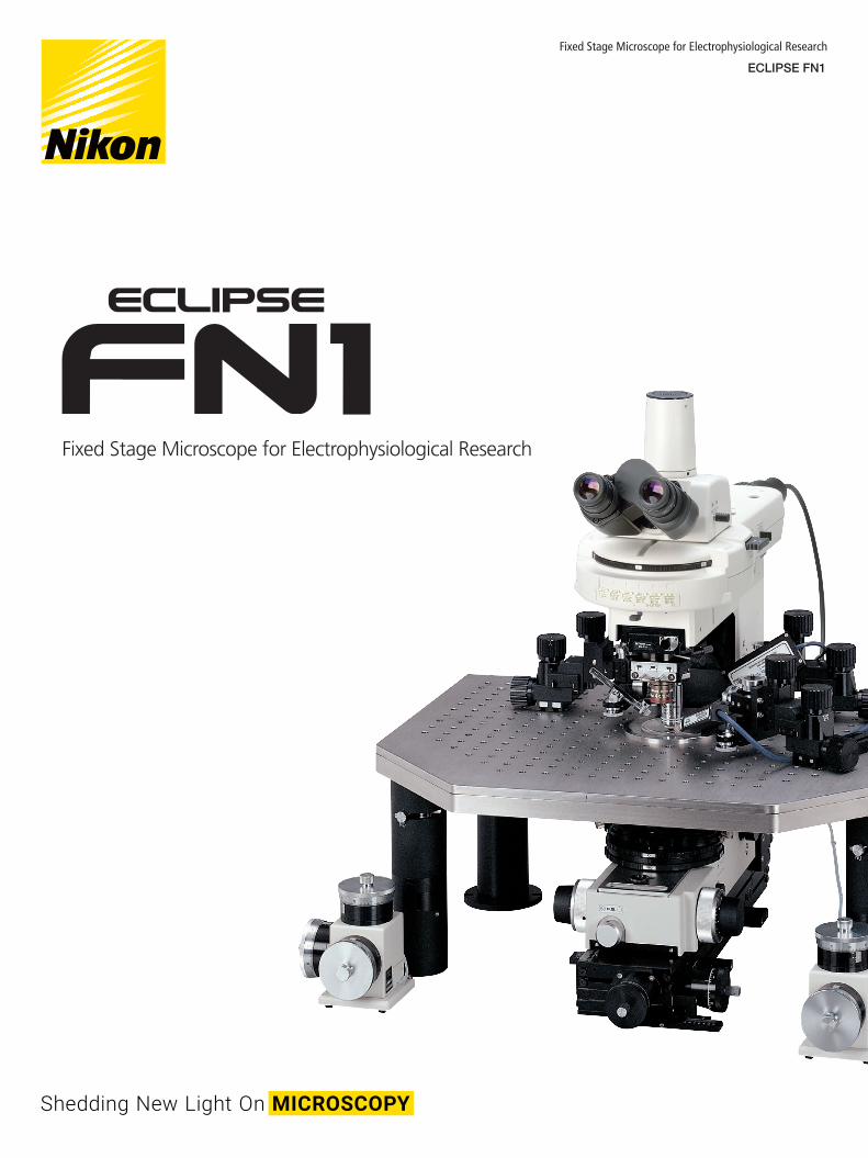
Fixed Stage Microscope for Electrophysiological Research
ECLIPSE FN1
Fixed Stage Microscope for Electrophysiological Research
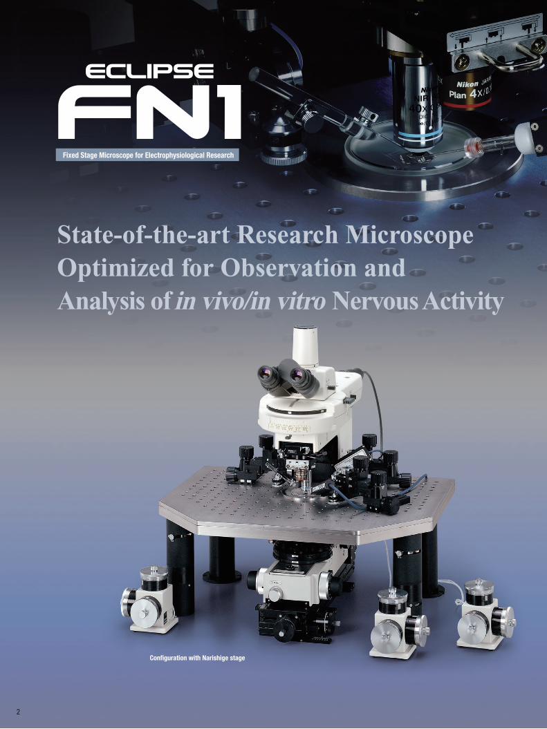
State-of-the-art Research Microscope Optimized for Observation and Analysis of in vivo/in vitro Nervous Activity
Configuration with Narishige stage
2
Fixed Stage Microscope for Electrophysiological Research
State-of-the-art Research Microscope Optimized for Observation and Analysis of in vivo/in vitro Nervous Activity
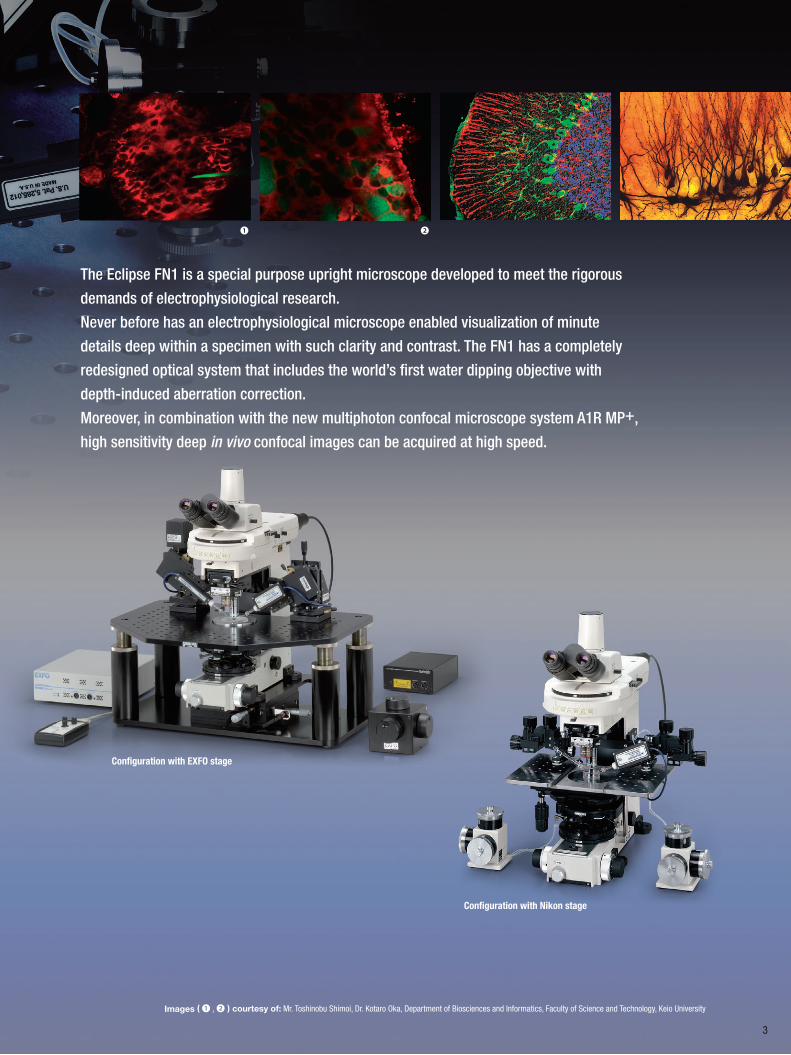
The Eclipse FN1 is a special purpose upright microscope developed to meet the rigorous
demands of electrophysiological research.
Never before has an electrophysiological microscope enabled visualization of minute
details deep within a specimen with such clarity and contrast. The FN1 has a completely
redesigned optical system that includes the world’s first water dipping objective with
depth-induced aberration correction.
Moreover, in combination with the new multiphoton confocal microscope system A1R MP+,
high sensitivity deep in vivo confocal images can be acquired at high speed.
Configuration with EXFO stage
Configuration with Nikon stage
❶ ❷
3
Images ( ❶ , ❷ ) courtesy of: Mr. Toshinobu Shimoi, Dr. Kotaro Oka, Department of Biosciences and Informatics, Faculty of Science and Technology, Keio University
4
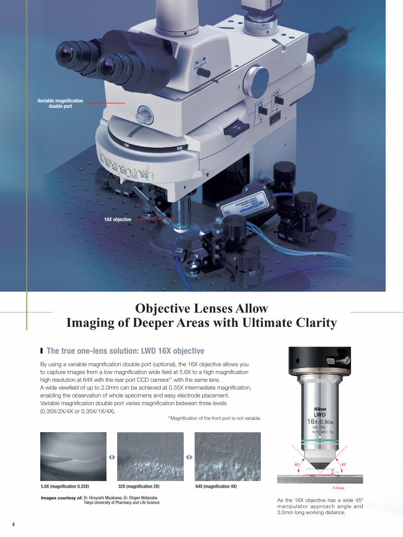
Objective Lenses Allow Imaging of Deeper Areas with Ultimate Clarity
By using a variable magnification double port (optional), the 16X objective allows you to capture images from a low magnification wide field at 5.6X to a high magnification high resolution at 64X with the rear port CCD camera*1 with the same lens. A wide viewfield of up to 2.0mm can be achieved at 0.35X intermediate magnification, enabling the observation of whole specimens and easy electrode placement.Variable magnification double port varies magnification between three levels (0.35X/2X/4X or 0.35X/1X/4X).
*1Magnification of the front port is not variable.
The true one-lens solution: LWD 16X objective
32X (magnification 2X) 64X (magnification 4X)5.6X (magnification 0.35X)
Images courtesy of: Dr. Hiroyoshi Miyakawa, Dr. Shigeo Watanabe, Tokyo University of Pharmacy and Life Science As the 16X objective has a wide 45º
manipulator approach angle and 3.0mm long working distance.
Variable magnification double port
16X objective
Images courtesy of: Hiroyuki Hakozaki MS, University of California, San Diego
45º approach angle, long working distance
45˚ 45˚
3.5mm
5
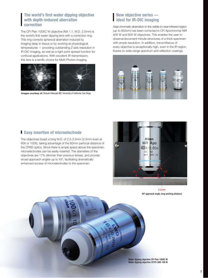
Water dipping objective CFI Plan 100XC WWater dipping objective CFI75 LWD 16X W
The CFI Plan 100XC W objective (NA 1.1, W.D. 2.5mm) is the world’s first water dipping lens with a correction ring. This ring corrects spherical aberration induced by imaging deep in tissue or by working at physiological temperatures — providing outstanding Z-axis resolution in IR-DIC imaging, as well as a tight point spread function for confocal applications. With excellent IR transmission, this lens is a terrific choice for Multi-Photon imaging.
The world’s first water dipping objective with depth-induced aberration correction Axial chromatic aberration in the visible to near-infrared region
(up to 850nm) has been corrected in CFI Apochromat NIR 40X W and 60X W objectives. This enables the user to observe/document minute structures of a thick specimen with ample resolution. In addition, transmittance of every objective is exceptionally high, even in the IR region, thanks to wide-range spectrum anti-reflection coatings.
New objective series — ideal for IR-DIC imaging
The objectives boast a long W.D. of 2.5-3.5mm (2.5mm even at 60X or 100X), taking advantage of the 60mm parfocal distance of the CFI60 optics. Since there is ample space above the specimen, microelectrodes can be easily inserted. The diameters of the objectives are 17% slimmer than previous lenses, and provide broad approach angles up to 45º, facilitating dramatically enhanced access of microelectrodes to the specimen.
Easy insertion of microelectrode
Front/back sliding objective changeover
Objective retraction mechanism Simple lever operation ensures safe dipping Objective up/down lever
6
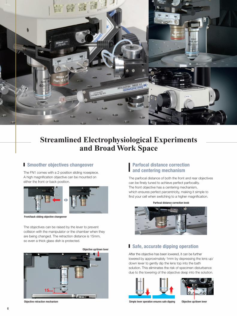
The objectives can be raised by the lever to prevent collision with the manipulator or the chamber when they are being changed. The retraction distance is 15mm, so even a thick glass dish is protected.
Objective up/down lever
Streamlined Electrophysiological Experiments and Broad Work Space
The FN1 comes with a 2-position sliding nosepiece. A high magnification objective can be mounted on either the front or back position.
Smoother objectives changeover
The parfocal distance of both the front and rear objectives can be finely tuned to achieve perfect parfocality. The front objective has a centering mechanism, which ensures perfect parcentricity, making it simple to find your cell when switching to a higher magnification.
Parfocal distance correction and centering mechanism
Parfocal distance correction knob
After the objective has been lowered, it can be further lowered by approximately 1mm by depressing the lens up/down lever to gently dip the lens top into the bath solution. This eliminates the risk of specimen disturbance due to the lowering of the objective deep into the solution.
Safe, accurate dipping operation
7
The condenser and polarizer turret can be simply and quickly removed.
The simple and slim I-shaped body has no projection on the body other than the focus knob, so there is more space in the working area for your experiment. This also provides better access around the microscope to position manipulators and other peripherals. With the eye-point of the body 25mm lower than conventional models, you can work in greater comfort.
I-shaped slimline body creates more space above and below the stage
Observed under oblique illumination Observed under IR-DIC illumination
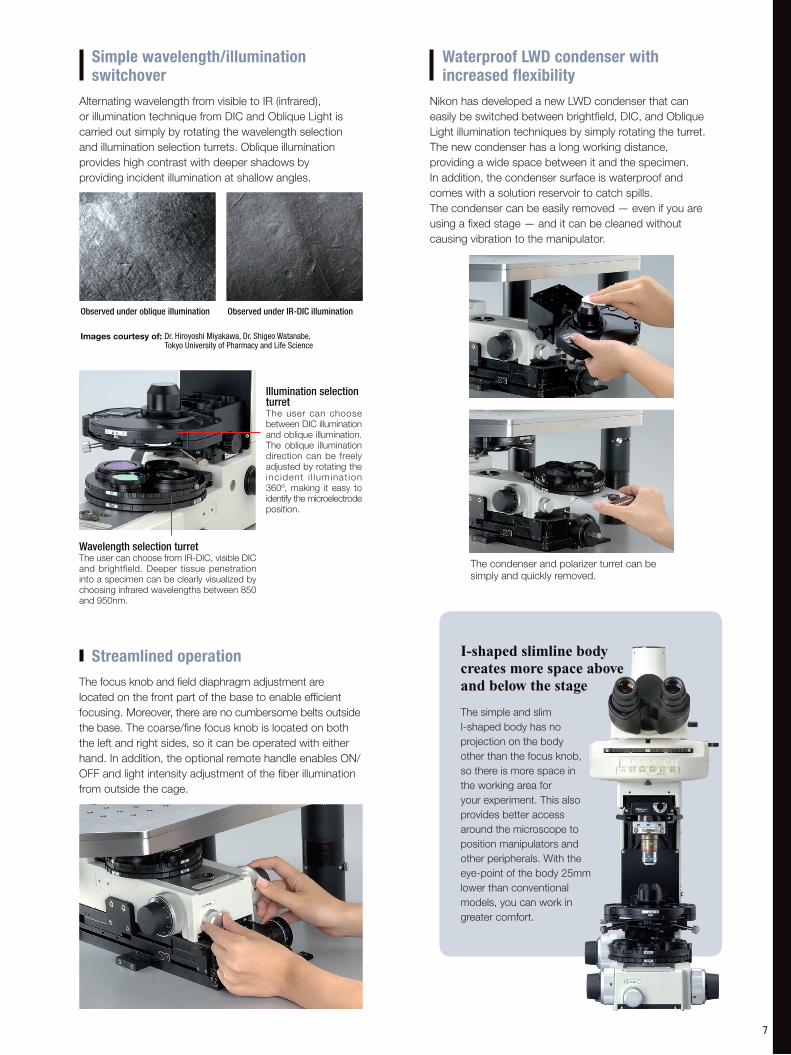
Illumination selection turretThe user can choose between DIC illumination and oblique illumination. The oblique illumination direction can be freely adjusted by rotating the incident i l luminat ion 360º, making it easy to identify the microelectrode position.
Wavelength selection turretThe user can choose from IR-DIC, visible DIC and brightfield. Deeper tissue penetration into a specimen can be clearly visualized by choosing infrared wavelengths between 850 and 950nm.
Alternating wavelength from visible to IR (infrared), or illumination technique from DIC and Oblique Light is carried out simply by rotating the wavelength selection and illumination selection turrets. Oblique illumination provides high contrast with deeper shadows by providing incident illumination at shallow angles.
Simple wavelength/illumination switchover
Images courtesy of: Dr. Hiroyoshi Miyakawa, Dr. Shigeo Watanabe, Tokyo University of Pharmacy and Life Science
The focus knob and field diaphragm adjustment are located on the front part of the base to enable efficient focusing. Moreover, there are no cumbersome belts outside the base. The coarse/fine focus knob is located on both the left and right sides, so it can be operated with either hand. In addition, the optional remote handle enables ON/OFF and light intensity adjustment of the fiber illumination from outside the cage.
Streamlined operation
Nikon has developed a new LWD condenser that can easily be switched between brightfield, DIC, and Oblique Light illumination techniques by simply rotating the turret. The new condenser has a long working distance, providing a wide space between it and the specimen. In addition, the condenser surface is waterproof and comes with a solution reservoir to catch spills. The condenser can be easily removed — even if you are using a fixed stage — and it can be cleaned without causing vibration to the manipulator.
Waterproof LWD condenser with increased flexibility
8
Spacers Can be raised by 10mm to 40mm
System Expansion
Enhanced Noise Reduction and High Responsiveness
to a Broad Range of Experimental Needs
Nikon has succeeded in significantly reducing electrical noise by utilizing fiber illumination to bring light into the microscope from outside the cage. Noise can be dramatically reduced by connecting ground pins to all main parts of the microscope.
Minimizing electronic noise
Nikon has achieved both improved rigidity and vibration resistance for the FN1 body by undertaking critical measurement and simulation analysis of its structure. Nikon has succeeded in suppressing the vibration generated when the nosepiece or the magnification module is switched.
Ultimate vibration noise reduction
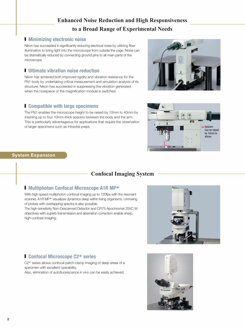
With high speed multiphoton confocal imaging up to 720fps with the resonant scanner, A1R MP+ visualizes dynamics deep within living organisms. Unmixing of probes with overlapping spectra is also possible.The high-sensitivity Non-Descanned Detector and CFI75 Apochromat 25XC W objectives with superb transmission and aberration correction enable sharp, high-contrast imaging.
Multiphoton Confocal Microscope A1R MP+
C2+ series allows confocal patch-clamp imaging of deep areas of a specimen with excellent operability.Also, elimination of autofluorescence in vivo can be easily achieved.
Confocal Microscope C2+ series
The FN1 enables the microscope height to be raised by 10mm to 40mm by inserting up to four 10mm-thick spacers between the body and the arm. This is particularly advantageous for applications that require the observation of larger specimens such as intravital preps.
Compatible with large specimens
Confocal Imaging System
9
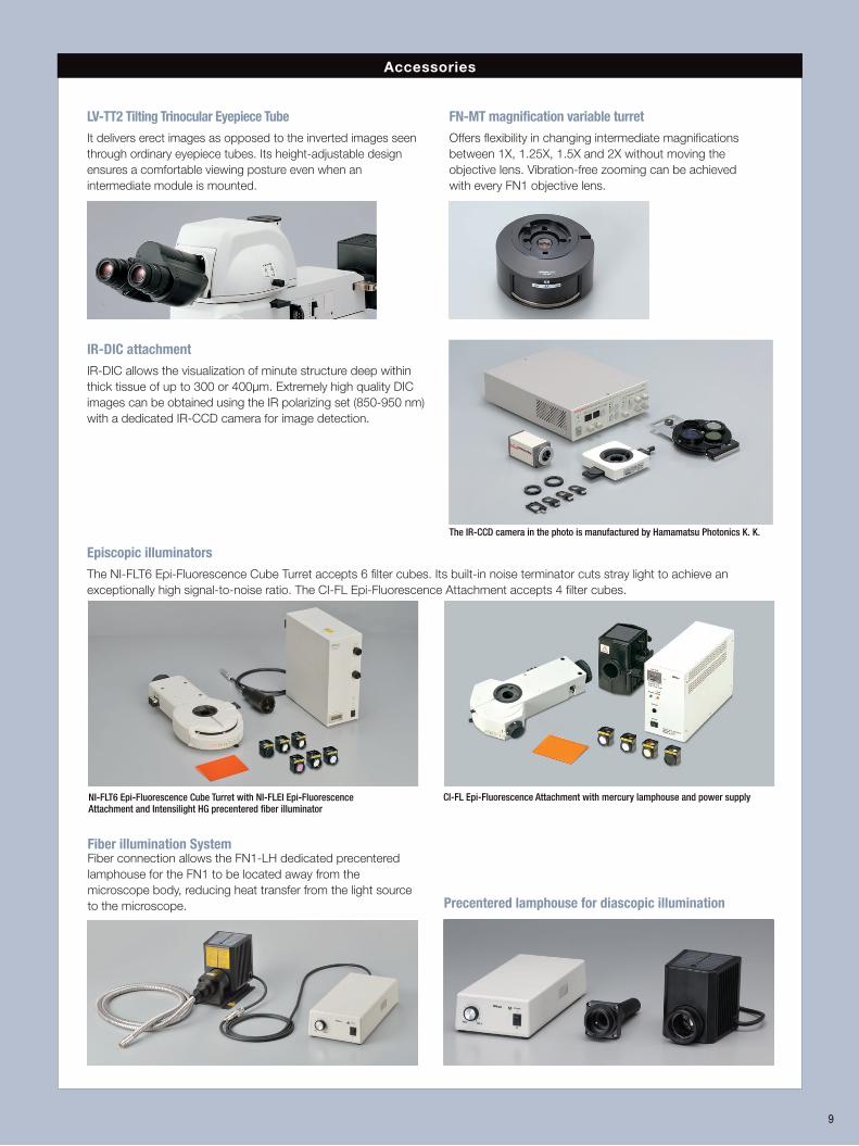
Accessories
Fiber illumination System Fiber connection allows the FN1-LH dedicated precentered lamphouse for the FN1 to be located away from the microscope body, reducing heat transfer from the light source to the microscope. Precentered lamphouse for diascopic illumination
NI-FLT6 Epi-Fluorescence Cube Turret with NI-FLEI Epi-Fluorescence Attachment and Intensilight HG precentered fiber illuminator
CI-FL Epi-Fluorescence Attachment with mercury lamphouse and power supply
Episcopic illuminators The NI-FLT6 Epi-Fluorescence Cube Turret accepts 6 filter cubes. Its built-in noise terminator cuts stray light to achieve an exceptionally high signal-to-noise ratio. The CI-FL Epi-Fluorescence Attachment accepts 4 filter cubes.
FN-MT magnification variable turretOffers flexibility in changing intermediate magnifications between 1X, 1.25X, 1.5X and 2X without moving the objective lens. Vibration-free zooming can be achieved with every FN1 objective lens.
LV-TT2 Tilting Trinocular Eyepiece Tube It delivers erect images as opposed to the inverted images seen through ordinary eyepiece tubes. Its height-adjustable design ensures a comfortable viewing posture even when an intermediate module is mounted.
IR-DIC attachmentIR-DIC allows the visualization of minute structure deep within thick tissue of up to 300 or 400µm. Extremely high quality DIC images can be obtained using the IR polarizing set (850-950 nm) with a dedicated IR-CCD camera for image detection.
The IR-CCD camera in the photo is manufactured by Hamamatsu Photonics K. K.
Accessories
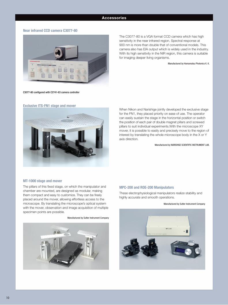
MT-1000 stage and mover The pillars of this fixed stage, on which the manipulator and chamber are mounted, are designed as modular, making them compact and easy to customize. They can be freely placed around the mover, allowing effortless access to the microscope. By translating the microscope’s optical system with the mover, observation and image acquisition of multiple specimen points are possible.
Manufactured by Sutter Instrument Company
MPC-200 and ROE-200 ManipulatorsThese electrophysiological manipulators realize stability and highly accurate and smooth operations.
Manufactured by Sutter Instrument Company
10
Exclusive ITS-FN1 stage and moverWhen Nikon and Narishige jointly developed the exclusive stage for the FN1, they placed priority on ease of use. The operator can easily sustain the stage in the horizontal position or switch the position of each pair of double magnet pillars and screwed pillars to suit individual experiments.With the microscope XY mover, it is possible to easily and precisely move to the region of interest by translating the whole microscope body in the X or Y axis direction.
Manufactured by NARISHIGE SCIENTIFIC INSTRUMENT LAB.
Near infrared CCD camera C3077-80
The C3077-80 is a VGA format CCD camera which has high sensitivity in the near infrared region. Spectral response at 900 nm is more than double that of conventional models. This camera also has EIA output which is widely used in the industry. With its high sensitivity in the NIR region, this camera is suitable for imaging deeper living organisms.
Manufactured by Hamamatsu Photonics K. K.
C3077-80 configured with C2741-63 camera controller
N1 Dr y
JAPAN
JAPANFN-P
A.S
FN-PT4X 4XSupport Lens
6
6
6
1 2
8 9
3
1
4
7
7
7
7
598
JAPANLV-TV
55
1 3
4 4
2
1
3
7
7
7
5
2
1 2 3
44
Y-TV55TV Tube0.55X
IntensilightHG PrecenteredFiber Illuminator
YM-EPIExtension Cable
FN-FA for Fiber Illuminator Adapterwith Fiber for FN1 Fiber Illuminator
ITS-FN1 Stage (Narishige)
TE-PS 100WPower Supply
100-240V
XY Mover (NARISHIGE)Z-axis Motorized Drive Unit
ECLIPSE FN1
C-mount Camera
FN-DP Mag. Variable Double Port
FN-MTMag. Variable Turret C-ISA Simple Analyzer FN IR/ISA DIC IR Analyzer
DIC SliderFN-S2N Sliding Nosepiece
CFI60 Objective Lens
D-C DIC Slider 16XFN-MN-H
Single Objective Holder
CFI75 LWD 16X W Objective Lens
D-C DIC Module
FN-IRP IR Polarizer/FN-P Polarizer
FN-PT Polarizer Turret
FN-C LWD Condenser
TE-ATDouble Lamphouse
Adapter
Hg Lamphouse HMX-4B
C-HGFIBHG 100WAdapter R
FN-3PS2 FN1 Rectangular Stage
FN-LPALamphouse Adapter
FN-LHPrecentered Lamphouse
* For the DS-Fi3 camera, a C-TEP3 DSC Port C-0.55X for Ergonomic Binocular Tube is recommended
C-HGFIF 30HG Fiber
Spacer
TE-ATDouble Lamphouse
Adapter
LV-TI3Trinocular Tube
ESDLV-TT2
Trinocular Tube
Y-TTV Tube
Y-QTDouble TVAdapter LV-TV Tube Adapter
T-BPAPhoto
Adapter
C-mountAdapter0.35X,0.45X,0.6X
C-mountTV Relay Lens
VM2.5X
C-DAC-mountAdapter
C-mountTV Relay Lens
VM4X
C-0.7XDXM Relay
Lens
C-0.55XDS Relay
Lens
C-mount Camera F-mount Camera
C-TF Trinocular
Tube F
8 9 8 98 9
8
C-TT Trinocular Tube T
TI-A DIC Analyzer
Cube
A1-FN1-IRAnalyzerCube IR
C-FL Epi-fl Filter Cube
C-FL Epi-fl Filter Cube
NI-FLT6 Epi-fluorescence Cube Turret
F.STOP A.STOP
EX.ADJ.
NI-FLEI EPI-Fluorescence AttachmentNI-FA FL/
DIC Analyzer
C-TEP2 DSC Portfor Ergonomic Binocular Tube*(for C-mount Camera)
C-TE2 Ergonomic Binocular Tube
CI-FL Epi-Fluorescence Attachment
C-TEPF2.5 DSC Port F2.5Xfor Ergonomic Binocular Tube
(for F-mount Camera)
CFI 10X CFI UW 10X
C-CT Centering Telescope
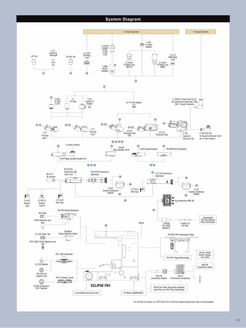
System Diagram
11
En
100
94 106.5107
205.5 428115
Ø9
21.8
226.8Ø9
Ø9 Ø9
Specimen Plane
Center of optical axis
1 (coverslip)
496.2
4
170
156
151.4
8.2
185.5
159
67
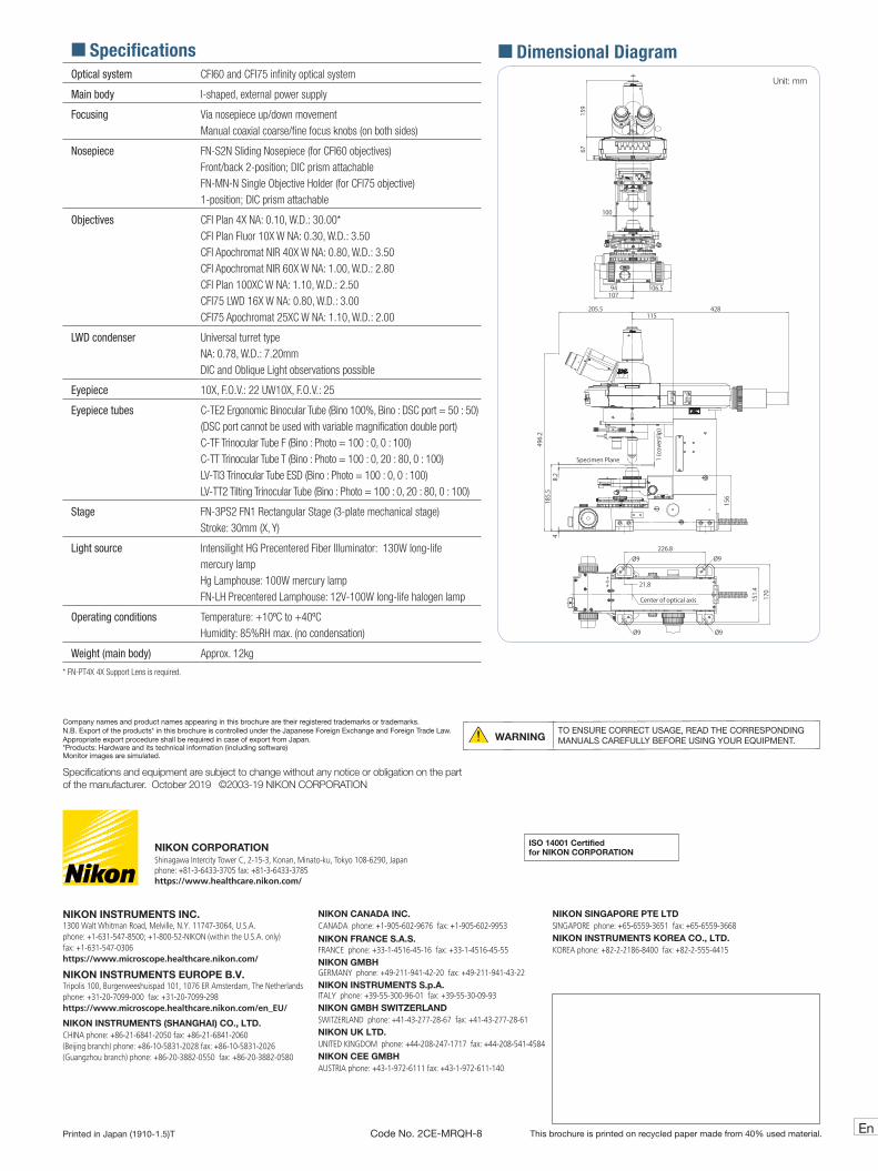
� Dimensional DiagramUnit: mm
Printed in Japan (1910-1.5)T Code No. 2CE-MRQH-8
Specifications and equipment are subject to change without any notice or obligation on the part of the manufacturer. October 2019 ©2003-19 NIKON CORPORATION
Company names and product names appearing in this brochure are their registered trademarks or trademarks.N.B. Export of the products* in this brochure is controlled under the Japanese Foreign Exchange and Foreign Trade Law. Appropriate export procedure shall be required in case of export from Japan.*Products: Hardware and its technical information (including software)Monitor images are simulated.
WARNINGTO ENSURE CORRECT USAGE, READ THE CORRESPONDING MANUALS CAREFULLY BEFORE USING YOUR EQUIPMENT.
This brochure is printed on recycled paper made from 40% used material.
� SpecificationsOptical system CFI60 and CFI75 infinity optical system
Main body I-shaped, external power supply
Focusing Via nosepiece up/down movementManual coaxial coarse/fine focus knobs (on both sides)
Nosepiece FN-S2N Sliding Nosepiece (for CFI60 objectives) Front/back 2-position; DIC prism attachable FN-MN-N Single Objective Holder (for CFI75 objective) 1-position; DIC prism attachable
Objectives CFI Plan 4X NA: 0.10, W.D.: 30.00*CFI Plan Fluor 10X W NA: 0.30, W.D.: 3.50CFI Apochromat NIR 40X W NA: 0.80, W.D.: 3.50CFI Apochromat NIR 60X W NA: 1.00, W.D.: 2.80CFI Plan 100XC W NA: 1.10, W.D.: 2.50CFI75 LWD 16X W NA: 0.80, W.D.: 3.00CFI75 Apochromat 25XC W NA: 1.10, W.D.: 2.00
LWD condenser Universal turret typeNA: 0.78, W.D.: 7.20mm DIC and Oblique Light observations possible
Eyepiece 10X, F.O.V.: 22 UW10X, F.O.V.: 25
Eyepiece tubes C-TE2 Ergonomic Binocular Tube (Bino 100%, Bino : DSC port = 50 : 50)(DSC port cannot be used with variable magnification double port)C-TF Trinocular Tube F (Bino : Photo = 100 : 0, 0 : 100)C-TT Trinocular Tube T (Bino : Photo = 100 : 0, 20 : 80, 0 : 100) LV-TI3 Trinocular Tube ESD (Bino : Photo = 100 : 0, 0 : 100) LV-TT2 Tilting Trinocular Tube (Bino : Photo = 100 : 0, 20 : 80, 0 : 100)
Stage FN-3PS2 FN1 Rectangular Stage (3-plate mechanical stage) Stroke: 30mm (X, Y)
Light source Intensilight HG Precentered Fiber Illuminator: 130W long-life mercury lamp Hg Lamphouse: 100W mercury lampFN-LH Precentered Lamphouse: 12V-100W long-life halogen lamp
Operating conditions Temperature: +10ºC to +40ºCHumidity: 85%RH max. (no condensation)
Weight (main body) Approx. 12kg
* FN-PT4X 4X Support Lens is required.
NIKON CORPORATIONShinagawa Intercity Tower C, 2-15-3, Konan, Minato-ku, Tokyo 108-6290, Japan phone: +81-3-6433-3705 fax: +81-3-6433-3785https://www.healthcare.nikon.com/
ISO 14001 Certifiedfor NIKON CORPORATION
NIKON INSTRUMENTS INC.1300 Walt Whitman Road, Melville, N.Y. 11747-3064, U.S.A.phone: +1-631-547-8500; +1-800-52-NIKON (within the U.S.A. only) fax: +1-631-547-0306https://www.microscope.healthcare.nikon.com/
NIKON INSTRUMENTS EUROPE B.V.Tripolis 100, Burgerweeshuispad 101, 1076 ER Amsterdam, The Netherlandsphone: +31-20-7099-000 fax: +31-20-7099-298https://www.microscope.healthcare.nikon.com/en_EU/
NIKON INSTRUMENTS (SHANGHAI) CO., LTD.CHINA phone: +86-21-6841-2050 fax: +86-21-6841-2060(Beijing branch) phone: +86-10-5831-2028 fax: +86-10-5831-2026(Guangzhou branch) phone: +86-20-3882-0550 fax: +86-20-3882-0580
NIKON CANADA INC.CANADA phone: +1-905-602-9676 fax: +1-905-602-9953
NIKON FRANCE S.A.S.FRANCE phone: +33-1-4516-45-16 fax: +33-1-4516-45-55NIKON GMBHGERMANY phone: +49-211-941-42-20 fax: +49-211-941-43-22NIKON INSTRUMENTS S.p.A.ITALY phone: +39-55-300-96-01 fax: +39-55-30-09-93NIKON GMBH SWITZERLANDSWITZERLAND phone: +41-43-277-28-67 fax: +41-43-277-28-61NIKON UK LTD.UNITED KINGDOM phone: +44-208-247-1717 fax: +44-208-541-4584NIKON CEE GMBHAUSTRIA phone: +43-1-972-6111 fax: +43-1-972-611-140
NIKON SINGAPORE PTE LTD SINGAPORE phone: +65-6559-3651 fax: +65-6559-3668NIKON INSTRUMENTS KOREA CO., LTD.KOREA phone: +82-2-2186-8400 fax: +82-2-555-4415