first report of heterodermia squamulosa (lichenized ascomycota ... … · first report of...
TRANSCRIPT

Mycobiology 36(3) : 190-192 (2008)
© The Korean Society of Mycology
190
First Report of Heterodermia squamulosa (Lichenized Ascomycota, Physciaceae)in South Korea
Xin Yu Wang1
, Hyun Hur2
, You Mi Lee3
, Young Jin Koh1
and Jae-Seoun Hur1
*
1
Korean Lichen Research Institute, Sunchon National University, Sunchon 540-742, Korea2
Department of Biology, Dongguk University, Seoul 100-715, Korea3
Division of Specimen and Genetic Resources, Korea National Arboretum 487-821, Korea
(Received August 18, 2008. Accepted September 12, 2008)
Heterodermia squamulosa (Degel.) W.L. Culb. was found in the mountain of Gariwang, Gangwon province, in 2008. It is
characterized by numerous squamules along the margin, decorticate and white lower surface, rhizines along the margin,
black and densely squarrosely branched, usually forming a dense mat under the thallus. Apothecia margins densely squa-
mulose, ascospores 12~15 × 25~30 µm. Atranorin and zeorin contained in thallus. This is the first record of this species in
South Korea.
KEYWORDS : Foliose lichen, Korea, Lichen-forming fungus, New record
The genus Heterodermia Trev. belongs to the lichenized
ascomycete family Physciaceae Zahl., and is mainly char-
acterized by the linear habitat of the thallus and Pachyspo-
raria- or Polyblastidia-type spores (Poelt, 1965; Brodo et
al., 2001). Heterodermia was included in Anaptychia
Körb (Kurokawa, 1962) until the former was separated in
1965 based on the characteristics of thick walled spores
and the presence of atranorin (Poelt, 1965; Swinscow and
Krog, 1976). According to the most recent checklist of
Korean lichens (Hur et al., 2005), there are 20 species of
Heterodermia recorded in the Korean peninsula. This is
the first record of this species in South Korea.
The specimens for this study were deposited in the
Korean Lichen Research Institute (KoLRI) in Sunchon
National University. The phenotypic characteristics were
based on air-dried material. Morphological characteristics
were observed using the dissecting microscope (Nikon
SMZ 1500), and anatomy was examined using the com-
pound microscope (Olympus BX 50). Lichen substances
were detected by color reagents and thin-layer chromatog-
raphy (Culberson, 1972; White and James, 1985).
Heterodermia squamulosa (Degel.) W.L. Culb. (1967).
(Figs. 1A~1E).
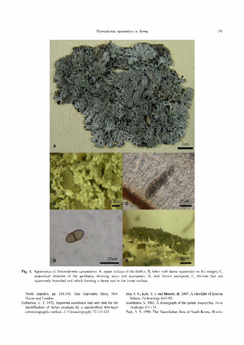
Morphology. Thallus foliose, grayish white when dried,
greenish when wet, 6~10 cm in diameter, laciniate;
laciniae dichotomously or subirregularly branched, about
0.5~1.5 mm wide, with numerous squamules along the
margin, sometimes also on the upper surface, lower sur-
face of the squamulse decorticate, with the same color
of the thallus; lower surface of the thallus decorticate
and white. Rhizines along the margin, black and
densely squarrosely branched, 1~1.5 mm long, usually
forming a dense mat under the thallus. Apothecia lami-
nal and sessile, dark reddish brown, 1~4 mm in diame-
ter, margins densely squamulose; hymenium 140~150
µm thick; ascospores dark brown, two cells, 12~15 ×
25~30 µm.
Chemistry. Cortex and medulla K+ yellow, P-. C-, KC-;
containing atranorin and zeorin.
Distribution. This species mostly occurs in the woods
and on rocks. It has previously been reported in Malaysia
(Sipman, 1993), but not in South Korea.
Specimens examined. The sample location was Mt Gari-
wang (37o
24'05.0" N, 128o
32'39.5" E), on moss rock. Hur
080032, May 10, 2008.
Remarks. This species is very similar to H. hypoleuca,
could be easily distinguished by the presence of the mar-
ginal squamules. It has another closely related species H.
microphylla (Kurokawa, 1962). The latter contains sore-
dia or granules among the squamules, and the spores,
which measure about 10~20 µm, are smaller than those of
H. squamulosa.
Acknowledgement
This work was supported by a grant from Korea National
Research Resource Center Program (Grant R21-2007-000-
10033-0), and Korean Forest Service Program (KNA
2008) through Korea National Arboretum.
References
Brodo, I. M., Sharnoff, D. S. and Sharnoff, S. 2001. Lichens of*Corresponding author <E-mail : [email protected]>
Heterodermia squamulosa in Korea 191
North America, pp. 334-341. Yale University Press, New
Haven and London.
Culberson, C. F. 1972. Improved conditions and new data for the
identification of lichen products by a standardized thin-layer
chromatographic method. J. Chromatography 72:113-125.
Hur, J. S., Koh, Y. J. and Harada, H. 2005. A checklist of Korean
lichens. Lichenology 4:65-95.
Kurokawa, S. 1962. A monograph of the genus Anaptychia. Nova
Hedwigia 6:1-115.
Park, Y. S. 1990. The Macrolichen flora of South Korea. Bryolo-
Fig. 1. Appearance of Heterodermia squamulosa. A, upper surface of the thallus; B, lobes with dense squamules on the margin; C,
anatomical structure of the apothecia, showing ascus and ascospores; D, dark brown ascospore; E, rhizines that are
squarrosely branched and which forming a dense mat in the lower surface.
192 Wang et al.
gist 93:105-160.
Poelt, J. 1965. Zur Systematik der Flechtenfamilie Physciaceae.
Nova Hegwigia 9:21-32.
Sipman, H. J. M. 1993. Lichens from Mount Kinabalu. Trop.
Bryol. 8:288.
Swinscow, T. D. V. and Krog, H. 1976. The genera Anaptychia
and Heterodermia in East Africa. Lichenologist 8:103-138.
White, F. J. and James, P. W. 1985. A new guide to microchemi-
cal techniques for the identification of lichen substances. Brit.
Lichen Soc. Bull. 57:1-41.