fine structural aspects of the urothelium in the mouse
TRANSCRIPT

Hiroshima J. Med. Sci. Vol. 51, No. 2, 41-48, June, 2002 HIJM 51-6
41
Fine Structural Aspects of the Urothelium in the Mouse Ureter with Special Reference to Cell Kinetics
Keisuke YAMASHITA Department of Anatomy, Hiroshima University School of Medicine, 1-Chome 2-3, Kasumi, Minami-ku, Hiroshima 734-8551, Japan and Core Research for Evolutional Science and Technology (CREST), Japan Science and Technology, Tokyo, Japan
ABSTRACT The present study aimed at clarifying the cell kinetics of the mouse ureteral epithelium by
focusing on vesicle maturation in the cells and labeling with bromodeoxyuridine (BrdU). Electron microscopically, superficial cells were characterized by concave plaques in the apical plasma membrane and numerous fusiform vesicles in the cytoplasm. Intermediate cells were laden with ellipsoid vesicles, and basal cells had a few or no round vesicles. From the difference in number and form of vesicles among the three types of cells, it can be inferred that intermediate cells are immature in comparison with superficial cells, and likewise basal cells in comparison with intermediate cells. When BrdU was injected intraperitoneally once a day for seven days, most BrdU-labeled cells were located in the basal layer. Twelve days after the last injection, BrdU was detected in the intermediate or superficial layer in addition to the basal layer. These findings suggest that the basal cell is a progenitor cell giving rise to daughter cells that migrate upward to replace intermediate and superficial cells.
Key words: Ureter, Epithelium, Cell kinetics, Mice
The pathogenesis of the hydronephrosis (dilated renal pelvis) in mouse embryos is ascribed to hyperplasia of the ureteral epithelium when pregnant dams are given dioxin l,lS). The cell kinetics of the ureteral epithelium still remains unclear in normal as well as pathological conditions.
The epithelium of the urinary tract, also called the urothelium, is composed of three cell layers: the basal, intermediate and superficial cell layers4). In the mouse, there are about five layers of epithelial cells in the ureter, compared with 2-3 in the urinary bladder10l. The urothelium is required to resist the high osmolarity of urine. In addition, it must accommodate the contractionexpansion cycle due to volume change. The permeability barrier to high osmolarity and accommodation to volume change are considered to be maintained by peculiar structures called asymmetric unit membranes4) as well as by the lamina propria 17l.
In a normal steady state, the cell kinetics of the urothelium in adult animals is quiescent, as revealed by 3H-thymidine autoradiography2'3'6'11,12,19), by mitotic figuress,7,11), and by immunohistochemistry for proliferating antigens5l.
Correspondence to: Dr. Keisuke Yamashita,
Martin11) confirmed by 3H-thymidine autoradiography that basal cells migrated upward, differentiated into intermediate cells, and finally replaced superficial cells. Spicer et al17) pointed out that the distribution of high molecular weight keratin and Na+, K+-ATPase in the urothelium was similar to that in stratified squamous epithelium, suggesting the stratified nature of the urothelium.
The superficial cell is characterized by numerous fusiform vesicles and peculiar apical plasma membranes with concave plaques4). The turnover of the vesicles has been well studied 16,18). Concave plaques in the apical plasma membrane are endocytosed and fuse to form fusiform vesicles. These vesicles are incorporated in the apical plasma membrane when the lumen is extended in order to satisfy the demand of the membrane in the apical domain. Thus the fusiform vesicles form a turnover or shuttling cycle between the apical plasma membrane and the cytoplasm. However, these previous studies paid little attention to the nature of the vesicles in basal and intermediate cells, although the presence of fusiform vesicles was reported in intermediate cells8,17).
In the present study, vesicles in the urothelial
Department of Anatomy, Hiroshima University School of Medicine, 1-Chome 2-3, Kasumi, Minami-ku, Hiroshima 734-8551,Japan TEL 082-257-5111, or -5112, FAX 082-257-5114 e-mail address: [email protected]

42 K. Yamashita
cells were precisely examined in relation to the cell kinetics. In addition, bromodeoxyuridine (BrdU) was applied to label S-phase nuclei.
MATERIALS AND METHODS 1) Animals
Colony-bred female Jcl: ICR mice (Clea Japan, Inc., Tokyo, Japan) were acclimatized and bred in our laboratory at 22 ± 2°C with 50 ± 10% humidity. 2) Electron microscopy
The mice (8-12 week old), anesthetized with an intraperitoneally injected overdose of pentobarbital, were perfusion-fixed with 2.5% glutaraldehyde in 0.1 M Millonig's phosphate buffer (pH 7.4). The ureter was divided into three portions from the lower end of the renal pelvis to the urinary bladder and was collected separately. Since the histology of the three portions is not different, the middle portion of the ureter was served for observation in this experiment. The ureteric pieces were fixed by immersion in the same glutaraldehyde fixative for 2 hours at 4°C, and postfixed in 1 % osmium tetroxide buffered with 0.1 M Millonig's phosphate containing sucrose (5% weight/volume) for 1 hour at 4 °C. They were rinsed five times .with 10% sucrose solution and stained en bloc in 3% uranyl acetate for 1 hour at room temperature. The specimen was dehydrated in graded ethanol series and propylene oxide, infiltrated in a mixture of Epon and propylene oxide (1 : 1, volume : volume), and embedded in Epon
mixture. Transverse ultrathin sections were cut in a Porter-Blum ultramicrotome (MT-1 type), doubly stained with 3% uranyl acetate and Reynolds' lead citrate, and examined in a Hitachi H7100-type transmission electron microscope at 75 kV. 3) Labeling with BrdU and immunohisto
chemical detection In order to label S-phase cells, a cell prolifera
tion kit (RPN20, Amersham Life Science, England) was used. In each labeling regimen, a labeling reagent containing 5-bromo-2'-deoxyuridine (BrdU) was administered intraperitoneally (i. p.) to three female mice (8-12 week old) for each experimental group as shown below. The injected dose was 3 mg/kg body weight/day. We made three
groups following Jost6>. a) Single-pulse labeling. Mice were given an i. p. injection of the BrdU labeling reagent at 10 a.m. The mice were sacrificed two hours after the injection. b) Seven-time labeling. BrdU was injected once a day at 10 a.m. for 7 days. The mice were sacrificed two hours after the last injection. c) Trace labeling. After the seven-time labeling, the mice were allowed to survive for the next 12 days and sacrificed in order to study the fate of the labeled cells.
The mice were fixed by perfusion with 10% neutral formalin in 0.1 M phosphate buffered saline. The ureter was taken out and embedded in paraffin. Cross sections of ureters were soaked in 1 N hydrochloric acid for 20 min at room temperature. Endogenous peroxidase activity was blocked by soaking the sections in 0.3% hydrogen peroxide/ methanol for 30 min. The sections were incubated in a mouse anti-BrdU antibody (RPN20, Amersham Life Science, England) for one hour at room temperature, and in a caprine anti-mouse IgG2a antibody conjugated with horse-radish peroxidase (RPN20, Amersham Life Science, England) for 30 min at room temperature. The sections were incubated in a medium containing hydrogen peroxide and 3,3'-diaminobenzidine tetrahydrochloride (RPN20, Amersham Life Science, England) for 10 min at room temperature, and counter-stained with eosin for 5 min.
RESULTS 1) The epithelial layer
The epithelial layer was four-cell thick on the basal lamina (Fig. 1), and composed of basal, intermediate, and superficial cells. a) The basal cell (Figs. 1 and 2)
Basal cells were small and had a relatively high nuclear to cytoplasmic ratio (N/C ratio) compared with intermediate or superficial cells. In the present observation, no mitotic figures were seen. The rough endoplasmic reticulum was poorly developed. Some basal cells had small round vesicles, 150-200 nm in diameter, in the supranuclear cytoplasm (Figs. 2b and 2c), while many basal cells lacked such vesicles. The plasma membrane of
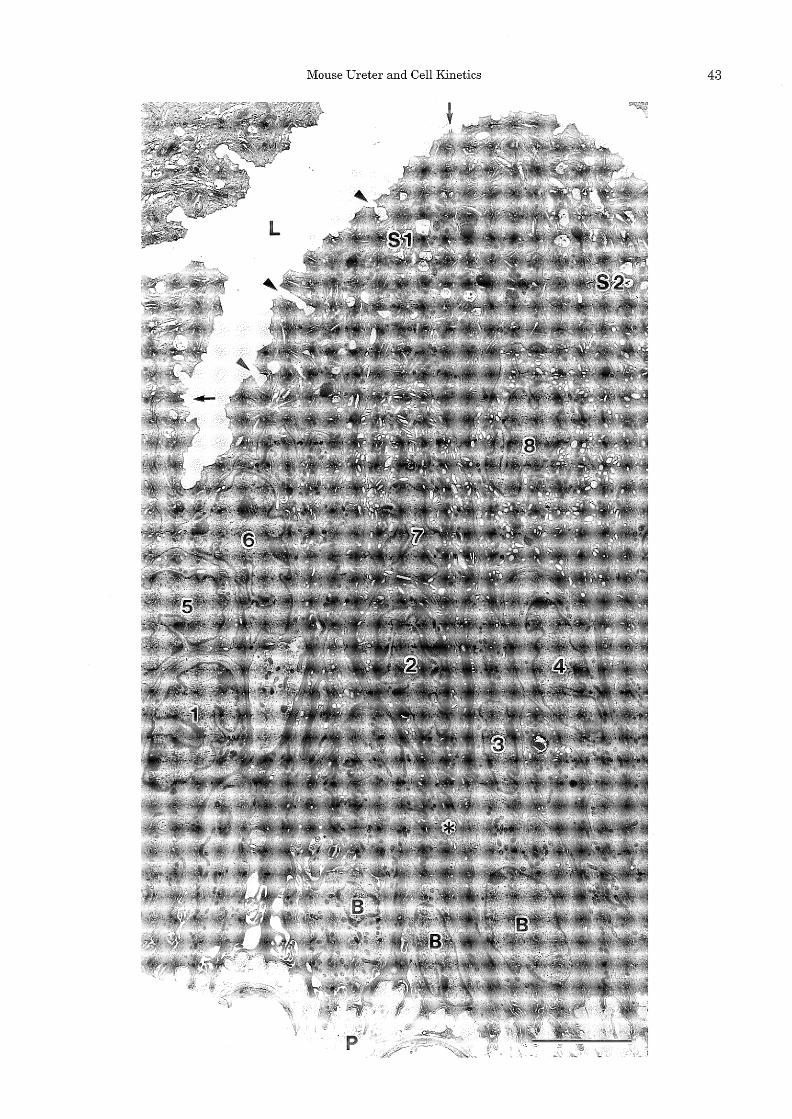
Fig. 1. Electron micrograph of the epithelium in a cross-sectioned mouse ureter. An epithelial fold protrudes into the lumen (L) on the right side of the micrograph. The urothelium is four-cell thick in the fold as well as in the thin area (the left side of the micrograph). Basal cells (B) are rather small and have faintly stained cytoplasm. According to the location of cell nuclei, the intermediate layer (cells 1-8) is further subdivided into two layers: a layer located rather basally (cells 1-4) (the second layer) and a layer located more superficially (cells 5-8) (the third layer). Among intermediate cells, cells 1, 3 and 4 contain round vesicles whereas cell 2 has both round and oval vesicles. Cell 2 extends a cell process (asterisk) onto the basal lamina. The third layer intermediate cells (cells 5-8) contain a larger number of oval to ellipsoid vesicles. The cells in the thin area (cells 5 and 6) contain a smaller number of vesicles than those in the fold area (cells 7 and 8). Large superficial cells (81 and 82) cover the lumenal surface. The lateral borders of a superficial cell (81) are shown by two arrows. In superficial cells, numerous vacuoles (multivesicular bodies) and dense bodies are apparent in addition to fusiform vesicles. Invaginations (arrowheads) are found along the apical surface. Abbreviation: P, lamina propria. Scale bar: 5 µm. All the following figures are shown with the lumenal side upwards.
Mouse Ureter and Cell Kinetics 43

44 K. Yamashita
basal cells was symmetrically thick. The unit membrane of the round vesicles was also symmetric (Fig. 2c). The term "symmetric" signifies a normal cell membrane, where the electron-dense outer (lumenal) leaflet and inner (cytoplasmic) leaflet are identically thick. b) The intermediate cell (Figs. 1 and 3)
Intermediate cells were located in the middle layer of the epithelium. As shown in Fig. 1, this layer was two-cell thick: intermediate cells located basally (the second layer) and more superficially (the third layer). Some of the second layer inter-
Fig. 2. Electron micrographs of basal cells (B).
mediate cells extended cell processes onto the basal lamina (Fig. 1, cell 2). A large number of oval to ellipsoid vesicles were prominent in the cytoplasm of intermediate cells (Fig. 3a). At higher magnifications, the vesicles were shown to have asymmetrically thick unit membranes (Fig. 3b and 3c). The lumenal leaflet was thicker than the cytoplasmic one. In addition to these oval to ellipsoid vesicles, intermediate cells contained round vesicles with symmetric unit membranes. When the number and shape of the vesicles were compared between intermediate cells in the second layer and
a: The nucleus/cytoplasm ratio is large. No cytoplasmic vesicle is seen. The Golgi apparatus (G) is small and does not show any sign of vesicle formation. Neighboring intermediate cells contain round or oval vesicles (arrows). Capillary endothelial cells, ensheathed by processes of pericytes, are poor in fenestration. Abbreviations: C, capillary; P, lamina propria; arrowheads, basal lamina. Scale bar: 2 µm. b: A somewhat matured basal cell (B) contains small round vesicles (arrows) in the supranuclear region. Some round and oval vesicles have formed in the Golgi area. Intermediate cells (I) contain round to ellipsoid vesicles in areas shown by asterisks. Scale bar: 2 µm. c: In a basal cell, both the round vesicles (V) and opposing plasma membranes (arrowheads) are symmetrically thick. Scale bar: 100 nm.

Mouse Ureter and Cell Kinetics 45
those in the third layer, the ellipsoid vesicles were more numerous and more elongated toward the fusiform in the third layer intermediate cells (Fig. 1, cells 7 and 8). c) The superficial cell (Figs. 1, 3a and 4)
Superficial cells were large cells lining the lumenal surface of the epithelium and were often characterized by large nuclei. Superficial cells extending cytoplasmic processes to the basal lamina were not seen. Neighboring superficial cells were connected by junctional complexes and desmosomes (Fig. 4a). The apical plasma membrane showed a scalloped appearance with con-
cave plaques and interplaque regions (Fig. 4b). The unit membrane of the concave plaque was about 120 A in thickness and its outer (lumenal) leaflet was thicker than the inner (cytoplasmic) leaflet. On the contrary, in the interplaque ridge, the outer leaflet looked thin (Fig. 4b). Various numbers of plaques were fused to form invaginations along the lumenal surface (Fig. 1, arrowheads). The basolateral plasma membrane was symmetric.
Fusiform vesicles were quite numerous in the cytoplasm. Their limiting membranes were asymmetrically thick except in the hinge region, where
Fig. 3. a: Electron micrographs of a third layer intermediate cell (I) and a superficial cell (S) containing numerous vesicles. Vesicles (asterisks) in the intermediate cell are oval to ellipsoid, while those in the superficial cell are fusiform (arrowheads). Abbreviation: L, ureteric lumen. Scale bar: 2 µm. b: Oval to ellipsoid vesicles (o) and round vesicles (r) in intermediate cells. The vesicles contain flocculent materials in the lumen. Arrowheads depict plasma membranes of two opposing intermediate cells. Scale bar: 200 nm. c: Higher magnification of an oval vesicle in an intermediate cell. The lumenal leaflet of the membrane (arrowheads) is thicker than the cytoplasmic leaflet. The thickness of the unit membrane is 12 nm. Scale bar: 100 nm.

46 K. Yamashita
the membrane was symmetrically thick (Fig. 4c). A large number of multivesicular bodies was
found in the cytoplasm. Electron dense bodies, probably primary and secondary lysosomes, were also seen. The rough endoplasmic reticulum was poorly developed, and mitochondria were abundant.
Fig. 4. Electron micrographs of superficial cells.
2) Labeling with BrdU (Fig. 5) Few epithelial cells were labeled with BrdU in
the single-pulse labeling group. In the seven-time labeling group, some basal cells were labeled (Fig. 5a). The labeling index in basal cells was 3.9 ± 2.8%. The number of positively labeled cells in a ureteric cross section was 2.0 ± 1.5 (mean± S. D.,
a: The junctional complex between neighboring superficial cells consists of zonula occludens (Z), zonula adherens (A) and desmosomes (D). The apical plasma membrane in concave plaques is asymmetrically thick (arrowheads). The lateral plasma membrane is symmetric. Part of a fusiform vesicle (F) is seen in the right lower corner of the micrograph. The unit membrane of the fusiform vesicle is asymmetrically thick, while the membrane is not clear at the edge. Abbreviation: L, ureteric lumen. Scale bar: 100 nm. b: Concave plaques and an interplaque ridge along the apical plasma membrane. The outer (lumenal) membrane leaflet of concave plaques is thicker than the inner (cytoplasmic) leaflet (arrowheads), while the outer leaflet of the interplaque ridge is thin (arrow). Scale bar: 100 nm. c: Fusiform vesicles (F). The lumenal membrane leaflet (arrowheads) is thicker than the cytoplasmic leaflet. The unit membrane is symmetrical in the hinge region (arrow). Scale bar: 100 nm.

Mouse Ureter and Cell Kinetics 47
Fig. 5. Cross-sections of the ureter with BrdU/antiBrdU immunohistochemistry. a: Ureter after the seven-time labeling with BrdU. Two labeled cells are seen in the basal layer of the urothelium. Arrowheads show the location of the basal lamina. band c: Ureter after the trace labeling. Two labeled nuclei are seen in the superficial layer (b). A labeled cell is seen also in the basal layer (c). Abbreviations: L, ureteric lumen; P, lamina propria; M, smooth muscle layer. Scale bar: 10 µm.
n = 33 sections). The mean number of basal cells per section was 51.6. Only a small number of intermediate cells and no superficial cells were labeled in this group.
In the trace labeling group, where mice were allowed to live 12 days after seven-time labeling with BrdU, some nuclei of superficial cells were labeled (Fig. 5b) in addition to some basal and intermediate cells (Fig. 5c). When BrdU was administered once a day for 19 days (total 20 times), all three types of urothelial cells were labeled (data not shown).
DISCUSSION The present study revealed that the urothelium
is fundamentally a kind of stratified epithelium: the basal cell is the progenitor cell, it goes up to the intermediate layer and finally matures into superficial cells. It was proposed in previous studies that every type of cell, including superficial cells, extends its cell processes onto the basal lamina in the urothelium15,20). However, superficial cells in contact with the basal lamina were never found. The present study showed that neighboring superficial cells are connected by a tight junction. Intermediate cells and basal cells neither face the lumen nor have a tight junction. Thus the cells in the urothelium can be categorized into two types: superficial and non-superficial cells. Whether superficial cells extend cell processes onto the basal lamina or not, urothelial cells are stratified in this sense.
The membrane kinetics of the superficial cell has been extensively studied. Fusiform vesicles are incorporated into the apical plasma membrane
in order to extend the apical cell surface9,14). The present study is the first to precisely examine various vesicles in the three types of cells in relation to cell kinetics. Severs and Hicks16) showed by freeze fracture replicas that membrane components matured into vesicles after they left the Golgi apparatus, and vesicle membranes incorporated some subunits during this process, to acquire asymmetricity. It is considered that ellipsoid vesicles in intermediate cells are not incorporated into the plasma membrane, but incorporated into the apical plasma membrane, when the cells are exposed to the lumen to be superficial cells.
This is the first report demonstrating the label of superficial cells with DNA precursors or their analogs. Basal cells were labeled in the seven-time labeling group, and superficial cells only in the trace labeling group. These facts suggest that the superficial cell is a descendant of the basal cell.
Exfoliation of superficial cells from the urothelium could not be found in the present study. This may be attributed to the quiescent nature of the urothelium. The reason why superficial cells are sometimes binuclear could not be explained.
In summary, the present study indicates that the urothelium is a kind of stratified epithelium. It is also suggested by the maturation of vesicles and BrdU labeling that the superficial cell is the final descendant of the basal cell which proliferates very slowly.
ACKNOWLEDGMENTS This work was supported in part by Grants-in
Aid for Scientific Research from the Ministry of Education, Culture, Sports, Science and Technology of Japan, and by Health Science Research Grants for Research on Environmental Health from the Ministry of Health, Labour and Welfare of Japan. I am very grateful to Dr. Katsuko ~ of Department of Anatomy, Hiroshi:dia University School of Medicine for her encouragement and critical reading of the manuscript. I also wish to express thanks to Ms. Saori Okamura (CREST, JTS), Mr. Hiroshi Ishihara, and Mr. Nobuki Shimizu for their technical assistance.
(Received October 4, 2001) (Accepted March 22, 2002)
REFERENCES 1. Abbott, B.D., Birnbaum, L.S. and Pratt, R.M.
1987. TCDD-induced hyperplasia of the ureteral epithelium produces hydronephrosis in murine fetuses. Teratology 35: 329-334.
2. Blenkinsopp, W.K. 1969. Cell proliferation in the epithelium of the oesophagus, trachea and ureter in mice. J. Cell Sci. 5: 393-401.
3. Farsund, T. 1975. Cell kinetics of mouse urinary bladder epithelium. I. Circadian and age variations

48 K. Yamashita
m cell proliferation and nuclear DNA content. Virchows Arch. (Cell Pathol.) 18: 35-49.
4. Hicks, R.M. 1975. The mammalian urinary bladder: An accommodating organ. Biol. Rev. Camb. Philos. Soc. 50: 212-246.
5. Holstein, A.-F., Sandmann, J., Bresse!, M. and Davidoff, M.S. 1994. Reinvestigation of the transitional epithelium (urothelium) of the human ureter. Ann. Anat.176: 109-117.
6. Jost, S.P. 1986. Renewal of normal urothelium in adult mice. Virchows Arch. (Cell Pathol.) 51: 65-70.
7. Jost, S.P. and Potten, C.S. 1986. Urothelial proliferation in growing mice. Cell Tissue Kinet. 19: 155-160.
8. Koss, L.G. 1969. The asymmetric unit membranes of the epithelium of the urinary bladder of the rat. An electron microscopic study of a mechanism of epithelial maturation and function. Lab. Invest. 21: 154-168.
9. Lewis, S.A. and De Moura, J.L.C. 1982. Incorporation of cytoplasmic vesicles into apical membrane of mammalian urinary bladder epithelium. Nature 297: 685-688.
10. Martin, B.F. 1958. Histological and histochemical studies on the bladder and ureter, with special reference to alkaline phosphatase and Golgi material. J. Anat. 92: 286-297.
11. Martin, B.F. 1972. Cell replacement and differentiation in transitional epithelium: A histological and autoradiographic study of guinea-pig bladder and ureter. J. Anat. 112: 433-455.
12. Messier, B. and Leblond, C.P. 1960. Cell proliferation and migration as revealed by radioautography after injection of thymidine-H3 into male rats and mice. Am. J. Anat. 106: 247-285.
13. Mimura, J., Yamashita, K., Nakamura, K.,
Morita, M., Takagi, T.N., Nakao, K., Ema, M., Sogawa, K., Yasuda, M., Katsuki, M. and FujiiKuriyama, Y. 1997. Loss of teratogenic response to 2, 3, 7, 8-tetrachlorodibenzo-p-dioxin (TCDD) in mice lacking the Ah (dioxin) receptor. Genes to Cells 2: 645-654.
14. Minsky, B.D. and Chlapowski, F.J. 1978. Morphometric analysis of the translocation of lumenal membrane between cytoplasm and cell surface of transitional epithelial cells during the expansioncontraction cycles of mammalian urinary bladder. J. Cell Biol. 77: 685-697.
15. Petry, G. and Amon, H. 1966. Licht- und Elektronenmikroskopische Studien i.iber Struktur und Dynamik des Ubergangsepithels. Zeitschrift Zellforsch. 69: 587-612.
16. Severs, N.J. and Hicks, R.M. 1979. Analysis of membrane structure in the transitional epithelium of rat urinary bladder. 2. The discoidal vesicles and Golgi apparatus: Their role in luminal membrane biogenesis. J. Ultrastr. Res. 69: 279-296.
17. Spicer, S.S., Ge, Z.H. and Siegel, G.J. 1987. Evidence for the blood-urine barrier depending on urothelium and carbonic anhydrase positive fibroblasts. Lab. Invest. 57: 535-545.
18. Staehelin, L.A., Chlapowski, F.J. and Bonneville, M.A. 1972. Lumenal plasma membrane of the urinary bladder. I. Three-dimensional reconstruction from freeze-etch images. J. Cell Biol. 53: 73-91.
19. Stewart, F.A., Denekamp, J. and Hirst, D.G. 1980. Proliferation kinetics of the mouse bladder after irradiation. Cell Tissue Kinet. 13: 75-89.
20. Tanaka, K. 1962. Polarisationsoptische Analyse der Ubergangsepithelien des Menschen. Arch. histol. jpn. 22: 229-236.