fine quantitation of novel trace taxans in suspension...
TRANSCRIPT

1963
*Corresponding author E-mail address: [email protected] Received: October, 2016 Accepted: January, 2017
Fine quantitation of novel trace taxans in suspension-cultured Corylus avellana L.
cells by photo diode array HPLC technique
Naba Alnajjar1, Faezeh Ghanati1*, Mehrdad Behmanesh2, Hassan Ahmadi Gavlighi3
1. Department of Plant Biology, Faculty of Biological Science, Tarbiat Modares University (TMU), POB:14115-154, Tehran, Iran
2. Department of Genetics, Faculty of Biological Science, Tarbiat Modares University (TMU), POB:14115-154, Tehran, Iran
3. Department of Food Science and Technology, Faculty of Agriculture, Tarbiat Modares University, 4. P. O. Box 14115-336, Tehran, Iran
________________________________________________________________________________
Abstract
Taxanes are widely known as great family of antitumor compounds. Identification of certain taxanes, particularly taxol, has opened new perspectives for further researches in plant and medical sciences. The aim of the present study was to manipulate chromatographic method in order to detect and quantified novel trace taxanes in suspension-cultured hazel (Corylus avellana L.) cells. A rapid growing cell line established from hazel seeds were grown in a modified LS media. The cells were harvested and extracted at days 10 and 15 when based on growth curve they were in the second half of logarithmic growth phase. Separation of taxanes was conducted by a coupled gradient-isocratic high performance liquid chromatography, equipped with a photo diode array detector. Base on chromatographic behavior and UV spectrum, the method enabled us to determine and quantified not only previously reported taxoides i.e., taxol, 10-deacetylbaccatin III, baccatin III, but also trace novel ones e.g., cephalomannine, 7-epi-taxol, 7-epi-10-deacetyltaxol and 10-deacetyltaxol. Certain taxanes i.e., 10-deacetylbaccatin III, baccatin III, taxol, and 10-deacetyltaxol were the most abundant taxanes detected at day 10, while 7-epitaxol, 7-epi-10-deacetyltaxol, cephalomannine were identified and quantified at day 15. Moreover, total content of taxanes was higher in day 15 than day 10. It should be noted that different taxanes bear different applications, for example taxol is directly used in medicine while 10- deacetylbaccatin III, baccatin III, 10-deacetyltaxol are more considered as valuable precursors in semisynthetic production of other taxanes. Therefore, the results presented here can provide approaches in decision making and time management toward extract maximum amounts of a desired taxane. Key words: cephalomannine, Corylus avellana, 10-deacetyltaxol, 7-epi-10-deacetyltaxol, 7-epi-taxol, HPLC-PDA, novel trace taxanes Alnajjara, N., F. Ghanatia, M. Behmaneshb and H. Ahmadi Gavlighic. 2017. 'Fine quantitation of novel trace taxans in suspension-cultured Corylus avellana L. cells by photo diode array HPLC technique'. Iranian Journal of Plant Physiology 7 (2), 1963-1969.
________________________________________________________________________________

1964 Iranian Journal of Plant Physiology, Vol (7), No (2)
Introduction
Taxanes are known as great bioactive diterpenoide compounds and have been intensively studied during the past 50 years. The naturally occurring paclitaxel, was the first taxane to be isolated from the bark of the Pacific yew tree in the 1960s, have attracted much attention from scientific laboratories due to their powerful antitumor properties (Kingston et al., 1990 and Mroczek et al., 2000). Paclitaxel was approved for use in 1992 as potent chemotherapeutic agent against solid tumors (Crown and O’Leary, 2000). So far, more than 500 natural taxoides have been discovered in different Taxus spp. (Wang et al., 2011).
Important concern about the environmental and economic limitation of yew trees sources, the very low yield of taxanes and complexity of fully chemical synthesis of these compounds, changed the direction of the studies to find alternative natural sources (Charlwood and Rhodes, 1990). Hopefully but unexpectedly, taxol and related taxanes were found in hazel (Corylus avellana L.) plants by high performance liquid chromatography equipped with mass spectrometry (HPLC-MS) analysis of the methanolic extract of different plant parts. This new finding was further confirmed by a number of studies (Hoffman et al., 1998; Bestoso et al. 2006; Hoffman and Shahidi, 2009). Paclitaxel, 10-deacetylbaccatin III, baccatin III, paclitaxel C, and 7-epipaclitaxel were detected as the main and 10-deacetyl-7-xylosylcephalomannine, 10-deacetyl-7-xylosylpaclitaxel, taxinine M, 10-deacetyl-7-xylosylpaclitaxel C, 10-deacetylpaclitaxel, 7-xylosylpaclitaxel; cephalomannine and 10- deacetyl-7-epipaclitaxel as trace taxanes have been found in hazel tissues (Otaggio et al., 2008).
Establishment of fast growing lines of C. avellana cells in culture system provides the researchers with a widely available resource for taxanes, although their contents was so less than Taxus spp. (Rezaei et al., 2011). Recent investigations also demonstrated that the hazel cell extract was more effective than pure taxol, may be due to the enriched matrix of various taxanes (Bemani et al., 2012).
HPLC is the most general analytical separation technique applied for both quantification and qualification of taxanes, while HPLC-photodiode array (PDA) has been applied to simultaneous quantitative analysis of taxanes. HPLC-MS has been widely used with the aim of determination and characterization of taxoides (Hoffman et al., 1998; Otaggio et al., 2008). Concentrations less than 1 µg mL-1 level are typically referred as trace compounds and therefore their identification and quantification is so difficult. The applied methodology is very critical for detection of trace compounds since resolution and selectivity are affected by many factors. Application of a gradient solvent flow has been developed as the most preferred mode for complex samples with diverse polarities. Considering strong UV chromophores of taxol and related taxanes, so far analysis of taxanes has been conducted by a fixed wavelength DAD detector. However, regard to the fact that toxoides include compounds with various structures whose difference results in different wavelength of maximum absorbance, their detection via HPLC coupled with a PDA detector with a range of wavelengths scanning seems to be more reasonable and convictional. The present study was undertaken in order to simultaneous detection and quantification of trace taxanes in methanolic extracts of suspension-cultured hazel cells by HPLC-PDA.
Material and Method
Plant material and sample preparation
Suspension culture of hazel (Corylus avellana L.) cells grown in a modified LS medium and supplemented with NAA 3 mgL-1, IAA 3 mgL-1, and mg kinetin l mgL-1 were used (Rezaei et al., 2013). The cell suspensions were maintained in 250 mL flasks at 25 °C in the dark on an orbital shaker at 120 rpm and were renewed every 12 days. Before starting for analysis, definite volumes of the cells were withdrawn, washed thoroughly with distilled water, and weighed daily. Growth curve of the cells was drowning subsequently. The cells were harvested either 10 or 15 days of subculture, washed thoroughly with distilled water and freeze-dried. Intracellular

Novel trace taxans in hazel cells detected by PDA-HPLC 1965
taxanes were sequentially extracted from freeze dried cells (500 mg) with MeOH and methylene chloride/water (1:1 v/v) as described previously (Wu and Lin 2003). The organic phase was dried, dissolved in 250 µL MeOH, and filtered through a 0.22 µm PVDF filter before analysis. HPLC analysis
A HPLC-PDA system (Knauer, Germany) equipped with UV detector (2.1 L) and a quaternary pump (6.1 L), C-18 column (Perfectsil Target ODS-3, 250×4.6 mm, with 5µm inner diameter, MZ-Analysentechnik, Mainz, Germany). The column temperature, injection volume, and flow rate were set as 25 °C, 20 µL, and 0.8 mL.min-1, respectively. A gradient mode composed of 0-20 min a linear gradient of 40-78% MeOH followed by an isocratic elution with 78% MeOH for 20-40 min, and finally (40-45 min) the decrease of MeOH to 40%, was achieved. Then a 5 min delay was maintained for equilibration of the column and stabilization of the baseline. The peaks were typically recorded at 227 nm for taxoides. The scanning of UV spectrum was performed from 190 to 700 nm. Quantification of Taxanes
Calibration curves were done by plotting each taxane area versus the 50-2 µgmL-1 serial dilution concentration, obtaining correlation coefficients (R2) higher than 0.99. Quantification of taxanes was accomplished by comparison of retention time and peak area with genuine standard (ChromaDex, USA). Quantification of taxanes were carried out by reference to a calibration curve by comparing the retention time. Statistical analysis
All of the experiments were carried out with independent repetitions three times with at least three samples each. SPSS (version 19, Chicago, IL, USA) were used for statistical analysis. LSD test was calculated for multiple means comparisons at a significance level of p ≤ 0.05.
Results
The growth curve of hazel cells is shown in Fig. I. Based on the curve, the cells were in
their logarithmic growth phase, when they were extracted for taxanes (Fig. I).
Regard to enriched matrix of hazel
extract from different chemicals accompanied with taxanes, wide range of attempts was achieved in order to clearly separate different taxanes in hazel cells extracts. The method developed in the present study, coupled a liner gradient to an isocratic mobile phase flow to take a clean chromatogram of taxanes as shown in Fig. II. The modification also led to preliminary elimination of polar and low weight molecules first, and then separation of more polar and low weight taxanes e.g., 10-deacetylbaccatin III and baccatin III along with the decrease of polarity of the eluent. The isocratic mode permitted complete separation of non-polar taxanes at the
optimum polarity of solvent elution (Fig. II). The linearity of the chromatographic
method was determined using a mixed standard solution of seven taxane compounds at 5 concentrations in the range of 0.5-25 µg mL-1. The correlation coefficient of all compound (R2) in their corresponding standard curve was higher than 0.99 (data not shown). The curves showed a good linearity over the measured range and allowed a good extrapolation of data. PDA spectrum for each taxane was illustrated by PDA
Fig. I. Growth curve of suspension-cultured hazel cells. Cultures were established with 6 g inoculum in 90 mL of modified LS media

1966 Iranian Journal of Plant Physiology, Vol (7), No (2)
mode in clarity chrome software. The reference wavelength was adjusted to 360 nm.
Taxol, DAB, and BAC were the major taxanes of hazel cells, in tandem. Moreover, additional taxanes i.e., 10-deacetyl taxol,
(a)
(b)
(c )
Fig. II. Isotopic view (a) and 3D-veiw (b) of mixed taxane standards (5 µgmL-1) and the 3D-veiw (c) of the taxane content of hazel cells
illustrated by clarity Chrom software on HPLC-PDA.

Novel trace taxans in hazel cells detected by PDA-HPLC 1967
cephalomannine, epi-10-deacetyl taxol, and 7- epitaxol were clearly detectable and comparable with corresponding standards (Fig. III).
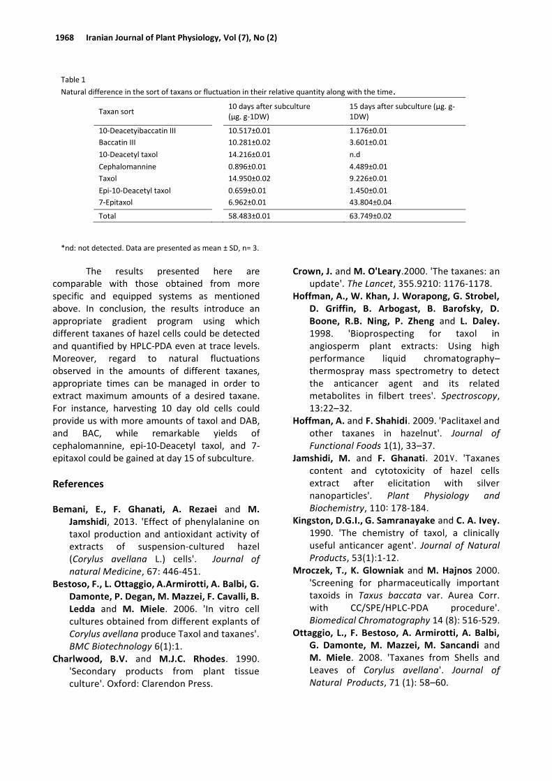
Natural difference in the sort of taxanes or fluctuation in their relative quantity along with the time, are presented in Table 1. The amounts of taxol, 10-deacetylbaccatin III and baccatin III at day 15 were respectively reduced to 62%, 11%, and 35% of their contents at day 10. While 10-Deacetyl taxol significantly reduced along with the time and was not detectable at day 15, other taxanes i.e., cephalomannine, epi-10-deacetyltaxol, and 7-epitaxol showed remarkable increases reaching to 5, 2, and 6 folds of their content at day 10 (Table 1(.
Discussion
The present study optimized a new sensitive method by HPLC-PDA in order to detect and accurately quantify natural taxanes in C. avellana cell suspension culture, even at the trace level. The procedure showed high resolution
detection of taxanes at different sections of hazel cells growth phases. Based on the growth curve, the cells were in their late of logarithmic growth phase between days 10 and 15. In this period, the intensity of production of secondary metabolism is rather prevalent than the primary one, allowing to detect different sorts of taxans along with the time.
Applying HPLC-UV technique, Razaei et al. (2011), Safari et el. (2012), and Bemani et al. (2013) found taxol in the filtered media as well as inside the suspension-cultured hazel cells. Although they confirmed the structure of taxol via LC-MS, due to the limitation in HPLC-UV technique set up, only taxol was detected in their experiment and quantified as 40-80 µgL-1, 10-14 day of subculture.
Relative changes of taxol and baccatin III in hazel cells along with the growth period was also reported by other researches using HPLC-UV technique (Jamshidi and Ghanati, 2017). They showed an increase of 43 to 95 µgL-1 for taxol from day 8 to 14 of subculture but a liner decrease from 38 to 14 µgL-1 for BAC at the same period of the time.
More detailed reports on taxanes of hazel have been provided by Ottaggio et al. (2008). It should be noted however, they eluted the extracts obtained from various parts of the plants collected from different areas on HPLC-MS system. The maximum amounts of taxanes in their report were as follows: paclitaxel (10.83 µg g-1), 10-deacetylbaccatin III (29.08 µgg-1), baccatin III (108.43 µg g-1), 10-deacetyl-7-xylosylcephalomannine and 10-deacetyl-7-xylosylpaclitaxel (206.26 µgg-1), taxinine M, 10-deacetyl-7-xylosylpaclitaxel C, 10-deacetylpaclitaxel, 7-xylosylpaclitaxel (36.63 µg g-
1), cephalomannine and 10- deacetyl-7-epipaclitaxel (52.56 µg g-1), paclitaxel C (6, 49 µg g-1),7-epipaclitaxel (1.08 µg g-1).
(a)
(b)
Fig. III. Elution profile of taxanes of hazel cells by HPLC-PDA (a), compared to that of mixed standards (5 µg mL-1) (b).
1968 Iranian Journal of Plant Physiology, Vol (7), No (2)
The results presented here are comparable with those obtained from more specific and equipped systems as mentioned above. In conclusion, the results introduce an appropriate gradient program using which different taxanes of hazel cells could be detected and quantified by HPLC-PDA even at trace levels. Moreover, regard to natural fluctuations observed in the amounts of different taxanes, appropriate times can be managed in order to extract maximum amounts of a desired taxane. For instance, harvesting 10 day old cells could provide us with more amounts of taxol and DAB, and BAC, while remarkable yields of cephalomannine, epi-10-Deacetyl taxol, and 7-epitaxol could be gained at day 15 of subculture.
References Bemani, E., F. Ghanati, A. Rezaei and M.
Jamshidi, 2013. 'Effect of phenylalanine on taxol production and antioxidant activity of extracts of suspension-cultured hazel (Corylus avellana L.) cells'. Journal of natural Medicine, 67: 446-451.
Bestoso, F., L. Ottaggio, A.Armirotti, A. Balbi, G. Damonte, P. Degan, M. Mazzei, F. Cavalli, B. Ledda and M. Miele. 2006. 'In vitro cell cultures obtained from different explants of Corylus avellana produce Taxol and taxanes'. BMC Biotechnology 6(1):1.
Charlwood, B.V. and M.J.C. Rhodes. 1990. 'Secondary products from plant tissue culture'. Oxford: Clarendon Press.
Crown, J. and M. O'Leary.2000. 'The taxanes: an update'. The Lancet, 355.9210: 1176-1178.
Hoffman, A., W. Khan, J. Worapong, G. Strobel, D. Griffin, B. Arbogast, B. Barofsky, D. Boone, R.B. Ning, P. Zheng and L. Daley. 1998. 'Bioprospecting for taxol in angiosperm plant extracts: Using high performance liquid chromatography–thermospray mass spectrometry to detect the anticancer agent and its related metabolites in filbert trees'. Spectroscopy, 13:22–32.
Hoffman, A. and F. Shahidi. 2009. 'Paclitaxel and other taxanes in hazelnut'. Journal of Functional Foods 1(1), 33–37.
Jamshidi, M. and F. Ghanati. 2017. 'Taxanes content and cytotoxicity of hazel cells extract after elicitation with silver nanoparticles'. Plant Physiology and Biochemistry, 110 : 178-184.
Kingston, D.G.I., G. Samranayake and C. A. Ivey. 1990. 'The chemistry of taxol, a clinically useful anticancer agent'. Journal of Natural Products, 53(1):1-12.
Mroczek, T., K. Glowniak and M. Hajnos 2000. 'Screening for pharmaceutically important taxoids in Taxus baccata var. Aurea Corr. with CC/SPE/HPLC-PDA procedure'. Biomedical Chromatography 14 (8): 516-529.
Ottaggio, L., F. Bestoso, A. Armirotti, A. Balbi, G. Damonte, M. Mazzei, M. Sancandi and M. Miele. 2008. 'Taxanes from Shells and Leaves of Corylus avellana'. Journal of Natural Products, 71 (1): 58–60.
Table 1
Natural difference in the sort of taxans or fluctuation in their relative quantity along with the time.
Taxan sort 10 days after subculture
(µg. g-1DW) 15 days after subculture (µg. g-1DW)
10-Deacetyibaccatin III 10.517±0.01 1.176±0.01
Baccatin III 10.281±0.02 3.601±0.01
10-Deacetyl taxol 14.216±0.01 n.d
Cephalomannine 0.896±0.01 4.489±0.01
Taxol 14.950±0.02 9.226±0.01
Epi-10-Deacetyl taxol 0.659±0.01 1.450±0.01
7-Epitaxol 6.962±0.01 43.804±0.04
Total 58.483±0.01 63.749±0.02
*nd: not detected. Data are presented as mean ± SD, n= 3.

Novel trace taxans in hazel cells detected by PDA-HPLC 1969
Rezaei, A., F. Ghanati, M. Behmanesh and M. Mokhtari-Dizaji 2011. 'Ultrasound-potentiated salicylic acid–induced physiological effects and production of taxol in hazelnut (Corylus avellana L.) cell culture'. Ultrasound in medicine & biology, 37(11): 1938-1947.
Safari, M., F. Ghanati, A. Hajnoruzi, A Rezaei, P. Abdolmaleki and M. Mokhtari-Dizaji. 2012. 'Maintenance of membrane integrity and increase of taxanes production in hazel (Corylus avellana L.) cells induced by low-intensity ultrasound'. Biotechnology letters, 34 (6): 1137-1141.
Wang, Y.F., Q.W. Shi, M. Dong, H. Kiyota, Y.C. Gu and B. Cong. 2011. 'Natural taxanes: developments since 1828'. Chemical reviews, 111(12):7652–709.
Wu, J. and L. Lin. 2003. 'Enhancement of paclitaxel production and release in Taxus chinensis cell cultures by ultrasound, methyl jasmonate and in situ solvent extraction'. Applied Microbiology and Biotechnology, 62: 151-155.