finding a needle in a haystack: the diagnosis of a rectal ... · ge port j gastroenterol....
TRANSCRIPT
GE Port J Gastroenterol. 2015;22(5):221---225
www.elsevier.pt/ge
CLINICAL CASE
Finding a Needle in a Haystack: The Diagnosis of aRectal Neuroendocrine Tumor by Transrectal ProstateBiopsy
Rosa Coelhoa,∗, Susana Rodriguesa, Roberto Silvab, Helena Baldaiab, Guilherme Macedoa
a Gastroenterology Department, Centro Hospitalar São João, Porto, Portugalb Pathology Department, Centro Hospitalar São João, Porto, Portugal
Received 20 February 2015; accepted 19 May 2015Available online 4 July 2015
KEYWORDSBiopsy;Carcinoma,Neuroendocrine;Prostate/pathology;Rectal Neoplasms
AbstractIntroduction: Prostate biopsy, usually performed by a transrectal approach, is executed whenthere is a suspicion of prostate cancer. Neuroendocrine tumors (NETs) are epithelial neoplasmswith predominant neuroendocrine differentiation and only 19% of them are localized in therectum.Case report: The authors describe a 73-year-old man without a significant past medical historythat underwent a prostate biopsy because of urinary complaints and elevated serum levelsof prostate specific antigen. The histology revealed a well-differentiated NET characterizedas a low-grade tumor (G1). A total colonoscopy revealed a 5 mm sessile rectal polyp and inthe splenic flexure a sessile lesion with central ulceration with 5 cm with histological featurescompatible with an adenocarcinoma.Conclusion: This is the first case reported in the literature of a rectal NET diagnosed by tran-srectal prostate biopsy. This case is particularly unique because the diagnosis of the NET leadto the subsequent timely detection of a colonic adenocarcinoma.© 2015 Sociedade Portuguesa de Gastrenterologia. Published by ElsevierEspaña, S.L.U. This is an open access article under the CC BY-NC-ND license(http://creativecommons.org/licenses/by-nc-nd/4.0/).
PALAVRAS-CHAVEBiopsia;CarcinomaNeuroendócrino;Neoplasias do Reto;Próstata/patologia
Tumor Neuroendócrino do Reto Diagnosticado por Biopsia Prostática
ResumoIntroducão: A biópsia prostática transretal é realizada na suspeita de cancro da próstata.Os tumores neuroendócrinos (TNE) são neoplasias epiteliais com diferenciacão predominanteneuroendócrina e em 19% dos casos localizam-se no reto.
Abbreviations: NET, neuroendocrine tumor; PSA, prostate specific antigen; TNM, tumor node metastases.∗ Corresponding author.
E-mail address: [email protected] (R. Coelho).
http://dx.doi.org/10.1016/j.jpge.2015.05.0052341-4545/© 2015 Sociedade Portuguesa de Gastrenterologia. Published by Elsevier España, S.L.U. This is an open access article under theCC BY-NC-ND license (http://creativecommons.org/licenses/by-nc-nd/4.0/).

222 R. Coelho et al.
Caso: Os autores descrevem o caso de um homem, 73 anos de idade, sem antecedentes médicosprévios, que por elevacão dos níveis séricos de antigénio específico prostático realizou biópsiaprostática transretal. A histologia revelou TNE bem diferenciado de baixo grau (G1). Foi real-izada posteriormente colonoscopia total onde se observou pólipo séssil de 5 mm no reto distal.No ângulo esplénico observou-se ainda um lesão séssil de 5 cm com ulceracão central cujasbiopsias foram compatíveis com o diagnóstico de adenocarcinoma.Conclusão: Este é o primeiro caso relatado na literatura de um TNE retal diagnosticado por bióp-sia prostática transretal. Este caso é peculiar dado que o diagnóstico do TNE do reto permitiua detecão de um adenocarcinoma do cólon num estadio inicial.© 2015 Sociedade Portuguesa de Gastrenterologia. Publicado por ElsevierEspaña, S.L.U. Este é um artigo Open Access sob a licença de CC BY-NC-ND(http://creativecommons.org/licenses/by-nc-nd/4.0/).
1. Introduction
Prostate cancer is the leading cancer site in males after lungcancer,1 however screening the general population remainsa controversial issue, since improved patient outcomes havenot been demonstrated.2---3 Prostate biopsy, typically per-formed by an urologist, is a minimally invasive procedurein which tissue samples are obtained through two differ-ent anatomic approaches: transrectal (the most common)and transperineal.4 Patients are referred to prostate biopsyin the presence of abnormal digital rectal exam or whenrepeated abnormal prostate specific antigen (PSA) valuesare found.5
Neuroendocrine tumors (NETs) are epithelial neoplasmswith predominant neuroendocrine differentiation.6 Thereare two major categories: well and poorly differentiatedgastrointestinal NETs. NETs of the digestive system are rel-atively rare and 19% of them are localized in the rectum.11
Despite the low frequency of colorectal NETs, they arefrequently associated with synchronous or metachronousother tumors, with an annual incidence reported of3---15%.7,8
The authors describe a case report where a polypoid rec-tum NET with 5 mm of major diameter was diagnosed byprostate transrectal biopsy. The patient was submitted to atotal colonoscopy that revealed not only the rectal lesion,that was removed with a snare, but also a colonic adenocar-cinoma in the splenic flexure. This is the first case reportedin the literature of a rectal NET diagnosed by transrectalprostate biopsy.
2. Case presentation
We describe the case of a 73-year-old man that com-plained of nocturia and pollakiuria and had high PSA level.A transrectal prostate biopsy was performed and revealed awell-differentiated NET characterized as a low-grade tumor(G1).
Total colonoscopy showed, in the distal rectum, a poly-poid, yellowish, well-circumscribed lesion, measuring 5 mm(Fig. 1) removed with a diathermic snare. Biopsies weretaken from a 5-cm ulcerated lesion observed in the splenic
Figure 1 Colonoscopic view, in retroflexed maneuver,showing in the lower rectum a polypoid, yellowish, well-circumscribed lesion, measuring 5 mm compatible with a rectalNET.
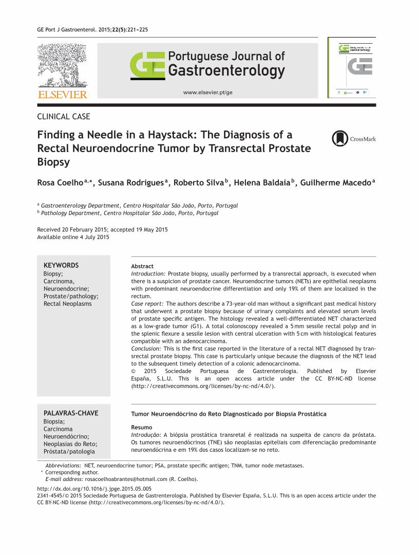
flexure occupying half of the circumference of the lumen(Fig. 2).
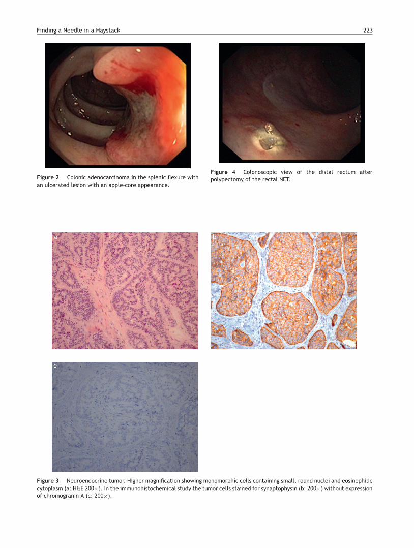
Histologically, the rectal lesion was a tumor centered insubmucosa and focally in the mucosa with a trabecular andacinar pattern composed by small and monomorphic cellswith round and hyperchromatic nuclei and eosinophilic cyto-plasm (Fig. 3a). There was no necrosis or lymphovascularinvasion. The immunohistochemical study revealed strongand diffuse staining of the tumor cells for synaptophysinwithout expression of chromogranin A (Fig. 3b and c). Mitoticindex was less than 1 mitosis per 10 high-power field. Theproliferative index by Ki-67 was less than 1%. With these his-tological and immunohistochemical features the diagnosis ofa well-differentiated of low-grade NET was made.
The rectal NET was completely excised with a diather-mic snare without complications (Fig. 4). Colonic biopsiesfrom the splenic flexure lesion showed tubulovillous ade-noma with high-grade dysplasia and foci of intramucosaladenocarcinoma (Fig. 5).
Finding a Needle in a Haystack 223
Figure 2 Colonic adenocarcinoma in the splenic flexure withan ulcerated lesion with an apple-core appearance.
Figure 4 Colonoscopic view of the distal rectum afterpolypectomy of the rectal NET.
Figure 3 Neuroendocrine tumor. Higher magnification showing monomorphic cells containing small, round nuclei and eosinophiliccytoplasm (a: H&E 200×). In the immunohistochemical study the tumor cells stained for synaptophysin (b: 200×) without expressionof chromogranin A (c: 200×).

224 R. Coelho et al.
Figure 5 High-grade dysplasia and foci of adenocarcinoma inthe splenic flexure (H&E 200×).
Thoracic, abdominal and pelvic computed tomogra-phy was performed and revealed no metastases norlymphadenopathies. Laparoscopic left hemicolectomy wassuccessfully performed and the colonic adenocarcinoma wasstaged as pT2N0M0.
3. Discussion
Prostate cancer is the second most frequent cancer in men,however screening the general population remains a con-troversial issue.1,2 Prostate biopsy is an invasive proceduremade usually through transrectal approach.3 We describea patient with a histological diagnosis of NET (G1) after aprostate biopsy. The diagnosis was confirmed performing acolonoscopy that revealed in the distal rectum a polypoid,yellowish, well-circumscribed lesion measuring 5 mm. Thecolonoscopy performed showed an ulcerated lesion in thesplenic flexure compatible with colonic adenocarcinoma.The authors describe a case of a successful diagnosis of a rec-tum NET made by prostate transrectal biopsy that allowedan adenocarcinoma colonic diagnosis.
The NET, traditionally referred as carcinoid, often has theappearance of well circumscribed round lesions in the sub-mucosa or extending to the muscular layer. As shown inFig. 3, the cut surface sometimes has a yellowish colorreflecting the high lipid content of these lesions. Thecolonoscopy performed in order to confirm the histologyof prostate biopsy revealed another neoplasm --- a colonicadenocarcinoma of the splenic flexure. In fact, as manyas 29% of gastrointestinal carcinoids exhibit a synchronousor metachronous association with other tumors, usuallyadenocarcinomas of the colon.9
Our patient performed thoracic and abdominopelviccomputed tomography that excluded the presence ofmetastasis. A somatostatin receptor scintigraphy was alsoperformed and did not reveal significant changes. Imagingby positron-emission tomography is not routinely recom-mended for NETs because most of these tumors are welldifferentiated and have a low metabolic rate.10
According to the TNM staging system of the AmericanJoint Committee on Cancer/Union for International CancerControl the tumor was classified as T2N0M0 (tumor invadesmuscularis propria without regional lymph node metastasisand no distant metastasis).11 The surgical resection is theprimary treatment modality for these patients with local-ized colorectal cancer. A laparoscopic left hemicolectomywas performed without complications.
Taking into consideration this case we think that patientscould be submitted to screening colonoscopy or flexible sig-moscopy before performing prostate biopsy. In fact, even ifit is low, the risk of tumor dissemination during the prostatebiopsy in the presence of rectal tumors must be considered.
4. Conclusion
To the best of our knowledge, in the literature this is thefirst rectum NET diagnosed by prostate transrectal biopsy.Although prostate biopsy is a relatively frequent procedure,this is the first case of a NET diagnosed in this manner. Thiscase is particularly unique because the diagnosis of the NETlead to the subsequent timely detection of a colonic adeno-carcinoma.
Ethical disclosures
Protection of human and animal subjects. The authorsdeclare that no experiments were performed on humans oranimals for this study.
Confidentiality of data. The authors declare that no patientdata appear in this article.
Right to privacy and informed consent. The authorsdeclare that no patient data appear in this article.
Conflicts of interest
The authors have no conflicts of interest to declare.
References
1. Siegel R, Ma J, Zou Z, Jemal A. Cancer statistics, 2014. CACancer J Clin. 2014;64:9---29.
2. Schröder FH, Hugosson J, Roobol MJ, Tammela TL, Ciatto S,Nelen V, et al. Screening and prostate-cancer mortality in arandomized European study. N Engl J Med. 2009;360:1320---8.
3. Andriole GL, Crawford ED, Grubb RL 3rd, Buys SS, Chia D, ChurchTR, et al. Mortality results from a randomized prostate-cancerscreening trial. N Engl J Med. 2009;360:1310---9.
4. Ismail MT, Gomella LG. Transrectal prostate biopsy. Urol Clin NAm. 2013;40:457---72.
5. Greene KL, Albertsen PC, Babaian RJ, Carter HB, Gann PH, HanM, et al. Prostate specific antigen best practice statement: 2009update. J Urol. 2013;189:S2---11.
6. Lawrence B, Gustafsson BI, Chan A, Svejda B, Kidd M, Modlin IM.The epidemiology of gastroenteropancreatic neuroendocrinetumors. Endocrinol Metab Clin N Am. 2011;40:1---18.
7. Moore JR, Greenwell B, Nuckolls K, Schammel D, Schisler N,Schammel C, et al. Neuroendocrine tumors of the rectum: a10-year review of management. Am Surg. 2011;77:198---200.

Finding a Needle in a Haystack 225
8. Ballantyne GH, Savoca PE, Flannery JT, Ahlman MH, Modlin IM.Incidence and mortality of carcinoids of the colon: data fromthe Connecticut Tumor Registry. Cancer. 1992;69:2400---5.
9. Modlin IM, Lye KD, Kidd M. A 5-decade analysis of 13,715 carci-noid tumors. Cancer. 2003;15:934---59.
10. Kocha W, Maroun J, Kennecke H, Law C, Metrakos P, OuelletJF, et al. Consensus recommendations for the diagnosis and
management of well-differentiated gastroenterohepatic neu-roendocrine tumours: a revised statement from a CanadianNational Expert Group. Curr Oncol. 2010;17:49---64.
11. AJCC American Joint Committee on Cancer. In: Edge SB, ByrdDR, Compton CC, editors. Cancer staging manual. 7th ed. NewYork: Springer; 2010. p. 143.