
ORIGINAL ARTICLE
Tunable nano-replication to explore the omniphobiccharacteristics of springtail skin
Rene Hensel1, Ralf Helbig1, Sebastian Aland2, Axel Voigt2, Christoph Neinhuis3,4 and Carsten Werner1,4
Springtails (Collembola) are wingless arthropods adapted to cutaneous respiration in temporarily rain-flooded and microbially
contaminated habitats by a non-wetting and antiadhesive skin surface that is mechanically rather stable. Recapitulating the
robust and effectively repellent surface characteristics of springtail skin in engineered materials may offer exciting opportunities
for demanding applications, but it requires a detailed understanding of the underlying design principles. Towards this aim and
based on our recent analysis of the structural features of springtail skin, we developed a tunable polymer replication process to
dissect the contributions of different structural elements and surface chemistry to the omniphobic performance of the cuticle.
The Cassie–Wenzel transition at elevated pressures was explored by in situ plastron collapse experiments and by numerical FEM
simulations. The results obtained unravel the decisive role of nanoscopic cuticle structures for the protection of springtails
against wetting, and explain how the evolved nanotopography enables the production of omniphobic surfaces even from
intrinsically hydrophilic polymer materials.
NPG Asia Materials (2013) 5, e37; doi:10.1038/am.2012.66; published online 1 February 2013
Keywords: Collembola; cuticle; omniphobic; replication
INTRODUCTION
Springtails (Collembola), wingless arthropods are probably the mostabundant hexapods on Earth1 and an integral component of the soilcommunity2 with more than 8000 species.3 Differing from insects,most springtail species respire through the skin.4,5 Consequently, theirsurvival is affected by transpiration, that is critically dependent onenvironmental humidity.6,7 In turn, complete wetting of their skinblocks gas exchange and results in suffocation.
The consequent repellence of aqueous media evolved by the spring-tail skin has been known for more than half a century.6,8,9 However, theremarkable wetting resistance of the skin surfaces even against wettingby low-surface-tension liquids such as alkanes or ethyl alcohol andagainst bacterial adhesion were only recently quantitatively described.10
These findings reflect the adaptation to habitats where water ismassively loaded with surface-active substances originating fromdecaying organic matter and from microorganisms.
A most striking feature of springtail skin is the hierarchicallyarranged and highly textured surface.9–11 Surface roughness has beenextensively studied in general and shown to affect the macroscopicsurface wetting properties significantly: In the Cassie state, liquids canbe sustained at the top of the asperities and air (designated asplastron) is trapped inside the grooves of the rough surfaceunderneath the liquid, resulting in a minimal solid–liquid contactarea.12,13 In the Wenzel state, liquids can penetrate the grooves and
wet the entire rough surface characterized by a maximal solid–liquidcontact area.14 The metastable Cassie state can be transferred to theWenzel state by means of supplied energy15,16 and geometricparameters of rough surfaces have been discussed to influence theenergy barrier that must be overcome for complete wetting.17
Analysing the resulting wetting behaviour of rough surfaces isclearly more demanding if the surface roughness is hierarchicallyassembled with at least two sets of rough structures,18–20 as iscommon for many naturally occurring water-repellent surfaces.21–24
Therefore, the identification and mechanistic understanding ofomniphobic and antiadhesive surface patterns evolved by livingorganisms still defines a challenge, where springtail skin represents aparticularly interesting example.
Towards this aim, we have now adapted a nanoimprint lithographyscheme as this recently developed methodology allows for the faithfulreplication of delicate morphologies with a few tens of nanometersresolution in polymeric materials of defined bulk and surfacechemistry.25,26 When applying elastomeric moulds, the approacheven permits the transfer of high aspect ratios, large overhangs27
and even closed loops.28 Successful replication of various differenthierarchically assembled biological templates such as plant leaves,29
insect corneas30,31 or grasshopper wings32 was recently demonstrated.Using this approach, we explored the role of distinct skin features
in creating the unique omniphobic characteristics of springtail cuticle.
1Max Bergmann Center of Biomaterials, Leibniz Institute of Polymer Research Dresden, Dresden, Germany; 2Institute of Scientific Computation and Applied Mathematics,Technische Universitat Dresden, Dresden, Germany; 3Institute of Botany, Technische Universitat Dresden, Dresden, Germany and 4B CUBE Innovation Center for MolecularBioengineering, Technische Universitat Dresden, Dresden, GermanyCorrespondence: Professor C Werner, Max Bergmann Center of Biomaterials, Leibniz Institute of Polymer Research Dresden, Dresden 01069, Germany.E-mail: [email protected]
Received 24 August 2012; revised 7 October 2012; accepted 2 November 2012
NPG Asia Materials (2013) 5, e37; doi:10.1038/am.2012.66& 2013 Nature Japan K.K. All rights reserved 1884-4057/13
www.nature.com/am

A tunable nanoimprint replication process was developed to translatethe skin structure of Tetrodontophora bielanensis (European giantspringtail) into polymer replicas with defined variations in nano-scopic surface morphology and surface chemistry. We performedstatic contact angle measurements and determined the in situ wettingresistance at elevated pressures. In addition, the dynamics of theCassie–Wenzel transition at the unravelled nanotopographies wereinvestigated in detail by numerical FEM simulation and the resultsobtained were compared with experimental data.
With this approach, we were able to decipher the protectionmechanism of springtail skin against wetting, and quantitativelydetermined the structural and chemical requirements for employingthis design concept in the development of artificial structures.
MATERIALS AND METHODS
AnimalsTetrodontophora bielanensis springtails were collected in the wooded mountains
of Saxony near Dresden, southeastern Germany. The animals were kept as
laboratory colonies in large petridishes using soil, litter, decaying wood and
moss from their original habitat as food source and substrate.
Replication process
Mould preparation. Individual animals were freshly prepared by freezing at
�20 1C for 30 min immediately before mould preparation was started. The
animal was ventrally dipped into Fluorolink MD700 (MW¼ 1500 g mol�1,
Solvay Solexis, Bollate, Italy) precursor solution containing 0.5wt% Irgacure
651 (CIBA, Basel, Switzerland) and mechanically fixed by exposing to UV-
irradiation 2 min by DELOLUX 04 (DELO, Windach, Germany) at each side.
Next, Fomblin MD40 (MW¼ 4000 g mol�1, Solvay Solexis) precursor solution
containing 0.5wt% Irgacure 651 was applied dorsally onto the animal skin and
exposed to vacuum of B1 hPa for 3 h to ensure infiltration of the entire skin
by the precursor solution. Fomblin MD40 was crosslinked by exposing to UV-
irradiation for 5 min under nitrogen atmosphere. Subsequently, the cured
elastomeric MD40 mould was gently peeled from the springtail skin.
Route A: Fabrication of faithful PEGda replicas. A PEGda (Mn¼ 700 g mol�1,
Sigma Aldrich, Deisenhofen, Germany) precursor solution containing 0.5wt%
Irgacure 651 was cast onto the prepatterned MD40 mould and exposed to
vacuum of 3� 10�3 hPa for 12 h. PEGda was crosslinked by exposing to UV-
irradiation for 5 min under nitrogen atmosphere. Subsequently, the PEGda
polymer replica was gently peeled from the MD40 mould.
Route B: Fabrication of PEGda replicas without primary granules. A high-
viscous PDMS Sylgard 184 (Dow Corning, Wiesbaden, Germany) precursor
solution was freshly mixed with the curing reagent in the ratio 10:1, cast onto
the prepatterned MD40 mould and exposed to vacuum of 1 hPa for 12 h. The
residuary PDMS crosslinking took place at room temperature over two more
days. The elastomeric PDMS replica was gently peeled from the MD40 mould
and used as template for the preparation of a further MD40 mould that, in
turn, was used to make PEGda polymer replicas using the same procedure as
described above in route A.
Surface treatmentFor Teflon-AF-coating, the PEGda polymer replicas were dipped (1 min) into a
solution of 0.6wt% of amorphous fluoropolymer Teflon (Teflon AF, DuPont,
Wilmington, DE, USA) diluted in a fully fluorinated solvent FC-77 (3M,
Haven, Belgium). After withdrawing, the replicas were dried (10 min) on a hot
plate at 60 1C.
Contact angle measurementsStatic contact angle measurements were performed using the contact angle
system OCA 30 (DataPhysics Instruments, Filderstadt, Germany). Droplets
(B2ml) of Milli-Q filtered water (Merck Millipore, Billerica, MA, USA) and
hexadecane (Sigma-Aldrich) were dorsally applied at the skin of T. bielanensis
and its polymer replicas. Dynamic contact angle measurements of the
Collembola skin and its polymer replicas were impossible due to the restricted
sample area. Smooth reference polymer films were used for the determination
of the intrinsic contact angles using dynamic contact angle measurements. The
static contact angle was estimated as Ystat¼ arccos cosYadv þ cosYrec
2
� �where Ystat
is the static contact angle, Yadv is the advancing contact angle, and is Yrec the
receding contact angle.
In situ plastron collapse testsPlastron collapse experiments were performed using a self-made setup that
consisted of an optical microscope, a water pump, a processing unit and a
water-flooded chamber where the samples were previously placed (cf.
Figure 3a). The liquid reservoir was linearly compressed 50 hPa s�1 by
increasing hydrostatic pressure and images were simultaneously recorded
using an optical microscope unit. To minimize the influence of the gas
solubility in water, the experiments were carried out at a water-air ratio of 10:1.
SEM and TEM imagingScanning electron microscopy studies were performed using a Gemini DSM
982 (Carl Zeiss SMT, Oberkochen, Germany). The animals were prepared by
freezing and subsequent air-drying without any fixation. All samples (animals,
moulds and replicas) were coated with 3 nm platinum (BAL-TEC SCD 500,
BalTec, Pfaffikon, Switzerland) to eliminate surface charging effects. Transmis-
sion electron microscopy studies were carried out using an EM 912 Omega
(Carl Zeiss SMT). The samples were fixed, stained and subsequently sliced into
ultrathin sections as described in Helbig, R. et al.10
RESULTS
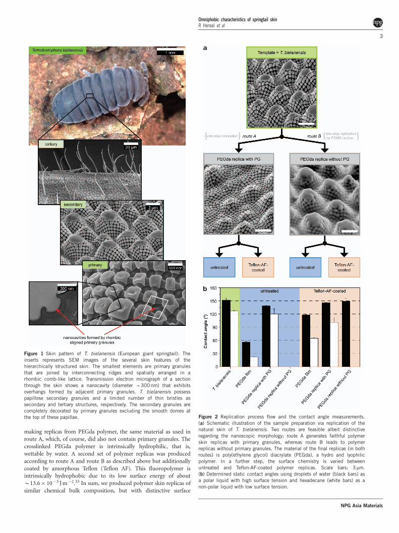
The hierarchical structure of the skin of T. bielanensis is depicted inFigure 1. At the nanoscopic level, primary granules are joined byinterconnecting ridges covering the entire body of the springtail in arhombic comb-like lattice. They form nanoscopic cavities with amean diameter of about 300 nm. The primary granules protrudeabove the ridges so that, in sectional view, the nanocavities exhibitoverhangs. The skin at the bottom of these cavities has to be highlypermeable to enable skin respiration.9 At the microscopic scale,T. bielanensis possess papillose secondary granules and a limitednumber of thin bristles as a tertiary structure. The secondary granulesare completely decorated by primary granules excluding the smoothdomes at the top of these papillae.
In order to resolve the impact of the individual structure elementson the characteristics of the springtail skin, we developed an adaptivereplication process that is depicted schematically in Figure 2a (formore SEM images of each replication step, see the SupplementarySection 1 and Supplementary Figure S1). Starting from the biologicaltemplate, T. bielanensis, the whole skin features were firstly capturedby an elastomeric perfluoropolyether dimethacrylate (PFPEdma)mould. The mould material has a high fluorine content B58wt%that inhibited chemical crossreactions with the biological templatedue to its chemical inertness.31 The PFPEdma mould as the negativereplica of the natural skin was then the starting point for thefabrication of polymer skin replicas by two different routes. Inroute A, poly(ethylene glycol) diacrylate (PEGda) was used as aliquid prepolymer. After crosslinking the PEGda and subsequentdemoulding, the polymer replicas possessed the entire superficialgranular surface structure of T. bielanensis but did not contain anybristles (see Supplementary Figure S2). In route B, polydimethylsilox-ane (PDMS) was used as the liquid prepolymer, with a significantlyhigher viscosity. The penetration of the more viscous prepolymer intothe smallest pores of the mould was nearly inhibited (for more details,see Supplementary Section 2). Thus, the replicas of route B did notcontain the nanoscopic primary granules and nanocavities. Further-more, the generated PDMS replicas were used as templates for
Omniphobic characteristics of springtail skinR Hensel et al
2
NPG Asia Materials
making replicas from PEGda polymer, the same material as used inroute A, which, of course, did also not contain primary granules. Thecrosslinked PEGda polymer is intrinsically hydrophilic, that is,wettable by water. A second set of polymer replicas was producedaccording to route A and route B as described above but additionallycoated by amorphous Teflon (Teflon AF). This fluoropolymer isintrinsically hydrophobic due to its low surface energy of aboutB13.6� 10�3 J m�2.33 In sum, we produced polymer skin replicas ofsimilar chemical bulk composition, but with distinctive surface
Figure 1 Skin pattern of T. bielanensis (European giant springtail). The
inserts represents SEM images of the several skin features of the
hierarchically structured skin. The smallest elements are primary granules
that are joined by interconnecting ridges and spatially arranged in a
rhombic comb-like lattice. Transmission electron micrograph of a section
through the skin shows a nanocavity (diameter B300nm) that exhibits
overhangs formed by adjacent primary granules. T. bielanensis possess
papillose secondary granules and a limited number of thin bristles as
secondary and tertiary structures, respectively. The secondary granules are
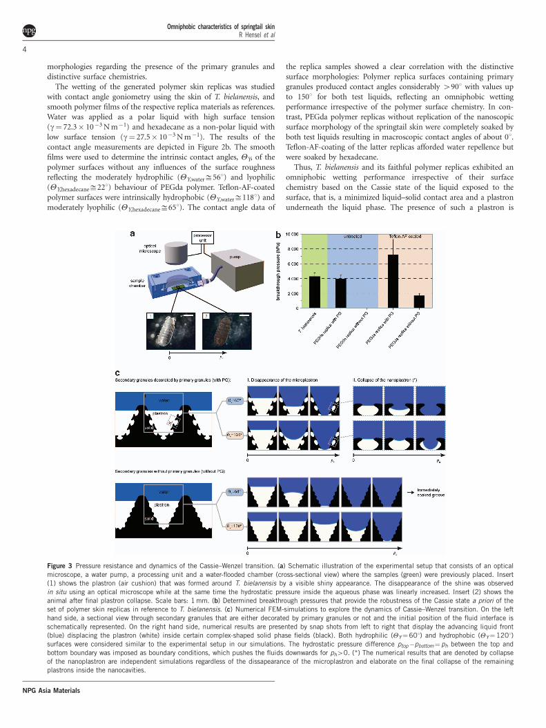
completely decorated by primary granules excluding the smooth domes atthe top of these papillae. Figure 2 Replication process flow and the contact angle measurements.
(a) Schematic illustration of the sample preparation via replication of the
natural skin of T. bielanensis. Two routes are feasible albeit distinctive
regarding the nanoscopic morphology; route A generates faithful polymer
skin replicas with primary granules, whereas route B leads to polymer
replicas without primary granules. The material of the final replicas (in both
routes) is poly(ethylene glycol) diacrylate (PEGda), a hydro and lyophilic
polymer. In a further step, the surface chemistry is varied between
untreated and Teflon-AF-coated polymer replicas. Scale bars: 3mm.
(b) Determined static contact angles using droplets of water (black bars) as
a polar liquid with high surface tension and hexadecane (white bars) as a
non-polar liquid with low surface tension.
Omniphobic characteristics of springtail skinR Hensel et al
3
NPG Asia Materials
morphologies regarding the presence of the primary granules anddistinctive surface chemistries.
The wetting of the generated polymer skin replicas was studiedwith contact angle goniometry using the skin of T. bielanensis, andsmooth polymer films of the respective replica materials as references.Water was applied as a polar liquid with high surface tension(g¼ 72.3� 10�3 N m�1) and hexadecane as a non-polar liquid withlow surface tension (g¼ 27.5� 10�3 N m�1). The results of thecontact angle measurements are depicted in Figure 2b. The smoothfilms were used to determine the intrinsic contact angles, YY, of thepolymer surfaces without any influences of the surface roughnessreflecting the moderately hydrophilic (YY,waterD561) and lyophilic(YY,hexadecaneD221) behaviour of PEGda polymer. Teflon-AF-coatedpolymer surfaces were intrinsically hydrophobic (YY,waterD1181) andmoderately lyophilic (YY,hexadecaneD651). The contact angle data of
the replica samples showed a clear correlation with the distinctivesurface morphologies: Polymer replica surfaces containing primarygranules produced contact angles considerably 4901 with values upto 1501 for both test liquids, reflecting an omniphobic wettingperformance irrespective of the polymer surface chemistry. In con-trast, PEGda polymer replicas without replication of the nanoscopicsurface morphology of the springtail skin were completely soaked byboth test liquids resulting in macroscopic contact angles of about 01.Teflon-AF-coating of the latter replicas afforded water repellence butwere soaked by hexadecane.
Thus, T. bielanensis and its faithful polymer replicas exhibited anomniphobic wetting performance irrespective of their surfacechemistry based on the Cassie state of the liquid exposed to thesurface, that is, a minimized liquid–solid contact area and a plastronunderneath the liquid phase. The presence of such a plastron is
Figure 3 Pressure resistance and dynamics of the Cassie–Wenzel transition. (a) Schematic illustration of the experimental setup that consists of an optical
microscope, a water pump, a processing unit and a water-flooded chamber (cross-sectional view) where the samples (green) were previously placed. Insert
(1) shows the plastron (air cushion) that was formed around T. bielanensis by a visible shiny appearance. The disappearance of the shine was observed
in situ using an optical microscope while at the same time the hydrostatic pressure inside the aqueous phase was linearly increased. Insert (2) shows the
animal after final plastron collapse. Scale bars: 1 mm. (b) Determined breakthrough pressures that provide the robustness of the Cassie state a priori of the
set of polymer skin replicas in reference to T. bielanensis. (c) Numerical FEM-simulations to explore the dynamics of Cassie–Wenzel transition. On the left
hand side, a sectional view through secondary granules that are either decorated by primary granules or not and the initial position of the fluid interface is
schematically represented. On the right hand side, numerical results are presented by snap shots from left to right that display the advancing liquid front
(blue) displacing the plastron (white) inside certain complex-shaped solid phase fields (black). Both hydrophilic (YY¼601) and hydrophobic (YY¼1201)
surfaces were considered similar to the experimental setup in our simulations. The hydrostatic pressure difference ptop�pbottom¼ ph between the top and
bottom boundary was imposed as boundary conditions, which pushes the fluids downwards for ph40. (*) The numerical results that are denoted by collapse
of the nanoplastron are independent simulations regardless of the dissapearance of the microplastron and elaborate on the final collapse of the remaining
plastrons inside the nanocavities.
Omniphobic characteristics of springtail skinR Hensel et al
4
NPG Asia Materials
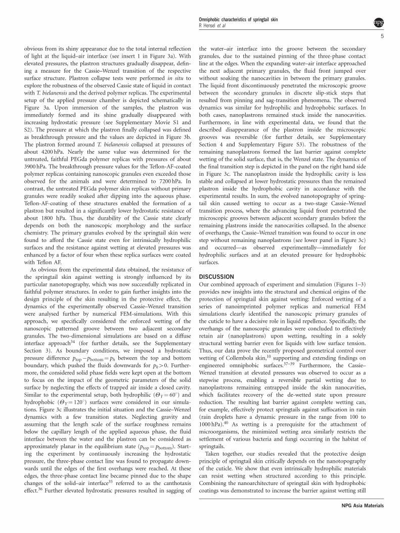
obvious from its shiny appearance due to the total internal reflectionof light at the liquid–air interface (see insert 1 in Figure 3a). Withelevated pressures, the plastron structures gradually disappear, defin-ing a measure for the Cassie–Wenzel transition of the respectivesurface structure. Plastron collapse tests were performed in situ toexplore the robustness of the observed Cassie state of liquid in contactwith T. bielanensis and the derived polymer replicas. The experimentalsetup of the applied pressure chamber is depicted schematically inFigure 3a. Upon immersion of the samples, the plastron wasimmediately formed and its shine gradually disappeared withincreasing hydrostatic pressure (see Supplementary Movie S1 andS2). The pressure at which the plastron finally collapsed was definedas breakthrough pressure and the values are depicted in Figure 3b.The plastron formed around T. bielanensis collapsed at pressures ofabout 4200 hPa. Nearly the same value was determined for theuntreated, faithful PEGda polymer replicas with pressures of about3900 hPa. The breakthrough pressure values for the Teflon-AF-coatedpolymer replicas containing nanoscopic granules even exceeded thoseobserved for the animals and were determined to 7200 hPa. Incontrast, the untreated PEGda polymer skin replicas without primarygranules were readily soaked after dipping into the aqueous phase.Teflon-AF-coating of these structures enabled the formation of aplastron but resulted in a significantly lower hydrostatic resistance ofabout 1800 hPa. Thus, the durability of the Cassie state clearlydepends on both the nanoscopic morphology and the surfacechemistry. The primary granules evolved by the springtail skin werefound to afford the Cassie state even for intrinsically hydrophilicsurfaces and the resistance against wetting at elevated pressures wasenhanced by a factor of four when these replica surfaces were coatedwith Teflon AF.
As obvious from the experimental data obtained, the resistance ofthe springtail skin against wetting is strongly influenced by itsparticular nanotopography, which was now successfully replicated infaithful polymer structures. In order to gain further insights into thedesign principle of the skin resulting in the protective effect, thedynamics of the experimentally observed Cassie–Wenzel transitionwere analysed further by numerical FEM-simulations. With thisapproach, we specifically considered the enforced wetting of thenanoscopic patterned groove between two adjacent secondarygranules. The two-dimensional simulations are based on a diffuseinterface approach34 (for further details, see the SupplementarySection 3). As boundary conditions, we imposed a hydrostaticpressure difference ptop�pbottom¼ ph between the top and bottomboundary, which pushed the fluids downwards for ph40. Further-more, the considered solid phase fields were kept open at the bottomto focus on the impact of the geometric parameters of the solidsurface by neglecting the effects of trapped air inside a closed cavity.Similar to the experimental setup, both hydrophilic (YY¼ 601) andhydrophobic (YY¼ 1201) surfaces were considered in our simula-tions. Figure 3c illustrates the initial situation and the Cassie–Wenzeldynamics with a few transition states. Neglecting gravity andassuming that the length scale of the surface roughness remainsbelow the capillary length of the applied aqueous phase, the fluidinterface between the water and the plastron can be considered asapproximately planar in the equilibrium state (ptop¼ pbottom). Start-ing the experiment by continuously increasing the hydrostaticpressure, the three-phase contact line was found to propagate down-wards until the edges of the first overhangs were reached. At theseedges, the three-phase contact line became pinned due to the shapechanges of the solid–air interface35 referred to as the canthotaxiseffect.36 Further elevated hydrostatic pressures resulted in sagging of
the water–air interface into the groove between the secondarygranules, due to the sustained pinning of the three-phase contactline at the edges. When the expanding water–air interface approachedthe next adjacent primary granules, the fluid front jumped overwithout soaking the nanocavities in between the primary granules.The liquid front discontinuously penetrated the microscopic groovebetween the secondary granules in discrete slip-stick steps thatresulted from pinning and sag-transition phenomena. The observeddynamics was similar for hydrophilic and hydrophobic surfaces. Inboth cases, nanoplastrons remained stuck inside the nanocavities.Furthermore, in line with experimental data, we found that thedescribed disappearance of the plastron inside the microscopicgrooves was reversible (for further details, see SupplementarySection 4 and Supplementary Figure S3). The robustness of theremaining nanoplastrons formed the last barrier against completewetting of the solid surface, that is, the Wenzel state. The dynamics ofthe final transition step is depicted in the panel on the right hand sidein Figure 3c. The nanoplastron inside the hydrophilic cavity is lessstable and collapsed at lower hydrostatic pressures than the remainedplastron inside the hydrophobic cavity in accordance with theexperimental results. In sum, the evolved nanotopography of spring-tail skin caused wetting to occur as a two-stage Cassie–Wenzeltransition process, where the advancing liquid front penetrated themicroscopic grooves between adjacent secondary granules before theremaining plastrons inside the nanocavities collapsed. In the absenceof overhangs, the Cassie–Wenzel transition was found to occur in onestep without remaining nanoplastrons (see lower panel in Figure 3c)and occurred—as observed experimentally—immediately forhydrophilic surfaces and at an elevated pressure for hydrophobicsurfaces.
DISCUSSION
Our combined approach of experiment and simulation (Figures 1–3)provides new insights into the structural and chemical origins of theprotection of springtail skin against wetting: Enforced wetting of aseries of nanoimprinted polymer replicas and numerical FEMsimulations clearly identified the nanoscopic primary granules ofthe cuticle to have a decisive role in liquid repellence. Specifically, theoverhangs of the nanoscopic granules were concluded to effectivelyretain air (nanoplastrons) upon wetting, resulting in a solelystructural wetting barrier even for liquids with low surface tension.Thus, our data prove the recently proposed geometrical control overwetting of Collembola skin,10 supporting and extending findings onengineered omniphobic surfaces.37–39 Furthermore, the Cassie–Wenzel transition at elevated pressures was observed to occur as astepwise process, enabling a reversible partial wetting due tonanoplastrons remaining entrapped inside the skin nanocavities,which facilitates recovery of the de-wetted state upon pressurereduction. The resulting last barrier against complete wetting can,for example, effectively protect springtails against suffocation in rain(rain droplets have a dynamic pressure in the range from 100 to1000 hPa).40 As wetting is a prerequisite for the attachment ofmicroorganisms, the minimized wetting area similarly restricts thesettlement of various bacteria and fungi occurring in the habitat ofspringtails.
Taken together, our studies revealed that the protective designprinciple of springtail skin critically depends on the nanotopographyof the cuticle. We show that even intrinsically hydrophilic materialscan resist wetting when structured according to this principle.Combining the nanoarchitecture of springtail skin with hydrophobiccoatings was demonstrated to increase the barrier against wetting still
Omniphobic characteristics of springtail skinR Hensel et al
5
NPG Asia Materials
further—beyond the characteristics of the natural template. Thenewly garnered understanding of the protective topography couldpave the way for synthetic materials that effectively and durably resistwetting and biofouling.
CONFLICT OF INTERESTThe authors declare no conflict of interest.
ACKNOWLEDGEMENTSWe are grateful to Julia Nickerl for substantial support concerning the biology
of Collembola, the TEM investigations and collecting the animals. We also
thank Jannik Baumer for collecting animals and assistance in preparing the
polymer replicas. Furthermore, we thank Roland Vogel for measuring the shear
viscosity of the used prepolymers. Hans-Georg Braun and Karina Grundke are
gratefully acknowledged for helpful discussions throughout the work.
1 Hopkin, S. P. Biology of the Springtails (Insecta: Collembola) (Oxford University Press,USA, 1997).
2 Rusek, J. Biodiversity of collembola and their functional role in the ecosystem.Biodivers. Conserv. 7, 1207–1219 (1998).
3 Bellinger, P., Christiansen, K. & Janssens, F. Collembola species catalogue. Checklistof the Collembola of the World http://www.collembola.org. Accessed 12 June 2012(1998–2012).
4 Davies, W. M. On the tracheal system of collembola, with special reference, to that ofsminthurus viridis, lubb. Q. J. Microsc. Sci. 71, 15–30 (1927).
5 Zinkler, D. Vergleichende untersuchungen zur atmungs-physiologie von collembolen(apterygota) und andereen bodenkleinarthropoden. Z. Vergl. Physiol. 52, 99–144(1966).
6 Noble-Nesbitt, J. Transpiration in podura aquatica l. (collembola, isotomidae) andwetting properties of its cuticle. J. Exp. Biol. 40, 681–700 (1963).
7 Joosse, E. N. G. & Groen, J. B. Relationship between saturation deficit and survival andlocomotory activity of surface dwelling collembola. Entomol. Exp. Appl. 13, 229–235(1970).
8 Ghiradella, H. & Radigan, W. Collembolan cuticle - wax layer and anti-wettingproperties. J. Insect. Physiol. 20, 301–306 (1974).
9 King, P. E., Pugh, P. J. A., Fordy, M. R., Love, N. & Wheeler, S. A. A comparison ofsome environmental adaptations of the littoral collembolans anuridella marina (willem)and anurida maritima (guerin). J. Nat. Hist. 24, 673–688 (1990).
10 Helbig, R., Nickerl, J., Neinhuis, C. & Werner, C. Smart skin patterns protectspringtails. PLoS ONE 6, e25105 (2011).
11 Hale, W. G. & Smith, A. L. Scanning electron microscope studies of cuticular structuresin the genus onychiurus (collembola). Rev. Ecol. Biol. Sol. 52, 343–354 (1966).
12 Cassie, A. B. D. & Baxter, S. Wettability of porous surfaces. Trans. Faraday Soc. 40,
546–551 (1944).13 Nakajima, A. Design of hydrophobic surfaces for liquid droplet control. NPG Asia
Mater. 3, 49–56 (2011).14 Wenzel, R. Resistance of solid surfaces to wetting by water. Ind. Eng. Chem. Res. 28,
988–994 (1936).15 Lafuma, A. & Quere, D. Superhydrophobic states. Nat. Mater. 2, 457–460 (2003).16 Quere, D. Wetting and roughness. Annu. Rev. Mater. Res. 38, 71–99 (2008).17 Barbieri, L., Wagner, E. & Hoffmann, P. Water wetting transition parameters of
perfluorinated substrates with periodically distributed flat-top microscale obstacles.Langmuir 23, 1723–1734 (2007).
18 Nosonovsky, M. Multiscale roughness and stability of superhydrophobic biomimeticinterfaces. Langmuir 23, 3157–3161 (2007).
19 Bormashenko, E. Wetting transitions on biomimetic surfaces. Phil. Trans. A Math.Phys. Eng. Sci. 368, 4695–4711 (2010).
20 Verho, T., Korhonen, J. T., Sainiemi, L., Jokinen, V., Bower, C., Franze, K., Franssila, S.,Andrew, P., Ikkala, O. & Ras, R. H. A. Reversible switching between superhydrophobicstates on a hierarchically structured surface. Proc. Natl Acad. Sci. USA 109, 10210–10213 (2012).
21 Neinhuis, C. & Barthlott, W. Characterization and distribution of water-repellent, self-cleaning plant surfaces. Ann. Bot. 79, 667–677 (1997).
22 Gao, X. & Jiang, L. Biophysics: water-repellent legs of water striders. Nature 432, 36(2004).
23 Bush, J. W. M., Hu, D. L. & Prakash, M. The integument of water-walking arthropods:Form and function. Adv. Insect Physiol. 34, 117–192 (2007).
24 Koch, K., Bhushan, B. & Barthlott, W. Diversity of structure, morphology and wetting ofplant surfaces. Soft Matter 4, 1943–1963 (2008).
25 Chou, S. Y., Krauss, P. R. & Renstrom, P. J. Imprint lithography with 25-nanometerresolution. Science 272, 85–87 (1996).
26 Williams, S. S., Retterer, S., Lopez, R., Ruiz, R., Samulski, E. T. & DeSimone, J. M.High-resolution pfpe-based molding techniques for nanofabrication of high-patterndensity, sub-20 nm features: A fundamental materials approach. Nano Lett. 10,
1421–1428 (2010).27 LaFratta, C. N., Baldacchini, T., Farrer, R. A., Fourkas, J. T., Teich, M. C., Saleh, B. E. .
A. & Naughton, M. J. Replication of two-photon-polymerized structures withextremely high aspect ratios and large overhangs. J. Phys. Chem. B 108,
11256–11258 (2004).28 LaFratta, C. N., Li, L. & Fourkas, J. T. Soft-lithographic replication of 3d
microstructures with closed loops. Proc. Natl Acad. Sci. USA 103, 8589–8594(2006).
29 Schulte, A. J., Koch, K., Spaeth, M. & Barthlott, W. Biomimetic replicas: Transfer ofcomplex architectures with different optical properties from plant surfaces ontotechnical materials. Acta Biomater. 5, 1848–1854 (2009).
30 Pulsifer, D. P., Lakhtakia, A., Martin-Palma, R. J. & Pantano, C. G. Mass fabricationtechnique for polymeric replicas of arrays of insect corneas. Bioinspir. Biomim. 5,
036001 (2010).31 Ko, D.-H., Tumbleston, J. R., Henderson, K. J., Euliss, L. E., DeSimone, J. M., Lopez,
R. & Samulski, E. T. Biomimetic microlens array with antireflective "moth-eye" surface.Soft Matter 7, 6404–6407 (2011).
32 Zhang, T., Li, M., Su, B., Ye, C., Li, K., Shen, W., Chen, L., Xue, Z., Wang, S. & Jiang,L. Bio-inspired anisotropic micro/nano-surface from a natural stamp: grasshopperwings. Soft Matter 7, 7973–7975 (2011).
33 Tavana, H., Petong, N., Hennig, A., Grundke, K. & Neumann, A. Contact angles andcoating film thickness. J. Adhes. 81, 29–39 (2005).
34 Aland, S., Lowengrub, J. & Voigt, A. Two-phase flow in complex geometries: a diffusedomain approach. Comput. Model. Eng. Sci. 57, 77–107 (2010).
35 Oliver, J. F., Huh, C. & Mason, S. G. Resistance to spreading of liquids by sharp edges.J. Colloid Interface Sci. 59, 568–581 (1977).
36 Berthier, J., Loe-Mie, F., Tran, V., Schoumacker, S., Mittler, F., Marchand, G. & Sarrut,N. On the pinning of interfaces on micropillar edges. J. Colloid Interface Sci. 338,
296–303 (2009).37 Tuteja, A., Choi, W., Ma, M. L., Mabry, J. M., Mazzella, S. A., Rutledge, G. C.,
McKinley, G. H. & Cohen, R. E. Designing superoleophobic surfaces. Science 318,
1618–1622 (2007).38 Cao, L. L., Hu, H. H. & Gao, D. Design and fabrication of micro-textures for inducing a
superhydrophobic behavior on hydrophilic materials. Langmuir 23, 4310–4314(2007).
39 Tuteja, A., Choi, W., Mabry, J. M., McKinley, G. H. & Cohen, R. E. Robust omniphobicsurfaces. Proc. Natl Acad. Sci. USA 105, 18200–18205 (2008).
40 Zheng, Q. S., Yu, Y. & Zhao, Z. H. Effects of hydraulic pressure on the stabilityand transition of wetting modes of superhydrophobic surfaces. Langmuir 21,
12207–12212 (2005).
This work is licensed under a Creative CommonsAttribution-NonCommercial-NoDerivs 3.0 Unported
License. To view a copy of this license, visit http://creativecommons.org/licenses/by-nc-nd/3.0/
Supplementary Information accompanies the paper on the NPG Asia Materials website (http://www.nature.com/am)
Omniphobic characteristics of springtail skinR Hensel et al
6
NPG Asia Materials


