Download - Screening Tests for the Rapid Detection of Diarrhetic Shellfish Toxins in Washington State
Mar. Drugs 2013, 11, 3718-3734; doi:10.3390/md11103718
marine drugs ISSN 1660-3397
www.mdpi.com/journal/marinedrugs
Article
Screening Tests for the Rapid Detection of Diarrhetic Shellfish
Toxins in Washington State
Bich-Thuy L. Eberhart 1,
*, Leslie K. Moore 1, Neil Harrington
2, Nicolaus G. Adams
1,
Jerry Borchert 3 and Vera L. Trainer
1
1 NOAA, Northwest Fisheries Science Center, Marine Biotoxins Laboratory, 2725 Montlake Blvd. E.,
Seattle, WA 98112, USA; E-Mails: [email protected] (L.K.M.);
[email protected] (N.G.A.); [email protected] (V.L.T.) 2 Jamestown S’Klallam Tribe, 1033 Old Blyn Highway, Sequim, WA 98392, USA;
E-Mail: [email protected] 3 Food Safety and Shellfish Program, Washington State Department of Health, 7171 Clearwater
Lane, Olympia, WA 98504, USA; E-Mail: [email protected]
* Author to whom correspondence should be addressed; E-Mail: [email protected];
Tel.: +1-206-860-3324; Fax: +1-206-860-3335.
Received: 19 August 2013; in revised form: 7 September 2013 / Accepted: 10 September 2013 /
Published: 30 September 2013
Abstract: The illness of three people due to diarrhetic shellfish poisoning (DSP) following
their ingestion of recreationally harvested mussels from Sequim Bay State Park in the
summer of 2011, resulted in intensified monitoring for diarrhetic shellfish toxins (DSTs) in
Washington State. Rapid testing at remote sites was proposed as a means to provide early
warning of DST events in order to protect human health and allow growers to test
“pre-harvest” shellfish samples, thereby preventing harvest of toxic product that would
later be destroyed or recalled. Tissue homogenates from several shellfish species collected
from two sites in Sequim Bay, WA in the summer 2012, as well as other sites throughout
Puget Sound, were analyzed using three rapid screening methods: a lateral flow
antibody-based test strip (Jellett Rapid Test), an enzyme-linked immunosorbent assay
(ELISA) and a protein phosphatase 2A inhibition assay (PP2A). The results were
compared to the standard regulatory method of liquid chromatography coupled with
tandem mass spectroscopy (LC-MS/MS). The Jellett Rapid Test for DSP gave an
unacceptable number of false negatives due to incomplete extraction of DSTs using the
manufacturer’s recommended method while the ELISA antibody had low cross-reactivity
OPEN ACCESS

Mar. Drugs 2013, 11 3719
with dinophysistoxin-1, the major toxin isomer in shellfish from the region. The PP2A test
showed the greatest promise as a screening tool for Washington State shellfish harvesters.
Keywords: diarrhetic shellfish poisoning (DSP); diarrhetic shellfish toxins (DSTs);
okadaic acid; rapid screening test; LC-MS/MS; protein phosphatase 2A (PP2A);
enzyme-linked immunosorbent assay (ELISA); Jellett rapid test; Puget Sound
1. Introduction
Diarrhetic shellfish poisoning (DSP) is an illness in humans caused by the ingestion of shellfish
contaminated by diarrhetic shellfish toxins (DSTs), including okadaic acid (OA) and the dinophysis
toxins (DTXs), which are lipophilic toxins produced by dinoflagellates in the genera Dinophysis and
Prorocentrum [1–3]. OA and its analogs (DTX-1, DTX-2 and DTX-3) are acid polyethers that inhibit
serine/threonine protein phosphatase activity by binding to its receptor site, resulting in a rapid
increase of phosphorylated proteins [4–6]. They are the only toxins of the DSP complex with
diarrheagenic effects in mammals [7]. Diarrhetic shellfish poisoning symptoms include diarrhea,
nausea, vomiting, and abdominal pain starting 30 min to a few hours after ingestion of the toxic
shellfish with complete recovery within three days [8]. Tumor-promoting, mutagenic and
immunosuppressive effects shown in animals to be associated with DSTs have not yet been confirmed
in humans [7] however several studies suggest that chronic exposure may increase the risk of
gastrointestinal cancers [9–11]. DSP events had been suspected, but not confirmed in the U.S.
until recently.
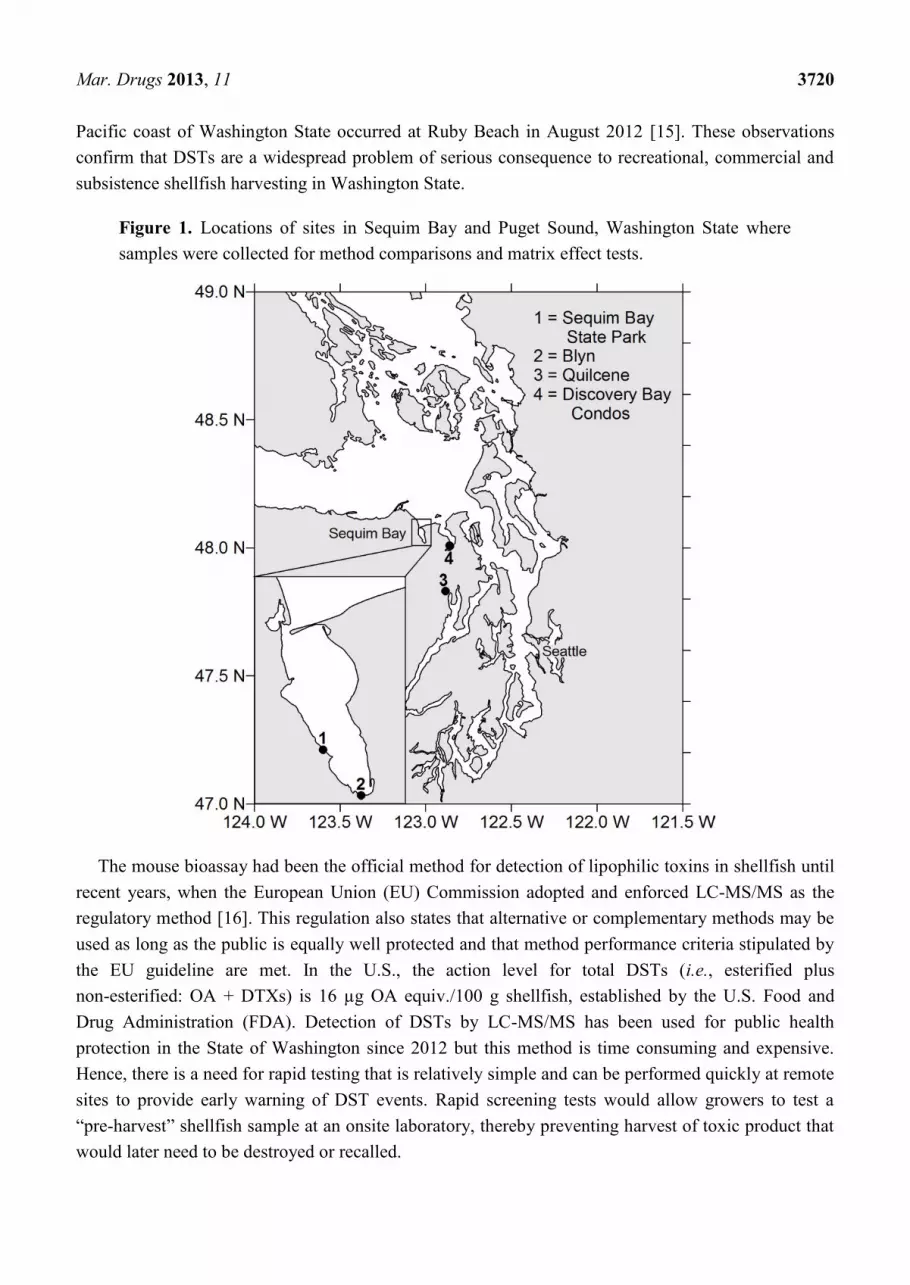
Three DSP cases were reported on 29 June 2011 in the U.S. Pacific Northwest from the
consumption of mussels collected from a pier at Sequim Bay State Park (Figure 1). Family members
aged 2, 5 and 45 years developed symptoms 4, 7, and 14 h, respectively, after consuming
8–15 mussels [12]. Diarrhetic shellfish poisoning symptoms that were exhibited by the individuals
included vomiting, diarrhea, body aches, fever and chills. Blue mussels collected within a few days of
the illnesses were found by liquid chromatography tandem mass spectrometry (LC-MS/MS) analysis
to contain levels of DSTs 2–10 times the action level of 16 μg OA equiv./100 g shellfish tissue. This
finding prompted product recalls and the closure of recreational and commercial shellfish harvesting
from Sequim Bay.
Additionally, in July–August 2011, 62 people suffered from DSP in British Columbia, Canada.
These illnesses were traced to the ingestion of Pacific coast mussels and were the first reports of DSP
in western Canada [13]. Almost 14,000 kg of product was recalled. Although the presence of
Dinophysis in Pacific Northwest coastal waters dates back many years [14], these events represented
the first time illnesses were reported in conjunction with DST levels deemed hazardous to
human health.
During the summer of 2012, a comprehensive analysis of DSTs was performed in several shellfish
species collected from numerous sites throughout Puget Sound [15]. Detection of DSTs by LC-MS/MS
above the regulatory action level resulted in widespread harvest closures of California mussels, varnish
clams, manila clams and Pacific oysters [15]. In addition, the first ever closure due to DSTs on the
Mar. Drugs 2013, 11 3720
Pacific coast of Washington State occurred at Ruby Beach in August 2012 [15]. These observations
confirm that DSTs are a widespread problem of serious consequence to recreational, commercial and
subsistence shellfish harvesting in Washington State.
Figure 1. Locations of sites in Sequim Bay and Puget Sound, Washington State where
samples were collected for method comparisons and matrix effect tests.
The mouse bioassay had been the official method for detection of lipophilic toxins in shellfish until
recent years, when the European Union (EU) Commission adopted and enforced LC-MS/MS as the
regulatory method [16]. This regulation also states that alternative or complementary methods may be
used as long as the public is equally well protected and that method performance criteria stipulated by
the EU guideline are met. In the U.S., the action level for total DSTs (i.e., esterified plus
non-esterified: OA + DTXs) is 16 µg OA equiv./100 g shellfish, established by the U.S. Food and
Drug Administration (FDA). Detection of DSTs by LC-MS/MS has been used for public health
protection in the State of Washington since 2012 but this method is time consuming and expensive.
Hence, there is a need for rapid testing that is relatively simple and can be performed quickly at remote
sites to provide early warning of DST events. Rapid screening tests would allow growers to test a
“pre-harvest” shellfish sample at an onsite laboratory, thereby preventing harvest of toxic product that
would later need to be destroyed or recalled.

Mar. Drugs 2013, 11 3721
Currently, there are several commercially available rapid screening test kits. These kits are based on
functional action of toxins on their receptors (e.g., protein phosphatase 2A isolated from human red
blood cells) or structural recognition of a common epitope (e.g., antibody-based assays, termed
enzyme-linked immunosorbent assay or ELISA). Here we describe the analysis of tissue homogenates
from several shellfish species collected in the summer of 2012. Samples were collected from two sites
in Sequim Bay, WA: Sequim Bay State Park on the western shore and Blyn in the south (Figure 1) and
other sites throughout Puget Sound (see Figure 1 in [15]). Three rapid screening methods were used
and the results were compared to those obtained from the standard LC-MS/MS regulatory method.
2. Results
2.1. Analysis of DSTs by LC-MS/MS
Figure 2A shows a typical LC-MS/MS chromatogram of OA, DTX-1, DTX-2 and yessotoxin
(YTX) standards. Although it is often present in shellfish, YTX is a DST that is not currently
monitored for regulatory purposes in Washington State. Dinophysistoxin-1 (detected as both free and
acyl ester forms) and YTX were found exclusively in the majority of shellfish samples we analyzed. A
representative chromatogram of a hydrolyzed blue mussel extract from Sequim Bay, WA is shown in
Figure 2B. Our findings confirmed previous survey of Puget Sound showing DTX-1 to be the primary
toxin found in shellfish from Puget Sound and the coast of Washington state [15].
Figure 2. LC-MS/MS chromatograms. Retention times are shown in parenthesis.
(A) certified reference standards okadaic acid (OA) (5.31 min), DTX-2 (5.67 min), DTX-1
(6.56 min) and yessotoxin (YTX, 8.37 min); (B) DTX-1 (6.55 min) and YTX (8.41 min) in
a hydrolyzed blue mussel extract from Sequim Bay, WA, USA.

Mar. Drugs 2013, 11 3722
2.2. Tissue Matrix Effects
The potential matrix effect from shellfish tissue, resulting in over- or under-estimation of the true
concentration of analytes present in the sample, was assessed by testing both hydrolyzed and
non-hydrolyzed shellfish extracts at several dilutions by LC-MS/MS. Blue mussel samples were
analyzed neat and at two dilutions (50%, 10%) for non-hydrolyzed extracts, and a single dilution
(50%) for hydrolyzed extracts (Table 1). The DST value of each diluted sample extract was corrected
by its dilution factor and compared to the neat extract.
Table 1. Diarrhetic shellfish toxins (DST) concentrations by LC-MS/MS analysis of
hydrolyzed and non-hydrolyzed blue mussel extracts at three dilutions. Data are corrected
for dilution factors. See Figure 1 for site locations.
Hydrolyzed DST concentration (μg total OA equiv./100 g shellfish)
Site OA DTX-2 DTX-1 OA DTX-2 DTX-1 OA DTX-2 DTX-1
(% extract) a 100 100 100 50 50 50 10 10 10
Quilcene bd bd 27.92 bd bd 30.99 na na na
Discovery Bay Condos bd bd 25.64 bd bd 25.61 na na na
Discovery Bay Condos bd bd 21.84 bd bd 22.23 na na na
Sequim Bay State Park bd bd 50.31 bd bd 46.32 na na na
Sequim Bay State Park bd bd 19.09 bd bd 16.91 na na na
Sequim Bay State Park bd bd 82.87 bd bd 82.48 na na na
Sequim Bay Blyn bd bd 37.08 bd bd 33.62 na na na
Sequim Bay Blyn bd bd 31.53 bd bd 29.87 na na na
Non-hydrolyzed DST concentration (μg OA equiv./100 g shellfish)
Site OA DTX-2 DTX-1 OA DTX-2 DTX-1 OA DTX-2 DTX-1
(% extract) a 100 100 100 50 50 50 10 10 10
Quilcene 1.33 bd 14.18 bd bd 13.72 bd bd 12.86
Discovery Bay Condos bd bd 13.53 bd bd 11.40 bd bd 10.72
Discovery Bay Condos bd bd 11.90 bd bd 10.40 bd bd 10.01
Sequim Bay State Park bd bd 31.65 bd bd 25.86 bd bd 16.84
Sequim Bay State Park bd bd 26.56 bd bd 25.92 bd bd 17.66
Sequim Bay State Park bd bd 44.46 bd bd 20.60 bd bd 25.42
Sequim Bay Blyn bd bd 20.98 bd bd 18.70 bd bd 14.23
Sequim Bay Blyn bd bd 20.21 bd bd 18.53 bd bd 14.52
a Indicates percentage of original extract. All shellfish extracts at less than 100% are diluted with 100% MeOH,
bd = below the analytical limit of detection (OA, DTX-1: 1.25 μg/100 g tissue; DTX-2: 1.00 μg/100 g tissue),
na = not analyzed.
Matrix effects were found to be low in hydrolyzed blue mussel extracts where DTX-1 values were
approximately 3% higher in the neat sample versus corrected values of the 50% diluted sample (Table 1),
within the calculated error of LC-MS/MS method. This difference was higher in non-hydrolyzed
extracts (12%). However, because toxin concentrations were lower in non-hydrolyzed extracts
compared to hydrolyzed extracts, the increased instrument or extraction variability at these lower
concentrations could be partially responsible for the difference between 100% and 50% (or 10%).

Mar. Drugs 2013, 11 3723
2.3. Comparison of Screening Methods to LC-MS/MS
Shellfish samples collected at various locations in Puget Sound and the Pacific coast of Washington
State (n = 110) were analyzed by PP2A test and compared to LC-MS/MS analysis (Figure 3). These
data include the samples shown in Table 2 that were re-extracted for this repeat analysis. The best fit
by linear regression was seen with blue mussel (Mytilus edulis; R2 = 0.82; n = 63) followed by
California mussel (Mytilus californianus; R2 = 0.74; n = 11), manila clam (Venerupis philippinarum;
R2 = 0.73; n = 10), Pacific oyster (R
2 = 0.51; n = 15), razor clam (Siliqua patula; R
2 = 0.52; n = 5; not
shown), and littleneck clam (Leukoma staminea; R2 = 0.06; n = 6; not shown).
Figure 3. Comparison between LC-MS/MS and PP2A results for total DSTs
(μg OA equiv./100 g shellfish extract).

Mar. Drugs 2013, 11 3724
Table 2. Methods comparison of LC-MS/MS, PP2A, ELISA and Jellett test strips in
samples extracted using the EU method and comparison of LC-MS/MS and Jellett test
strips in samples extracted using the Jellett Rapid Test method. DST concentrations are
shown (µg total OA equiv./100 g). Positive results: ≥16.0 µg OA equiv./100 g in bold
print. Negative result: <16.0 µg OA equiv./100 g in regular print. Jellett rapid test were
read visually and noted as positive (+) or negative (−) based on the manufacturer’s method.
EU Extracts Jellett Extracts
Species LC-MS/MS PP2A ELISA Jellett LC-MS/MS Jellett
1. Blue Mussel 96.5 48.9 51.8 − 39.8 +
2. Blue Mussel 63.6 38.2 52.3 − 31.7 +
3. Blue Mussel 60.6 38.1 58.8 − 32.4 +
4. Blue Mussel 38.7 35.7 35.7 − 18.3 +
5. Blue Mussel a 30.3 28.7 22.6 − 17.1 +
6. Blue Mussel 36.9 36.0 63.0 − 21.1 +
7. Blue Mussel 26.2 31.2 31.8 − 12.9 +
8. Blue Mussel 32.7 33.5 33.8 − 15.1 −
9. Blue Mussel 26.5 34.1 28.8 − 13.9 −
10. Blue Mussel 37.7 35.1 51.3 − 16.9 +
11. Blue Mussel 60.5 29.4 49.3 − 26.0 +
12. Blue Mussel 28.1 33.6 25.3 − 12.7 +
13. Blue Mussel a 31.5 34.5 20.5 − 18.4 +
14. Geoduck <LoQ <LoQ 10.9 − <LoQ −
15. Littleneck Clam <LoQ <LoQ 20.5 − 2.0 −
16. Manila Clam 36.1 16.9 28.8 − 16.2 −
17. Manila Clam 21.9 12.8 20.3 − 13.1 −
18. Pacific Oyster 25.3 16.5 67.8 − 14.5 −
19. Pacific Oyster 6.3 14.7 27.2 − 3.0 −
20. Pacific Oyster 3.7 <LoQ 23.6 − 2.2 −
21. Pacific Oyster 7.9 13.0 31.7 − 2.9 −
22. Pacific Oyster 37.7 13.1 61.3 − 14.4 −
23. Pacific Oyster 14.2 26.6 32.7 − 6.5 − a Extracts of the same sample, LoQ = Limit of Quantification.
To compare screening methods, shellfish samples (n = 23) were extracted by the EU method and
analyzed by LC-MS/MS, PP2A, ELISA, and Jellett Rapid Tests (Table 2, Figure 4). There were 5 false
positives (samples 15, 19, 20, 21, 23) by ELISA; 2 false negatives (samples 17, 22) and 1 false positive
(sample 23) by PP2A while all of Jellett Rapid Test results were negative. However, when this same
subset of shellfish extracts were re-tested using the Jellett extraction method and compared to
LC-MS/MS results, there were only 6 false negatives (samples 8, 9, 16, 17, 18, 22). The EU method
uses a double methanolic extract of shellfish homogenate whereas Jellett manufacturer recommends a
single extraction. Comparison of the LC-MS/MS results from samples extracted using the Jellett and
EU methods showed that the Jellett method extracted approximately 50% of the DSTs (Table 2).
Mar. Drugs 2013, 11 3725
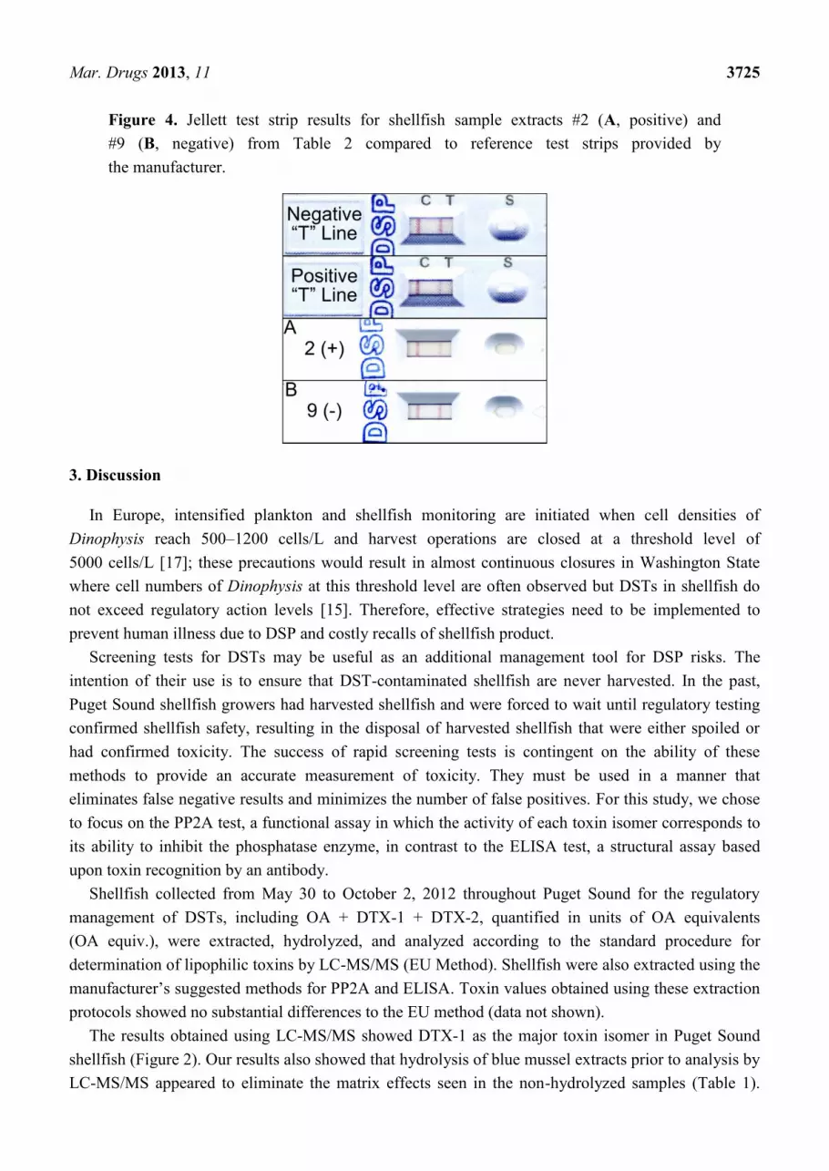
Figure 4. Jellett test strip results for shellfish sample extracts #2 (A, positive) and
#9 (B, negative) from Table 2 compared to reference test strips provided by
the manufacturer.
3. Discussion
In Europe, intensified plankton and shellfish monitoring are initiated when cell densities of
Dinophysis reach 500–1200 cells/L and harvest operations are closed at a threshold level of
5000 cells/L [17]; these precautions would result in almost continuous closures in Washington State
where cell numbers of Dinophysis at this threshold level are often observed but DSTs in shellfish do
not exceed regulatory action levels [15]. Therefore, effective strategies need to be implemented to
prevent human illness due to DSP and costly recalls of shellfish product.
Screening tests for DSTs may be useful as an additional management tool for DSP risks. The
intention of their use is to ensure that DST-contaminated shellfish are never harvested. In the past,
Puget Sound shellfish growers had harvested shellfish and were forced to wait until regulatory testing
confirmed shellfish safety, resulting in the disposal of harvested shellfish that were either spoiled or
had confirmed toxicity. The success of rapid screening tests is contingent on the ability of these
methods to provide an accurate measurement of toxicity. They must be used in a manner that
eliminates false negative results and minimizes the number of false positives. For this study, we chose
to focus on the PP2A test, a functional assay in which the activity of each toxin isomer corresponds to
its ability to inhibit the phosphatase enzyme, in contrast to the ELISA test, a structural assay based
upon toxin recognition by an antibody.
Shellfish collected from May 30 to October 2, 2012 throughout Puget Sound for the regulatory
management of DSTs, including OA + DTX-1 + DTX-2, quantified in units of OA equivalents
(OA equiv.), were extracted, hydrolyzed, and analyzed according to the standard procedure for
determination of lipophilic toxins by LC-MS/MS (EU Method). Shellfish were also extracted using the
manufacturer’s suggested methods for PP2A and ELISA. Toxin values obtained using these extraction
protocols showed no substantial differences to the EU method (data not shown).
The results obtained using LC-MS/MS showed DTX-1 as the major toxin isomer in Puget Sound
shellfish (Figure 2). Our results also showed that hydrolysis of blue mussel extracts prior to analysis by
LC-MS/MS appeared to eliminate the matrix effects seen in the non-hydrolyzed samples (Table 1).

Mar. Drugs 2013, 11 3726
Hydrolysis is required to convert DTX-3, the esterified form of DST toxins present in some shellfish to
the parent toxins OA, DTX-1 or DTX-2. Therefore, the hydrolyzed shellfish extracts were used for
comparison of toxin values obtained by LC/MS-MS with screening method results.
The first screening method that was assessed in this study was a commercial ELISA that is
currently sold in the U.S. by Abraxis (Warminster, PA, USA). Analysis of hydrolyzed shellfish
extracts using ELISA in comparison to LC-MS/MS data resulted 5 out of 23 (22%) false positive
values. The ELISA overestimated toxin concentrations in hydrolyzed extracts, an interesting finding
given that one would expect that the estimated 50% cross reactivity of DTX-1 would result in
underestimation of toxin concentrations. The stated LOD for ELISA (10 μg/100 g) combined with the
50% cross-reactivity for DTX-1 and DTX-2 means that when applied to shellfish samples containing
only DTX-1 (free and acylated forms), the effective LOD would be 20 μg/100 g, which is above the
regulatory action limit. This calls into question the use of the ELISA in Washington State. Other
commercial ELISAs, including the “DSP check kit” from Japan, based on the procedure of
Usagawa et al. [18], have been tested previously [19], however these kits are no longer commercially
available. An antibody-based DST test recently has been made available by Neogen (Reveal 2.0
for DSP), however, at the time this study was underway, it was not yet calibrated for use with
hydrolyzed samples.
We found that when used as recommended by the manufacturer, the Jellett Rapid Test for DSP gave
a high number of false negatives (Table 2), most likely due to incomplete extraction of DSTs by the
Jellett extraction method compared to the standard EU method (Table 2). Therefore, improved
extraction efficiency is necessary for the Jellett Rapid Test to be used as an accurate screening tool
with Washington State shellfish. The need for improving the efficiency of the Jellett Rapid Test
extraction method has been recognized by the developers of the test [20]. Once the extraction
efficiency is improved, we believe that these test strips will show promise as a screening tool, due to
their simplicity of use.
Several trials have been performed with protein phosphatase inhibition assays (PPIA). Different
labs have developed their own PPIA tests using absorbance or fluorescence-based endpoint
determinations [21–28] with relatively good results. Recently, the first comprehensive single lab
validation of a commercial PP2A kit (OkaTest is developed by ZEU-INMUNOTEC, Zaragoza, Spain
and distributed in the US by Abraxis LLC) was performed to determine the repeatability, precision,
and stability of reagents [29]. This test uses a purified human PP2A that has higher sensitivity than
other similar enzymes produced by genetic engineering [30]. The single-lab assessment, in which
shellfish were spiked with certified reference solutions and compared to both LC-MS/MS and mouse
bioassay results, demonstrated that all reagents were stable for >6 months. Smienk et al. [29]
determined that the IC50 was 1.6 nM for DTX-1 and 1.2 nM for both OA and DTX-2. They reasoned
that the lower affinity of the PP2A for DTX-1 would result in underestimation using the commercial
PP2A kit compared to LC-MS/MS. Smienk et al. [29] also observed that analysis of spiked,
hydrolyzed samples gave 146% and 163% recovery for mussel and king scallop, respectively. The
authors suggested that the reason for high recovery may be that reference standard materials were
certified only for non-hydrolyzed DSTs.
The single lab validation was followed by an interlaboratory validation [31] in which 16 labs were
given several species of spiked shellfish and blank samples to analyze. The results indicated that the

Mar. Drugs 2013, 11 3727
OkaTest was suitable for quantitative determination of the OA toxins group [31]. In our study, we
chose not to replicate this prior investigation, but rather to determine test precision, accuracy, and
potential matrix effect in naturally-contaminated shellfish from the Pacific Northwest.
The PP2A test results of the subset of shellfish samples (Table 2) showed 2 false negatives (manila
clam and Pacific oyster) and 1 false positive value (Pacific oyster). The 8% false negatives could be
reduced through the choice of a lower threshold value (e.g., 12 μg/100 g) for this screening test, which
would require that any samples at or above this conservative value be tested by the standard regulatory
method. In addition, some sample concentrations predicted by the PP2A test, especially those
>60 μg/100 g shellfish, were greatly underestimated using PP2A (Table 2). The manufacturer states
that values >35.2 μg/100 g are outside the linear range of the standard curve and suggests that these
values be treated as estimates. However, these underestimated high concentrations do not alter the
effectiveness of this test as a screening tool.
Some matrix effect was evident using the PP2A test, in that extracts with zero toxin concentrations
by LC-MS/MS showed measurable DSTs by PP2A (Figure 3, all panels, see symbols on the y-axis).
Matrix effects have been observed in past studies where toxin content was overestimated by enzyme
assay compared to LC-MS/MS analysis [32]. This effect has been attributed to the presence of
compounds such as methanol-soluble lipids that exert an unspecific inhibitory effect on the protein
phosphatase [22,29]. Many of these past studies used shellfish homogenates that were spiked with
known concentrations of OA and other DSTs. In the future, naturally contaminated samples must be
tested, including Pacific Northwest shellfish samples containing varied concentrations of DTX-1 and
DTX-3, in order to confirm the effectiveness of PP2A as a screening test.
Harvest closures in the State of Washington are implemented when toxins exceed the regulatory
action level [33]. Traditional monitoring programs established by shellfish authorities allow for timely
closures in state waters with minimal negative impacts on industry. However, such monitoring
programs are not always the most effective at remote sites given the time required for sample shipment
and analysis. For example, the Jamestown S’Klallam Tribe has had to purchase and ship shellfish from
other locations for their tribal potlatches because of the shellfish harvested from Sequim Bay could not
be tested for DSTs in time for the tribal feast. However, in order to use screening tools for rapid
assessment of toxins, these methods must be endorsed by the Interstate Shellfish Sanitation Conference
(ISSC) for specific applications in federal waters. An example of this process is the ISSC approval of
Abraxis shipboard ELISA for paralytic shellfish poisoning (PSP) for the offshore scallop fishery on
Georges Bank [34]. Since the Jamestown S’Klallam Tribe is an independent federal entity, they could
choose similar monitoring strategies to ensure the safety of shellfish intended for subsistence
consumption. The PP2A may provide a safety buffer for the harvest of large amounts of shellfish prior
to confirmatory regulatory testing, or may suffice as a tool for specific uses, for example, to maintain
an open status of the shellfish beds. Another example of rapid screening test application is the
combination of phytoplankton monitoring and ELISA screening tests for domoic acid that are used by
the Olympic Region Harmful Algal Bloom (ORHAB) partnership to maintain opening status of razor
clam beds. We envision that a combination of phytoplankton monitoring and rapid screening tests
might be implemented as a complement to regulatory monitoring, especially at remote sites in
Washington State.

Mar. Drugs 2013, 11 3728
4. Experimental Section
4.1. Shellfish Collection
For comparing LC-MS/MS and the three screening methods, shellfish, including blue mussel
(Mytilus edulis; n = 13), geoduck clam (Panopea generosa; n = 1), littleneck clam (Leukoma staminea;
n = 1), manila clam (Venerupis philippinarum; n = 2) and Pacific oyster (Crassostrea gigas; n = 6),
were collected at two locations in Sequim Bay in Washington State (Figure 1) and stored at −20 °C
until analysis. Samples for detailed comparison of PP2A and LC-MS/MS were collected from sites
throughout Puget Sound and included blue mussel (n = 63), California mussel (n = 11), manila clam
(n = 10), Pacific oyster (n = 15), razor clam (n = 5), and littleneck clam (n = 6).
4.2. Sample Preparation
Shellfish were rinsed with tap water and opened by cutting the adductor muscles. About
10–20 individual shellfish were pooled to make up at least 100 g of tissue per sample. Samples were
drained for a few minutes to remove any excess water and then homogenized in a glass blender for
1 min. The homogenates were stored in ultra-high performance polypropylene copolymer containers at
−20 °C until analysis.
4.3. Tissue Extraction
The method for shellfish tissue extraction was a modification of the EU-Harmonized Standard
Operating Procedure for the Determination of Lipophilic Marine Biotoxins in Mollusks by
LC-MS/MS [35]. Briefly, an aliquot of sample homogenate (2.5 g) was accurately weighed into a
50 mL centrifuge tube (BD Falcon, San Jose, CA, USA) and extracted with 12 ml of methanol by
vortex mixing for 3 min. The mixture was centrifuged at 2500× g for 10 min and the supernatant
transferred to a 25 mL volumetric flask. The residual pellet was re-extracted by homogenizing in
10 mL methanol for 1 min with a 10 mm stainless steel OmniProbe High Power Tissue Homogenizer
(Kennesaw, GA, USA) followed by centrifugation at 2500× g for 10 min. The supernatant was
combined with the first extract and brought to 25 mL with methanol. Samples also were extracted
using the method specified by the manufacturer of the Jellett Rapid Test to evaluate the extraction
efficiency of this method. Briefly, 1 g of shellfish sample was weighed into a 15 mL centrifuge tube
and extracted by vortex mixing in 3 mL 100% methanol for 3 min. The mixture was then centrifuged
at 2500× g for 10 min. The extract solutions were mixed well and aliquots were stored in amber glass
vials at −20 °C until analyzed by LC-MS/MS and other screening methods.
4.4. Tissue Extract Hydrolysis
The tissue extracts, from both EU and Jellett extraction methods, were hydrolyzed using a modified
method published by Mountfort et al. [23]. A 125 µL aliquot of a 2.5 N NaOH solution was mixed
with 1 mL of the extract and heated at 76 °C for 40 min in a tightly sealed 1.5 mL vial (Sun Brokers,
Inc., Wilmington, NC, USA) to avoid sample loss due to evaporation. The hydrolyzed sample was
neutralized with 125 µL of a 2.5 N HCl solution and filtered through a 0.22 µm PTFE syringe filter
Mar. Drugs 2013, 11 3729
(Fisher Scientific, Pittsburgh, PA, USA). Samples were analyzed by LC-MS/MS immediately after
hydrolysis or stored at 4 °C and analyzed by rapid methods within 2 days of hydrolysis. Total toxin
levels (free plus esterified) were calculated with correction for the 25% volume increase from the
additions of base and acid.
4.5. LC-MS/MS
Shellfish extracts, from both the EU and Jellett methods, were analyzed by Ultra-performance
Liquid Chromatography (UPLC; Acquity system, Waters Co., Milford, MA, USA) coupled with a
triple quadrupole tandem mass spectrometer (MS/MS, ABSciex 5500, Framingham, MA, USA). For
each sample, 10 µL of filtered extract was injected into the UPLC-MS/MS. The UPLC was equipped
with a 0.2 µm pre-filter followed by a 2.1 × 4 mm C8 Security guard cartridge and a 2.1 × 100 mm,
5 µm Luna C8 reverse phase column (Phenomenex, Torrance, CA, USA). The acidic chromatographic
conditions used in this study were described in the EU-Harmonised method [35] and by
McCarron et al. [36]. A description of the mobile phases, linear gradients, run time and analyte
detection are described in detail in Trainer et al. [15].
The analytes were quantified with individual seven-point external calibration curves prepared in
methanol from certified reference standards (CRM-OA-c, CRM-DTX-1-c, CRM-DTX-2-c and
CRM-YTX-b) purchased from National Research Council Canada (Halifax, Nova Scotia). A linear
best fit resulted in a correlation coefficient of R = 0.99 for each curve. Standard calibration curve
ranges were as follows: OA: 0.71 to 286 ng/mL; DTX-2: 0.44 to 177 ng/mL; DTX-1: 0.72 to 289 ng/mL;
YTX: 0.56 to 224 ng/mL A water blank and a check vial containing all 4 analytes was run after every
10 to 12 samples as quality control. The % relative standard deviation for OA = 2.2%, DTX-2 = 1.3%,
DTX-1 = 2.5% and YTX = 3.3%. Each extract was run in the hydrolyzed and non-hydrolyzed form.
Hydrolysis reduces esterified compounds to the parent form and allows the detection of total toxin for
OA, DTX-2, and DTX-1. Final reporting was in µg/100 g tissue for shellfish extracts. The limits of
detection (LOD) and limits of quantitation (LOQ) in shellfish tissue for monitored toxins were,
respectively: OA, 10 pg on column and 1.25 µg/100 g tissue; DTX-1, 10 pg on column and
1.25 µg/100 g tissue; and DTX-2, 8 pg on column and 1.00 µg/100 g tissue.
4.6. Enzyme-Linked Immunosorbent Assay (ELISA)
The DSP ELISA test is an immunoassay based on the recognition of OA and its isomers by rabbit
anti-OA antibodies, produced by Abraxis (DSP ELISA kit, PN 52001; Warminster, PA, USA).
Detailed instructions on its use can be found at the manufacturer’s website [37]. This test can detect
OA in shellfish and water samples; however, while the antibodies react with 100% affinity to OA, they
have only 50% cross-reactivity to DTX-1, DTX-2. The antibody reaction is initiated by adding
shellfish sample or standard and OA-HRP conjugate to a solution of the anti-OA antibodies. The OA
antibodies are then incubated with a secondary antibody (goat anti-rabbit) immobilized on the surface
of the 96-well microplate. The concentration of DSP toxins in a sample or standard is inversely
proportional to the intensity of the color signal produced by 3,3′,5,5′-tetramethylbenzidine (TMB)
substrate and determined by interpolation from OA standard curve constructed with each analysis. The

Mar. Drugs 2013, 11 3730
test has a 5-point standard curve ranging from 0.1 to 5.0 ppb OA in solution. The limit of detection for
OA by this test is 10 μg/100 g shellfish tissues.
4.7. Protein Phosphatase 2A (PP2A)
Protein-phosphatase inhibition assays were developed about a decade ago based on the finding that
serine/threonine protein phosphatases are inhibited by OA group of toxins. Of the four major classes of
this group of enzymes, PP1, PP2A, PP2B and PP2C, PP2A has the highest affinity for OA [38].
Commercially available PP2A test kits (OkaTest, ZEU, Spain) were developed using purified human
protein phosphatase type 2A and p-nitrophenylphosphate (pNPP) as a colorimetric substrate in a
96-well microplate format to measure OA and its analogs including DTX-1, DTX-2 and DTX-3.
Dinophysistoxin-3, acyl ester form of OA, DTX-1 or DTX-2, is converted to its free form by alkaline
hydrolysis of the methanolic extracts. Hydrolyzed sample extracts were diluted with assay buffer and
incubated with enzyme phosphatase solution at 30 °C. Toxins in sample extracts are directly
proportional to the color product, p-nitrophenol (p-NP) formed in the reaction between the uninhibited
enzyme and pNPP substrate that can be measured at 405 nm using a microplate reader. Total DST
concentration was determined in hydrolyzed shellfish extract based on a 5-point calibration standard
curve, ranging from 0.5 to 2.0 nM. LOD and LOQ for blank mussel are 4.4 and 5.6 μg/100 g,
respectively [29].
4.8. Jellett Rapid Test Strip
A rapid field test kit was developed to screen for DSTs in shellfish samples by Jellett Rapid
Testing, Ltd. (Nova Scotia, Canada). This test is based on the anti-OA antibodies used in the ELISA
method and was developed for simple field application. The Jellett Rapid Test for DSP is a lateral flow
immuno-chromatographic (LFI) system similar to the pregnancy test strips. In the strip format, the
three major DSP toxins (OA, DTX-1, DTX-2) are detected with similar affinity with the 50%
reduction in color intensity of the test line set at 5 nM to trigger a positive result compared to the
control line [20]. Hydrolyzed extracts were diluted with a running buffer solution supplied with the test
kit and 100 µL of the diluted sample was applied to the sample pad of a test strip. After 30 min, the test
and control lines were fully developed and the color intensity was compared to a color guide supplied
by the manufacturer. If the T(est) line was lighter than the C(ontrol) line, the result was positive; if it
had the same color intensity as the C line, then the result was negative (Figure 4).
5. Conclusions
In this study, the applicability of screening assays for the determination of DSTs in shellfish tissues
was compared to the standard regulatory LC-MS/MS method. Few other studies have explored the
effectiveness of screening assays using naturally contaminated samples, particularly those that contain
high concentrations of DTX-1 and acyl derivatives, collectively called DTX-3. Good agreement was
observed between the PP2A test and LC-MS/MS, however, when toxin levels exceeded the upper
working range of the assay, the PP2A test underestimated total DTX-1 compared to LC-MS/MS
values. The few false negatives (8%) observed with the PP2A test might be eliminated by choosing a
Mar. Drugs 2013, 11 3731
conservative threshold value (e.g., 12 μg/100 g) for screening. The ELISA showed relatively high false
positives (>20%) and no false negatives. The Jellett rapid test gave >30% false negative results due to
the incomplete extraction efficiency of the method used as recommended by the manufacturer. Finally,
our results suggest that PP2A is the screening test of choice for Washington State monitoring program
as it may provide a safety buffer for the harvest of large amounts of shellfish prior to confirmatory
regulatory testing, or may suffice as a tool to maintain an open status of the shellfish beds. However,
further screening tests must be conducted over the next few years using a variety of subsistence,
recreational, and commercial shellfish from the region in order to determine the most effective
scenarios for their use. In Washington State, we envision that the implementation of a strategy where
monitoring for the presence of Dinophysis species together with the application of toxin screening
would allow shellfish harvesters to avoid unnecessary disposal of harvested product and eliminate
costly recalls.
Acknowledgments
We thank all SoundToxins and ORHAB partners for their dedication to weekly sampling of coastal
and Puget Sound waters. We acknowledge University of Washington student intern Megan Stephens
and University of Washington Capstone Program student Matthew Choowong for their help in the
laboratory, and Nobuharu Inaba for reviewing this manuscript. We also thank four anonymous
reviewers for their helpful comments. This study was funded in part by the United States
Environmental Protection Agency (EPA) through their National Estuary Program, via a contract
(PC-00J32601) and by NOAA’s National Center for Coastal Ocean Sciences’ Event Response
program (NOAA ER). This is NOAA ER publication #17.
Conflict of Interest
The authors declare no conflict of interest.
References
1. Van Egmond, H.P.; Van Apeldoorn, M.E.; Speijers, G.J.A. Marine Biotoxins. In Food and
Nutrition; FAO: Rome, Italy, 2004; pp. 53–96.
2. Control of Communicable Diseases Manual, 19th ed.; Heymann, D.L., Ed.; American Public
Health Association Press: Washington, DC, USA, 2008.
3. Cembella, A.D. Occurrence of okadaic acid, a major diarrheic shellfish toxin, in natural
populations of Dinophysis spp. from the eastern coast of North America. J. Appl. Phycol. 1989, 1,
307–310.
4. Takai, A.; Bialojan, C.; Troschka, M.; Ruegg, J.C. Smooth-muscle myosin phosphate inhibition
and force enhancement by black sponge toxin. FEBS Lett. 1987, 217, 81–84.
5. Fujiki, H.; Suganuma, M. Tumor Promotion by Inhibitors of Protein Phosphatase-1 and
Phosphatase-2A—The Okadaic Acid Class of Compounds. Adv. Cancer Res. 1993, 61, 143–194.
6. Bialojan, C.; Takai, A. Inhibitory Effect of a Marine-Sponge Toxin, Okadaic Acid, on Protein
Phosphatases-Specificity and Kinetics. Biochem. J. 1988, 256, 283–290.

Mar. Drugs 2013, 11 3732
7. Burgess, V.; Shaw, G. Pectenotoxins—an issue for public health—A review of their comparative
toxicology and metabolism. Environ. Int. 2001, 27, 275–283.
8. Barceloux, D.G. Diarrhetic shellfish poisoning and okadaic acid. In Medical Toxicology of
Natural Substances: Foods, Fungi, Medicinal Herbs, Plants, and Venomous Animals; John Wiley
& Sons: Hoboken, NJ, USA, 2008; pp. 222–226.
9. Stamman, E.; Segar, D.A.; Davis, P.G. A Preliminary Epidemiological Assessment of the
Potential for Diarrhetic Shellfish Poisoning in the Northeast United States. In NOAA Technical
Memorandum NOS OMA 34; NOAA: Rockville, MD, USA, 1987.
10. Manerio, E.; Rodas, V.L.; Costas, E.; Hernandez, J.M. Shellfish consumption: A major risk factor
for colorectal cancer. Med. Hypotheses 2008, 70, 409–412.
11. Cordier, S.; Monfort, C.; Miossec, L.; Richardson, S.; Belin, C. Ecological analysis of digestive
cancer mortality related to contamination by diarrhetic shellfish poisoning toxins along the coasts
of France. Environ. Res. 2000, 84, 145–150.
12. Lloyd, J.; Duchin, J.; Borchert, J.; Flores-Quintana, H.; Robertson, A. Diarrhetic shellfish
poisoning in Washington State, USA, 2011. Emerg. Infect. Dis. 2013, in press.
13. Taylor, M.; McIntyre, L.; Ritson, M.; Stone, J.; Bronson, R.; Bitzikos, O.; Rourke, W.;
Galanis, E.; Team, O. Outbreak of Diarrhetic Shellfish Poisoning Associated with Mussels,
British Columbia, Canada. Mar. Drugs 2013, 11, 1669–1676.
14. Horner, R.A.; Garrison, D.L.; Plumley, F.G. Harmful algal blooms and red tide problems on the
U. S. west coast. Limnol. Oceanogr. 1997, 42, 1076–1088.
15. Trainer, V.; Moore, L.; Bill, B.; Adams, N.; Harrington, N.; Borchert, J.; da Silva, D.;
Eberhart, B.-T. Diarrhetic Shellfish Toxins and Other Lipophilic Toxins of Human Health
Concern in Washington State. Mar. Drugs 2013, 11, 1815–1835.
16. Commission Regulation (EU) No 15/2011 of 10 January 2011 amending Regulation (EC) No
2074/2005 as regards recognised testing methods for detecting marine biotoxins in live bivalve
molluscs. Off. J. Eur. Union 2011, 54, 3–6.
17. Anderson, D.M.; Andersen, P.; Bricelj, V.M.; Cullen, J.J.; Rensel, J.E.J. Monitoring and
Management Strategies for Harmful Algal Blooms in Coastal Waters; Technical Report No.
APEC #201-MR-01.1; Asia Pacific Economic Program, Singapore, and Intergovernmental
Oceanographic Commission: Paris, France, 2001.
18. Usagawa, T.; Nishimura, M.; Itoh, Y.; Uda, T.; Yasumoto, T. Preparation of
monoclonal-antibodies against okadaic acid prepared from the sponge Halichondria okadai.
Toxicon 1989, 27, 1323–1330.
19. Vale, P.; Sampayo, M.A.D. Comparison between HPLC and a commercial immunoassay kit for
detection of okadaic acid and esters in Portuguese bivalves. Toxicon 1999, 37, 1565–1577.
20. Laycock, M.V.; Jellett, J.F.; Easy, D.J.; Donovan, M.A. First report of a new rapid assay for
diarrhetic shellfish poisoning toxins. Harmful Algae 2006, 5, 74–78.
21. Tubaro, A.; Florio, C.; Luxich, E.; Sosa, S.; DellaLoggia, R.; Yasumoto, T. A protein phosphatase
2A inhibition assay for a fast and sensitive assessment of okadaic acid contamination in mussels.
Toxicon 1996, 34, 743–752.

Mar. Drugs 2013, 11 3733
22. Honakanen, R.E.; Moody, D.E.; Dickey, R.W. Detection of DSP-toxins, okadaic acid, and
dinophysis toxin-1 in shellfish by serine/threonine protein phosphatase assay. J. AOAC Int. 1996,
79, 1336–1343.
23. Mountfort, D.O.; Suzuki, T.; Truman, P. Protein phosphatase inhibition assay adapted for
determination of total DSP in contaminated mussels. Toxicon 2001, 39, 383–390.
24. Ramstad, H.; Shen, J.L.; Larsen, S.; Aune, T. The validity of two HPLC methods and a
colorimetric PP2A assay related to the mouse bioassay in quantification of diarrhetic toxins in
blue mussels (Mytilus edulis). Toxicon 2001, 39, 1387–1391.
25. Simon, J.F.; Vemoux, J.-P. Highly sensitive assay of okadaic acid using protein phosphatase and
paranitrophenyl phosphate. Nat. Toxins 1994, 2, 293–301.
26. Vieytes, M.R.; Fontal, O.I.; Leira, F.; deSousa, J.; Botana, L.M. A fluorescent microplate assay
for diarrheic shellfish toxins. Anal. Biochem. 1997, 248, 258–264.
27. Gonzalez, J.C.; Fontal, O.I.; Vieytes, M.R.; Vieytes, J.M.; Botana, L.M. Basis for a new
procedure to eliminate diarrheic shellfish toxins from a contaminated matrix. J. Agric. Food
Chem. 2002, 50, 400–405.
28. Prassopoulou, E.; Katikou, P.; Georgantelis, D.; Kyritsakis, A. Detection of okadaic acid and
related esters in mussels during diarrhetic shellfish poisoning (DSP) episodes in Greece using the
mouse bioassay, the PP2A inhibition assay and HPLC with fluorimetric detection. Toxicon 2009,
53, 214–227.
29. Smienk, H.G.F.; Calvo, D.; Razquin, P.; Dominguez, E.; Mata, L. Single Laboratory Validation of
A Ready-to-Use Phosphatase Inhibition Assay for Detection of Okadaic Acid Toxins. Toxins
2012, 4, 339–352.
30. Sassolas, A.; Catanante, G.; Hayat, A.; Marty, J.L. Development of an efficient protein
phosphatase-based colorimetric test for okadaic acid detection. Anal. Chim. Acta 2011, 702,
262–268.
31. Smienk, H.; Dominguez, E.; Rodriguez-Velasco, M.L.; Clarke, D.; Kapp, K.; Katikou, P.;
Cabado, A.G.; Otero, A.; Vieites, J.M.; Razquin, P.; et al. Quantitative Determination of the
Okadaic Acid Toxins Group by a Colorimetric Phosphatase Inhibition Assay: Interlaboratory
Study. J. AOAC Int. 2013, 96, 77–85.
32. Garibo, D.; Damaso, E.; Eixarch, H.; de la Iglesia, P.; Fernandez-Tejedor, M.; Diogene, J.;
Pazos, Y.; Campas, M. Protein phosphatase inhibition assays for okadaic acid detection in
shellfish: Matrix effects, applicability and comparison with LC-MS/MS analysis. Harmful Algae
2012, 19, 68–75.
33. Annual Report 2011: Commercial and Recreational Shellfish Areas in Washington State; Office
of Shellfish and Water Protection: Olympia, WA, USA, 2012.
34. DeGrasse, S.; Conrad, S.; DiStefano, P.; Vanegas, C.; Wallace, D.; Jensen, P.; Hickey, J.M.;
Cenci, F.; Pitt, J.; Deardorff, D.; et al. Onboard screening dockside testing as a new means of
managing paralytic shellfish poisoning risks in federally closed waters. Deep Sea Res. Part II:
Top. Stud. Oceanogr. 2013, in press.

Mar. Drugs 2013, 11 3734
35. EU-Harmonized Standard Operation Procedure for Determination of Lipophilic Marine
Biotoxins in Molluscs by LC-MS/MS, Version 4; European Union Reference Laboratory for
Marine Biotoxins (EU-RL-MB), Agencia Esponola de Seguridad Alimentaria y Nutricion
(AESAN): Vigo, Spain, 2011. Available online: http://www.aesan.msps.es/CRLMB/docs/docs/
metodos_analiticos_de_desarrollo/EU-Harmonised-SOP-LIPO-LCMSMS_Version4.pdf (accessed
on 10 May 2013).
36. McCarron, P.; Giddings, S.D.; Quilliam, M.A. A mussel tissue certified reference material for
multiple phycotoxins. Part 2: Liquid chromatography-mass spectrometry, sample extraction and
quantitation procedures. Anal. Bioanal. Chem. 2011, 400, 835–846.
37. User’s Guide. Algal Toxins. Available online: http://www.abraxiskits.com/productByCat.php?
catId=36 (accessed on 9 July 2011).
38. Cohen, P. The Structure and Regulation of Protein Phosphatases. Annu. Rev. Biochem. 1989, 58,
453–508.
© 2013 by the authors; licensee MDPI, Basel, Switzerland. This article is an open access article
distributed under the terms and conditions of the Creative Commons Attribution license
(http://creativecommons.org/licenses/by/3.0/).


