
Am. J. Hum. Genet. 69:1022–1032, 2001
1022
Hermansky-Pudlak Syndrome Type 3 in Ashkenazi Jews and OtherNon–Puerto Rican Patients with Hypopigmentation and PlateletStorage-Pool DeficiencyMarjan Huizing,1 Yair Anikster,1 Diana L. Fitzpatrick,1 Anna B. Jeong,1 Maria D’Souza,1Melanie Rausche,1 Jorge R. Toro,3 Muriel I. Kaiser-Kupfer,2 James G. White,4and William A. Gahl1
1Section on Human Biochemical Genetics, Heritable Disorders Branch, National Institute of Child Health and Human Development,2Ophthalmic Genetics and Clinical Services Branch, National Eye Institute, and 3Genetic Epidemiology Branch, National Cancer Institute,National Institutes of Health, Bethesda; and 4Department of Laboratory Medicine, University of Minnesota, Minneapolis
Hermansky-Pudlak syndrome (HPS), consisting of oculocutaneous albinism and a bleeding diathesis due to theabsence of platelet dense granules, displays extensive locus heterogeneity. HPS1 mutations cause HPS-1 disease,and ADTB3A mutations cause HPS-2 disease, which is known to involve abnormal intracellular vesicle formation.A third HPS-causing gene, HPS3, was recently identified on the basis of homozygosity mapping of a genetic isolateof HPS in central Puerto Rico. We now describe the clinical and molecular characteristics of eight patients withHPS-3 who are of non–Puerto Rican heritage. Five are Ashkenazi Jews; three of these are homozygous for a1303+1GrA splice-site mutation that causes skipping of exon 5, deleting an RsaI restriction site and decreasingthe amounts of mRNA found on northern blotting. The other two are heterozygous for the 1303+1GrA mutationand for either an 1831+2TrG or a 2621-2ArG splicing mutation. Of 235 anonymous Ashkenazi Jewish DNAsamples, one was heterozygous for the 1303+1GrA mutation. One seven-year-old boy of German/Swiss extractionwas compound heterozygous for a 2729+1GrC mutation, causing skipping of exon 14, and resulting in a C1329Tmissense (R396W), with decreased mRNA production. A 15-year-old Irish/English boy was heterozygous for an89-bp insertion between exons 16 and 17 resulting from abnormal splicing; his fibroblast HPS3 mRNA is normalin amount but is increased in size. A 12-year-old girl of Puerto Rican and Italian background has the 3,904-bpfounder deletion from central Puerto Rico on one allele. All eight patients have mild symptoms of HPS; two Jewishpatients had received the diagnosis of ocular, rather than oculocutaneous, albinism. These findings expand themolecular diagnosis of HPS, provide a screening method for a mutation common among Jews, and suggest thatother patients with mild hypopigmentation and decreased vision should be examined for HPS.
Introduction
Human disorders of hypopigmentation can be dividedinto ocular albinism (OA), which is generally X-linked,and oculocutaneous albinism (OCA), which is usuallyinherited in an autosomal recessive fashion (King et al.2001). OCA1 (MIM 203100) results from deficiencyof tyrosinase, the primary pigment-forming enzyme,whereas OCA2 (MIM 203200) and “brown OCA” areresults of mutations in the P gene, whose function iscurrently under investigation. OCA3 (MIM 203290) re-sults from mutations in TYRP1, the gene coding foranother melanogenic protein, tyrosinase-related protein
Received July 30, 2001; accepted for publication August 27, 2001;electronically published October 3, 2001.
Address for correspondence and reprints: Dr. William A. Gahl, 10Center Drive, MSC 1830, Building 10, Room 9S-241, NICHD, NIH,Bethesda, MD 20892-1830. E-mail: [email protected]
� 2001 by The American Society of Human Genetics. All rights reserved.0002-9297/2001/6905-0011$02.00
1 (King et al. 2001). In addition, a variety of syn-dromes have hypopigmentation as one of their pri-mary manifestations. These include Chediak-Higashisyndrome (MIM 214500), which is characterized bya fatal infectious diathesis and mild bleeding (Introneet al. 1999), Griscelli syndrome (MIM 214450), whichis characterized by neurological and immunoprolifera-tive involvement (Griscelli et al. 1978), and Hermansky-Pudlak syndrome (HPS [MIM 203300]; Hermansky andPudlak 1959).
HPS is considered a disorder of the formation of in-tracellular vesicles, such as the melanosome in mela-nocytes, the dense body in platelets, and the lysosomein less-specialized cells (Shotelersuk et al. 1998; Huizinget al. 2000). This explains both the clinical manifesta-tions of the disorder (Gahl et al. 1998) and its locusheterogeneity (Hazelwood et al. 1997; Oh et al. 1998),because many different gene products are required forvesicle formation. Patients with HPS exhibit various de-grees of oculocutaneous albinism, characterized by con-

Huizing et al.: HPS-3 Disease 1023
genital nystagmus, decreased visual acuity, and hypo-pigmentation of the skin, hair, and irides (Simon et al.1982; Summers et al. 1988; Toro et al. 1999; Iwataet al. 2000). Their platelets lack dense bodies, ac-counting for the loss of a secondary aggregation re-sponse and for subsequent bleeding of mucous mem-branes and soft tissues. The absence of dense granulesin platelets examined by whole-mount electron micros-copy confirms the diagnosis of HPS (Witkop et al.1987). Lysosomal involvement in HPS is manifested byincreased accumulation within intracellular vesicles ofceroid lipofuscin, an amorphous lipid-protein complex(Witkop et al. 1988). Some patients with HPS developgranulomatous colitis (Schinella et al. 1980; Mahadeoet al. 1991) and a fatal pulmonary fibrosis (Garay etal. 1979; Harmon et al. 1994; Brantly et al. 2000), inaddition to the common manifestations of the disorder(Gahl et al. 1998).
HPS can be caused by mutations in any one of severalgenes, reflecting, in part, the situation in mice, where15 different genes cause hypopigmentation and plateletstorage-pool deficiency (Swank et al. 1998; Wilson etal. 2000). In humans, mutations in HPS1, which codesfor a 79.3-kD cytoplasmic protein of unknown function(Oh et al. 1996), cause HPS-1 disease (MIM 604982).This disorder is common in northwestern Puerto Rico,where ∼400 people are homozygous for a 16-bp du-plication in HPS1 (Witkop et al. 1990). All patients withHPS-1 are at increased risk of developing pulmonaryfibrosis (Gahl et al. 1998; Brantly et al. 2000).
Mutations in ADTB3A cause human HPS-2 disease(MIM 603401), which affects only three known indi-viduals in two families (Shotelersuk et al. 2000; Huizinget al. 2001b). Patients with HPS-2 have mild oculocutaneous albinism, a mild bleeding diathesis, per-sistent neutropenia, and recurrent childhood infec-tions. ADTB3A codes for the b3A subunit of adaptorcomplex–3 (AP-3), a coat protein that facilitates theformation of vesicles of lysosomal lineage from thetrans-Golgi network (Simpson et al. 1996; Dell’Angelicaet al. 1997a, 1997b; Simpson et al. 1997). This functionsuggests that all types of HPS may result from abnormalvesicle formation.
Recently, a third HPS-causing gene, HPS3 (MIM606118), was isolated using a combination of homo-zygosity mapping, genome database searches, northernblot analysis, and sequencing applied to patients withinan isolate of HPS from central Puerto Rico (Anikster etal. 2001). HPS3, on chromosome 3q24, has 17 exonsand a 3,015-bp open reading frame that codes for a1,004–amino acid protein of unknown function. Cen-tral Puerto Rican patients with HPS exhibit a foundereffect; all are homozygous for a 3,904-bp deletion re-moving the first exon of HPS3 and 12 kb of upstreamsequence (Anikster et al. 2001).
We now describe HPS-3 disease and HPS3 mutationsin eight non–Puerto Rican patients. HPS-3 disease man-ifests clinically with mild OCA, absent platelet densebodies, and little or no pulmonary disease. We docu-ment HPS3 mutations that include four splice-site mu-tations, a C1329T missense mutation, and an 89-bpinsertion, with variable degrees of mRNA expressionon northern blots. In addition, a 1303�1GrA splice-site mutation was evident in the homozygous state inthree Ashkenazi Jewish patients and in the heterozygousstate in two other Ashkenazi Jewish patients. Using adiagnostic assay based on restriction enzymes, we foundthis mutation in one of 235 DNA samples from unaf-fected Ashkenazi Jews. These findings suggest that con-sideration be given to screening for mutations in HPS3among patients with OCA—or even those with OA whohave any history of a bleeding diathesis.
Methods
Patients and Cells
All patients were enrolled in a protocol approved bythe National Institute of Child Health and Human De-velopment (NICHD) institutional review board to studythe clinical and molecular aspects of HPS. Informed con-sent was obtained from either the patient or the patient’sparents. The diagnosis of HPS was based on documen-tation of OCA (i.e., decreased visual acuity, nystagmus,and some degree of hypopigmentation relative to familymembers) and the absence of platelet dense bodies onwhole-mount electron microscopy (fig. 1). Patient num-bers correspond to a master file of all NICHD patientswith HPS. Primary cultures of skin fibroblasts, obtainedfrom a 4-mm punch biopsy, were grown in Dulbeccomodified Eagle medium supplemented with 10% fetalbovine serum containing 100 U/ml penicillin and 0.1 mg/ml streptomycin.
Electron Microscopy of Platelet Dense Bodies
Platelet-rich plasma, prepared from fresh citratedblood, was placed on copper grids and treated as de-scribed elsewhere (Witkop et al. 1987; Hazelwood et al.1997). The grids were air dried and examined using aPhilips model 301 electron microscope.
PCR Amplification and Sequencing
Standard PCR amplification procedures were used(Sambrook et al. 1989), with an annealing temperatureof 58�C for all primers. Automated sequencing was per-formed on a Beckman CEQ 2000, using the CEQ DyeTerminator Cycle Sequencing kit, according to the man-ufacturer’s protocols.

1024 Am. J. Hum. Genet. 69:1022–1032, 2001
Figure 1 Whole-mount electron micrographs of platelet dense bodies. A, Normal platelets showing two to six dense bodies per platelet.B, Platelets of patient 91, which are devoid of dense bodies. C, Platelets of patient 100, which are totally lacking dense granules.
Northern Blot Analysis
RNA (20 mg), which was isolated from human fi-broblasts by use of Trizol reagent (Gibco-BRL-LifeTechnologies), was electrophoresed on a 1.2% aga-rose/3% formaldehyde gel and was blotted onto a Ny-tran nylon membrane (Schleicher and Schuell) in thepresence of 20# sodium chloride (3 M)/sodium cit-rate (0.3 M). Northern blots of human immune systemand cancer cell lines were purchased from Clontech.All blots were hybridized and washed, as describedelsewhere (Hazelwood et al. 1997). The probe was a[32P]-dCTP (DuPont/New England Nuclear) randomprimer that was labeled and prepared from humanHPS3 cDNA, using forward primer 5′-TCCTTACCT-CATGTGGGCTATC-3′ (nt 1179–1200) and reverseprimer 5′-TTCAAGTGAAGTGCAAGCTCGGT-3′ (nt2341–2319). The same set of filters was also probedfor the b-actin gene.
Screening for HPS-1 and HPS-2
Screening for the northwest Puerto Rican 16-bp du-plication in the HPS1 gene (GenBank accession numberU65676) was performed by amplifying exon 15 of HPS1genomic DNA (forward primer 5′-GATGGTCCACAA-AGGACGAG-3′ and reverse primer 5′-GCGTGAAGG-AAGTACGGGCC-3′) and analyzing the products on a2% agarose gel (Oh et al. 1996; Hazelwood et al. 1997).Mutation detection for the entire HPS1 gene was per-formed on genomic DNA by amplifying and sequencingeach exon, as described elsewhere (Bailin et al. 1997).
Screening for defects in ADTB3A (GenBank acces-sion number U91931) was performed by western blotanalysis of protein extracts of the patients’ fibroblastsand the use of antibodies (generously provided by M.Robinson, Cambridge, U.K.) against the b3A subunitof AP-3 to detect defects in protein expression. Cellsthat showed aberrant expression of b3A protein sub-sequently had their cDNA or genomic DNA sequencedto detect defects in ADTB3A (Shotelersuk et al. 2000;Huizing et al. 2001b).
Screening for Mutations in HPS3
A multiplex PCR-amplification assay was performedon genomic DNA to screen for the common centralPuerto Rican deletion in HPS3 (Anikster et al. 2001).Primer pairs (nucleotide numbering according toHPS3 genomic DNA [GenBank accession numberAF375663]) across the deletion (nt 86–106, 5′-GGTG-TTGTTTAGAGATGCAGA-3′; nt 4639–4616, 5′-GCA-TAGCCACCAGCTTTTGCAACG-3′) and within thedeletion (nt 2581–2604, 5′-CGTGAACTCCACGTTG-AGATGTCA-3′; nt 2977-2954, 5′-CGTTCTGACAAT-TCATCATCTATC-3′) were employed using PCR con-ditions, as described elsewhere (Anikster et al. 2001).
For patients with no Puerto Rican ancestry, cDNAwas PCR amplified in three overlapping fragments andwas analyzed both on 1% agarose gels and by directsequencing. The following HPS3 cDNA (GenBank ac-cession number AY033141) primer pairs were used:fragment 1, 5′-CGGACGTCGGGATGGTGCAGC-3′
(nt 130–150) and 5′-GCAAAAATTGTGGCGTGAG-TAC-3′ (nt 1290–1269); fragment 2, 5′-TCCTTACCTC-ATGTGGGCTATC-3′ (nt 1179–1200) and 5′-TTCAA-GTGAAGTGCAAGCTCGGT-3′ (nt 2341–2319); andfragment 3, 5′-CCTCGGCTGTTGATTCAACAGA-3′
(nt 2177–2298) and 5′-CTCCTCTACTTGTACTTTG-GCA-3′ (nt 3234–3213). We confirmed mutations foundon cDNA by amplifying the exon in which the mutationwas detected. Primer pairs and conditions for amplifyingeach HPS3 exon on genomic DNA have been describedelsewhere (Anikster et al. 2001).
Assays for Specific Mutations
Demonstration of the splice-site mutation resulting inexon 14 skipping (which was observed in patient 26,patient 91, and the family of patient 91) involved cDNAamplification, using forward primer 5′-CCTCGGCT-GTTGATTCAACAGA-3′ (nt 2177–2298) and reverseprimer 5′-ACCACTACACGGTAAGGTAAGC-3′ (nt3335–3314). Illustration of the missense mutation in theother allele of patient 91 was provided by PCR ampli-
Huizing et al.: HPS-3 Disease 1025
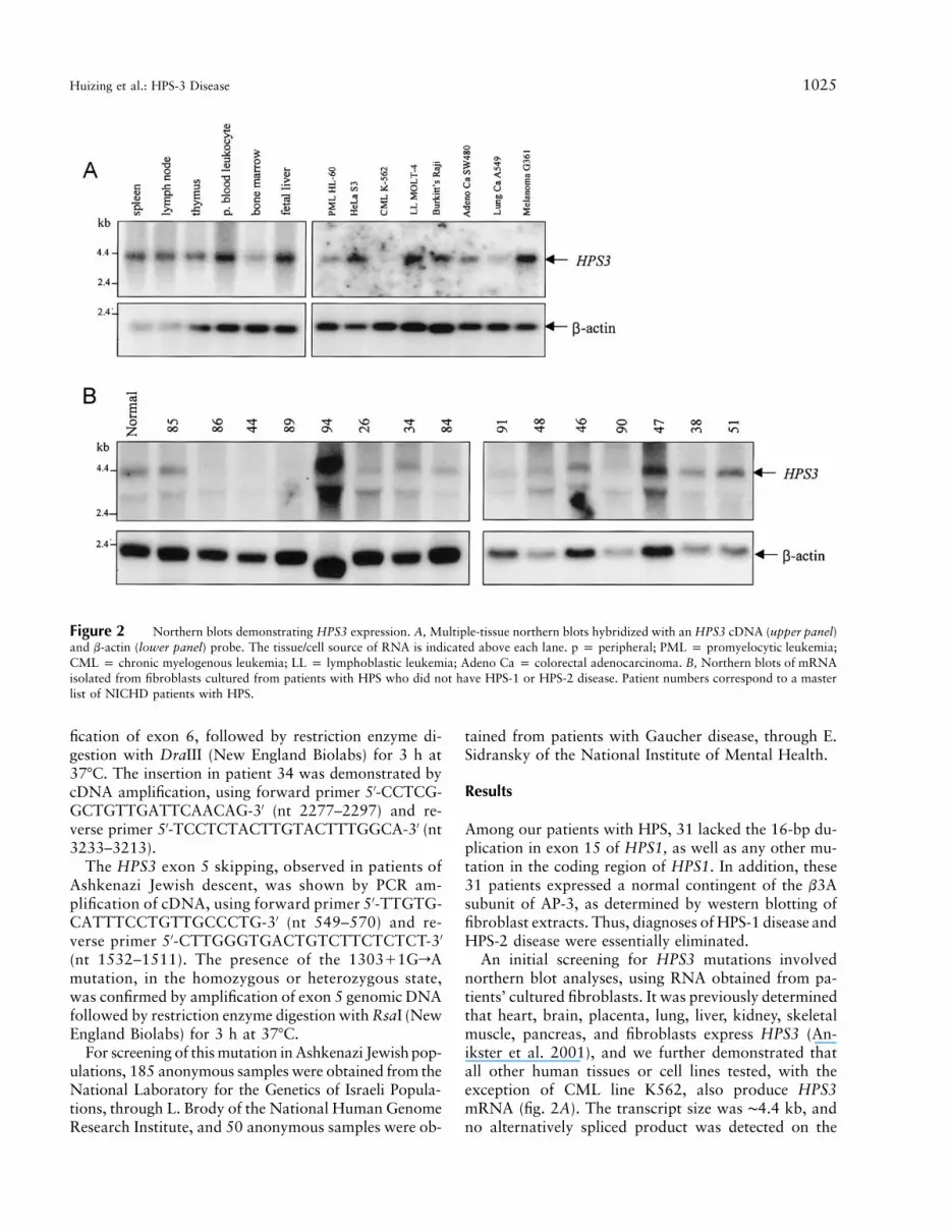
Figure 2 Northern blots demonstrating HPS3 expression. A, Multiple-tissue northern blots hybridized with an HPS3 cDNA (upper panel)and b-actin (lower panel) probe. The tissue/cell source of RNA is indicated above each lane. p p peripheral; PML p promyelocytic leukemia;CML p chronic myelogenous leukemia; LL p lymphoblastic leukemia; Adeno Ca p colorectal adenocarcinoma. B, Northern blots of mRNAisolated from fibroblasts cultured from patients with HPS who did not have HPS-1 or HPS-2 disease. Patient numbers correspond to a masterlist of NICHD patients with HPS.
fication of exon 6, followed by restriction enzyme di-gestion with DraIII (New England Biolabs) for 3 h at37�C. The insertion in patient 34 was demonstrated bycDNA amplification, using forward primer 5′-CCTCG-GCTGTTGATTCAACAG-3′ (nt 2277–2297) and re-verse primer 5′-TCCTCTACTTGTACTTTGGCA-3′ (nt3233–3213).
The HPS3 exon 5 skipping, observed in patients ofAshkenazi Jewish descent, was shown by PCR am-plification of cDNA, using forward primer 5′-TTGTG-CATTTCCTGTTGCCCTG-3′ (nt 549–570) and re-verse primer 5′-CTTGGGTGACTGTCTTCTCTCT-3′
(nt 1532–1511). The presence of the 1303�1GrAmutation, in the homozygous or heterozygous state,was confirmed by amplification of exon 5 genomic DNAfollowed by restriction enzyme digestion with RsaI (NewEngland Biolabs) for 3 h at 37�C.
For screening of this mutation in Ashkenazi Jewish pop-ulations, 185 anonymous samples were obtained from theNational Laboratory for the Genetics of Israeli Popula-tions, through L. Brody of the National Human GenomeResearch Institute, and 50 anonymous samples were ob-
tained from patients with Gaucher disease, through E.Sidransky of the National Institute of Mental Health.
Results
Among our patients with HPS, 31 lacked the 16-bp du-plication in exon 15 of HPS1, as well as any other mu-tation in the coding region of HPS1. In addition, these31 patients expressed a normal contingent of the b3Asubunit of AP-3, as determined by western blotting offibroblast extracts. Thus, diagnoses of HPS-1 disease andHPS-2 disease were essentially eliminated.
An initial screening for HPS3 mutations involvednorthern blot analyses, using RNA obtained from pa-tients’ cultured fibroblasts. It was previously determinedthat heart, brain, placenta, lung, liver, kidney, skeletalmuscle, pancreas, and fibroblasts express HPS3 (An-ikster et al. 2001), and we further demonstrated thatall other human tissues or cell lines tested, with theexception of CML line K562, also produce HPS3mRNA (fig. 2A). The transcript size was ∼4.4 kb, andno alternatively spliced product was detected on the
1026 Am. J. Hum. Genet. 69:1022–1032, 2001
Table 1
Characteristics of Patients with Mutations in HPS3
PATIENT
NUMBER
AGE
(YEARS),SEX ANCESTRYa
HPS3 MUTATIONVISUAL
ACUITYb
(OD, OS) BLEEDINGc
FVC(%)d GIe COMMENTSf1 2
26 3, F AJ/Iri-Ger 1303�1GrA 2621�2ArG 60, 60 E, Br NA � 11q24 Del91 7, M Ger/Swi 2729�1GrC C1329T 160, 200 E, Br 75 � …90 12, F PR/Ita Del3904bp Unknown 63, 80 Br 86 � Central PR34 15, M Iri/Eng 3027Ins89bp Unknown 100, 125 E, Br 90 � PDA86 25, F AJ/AJ 1303�1GrA 1831�2TrG 160, 100 E, Br, M 96 � OA44 30, F AJ/AJ 1303�1GrA 1303�1GrA 125, 100 E, Br, M 84 � Transfusion100 44, F AJ/AJ 1303�1GrA 1303�1GrA 100, 160 E, Br 92 � OA89 52, F AJ/AJ 1303�1GrA 1303�1GrA 80, 100 Br, M 93 � …
a AJ p Ashkenazi Jewish; Iri p Irish; Ger p German; Swi p Swiss; PR p Puerto Rican; Ita p Italian; Eng p English.b Visual acuity expressed in terms of vision at 20 feet (e.g., 160 indicates 20/160 acuity). OD p right eye; OS p left eye. Patient 26 had
an acuity of 10/30 when tested by use of Allen cards.c E p epistaxis; Br p bruising; M p menorrhagia.d FVC p forced vital capacity (expressed as percent of predicted value). Patient 91 may have been too young to provide optimal effort.e GI p gastrointestinal symptoms of colitis; � p absent.f Patient 26 has Jacobsen’s syndrome, with an 11q24-11qter deletion. The father of patient 90 is from the central Puerto Rican area where
the 3,904-bp deletion in HPS3 was found. PDA p patent ductus arteriosus. OA p ocular albinism. Patient 44 required a transfusion forbleeding after wisdom teeth removal.
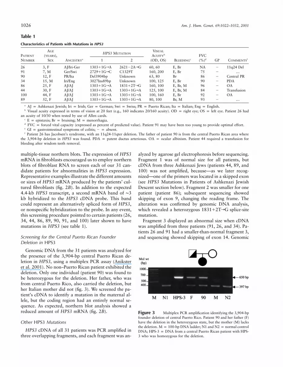
Figure 3 Multiplex PCR amplification identifying the 3,904-bpfounder deletion of central Puerto Rico. Patient 90 and her father (F)have the deletion in the heterozygous state, but the mother (M) lacksthe deletion. M p 100-bp DNA ladder; N1 and N2 p normal controlDNA; HPS-3 p DNA from a central Puerto Rican patient with HPS-3 who was homozygous for the deletion.
multiple-tissue northern blots. The expression of HPS3mRNA in fibroblasts encouraged us to employ northernblots of fibroblast RNA to screen each of our 31 can-didate patients for abnormalities in HPS3 expression.Representative examples illustrate the different amountsor sizes of HPS3 mRNA produced by the patients’ cul-tured fibroblasts (fig. 2B). In addition to the expected4.4-kb HPS3 transcript, a second mRNA band of ∼3kb hybridized to the HPS3 cDNA probe. This bandcould represent an alternatively spliced form of HPS3,or nonspecific hybridization to the probe. In any event,this screening procedure pointed to certain patients (26,34, 44, 86, 89, 90, 91, and 100) later shown to havemutations in HPS3 (see table 1).
Screening for the Central Puerto Rican FounderDeletion in HPS3
Genomic DNA from the 31 patients was analyzed forthe presence of the 3,904-bp central Puerto Rican de-letion in HPS3, using a multiplex PCR assay (Aniksteret al. 2001). No non–Puerto Rican patient exhibited thedeletion. Only one individual (patient 90) was found tobe heterozygous for the deletion. Her father, who wasfrom central Puerto Rico, also carried the deletion, buther Italian mother did not (fig. 3). We screened the pa-tient’s cDNA to identify a mutation in the maternal al-lele, but the coding region had an entirely normal se-quence. As expected, northern blot analysis showed areduced amount of HPS3 mRNA (fig. 2B).
Other HPS3 Mutations
HPS3 cDNA of all 31 patients was PCR amplified inthree overlapping fragments, and each fragment was an-
alyzed by agarose gel electrophoresis before sequencing.Fragment 1 was of normal size for all patients, butcDNA from three Ashkenazi Jews (patients 44, 89, and100) was not amplified, because—as we later recog-nized—one of the primers was located in a skipped exon(see HPS3 Mutations in Patients of Ashkenazi JewishDescent section below). Fragment 2 was smaller for onepatient (patient 86); subsequent sequencing showedskipping of exon 9, changing the reading frame. Thealteration was confirmed by genomic DNA analysis,which revealed a heterozygous 1831�2TrG splice-sitemutation.
Fragment 3 displayed an abnormal size when cDNAwas amplified from three patients (91, 26, and 34). Pa-tients 26 and 91 had a smaller-than-normal fragment 3,and sequencing showed skipping of exon 14. Genomic
Huizing et al.: HPS-3 Disease 1027
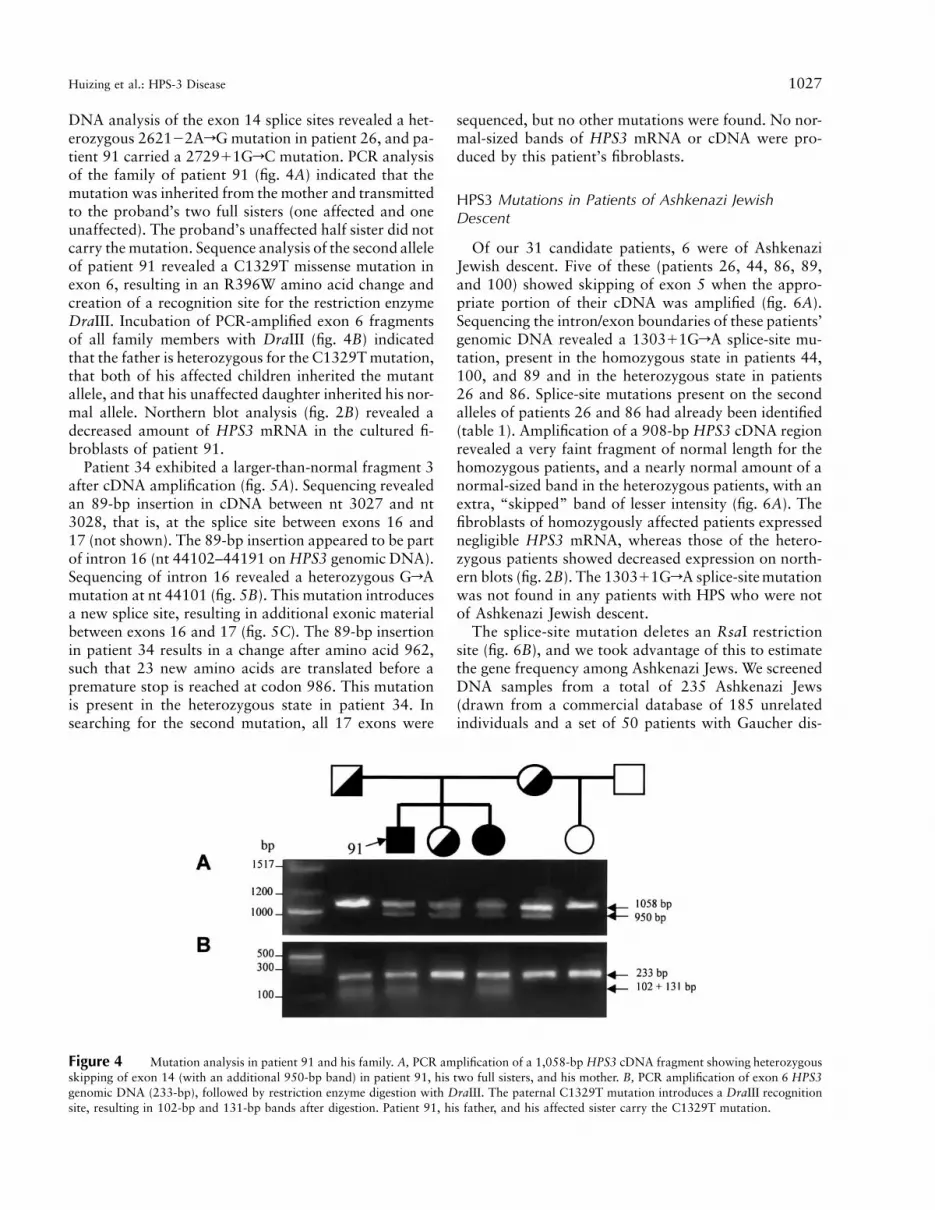
Figure 4 Mutation analysis in patient 91 and his family. A, PCR amplification of a 1,058-bp HPS3 cDNA fragment showing heterozygousskipping of exon 14 (with an additional 950-bp band) in patient 91, his two full sisters, and his mother. B, PCR amplification of exon 6 HPS3genomic DNA (233-bp), followed by restriction enzyme digestion with DraIII. The paternal C1329T mutation introduces a DraIII recognitionsite, resulting in 102-bp and 131-bp bands after digestion. Patient 91, his father, and his affected sister carry the C1329T mutation.
DNA analysis of the exon 14 splice sites revealed a het-erozygous 2621�2ArG mutation in patient 26, and pa-tient 91 carried a 2729�1GrC mutation. PCR analysisof the family of patient 91 (fig. 4A) indicated that themutation was inherited from the mother and transmittedto the proband’s two full sisters (one affected and oneunaffected). The proband’s unaffected half sister did notcarry the mutation. Sequence analysis of the second alleleof patient 91 revealed a C1329T missense mutation inexon 6, resulting in an R396W amino acid change andcreation of a recognition site for the restriction enzymeDraIII. Incubation of PCR-amplified exon 6 fragmentsof all family members with DraIII (fig. 4B) indicatedthat the father is heterozygous for the C1329T mutation,that both of his affected children inherited the mutantallele, and that his unaffected daughter inherited his nor-mal allele. Northern blot analysis (fig. 2B) revealed adecreased amount of HPS3 mRNA in the cultured fi-broblasts of patient 91.
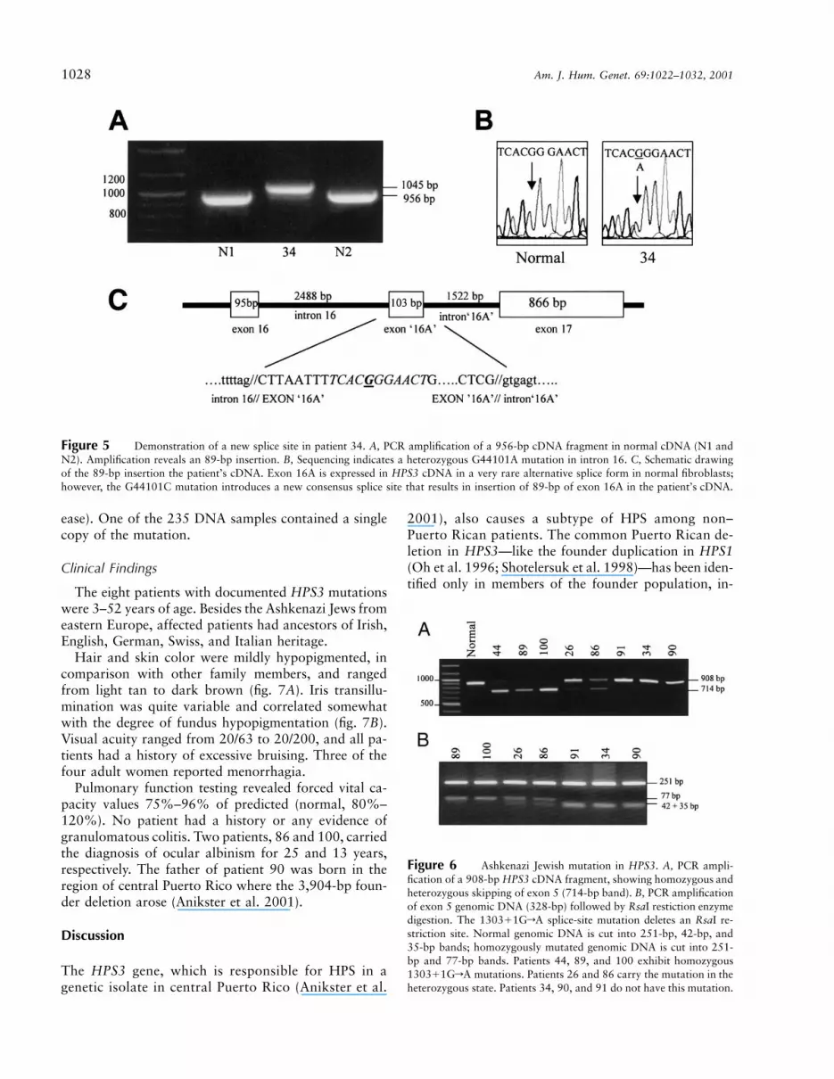
Patient 34 exhibited a larger-than-normal fragment 3after cDNA amplification (fig. 5A). Sequencing revealedan 89-bp insertion in cDNA between nt 3027 and nt3028, that is, at the splice site between exons 16 and17 (not shown). The 89-bp insertion appeared to be partof intron 16 (nt 44102–44191 on HPS3 genomic DNA).Sequencing of intron 16 revealed a heterozygous GrAmutation at nt 44101 (fig. 5B). This mutation introducesa new splice site, resulting in additional exonic materialbetween exons 16 and 17 (fig. 5C). The 89-bp insertionin patient 34 results in a change after amino acid 962,such that 23 new amino acids are translated before apremature stop is reached at codon 986. This mutationis present in the heterozygous state in patient 34. Insearching for the second mutation, all 17 exons were
sequenced, but no other mutations were found. No nor-mal-sized bands of HPS3 mRNA or cDNA were pro-duced by this patient’s fibroblasts.
HPS3 Mutations in Patients of Ashkenazi JewishDescent
Of our 31 candidate patients, 6 were of AshkenaziJewish descent. Five of these (patients 26, 44, 86, 89,and 100) showed skipping of exon 5 when the appro-priate portion of their cDNA was amplified (fig. 6A).Sequencing the intron/exon boundaries of these patients’genomic DNA revealed a 1303�1GrA splice-site mu-tation, present in the homozygous state in patients 44,100, and 89 and in the heterozygous state in patients26 and 86. Splice-site mutations present on the secondalleles of patients 26 and 86 had already been identified(table 1). Amplification of a 908-bp HPS3 cDNA regionrevealed a very faint fragment of normal length for thehomozygous patients, and a nearly normal amount of anormal-sized band in the heterozygous patients, with anextra, “skipped” band of lesser intensity (fig. 6A). Thefibroblasts of homozygously affected patients expressednegligible HPS3 mRNA, whereas those of the hetero-zygous patients showed decreased expression on north-ern blots (fig. 2B). The 1303�1GrA splice-site mutationwas not found in any patients with HPS who were notof Ashkenazi Jewish descent.
The splice-site mutation deletes an RsaI restrictionsite (fig. 6B), and we took advantage of this to estimatethe gene frequency among Ashkenazi Jews. We screenedDNA samples from a total of 235 Ashkenazi Jews(drawn from a commercial database of 185 unrelatedindividuals and a set of 50 patients with Gaucher dis-
1028 Am. J. Hum. Genet. 69:1022–1032, 2001
Figure 5 Demonstration of a new splice site in patient 34. A, PCR amplification of a 956-bp cDNA fragment in normal cDNA (N1 andN2). Amplification reveals an 89-bp insertion. B, Sequencing indicates a heterozygous G44101A mutation in intron 16. C, Schematic drawingof the 89-bp insertion the patient’s cDNA. Exon 16A is expressed in HPS3 cDNA in a very rare alternative splice form in normal fibroblasts;however, the G44101C mutation introduces a new consensus splice site that results in insertion of 89-bp of exon 16A in the patient’s cDNA.
Figure 6 Ashkenazi Jewish mutation in HPS3. A, PCR ampli-fication of a 908-bp HPS3 cDNA fragment, showing homozygous andheterozygous skipping of exon 5 (714-bp band). B, PCR amplificationof exon 5 genomic DNA (328-bp) followed by RsaI restiction enzymedigestion. The 1303�1GrA splice-site mutation deletes an RsaI re-striction site. Normal genomic DNA is cut into 251-bp, 42-bp, and35-bp bands; homozygously mutated genomic DNA is cut into 251-bp and 77-bp bands. Patients 44, 89, and 100 exhibit homozygous1303�1GrA mutations. Patients 26 and 86 carry the mutation in theheterozygous state. Patients 34, 90, and 91 do not have this mutation.
ease). One of the 235 DNA samples contained a singlecopy of the mutation.
Clinical Findings
The eight patients with documented HPS3 mutationswere 3–52 years of age. Besides the Ashkenazi Jews fromeastern Europe, affected patients had ancestors of Irish,English, German, Swiss, and Italian heritage.
Hair and skin color were mildly hypopigmented, incomparison with other family members, and rangedfrom light tan to dark brown (fig. 7A). Iris transillu-mination was quite variable and correlated somewhatwith the degree of fundus hypopigmentation (fig. 7B).Visual acuity ranged from 20/63 to 20/200, and all pa-tients had a history of excessive bruising. Three of thefour adult women reported menorrhagia.
Pulmonary function testing revealed forced vital ca-pacity values 75%–96% of predicted (normal, 80%–120%). No patient had a history or any evidence ofgranulomatous colitis. Two patients, 86 and 100, carriedthe diagnosis of ocular albinism for 25 and 13 years,respectively. The father of patient 90 was born in theregion of central Puerto Rico where the 3,904-bp foun-der deletion arose (Anikster et al. 2001).
Discussion
The HPS3 gene, which is responsible for HPS in agenetic isolate in central Puerto Rico (Anikster et al.
2001), also causes a subtype of HPS among non–Puerto Rican patients. The common Puerto Rican de-letion in HPS3—like the founder duplication in HPS1(Oh et al. 1996; Shotelersuk et al. 1998)—has been iden-tified only in members of the founder population, in-

Huizing et al.: HPS-3 Disease 1029
Figure 7 Hypopigmentation of hair, iris, and retina in patients with HPS-3. A, Hair pigmentation in patients of increasing age (see table1). Color ranges from tan (patient 34) to dark brown (patient 90). Patient 100 tints her hair lighter, but her eyebrows are natural in color. B,Increasing severity of iris transillumination (top panel) and retinal hypopigmentation (bottom panel) in four patients with HPS-3. The pictureon the left shows the retina of an unaffected control subject. Iris transillumination consists of light transmitted through the iris as a result ofreduced pigment in that tissue. The normal iris remains dark when light is shone through the pupil. In the HPS-3 retinas, patchy hypopigmentationresults from a reduction of pigment epithelium compared with normal retina (left). The degree of fundus hypopigmentation correspondssomewhatwith the extent of iris transillumination.
cluding one Puerto Rican/Italian patient (patient 90)who inherited the deletion from her father (fig. 3).Among non–Puerto Rican patients with HPS, however,other HPS3 mutations give rise to HPS-3 disease.
For example, patient 91 is heterozygous for a splicingmutation that removes exon 14, consisting of 108 bp.The 36 amino acids eliminated by this in-frame deletionare relatively conserved (81%) between humans andmice (Huizing et al. in press), indicating the importanceof the region. The decrease in HPS3 mRNA (fig. 2B)suggests that the truncated message is unstable. Thepatient’s second mutation consists of an R397W mis-sense mutation involving an arginine residue that is con-served from mice to humans but is not part of a specificsignaling or binding domain. PCR amplification andrestriction-site analysis verified that the patients’ twomutations segregate with HPS-3 disease in the family(fig. 4).
Patient 34 exhibited an 89-bp insertion of a portionof intron 16. In attempting to explain how this insertionarose, we noted that FLJ22704, a cDNA clone codingfor a protein of unknown function, contains a 103-bpinsertion between HPS exons 16 and 17 (called “exon16A,” nt 44089–44191). We speculate that this clonerepresents an alternatively spliced form of HPS3 that isnormally very weakly expressed. (In fact, we were ableto amplify a band from fibroblast cDNA, using a primerlocated in exon 16A). In patient 34, a GrA mutationat nt 44101, within exon 16A, creates a very strong new
splice site, resulting in abundant expression of the 89-bp insertion. The mutated RNA, which contains 23 newamino acids and a premature stop at codon 986, ap-pears stable, because northern blot analysis shows anmRNA signal slightly larger than the normal 4.4-kbHPS3 mRNA transcript. It is even possible that trans-lation of a truncated protein takes place in this patient.
The second mutation of patient 34, a mutation thatis as yet unidentified, is probably severe, because nonormal-sized HPS3 mRNA is expressed by the patient’sfibroblasts (fig. 2B) and because no normal band ap-pears on PCR amplification of the cDNA region thatincludes exons 16 and 17 (fig. 5A). This suggests a largedeletion or a promoter mutation.
Perhaps our most significant finding is the existenceof a common mutation in HPS3 among patients of Ash-kenazi Jewish descent. Five of our six Ashkenazi pa-tients with HPS had mutations in HPS3, and 8 of their10 alleles carried the 1303�1GrA splice-site mutationthat removes exon 5. This finding, along with the iden-tification of one heterozygous carrier of the mutationamong 235 DNA samples from anonymous AshkenaziJews, suggests a founder effect among the Ashkenazim.HPS is not recognized as a disorder that affects Jews(Zlotogora et al. 2000), nor has it been listed amongthe types of albinism in Israel (Gershoni-Baruch et al.1994). Certainly, the mildness of the patients’ hypopig-mentation and bleeding contributes to this situation.Nevertheless, our findings show a need for heightened

1030 Am. J. Hum. Genet. 69:1022–1032, 2001
awareness of the possibility of HPS among AshkenaziJews, particularly those with some degree of albinismor platelet storage-pool deficiency. For these individuals,HPS-3 disease and the 1303�1GrA splicing mutationin HPS3 should be considered first. Further screeningof Ashkenazi Jewish populations may help to determinethe true frequency of the exon 5 splicing mutation inthis population, and haplotype analysis may allow es-timation of when the putative founder mutation oc-curred (Luria and Delbruck 1943; Anikster et al. 2001).
The preponderance of splicing mutations in patientswho have HPS-3 along with drastically reduced mRNAproduction suggests that, in general, missense mutationsmay not result in a clinically recognizable phenotype.The HPS-3 disease associated with severe HPS3 mu-tations is relatively mild, so patients with less-severemutations may be indistinguishable from individualswithout HPS3 mutations.
The non–Puerto Rican HPS-3 patients we describehere enhance our understanding of HPS in other ways.We know that the HPS3 founder mutation causes rel-atively mild disease among the few central Puerto Ri-cans we have examined (Hazelwood et al. 1997; An-ikster et al. 2001). Clearly, the same is true for otherHPS3 mutations affecting non–Puerto Ricans. Hair hy-popigmentation (fig. 7A), iris transillumination (fig.7B), and visual acuity deficits (table 1) were less severethan those typically seen among patients with HPS-1(Iwata et al. 2000). In the present study, two individuals(patients 86 and 100) had such mild skin and hair hy-popigmentation that they carried the diagnosis of oc-ular, rather than oculocutaneous, albinism for morethan a decade. The patients’ histories of bleeding werenot impressive; reduction in forced vital capacity wasminimal, if present at all; and no history of colitis waselicited. This sample of patients with HPS-3 is small,however, and more and older patients must be examinedto determine whether pulmonary and gastrointestinaldisease will accompany HPS3 mutations.
Strategies for the molecular diagnosis of patients withHPS are now becoming better defined. Obviously, pa-tients of northwest Puerto Rican ancestry should bestudied by PCR amplification for the 16-bp duplicationin HPS1 (Oh et al. 1996), and patients from centralPuerto Rico should be analyzed by use of multiplex PCRfor the 3,904-bp deletion in HPS3 (Anikster et al. 2001).The few patients with HPS who have histories of child-hood infections or neutropenia can be suspected of hav-ing HPS-2, with mutations in ADTB3A (Dell’Angelicaet al. 1999; Shotelersuk et al. 2000). Ashkenazi Jewishindividuals, especially those with mild phenotypes,should be examined using restriction enzyme analysisof genomic DNA to detect the 1303�1GrA splice-sitemutation. Other mildly affected patients can be inves-tigated by amplifying HPS3 cDNA in three overlapping
fragments or, if RNA is not available, by amplifyingand sequencing genomic DNA exon by exon. HPS1mutations can be pursued in a similar fashion. As anestimate of the relative likelihood of HPS-1, HPS-2, andHPS-3 disease, we found 7 patients (6 families) withHPS1 mutations, 3 patients (2 families) with ADTB3Amutations, and 8 patients (8 families) with HPS3 mu-tations among 43 patients with HPS (38 families) nothomozygous for the founder HPS1 or HPS3 mutations.
We expect that each HPS-causing gene will provideunique insights into the cell biology of vesicle formation,specifically the creation of melanosomes, dense bodies,and lysosomes from the trans-Golgi network. These or-ganelles are considered to have a common genesis andare known to share certain integral membrane proteins(Shotelersuk et al. 1998; Huizing et al. 2000); they arealso abnormal to various degrees in patients with HPS.Patients with HPS-2 provide the most-direct evidencethat HPS is a disorder of vesicle formation. In theseindividuals, ADTB3A mutations cause defective pro-duction of b3A, a subunit of the coat protein AP-3. Thisheterotetrameric complex mediates the formation ofvesicles, apparently including the melanosome, densebody, and lysosome. HPS-2 fibroblasts display mistraf-ficking of the lysosomal membrane protein CD63through the plasma membrane (Dell’Angelica et al.1999). HPS-2 melanocytes exhibit misrouting of tyros-inase, which does not display a normal melanosomaldistribution (Huizing et al. 2001a). The association ofb3A mutations with HPS and with membrane-traffick-ing defects suggests that the products of HPS1 andHPS3 also function in vesicle formation, although nodirect evidence to this effect has been forthcoming.It will be of critical importance to determine whetherthe HPS3 gene product interacts with either the HPS1or the ADTB3A gene product, and these studies areongoing.
Because the phenotype of HPS can be extremely mildwith respect to both pigmentation and bleeding, it maybe appropriate to screen for HPS among patients whohave some degree of hypopigmentation. Three separateHPS-causing genes are known, so molecular studiescould confirm the initial diagnosis. Identification ofpatients with a bleeding diathesis among those withhypopigmentation would assist in prognosis as well asprophylaxis against serious bleeding episodes or ex-posure to lung toxins in selected patients. We suggestthat HPS screening be performed by whole-mount elec-tron microscopy for identification of platelet dense gran-ules in individuals with oculocutaneous, or even ocular,albinism. A recent study showed that 35% of a Germanalbino population had no mutations in either the ty-rosinase or the P gene (Passmore et al. 1999); thesepatients are candidates for having mild HPS.

Huizing et al.: HPS-3 Disease 1031
Acknowledgments
Lessie McCain of the National Eye Institute provided superbpersonalized ophthalmic nursing care to our patients. The au-thors also appreciate the skill and expertise in ophthalmic pho-tography provided by Ernest Kuehl, Patrick Ciatto, and Mar-ilois Palmer of the National Eye Institute. Isa Bernardiniprovided excellent technical assistance. Drs. Ellen Sidransky,of the National Institute of Mental Health, and Dr. LawrenceBrody, of the National Human Genome Research Institute,kindly supplied anonymous Ashkenazi Jewish DNA samples.Y.A. is a Howard Hughes Medical Institute Physician Post-doctoral Fellow.
Electronic-Database Information
Accession numbers and URLs for data in this article are asfollows:
GenBank, http://www.ncbi.nlm.nih.gov/Genbank/ (forHSP1 [accession number U65676], ADTB3A [accessionnumber U91931], HPS3 genomic DNA [accession num-ber AF375663], and HPS3 cDNA [accession numberAY033141])
Online Mendelian Inheritance in Man (OMIM), http://www.ncbi.nlm.nih.gov/Omim/ (for OCA1 [MIM 203100],OCA2 [MIM 203200], OCA3 [MIM 203290], Chediak-Higashi syndrome [MIM 214500], Griscelli syndrome[MIM 214450], Hermansky-Pudlak syndrome [MIM203300], HPS-1 [MIM 604982], HPS-2 [MIM 603401],and HPS-3 [MIM 606118])
References
Anikster Y, Huizing M, White J, Shevchenko YO, FitzpatrickDL, Touchman JW, Compton JG, Bale SJ, Swank RT, GahlWA, Toro JR (2001) Mutation of a new gene causes a uniqueform of Hermansky-Pudlak syndrome in a genetic isolate ofcentral Puerto Rico. Nat Genet 28:376–380
Bailin T, Oh J, Feng GH, Fukai K, Spritz RA (1997) Organ-ization and nucleotide sequence of the human Hermansky-Pudlak syndrome (HPS) gene. J Invest Dermatol 108:923–927
Brantly M, Avila NA, Shotelersuk V, Lucero C, Huizing M,Gahl WA (2000) Pulmonary function and high-resolutionCT findings in patients with an inherited form of pulmonaryfibrosis, Hermansky-Pudlak syndrome, due to mutations inHPS-1. Chest 117:129–136
Dell’Angelica EC, Ohno H, Ooi CE, Rabinovich E, Roche KW,Bonifacino JS (1997a) AP-3: an adaptor-like protein com-plex with ubiquitous expression. EMBO J 16:917–928
Dell’Angelica EC, Ooi CE, Bonifacino JS (1997b) b3A-adap-tin, a subunit of the adaptor-like complex AP-3. J Biol Chem272:15078–15084
Dell’Angelica EC, Shotelersuk V, Aguilar RC, Gahl WA, Bon-ifacino JS (1999) Altered trafficking of lysosomal proteinsin Hermansky-Pudlak syndrome due to mutations in theb3A subunit of the AP-3 adaptor. Mol Cell 3:11–21
Gahl WA, Brantly M, Kaiser-Kupfer MI, Iwata F, HazelwoodS, Shotelersuk V, Duffy LF, Kuehl EM, Troendle J, Bernar-
dini I (1998) Genetic defects and clinical characteristics ofpatients with a form of oculocutaneous albinism (Herman-sky-Pudlak syndrome). N Engl J Med 338:1258–1264
Garay SM, Gardella JE, Fazzini EP, Goldring RM (1979) Her-mansky-Pudlak syndrome: pulmonary manifestations of aceroid storage disorder. Am J Med 66:737–747
Gershoni-Barush R, Rosenmann A, Droetto S, Holmes S, Tri-pathi RK, Spritz RA (1994) Mutations of the tyrosinase genein patients with oculocutaneous albinism from various eth-nic groups in Israel. Am J Hum Genet 54:586–594
Griscelli C, Durandy A, Guy-Grand D, Daguillard F, HerzogC, Prunicras M (1978) A syndrome associating partial al-binism and immunodeficiency. Am J Med 65:691–702
Harmon KR, Witkop CJ, White JG, King RA, Peterson M,Moore D, Tashjian J, Marinelli WA, Bitterman PB (1994)Pathogenesis of pulmonary fibrosis: platelet-derived growthfactor precedes structural alterations in the Hermansky-Pud-lak syndrome. J Lab Clin Med 123:617–627
Hazelwood S, Shotelersuk V, Wildenberg SC, Chen D, IwataF, Kaiser-Kupfer MI, White JG, King RA, Gahl WA (1997)Evidence for locus heterogeneity in Puerto Ricans with Her-mansky-Pudlak syndrome. Am J Hum Genet 61:1088–1094
Hermansky F, Pudlak P (1959) Albinism associated with hem-orrhagic diathesis and unusual pigmented reticular cells inthe bone marrow: report of two cases with histochemicalstudies. Blood 14:162–169
Huizing M, Anikster Y, Gahl WA (2000) Hermansky-Pudlaksyndrome and related disorders of organelle formation. Traf-fic 1:823–835
Huizing M, Anikster Y, White JG, Gahl WA. Characterizationof the murine gene corresponding to human Hermansky-Pudlak syndrome type 3: exclusion of the subtle gray (sut)locus. Mol Genet Metab, in press
Huizing M, Saranjarajan R, Strovel E, Zhao Y, Gahl WA,Boissy RE (2001a) AP-3-dependent vesicles carry tyrosinase,but not TRP-1, in cultured human melanocytes. Mol BiolCell 12:2075–2085
Huizing M, Scher CD, Strovel E, Fitzpatrick DL, Anikster Y,Gahl WA (2001b) A new case of Hermansky-Pudlak syn-drome type 2 due to nonsense mutations in the b3A subunitof adaptor complex-3. Pigment Cell Res 14:228
Introne W, Boissy RE, Gahl WA (1999) Clinical, molecular,and cell biological aspects of Chediak-Higashi syndrome.Mol Genet Metab 68:283–303
Iwata F, Reed GF, Caruso RC, Kuehl EM, Gahl WA, Kaiser-Kupfer MI (2000) Correlation of visual acuity and ocularpigmentation with the 16-bp duplication in the HPS-1 geneof Hermansky-Pudlak syndrome, a form of albinism. Oph-thalmology 107:783–789
King RA, Hearing VJ, Creel DJ, Oetting WS (2001) Albinism.In: Scriver CR, Beaudet AL, Sly WS, Valle DL (eds) Themetabolic and molecular bases of inherited disease, 8th ed.McGraw-Hill, New York, pp 5587–5627
Luria SE, Delbruck M (1943) Mutations of bacteria from virussensitivity to virus resistance. Genetics 28:491–511
Mahadeo R, Markowitz J, Fisher S, Daum F (1991) Herman-sky-Pudlak syndrome with granulomatous colitis in chil-dren. J Pediatr 118:904–906
Oh J, Bailin T, Fukai K, Feng GH, Ho L, Mao J-i, Frenk E,Tamura N, Spritz RA (1996) Positional cloning of a gene

1032 Am. J. Hum. Genet. 69:1022–1032, 2001
for Hermansky-Pudlak syndrome, a disorder of cytoplasmicorganelles. Nat Genet 14:300–306
Oh J, Ho L, Ala-Mello S, Amato D, Armstrong L, Bellucci S,Carakushansky G, Ellis JP, Fong C-T, Green JS, Heon E,Legius E, Levin AV, Nieuwenhuis HK, Pinckers A, TamuraN, Whiteford ML, Yamasaki H, Spritz RA (1998) Mutationanalysis of patients with Hermansky-Pudlak syndrome: aframeshift hot spot in the HPS gene and apparent locusheterogeneity. Am J Hum Genet 62:593–598
Passmore LA, Kaesmann-Kellner B, Weber BHF (1999) Noveland recurrent mutations in the tyrosinase gene and the Pgene in the German albino population. Hum Genet 105:200–210
Sambrook J, Fritsch EF, Maniatis T (1989) Molecular cloning:a laboratory manual, 2nd ed. Cold Spring Harbor Labo-ratory, Cold Spring Harbor, NY
Schinella RA, Greco MA, Cobert BL, Denmark LW, Cox RP(1980) Hermansky-Pudlak syndrome with granulomatouscolitis. Ann Intern Med 92:20–23
Shotelersuk V, Dell’Angelica EC, Hartnell L, Bonifacino JS,Gahl WA (2000) A new variant of Hermansky-Pudlak syn-drome due to mutations in a gene responsible for vesicleformation. Am J Med 108:423–427
Shotelersuk V, Gahl WA (1998) Hermansky-Pudlak syndrome:models for intracellular vesicle formation. Mol Genet Metab65:85–96
Simon JW, Adams RJ, Calhoun JH, Shapiro SS, Ingerman CM(1982) Ophthalmic manifestations of the Hermansky-Pud-lak syndrome (oculocutaneous albinism and hemorrhagicdiathesis). Am J Ophthalmol 93:71–77
Simpson F, Bright NA, West MA, Newman LS, Darnell RB,Robinson MS (1996) A novel adaptor-related protein com-plex. J Cell Biol 133:749–760
Simpson F, Peden AA, Christopoulou L, Robinson MS (1997)
Characterization of the adaptor-related protein complex,AP-3. J Cell Biol 137:835–845
Summers CG, Knobloch WH, Witkop CJ, King RA (1988)Hermansky-Pudlak syndrome: ophthalmic findings. Oph-thalmology 95:545–554
Swank RT, Novak EK, McGarry MP, Rusiniak ME, Feng L(1998) Mouse models of Hermansky-Pudlak syndrome: areview. Pigment Cell Res 11:60–80
Toro J, Turner M, Gahl WA (1999) Dermatologic manifes-tations of Hermansky-Pudlak syndrome in patients with andwithout a 16-base pair duplication in the HPS1 gene. ArchDermatol 135:774–780
Wilson SM, Yip R, Swing DA, O’Sullivan TN, Zhang Y, NovakE, Swank RT, Russell LB, Copeland NG, Jenkins NA (2000)A mutation in Rab27a causes the vesicle transport defectsobserved in ashen mice. Proc Natl Acad Sci USA 97:7933–7938
Witkop CJ, Babcock MN, Rao GHR, Gaudier F, Summers CG,Shanahan F, Harmon KR, Townsend DW, Sedano HO, KingRA, Cal SX, White JG (1990) Albinism and Hermansky-Pudlak syndrome in Puerto Rico. Bol Asoc Med P Rico-Agosto 82:333–339
Witkop CJ, Krumwiede M, Sedano H, White JG (1987) Re-liability of absent platelet dense bodies as a diagnostic cri-terion for Hermansky-Pudlak syndrome. Am J Hematol 26:305–311
Witkop CJ Jr, White JG, Townsend D, Sedano HO, Cal SX,Babcock M, Krumwiede M, Keenan K, Love JE, Wolfe LS(1988) Ceroid storage disease in Hermansky-Pudlak syn-drome: induction in animal models. In: Zs.-Nagy I (ed) Li-pofuscin-1987: state of the art. Amsterdam, Elsevier, p 413
Zlotogora J, Bach G, Munnich A (2000) Molecular basis ofMendelian disorders among Jews. Mol Genet Metab 69:169–180


