University of Tennessee, Knoxville University of Tennessee, Knoxville
TRACE: Tennessee Research and Creative TRACE: Tennessee Research and Creative
Exchange Exchange
Doctoral Dissertations Graduate School
12-2007
Exploring the Mechanism of Meiosis in Exploring the Mechanism of Meiosis in Drosophila melanogaster: :
Meiotic Functions of a Novel Cohesion Protein SOLO and a Meiotic Functions of a Novel Cohesion Protein SOLO and a
Translation Initiation Factor VASA Translation Initiation Factor VASA
Rihui Yan University of Tennessee - Knoxville
Follow this and additional works at: https://trace.tennessee.edu/utk_graddiss
Part of the Biochemistry, Biophysics, and Structural Biology Commons
Recommended Citation Recommended Citation Yan, Rihui, "Exploring the Mechanism of Meiosis in Drosophila melanogaster: Meiotic Functions of a Novel Cohesion Protein SOLO and a Translation Initiation Factor VASA. " PhD diss., University of Tennessee, 2007. https://trace.tennessee.edu/utk_graddiss/280
This Dissertation is brought to you for free and open access by the Graduate School at TRACE: Tennessee Research and Creative Exchange. It has been accepted for inclusion in Doctoral Dissertations by an authorized administrator of TRACE: Tennessee Research and Creative Exchange. For more information, please contact [email protected].
To the Graduate Council:
I am submitting herewith a dissertation written by Rihui Yan entitled "Exploring the Mechanism
of Meiosis in Drosophila melanogaster: Meiotic Functions of a Novel Cohesion Protein SOLO
and a Translation Initiation Factor VASA." I have examined the final electronic copy of this
dissertation for form and content and recommend that it be accepted in partial fulfillment of the
requirements for the degree of Doctor of Philosophy, with a major in Biochemistry and Cellular
and Molecular Biology.
Bruce D. McKee, Major Professor
We have read this dissertation and recommend its acceptance:
Ranjan Ganguly, Jae Park, Mariano Labrador, Yisong Wang
Accepted for the Council:
Carolyn R. Hodges
Vice Provost and Dean of the Graduate School
(Original signatures are on file with official student records.)
To the graduate Council: I am submitting herewith a dissertation written by Rihui Yan entitled “Exploring the mechanism of meiosis in Drosophila melanogaster: Meiotic functions of a novel cohesion protein SOLO and a translation initiation factor VASA.” I have examined the final electronic copy of this dissertation for form and content and recommend that it be accepted in partial fulfillment of the requirements for the degree of Doctor of Philosophy, with a major in Biochemistry, Cellular and Molecular Biology. Bruce D. McKee Major Professor We have read this dissertation and recommend its acceptance: Ranjan Ganguly Jae Park Mariano Labrador Yisong Wang Accepted for the Council: Carolyn R. Hodges
Vice Provost and Dean of the Graduate School
(Original signatures are on file with official student records)

EXPLORING THE MECHANISM OF MEIOSIS IN DROSOPHILA
MELANOGASTER: MEIOTIC FUNCTIONS OF A NOVEL COHESION PROTEIN SOLO AND A TRANSLATION INITIATION FACTOR VASA
A Dissertation
Presented for the Doctor of Philosophy Degree
The University of Tennessee, Knoxville
Rihui Yan December 2007
ii
DEDICATION
To my wife, Hongmei Jia, for your endless love, support, and encouragement; my
parents, Shuliang Yan and Zhengmei Zhang for your faithful support; my
precious daughter Andrea Yan.
iii
ACKNOWLEDGEMENTS
This dissertation would have been impossible without the generous help
and support from many people. I would like to express my gratitude to all those
who gave me the possibility to complete this work.
First of all, I am deeply indebted to my advisor, Dr. Bruce McKee. Without
his excellent guidance, this work would not be possible. Above all and most
needed, he provided me constant encouragement, help, and support in various
ways. His advice and patience are deeply appreciated. His exceptional ability to
think deeply and creatively and his enthusiasm in science have greatly influenced
me and inspired me to grow not only as a student, but also a researcher and a
scientist.
I am particularly grateful to the members of my dissertation committee, Dr.
Ranjan Ganguly, Dr. Jae Park, Dr. Mariano Labrador and Dr. Yisong Wang for
the valuable suggestions, time, and support in my progression.
I wish to thank both past and present members of Dr. McKee’s laboratory
for their friendship and help: Dr. Sharon Thomas, Dr. Morvarid Bejnood, Hueiwen
Yeh, Jue-He Tsai, Jian Ma, and Jennifer Vandiver.
Finally, I would like to express my deepest gratitude for the constant
support, understanding and love from my wife Hongmei, my daughter Andrea,
and my parents during the past years.
iv
ABSTRACT
Sister chromatid cohesion is essential for proper chromosome segregation
during meiosis. However, the mechanism of meiotic cohesion in Drosophila is
unclear.
We describe a novel protein, SOLO (Sisters On the LOose) that is
essential for meiotic cohesion in Drosophila melanogaster. solo mutations cause
high nondisjunction of sister and homologous chromatids of sex chromosomes
and autosomes in both sexes. In solo males, sister chromatids separate
prematurely and segregate randomly during meiosis II. Although bivalents
appear intact throughout meiosis I, sister centromeres lose cohesion prior to
prometaphase I and orient nearly randomly on the meiosis I spindle.
Centromeric foci of SMC1 are absent in solo males at all meiotic stages. SOLO
and the cohesin protein SMC1 co-localize to meiotic centromeres from early
prophase I until anaphase II in wild-type males but both proteins are removed
prematurely from centromeres at anaphase I in mei-S332 mutants, coincident
with premature loss of cohesion in those mutants.
solo mutations in females cause reduced frequency of homologous
recombination between X chromosomes and autosomes, partially due to the loss
of inhibition of sister chromatid exchange. Synaptonemal complex assembly is
severely disrupted in early meiotic stage in solo females. SOLO colocalizes with
SMC1 and C(3)G in meiosis. Additionally, SOLO is required for stabilizing
chiasmata generated from residual recombination events.
The data about the phenotypes of solo males and females and
v
colocalization patterns of SOLO strongly suggest SOLO is a component of
potential cohesin in Drosophila meiosis.
Drosophila males undergo meiosis without recombination. However, the
underlying mechanism is not known. Mutations of vasa cause high frequency of
X-Y exchange in meiosis. Chromatin bridges at anaphase I and II, due to
dicentric recombination events, were observed in vasa males. vas and solo
double mutant showed precocious segregation of homologs at metaphase I
besides chromatin bridge at anaphase I and II. Our data thus for the first time
demonstrate that inhibition of meiotic recombination during male meiosis requires
vas function and interactions between vas and solo regulate chromosome
dynamics in male meiosis.
vi
TABLE OF CONTENTS CHAPTER 1 – GENERAL INTRODUCTION........................................................ 1 CHAPTER 2 - SOLO IS A NOVEL PROTEIN REQUIRED FOR SISTER CHROMATID COHESION, SISTER CENTROMERE CO-ORIENTATION, AND CENTROMERIC LOCALIZATION OF SMC1 IN DROSOPHILA MEIOSIS ........ 35
INTRODUCTION ............................................................................................ 37 MATERIALS AND METHODS ........................................................................ 42 RESULTS ....................................................................................................... 49 DISCUSSION.................................................................................................. 85
CHAPTER 3 - SOLO IS A COHESION PROTEIN REQUIRED FOR FORMATION OF SYNAPTONEMAL COMPLEX, MAINTENANCE OF CHIASMATA, AND PROMOTING HOMOLOG RECOMBINATION IN DROSOPHILA MEIOSIS .................................................................................... 94
INTRODUCTION ............................................................................................ 96 MATERIALS AND METHODS ...................................................................... 100 RESULTS ..................................................................................................... 108 DISCUSSION................................................................................................ 130 APPENDICES............................................................................................... 137
CHAPTER 4 - NOVEL ROLES OF VASA IN DROSOPHILA MALE MEIOSIS: HOMOLOGOUS RECOMBINATION AND CHROMOSOME SEGREGATION 138
INTRODUCTION .......................................................................................... 140 MATERIALS AND METHODS ...................................................................... 144 RESULTS ..................................................................................................... 147 DISCUSSION................................................................................................ 163
CHAPTER 5 – GENERAL CONCLUSIONS AND FUTURE DIRECTIONS ...... 167
GENERAL CONCLUSIONS.......................................................................... 168 FUTURE DIRECTIONS ................................................................................ 172
LIST OF REFERENCES .................................................................................. 175 VITA ................................................................................................................. 191
vii
LIST OF FIGURES Chapter 1 – General introduction Figure 1-1. Stages of meiosis. ............................................................................. 3 Figure 1-2. Nondisjunction of meiotic chromosomes. .......................................... 6 Figure 1-3. The structure of rDNA region on X and Y chromosomes. ............... 29 Figure 1-4. Schematic drawings of Drosophila ovariole and germarium............ 31 Chapter 2 – SOLO is a novel protein required for sister chromatid cohesion, sister centromere co-orientation, and centromeric localization of SMC1 in Drosophila meiosis Figure 2-1. Chromosome segregation in solo and solo; snm spermatocytes. ... 53 Figure 2-2. Sister centromeres separate prematurely in solo mutants. ............. 58 Figure 2-3. SNM localizes normally to the X-Y bivalent during meiosis I in solo
mutants........................................................................................................ 61 Figure 2-4. Localization of SMC1 in wild-type and solo spermatocytes............. 64 Figure 2-5. Arm cohesion in spermatogonia and early prophase I spermatocytes
in solo males hemizygous for an inserted lacO array on chromosome 2..... 67 Figure 2-6. Molecular characterization of solo. .................................................. 69 Figure 2-7. Co-localization of Venus-SOLO and CID on meiotic centromeres. . 74 Figure 2-8. Venus-SOLO foci in wild-type (A) and mei-S332 (B) spermatocytes.
.................................................................................................................... 76 Figure 2-9. Diffuse Venus-SOLO foci during late prophase I............................. 79 Figure 2-10. Co-localization of Venus-SOLO and SMC1 foci on centromeres in
wild-type and mei-S332 spermatocytes....................................................... 80 Figure 2- 11. SOLO is absent at centromeres from early prophase I but present
in spermatogonia......................................................................................... 83 Chapter 3 – SOLO is a cohesion protein required for formation of synaptonemal complex, maintenance of chiasmata, and promoting homolog recombination in Drosophila meiosis
Figure 3-1. The chromosome segregation pattern and chiasmata stability test in
solo females when recombination occurs.................................................. 102 Figure 3-2. solo mutations disrupt the assembly and maintenance of the
synaptonemal complex.............................................................................. 118 Figure 3-3. Venus-SOLO foci are abundant at centromeres. .......................... 122 Figure 3-4. Thread-like SOLO structures localizes to SC. ............................... 124 Figure 3-5. SOLO and SMC1 colocalize together............................................ 128 Chapter 4 – novel roles of VASA in Drosophila male meiosis: homologous recombination and chromosome segregation
viii
Figure 4-1. Abdomen pigment in different males. ............................................ 149 Figure 4-2. The structure of X and Y chromosomes used in crosses and the
predicted exchange patterns at rDNA loci. ................................................ 153 Figure 4-3. vas spermatocytes exhibit chromatin bridges and chromatin mass at
anaphase I and II....................................................................................... 158 Figure 4-4. Localization of MNM in vas6356-001 spermatocytes. ........................ 161 Figure 4-5. Cytological phenotypes of vas7 spermatocytes. ............................ 162
ix
LIST OF TABLES
Chapter 1 – General introduction Table 1-1. Cohesin complex across species in mitosis and meiosis. ................ 14 Chapter 2 – SOLO is a novel protein required for sister chromatid cohesion, sister centromere co-orientation, and centromeric localization of SMC1 in Drosophila meiosis
Table 2-1. Sex chromosome nondisjunction in solo males................................ 50 Table 2-2. 2nd chromosome NDJ...................................................................... 51 Table 2-3. Transgene rescue data..................................................................... 72 Chapter 3 – SOLO is a cohesion protein required for formation of synaptonemal complex, maintenance of chiasmata, and promoting homolog recombination in Drosophila meiosis
Table 3-1. X chromosome recombination and nondisjunction in solo females.109 Table 3-2. Second chromosome NDJ.............................................................. 110 Table 3-3. 2nd chromosome recombination in solo and control females......... 112 Table 3-4. Sister chromatid exchange is reduced in solo mutants................... 114 Table 3-5. Chiasmata stability is defective in solo mutant. .............................. 116 Table 3-6. Venus-SOLO transgenes completely rescue solo phenotypes in
females...................................................................................................... 120 Table A-1. Ring/Rod and NDJ test for solo mutants ......................................... 137 Chapter 4 – novel roles of VASA in Drosophila male meiosis: homologous recombination and chromosome segregation
Table 4-1. X-Y NDJ and exchange in crosses of +/BSYy+; vas/Df males X y w
females...................................................................................................... 148
1
CHAPTER 1 – GENERAL INTRODUCTION
2
Overview: meiosis is a specialized cell division
Accurate segregation of chromosomes during meiosis is required for the
proper transmission of genetic material during sexual reproduction. Errors in
meiotic chromosome segregation result in aneuploidy, an aberrant number of
chromosomes. Aneuploidy is the primary cause of miscarriage in human beings.
Approximately 35% of all miscarriages result from aneuploidy. Moreover,
aneuploidy is the leading genetic cause of developmental and mental disorders,
such as Down syndrome, which is caused by an extra copy of chromosome 21
(Hassold and Hunt, 2001). The study of the mechanism of meiosis thus has
potential clinical relevance to human beings.
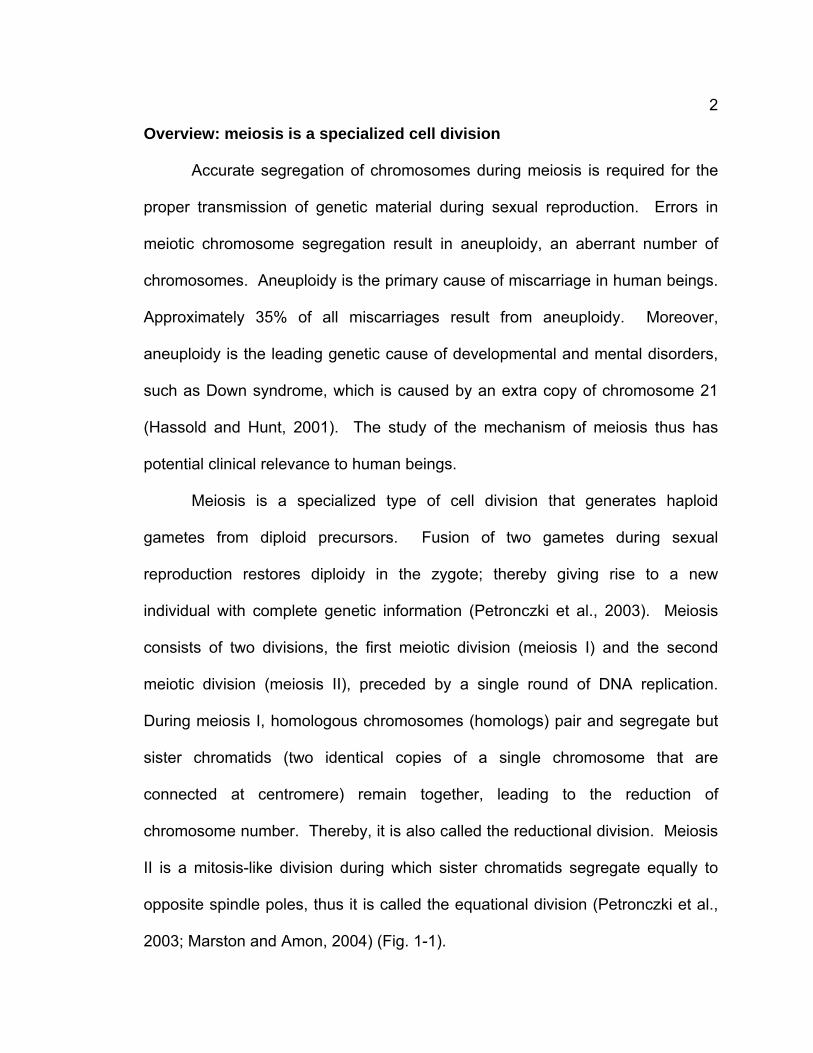
Meiosis is a specialized type of cell division that generates haploid
gametes from diploid precursors. Fusion of two gametes during sexual
reproduction restores diploidy in the zygote; thereby giving rise to a new
individual with complete genetic information (Petronczki et al., 2003). Meiosis
consists of two divisions, the first meiotic division (meiosis I) and the second
meiotic division (meiosis II), preceded by a single round of DNA replication.
During meiosis I, homologous chromosomes (homologs) pair and segregate but
sister chromatids (two identical copies of a single chromosome that are
connected at centromere) remain together, leading to the reduction of
chromosome number. Thereby, it is also called the reductional division. Meiosis
II is a mitosis-like division during which sister chromatids segregate equally to
opposite spindle poles, thus it is called the equational division (Petronczki et al.,
2003; Marston and Amon, 2004) (Fig. 1-1).
3
Figure 1-1. Stages of meiosis.
Through meiosis, one diploid parental cell divides into four haploid daughter
cells. See the text for details.
Meiosis I: homologous chromosomes segregate
Meiosis II: sister chromatids segregate
S phase: DNA replication
4
The reductional division is divided into five stages: prophase I,
prometaphase I, metaphase I, anaphase I, telophase I. Prophase I is the first
stage in meiosis I when diploid cells enter meiosis. During prophase I homologs
pair and form synaptonemal complexes (SCs, the proteinaceous structures that
form between homologs during prophase I), recombine and form crossovers in
most organisms. The crossovers lead to the formation of chiasmata, which
connect homologous chromosomes when SCs are gone. The paired
chromosomes are called bivalents, each consisting of two chromosomes each
with two sister chromatids. Chromosomes are condensed during prophase I,
allowing them to be seen under the microscope. During prometaphase I the
nuclear membrane breaks down and homologous centromeres attach to the
microtubules emanating from the spindle poles. The bivalents congress and
align at the equatorial plate at metaphase I. At anaphase I chiasmata are
resolved and homologous chromosomes, each with two sister chromatids,
segregate to opposite spindle poles. At telophase I, the nuclear membrane may
reform and DNA may be decondensed to some extent or the cells quickly enter
into meiosis II. At this point, meiosis I ends and each daughter cell has half the
number of chromosomes compared to that of the parental cell. Before entering
meiosis II, some organisms may undergo a special stage called cytokinesis
during which two daughter cells completely form.
Meiosis II is also divided into five stages: prophase II, prometaphase II,
metaphase II, anaphase II, telophase II. During prophase II, sister chromatids
condense again, showing shortening and thickening of chromosomes. Nuclear
5
membrane breaks down and disappears again at prometaphase II; thereby
microtubules can interact with chromosomes and chromosomes congress again.
Chromosomes align at equatorial plate at metaphase II. At anaphase II, the
centromeres of the two sister chromatids separate and sister chromatids
segregate and move to opposite spindle poles. Meiosis ends with telophase II
during which chromosomes uncoil and lengthen into chromatin as microtubules
disappear and nuclear envelopes reform. Through a complete meiosis, one
parental cell produces four daughter cells with one copy of every unique
chromosome (there are two copies of each chromosome in parental cell).
Although chromosomes properly separate into the four haploid daughter
cells in almost all cases, they occasionally fail to do so. This is called
nondisjunction (NDJ), the failure of chromosomes to properly disjoin during
meiosis. NDJ leads to the generation of aneuploid zygotes that have one copy
(monosomic) or three copies (trisomic) of the affected chromosome. NDJ is the
major cause of human aneuploidy. NDJ can happen during either meiosis I or II.
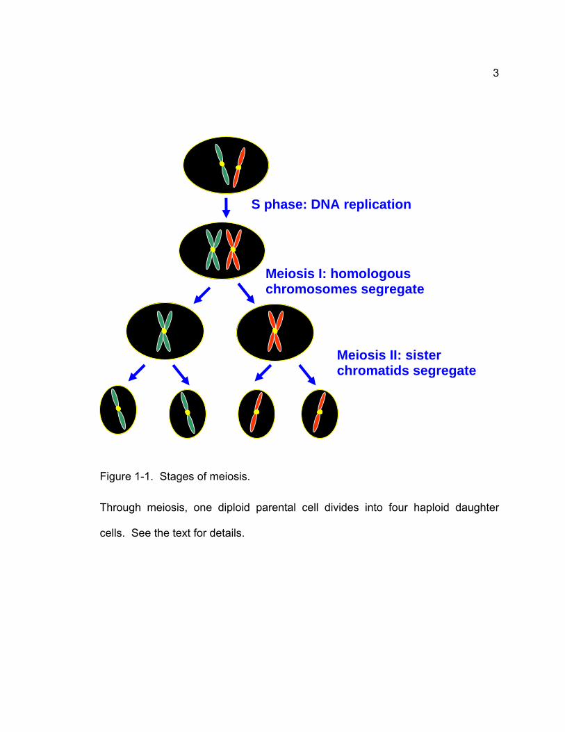
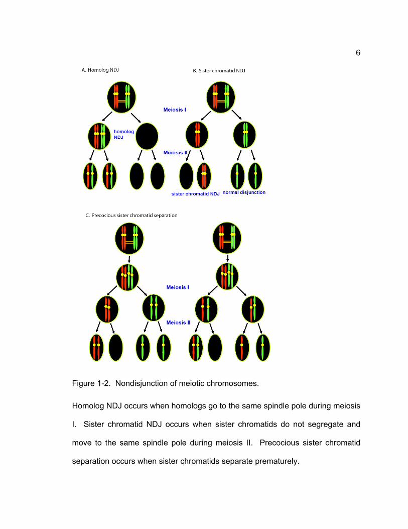
There are three types of NDJ: homolog NDJ in which homologs go to the same
spindle pole, sister chromatid NDJ in which sister chromatids move to the same
spindle pole, and precocious sister chromatid separation (PSCS) in which sister
chromatids precociously separate before anaphase (Fig. 1-2). Although
spontaneous NDJ occurs rarely in wild-type Drosophila, the frequency of NDJ
can be very high in some of meiotic mutants. Our studies aim to understand the
mechanism of meiosis through studying the factors involved in this process by
genetic, molecular, and cytological methods and tools currently available.
6
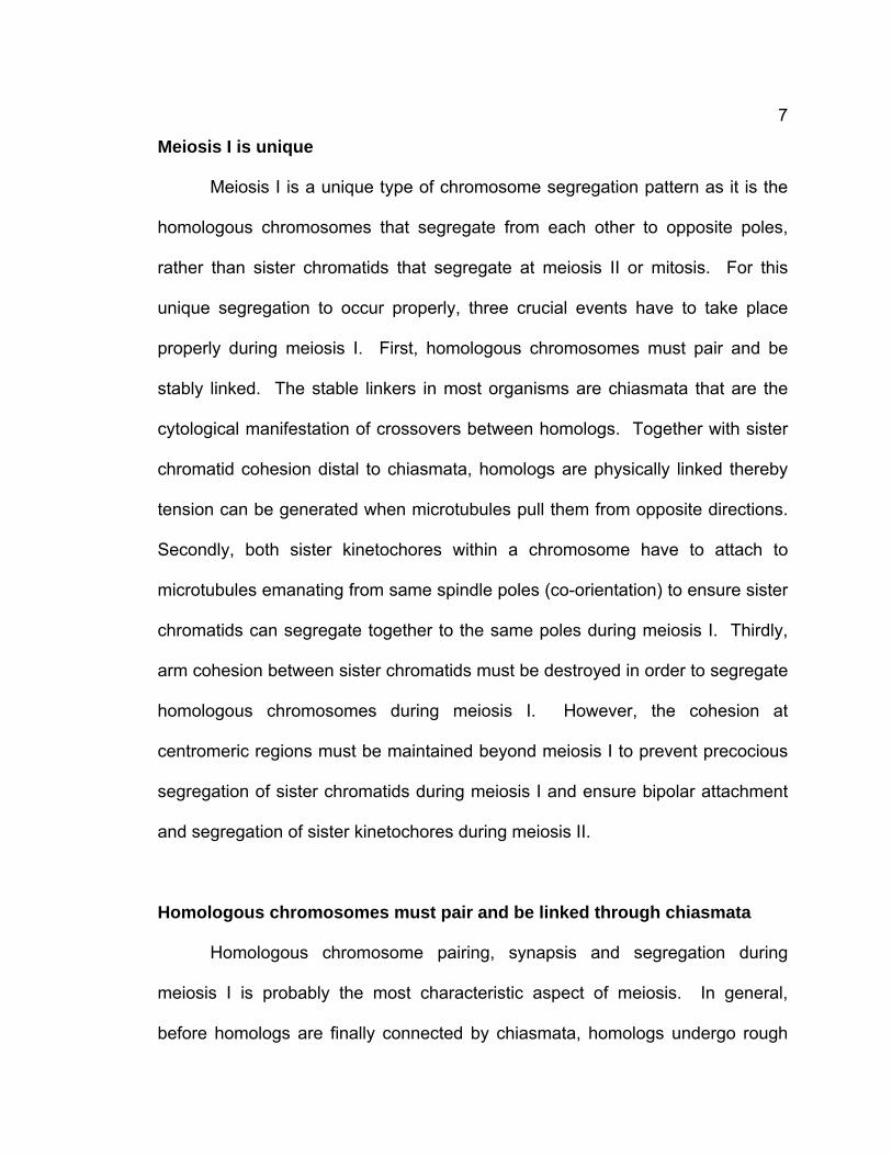
Figure 1-2. Nondisjunction of meiotic chromosomes.
Homolog NDJ occurs when homologs go to the same spindle pole during meiosis
I. Sister chromatid NDJ occurs when sister chromatids do not segregate and
move to the same spindle pole during meiosis II. Precocious sister chromatid
separation occurs when sister chromatids separate prematurely.
7
Meiosis I is unique
Meiosis I is a unique type of chromosome segregation pattern as it is the
homologous chromosomes that segregate from each other to opposite poles,
rather than sister chromatids that segregate at meiosis II or mitosis. For this
unique segregation to occur properly, three crucial events have to take place
properly during meiosis I. First, homologous chromosomes must pair and be
stably linked. The stable linkers in most organisms are chiasmata that are the
cytological manifestation of crossovers between homologs. Together with sister
chromatid cohesion distal to chiasmata, homologs are physically linked thereby
tension can be generated when microtubules pull them from opposite directions.
Secondly, both sister kinetochores within a chromosome have to attach to
microtubules emanating from same spindle poles (co-orientation) to ensure sister
chromatids can segregate together to the same poles during meiosis I. Thirdly,
arm cohesion between sister chromatids must be destroyed in order to segregate
homologous chromosomes during meiosis I. However, the cohesion at
centromeric regions must be maintained beyond meiosis I to prevent precocious
segregation of sister chromatids during meiosis I and ensure bipolar attachment
and segregation of sister kinetochores during meiosis II.
Homologous chromosomes must pair and be linked through chiasmata
Homologous chromosome pairing, synapsis and segregation during
meiosis I is probably the most characteristic aspect of meiosis. In general,
before homologs are finally connected by chiasmata, homologs undergo rough
8
alignment based on DNA sequence, and synapsis, a very intimate association
that is stabilized by the synaptonemal complex (SC) that lies between homologs
and connects them along their entire length. In most organisms, including
Drosophila females (Drosophila males use a different mechanism to hold
homologs, which will be discussed in more detail later), chiasmata that hold
homologs are generated by meiotic recombination. Although the mechanism of
how meiotic recombination is processed to form chiasmata is not completely
understood and controversial to some extent, DNA double strand breaks (DSBs)
have been found to be required for this process.
DSB generation
In most organisms, DSBs are required for homolog pairing during early
prophase I. In Saccharomyces cerevisiae DSBs are generated by Spo11
protein, which is related to a type II-like topoisomerase from archaebacteria
(Keeney et al., 1997; Bergerat et al., 1997). In Drosophila females, mei-W68
encodes the ortholog of Spo11 (McKim and Hayashi-Hagihara, 1998). Similarly,
Spo11 orthologs have been identified in many organisms, including
Schizoacchromyces pombe (Steiner et al., 2002), Caenorhabditis elegans
(Dernburg et al., 1998), Arabidopsis thaliana (Grelon et al., 2001), mouse
(Keeney et al., 1999), and human beings (Romanienko and Camerini-Otero,
1999). In all of these organisms, mutations in spo11 lead to the failure of DSB
formation, absence of meiotic recombination, and random chromosome
segregation during meiosis I. When DSBs were generated by other means, like
9
X-irradiation, in spo11 mutants lacking Spo11-induced DSBs in S, cerevisiae
(Thorne and Byers, 1993), C. elegans (Dernburg et al., 1998), and mouse
(Romanienko and Camerini-Otero, 2000), meiotic recombination and homolog
synapsis were restored to some extent. These studies showed that DSBs
generated by Spo11 are required for meiotic recombination in most or all
organisms.
DSB repair and generation of crossover
The DSBs generated by Spo11 can be repaired to form two types of
products, either a crossover or a non-crossover. Crossovers result from the
reciprocal exchange between homologous chromosomes when the DSBs are
repaired using one of sister chromatids from the homologous chromosome as the
repairing DNA template. In contrast, non-crossovers are the repair products
when DSBs are repaired without reciprocal exchange between homologous
chromosomes.
The production of crossovers is a tightly regulated process as shown by
the frequency and non-random distribution of crossovers. Under normal
conditions, at least one crossover per pair of homologs is generated. In budding
yeast, the crossover level is maintained at wild-type level at the expense of non-
crossover in the genome during meiosis when DSBs were reduced, a
phenomenon termed crossover homeostasis (Martini et al., 2006). In addition,
multiple crossovers are rarely close to each other when more than one
crossovers are produced, a phenomenon called interference (Muller, 1916).
10
Crossover interference has been extensively studied in Drosophila. Many
meiotic proteins, including, SC components, C(3)G (crossover suppressor on 3 of
Gowen) (Page and Hawley, 2001) and C(2)M (crossover suppressor on 2 of
Manheim) (Manheim and McKim, 2003), cohesion protein ord (orientation
disruptor) (Miyazaki and Orr-Weaver, 1992; Bickel et al., 2002), mei-W68 (spo11
ortholog in Drosophila) (McKim and Hayashi-Hajihara, 1998), are required for
crossover interference. In yeast, a component of synaptonemal complex, ZIP1,
is essential for crossover interference (Sym and Roeder, 1994). Furthermore, in
Drosophila females, sister chromatid cohesion has been shown to limit the
exchange between sister chromatids in a chromosome but stimulate the
exchange between homologs (Webber et al., 2004). Similar homolog bias was
observed in budding yeast. red1 and dmc1 were required for inter-homolog
meiotic recombination and the homolog bias was probably established prior to or
during DSB formation (Schwacha and Klener, 1997).
Crossover formation in Drosophila is SC-dependent
In most organisms, homolog pairing is stabilized by a tighter association
called synapsis that is defined by the formation of SC. Generally, the physical
structure of SC is conserved among diverse organisms although their protein
sequence similarity is very low. It consists of two lateral elements that run along
the entire length of each chromosome within homologs, a central element that is
midway between the lateral elements, and transverse filaments that connect the
lateral elements to the central element (Page and Hawley, 2004).
11
In many organisms SC formation is dependent on DSBs, e.g., in S.
cerevisiae (Keeney et al., 1997), A. thaliana (Grelon, et al., 2001), and mouse
(Romanienko and Camerini-Otero, 2000). In S. cerevisiae the crossover
frequency is reduced to half of the wild type level when SC does not form due to
mutations of zip1, a component of the transverse filament of SC (Sym et al.,
1993). In contrast, in Drosophila females, SC forms normally in the absence of
DSBs. Mutations in mei-W68 and mei-P22, which eliminate both meiotic
crossovers and gene conversion, have no effect on SC formation (McKim et al.,
1998). However, null mutations in C(3)G, the putative transverse filament
protein in Drosophila, eliminate SC formation, meiotic crossing over (Hall, 1972;
Rasmussen, 1975; Page and Hawley, 2001), intragenic exchange and gene
conversion (Carlson, 1972). Moreover, defects of C(2)M, a putative component
of lateral element of SC, cause significant decrease of meiotic crossover
(Manheim and McKim, 2003). These studies suggest that crossovers in
Drosophila females are processed by SC-dependent pathway.
Chiasmata hold homologs together with sister chromatid cohesion
After chiasmata are generated through meiotic recombination, they hold
each pair of homologous chromosomes together. However, only chiasmata are
insufficient to hold homologous chromosomes, sister chromatid cohesion distal to
chiasmata is required to stabilize the interactions between homologs mediated by
chiasmata. The loss of arm cohesion between sister chromatids thus can allow
homologs to segregate at anaphase I when chiasmata are dissolved (Petronczki
12
et al., 2003). Mutations of cohesion proteins, like ORD in Drosophila and Rec8 in
yeast, are required for maintaining chiasmata (Bickel et al., 2002; Buonomo et
al., 2000).
Cohesion is provided by a multi-protein complex called cohesin
Sister chromatid cohesion is mediated by a multisubunit complex called
cohesin in mitosis and meiosis. Cohesin consists of four proteins: SMC1, SMC3,
SCC1/Mcd1/RAD21, and SCC3/SA (Nasmyth, 2001; Losada and Hirano, 2005).
SMC1 and SMC3 belong to structural maintenance of chromosomes (SMC)
superfamily that is widely conserved. The N- and C-terminal halves of each
SMC1 and SMC3 fold back on themselves to form 50nm-long antiparallel coiled-
coils. The N and C termini of SMC1 or SMC3 together form an ATP-binding
cassette (ABC)-like “head” domain at one end of the coiled-coil, while their
central sequences form a “hinge” domain at the other end. SMC1 and SMC3
associate with each other through their hinge domains, generating a V-shaped
heterodimer (Melby et al., 1998; Anderson et al., 2002; Haering et al., 2002).
SCC1 is a member of the α-kleisin superfamily (Schleiffer et al., 2003). The N-
and C- terminal domains of SCC1 bind to the heads of SMC3 and SMC1
(Uhlmann et al., 2000), respectively, thus closing SMC1 and SMC3 heterodimer
to form a tripartite ring (Haering et al., 2002), which functions by topologically
encircling either a single chromatid, prior to S phase, or a pair of sister
chromatids following replication (Uhlmann, 2004). Significantly, the central
domain of SCC1 that connects its N- and C- termini contains a site for cleavage
13
by Separase, a cysteine protease conserved in many organisms. The
connection of SMC1 and SMC3 heads provided by SCC1 is essential for sister
chromatid cohesion. Proteolytic cleavage of SCC1 by Separase at the onset of
mitotic anaphase destroys cohesion between sister chromatids, allowing sister
chromatids to disjoin to opposite spindle poles (Nasmyth, 2001). Recent studies
show that cohesin’s hinge domains are essential not only for dimerization but
also for cohesin’s association with chromosomes. Transient dissociation of
SMC1 and SMC3 hinge domains is required for entry of DNA into cohesin ring
(Gruber et al., 2006).
Meiotic cohesins often contain novel subunits that are paralogs of the mitotic
subunits (Table 1-1). SMC1β is a meiosis-specific homolog of SMC1 in
mammals (Revenkova et al., 2001) and is essential for recombination, synapsis,
and sister chromatid cohesion (Revenkova et al., 2004). A meiosis-specific α-
kleisin, REC8, replaces SCC1/RAD21 in most meiotic cohesin complexes in
many eukaryotes and is necessary for the delayed release of centromeric
cohesion as well as for other meiosis-specific cohesive functions. In yeast,
cleavage of REC8 by Separase occurs at both AI, in chromosome arms, and at
AII, at centromeres. Mutations in the rec8 genes of budding yeast, fission yeast,
C. elegans, Arabidopsis and mice exhibit similar pleiotropic phenotypes,
including failure of synapsis, reduced homologous recombination, absence of
chiasmata and either premature sister chromatid separation or equational
segregation during meiosis I (Klein et al., 1999; Watanable and Nurse, 1999;
Eijpe et al., 2003; Cai et al., 2003; Chelysheva et al., 2005).
14
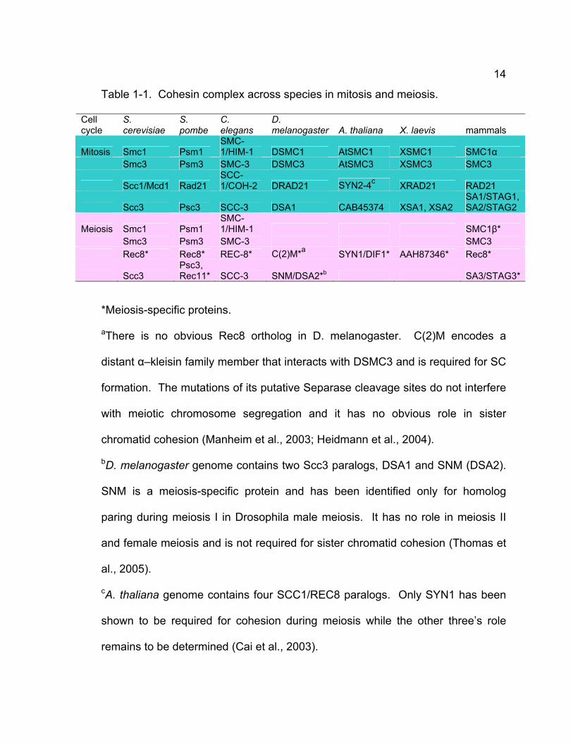
Table 1-1. Cohesin complex across species in mitosis and meiosis.
Cell cycle
S. cerevisiae
S. pombe
C. elegans
D. melanogaster A. thaliana X. laevis mammals
Mitosis Smc1 Psm1 SMC-1/HIM-1 DSMC1 AtSMC1 XSMC1 SMC1α
Smc3 Psm3 SMC-3 DSMC3 AtSMC3 XSMC3 SMC3
Scc1/Mcd1 Rad21 SCC-1/COH-2 DRAD21 SYN2-4c XRAD21 RAD21
Scc3 Psc3 SCC-3 DSA1 CAB45374 XSA1, XSA2 SA1/STAG1, SA2/STAG2
Meiosis Smc1 Psm1 SMC-1/HIM-1 SMC1β*
Smc3 Psm3 SMC-3 SMC3 Rec8* Rec8* REC-8* C(2)M*a SYN1/DIF1* AAH87346* Rec8*
Scc3 Psc3, Rec11* SCC-3 SNM/DSA2*b SA3/STAG3*
*Meiosis-specific proteins.
aThere is no obvious Rec8 ortholog in D. melanogaster. C(2)M encodes a
distant α–kleisin family member that interacts with DSMC3 and is required for SC
formation. The mutations of its putative Separase cleavage sites do not interfere
with meiotic chromosome segregation and it has no obvious role in sister
chromatid cohesion (Manheim et al., 2003; Heidmann et al., 2004).
bD. melanogaster genome contains two Scc3 paralogs, DSA1 and SNM (DSA2).
SNM is a meiosis-specific protein and has been identified only for homolog
paring during meiosis I in Drosophila male meiosis. It has no role in meiosis II
and female meiosis and is not required for sister chromatid cohesion (Thomas et
al., 2005).
cA. thaliana genome contains four SCC1/REC8 paralogs. Only SYN1 has been
shown to be required for cohesion during meiosis while the other three’s role
remains to be determined (Cai et al., 2003).
15
Cohesion is lost only at chromosome arms in meiosis I
Sister chromatids are held together by cohesion that is provided by the
complex cohesin. Cohesin is generated during DNA replication stage and forms
a ring to encircle the pair of sister chromatids (Nasmyth, 2001). During mitosis,
proteolytic cleavage of one of the cohesin subunits Scc1/Rad21 by Separase
eliminates the cohesion between sister chromatids. Separase activity is inhibited
by its inhibitor chaperone securin until the onset of anaphase. Separase is
activated when its inhibitor securin is degraded by proteasome mediated by the
anaphase-promoting complex/cyclosome (APC/C) together with Cdc20 (Cohen-
Fix et al., 1996; Funabiki et al., 1996). The loss of sister chromatid cohesion
allow them to separate to opposite spindle poles.
Since meiosis consists of two consecutive rounds of chromosome
segregation, cohesion between sister chromatids is required to be lost in a
stepwise manner. Loss of cohesion on chromosome arm can abolish the
association between homologs and allow them to separate to opposite spindle
poles in meiosis I whereas cohesion at centromeric region is maintained to
ensure sister chromatids are not separated at meiosis I but can segregate to
opposite spindle poles at anaphase II. Studies in many organisms have shown
that cohesion loss is performed exactly in the stepwise manner as reasoned
above. Rec8 is lost from chromosome arms at anaphase I but maintained at
centromeres until meiosis II in S. cerevisiae (Klein et al, 1999), S. pombe
(Watanabe and Nurse, 1999), C. elegans (Pasierbek et al., 2001), and mouse
(Lee et al., 2003, Lee et al., 2006). Cohesion is released at chromosome arms
16
during meiosis I through degradation of Rec8, a meiotic-specific paralog of Scc1,
by Separase through a similar mechanism in mitosis discussed above (Buonomo
et al., 2000; Siomos et al., 2001, Kitajima et al., 2003a).
The regulation of the only loss of arm cohesion at meiosis I is an
interesting issue. One can imagine at two mechanisms: the composition of
cohesion along arm and at centromeric region is different, or centromeric
cohesion is protected whereas arm cohesion is not.
Fission yeast has two Scc3 homologue proteins: Psc3 that is required for
sister chromatid cohesion by forming a complex with Rad21, and Rec11 that is
meiosis-specific and reduce recombination when it is mutated. Watanabe and
his collaborators found that Rec8 forms a cohesion complex with Psc3 at
centromeres but with Rec11 on chromosome arms (Kitajima et al., 2003b). This
spatially distinct organization of cohesion complex on chromosome may
contribute to the temporally-regulated loss of cohesion during meiosis. Similar
observations were made in mammals. In diplotene stage STAG2 associates
with decondensed chromatin but not with chromosome axis which STAG3
localizes to while Rad21 associates with decondensed chromatin and
chromosome axis of desynapsed SCs. Furthermore, Rad21 localizes to the
desynapsed chromosome region in which STAG3 shows weak or little signals
(Prieto et al., 2002).
17
Centromeric cohesion is protected from cleavage during meiosis I
Studies have shown that in many organisms centromeric cohesion is
retained during meiosis I and until anaphase II and that its protection during
meiosis I is essential for bi-orientation and segregation of sister chromatids
during meiosis II (Watanabe, 2005). How is centromeric cohesion protected
during meiosis I? Several factors have been shown to be required for
maintaining centromeric cohesion during meiosis I. Rec8 is essential for the
meiosis-specific cohesin at centromeres escaping the cleavage at anaphase I.
The replacement of Rec8 with Scc1/Rad21 results in the loss of cohesins along
the entire chromosomes at anaphase I in budding and fission yeast (Toth et al.,
2000; Yokobayashi et al., 2003) whereas centromeric cohesin is normally
protected at anaphase I. Spo13, a meiosis-specific protein without conserved
motif in current database, is required for maintaining centromeric cohesion during
meiosis I in budding yeast (Shonn et al, 2002). The ability to maintain Rec8 at
centromeric region during meiosis I is impaired in spo13 cells and sister
chromatid cohesion at centromeres is not protected effectively (Klein et al., 1999;
Shonn et al., 2002; Katis et al., 2004b; Lee et al., 2004). Recent studies have
shed lights into the mechanisms of protection of centromeric cohesion: a
shugoshin protein family play a major role in protecting meiosis-specific cohesin
during meiosis I.
The protector of centromeric cohesion was identified in an elegantly
designed genomic screen by Kitajima et al. (2004). The authors reasoned that if
Rec8 was forcibly co-expressed with a centromeric cohesion protector in mitosis,
18
it might be toxic to cells since sister chromatids could not segregate efficiently.
They identified such a gene called Sgo1 (Shugoshin) that is a distant relative of
Mei-S332 in Drosophila, which has long been thought to be a candidate for a
protector of centromeric cohesion during meiosis I based on its localization,
timing and phenotype (Kerrebrock et al., 1992; Kerrebrock et al., 1995). In
sgo1Δ cells, Rec8 is not retained at centromeric regions from late anaphase I
and sister chromatids segregate precociously. Moreover, Sgo1’s localization to
centromeres is regulated by a conserved centromere-associated kinase Bub1
that is involved spindle checkpoint for delaying activation of the APC/C until all
chromosomes are under tension on metaphase plate and is required for
protecting centromeric cohesion (Bernard et al., 2001). Bub1 deletion leads to
disappearance of punctuate foci of Sgo1 at centromeres, suggesting the
protection of centromeric cohesion by Bub1 is achieved by recruiting Sgo1 to
centromeres. Three other screens carried out in fission yeast and budding yeast
yielded similar results (Katis et al., 2004a; Marston et al., 2004; Rabitsch et al.,
2004). A minor difference is that Sgo1 seems only exist at meiosis I in fission
yeast while Sgo1 exists until metaphase II in budding yeast (Kitajima et al., 2004;
Rabitsch et al., 2004). Studies show that cohesion mediated by Rec8 is properly
established in sgo1 deletion mutants and the precocious loss of cohesion at
centromeric region in sgo1 mutants is due to the failure to protect Rec8 from
cleavage by Separase (Rabitsch et al., 2004). Interestingly, Spo13 functions
independently of Sgo1 to protect centromeric cohesion since its depletion has
little or no effect on localization of Sgo1 to centromeres (Katis et al., 2004; Lee et
19
al., 2004). Recent studies show that PP2A, a serine/threonine protein
phosphatase 2A, co-operates with Sgo1 to protect centromeric cohesion in both
fission and budding yeast (Kitajima et al., 2006; Riedel et al., 2006; Tang et al,
2006). In Drosophila, Polo kinase is required for dissociation of MEI-S332 from
centromeres (Clarke et al., 2005) while Aurora B kinase is necessary for loading
MEI-S332 to centromeres (Resnick et al., 2006), suggesting that MEI-
S332/Sgo1’s activity is regulated by phosphorylation. However, how these
phosphorylation and/or dephosphorylation events are regulated to make MEI-
S332/Shugoshin turn on/off at different time windows is not known yet.
Co-orientation of sister kinetochores
In meiosis II or mitosis, sister kinetochores (kinetochores of sister
chromatids) are attached to microtubules from opposite poles (bi-orientation).
However, sister kinetochores must attach to microtubules from the same pole in
meiosis I to ensure that sister chromatids move together, a phenomenon called
co-orientation (or mono-orientation).
The observation that homologs taken from grasshopper spermatocytes in
meiosis I can segregate in a reduction-like manner (co-orientation of sister
chromatids) when they were transported into a meiosis-II like spindle (Paliulis
and Nicklas, 2000), suggests that kinetochores in meiosis I are modified to
ensure co-orientation of sister centromeres (or sister chromatids). In Drosophila
melanogaster, sister kinetochores are fused at early prometaphase I when
microtubules begin to attach, but become two distinct kinetochores before the
20
onset of anaphase I (Goldstein, 1981).
It seems that chiasmata do not play a role in co-orientation of sister
kinetochores. In budding and fission yeast, deletions of the spo11 or rec12 genes
that generate DSB breaks do not interfere with co-orientation of sister
kinetochores (Klein et al., 1999; Kitajima et al., 2003b). In Drosophila
melanogaster females, mutations of the gene c(2)M that is required for SC
formation result in failure of synapsis (thus leading to defects in chiasma
formation) but show little significant defects in sister chromatid segregation
(Manheim and McKim, 2003).
Sister chromatid cohesion has been shown to play an important role in co-
orientation of sister kinetochores in both budding yeast and fission yeast. Loss of
cohesion due to rec8 mutations leads to failure of sister kinetochore co-
orientation and random sister chromatid segregation during meiosis I in budding
yeast (Klein et al., 1999). In fission yeast, loss of rec8 functions causes
predominantly equational orientation of sister kinetochores at meiosis I
(Watanabe and Nurse, 1999; Yokobayashi and Watanabe, 2005).
In addition to cohesin, other specific complexes have been shown to be
involved in co-orientation of sister chromatids in budding yeast and fission yeast.
The Monopolin complex containing Mam1 (monopolar microtubule attachment
during meiosis I), Lrs4 (loss of rDNA silencing-4), and Csm1 (chromosome
segregation in meiosis I), is required for co-orientation of sister kinetochores in
budding yeast. Mam1 is a meiosis-specific protein that resides at kinetochores
from pachytene to metaphase I (Toth et al., 2000) while Lrs4 and Csm1 are
21
expressed during both meiosis and mitosis. Lrs4 and Csm1 localize in the
nucleolus until G2 when they are released by the polo kinase Cdc5 and then
form a monopolin complex with Mam1 and bind to kinetochores (Clyne et al.,
2003; Rabitsch et al., 2003). Spo13 is also required for co-orientation of sister
kinetochores. The monopolin complex initially associates with kinetochores but
cannot be maintained in spo13Δ cells (Katis et al., 2004b; Lee et al., 2004).
Recently, Hrr25, a highly conserved casein kinase was found binding to Mam1 to
facilitate co-orientation of sister kinetochores (Petronczki et al., 2006). However,
how the monopolin complex can promote co-orientation of sister kinetochores is
still elusive.
Pcs1, the homolog of monopolin component Csm1, has been identified in
fission yeast. Surprisingly, Pcs1 is not required for co-orientation of sister
kinetochores during meiosis I but is essential for proper chromosome
segregation in meiosis II and mitosis (Rabitsch et al., 2003). Recent study has
found that Moa1 (monopolar attachment) is essential for co-orientation of sister
kinetochores in fission yeast (Yokobayashi and Watanabe, 2005). Moa1 is a
meiosis-specific protein that localizes to the central core of centromeres, which
the cohesin containing Rec8 binds to. In haploid meiosis sister chromatids
usually segregate reductionally but in moa1Δ haploids, sister chromatids
segregate equationally. Interestingly, Moa1 interacts with Rec8 in vitro and in
vivo, suggesting that their cooperation promotes co-orientation (Yokobayashi and
Watanabe, 2005).
How does the monopolin complex or Moa1 promote sister kinetochore co-
22
orientation? A recent study by Amon and her collaborators found that Aurora B
kinase plays a key role in this process (Monje-Casas et al., 2007). Homologous
chromosomes segregate to the same spindle pole in meiosis I cells in which Ipl1
(homolog of Aurora B kinase in budding yeast) is depleted while homologous
chromosomes normally segregate to opposite poles in wild type cells. Depletion
of Ipl1 in mam1Δ cells causes sister chromatids to segregate to one spindle pole
while sister chromatids segregate to opposite poles in the first division in single
mam1Δ diploid cells, suggesting Ipl1 depletion suppresses the co-orientation
defects of cells. These studies suggest that Ipl1 probably severs microtubule-
kinetochore attachments that are not under tension by phosphorylating
kinetochore components, as it does in mitosis (Cheeseman et al., 2002; Tanaka
et al., 2002, Dewar et al., 2004). By contrast with mitosis, the presence of
monopolin alters sister kinetochores such that they are under tension only when
homologous chromosomes are bi-oriented.
Meiosis II is an equational meiotic division
Chromosome segregation in meiosis II is similar with that in mitosis.
Sister kinetochores are bi-oriented, in contrast to their co-orientation in meiosis I.
Centromeric cohesion, which is protected from cleavage by Shugoshin during
meiosis I, can resist the pulling forces from opposite poles before anaphase II,
thereby prevent sister chromatids from precocious separation. At the onset of
anaphase II centromeric cohesion is degraded by Separase due to absence of
Shugoshin, allowing sister chromatids segregate to opposite poles.
23
Meiosis in Drosophila melanogaster
The fruit fly Drosophila melanogaster is an excellent model organism to
study meiosis during spermatogenesis and oogenesis. Drosophila has a short
life cycle. As early as 11 days after mating the next generation of adult offspring
can emerge under standard culture conditions. Drosophila melanogaster has
only four pairs of chromosomes, which can be easily seen during male meiosis at
cytological level. The abundant collections of stocks, constructs, and clones of
Drosophila melanogaster are invaluable to Drosophila researchers who work on
various fields. Drosophila females have the “standard” meiotic system with
formation of SC, recombination, and chiasmata and is used to study common
meiotic events while Drosophila males undergo meiosis without SC,
recombination, or chiasmata, and thus become a good model to study
achiasmate meiosis. This is valuable for researchers to learn how meiosis
processes when normal SC formation, recombination, and chiasmata are absent.
Furthermore, Drosophila females have a distributive (“back-up”) system for
pairing and segregation of chromosomes that do not exchange, like the fourth
chromosomes that never recombine. Moreover, the lineages of spermatogenesis
and oogenesis are beneficial to the study of control of cell cycle and other
developmental mechanism.
Meiosis in male Drosophila
Cytological aspects of spermatogenesis
Spermatogenesis in Drosophila males has been well characterized at
24
cytological level by staining DNA and spindle structure. At the apex of the testis,
primary spermatogonia are generated by stem cells through asymmetric
divisions. They are surrounded by somatic cyst cells derived from progenitor
cyst cells. The primary spermatogonia undergo four rapid mitotic divisions and
generate cysts containing 2, 4, or 8 secondary spermatogonia and 16 primary
spermatocytes. Secondary spermatogonia are easily recognized by the cell
number in one cyst and the nuclei that are almost entirely occupied by chromatin.
The primary spermatocytes undergo pre-meiotic S phase immediately after the
last gonial mitotic division, followed by a 4-day growth period featuring high levels
of transcription and a series of characteristic changes in nuclear morphology that
lead to 25-fold increase in primary spermatocyte size (Cenci et al., 1994; McKee,
2004).
A detailed cytological analysis of spermatocyte growth and meiotic stages
has been carried out by Cenci et al. (1994). Chromosomes are initially clustered
as a compact chromatin mass in the center of the nucleus during S1-S2a stages
(considered as early prophase I). Chromosomes resolve into three different
nuclear regions known as “territories”, which are associated with the inside of
nuclear membrane by stage S3. These separate territories are evident
throughout mid prophase I (stages S3 and S4) and late prophase I (stages S5
and S6) (Cenci et al., 1994). Chromosomes begin condensing in stage S6 and
the territories persist until prometaphase I when nuclear membrane breaks down.
At prometaphase I, three big DNA clumps that represent bivalents of sex
chromosomes, chromosome 2 and 3 are often seen and sometimes a fourth tiny
25
DNA clumps that represents the chromosome 4 bivalent. Two microtubule asters
are evident from prometaphase I. At metaphase I one big DNA clump is usually
seen at the center of the nucleus since bivalents are aligned at metaphase plate.
At anaphase I each of two DNA clumps can be seen at opposite poles due to
segregated bivalents. At telophase I chromatin is relatively decondensed and
microtubules appear to decrease. At this point one intact cyst contains 32
secondary spermatocytes. The chromatin and microtubules undergo similar
dynamics during meiosis II and 64 spermatids are produced (Cenci et al., 1994).
Achiasmate meiosis in Drosophila males
Most organisms undergo meiosis with recombination, synapsis and
ensuing chiasmata. However, a non-typical meiosis, achiasmate meiosis, i.e.
meiosis without chiasmata, exists in numerous eukaryotes, such as lepidopteran
females and dipteran males, although homologous pairing is also essential for
their meiosis. In Drosophila males, which provide the best-studied model for
achiasmate meiosis, recombination is completely absent and no SC and
chiasmata form, but chromosome pair and segregate regularly to opposite
spindle poles (Hawley, 2002; McKee, 2004).
How do the homologs pair in Drosophila males? Although homologs do
pair at prophase I without recombination and chiasmata formation, the limitations
of available cytological methods have hindered study of the mechanism of
pairing. Use of GFP-Lac repressor/Lac operator (LacI/O) system in which GFP-
LacI can target to LacO sequences inserted on euchromatic regions of
26
chromosomes, allowed Vazquez and his collaborators to characterize the
dynamics of pairing and to track the movements of chromosome arms (Vazquez
et al., 2002. In males homozygous for a single LacO insertion, the presence of a
single GFP-LacI spot indicates pairing whereas the presence of two separate
spots indicates that the marked loci are unpaired. During G2, one to four
separate spots can be seen if sister chromatid cohesion is absent. In live
spermatogonia and spermatocytes Vazquez et al. observed that about half of
pre-meiotic spermatogonia were paired (one spot) and half of them were
unpaired (two spots). By contrast, more than 95% of young primary
spermatocytes (stages S1 and S2) exhibited pairing as an evident fluorescent
spot, suggesting a tight connection along chromosome arms. This tight pairing
disappears and four separate spots become evident at stage S3, the beginning
of mid prophase I when distinct territories begin to form. This result indicates that
sister chromatid arms as well as homologs are separate. Interestingly,
homologous and sister foci remain with a common territory throughout the late
prophase I (stage S5 and S6). However, the possibility that homologs are still
paired at specific regions, like rDNA region, cannot be ruled out. Further
investigation is needed to elucidate this issue.
In contrast to the loss of tight pairing of sister chromosome arms separate
at S3, Vazquez et al. (2002) found that sister centromeres are tightly paired
throughout the prophase I and actually majority of sister centromeres are
clustered at early prophase I.
The observation that homologs are paired in young spermatocytes
27
provides direct evidence that Drosophila males enter meiosis with already paired
homologs, which is consist with the previous observation that homolog pairing
occurs in pre-meiotic cells, as early as anaphase of the last mitotic gonial division
(Metz, 1926). However, whether homolog pairing in meiosis originates directly
from the pairing at mitotic stage is not determined. Homolog pairing established
in mitosis may be lost since there is S phase between the last mitotic division and
meiosis and then is re-established in the first beginning of meiosis. The fact that
only half of spermatogonia are paired support the idea that at least homolog
pairing in some spermatocytes is not established in the last mitosis division.
rDNA as pairing sites for X-Y chromosomes
Studies have shown that some specific sites or regions may facilitate
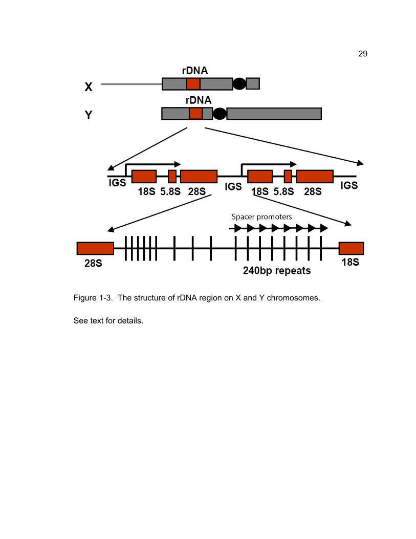
pairing. The best characterized such site is the X-Y pairing site that has been
mapped to the rDNA loci, which consist of 200-250 tandem copies of the genes
for the 18S, 5.8S and 28S ribosomal RNAs (rRNAs). The rDNA arrays are
located in the middle of the proximal heterochromatin of X chromosome and near
the centromere on the short arm of Y chromosome, respectively. Deletions of X
chromosome heterochromatin that remove all rDNA cause failure of X-Y pairing
and X-Y nondisjunction during meiosis I, whereas incomplete deletions that leave
as few as 6-8 rDNA repeats do not affect pairing and segregation of X and Y
chromosomes (McKee and Lindsley, 1987). Moreover, transgenes with a single
ribosomal RNA gene can partially restore X-Y pairing and disjunction when
inserted into a rDNA-deficient X chromosome (McKee and Karpen, 1990).
28
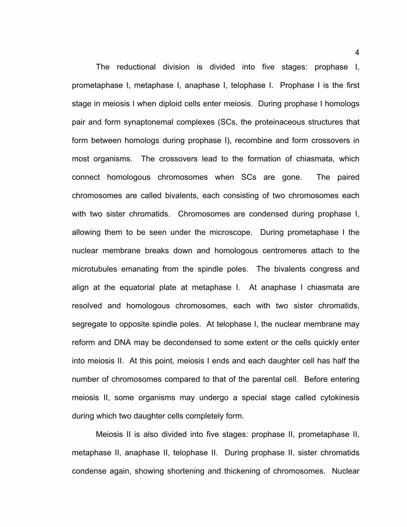
Elegant experiments have shown that the 240bp repeat sequence in the
intergenic spacer between ribosomal RNA genes, which may present in 1000-
2000 copies in total, is the primary site for pairing and segregation of X and Y
chromosomes (McKee et al., 1992; Merrill et al., 1992; McKee, 1996) (Fig. 1-3).
In their experiments, seven tandem 240bp repeats can effectively stimulate X-Y
pairing and segregation even if the rRNA transcription unit is completely
removed. In contrast, the rRNA transcription units without 240bp repeats fail to
stimulate X-Y pairing and disjunction.
A recent study identified two proteins that appear to act at the X-Y pairing
sites of Drosophila males: Stromalin in Meiosis (SNM), and Mod(mdg4) in
Meiosis (MNM) (Thomas et al., 2005). SNM shares homology with SCC3/SA,
which is a component of sister chromatid cohesion complex. MNM is a protein
with BTB domain, which is involved in many protein-protein interactions. During
prophase I both SNM and MNM localize to multiple foci in the nucleolus,
representing the rDNA region that contains the 240bp repeats. SNM and MNM
colocalize and form a dense focus that is associated the 240bp repeat during
prometaphase I and metaphase I but disappear at anaphase I, suggesting that in
achiasmate meiosis SNM and MNM may substitute for chiasmata to hold
homologs. Using heterochromatic mini-X chromosomes that lack of native rDNA
but carry transgenic 240bp repeat arrays, Thomas and McKee (2007) found that
mini-X chromosomes segregate primarily from normal sex chromosomes and
from each other and the mini-X chromosome pairs associate with the X-Y
bivalent to form trivalents and quadrivalents but do not form an
29
Figure 1-3. The structure of rDNA region on X and Y chromosomes.
See text for details.
30
additional pair of chromosomes. Furthermore they found that both SNM and
MNM are required for disjunction of mini-X chromosome pairs and multivalent
formation. This study strongly suggests 240bp repeat is the biding site for SNM
and MNM to mediate the association among sex chromosomes.
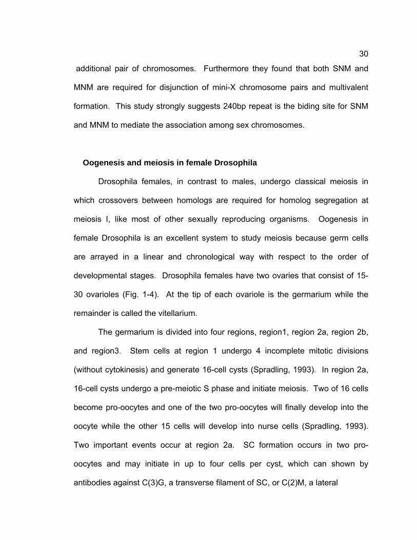
Oogenesis and meiosis in female Drosophila
Drosophila females, in contrast to males, undergo classical meiosis in
which crossovers between homologs are required for homolog segregation at
meiosis I, like most of other sexually reproducing organisms. Oogenesis in
female Drosophila is an excellent system to study meiosis because germ cells
are arrayed in a linear and chronological way with respect to the order of
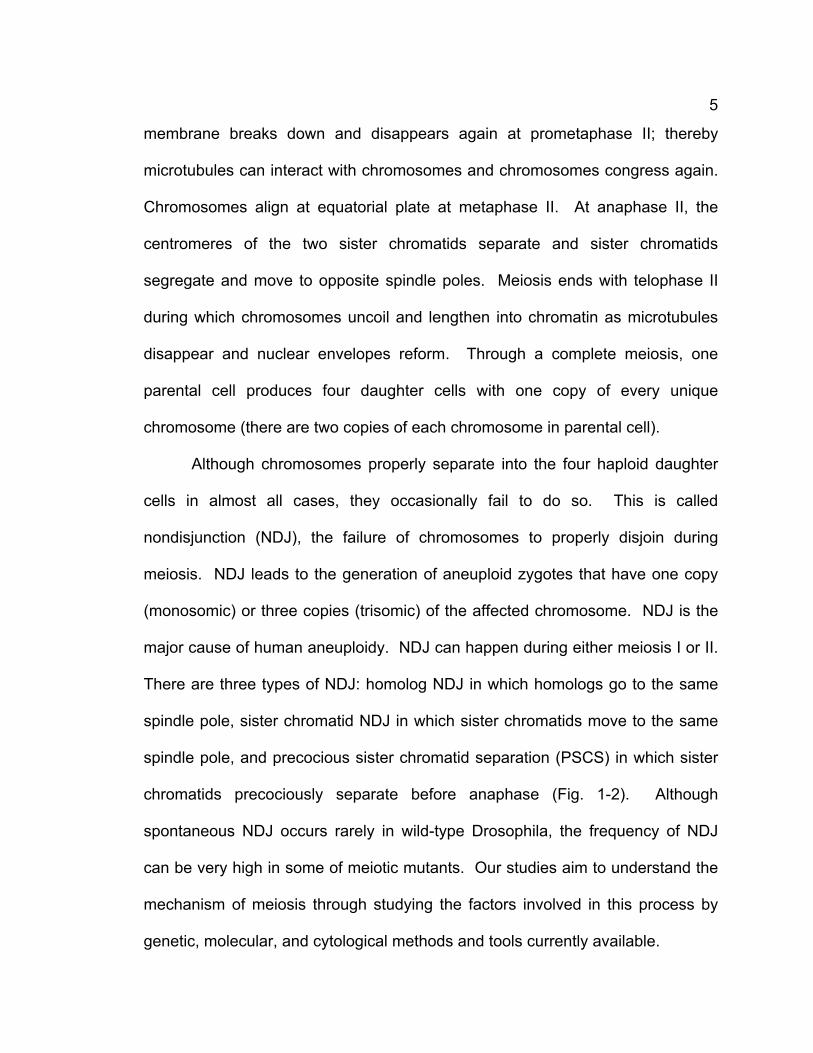
developmental stages. Drosophila females have two ovaries that consist of 15-
30 ovarioles (Fig. 1-4). At the tip of each ovariole is the germarium while the
remainder is called the vitellarium.
The germarium is divided into four regions, region1, region 2a, region 2b,
and region3. Stem cells at region 1 undergo 4 incomplete mitotic divisions
(without cytokinesis) and generate 16-cell cysts (Spradling, 1993). In region 2a,
16-cell cysts undergo a pre-meiotic S phase and initiate meiosis. Two of 16 cells
become pro-oocytes and one of the two pro-oocytes will finally develop into the
oocyte while the other 15 cells will develop into nurse cells (Spradling, 1993).
Two important events occur at region 2a. SC formation occurs in two pro-
oocytes and may initiate in up to four cells per cyst, which can shown by
antibodies against C(3)G, a transverse filament of SC, or C(2)M, a lateral
31
Figure 1-4. Schematic drawings of Drosophila ovariole and germarium.
Ovary consists of 15-30 ovarioles (A). Red ovals represent oocytes. Each
ovarioles consists of a germarium (B) connected to series of a developing egg
chambers. Mitotic divisions occur at region 1. In region 2a, meiosis initiates,
synaptonemal complex (SC) assembly begins and meiotic recombination begins.
In region 3 SC is restricted to the oocytes and recombination is completed. Red
thread-like structures represent SC. See text for details.
32
element of SC. In addition, meiotic recombination is initiated in region 2a since
DSBs appear and can be detected by using antibodies against phosphorylated
H2Av (γ-H2Av). In region 2b, the cysts become flattened out. SCs still exist in
two pro-oocytes. However, γ-H2Av foci disappear, suggesting meiotic
recombination is finished or almost finished. As early as region 2bb but not later
than region 3, the fates of the two pro-oocytes are determined and complete SC
is restricted to the oocyte, which is located at the end of germarium (McKim et
al., 2002). As cysts continue to mature, they move toward the posterior part of
the ovariole. The oocyte remains in pachytene with full-length SC until stage 6.
After 14-stage development, the cyst arrives at the end of the ovariole and
arrests at metaphase I (stage 14). The other 15 nurse cells in each cyst undergo
multiple round of S phase DNA synthesis but lacking of mitosis, leading to
polyploid DNA in the cells (Dej and Spradling, 1999).
Meiotic cohesion in Drosophila is not clear
The knowledge about cohesion in higher eukaryotes is still limited and
controversial to some extent although the mechanisms of cohesion in budding
and fission yeast have been well characterized. The mechanism of meiotic
cohesion in Drosophila is particularly elusive since REC8-containing cohesin is
not identified and only limited mutants of cohesion genes are available.
The genome of Drosophila melanogaster has one single copy of SMC1
and SMC3, two members of SCC1 family (RAD21 and C(2)M), two members of
SCC3 family (SA and SNM), but no functional REC8 ortholog (Adams et al.,
33
2000). No viable SMC1 and SMC3 mutants have been available for studying
meiosis until now. RAD21’s role in mitosis has been studied while whether it is
involved in meiosis is not known (Warren et al., 2000a; Warren et al., 200b; Vass
et al., 2003). Similar to RAD21, SA’s role in mitosis is characterized while its role
in meiosis is not known (Valdeolmillos et al., 2004). C(2)M promotes SC
formation at prophase I but it shows no or little role in sister chromatid cohesion
in female meiosis and it is not required for male meiosis (Manheim and McKim,
2003; Heidmann et al., 2004; Khetani and Bickel, 2007). A recent study by
McKee lab has identified SNM, a SCC3/SA paralog, that is required for
maintaining homolog pairing but is not required for sister chromatid cohesion in
male meiosis. In addition, SNM has no role in female meiosis (Thomas et al.,
2005). The studies have not provided any clue to meiotic cohesin in Drosophila:
is there a novel cohesin? or is a classical cohesin just not identified?
Until now, only a few meiotic proteins required for maintaining but not
establishing cohesion in both males and females have been characterized. One
of them is mei-S332, a member of the Shugoshin family. MEI-S332 is required to
prevent centromeric cohesion from degradation at meiosis I (Kerrebrock et al.,
1992; Kerrebrock et al., 1995; Katis et al., 2004a; Rabitsch et al., 2004). ord is
required for maintaining centromeric cohesion in male meiosis and is essential
for maintaining SC and meiotic recombination in female meiosis (Miyazaki and
Orr-Weaver et al., 1992; Bickel et al., 1997; Webber et al., 2004). INCENP (inner
centromere protein), a component of chromosomal passenger complex that is
required in mitosis for chromosome condensation, spindle attachment, and
34
cytokinesis (Adams et al., 2001; Carmena and Earnshaw, 2003; Vagnarelli and
Earnshaw, 2004), is essential for successful meiosis. Mutations of incenp lead to
premature loss of sister chromatid cohesion in meiosis (Resnick et al., 2006). A
recent study has shown that BubR1, a protein required for the spindle checkpoint
during mitosis, is also essential for maintaining sister chromatid cohesion at
centromeres at anaphase I (Malmanche et al., 2007). However, whether BubR1
in Drosophila has a similar role with Bub1 in fission yeast to recruit MEI-
S332/Shugoshin has not been determined. Other than that, no cohesion protein
is identified and severely hindering the study of meiotic cohesion in Drosophila.
35
CHAPTER 2 - SOLO IS A NOVEL PROTEIN REQUIRED
FOR SISTER CHROMATID COHESION, SISTER
CENTROMERE CO-ORIENTATION, AND CENTROMERIC
LOCALIZATION OF SMC1 IN DROSOPHILA MEIOSIS
This part is modified from the manuscript that has been submitted to Current
Biology and is under revision now.
Rihui Yan’s primary contributions: identified the gene solo, analyzed some
genetic phenotypes of solo, analyzed solo phenotypes at cytological level, cloned
solo gene, analyzed its localization pattern in wild type and cohesion mutants
,and wrote the manuscript draft.
36
Abstract
Sister chromatid cohesion plays several essential roles in meiotic
chromosome segregation, including maintenance of stable connections between
homologs and sister chromatids, and establishment of correct sister centromere
orientation patterns on the meiosis I and II spindles. Cohesin has been proposed
as the key factor; however, its mechanism in higher eukaryotes is still elusive.
We describe a novel protein, SOLO (Sisters On the LOose) that is essential for
meiotic cohesion in Drosophila melanogaster. solo mutations cause high
nondisjunction of sister and homologous chromatids of sex chromosomes and
autosomes. In solo males, sister chromatids separate prematurely and
segregate randomly during meiosis II. Although bivalents remain intact
throughout meiosis I, sister centromeres lose cohesion prior to prometaphase I
and orient nearly randomly on the meiosis I spindle. SOLO and the cohesin
protein SMC1 co-localize to meiotic centromeres from early prophase I until
anaphase II in wild-type males but both proteins are removed prematurely from
centromeres at anaphase I in mei-S332 mutants, coincident with premature loss
of cohesion in those mutants. In addition, centromeric foci of SMC1 are absent
in solo mutants at all stages of meiosis. The mutant phenotypes and localization
patterns of SOLO and SMC1 indicate that they function together to maintain
sister chromatid cohesion in Drosophila meiosis. Our data also show that MEI-
S332 protects cohesin from premature removal at anaphase I, similar to its
ortholog Shugoshin’s functions in yeast.
37
INTRODUCTION
Meiosis consists of two divisions, a reductional division at meiosis I in
which homologous chromosomes (homologs) pair and segregate to opposite
spindle poles, and an equational division at meiosis II in which sister chromatids
segregate. Cohesion between sister chromatids is essential for proper
chromosome segregation at both meiotic divisions (Page and Hawley, 2003;
Petronczki et al., 2003; McKee, 2004). Several roles of sister chromatid
cohesion in meiosis I have been defined in yeast. First, during prophase I,
cohesion between sister chromatid arms is essential for formation of lateral
elements of synaptonemal complexes; consequently cohesion mutations disrupt
synapsis. Second, arm cohesion distal to crossover sites is required to stabilize
chiasmata during late prophase I and metaphase I. Third, cohesion between
sister centromeres is required for their “co-orientation” to the same pole on the
meiosis I spindle, which is a prerequisite for bi-orientation of homologous
centromeres. Sister chromatid cohesion is also required to prevent sister
chromatids from separating prematurely prior to anaphase I, and to enable sister
centromeres to orient to opposite poles (bi-orient) on the meiosis II spindle. The
multiple functions of cohesion in meiosis require it to be released in a stepwise
manner. Arm cohesion is released at anaphase I, destabilizing chiasmata and
allowing segregation of homologs, whereas centromere cohesion is released
until anaphase II, allowing segregation of sister chromatids (Lee and Orr-Weaver,
2001; Nasmyth, 2001; Petronczki et al., 2003; Page and Hawley, 2004; Hauf and
38
Watanabe, 2004; Watanabe, 2005).
In both mitosis and meiosis, cohesion is mediated by a complex called
cohesin that consists of four proteins, two SMC (Structural Maintenance of
Chromosomes) subunits, SMC1 and SMC3, and two non-SMC subunits,
SCC1/RAD21 and SCC3/SA (Nasmyth, 2001; Schleiffer et al., 2003; Losada and
Hirano, 2005). In mitosis, SMC1 and SMC3, and SCC1/RAD21, a member of the
kleisin superfamily (Schleiffer et al., 2003), form a tripartite ring that topologically
encircles either a single chromatid, prior to S phase, or a pair of sister chromatids
following replication. Proteolytic cleavage of SCC1 by Separase at the onset of
mitotic anaphase destroys cohesion between sister chromatids, allowing sister
chromatids to disjoin to opposite spindle poles (Uhlmann, 2004).
Meiotic cohesins contain novel subunits that are paralogs of mitotic
subunits (Petronczki et al., 2003; Losada and Hirano, 2005). REC8 replaces
SCC1/RAD21 in most meiotic cohesin complexes and is necessary for the
delayed release of centromeric cohesion as well as for other meiosis-specific
cohesive functions. In yeast, cleavage of REC8 by Separase occurs at both
anaphase I, on chromosome arms, and at anaphase II, at centromeres
(Petronczki et al., 2003).
Cohesin genes are conserved throughout the eukaryotes (Losada and
Hirano, 2005; Schleiffer et al., 2003). Although the role of cohesin in meiosis is
less well-defined in higher eukaryotes, there is considerable evidence for
functions related to those in yeast (Pasierbek et al., 2001; Siomos et al., 2001;
Cai et al., 2003; Chelysheva et al., 2005; Xu et al., 2005; Kudo et al., 2006).
39
Recently, centromere proteins called Shugoshins that protect centromeric REC8
cohesin from cleavage at meiosis I have been described (Katis et al., 2004;
Kitajima et al., 2004; Rabitsch et al., 2004).
However, the universality of cohesin as a mediator of meiotic cohesion
has not been established in higher eukaryotes. Although immunocytological and
genetic analyses have demonstrated a major role of cohesins in SC formation
and chiasma function in several higher eukaryotes (Pasierbek et al., 2001; Prieto
et al., 2001; Siomos et al., 2001; Cai et al., 2003; Chelysheva et al., 2005; Xu et
al., 2005; Kudo et al., 2006), the mechanism underlying meiotic centromere
cohesion and centromere orientation during meiosis I are poorly understood
(Toth et al., 2000; Parra et al., 2004; Chelysheva et al., 2005; Yokobayashi et al.,
2005).
Cohesin and its role in Drosophila meiosis have been particularly murky.
The Drosophila genome includes single SMC1 and SMC3 genes and two
members each of the SCC1 (RAD21 and C(2)M), and SCC3/SA (SA and SNM)
families, but there is no clear functional REC8 ortholog (Adams et al., 2000).
Rad21’s functions in mitosis have been examined (Warren et al., 2000a, Warren
et al., 2000b; Vass et al., 2003; Valdeolmillos et al., 2004) but a role in meiotic
cohesion has not been reported. C(2)M, which exhibits weak similarity to SCC1
and REC8, localizes to the lateral elements of synaptonemal complex during
prophase I in oocytes and is required for synapsis, but is absent after mid-
prophase I and dispensable for chiasma stability, sister chromatid cohesion and
recruitment of centromeric cohesin (Manheim and McKim, 2003; Heidmann et al.,
40
2004; Khetani and Bickel, 2007). SNM, a meiosis-specific SCC3/SA paralog, is
required for stable homolog pairing in achiasmate meiosis of Drosophila males in
which homologs pair and segregate without crossing over, SC or chiasmata, but
is not required for sister chromatid cohesion in males or for any aspect of female
meiosis (Thomas et al., 2005).
Although cohesin mutants are lacking, mutations in three Drosophila
genes have been shown to disrupt meiotic cohesion. Mutations in mei-S332,
which encodes a Shugoshin homolog that localizes to meiotic centromeres from
prometaphase I through anaphase II, cause precocious sister chromatid
separation (PSCS) beginning at anaphase I and result in high frequencies of
meiosis II NDJ (Kerrebrock et al., 1992; Kerrebrock et al., 1995). Mutations in
orientation disruptor (ord), which encodes a meiosis-specific protein that localizes
to centromeres from late prophase I through anaphase II, cause PSCS and
chromatid mis-segregation in both meiosis I and II. ORD has no recognizable
domains and no orthologs outside of the genus Drosophila and its molecular
function is unclear (Miyazaki and Orr-Weaver, 1992). Mutations in the INCENP
protein, a component of the Aurora B kinase complex present on both mitotic and
meiotic centromeres, disrupt cohesion prior to metaphase I, leading to chromatid
nondisjunction (NDJ) at both meiosis I and meiosis II (Carmena and Earnshaw,
2003; Resnick et al., 2006); however, the cohesion component which INCENP
interacts with has not been identified.
Here we describe a novel Drosophila protein, SOLO (Sisters On the
LOose), that is required for sister chromatid cohesion in both meiosis I and
41
meiosis II and for sister centromere co-orientation during meiosis I. Mutations in
solo cause high nondisjunction of sex chromosomes and autosomes in both
meiotic divisions and premature separation of sister centromeres during
prophase I. SOLO and the cohesin protein SMC1 co-localize to meiotic
centromeres from early prophase I until anaphase II, and both proteins are
removed prematurely from centromeres in mei-S332 mutants. Moreover, SOLO
is required for centromere localization of SMC1 at all stages of meiosis. Our data
indicate that both SOLO and SMC1 play direct roles in sister chromatid cohesion
during Drosophila meiosis.
42
MATERIALS AND METHODS
Fly stocks, special chromosomes and Drosophila culture methods.
The solo mutations in this paper were from the Zuker-2 (Z2) collection of
more than 6000 lines with EMS-mutagenized second chromosomes
(Koundakjian et al., 2004). The Z2 lines used in this study were identified in a
screen for paternal 4th chromosome loss and were kindly provided by B.
Wakimoto (Wakimoto et al., 2004). vas alleles were obtained from M. Ashburner
(Cambridge University), P. Lasko (McGill University), D. Montell (John Hopkins
University), and the Bloomington Stock Center at the University of Indiana. mei-
S332 alleles were kindly donated by T. Orr-Weaver (Whitehead Institute, MIT).
ord alleles and its deficiency were kindly provided by S. Bickel at (Dartmouth
College). Compound chromosomes and markers are described in Flybase
(2007a). Unless otherwise specified, tested males were crossed singly to two or
three females in shell vials. All flies were maintained at 23°C on standard
cornmeal molasses medium. Parents were removed from the vial on day 10 and
progeny were counted between day 13 and day 22.
Sex chromosome NDJ assays
To measure X-Y NDJ, +/BsYy+ males were crossed singly to 2-3 females
carrying structurally normal X chromosomes marked with y1 and w1118. Regular
progeny are + females and y+ w BS males; paternal NDJ generates y+ w+ BS
43
female and y w B+ male progeny. %NDJ = 100 x (y+ w+ BS ♀♀ + y w B+
♂♂)/N.
To discriminate between NDJ of homologs and sister chromatids, +/BsYy+ males
were crossed singly to 2-3 C(1)RM, y2 su(wa) wa/0 females in which both X
chromosomes are attached to a single centromere. These females produce eggs
that are diplo-X and nullo-X at approximately equal frequencies. When nullo-X
eggs are fertilized, the cross yields progeny derived from both XX sperm and XY
sperm (+ females and BS males, respectively), which are diagnostic of sister
chromatid and homolog NDJ, respectively, as well as progeny of nullo-XY (O)
sperm (y2 su(wa) wa) females), which can result from either type of NDJ.
Progeny from XXY, XYY and XXYY sperm were very rare and thus they were
neglected in the analysis.
Measuring 2nd chromosome NDJ
soloZ2-0198, cn bw/b vas7 pr males were crossed singly with three C(2)EN,
bw sp females. vas7 is null for both vas and solo function. C(2)EN females carry
two copies of each arm of chromosome 2 attached to a single centromere and
produce only diplo-2 (2^2) and nullo-2 (O) eggs, so the only viable progeny are
the products of paternal chromosome 2 NDJ (2/2 and O sperm). Thus the
frequency of 2nd chromosome NDJ is proportional to progeny per male. Since
sibling conrtol males produce one chromosome if there is no autonomous NDJ,
the sperm with one paternal second chromosome combining with the eggs with
either 2 second chromosomes or null chromosome would produce no progeny.
The control experiment confirmed that in 54 males tested only one male
44
produced 1 progeny due to autonomous NDJ in male. Since the paternal second
chromosomes carry different recessive markers (cn bw and b pr, respectively),
the relative frequencies of sister chromatid NDJ and homolog NDJ can be
estimated from the proportions of NDJ progeny derived from 2/2 sperm that
express the heterozygous markers. cn bw and b pr progeny result from sister
chromatid NDJ whereas wild-type (WT, cn bw/b pr) progeny result from homolog
NDJ.
Mapping and identification of solo mutations
solo alleles were mapped by deficiency complementation against the
“deficiency kit” obtained from the National Drosophila Stock Center at Indiana
University, Bloomington, using the X-Y NDJ phenotype. solo was mapped to the
35B region where vas is located. More detailed deficiency mapping using vas-
region deficiencies (Ashburner et al., 1990 obtained from M. Ashburner
(Cambridge University) demonstrated that the solo mutations lie within the vas
locus (data not shown).
All exons and the third intron of vas were amplified from genomic DNA of
flies homozygous for each of the three solo mutations and sequenced using ABI
BigDye Terminator v3.1 Cycle sequencing kit (Applied Biosystems). No
mutations were detected in vas exons but the two large ORFs in the third intron
of vas contained single mutations in each of the three solo alleles, each of which
result in a nonsense mutation (shown in Fig. 2-4). Sequencing also showed that
the pre-existing vasRJ36 allele (Schupbach and Wieschaus, 1991) has an 8bp
45
insertion in the first intronic ORF resulting in a frame shift mutation.
Characterization of solo transcripts
To characterize the solo transcription unit, total RNA was prepared from
wild type (strain Zuker-2, cn bw) adults using TRI Reagent (Sigma). After DNase
treatment, the total RNA was used for cDNA synthesis using SuperscriptTM First-
Strand Synthesis System (Invitrogen). Then solo cDNAs were amplified by PCR
using primers: GTGAGAACTTTGTCACTCGG and
TTTATGGGAGGCAGTAAGGC. A following nested PCR reaction was carried
out using primers: CAATTCGAGTAGTGGTCAGC and
GAATCCGAATACCCTGTTGC. This procedure yielded a specific amplification
product of 972 base pairs that contains parts of the two large ORFs from intron 3
of vas spliced together to generate a continuous reading frame. To identify the 5’
and 3’ ends of the solo transcript, 5’ RACE and 3’ RACE reactions were
performed (BD SMART RACE cDNA Amplification Kit) and a cDNA (EST clone
AT08465) obtained from Berkeley Drosophila Genome Project (BDGP) was
sequenced. These experiments revealed that the second intronic ORF
terminates at a stop codon located 92 bases upstream of a poly-A site and 294
bases upstream of the fourth exon of vas. At the 5’ end, AT08645 includes all
sequences in the first three exons of vas except for the first 22 base pairs of exon
1. It is not clear if this difference reflects different transcription start sites for the
two genes or if AT08645 is incomplete at the 5’ end. The primer sequences used
in the RACE experiments are available upon request.
46
The sequence of the SOLO cDNA reported in this paper has been
deposited in Genbank as accession # DQ851162.
Construction of SOLO fusion clones and generation of transgenic flies
Two SOLO fusion constructs, UASp:Venus-SOLO and UASp:SOLO-
Venus, were made. For Venus-SOLO, the solo coding sequence and 3’UTR
were amplified from the EST clone AT08465 using Pfx polymerase (Invitrogen)
and primers CACCATGTCTGACGACTGGGATG and
CACCCGACATAGATGCCTCG. For SOLO-Venus, the following primers were
used: CACCATGTCTGACGACTGGGATG and
GAGCAGCCCGAAAAATCTACC. The PCR products were cloned into the
pENTRTM/D-TOPO entry vector (Invitrogen) and the resulting products were
sequenced.
Both entry constructs were recombined into Gateway P-element vectors
pPVW and pPWV (from Drosophila Genomics Resource Center (BDGC)),
generating the germ-line transformation vectors P[w+mC, UASp:Venus-SOLO] and
P[w+mC, UASp:SOLO-Venus]. Both vectors include Venus, UAS sequences for
transcriptional activation by GAL4 and mini-white to detect germ-line
transformants. Both constructs were transformed into w1118 flies (BestGene Inc.).
Transformants were mapped by standard procedures. Transformant lines
carrying UASp:SOLO-Venus are named 1910-1-# and transformant lines
carrying UASp:Venus-SOLO are named 1910-2-#.
47
Transgene rescue experiments
+/BSYy+; Df(2L)A267, cn/soloZ2-0198, cn; [UASp:Venus-
SOLO]/[nanos:Gal4-VP16] males and sibling controls without [UASp:Venus-
SOLO] or [nanos:Gal4-VP16] were crossed to y w females to measure sex
chromosome NDJ. The rescue experiments for UASp:SOLO-Venus transgenic
flies were carried out by similar methods.
Testis Immunostaining
α-tubulin/DAPI staining of testes was carried out as described (Thomas et
al., 2005). Immunostaining was performed with modification according to
(Bonaccorsi et al., 2000). The primary antibodies used: 1:500 anti-CID (chicken)
(provided by Dr. G. Karpen), 1:1000 anti-CID (rabbit) (Abcam), 1:250 anti-SNM
C-terminal (rabbit) (Thomas et al., 2005), 1:250 Anti-SMC1 (rabbit) (Thomas et
al., 2005), 1:500 anti-GFP (rabbit) (Molecular Probes). The secondary antibodies
used: Alexa Fluor 555 goat anti-chicken IgG (H+L) (Molecular Probes), Alexa
Fluor 546 goat anti-rabbit IgG (H+L) (Molecular Probes), Alexa Fluor 647 goat
anti-rabbit IgG (H + L) (Molecular Probes). Venus-SOLO expression was
induced by nanos:Gal4-VP16 (Doren et al., 1998) and fluorescent signals were
detected in the FITC channel or detected using anti-GFP antibody. Acetic orcein
staining of male meiotic chromosomes was carried out according to Stapleton et
al. (2001).
48
Assaying arm cohesion in spermatogonia and spermatocytes
Arm cohesion was assayed by counting GFP spots in spermatogonia and
spermatocytes from males hemizygous for a chromosome 2 transgene carrying a
256mer tandem array of lacO repeats and heterozygous for a transgene (also on
chromosome 2) expressing a GFP-LacI chimeric protein under control of the
hsp83 promoter (Robinett et al., 1996; Straight et al., 1996; Vaquez et al., 2002;
Thomas et al., 2005). The genotype of the tested males was w1118/Y;
Df(2L)A267, [GFP-LacI] [lacO]/soloZ2-0198. Testes were dissected from third instar
larvae, pupae or young adults in testes buffer (183 mM KCl, 47 mM NaCl, 10 mM
Tris-HCl, 1 mM EDTA, 1 mM PMSF) and gently squashed in testes buffer. GFP-
LacI foci were imaged by native fluorescence. DNA was stained with DAPI.
Microscopy and image processing
All images were collected using an Axioplan (ZEISS) microscope equipped with
an HBO 100-W mercury lamp and high-resolution CCD camera (Roper). Image
data were collected and merged using Metamorph Software (Universal Imaging
Corporation). For CID signals and some SMC1 and Venus-SOLO images, sum
or maximum projections of deconvolved Z-series planes were applied using
Metamorph Software. Images were processed with Adobe Photoshop CS2.
49
RESULTS
NDJ of homologous and sister chromatids in solo males
We identified three alleles of solo among a group of EMS-induced
mutations that interfere with paternal transmission of the small 4th chromosome
(Koundakjian et al., 2004; Wakimoto et al., 2004) and mapped solo to the 35B2-
35C1 region of chromosome arm 2L by deficiency complementation.
To determine whether solo mutations disrupt segregation of the sex
chromosomes in male meiosis, and to discriminate between homolog versus
sister chromatid NDJ, solo males carrying a dominantly marked Y chromosome
were crossed to females carrying the attached-X (X^X) chromosome C(1)RM
(Table 2-1). Males hemizygous for all three solo alleles exhibited similar NDJ
frequencies, averaging 55.8%. Similar NDJ frequencies were observed in trans-
heterozygous (Table 2-1) and homozygous males (data not shown), indicating
that all three alleles are genetically null. XY, XX and nullo-XY sperm were
generated at frequencies averaging 10.7%, 4.4% and 36%, respectively. Since
XY and XX sperm result exclusively from homolog and sister chromatid NDJ,
respectively, these data indicate that solo mutations perturb segregation of both
homologous and sister chromatids. solo males also exhibited high sister
chromatid and homolog NDJ frequencies for the autosomal 2nd and 4th
chromosomes (Table 2-2 and not shown). Taken together, these results suggest
the importance of solo in both homolog and sister chromatid segregation.
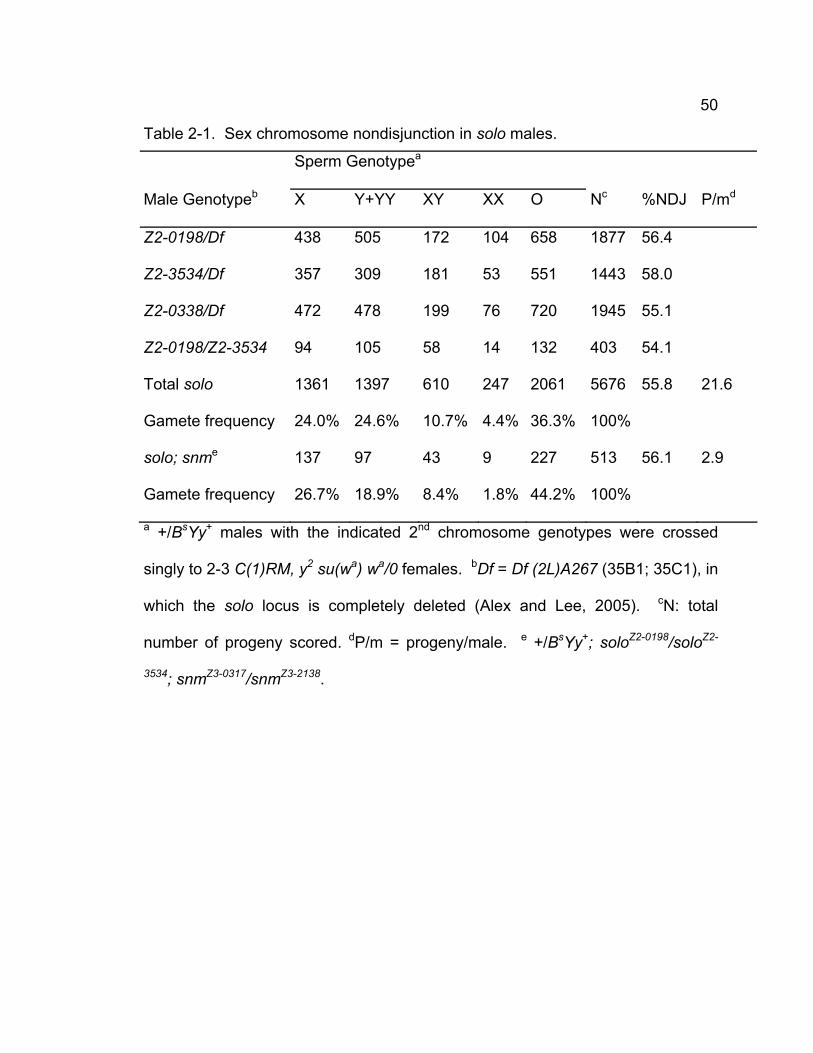
50
Table 2-1. Sex chromosome nondisjunction in solo males.
Sperm Genotypea
Male Genotypeb X Y+YY XY XX O Nc %NDJ P/md
Z2-0198/Df 438 505 172 104 658 1877 56.4
Z2-3534/Df 357 309 181 53 551 1443 58.0
Z2-0338/Df 472 478 199 76 720 1945 55.1
Z2-0198/Z2-3534 94 105 58 14 132 403 54.1
Total solo 1361 1397 610 247 2061 5676 55.8 21.6
Gamete frequency 24.0% 24.6% 10.7% 4.4% 36.3% 100%
solo; snme 137 97 43 9 227 513 56.1 2.9
Gamete frequency 26.7% 18.9% 8.4% 1.8% 44.2% 100%
a +/BsYy+ males with the indicated 2nd chromosome genotypes were crossed
singly to 2-3 C(1)RM, y2 su(wa) wa/0 females. bDf = Df (2L)A267 (35B1; 35C1), in
which the solo locus is completely deleted (Alex and Lee, 2005). cN: total
number of progeny scored. dP/m = progeny/male. e +/BsYy+; soloZ2-0198/soloZ2-
3534; snmZ3-0317/snmZ3-2138.
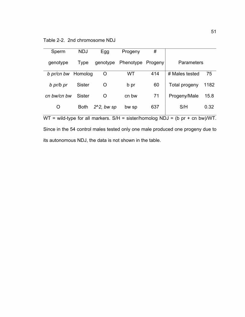
51
Table 2-2. 2nd chromosome NDJ
Sperm
genotype
NDJ
Type
Egg
genotype
Progeny
Phenotype
#
Progeny
Parameters
b pr/cn bw Homolog O WT 414 # Males tested 75
b pr/b pr Sister O b pr 60 Total progeny 1182
cn bw/cn bw Sister O cn bw 71 Progeny/Male 15.8
O Both 2^2, bw sp bw sp 637 S/H 0.32
WT = wild-type for all markers. S/H = sister/homolog NDJ = (b pr + cn bw)/WT.
Since in the 54 control males tested only one male produced one progeny due to
its autonomous NDJ, the data is not shown in the table.
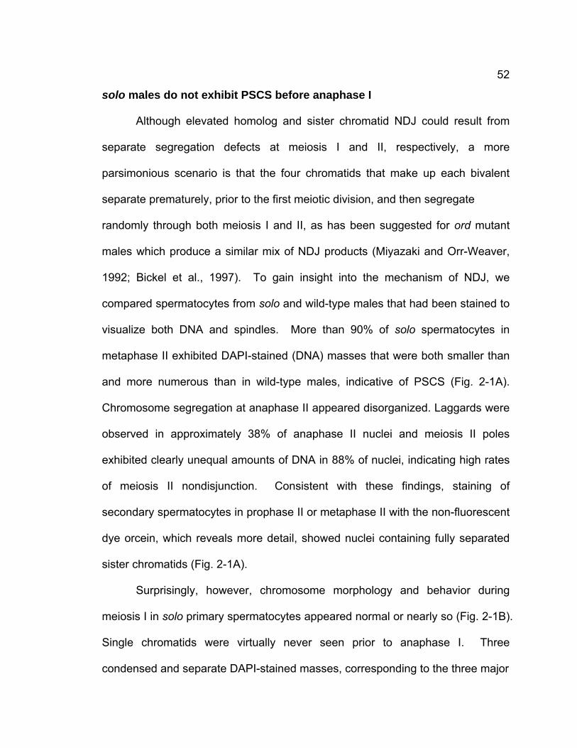
52
solo males do not exhibit PSCS before anaphase I
Although elevated homolog and sister chromatid NDJ could result from
separate segregation defects at meiosis I and II, respectively, a more
parsimonious scenario is that the four chromatids that make up each bivalent
separate prematurely, prior to the first meiotic division, and then segregate
randomly through both meiosis I and II, as has been suggested for ord mutant
males which produce a similar mix of NDJ products (Miyazaki and Orr-Weaver,
1992; Bickel et al., 1997). To gain insight into the mechanism of NDJ, we
compared spermatocytes from solo and wild-type males that had been stained to
visualize both DNA and spindles. More than 90% of solo spermatocytes in
metaphase II exhibited DAPI-stained (DNA) masses that were both smaller than
and more numerous than in wild-type males, indicative of PSCS (Fig. 2-1A).
Chromosome segregation at anaphase II appeared disorganized. Laggards were
observed in approximately 38% of anaphase II nuclei and meiosis II poles
exhibited clearly unequal amounts of DNA in 88% of nuclei, indicating high rates
of meiosis II nondisjunction. Consistent with these findings, staining of
secondary spermatocytes in prophase II or metaphase II with the non-fluorescent
dye orcein, which reveals more detail, showed nuclei containing fully separated
sister chromatids (Fig. 2-1A).
Surprisingly, however, chromosome morphology and behavior during
meiosis I in solo primary spermatocytes appeared normal or nearly so (Fig. 2-1B).
Single chromatids were virtually never seen prior to anaphase I. Three
condensed and separate DAPI-stained masses, corresponding to the three major
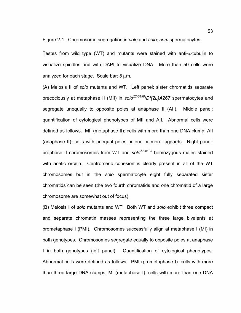
53
Figure 2-1. Chromosome segregation in solo and solo; snm spermatocytes.
Testes from wild type (WT) and mutants were stained with anti-α-tubulin to
visualize spindles and with DAPI to visualize DNA. More than 50 cells were
analyzed for each stage. Scale bar: 5 μm.
(A) Meiosis II of solo mutants and WT. Left panel: sister chromatids separate
precociously at metaphase II (MII) in soloZ2-0198/Df(2L)A267 spermatocytes and
segregate unequally to opposite poles at anaphase II (AII). Middle panel:
quantification of cytological phenotypes of MII and AII. Abnormal cells were
defined as follows. MII (metaphase II): cells with more than one DNA clump; AII
(anaphase II): cells with unequal poles or one or more laggards. Right panel:
prophase II chromosomes from WT and soloZ2-0198 homozygous males stained
with acetic orcein. Centromeric cohesion is clearly present in all of the WT
chromosomes but in the solo spermatocyte eight fully separated sister
chromatids can be seen (the two fourth chromatids and one chromatid of a large
chromosome are somewhat out of focus).
(B) Meiosis I of solo mutants and WT. Both WT and solo exhibit three compact
and separate chromatin masses representing the three large bivalents at
prometaphase I (PMI). Chromosomes successfully align at metaphase I (MI) in
both genotypes. Chromosomes segregate equally to opposite poles at anaphase
I in both genotypes (left panel). Quantification of cytological phenotypes.
Abnormal cells were defined as follows. PMI (prometaphase I): cells with more
than three large DNA clumps; MI (metaphase I): cells with more than one DNA
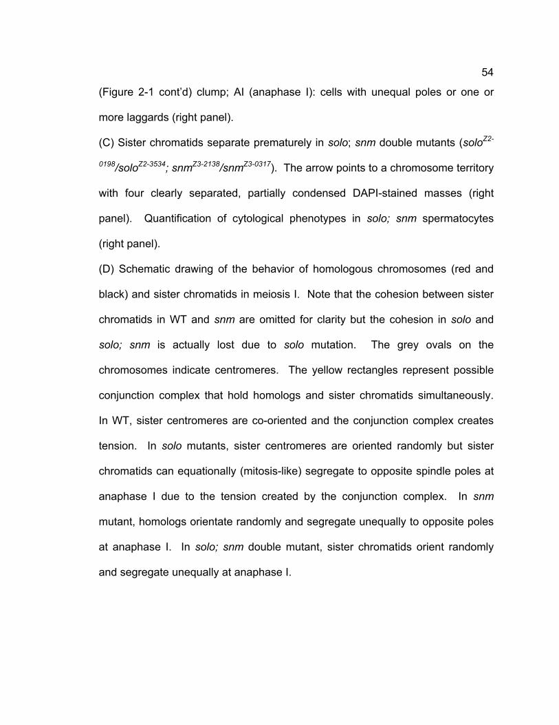
54
(Figure 2-1 cont’d) clump; AI (anaphase I): cells with unequal poles or one or
more laggards (right panel).
(C) Sister chromatids separate prematurely in solo; snm double mutants (soloZ2-
0198/soloZ2-3534; snmZ3-2138/snmZ3-0317). The arrow points to a chromosome territory
with four clearly separated, partially condensed DAPI-stained masses (right
panel). Quantification of cytological phenotypes in solo; snm spermatocytes
(right panel).
(D) Schematic drawing of the behavior of homologous chromosomes (red and
black) and sister chromatids in meiosis I. Note that the cohesion between sister
chromatids in WT and snm are omitted for clarity but the cohesion in solo and
solo; snm is actually lost due to solo mutation. The grey ovals on the
chromosomes indicate centromeres. The yellow rectangles represent possible
conjunction complex that hold homologs and sister chromatids simultaneously.
In WT, sister centromeres are co-oriented and the conjunction complex creates
tension. In solo mutants, sister centromeres are oriented randomly but sister
chromatids can equationally (mitosis-like) segregate to opposite spindle poles at
anaphase I due to the tension created by the conjunction complex. In snm
mutant, homologs orientate randomly and segregate unequally to opposite poles
at anaphase I. In solo; snm double mutant, sister chromatids orient randomly
and segregate unequally at anaphase I.
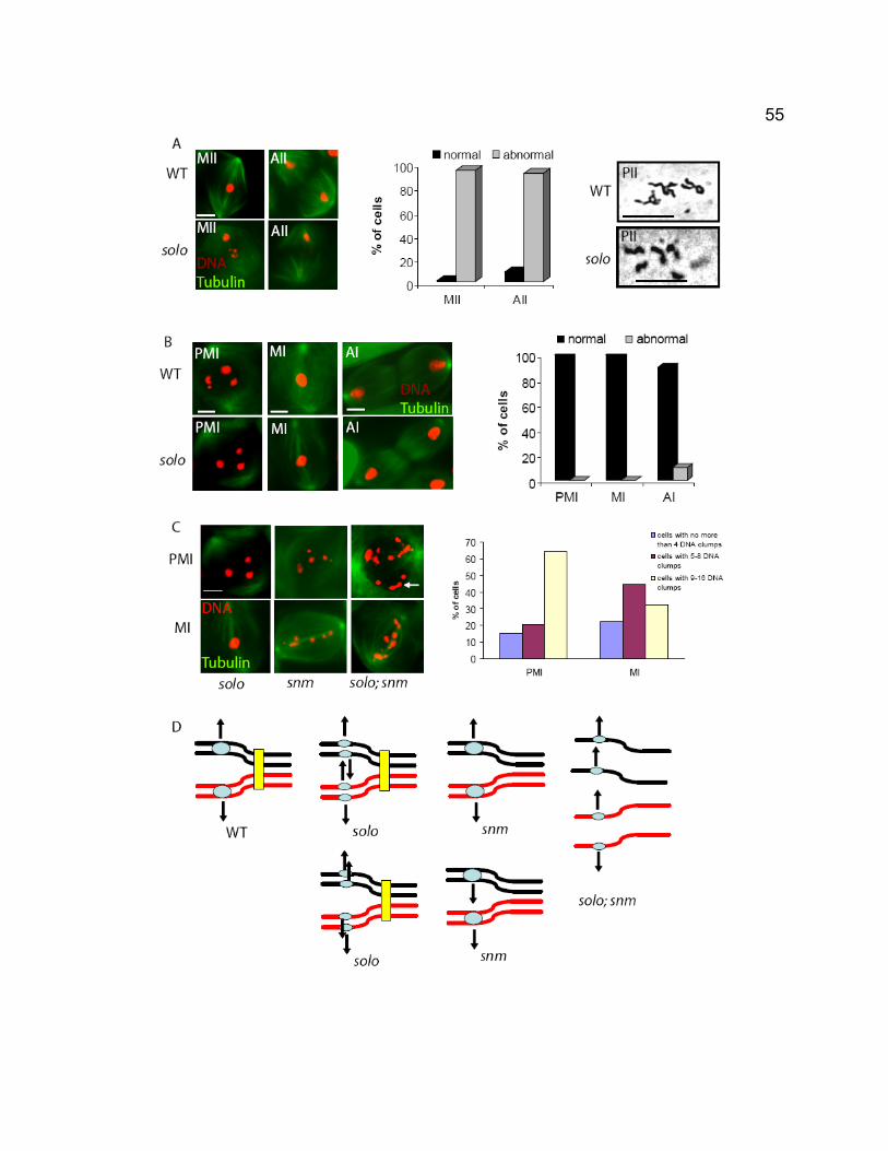
55
56
bivalents, were regularly seen (176/176) in prometaphase I. The bivalents
congressed normally, forming compact metaphase I configurations with the
bivalents equidistant from the two poles, then segregated synchronously,
generating poles of roughly equal DNA content in 62/69 anaphase I
spermatocytes, suggesting that approximately equal numbers of chromatids
segregate to each pole at the first division. Thus despite the genetic data
indicating that homologous chromatids (we use the term “homologous
chromatids” to refer to two chromatids that are from either of homologous
chromosomes, such as X and Y chromatids) segregate to the same pole at high
frequencies in solo males, the cytological data revealed visible PSCS only in
meiosis II but not before anaphase I and provided little evidence for disturbed
segregation at meiosis I.
Sister centromere cohesion is lost prior to PMI in solo spermatocytes
Although sister chromatids do not dissociate prior to anaphase I in solo
spermatocytes, sister centromeres might nevertheless separate prematurely due
to loss of cohesion. This precocious separation of centromeres could cause
failure of co-orientation of sister centromeres and thereby perturb the reductional
division, causing homologous chromatids move to the same spindle poles at
anaphase I, as revealed by the cross data. Although the mechanism of sister
centromere co-orientation is poorly understood, in both S. pombe and
Arabidopsis cohesion between sister centromeres is required for their co-
orientation and the failure of co-orientation shift the reductional pattern of
57
chromosome segregation at meiosis I to equational chromosome segregation
(Watanabe and Nurse, 1999; Watanabe et al., 2001; Chelysheva et al., 2005).
To test for effects of solo mutations on centromere cohesion, we monitored
centromere behavior in wild-type and solo males with an antibody against CID
(centromere identifier), a centromere-specific histone H3-like protein (Ahmad and
Henikoff, 2001; Blower and Karpen, 2001) (Fig. 2-2). In wild-type spermatocytes,
the number of anti-CID foci per nucleus never exceeded the number of
homologous chromosomes (eight in meiosis I and four in meiosis II). During late
prophase I when the four bivalents occupy well-separated territories, two CID
spots could often be seen in each chromosome territory.
The numbers of anti-CID foci in solo mutants were similar to wild-type
throughout early and mid-prophase I (stages S1-S4 (Cenci et al., 1994).
However, from late prophase I (stages S5 & S6, (Cenci et al., 1994)) through
metaphase I, many bivalents in solo spermatocytes exhibited 3 or 4 spots instead
of the normal 2. Virtually all nuclei exhibited more than 8 spots, with the number
ranging up to 16, the number of sister centromeres in a diploid nucleus (Fig. 2-
2C). solo mutants also exhibited too many CID spots during meiosis II, up to
eight instead of four. These observations indicate that sister chromatid cohesion
is lost prior to prometaphase I in solo mutants.
Thus although bivalents remain intact in solo males until anaphase I and
align properly on the meiosis I spindle, sister centromeres separate from one
another much earlier in meiosis I than anaphase I. These observations strongly
suggest that the aberrant meiosis I segregation patterns apparent in the cross
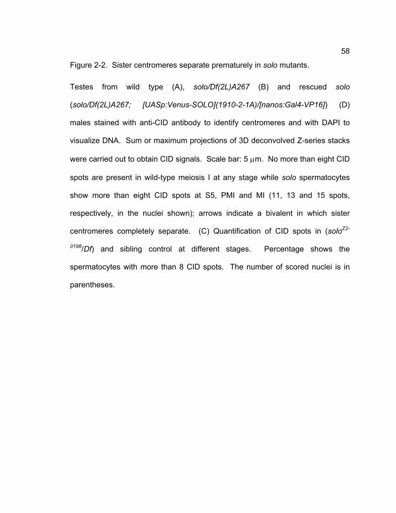
58
Figure 2-2. Sister centromeres separate prematurely in solo mutants.
Testes from wild type (A), solo/Df(2L)A267 (B) and rescued solo
(solo/Df(2L)A267; [UASp:Venus-SOLO](1910-2-1A)/[nanos:Gal4-VP16]) (D)
males stained with anti-CID antibody to identify centromeres and with DAPI to
visualize DNA. Sum or maximum projections of 3D deconvolved Z-series stacks
were carried out to obtain CID signals. Scale bar: 5 μm. No more than eight CID
spots are present in wild-type meiosis I at any stage while solo spermatocytes
show more than eight CID spots at S5, PMI and MI (11, 13 and 15 spots,
respectively, in the nuclei shown); arrows indicate a bivalent in which sister
centromeres completely separate. (C) Quantification of CID spots in (soloZ2-
0198/Df) and sibling control at different stages. Percentage shows the
spermatocytes with more than 8 CID spots. The number of scored nuclei is in
parentheses.
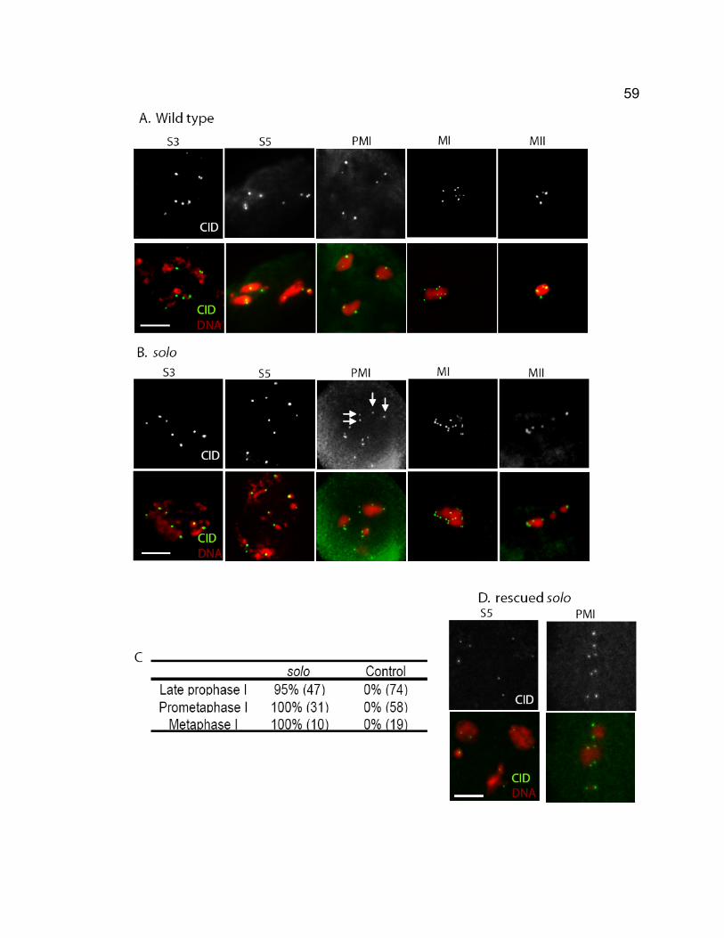
59
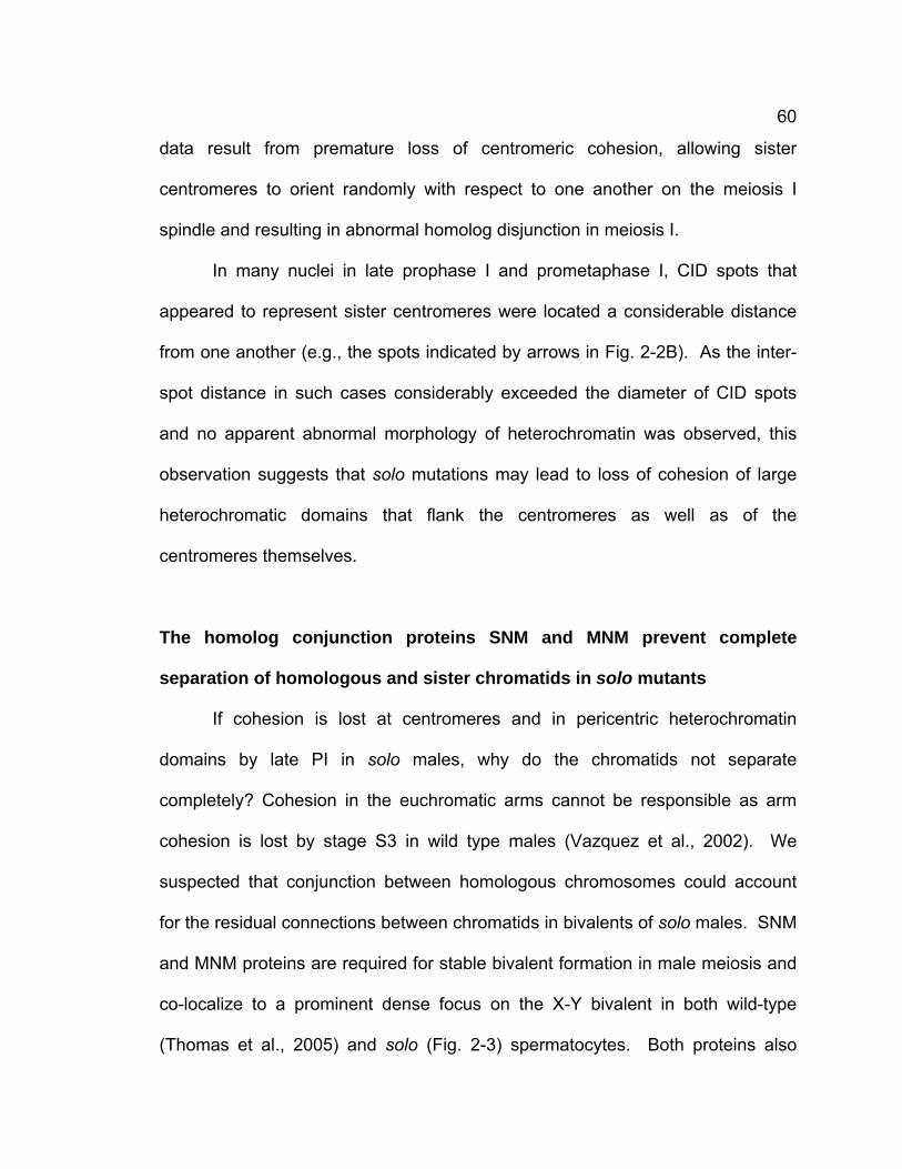
60
data result from premature loss of centromeric cohesion, allowing sister
centromeres to orient randomly with respect to one another on the meiosis I
spindle and resulting in abnormal homolog disjunction in meiosis I.
In many nuclei in late prophase I and prometaphase I, CID spots that
appeared to represent sister centromeres were located a considerable distance
from one another (e.g., the spots indicated by arrows in Fig. 2-2B). As the inter-
spot distance in such cases considerably exceeded the diameter of CID spots
and no apparent abnormal morphology of heterochromatin was observed, this
observation suggests that solo mutations may lead to loss of cohesion of large
heterochromatic domains that flank the centromeres as well as of the
centromeres themselves.
The homolog conjunction proteins SNM and MNM prevent complete
separation of homologous and sister chromatids in solo mutants
If cohesion is lost at centromeres and in pericentric heterochromatin
domains by late PI in solo males, why do the chromatids not separate
completely? Cohesion in the euchromatic arms cannot be responsible as arm
cohesion is lost by stage S3 in wild type males (Vazquez et al., 2002). We
suspected that conjunction between homologous chromosomes could account
for the residual connections between chromatids in bivalents of solo males. SNM
and MNM proteins are required for stable bivalent formation in male meiosis and
co-localize to a prominent dense focus on the X-Y bivalent in both wild-type
(Thomas et al., 2005) and solo (Fig. 2-3) spermatocytes. Both proteins also
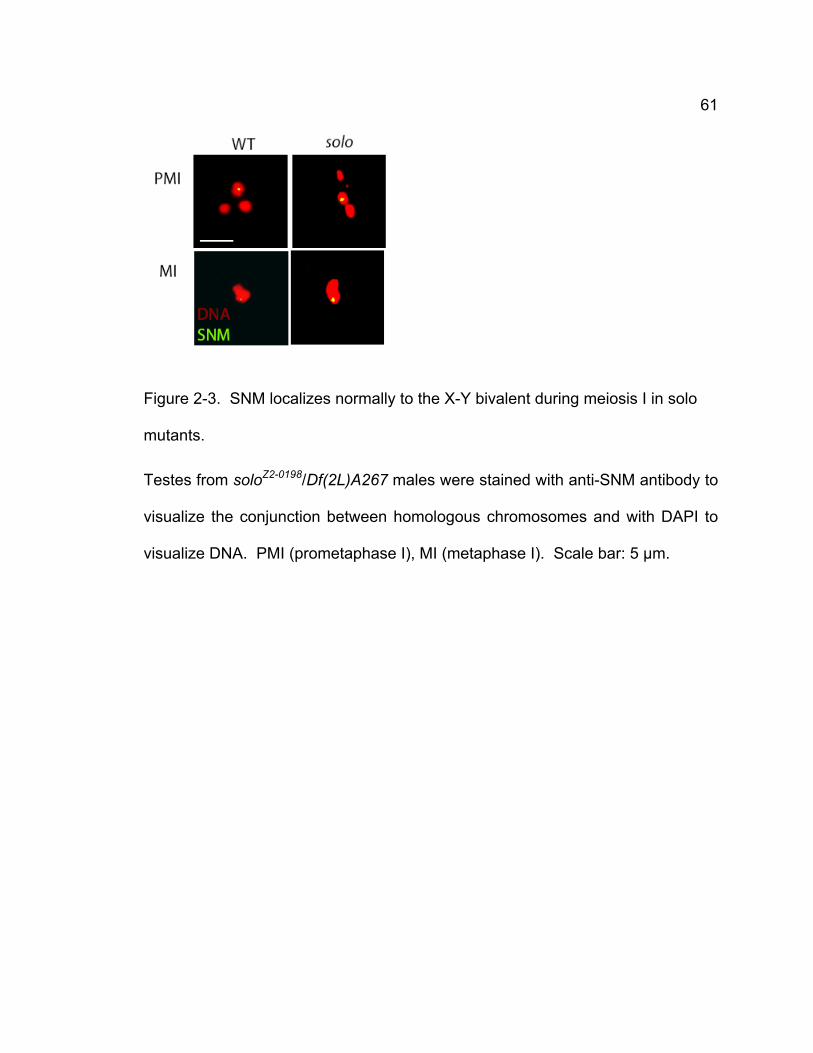
61
Figure 2-3. SNM localizes normally to the X-Y bivalent during meiosis I in solo
mutants.
Testes from soloZ2-0198/Df(2L)A267 males were stained with anti-SNM antibody to
visualize the conjunction between homologous chromosomes and with DAPI to
visualize DNA. PMI (prometaphase I), MI (metaphase I). Scale bar: 5 μm.
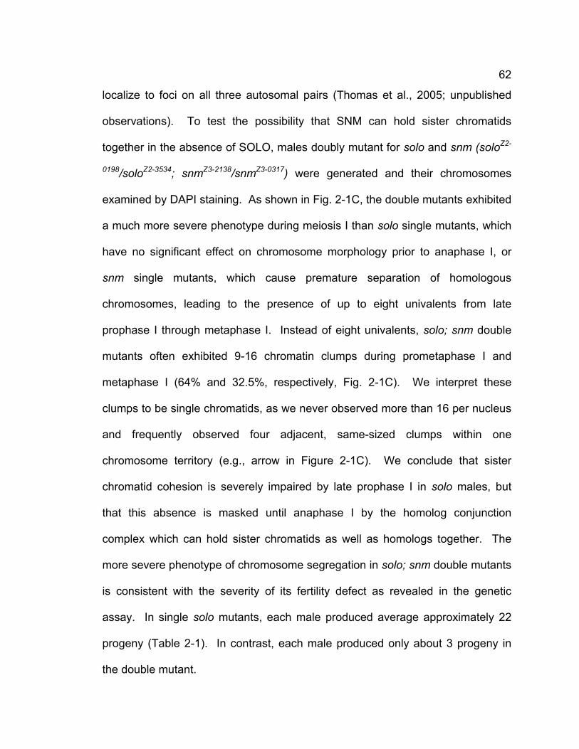
62
localize to foci on all three autosomal pairs (Thomas et al., 2005; unpublished
observations). To test the possibility that SNM can hold sister chromatids
together in the absence of SOLO, males doubly mutant for solo and snm (soloZ2-
0198/soloZ2-3534; snmZ3-2138/snmZ3-0317) were generated and their chromosomes
examined by DAPI staining. As shown in Fig. 2-1C, the double mutants exhibited
a much more severe phenotype during meiosis I than solo single mutants, which
have no significant effect on chromosome morphology prior to anaphase I, or
snm single mutants, which cause premature separation of homologous
chromosomes, leading to the presence of up to eight univalents from late
prophase I through metaphase I. Instead of eight univalents, solo; snm double
mutants often exhibited 9-16 chromatin clumps during prometaphase I and
metaphase I (64% and 32.5%, respectively, Fig. 2-1C). We interpret these
clumps to be single chromatids, as we never observed more than 16 per nucleus
and frequently observed four adjacent, same-sized clumps within one
chromosome territory (e.g., arrow in Figure 2-1C). We conclude that sister
chromatid cohesion is severely impaired by late prophase I in solo males, but
that this absence is masked until anaphase I by the homolog conjunction
complex which can hold sister chromatids as well as homologs together. The
more severe phenotype of chromosome segregation in solo; snm double mutants
is consistent with the severity of its fertility defect as revealed in the genetic
assay. In single solo mutants, each male produced average approximately 22
progeny (Table 2-1). In contrast, each male produced only about 3 progeny in
the double mutant.
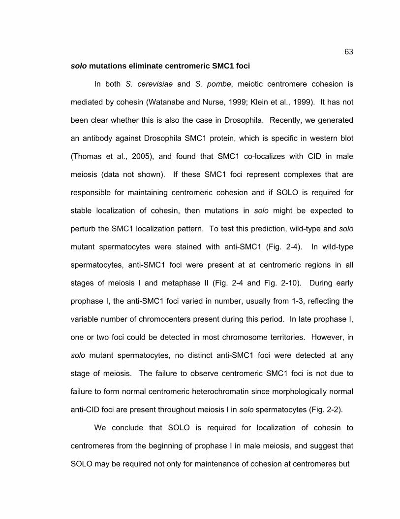
63
solo mutations eliminate centromeric SMC1 foci
In both S. cerevisiae and S. pombe, meiotic centromere cohesion is
mediated by cohesin (Watanabe and Nurse, 1999; Klein et al., 1999). It has not
been clear whether this is also the case in Drosophila. Recently, we generated
an antibody against Drosophila SMC1 protein, which is specific in western blot
(Thomas et al., 2005), and found that SMC1 co-localizes with CID in male
meiosis (data not shown). If these SMC1 foci represent complexes that are
responsible for maintaining centromeric cohesion and if SOLO is required for
stable localization of cohesin, then mutations in solo might be expected to
perturb the SMC1 localization pattern. To test this prediction, wild-type and solo
mutant spermatocytes were stained with anti-SMC1 (Fig. 2-4). In wild-type
spermatocytes, anti-SMC1 foci were present at at centromeric regions in all
stages of meiosis I and metaphase II (Fig. 2-4 and Fig. 2-10). During early
prophase I, the anti-SMC1 foci varied in number, usually from 1-3, reflecting the
variable number of chromocenters present during this period. In late prophase I,
one or two foci could be detected in most chromosome territories. However, in
solo mutant spermatocytes, no distinct anti-SMC1 foci were detected at any
stage of meiosis. The failure to observe centromeric SMC1 foci is not due to
failure to form normal centromeric heterochromatin since morphologically normal
anti-CID foci are present throughout meiosis I in solo spermatocytes (Fig. 2-2).
We conclude that SOLO is required for localization of cohesin to
centromeres from the beginning of prophase I in male meiosis, and suggest that
SOLO may be required not only for maintenance of cohesion at centromeres but
64
Figure 2-4. Localization of SMC1 in wild-type and solo spermatocytes.
SMC1 foci were detected by anti-SMC1 and DNA was stained with DAPI.
Centromeric SMC1 foci are visible throughout prophase I in wild-type (A) but are
completely absent in solo spermatocytes (B). Mutant spermatocytes are from
soloZ2-0198/soloZ2-0198 adult males. All images are sum projections of 3D
deconvolved Z-series planes. Size bar: 5 μm.
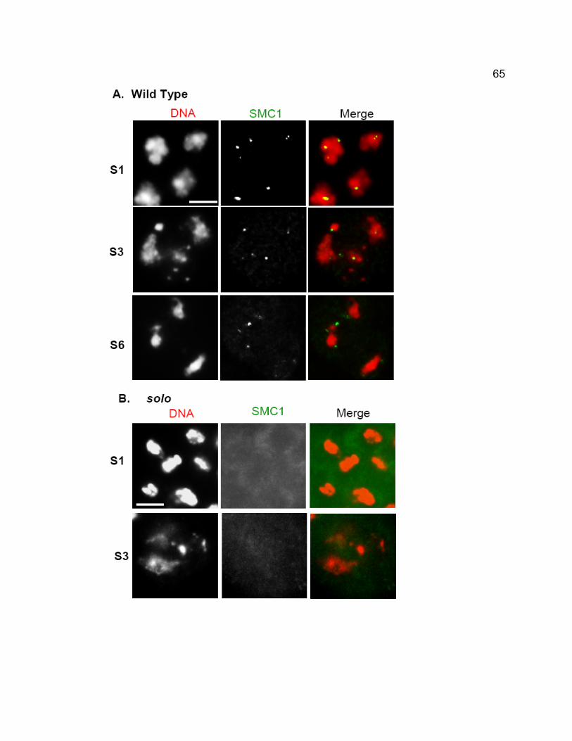
65
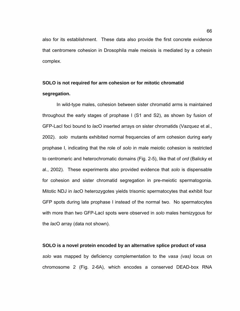
66
also for its establishment. These data also provide the first concrete evidence
that centromere cohesion in Drosophila male meiosis is mediated by a cohesin
complex.
SOLO is not required for arm cohesion or for mitotic chromatid
segregation.
In wild-type males, cohesion between sister chromatid arms is maintained
throughout the early stages of prophase I (S1 and S2), as shown by fusion of
GFP-LacI foci bound to lacO inserted arrays on sister chromatids (Vazquez et al.,
2002). solo mutants exhibited normal frequencies of arm cohesion during early
prophase I, indicating that the role of solo in male meiotic cohesion is restricted
to centromeric and heterochromatic domains (Fig. 2-5), like that of ord (Balicky et
al., 2002). These experiments also provided evidence that solo is dispensable
for cohesion and sister chromatid segregation in pre-meiotic spermatogonia.
Mitotic NDJ in lacO heterozygotes yields trisomic spermatocytes that exhibit four
GFP spots during late prophase I instead of the normal two. No spermatocytes
with more than two GFP-LacI spots were observed in solo males hemizygous for
the lacO array (data not shown).
SOLO is a novel protein encoded by an alternative splice product of vasa
solo was mapped by deficiency complementation to the vasa (vas) locus on
chromosome 2 (Fig. 2-6A), which encodes a conserved DEAD-box RNA
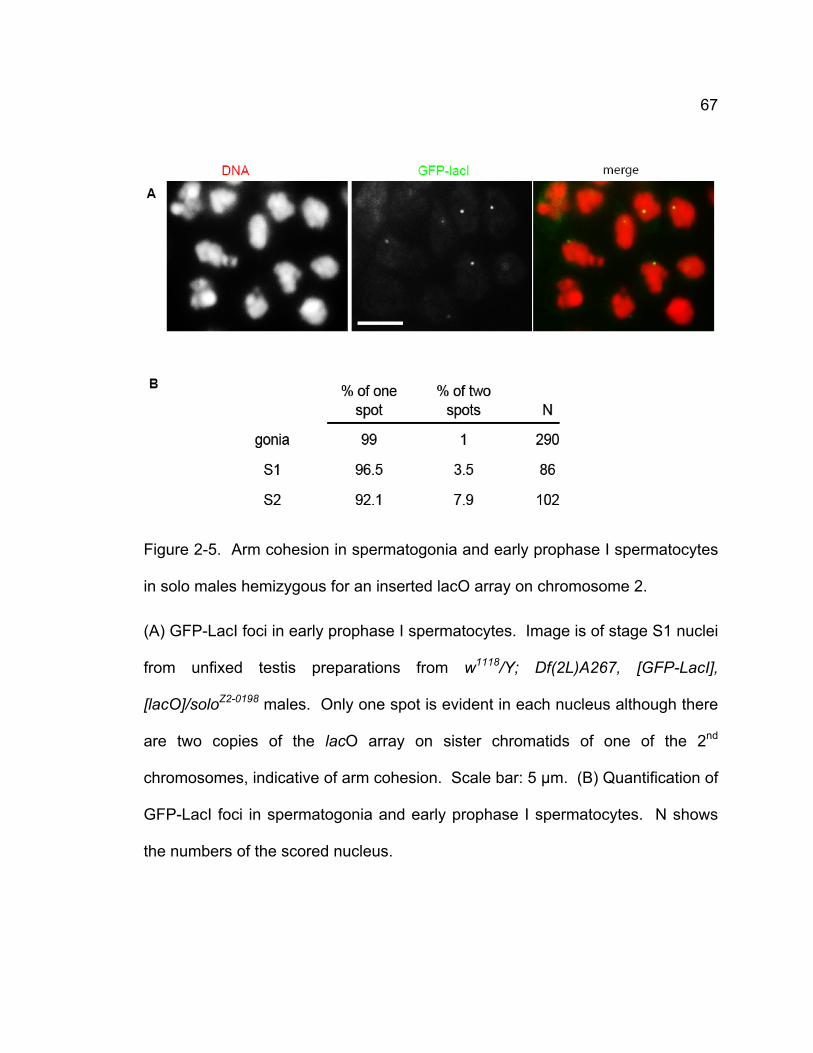
67
Figure 2-5. Arm cohesion in spermatogonia and early prophase I spermatocytes
in solo males hemizygous for an inserted lacO array on chromosome 2.
(A) GFP-LacI foci in early prophase I spermatocytes. Image is of stage S1 nuclei
from unfixed testis preparations from w1118/Y; Df(2L)A267, [GFP-LacI],
[lacO]/soloZ2-0198 males. Only one spot is evident in each nucleus although there
are two copies of the lacO array on sister chromatids of one of the 2nd
chromosomes, indicative of arm cohesion. Scale bar: 5 μm. (B) Quantification of
GFP-LacI foci in spermatogonia and early prophase I spermatocytes. N shows
the numbers of the scored nucleus.
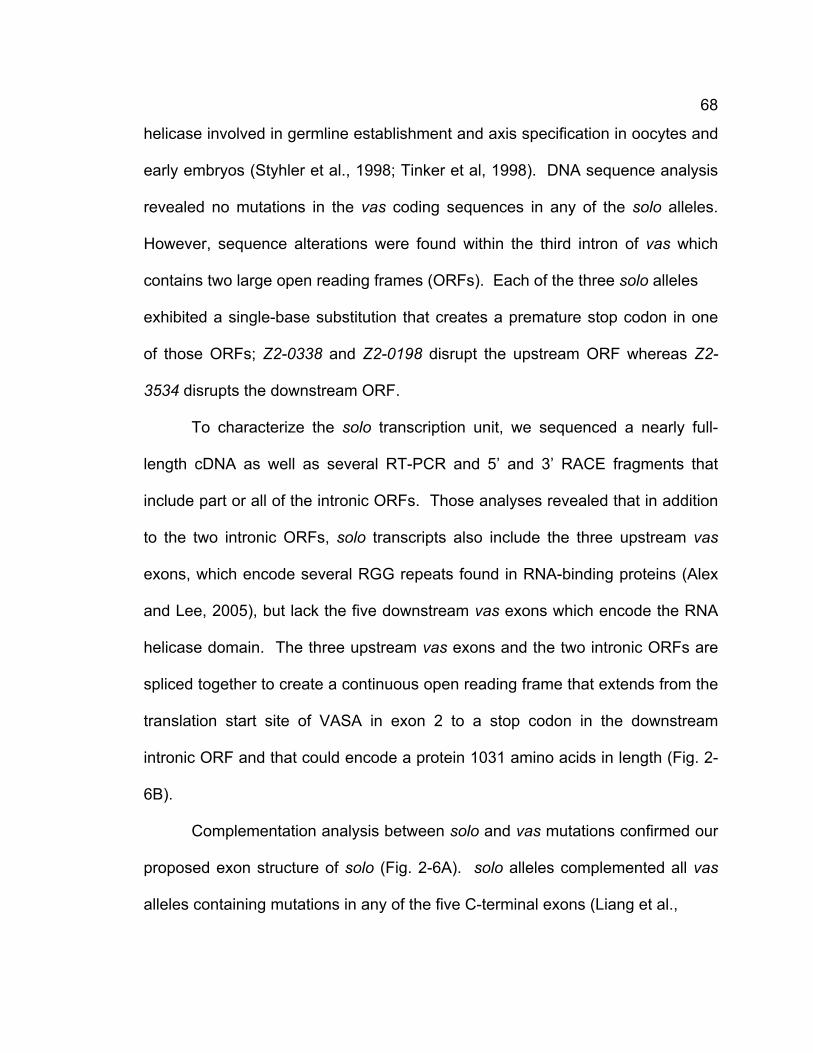
68
helicase involved in germline establishment and axis specification in oocytes and
early embryos (Styhler et al., 1998; Tinker et al, 1998). DNA sequence analysis
revealed no mutations in the vas coding sequences in any of the solo alleles.
However, sequence alterations were found within the third intron of vas which
contains two large open reading frames (ORFs). Each of the three solo alleles
exhibited a single-base substitution that creates a premature stop codon in one
of those ORFs; Z2-0338 and Z2-0198 disrupt the upstream ORF whereas Z2-
3534 disrupts the downstream ORF.
To characterize the solo transcription unit, we sequenced a nearly full-
length cDNA as well as several RT-PCR and 5’ and 3’ RACE fragments that
include part or all of the intronic ORFs. Those analyses revealed that in addition
to the two intronic ORFs, solo transcripts also include the three upstream vas
exons, which encode several RGG repeats found in RNA-binding proteins (Alex
and Lee, 2005), but lack the five downstream vas exons which encode the RNA
helicase domain. The three upstream vas exons and the two intronic ORFs are
spliced together to create a continuous open reading frame that extends from the
translation start site of VASA in exon 2 to a stop codon in the downstream
intronic ORF and that could encode a protein 1031 amino acids in length (Fig. 2-
6B).
Complementation analysis between solo and vas mutations confirmed our
proposed exon structure of solo (Fig. 2-6A). solo alleles complemented all vas
alleles containing mutations in any of the five C-terminal exons (Liang et al.,
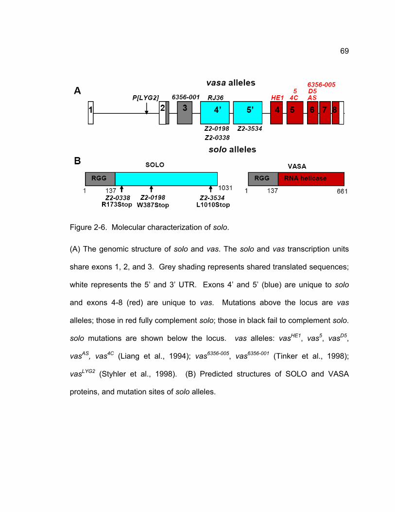
69
Figure 2-6. Molecular characterization of solo.
(A) The genomic structure of solo and vas. The solo and vas transcription units
share exons 1, 2, and 3. Grey shading represents shared translated sequences;
white represents the 5’ and 3’ UTR. Exons 4’ and 5’ (blue) are unique to solo
and exons 4-8 (red) are unique to vas. Mutations above the locus are vas
alleles; those in red fully complement solo; those in black fail to complement solo.
solo mutations are shown below the locus. vas alleles: vasHE1, vas5, vasD5,
vasAS, vas4C (Liang et al., 1994); vas6356-005, vas6356-001 (Tinker et al., 1998);
vasLYG2 (Styhler et al., 1998). (B) Predicted structures of SOLO and VASA
proteins, and mutation sites of solo alleles.
70
1994), which encode the VASA helicase domain, indicating that the C-terminus
of VASA is not shared by SOLO. However, vas mutations that map upstream of
the SOLO-specific ORFs, including one nonsense mutation in exon 3, vas6356-001
(Tinker et al., 1998) failed to complement the solo alleles, indicating that the 137
amino acids encoded by the upstream exons are present in both proteins. It is
unlikely that the SOLO-specific exons are expressed independently of vas in
addition to being expressed as a fusion product with the N terminus of vas, as
vas6356-001 behaves as a null allele of solo, giving X-Y NDJ frequencies of 41-44%
in trans with solo alleles. We conclude that solo encodes a protein that includes
the N-terminal 137 amino acids of VASA fused to 894 amino acids encoded
within the 3rd intron of vas.
Single homologs of SOLO were identified by BLAST analysis in the
genomes of all 10 Drosophila species for which sequenced genomes were
available (Flybase, 2007b). Overall conservation is fairly low; D. melanogaster
SOLO exhibits only around 30% amino acid identity with its homologs in D. virilis
and D. pseudoobscura. However, in all of the Drosophila genomes, the solo
sequences are nested within the third intron of vas, and SOLO appears capable
of being expressed by the same alternative splice mechanism used in D.
melanogaster.
No homologs of SOLO were identified outside of the genus Drosophila,
not even in the genome of the mosquito Anopheles gambiae, another Dipteran
insect. While it is possible that solo exists in Anopheles but is unrecognizable
due to divergence, it would have to be located elsewhere in the genome as there
71
are no large ORFs nested within introns of the Anopheles vas gene. Other than
the RGG motifs in the common N-terminus, SOLO exhibits no significant
homologies with other proteins in the sequence database.
Venus-SOLO co-localizes with CID and SMC1 from early prophase I until
anaphase II
To study the intracellular localization pattern of SOLO, transgenic
insertions of two P-element constructs containing the solo cDNA tagged at its N
or C terminus with Venus (an enhanced yellow-fluorescent protein) cloned
downstream of yeast UAS sequences were generated (see Supplementary
Experimental Procedures). Expression of the fusion proteins was induced by the
GAL4-VP16 transcription activator under control of the Drosophila nanos
promoter, which is active in most male germ cells (Doren et al., 1998). Two 3rd
chromosome insertions of [UASp:Venus-SOLO] and one 3rd chromosome
insertion of [UASp:SOLO-Venus] were tested for ability to complement the
meiotic phenotypes of solo mutants. One copy of each SOLO transgene sufficed
to provide virtually complete rescue of solo meiotic phenotypes. Sex
chromosome NDJ was reduced to background levels (Table 2-3) and cytological
analysis indicated that meiosis II segregation is regular in solo/Df; [UASp:Venus-
SOLO]/[nanos:Gal4-VP16] males (data not shown). Venus-SOLO also
suppressed the centromere cohesion defect of solo mutants. Late prophase I
and prometaphase I nuclei from rescued males showed a maximum of two CID
spots per bivalent (Fig. 2-2D), whereas nuclei from unrescued sibling solo males
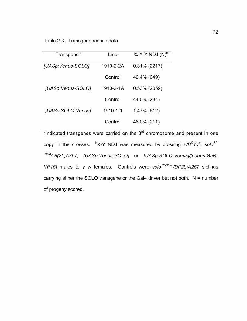
72
Table 2-3. Transgene rescue data.
Transgenea Line % X-Y NDJ (N)b
[UASp:Venus-SOLO] 1910-2-2A 0.31% (2217)
Control 46.4% (649)
[UASp:Venus-SOLO] 1910-2-1A 0.53% (2059)
Control 44.0% (234)
[UASp:SOLO-Venus] 1910-1-1 1.47% (612)
Control 46.0% (211)
aIndicated transgenes were carried on the 3rd chromosome and present in one
copy in the crosses. bX-Y NDJ was measured by crossing +/BSYy+; soloZ2-
0198/Df(2L)A267; [UASp:Venus-SOLO] or [UASp:SOLO-Venus]/[nanos:Gal4-
VP16] males to y w females. Controls were soloZ2-0198/Df(2L)A267 siblings
carrying either the SOLO transgene or the Gal4 driver but not both. N = number
of progeny scored.
73
typically showed three or four CID spots per bivalent (data not shown). These
data indicate that the tagged SOLO proteins function similarly to endogenous
SOLO in male meiosis.
The localization pattern of Venus-SOLO in spermatocytes was examined
using [nanos:Gal4-VP16] to induce expression. Bright Venus foci were seen in
nuclei of mitotic spermatogonia and meiotic spermatocytes of all stages up to
and including metaphase II but were absent at anaphase II and subsequent
stages (Fig. 2-7 and Fig. 2-8A). Moreover, at all stages, the Venus foci
overlapped CID foci (Figure 5), indicating that Venus-SOLO localizes to
centromeres. The number of Venus foci per nucleus varied with stage. Young
primary spermatocytes (stages S1 and S2), in which non-homologous
centromeres form variable numbers of clusters, typically exhibited one to four
foci, whereas spermatocytes in mid-late prophase I (stages S3-S6) and in
prometaphase I and metaphase I exhibited up to eight foci per nucleus, typically
two foci per bivalent.
Secondary spermatocytes typically exhibited 3-4 foci. SOLO-Venus
exhibited a similar localization pattern (data not shown). In addition, nuclei in late
prophase I often exhibited diffuse Venus-SOLO foci that localized to
chromosomal domains considerably larger than the centromeres (Fig. 2-9). This
observation is consistent with the idea that SOLO localizes not only to
centromere regions but more generally to heterochromatic domains of
spermatocyte chromosomes.
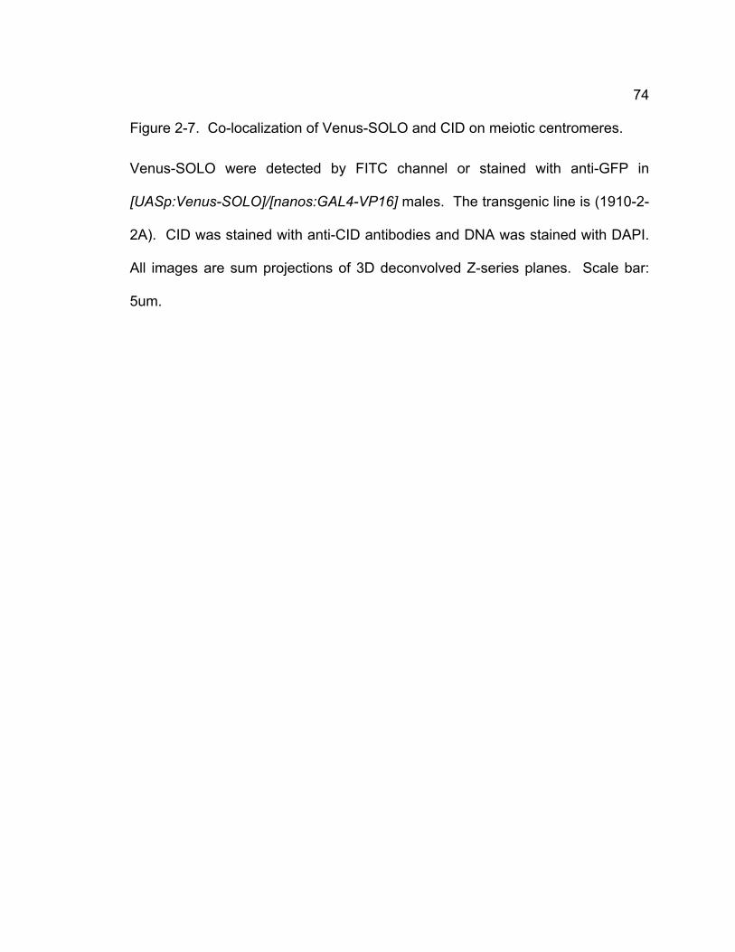
74
Figure 2-7. Co-localization of Venus-SOLO and CID on meiotic centromeres.
Venus-SOLO were detected by FITC channel or stained with anti-GFP in
[UASp:Venus-SOLO]/[nanos:GAL4-VP16] males. The transgenic line is (1910-2-
2A). CID was stained with anti-CID antibodies and DNA was stained with DAPI.
All images are sum projections of 3D deconvolved Z-series planes. Scale bar:
5um.
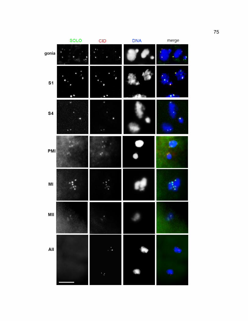
75
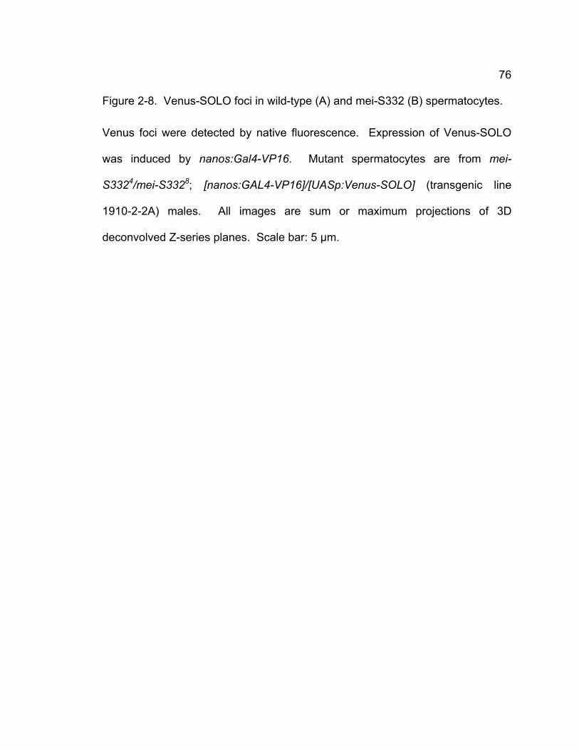
76
Figure 2-8. Venus-SOLO foci in wild-type (A) and mei-S332 (B) spermatocytes.
Venus foci were detected by native fluorescence. Expression of Venus-SOLO
was induced by nanos:Gal4-VP16. Mutant spermatocytes are from mei-
S3324/mei-S3328; [nanos:GAL4-VP16]/[UASp:Venus-SOLO] (transgenic line
1910-2-2A) males. All images are sum or maximum projections of 3D
deconvolved Z-series planes. Scale bar: 5 μm.
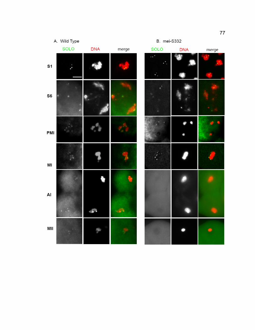
77
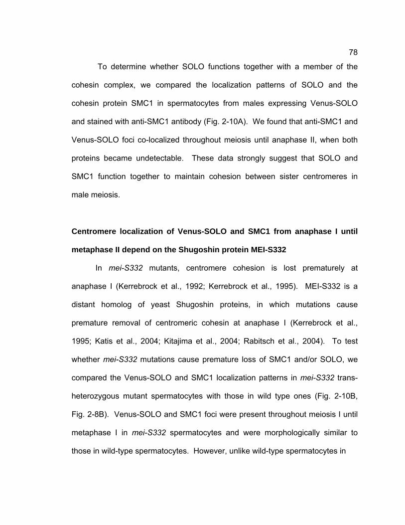
78
To determine whether SOLO functions together with a member of the
cohesin complex, we compared the localization patterns of SOLO and the
cohesin protein SMC1 in spermatocytes from males expressing Venus-SOLO
and stained with anti-SMC1 antibody (Fig. 2-10A). We found that anti-SMC1 and
Venus-SOLO foci co-localized throughout meiosis until anaphase II, when both
proteins became undetectable. These data strongly suggest that SOLO and
SMC1 function together to maintain cohesion between sister centromeres in
male meiosis.
Centromere localization of Venus-SOLO and SMC1 from anaphase I until
metaphase II depend on the Shugoshin protein MEI-S332
In mei-S332 mutants, centromere cohesion is lost prematurely at
anaphase I (Kerrebrock et al., 1992; Kerrebrock et al., 1995). MEI-S332 is a
distant homolog of yeast Shugoshin proteins, in which mutations cause
premature removal of centromeric cohesin at anaphase I (Kerrebrock et al.,
1995; Katis et al., 2004; Kitajima et al., 2004; Rabitsch et al., 2004). To test
whether mei-S332 mutations cause premature loss of SMC1 and/or SOLO, we
compared the Venus-SOLO and SMC1 localization patterns in mei-S332 trans-
heterozygous mutant spermatocytes with those in wild type ones (Fig. 2-10B,
Fig. 2-8B). Venus-SOLO and SMC1 foci were present throughout meiosis I until
metaphase I in mei-S332 spermatocytes and were morphologically similar to
those in wild-type spermatocytes. However, unlike wild-type spermatocytes in
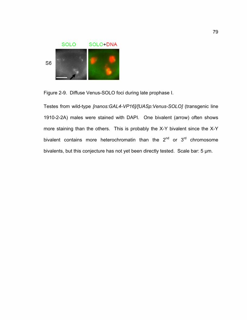
79
Figure 2-9. Diffuse Venus-SOLO foci during late prophase I.
Testes from wild-type [nanos:GAL4-VP16]/[UASp:Venus-SOLO] (transgenic line
1910-2-2A) males were stained with DAPI. One bivalent (arrow) often shows
more staining than the others. This is probably the X-Y bivalent since the X-Y
bivalent contains more heterochromatin than the 2nd or 3rd chromosome
bivalents, but this conjecture has not yet been directly tested. Scale bar: 5 μm.
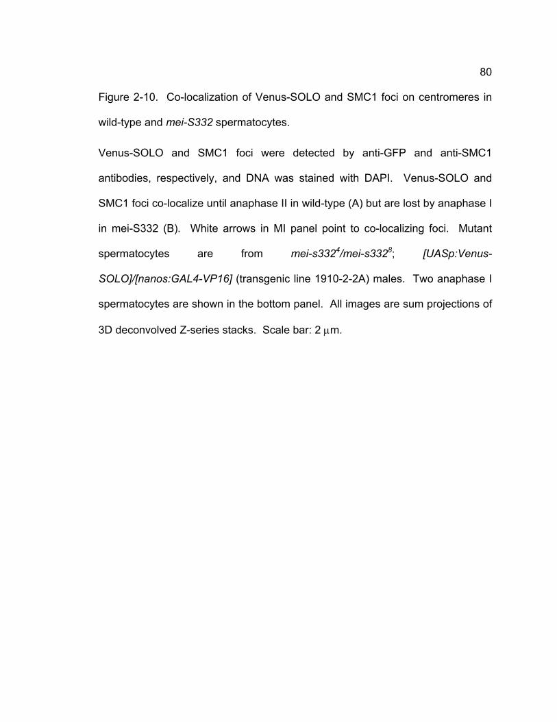
80
Figure 2-10. Co-localization of Venus-SOLO and SMC1 foci on centromeres in
wild-type and mei-S332 spermatocytes.
Venus-SOLO and SMC1 foci were detected by anti-GFP and anti-SMC1
antibodies, respectively, and DNA was stained with DAPI. Venus-SOLO and
SMC1 foci co-localize until anaphase II in wild-type (A) but are lost by anaphase I
in mei-S332 (B). White arrows in MI panel point to co-localizing foci. Mutant
spermatocytes are from mei-s3324/mei-s3328; [UASp:Venus-
SOLO]/[nanos:GAL4-VP16] (transgenic line 1910-2-2A) males. Two anaphase I
spermatocytes are shown in the bottom panel. All images are sum projections of
3D deconvolved Z-series stacks. Scale bar: 2 μm.
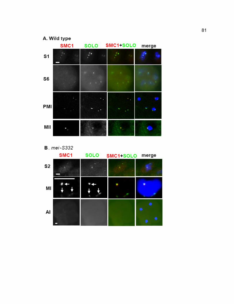
81
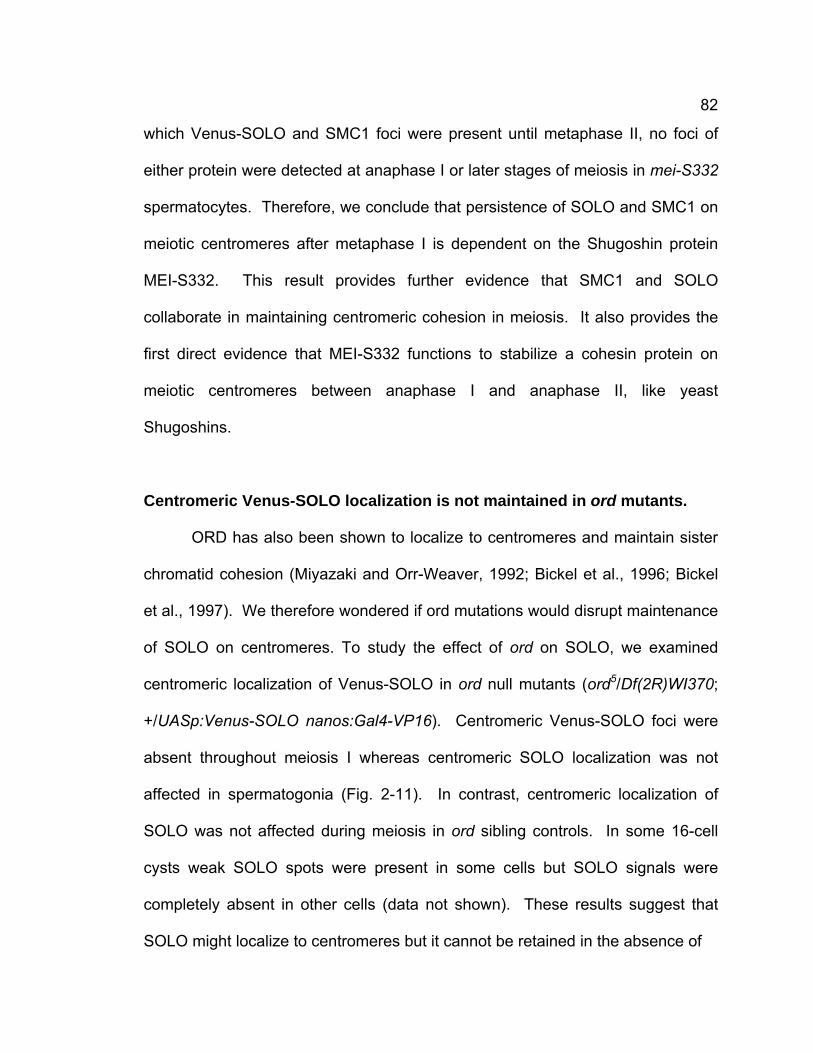
82
which Venus-SOLO and SMC1 foci were present until metaphase II, no foci of
either protein were detected at anaphase I or later stages of meiosis in mei-S332
spermatocytes. Therefore, we conclude that persistence of SOLO and SMC1 on
meiotic centromeres after metaphase I is dependent on the Shugoshin protein
MEI-S332. This result provides further evidence that SMC1 and SOLO
collaborate in maintaining centromeric cohesion in meiosis. It also provides the
first direct evidence that MEI-S332 functions to stabilize a cohesin protein on
meiotic centromeres between anaphase I and anaphase II, like yeast
Shugoshins.
Centromeric Venus-SOLO localization is not maintained in ord mutants.
ORD has also been shown to localize to centromeres and maintain sister
chromatid cohesion (Miyazaki and Orr-Weaver, 1992; Bickel et al., 1996; Bickel
et al., 1997). We therefore wondered if ord mutations would disrupt maintenance
of SOLO on centromeres. To study the effect of ord on SOLO, we examined
centromeric localization of Venus-SOLO in ord null mutants (ord5/Df(2R)WI370;
+/UASp:Venus-SOLO nanos:Gal4-VP16). Centromeric Venus-SOLO foci were
absent throughout meiosis I whereas centromeric SOLO localization was not
affected in spermatogonia (Fig. 2-11). In contrast, centromeric localization of
SOLO was not affected during meiosis in ord sibling controls. In some 16-cell
cysts weak SOLO spots were present in some cells but SOLO signals were
completely absent in other cells (data not shown). These results suggest that
SOLO might localize to centromeres but it cannot be retained in the absence of
83
Figure 2- 11. SOLO is absent at centromeres from early prophase I but present
in spermatogonia.
Venus-SOLO (green) was detected by FITC channel and DNA was stained with
DAPI (red). Venus-SOLO foci is present in spermatogonia (A) but absent in
S1(B) and S6 stages of prophase I (C). Scale bar: 5μm.
84
ORD. Previous genetic studies have shown ord functions earlier than mei-S332
in maintaining cohesion during meiosis. Our results in which SOLO disappears
at early prophase I in ord mutants while it is lost until anaphase I in mei-S332
provide further evidence at cytological level.
85
DISCUSSION
SOLO is required for centromere cohesion and for co-orientation of sister
centromeres.
Our results show that SOLO is required in male meiosis for sister
centromere cohesion during both meiosis I and meiosis II. Homologous and
sister chromatids of both sex chromosome and autosomal bivalents segregate
approximately randomly from one another in genetic crosses of solo males.
Although bivalents remain intact through meiosis I, sister centromeres are visibly
separated in most bivalents by stage S5 of prophase I and sister chromatids are
fully separated during meiosis II.
Taken together, these observations indicate that both sister centromere
cohesion and co-orientation of sister centromeres during meiosis I are disrupted
by solo mutations. We presume that the failure of sister centromeres to co-orient
is a consequence of their premature loss of cohesion, rather than an indication of
a second function for SOLO. In both S. cerevisiae and S. pombe, there are
specialized proteins required for sister centromere co-orientation, Monopolin in S.
cerevisiae and Moa1 in S. pombe (Toth et al., 2000; Yokobayashi and
Watanabe, 2005). However, in both organisms centromeric cohesion is also
essential, as loss of rec8 function leads either to random chromatid orientation
(S. cerevisiae) or to predominantly equational orientation (S. pombe) at meiosis I
(Klein et al., 1999; Watanabe and Nurse, 1999; Watanabe et al., 2001). Similar
86
observations have been made in rec8 mutants in both C. elegans and
Arabidopsis (Pasierbek et al., 2001; Xu et al., 2005). It remains to be determined
whether other proteins analogous to Monopolin or Moa1 are also required for
centromere co-orientation in Drosophila.
Since Venus-SOLO foci were present by stage S1 of prophase I and solo
mutations abolished centromere localization of SMC1 from stage S1 on, it is
surprising that sister centromere CID foci were not visibly separate prior to stage
S5 in solo mutants. Why the loss of cohesion due to solo mutations does not
cause separation of sister centromeres at early and mid prophase I is at present
unclear. The complete separation of sister centromeres at later meiotic stages
argues against residual activity of solo being responsible for holding sister
centromeres together at early and mid prophase I. The molecular data showing
that all three solo are null alleles also confirm that there is no residual SOLO
activity. Other pathways unrelated to cohesion, like catenation between sister
centromeres, might account for the lack of apparent separation of sister
centromeres at early and mid prophase I. We note that our finding is consistent
with observations in S. pombe (Molnar et al., 1995), where rec8 mutations did not
affect centromere clustering at meiotic prophase.
The roles of SMC1, SOLO and ORD in meiotic cohesion
In yeast, multiple meiotic cohesion functions are carried out by cohesin
complexes that include meiosis-specific subunits such as REC8, which replaces
the mitotic kleisin subunit RAD21. REC8 is conserved among most eukaryotes
87
and has been shown in several model plants and animals to be critical for many
of the same meiotic functions identified in yeast (Pasierbek et al., 2001; Cai et
al., 2003; chelysheva et al., 2003; Petronczki et al., 2003; Schleiffer et al., 2003;
Xu et al., 2005). In Drosophila, however, no true REC8 ortholog has been
identified and no mutations in cohesin genes have been available. Thus the role
of cohesin in Drosophila meiotic cohesion has been unclear.
The phenotypes of solo mutations are similar to those of ord mutations in
Drosophila and rec8 mutations in other eukaryotes. Both solo and ord mutations
cause premature loss of centromere cohesion during meiosis I, leading to mis-
segregation of both homologous and sister chromatids (Miyazaki and Orr-
Weaver, 1992; Bickel et al., 1997). Like SOLO, ORD is a centromere protein, but
there are significant differences in the localization patterns of the two proteins in
spermatocytes. ORD localizes predominantly to the interchromosomal spaces in
early prophase I of male meiosis, then to the chromosome arms in late prophase
I (after the loss of arm cohesion in mid-prophase I) and finally concentrates on
centromeres at prometaphase I where it remains until anaphase II (Balicky et al.,
2002). SOLO localizes to centromeres during pre-meiotic stages and the earliest
stages of prophase I and remains on the centromeres until anaphase II, similar to
the timing of REC8 localization in budding yeast (Watanabe et al., 2001). The
fact that some SOLO-Venus foci were markedly more extended than CID foci
suggests that SOLO may also localize to non-centromeric heterochromatin as
well as to centromeres. However, we found no evidence that SOLO localizes to
euchromatic arms or to inter-chromosomal spaces. Interestingly, centromeric
88
foci of SOLO are absent throughout meiosis in ord mutants although ORD does
not localize to centromeres at prophase I. Thus these data suggest that ORD
may affect SOLO indirectly. It seems unlikely that they work as cohesin
partners.
Our localization data strongly suggest that SOLO and SMC1 function as
partners in mediating centromere cohesion in Drosophila meiosis. Anti-SMC1
and Venus-SOLO foci overlap extensively on centromeres throughout meiosis
until anaphase II when both proteins disappear. In addition, both Venus-SOLO
and anti-SMC1 foci disappear prematurely at anaphase I in mei-S332 mutants,
coincident with the loss of centromere cohesion in that genotype. Finally,
centromere localization of SMC1 is abolished at all stages of meiosis in solo
spermatocytes. These results suggest that SOLO is required both for the
establishment of cohesin-mediated centromere cohesion and for its maintenance
throughout meiosis.
The exact role of SOLO in meiotic cohesion remains to be determined.
One possibility is that it is a regulatory protein required for stable localization of
cohesin to centromeres. Several cohesin co-factors have been described that
are required for specific aspects of cohesin function, such as chromosomal
loading, establishment of cohesion, removal of cohesin during prophase,
protection of centromeric cohesin or for undetermined functions (Lee and Orr-
Weaver, 2001; Nasmyth, 2001; Petronczki et al., 2003; Hauf and Watanabe,
2004; Uhlmann, 2004; Watanabe, 2005). SOLO appears to play a more general
role than most of these co-factors: it is involved both in stable chromosome
89
association of cohesin and in the establishment and maintenance of cohesion
throughout meiosis. Moreover, unlike the known cohesin co-factors which
associate with cohesin during certain stages of the cell cycle, SOLO co-localizes
with SMC1 throughout meiosis. Thus, except for the lack of homology to any of
the four families of cohesin proteins, our data are strongly consistent with the
possibility that SOLO is a novel and essential component of a meiosis-specific
cohesin complex. It will be of considerable interest to determine the subunit
composition of the meiotic cohesin complex(es) in Drosophila and to investigate
what proteins SOLO interacts with.
The role of mei-S332 in meiotic cohesion
Mutations in mei-S332 lead to premature loss of centromere cohesion
during anaphase I, resulting in high frequencies of sister chromatid NDJ during
meiosis II (Kerrebrock et al., 1992). Although mei-S332 was recently shown to
be a distant homolog of Shugoshin proteins (Watanabe, 2005), its precise
molecular function has remained unclear. Mutations in yeast Shugoshins lead to
premature, Separase-dependent removal of REC8 cohesin from meiotic
centromeres at anaphase I, indicating that Shugoshins function to protect REC8
cohesin from cleavage by Separase (Watanabe, 2005). However, because
Drosophila meiotic cohesins have not previously been identified, it has not been
clear whether MEI-S332 plays a similar role in Drosophila meiosis. Our data
indicate that MEI-S332 functions like other Shugoshins to protect a meiotic
cohesin complex from premature removal from centromeres at anaphase I.
90
Whether it does so by preventing Separase-mediated cleavage of a kleisin
subunit of such a complex, or by some other means, remains to be determined.
A role for SNM and MNM in connecting sister chromatids?
We have previously shown that SNM and MNM are required for homolog
conjunction and segregation during meiosis I but not for sister chromatid
cohesion during meiosis II or sister kinetochore orientation during meiosis I
(Thomas et al., 2005). Here we have shown that SNM localizes normally to
meiosis I chromosomes in solo mutants and that solo mutations do not greatly
perturb bivalent stability during meiosis I. Taken together, these observations
suggest that the homolog conjunction and sister centromere cohesion pathways
are largely independent in Drosophila male meiosis. However, simultaneous loss
of both solo and snm causes complete separation of sister chromatids prior to
prometaphase I, a phenotype never seen in either solo or snm single mutants.
This implies that the homolog conjunction complex is able to maintain
connections between sister chromatids in a bivalent as well as between
homologs in the absence of SOLO. These SNM/MNM-mediated connections are
evidently restricted to non-centromeric sites since both centromeres are often
well-separated during late prophase I in solo spermatocytes. In addition, they
differ from SOLO-mediated sister connections in being unable to fully support
sister centromere co-orientation. The location and nature of the sister chromatid
connections mediated by SNM and MNM are at present unknown. Perhaps SNM
and MNM connect sister chromatids at the same chromosomal sites at which
91
they connect homologs (e.g. rDNA locus on X and Y chromosomes (Thomas et
al., 2005), and might not distinguish between sister and homologous chromatids.
However, we cannot rule out more complex scenarios in which the SNM/MNM
complex has separate functions to connect homologs and sister chromatids
simultaneously.
A surprising feature of meiosis in solo mutants is that anaphase I poles
usually contain approximately equal amounts of DNA despite the presence of
four separate centromeres at prometaphase I and despite the evidence from
genetic crosses that sister and homologous chromatids segregate nearly
randomly. We propose that the conjunction complex containing SNM and MNM
still holds all four sister chromatids in a bivalent at pairing sites when the SOLO-
containing complex that holds sister chromatids at centromeres is absent. The
conjunction complex creates tension when the four sister centromeres in a
bivalent are attached by microtubules from opposite spindle poles. A checkpoint
with reduced-efficiency may exist in Drosophila male meiosis (Basu et al., 1999;
Rebollo and Gonzalez, 2000; Malmanche et al., 2006; Malmanche et al., 2007) to
monitor the presence or absence of bipolar tension at kinetochores. The
checkpoint causes tension balanced among four sister chromatids in a bivalent.
This would shift reductional division at meiosis I to an equal but random
chromosome segregation pattern of four chromatids, i.e. any combinations of two
chromatids segregate to one spindle pole while the remaining two go to the
opposite pole (Fig. 2-1D). The equal but random segregation of chromatids lead
to equal DNA amount to each pole at anaphase I, as shown in Fig. 2-1B. The
92
model showed in Fig. 2-1D predicts that cytologically detectable PSCS will not
occur until anaphase I, which is confirmed by our observations that obvious
PSCS can be seen at prophase II but not before anaphase I. This model also
predicts PSCS can be seen at telophase I, however, decondensation of
chromosomes at telophase I hinders the cytological analyses of PSCS at this
stage.
Conclusions
SOLO is a novel cohesion protein required for meiotic sister chromatid
cohesion in Drosophila. solo mutations disrupt centromere cohesion during both
meiosis I and meiosis II and randomize orientation of sister centromeres on the
meiosis I spindle. Since SOLO localizes to centromeres throughout meiosis I and
meiosis II but is absent from anaphase II on, and since its persistence on
centromeres after anaphase I is dependent on the Shugoshin protein MEI-S332,
we propose that SOLO is a component of the machinery that acts to maintain
cohesion at centromeres in Drosophila meiosis. Further, since SOLO colocalizes
with SMC1 on meiotic centromeres and is required for centromere localization of
SMC1, we suggest that SOLO is either a regulatory protein essential for stable
localization of cohesin to meiotic centromeres or an essential, albeit non-
canonical, member of a meiosis-specific cohesin complex in Drosophila.
Acknowledgments
We thank B. Wakimoto, P. Lasko, M. Ashburner, D. Montell, T. Orr-

93
Weaver, S. Bickel and Bloomington Stock Center for providing fly stocks and G.
Karpen for providing chicken anti-CID antibody. This work was supported by NIH
grant # R01 GM40489 to B.D.M.
94
CHAPTER 3 - SOLO IS A COHESION PROTEIN
REQUIRED FOR FORMATION OF SYNAPTONEMAL
COMPLEX, MAINTENANCE OF CHIASMATA, AND
PROMOTING HOMOLOG RECOMBINATION IN
DROSOPHILA MEIOSIS
95
Abstract
Sister chromatid cohesion is essential for proper chromosome segregation
during meiosis. It is required for normal homologous recombination, homolog
synapsis, and chiasmata maintenance. However, the mechanism of sister
chromatid cohesion in these processes in Drosophila is not well understood.
Mutations of solo cause severely reduced fertility and high nondisjunction (NDJ)
of sex chromosomes and autosomes in Drosophila female meiosis. Homolog
and sister chromatid NDJs of autosomes were observed in solo mutants. The
frequencies of homologous recombination of X chromosomes and autosomes
are reduced and the distribution of crossovers is altered in solo females. In
contrast, the level of exchanges between sister chromatids increases in the
absence of SOLO. Our cytological evidence shows that SOLO appears before
the meiotic stages and colocalizes with SMC1 and C(3)G in meiosis. SC
assembly is severely disrupted in the earliest meiotic stage in solo mutants.
These data suggest SOLO is a component of cohesin and synaptonemal
complex (SC) and is required for SC formation. Additionally, SOLO is required
for stabilizing chiasmata generated from residual recombination events. Our
studies about SOLO in Drosophila female meiosis suggest that SOLO acts as a
cohesin protein to promote formation of crossovers and following chiasmata.
Furthermore, the timing of SOLO expression and disruption of SC in solo
mutants suggest SOLO may be involved in initiation of SC assembly.
96
INTRODUCTION
Sister chromatid cohesion is essential for chromosome segregation during
meiosis and mitosis. Meiotic cohesion is not only necessary for the distinct
dynamic behavior of sister chromatids in both divisions but also is essential for
proper progression of homologous chromosomes (homologs) during meiosis I
(Lee and Orr-Weaver, 2001; Nasmyth 2001).
Meiotic recombination is required for correct homolog segregation in
Drosophila females. DSBs generated by Mei-W68 (an ortholog of spo11 in
Drosophila) are repaired to generate crossovers that are required for connecting
and orienting homologs in order to ensure their segregation at meiosis I (McKim
and Hayashi-Hagihara, 1998). Studies have shown that DSBs and crossovers
are not random events but are finely regulated (McKim et al., 2002). The
frequency and distribution of crossovers varies irregularly along chromosomes
and with genetic background. Under normal conditions, the recombination
frequency is lower near centromeres and telomeres than in medial regions. In
addition, crossovers rarely occur close to an existing crossover, a phenomenon
called interference (Muller, 1916). The changes of genetic context may result in
alterations of the distribution of crossovers besides changing recombination
frequency, like precondition mutations that are widely studied (Carpenter and
Sandler, 1974; Bhagat et al., 2004). Mutations of any genes involved in meiotic
recombination would lead to chaotic chromosome segregation during meiosis I.
97
In Drosophila females, DSBs and crossovers occur in the context of
synaptonemal complex (SC). SC is a conserved proteinaceous structure that is
assembled between two homologs during prophase I (Page and Hawley, 2004).
At early prophase I, sister chromatid axes undergo shortening and are
assembled into lateral elements (LEs). When homologs achieve synapsis at
pachytene, transverse filaments connect LEs and central elements that are
midway between two LEs (van Heemst and Heyting, 2000). In Drosophila, C(3)G
and C(2)M have been identified as components of the transverse filaments and
lateral elements, respectively (Page and Hawley, 2001; Manheim and McKim,
2003). C(2)M is necessary for assembly of C(3)G into SC (Manheim and McKim,
2003). Sister chromatid cohesion is essential for the assembly of SC because
the mutations that disrupt cohesion reduce SC formation or maintenance and
eventually reduce meiotic crossovers, which could affect homolog segregation.
Studies from yeast, flies, worms, plants, and mammals have confirmed the
essential role of cohesion in SC assembly and revealed that cohesins are
components of lateral elements of SC (Klein, et al., 1999; Cai et al., 2003; Chan
et al., 2003; Eijpe, et al., 2003; Mercier et al., 2003; Revenkova, et al., 2004;
Webber et al., 2004).
SC is disassembled at mid prophase I and the resulting chiasmata
(cytological manifestations of crossovers) generated by meiotic repair process
serve to link homologs (Petronczki et al., 2003). However, chiasmata alone are
not enough to hold homologs together, sister chromatid cohesion distal to
chiamata is necessary to stabilize chiasmata (Pentroczki et al., 2003).
98
In Drosophila, ORD (Orientation disruptor), a cohesion protein, is required
for maintaining meiotic sister chromatid cohesion in both sexes (Miyazaki and
Orr-Weaver, 1992; Bickel et al., 1997). The lack of ORD causes random
chromosome segregation in both meiotic divisions (Miyazaki and Orr-Weaver,
1992). In addition, ord is necessary for SC maintenance but not for initiating of
SC assembly during prophase I and ord mutations lead to reduced recombination
between homologs (Bickel et al., 2002; Webber et al., 2004). Furthermore, the
loading of SMC1 and SMC3 to centromeres in oocytes requires functional ORD
(Khetani et al., 2007). Correspondingly, ORD localizes to centromeres and
chromosome arms during meiosis (Webber, et al., 2004; Khetani et al., 2007).
SMC1 and SMC3 have recently been shown to localize to SC (Khetani et al.,
2007), probably as components of the lateral elements. A recent study showed a
spindle checkpoint protein BubR1 is also essential for maintenance of SC
(Malmanche et al., 2007), suggesting complexity of regulation of SC. However,
these studies do not answer the question whether meiotic cohesion is required
for initiating (establishment) of SC.
We have identified a novel cohesion protein SOLO (Sisters On the LOose)
that is required for sister chromatid cohesion in both meiotic divisions in
Drosophila males, which undergo meiosis without SC, recombination or
chiasmata. SOLO localizes to centromeres and colocalizes with centromeric foci
of SMC1. The mutations of solo cause loss of centromeric cohesion at the first
meiotic division and disrupt the co-orientation of sister centromeres, thereby
leading to high nondisjunction (NDJ) of chromosome segregation. In mei-S332
99
mutants SOLO and SMC1 centromeric foci disappear at anaphase I while they
are protected in normal situations.
Here, we show SOLO’s key role in homolog synapsis in Drosophila female
meiosis. solo mutations cause reduced female fertility and high nondisjunction
(NDJ) on X chromosomes and autosomes. Both homolog and sister chromatid
nondisjunction occurs in solo females. The homologous recombination
frequency is reduced and the distribution of crossovers is altered in the absence
of SOLO, probably due to the failure to form SC and to inhibit sister chromatid
exchange. A fluorescently tagged SOLO protein that completely rescues female
meiotic phenotypes localizes to centromeres in oocytes and colocalizes with
SMC1 and C(3)G, suggesting that SOLO is a cohesion protein and is a
component of SC. Moreover, the few chiasmata that are generated in solo
females are not effectively maintained. Our studies provide further evidence for
cohesion’s key role in successful meiosis in Drosophila.
100
MATERIALS AND METHODS
Fly strains and culture methods
The solo mutations used in this paper were from the Zuker-2 (Z2)
collection of more than 6000 lines with EMS-mutagenized second chromosomes
(Koundakjian et al., 2004) and have been described in Chapter 2. b vas7 pr
stock was obtained from M. Ashburner (Cambridge University, England). Other
flies are from Bloomington Stock Center at the University of Indiana. Unless
otherwise specified, the females being tested were crossed singly to two or three
males in shell vials. All flies were maintained at 23°C on standard cornmeal
molasses medium. Parents were removed from the vial on day 10 and progeny
were counted between day 13 and day 22.
Assaying X chromosome NDJ and recombination in females
+/y pn cv m f females were crossed with YSX.YL, In(1)EN, y B/Y males.
The regular progeny from this cross are: (a) B females and (b) B+ males. Female
NDJ yields (c) B+ females and (d) y B males. % X NDJ = 2(c + d)/(N + c + d).
Recombination along the marked X was scored in the regular (B+) sons.
Assaying 2nd chromosome recombination
2nd chromosome recombination was assayed among regular disjunctional
progeny by crossing soloZ2-0198, cn bw/b vas7 pr females to b cn bw males and
101
scoring the progeny for the frequency of crossovers between b and cn which are
on opposite sides of the centromere in proximal 2L and proximal 2R,
respectively, and between cn and bw, which flank most of 2R. vas7 is a strong
vas allele (Liang et al., 1994) that also acts as a null allele of solo (data not
shown).
Assaying NDJ and chiasmata instability on chromosome 2
soloZ2-0198, cn bw/b vas7 pr females were crossed singly to two or three
C(2)EN, bw sp males. C(2)EN flies generate only diplo-2 (22), bw sp and nullo-2
(0) eggs, so viable, euploid progeny are produced only from fertilization by
reciprocally aneuploid NDJ eggs. In the absence of recombination, four classes
of progeny are recovered: a) cn bw, b) b pr, c) +, and d) bw sp. Class a and b
progeny result from 22 gametes that are homozygous for either of the two
paternal 2nd chromosomes and thus represent sister chromatid NDJ. Class c
progeny are heterozygous for the two paternal 2nd chromosomes and thus
represent homolog NDJ. Class d progeny result from nullo-2 sperm that can
arise from either type of NDJ. Since regular haplo-2 sperm are not recovered,
there is no direct measure of total NDJ. However, crosses with wild-type males
produce less than one progeny per male and fecundity in this cross is roughly
proportional to the chromosome 2 NDJ frequency.
In the presence of recombination, additional classes arise as a result of
recombination prior to NDJ (Fig. 3-1). In particular, recombination within the long
cn-bw interval followed by NDJ of sister centromeres yields b+ pr+ cn bw/b+ pr+ cn
102
Figure 3-1. The chromosome segregation pattern and chiasmata stability test in
solo females when recombination occurs.
Residual recombination may occur, yielding recombinant chromatids. The b+ pr+
cn+ bw+ F1 males from the cross of soloZ2-0198, cn bw/b vas7 pr females to
C(2)EN, bw sp males were crossed to b cn bw females. The F2 progeny were
scored for the presence and distribution of the markers b, cn and bw. The
presence of two reciprocal exchange chromosomes in the progeny (chromatid 2
and 3 in the figure) indicates that chiasma is not effectively maintained in solo
females.
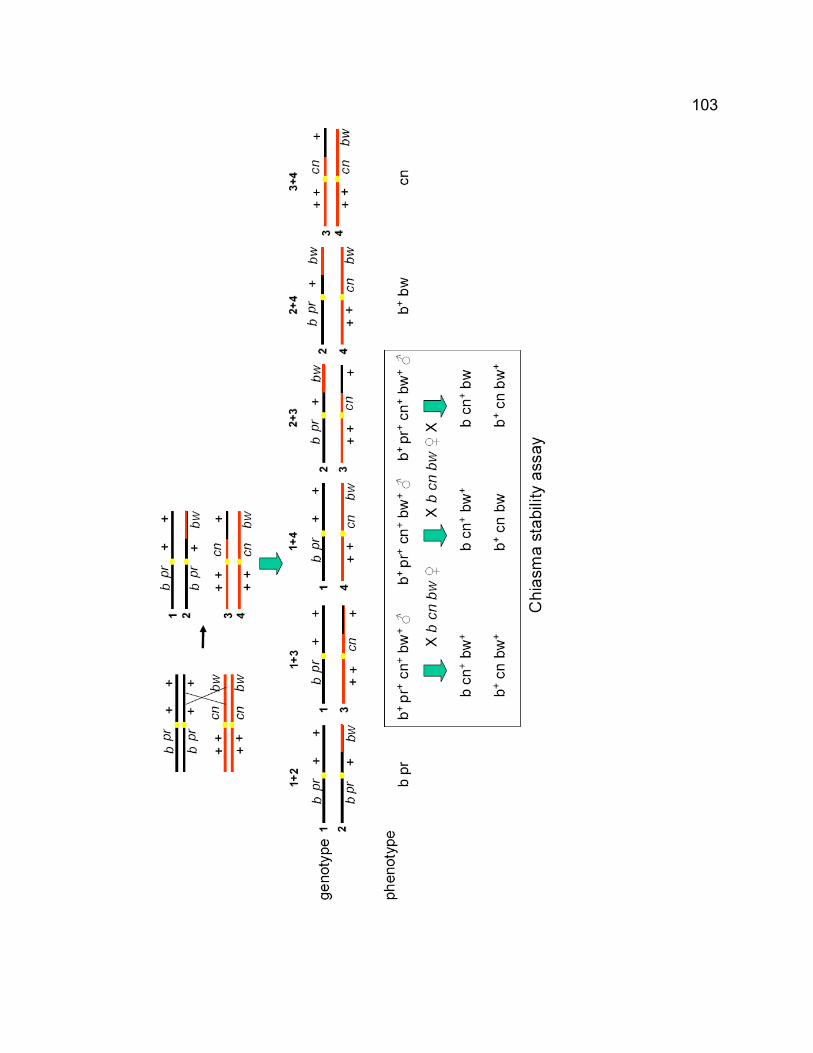
103
104
bw+ progeny (cinnabar eyes) that are readily distinguished from other classes,
and b pr cn+ bw/ b pr cn+ bw+ progeny that show the same phenotype as sister
chromatid NDJ of b pr in the absence of recombination. Recombination within
the cn-bw interval followed by homologous NDJ generates b pr cn+ bw/b+ pr+ cn
bw along with three classes of b+ pr+ cn+ bw+ progeny, of which the former are
readily distinguished from other classes. The ratio of sister chromatid NDJ to
homolog NDJ under the condition of meiotic recombination was calculated by the
ratio of cn bw+ progeny to b+ bw progeny.
In order to obtain an estimate of cn-bw recombination in the NDJ progeny,
the total number of cn bw+ and b+ bw recombinants were multiplied by 3 (to
account for the fact that cn bw+ recombinants are detectable only when they
segregate with the cn bw chromatid, but not when they segregate with the cn+ bw
or + + chromatids, which segregations are presumed to be equally likely) and
then divided by the number of recovered chromatids (2XN).
To investigate chiasma instability on chromosome 2, the b+ pr+ cn+ bw+ F1
males from the cross of soloZ2-0198, cn bw/b vas7 pr females to C(2)EN, bw sp
males were crossed to b cn bw females with structurally normal 2nd
chromosomes. The F2 progeny were scored for the presence and distribution of
the markers b, cn and bw. b+ pr+ cn+ bw+ F1 males that resulted from homolog
NDJ may contain 0, 1 and 2 exchange chromosomes. The presence of two
reciprocal exchange chromosomes in the progeny indicates that the absence of
an effective chiasma in meiosis in solo females.
105
Assaying sister chromatid exchange
Sister chromatid exchange in solo and wild type females was assayed by
monitoring the ratio of a Ring X chromosome to a Rod (normally linear) X
chromosome in the progeny. Since an odd number of crossovers occurs
between Ring sister chromatids generate a dicentric double ring chromosome
which cannot be transmitted to the progeny, reduced recovery of the Ring X
chromosome from solo female mutants then indicates the increased level of
sister chromatid exchange.
R(1)2, y1 f1/BSYy+ males (Bloomington, stock # 4330) were crossed to
Df(2L)A267, b cn bw/CyO, cn females. The R(1)2, y1 f1/+; Df(2L)A267, b cn bw/+
female progeny were crossed to y w/Y; solo, cn bw/CyO, cn males to generate
R(1)2, y1 f1/y w; Df(2L)A267, b cn bw/solo, cn bw females and the sibling control
R(1)2, y1 f1/y w; +/CyO, cn females. These females were crossed to w1118/Y
males to test sister chromatid exchange. The crosses were carried out without
an X chromosome balancer to estimate changes of sister chromatid exchange
under the condition in which both homolog and sister chromatid exchange occur
in mutant and wild type. The progeny classes were distinguished by eye color,
body color and bristle phenotype. The recovered Ring X chromosome generated
y+ w+ f+ females and y w+ f males (Ring progeny). In contrast, the recovered Rod
X chromosome generated y+ w f+ females and y w f+ males (Rod progeny).
These four categories were used to calculate the ratio of Ring/Rod. Additionally,
sister chromatid and homolog NDJ generated y w+ f or y w f+, and y w+ f+ females,
respectively. y+ w f+ XO males are recovered from null oocytes. Rarely, y w f
106
and y w+ f+ males were observed due to double exchanges between homologous
ring and rod sister chromatids. Since R(1), y f is actually bobbed and the R(1), y
f homozygotes grow slower than normal females, the progeny that were from
sister chromatid NDJ were not used for evaluating sister chromatid exchange in
this cross experiment.
Rescue experiments
+/w; Df(2L)A267, cn/soloZ2-0198, cn; [UAS:Venus-SOLO]/[nanos:Gal4-
VP16] and sibling control females (lacking [UAS:Venus-SOLO] were crossed to
X∧Y, y B males to measure X chromosome NDJ. The rescue experiments for
UAS:SOLO-Venus transgenic flies were carried out by similar methods.
Immunostaining in whole-mount ovaries
Newly eclosed females were fattened 2-3 days in vials with yeast paste
and males and then ovaries were dissected in 1 X PBS. Ovary immunostaining
was performed according to Page and Hawley (2001). After immunostaining,
ovaries were separated into individual ovarioles and transferred to slides and
mounted with Prolong Antifade reagent (Invitrogen). Venus-SOLO expression
was induced by nanos:Gal4-VP16 (Doren et al., 1998) and fluorescent signals
were detected in the FITC channel or detected using anti-GFP antibody.
107
Chromosome spread
Chromosome spreads were performed according to Webber et al. (2004).
This method takes advantages of drying-down techniques to get higher
resolution of meiotic cells, and thus is beneficial to better understand the
localization of SOLO in females.
Antibodies used
Primary antibodies used : 1:500 anti-C(3)G mouse monoclonal antibody
(provided by R.S. Hawley), 1:500 rabbit anti-GFP polyclonal antibody
(Invitrogen), 1:1000 rabbit anti-CID polyclonla antibody (Abcam), 1:250 anti-
SMC1 rabbit polyclonal antibody. Secondary antibodies used: Alexa Fluor 555
donkey anti-mouse IgG (H+L) (Molecular Probes), Alexa Fluor 647 goat anti-
rabbit IgG (H+L) (Molecular Probes), Alexa Fluor 546 goat anti-rabbit IgG (H+L)
(Molecular Probes).
Microscopy and image processing
All images were collected using an Axioplan (ZEISS) microscope
equipped with an HBO 100-W mercury lamp and high-resolution CCD camera
(Roper). Image data were collected and merged using Metamorph Software
(Universal Imaging Corporation). For signals and some Venus-SOLO images,
maximum or sum projections of deconvolved Z-series were applied using
Metamorph Software. Adobe photoshop CS2 and Illustrator CS2 were used to
process images.
108
RESULTS
solo mutations cause reduced fertility and elevated NDJ in female meiosis
solo mutations have been shown to cause high nondisjunction of sex
chromosomes and autosomes in Drosophila male meiosis. To assess the effects
of solo mutations on sex chromosome segregation in female meiosis, females
trans-heterozygous for two different pairs of solo alleles were tested for X
chromosome NDJ. The results (Table 3-1) show that solo females are semi-
sterile, producing only about 3.5 progeny per female, compared to 50-80 progeny
per female in parallel crosses involving solo/+ sibling control females. X NDJ
frequencies were also highly elevated in solo mutant females: 36.0% in Z2-
3534/Z2-0198 females and 53.0% in Z2-0338/Z2-0198 compared to less than 1%
in the wild-type control crosses. Elevated 2nd chromosome NDJ was also
observed in solo females. Diplo-2 eggs that carry sister chromatids and
homologous chromatids (we use the term “homologous chromatids” to refer to
two chromatids that are from either of homologous chromosomes, such as X and
Y chromatids) were detected in the test, indicating that solo mutations cause both
homolog and sister chromatid NDJ in females (Table 3-2).
solo mutations reduce recombination
solo females also exhibited reduced recombination frequencies on both
the X and 2nd chromosomes. Recombination between the pn and f loci, which
are near the distal and proximal ends of the X euchromatin, totaled only 18.0cM
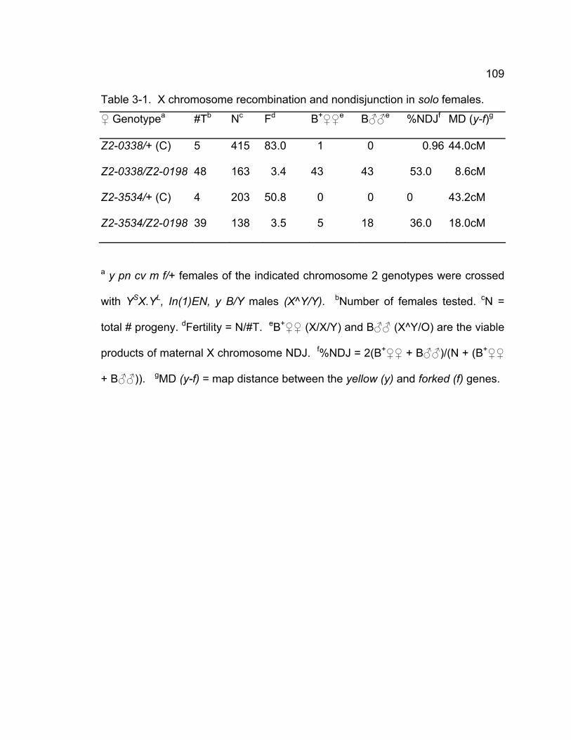
109
Table 3-1. X chromosome recombination and nondisjunction in solo females.
♀ Genotypea #Tb Nc Fd B+♀♀e B♂♂e %NDJf MD (y-f)g
Z2-0338/+ (C) 5 415 83.0 1 0 0.96 44.0cM
Z2-0338/Z2-0198 48 163 3.4 43 43 53.0 8.6cM
Z2-3534/+ (C) 4 203 50.8 0 0 0 43.2cM
Z2-3534/Z2-0198 39 138 3.5 5 18 36.0 18.0cM
a y pn cv m f/+ females of the indicated chromosome 2 genotypes were crossed
with YSX.YL, In(1)EN, y B/Y males (X^Y/Y). bNumber of females tested. cN =
total # progeny. dFertility = N/#T. eB+♀♀ (X/X/Y) and B♂♂ (X^Y/O) are the viable
products of maternal X chromosome NDJ. f%NDJ = 2(B+♀♀ + B♂♂)/(N + (B+♀♀
+ B♂♂)). gMD (y-f) = map distance between the yellow (y) and forked (f) genes.
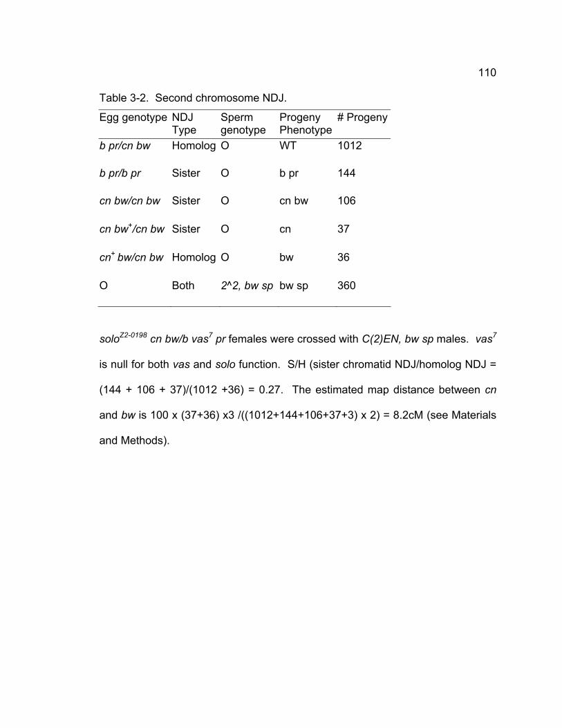
110
Table 3-2. Second chromosome NDJ.
Egg genotype NDJ Type
Sperm genotype
Progeny Phenotype
# Progeny
b pr/cn bw Homolog O WT 1012
b pr/b pr Sister O b pr 144
cn bw/cn bw Sister O cn bw 106
cn bw+/cn bw Sister O cn 37
cn+ bw/cn bw Homolog O bw 36
O Both 2^2, bw sp bw sp 360
soloZ2-0198 cn bw/b vas7 pr females were crossed with C(2)EN, bw sp males. vas7
is null for both vas and solo function. S/H (sister chromatid NDJ/homolog NDJ =
(144 + 106 + 37)/(1012 +36) = 0.27. The estimated map distance between cn
and bw is 100 x (37+36) x3 /((1012+144+106+37+3) x 2) = 8.2cM (see Materials
and Methods).
111
and 8.6cM in soloZ2-3534/soloZ2-0198 and soloZ2-0338/soloZ2-0198 females, respectively,
compared to 43.2cM and 44.0cM in the corresponding wild-type control crosses
(Table 3-1). On chromosome 2, recombination in the large euchromatic intervals
flanked by the cinnabar (cn) and brown (bw) loci was strongly reduced in soloZ2-
0198 females (6.8cM (Table 3-3) and 8.2cM (Table 3-2) compared to wild-type
control females (41.9cM) (Table 3-3). However, recombination was actually
slightly higher in solo than in wild-type females (5.1 cM versus 3.9 cM) in the b-cn
interval that flanks the centromere, indicating that the requirement for solo
function in recombination is much greater in distal euchromatic regions than in
centromere-proximal regions. This pattern is typical of precondition mutations
that reduce recombination and alter crossover distribution (Carpenter and
Sandler, 1974; Bhagat et al., 2004).
Sister chromatid exchange is elevated in solo mutants
The reduced meiotic recombination frequency could be due to a decrease
of all recombination events including both sister chromatid and homologous
recombination. Alternatively, if SOLO is required for the meiotic “homolog bias”,
sister chromatid exchange could be increased at the expense of homologous
recombination in solo mutants. To explore this possibility, we tested the
transmission of a Ring X chromosome during meiosis in solo mutants. A ring
chromosome cannot be transmitted efficiently when an odd number of
crossovers occurs between two Ring sister chromatids because it creates a
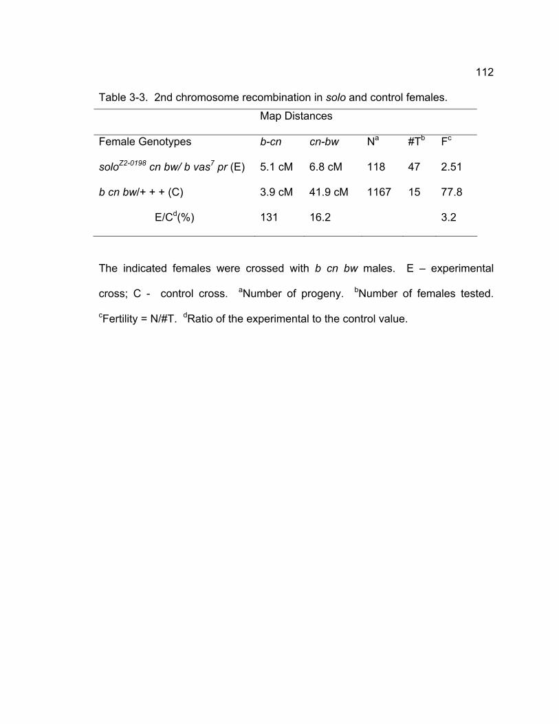
112
Table 3-3. 2nd chromosome recombination in solo and control females.
Map Distances
Female Genotypes b-cn cn-bw Na #Tb Fc
soloZ2-0198 cn bw/ b vas7 pr (E) 5.1 cM 6.8 cM 118 47 2.51
b cn bw/+ + + (C) 3.9 cM 41.9 cM 1167 15 77.8
E/Cd(%) 131 16.2 3.2
The indicated females were crossed with b cn bw males. E – experimental
cross; C - control cross. aNumber of progeny. bNumber of females tested.
cFertility = N/#T. dRatio of the experimental to the control value.
113
dicentric Ring chromosome, whereas Ring chromosomes can be transmitted
efficiently when an even number of crossovers occurs between two Ring sister
chromatids. In contrast, Rod X chromosomes (linear chromosomes) can be
transmitted efficiently following sister chromatid crossovers. Although the
exchanges between Ring and Rod also produce dicentric products, they equally
decrease the transmission of Ring and Rod chromosomes. Therefore, if the
inhibition of sister chromatid exchange is lost in solo mutants, the progeny
derived from Ring X chromosome-bearing eggs will be greatly decreased related
to those derived from normal Rod chromosome-bearing eggs.
We monitored the meiotic transmission of the R(1), y f chromosome in wild
type and hemizygous solo females. As shown in Table 3-4, the recovered ratio
of Ring/Rod is approximately 0.35 among the progeny of both soloZ2-0198 and
soloZ2-3534 females, which is significantly reduced compared to that in wild type
(0.83), strongly suggesting that SOLO is required for inhibiting sister chromatid
recombination to promote homologous recombination in Drosophila female
meiosis.
Chiasma stability is defective in solo mutants
Chiasmata must be effectively maintained before anaphase I in order to
separate homologs properly. Otherwise, homologs cannot be correctly disjoined
to opposite spindle poles. Our previous study has shown vas7 is not only a vas
mutant but also a null allele of solo. solo females with one vas7 chromosome
marked with b and pr and the other soloz2-0198 marked with cn bw were crossed
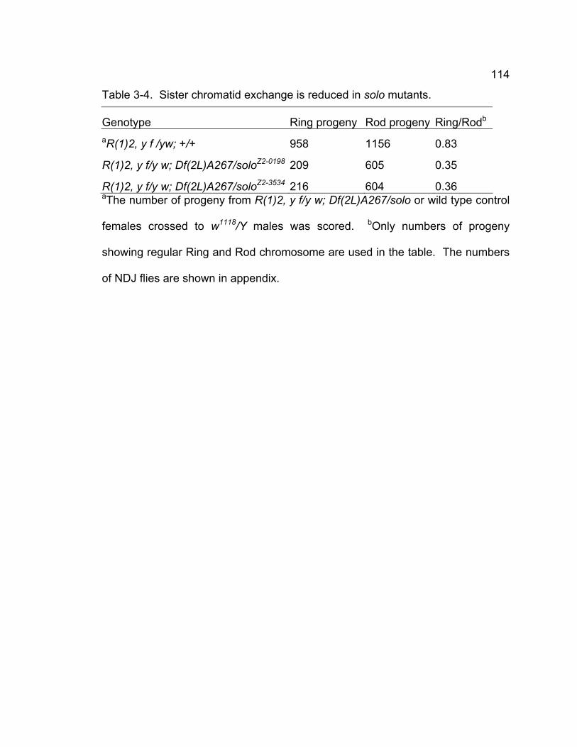
114
Table 3-4. Sister chromatid exchange is reduced in solo mutants.
Genotype Ring progeny Rod progeny Ring/Rodb aR(1)2, y f /yw; +/+ 958 1156 0.83
R(1)2, y f/y w; Df(2L)A267/soloZ2-0198 209 605 0.35
R(1)2, y f/y w; Df(2L)A267/soloZ2-3534 216 604 0.36 aThe number of progeny from R(1)2, y f/y w; Df(2L)A267/solo or wild type control
females crossed to w1118/Y males was scored. bOnly numbers of progeny
showing regular Ring and Rod chromosome are used in the table. The numbers
of NDJ flies are shown in appendix.
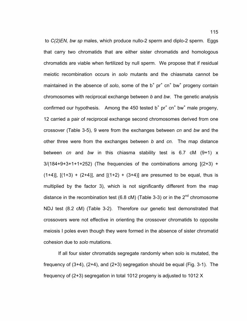
115
to C(2)EN, bw sp males, which produce nullo-2 sperm and diplo-2 sperm. Eggs
that carry two chromatids that are either sister chromatids and homologous
chromatids are viable when fertilized by null sperm. We propose that if residual
meiotic recombination occurs in solo mutants and the chiasmata cannot be
maintained in the absence of solo, some of the b+ pr+ cn+ bw+ progeny contain
chromosomes with reciprocal exchange between b and bw. The genetic analysis
confirmed our hypothesis. Among the 450 tested b+ pr+ cn+ bw+ male progeny,
12 carried a pair of reciprocal exchange second chromosomes derived from one
crossover (Table 3-5), 9 were from the exchanges between cn and bw and the
other three were from the exchanges between b and cn. The map distance
between cn and bw in this chiasma stability test is 6.7 cM (9+1) x
3/(184+9+3+1+1+252) (The frequencies of the combinations among [(2+3) +
(1+4)], [(1+3) + (2+4)], and [(1+2) + (3+4)] are presumed to be equal, thus is
multiplied by the factor 3), which is not significantly different from the map
distance in the recombination test (6.8 cM) (Table 3-3) or in the 2nd chromosome
NDJ test (8.2 cM) (Table 3-2). Therefore our genetic test demonstrated that
crossovers were not effective in orienting the crossover chromatids to opposite
meiosis I poles even though they were formed in the absence of sister chromatid
cohesion due to solo mutations.
If all four sister chromatids segregate randomly when solo is mutated, the
frequency of (3+4), (2+4), and (2+3) segregation should be equal (Fig. 3-1). The
frequency of (2+3) segregation in total 1012 progeny is adjusted to 1012 X
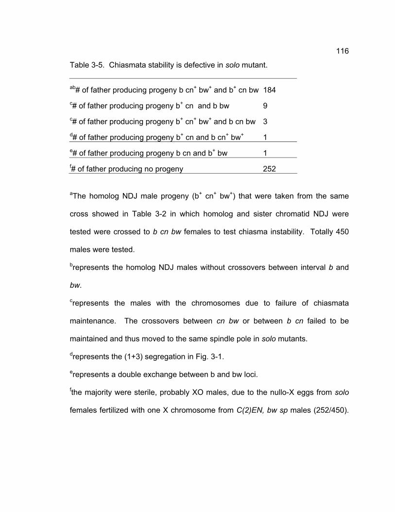
116
Table 3-5. Chiasmata stability is defective in solo mutant.
ab# of father producing progeny b cn+ bw+ and b+ cn bw 184 c# of father producing progeny b+ cn and b bw 9 c# of father producing progeny b+ cn+ bw+ and b cn bw 3 d# of father producing progeny b+ cn and b cn+ bw+ 1 e# of father producing progeny b cn and b+ bw 1 f# of father producing no progeny 252
aThe homolog NDJ male progeny (b+ cn+ bw+) that were taken from the same
cross showed in Table 3-2 in which homolog and sister chromatid NDJ were
tested were crossed to b cn bw females to test chiasma instability. Totally 450
males were tested.
brepresents the homolog NDJ males without crossovers between interval b and
bw.
crepresents the males with the chromosomes due to failure of chiasmata
maintenance. The crossovers between cn bw or between b cn failed to be
maintained and thus moved to the same spindle pole in solo mutants.
drepresents the (1+3) segregation in Fig. 3-1.
erepresents a double exchange between b and bw loci.
fthe majority were sterile, probably XO males, due to the nullo-X eggs from solo
females fertilized with one X chromosome from C(2)EN, bw sp males (252/450).
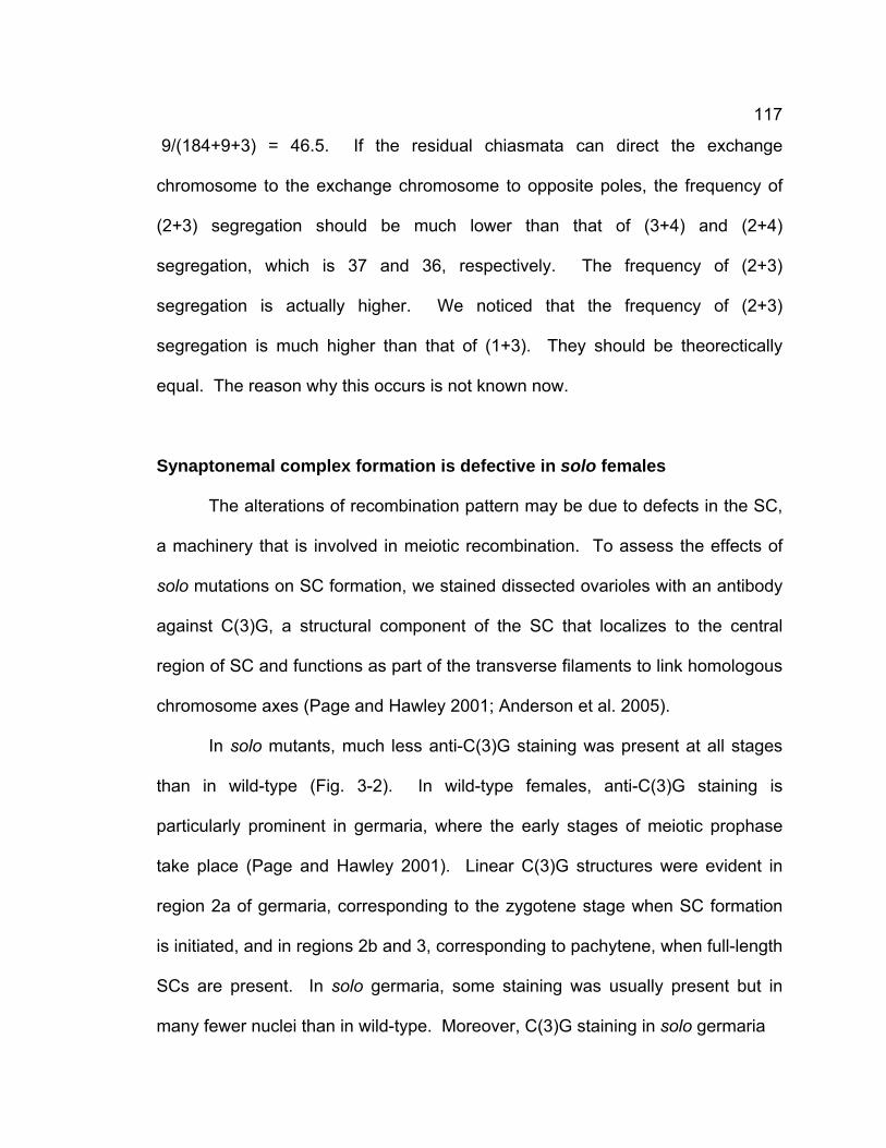
117
9/(184+9+3) = 46.5. If the residual chiasmata can direct the exchange
chromosome to the exchange chromosome to opposite poles, the frequency of
(2+3) segregation should be much lower than that of (3+4) and (2+4)
segregation, which is 37 and 36, respectively. The frequency of (2+3)
segregation is actually higher. We noticed that the frequency of (2+3)
segregation is much higher than that of (1+3). They should be theorectically
equal. The reason why this occurs is not known now.
Synaptonemal complex formation is defective in solo females
The alterations of recombination pattern may be due to defects in the SC,
a machinery that is involved in meiotic recombination. To assess the effects of
solo mutations on SC formation, we stained dissected ovarioles with an antibody
against C(3)G, a structural component of the SC that localizes to the central
region of SC and functions as part of the transverse filaments to link homologous
chromosome axes (Page and Hawley 2001; Anderson et al. 2005).
In solo mutants, much less anti-C(3)G staining was present at all stages
than in wild-type (Fig. 3-2). In wild-type females, anti-C(3)G staining is
particularly prominent in germaria, where the early stages of meiotic prophase
take place (Page and Hawley 2001). Linear C(3)G structures were evident in
region 2a of germaria, corresponding to the zygotene stage when SC formation
is initiated, and in regions 2b and 3, corresponding to pachytene, when full-length
SCs are present. In solo germaria, some staining was usually present but in
many fewer nuclei than in wild-type. Moreover, C(3)G staining in solo germaria
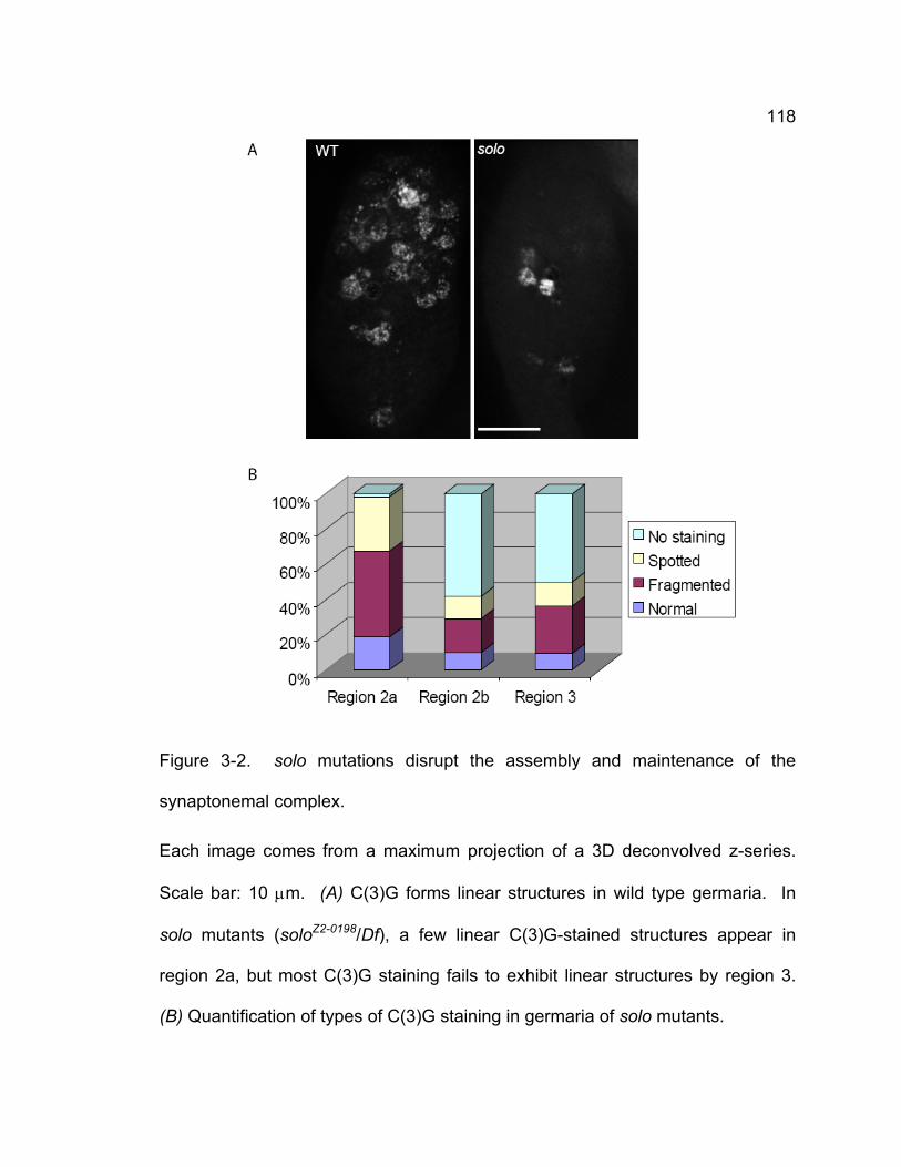
118
Figure 3-2. solo mutations disrupt the assembly and maintenance of the
synaptonemal complex.
Each image comes from a maximum projection of a 3D deconvolved z-series.
Scale bar: 10 μm. (A) C(3)G forms linear structures in wild type germaria. In
solo mutants (soloZ2-0198/Df), a few linear C(3)G-stained structures appear in
region 2a, but most C(3)G staining fails to exhibit linear structures by region 3.
(B) Quantification of types of C(3)G staining in germaria of solo mutants.
119
was often punctate rather than linear. Occasionally, normal-looking short linear
structures were seen in regions 2a and 2b but full-length SCs were very rarely
seen at any stage. Overall, normal C(3)G staining was observed in less than
20% of nuclei in region 2A and less than 10% of nuclei in regions 2B and 3 of the
germarium (Fig. 3-2B). These data indicate that SOLO is required for synapsis.
In addition, germ cells in ovarioles of solo mutants were significantly reduced in
number compared to wild type, suggesting a defect in germ cell proliferation or
development, consistent with the poor fertility of solo females.
Venus-SOLO form bright foci at centromeres in oocytes
We generated transgene constructs expressing UAS:Venus-SOLO and
UAS:SOLO-Venus and transformed them into flies. One copy of each construct
was previously shown to completely rescue male phenotype when their
expression were induced by nanos (nos):Gal4-VP16. Similar rescue
experiments showed that UAS:Venus-SOLO and UAS:SOLO-Venus also
rescued female NDJ to background level induced by nos:Gal4-VP16 (Table 3-6),
suggesting that Venus-SOLO is fully functional in both male and female meiosis.
SOLO localizes to centromeres in Drosophila males, corresponding to its role in
centromeric cohesion. In order to explore the localization of SOLO in female,
Venus-SOLO was induced by nos:Gal4-VP16. We found that Venus-SOLO
formed one to three very bright foci in nuclei of female germ cells of various
stages. The and Venus-SOLO foci co-localized with foci of anti-CID, which
detects a centromere-specific variant of histone H3. In germaria, Venus-SOLO
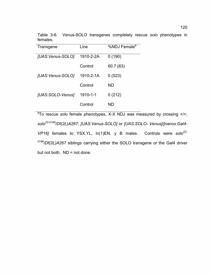
120
Table 3-6. Venus-SOLO transgenes completely rescue solo phenotypes in females.
Transgene Line %NDJ Femalea
[UAS:Venus-SOLO] 1910-2-2A 0 (190)
Control 60.7 (63)
[UAS:Venus-SOLO] 1910-2-1A 0 (523)
Control ND
[UAS:SOLO-Venus] 1910-1-1 0 (212)
Control ND
aTo rescue solo female phenotypes, X-X NDJ was measured by crossing +/+;
soloZ2-0198/Df(2L)A267; [UAS:Venus-SOLO] or [UAS:SOLO- Venus]/[nanos:Gal4-
VP16] females to YSX.YL, In(1)EN, y B males. Controls were soloZ2-
0198/Df(2L)A267 siblings carrying either the SOLO transgene or the Gal4 driver
but not both. ND = not done.
121
and CID colocalized in oogonia undergoing mitosis in region 1, and in pro-
oocytes, oocytes and nurse cells in regions 2 and 3 (Fig. 3-3A). In later meiotic
stages, Venus-SOLO continued to colocalize with CID in oocytes and nurse cells
(Fig. 3-3B and 3C). No Venus-SOLO foci were detected in the somatic follicle
cells.
SOLO localizes to synaptonemal complexes
Meiotic recombination in Drosophila females occurs in the context of SC (Page
and Hawley 2003). The reduced recombination frequency, altered distribution of
crossovers, and the defects of SC in solo mutants suggest that SOLO is probably
a component of SC. To test this possibility, we simultaneously stained germaria
for SOLO and C(3)G, a component of the transverse filaments of SC (Fig. 3-4).
In addition to forming bright foci in nuclei of female germ cells at centromeres as
shown in Fig. 3-3, Venus-SOLO also localized more generally within germ cell
nuclei and appeared to be especially abundant in region 2 of the germarium,
where germ cells undergo early meiotic prophase. Besides very bright staining
at centromeres, weak thread-like Venus-SOLO staining could sometimes be
seen in regions 2a, region 2b, and region 3. The thread-like SOLO staining
appeared to overlap with C(3)G. In region 2a, thread-like SOLO staining
colocalizes with C(3)G in several nuclei. The colocalization of SOLO and C(3)G
occurred in pro-oocytes, oocytes, and nurse cells. This is not surprising since
previous studies have shown that temporary SC formation
122
Figure 3-3. Venus-SOLO foci are abundant at centromeres.
Expression of Venus-SOLO was induced by nanos:Gal4-VP16 in [UAS:Venus-
SOLO]/[nanos:Gal4-VP16] females and detected at FITC channal. Centromeres
were visualized with CID staining and DNA was stained with DAPI. The
transgenic line is 1910-2-2A. Venus-SOLO colocalizes with CID in oogonia, pro-
oocytes, oocytes, and nurse cells of germaria. The images represent sum
projections of 3D-deconvolved Z-series (A). Venus-SOLO colocalizes with CID
in oocytes and nurse cells at stage 2 (B) and stage 4(C). Arrow heads indicate
the oocytes, showing colocalization of SOLO and CID at centromeres. No
Venus-SOLO foci but CID foci appear on follicle cells. Scale bars: 10 μm.
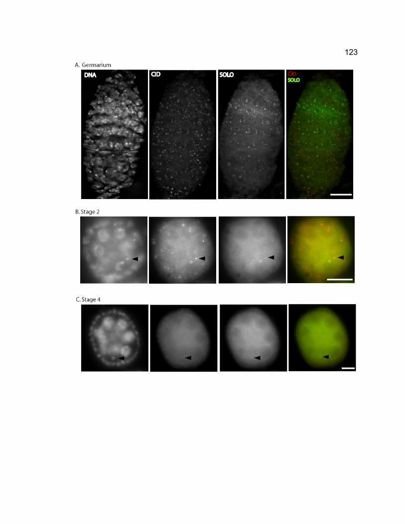
123
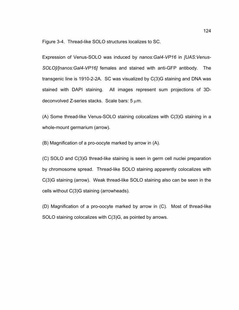
124
Figure 3-4. Thread-like SOLO structures localizes to SC.
Expression of Venus-SOLO was induced by nanos:Gal4-VP16 in [UAS:Venus-
SOLO]/[nanos:Gal4-VP16] females and stained with anti-GFP antibody. The
transgenic line is 1910-2-2A. SC was visualized by C(3)G staining and DNA was
stained with DAPI staining. All images represent sum projections of 3D-
deconvolved Z-series stacks. Scale bars: 5 μm.
(A) Some thread-like Venus-SOLO staining colocalizes with C(3)G staining in a
whole-mount germarium (arrow).
(B) Magnification of a pro-oocyte marked by arrow in (A).
(C) SOLO and C(3)G thread-like staining is seen in germ cell nuclei preparation
by chromosome spread. Thread-like SOLO staining apparently colocalizes with
C(3)G staining (arrow). Weak thread-like SOLO staining also can be seen in the
cells without C(3)G staining (arrowheads).
(D) Magnification of a pro-oocyte marked by arrow in (C). Most of thread-like
SOLO staining colocalizes with C(3)G, as pointed by arrows.
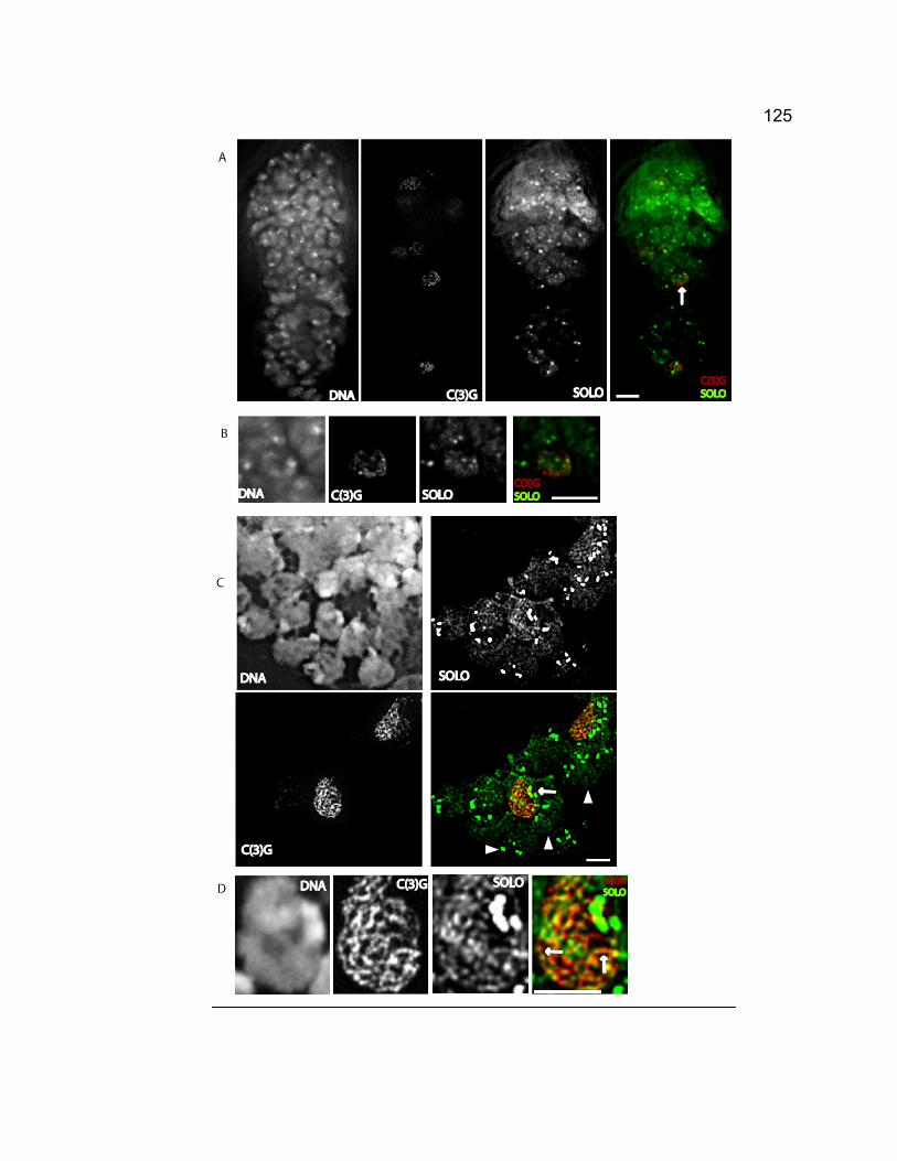
125
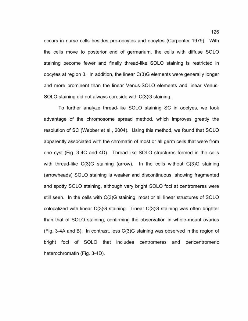
126
occurs in nurse cells besides pro-oocytes and oocytes (Carpenter 1979). With
the cells move to posterior end of germarium, the cells with diffuse SOLO
staining become fewer and finally thread-like SOLO staining is restricted in
oocytes at region 3. In addition, the linear C(3)G elements were generally longer
and more prominent than the linear Venus-SOLO elements and linear Venus-
SOLO staining did not always coreside with C(3)G staining.
To further analyze thread-like SOLO staining SC in ooctyes, we took
advantage of the chromosome spread method, which improves greatly the
resolution of SC (Webber et al., 2004). Using this method, we found that SOLO
apparently associated with the chromatin of most or all germ cells that were from
one cyst (Fig. 3-4C and 4D). Thread-like SOLO structures formed in the cells
with thread-like C(3)G staining (arrow). In the cells without C(3)G staining
(arrowheads) SOLO staining is weaker and discontinuous, showing fragmented
and spotty SOLO staining, although very bright SOLO foci at centromeres were
still seen. In the cells with C(3)G staining, most or all linear structures of SOLO
colocalized with linear C(3)G staining. Linear C(3)G staining was often brighter
than that of SOLO staining, confirming the observation in whole-mount ovaries
(Fig. 3-4A and B). In contrast, less C(3)G staining was observed in the region of
bright foci of SOLO that includes centromeres and pericentromeric
heterochromatin (Fig. 3-4D).
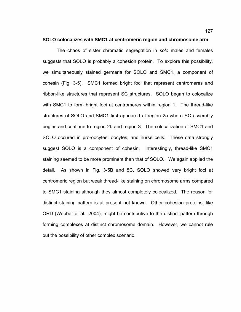
127
SOLO colocalizes with SMC1 at centromeric region and chromosome arm
The chaos of sister chromatid segregation in solo males and females
suggests that SOLO is probably a cohesion protein. To explore this possibility,
we simultaneously stained germaria for SOLO and SMC1, a component of
cohesin (Fig. 3-5). SMC1 formed bright foci that represent centromeres and
ribbon-like structures that represent SC structures. SOLO began to colocalize
with SMC1 to form bright foci at centromeres within region 1. The thread-like
structures of SOLO and SMC1 first appeared at region 2a where SC assembly
begins and continue to region 2b and region 3. The colocalization of SMC1 and
SOLO occured in pro-oocytes, oocytes, and nurse cells. These data strongly
suggest SOLO is a component of cohesin. Interestingly, thread-like SMC1
staining seemed to be more prominent than that of SOLO. We again applied the
detail. As shown in Fig. 3-5B and 5C, SOLO showed very bright foci at
centromeric region but weak thread-like staining on chromosome arms compared
to SMC1 staining although they almost completely colocalized. The reason for
distinct staining pattern is at present not known. Other cohesion proteins, like
ORD (Webber et al., 2004), might be contributive to the distinct pattern through
forming complexes at distinct chromosome domain. However, we cannot rule
out the possibility of other complex scenario.
128
Figure 3-5. SOLO and SMC1 colocalize together.
Expression of Venus-SOLO was induced by nanos:Gal4-VP16 in [UAS:Venus-
SOLO]/[nanos:Gal4-VP16] females and stained with anti-GFP antibody. The
transgenic line is 1910-2-2A. SMC1 was stained with anti-SMC1 antibody and
DNA was visualized with DAPI staining. All images represent sum projections of
3D-deconvolved Z-series stacks. Scale bars: 5 μm.
(A) Both SOLO and SMC1 form bright foci at centromeres and thread-like
staining at chromosome arms in a whole-mount germarium and colocalize. Both
of them localize to centromeres but do not form thread-like structures at region 1
(arrows). SOLO and SMC1 form thread-like structures at chromosome arm while
form bright foci at centromeric region at region 2a, 2b and 3 (arrowheads).
(B) Centromeric foci and thread-like staining of SOLO and SMC1 appear in germ
cell nuclei preparation by chromosome spread. SOLO forms relatively brighter
foci compared to SMC1 at centromeres whereas the thread-like staining of SOLO
is relatively weaker compared to SMC1 although both of them colocalize at
centromeres and chromosome arms.
(C) Magnification of a pro-oocyte marked by arrowheads in (B). SOLO and
SMC1 apparently colocalize (arrowheads).
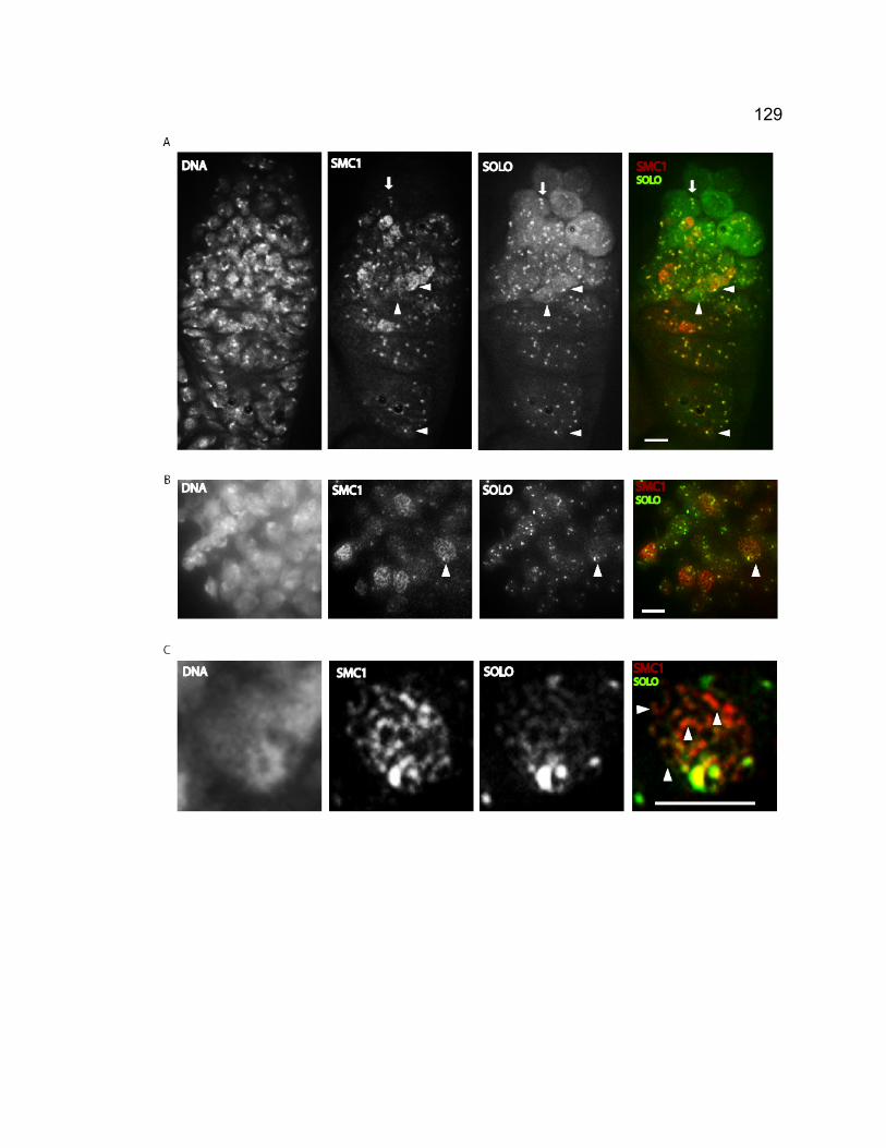
129
130
DISCUSSION
Although sister chromatid cohesion is required for the generation of
crossovers during meiosis, which is essential for homolog segregation, the
mechanism of how sister chromatid cohesion functions in meiotic recombination
is not well understood in higher eukaryotes. Here we show that meiotic cohesion
is required for formation of SC, promoting homologous recombination by
inhibiting recombination between sister chromatids, and maintaining chiasmata
stability for homolog segregation, thus providing evidence for cohesion’s key role
in homolog segregation during meiosis I.
SOLO is a cohesion protein that is required for normal homolog and sister
chromatid segregation.
We have showed that SOLO is essential for homolog and sister chromatid
segregation in Drosophila male meiosis. Our results show that in females both
homolog and sister chromatid NDJ occur when solo is not functional, suggesting
SOLO’s role in Drosophila meiosis is not sex-specific but it is universally required
for both homolog and sister chromatid segregation in meiosis of both sexes
although they undergo meiosis through different pathways. The colocalization of
SMC1 and SOLO from pre-meiotic stages to late prophase I in females and their
colocalization from pre-meiotic stages to anaphase II in males strongly suggest
that SOLO is probably a component of cohesin that requires SMC1.
Interestingly, the strength of SOLO and SMC1 staining is distinct at distinct
131
chromosome domains. SOLO usually shows weaker thread-like staining at
chromosome arms compared to SMC1. Although it could be a reflection of three-
dimensional distortion of cohesin due to the twisted SC as some studies
suggested (Carpenter 1979; Zickler and Kleckner 1999), it is unlikely since, if
cohesin is twisted, SOLO would be expected to be brighter than SMC1 at some
chromosome arm regions, but actually SOLO is rarely brighter than SMC1 on
chromosome arms. Alternatively, SMC1 may form different cohesion complexes
with different cohesion proteins at distinct regions depending on their different
functions. Besides SOLO, SMC1 may assemble cohesion complex on
chromosome arms with C(2)M, an α-Kleisin protein, which is not required for
sister chromatid cohesion and does not form bright foci at centromeres but is a
component of the lateral elements of SC (Manheim and McKim 2003; Heidmann
et al., 2004). This idea that SOLO and C(2)M functions redundantly on
chromosome arms is supported by the recent study in which SMC1/SMC3
showed weak staining on chromosome arms in c(2)M mutant whereas the
staining is robust in wild type (Khetani and Bickel 2007). Moreover, this model
also gives an explanation to why about 20% residual recombination still exists in
solo or c(2)M single mutants. If our model is correct, the frequency of
recombination in solo and c(2)M double mutantswould be very low, much less
than 20%. Indeed, distinct cohesion complexes form at distinct chromosome
domain (kitajima et al., 2003). However, more complex scenario might be still
possible. It will be very intriguing to explore the mechanism underlying it through
identifying the distinct cohesion complex in Drosophila meiosis.
132
SOLO is required for promoting homologous recombination and inhibiting
sister chromatid exchange during Drosophila female meiosis.
Our results showed that SOLO is required for normal homologous
recombination occurred on X chromosomes and autosomes since the lack of
SOLO causes reduced recombination along chromosome arm. However, the
recombination frequency of intervals spanning centromeres actually increases,
showing that both of the distribution and frequency of recombination are altered.
Thus solo belongs to the precondition type of mutation. The phenotype of solo is
similar to ord, which alter distribution and frequency of recombination. Moreover,
the homolog recombination is completely abolished in the absence of C(3)G
(Page and Hawley, 2003), suggesting that SC is required for homolog
recombination. Thus the defects of SC due to the loss of SOLO disrupt the
tendency of SC to promote homolog recombination, i.e. make crossovers
between homologous chromatids from DSBs.
Cohesion is required for mitotic and meiotic recombination besides
chromosome segregation (Hirano 2000; Jessberger 2002). The cohesion-
dependent recombination in meiosis occurs preferentially between homologs
(Schwacha and Kleckner 1997; Webber et al. 2004) whereas the recombination
in mitosis is more apt to occur between sister chromatids (Johnson and Jasin
2000). Our Ring chromosome exchange assay argues that solo mutations
disrupt homolog recombination bias. Our study showed that Ring/Rod ratio in
wild type is less than 1, which is consistent with other studys (Manheim and
McKim, 2003; Webber et al., 2004; McKee personal communication) and
133
probably reflects the normal sister chromatid exchange level of Drosophila
female meiosis. However, the recovery of Ring/Rod ratios in solo mutants is
significantly lower than that in wild type. The increased sister chromatid
exchange is not due to the disruption of SC since previous studies have shown
that sister chromatid exchange level is not elevated in C(3)G mutants (Sandler et
al. 1974). The significantly elevated sister chromatid exchange is probably due to
the loss of sister chromatid cohesion when SOLO is absent.
Thus our data suggest the reduced homolog recombination in solo
mutants might result both from a failure to promote inter-homolog recombination,
since SC promotes homolog recombination and solo mutations disrupt SC
formation, and a loss of the inhibition of sister chromatid exchange, since sister
chromatid cohesion is required for inhibition of recombination between sister
chromatids and sister chromatid cohesion is lost in solo mutants.
SOLO is a novel component of SC required for meiotic recombination.
SOLO localizes to the entire chromosome arm besides forming bright foci
at centromeric regions, which is consistent with the genetic and cytological
observation that SOLO is required for arm and centromeric cohesion during
female meiosis. Although thread-like SOLO staining appears in pro-oocytes and
pro-nurse cells, the linear structure of SOLO colocalizes with C(3)G along
chromosome arms in the cells showing C(3)G thread-like staining, suggesting
SOLO is a novel SC component. The severe defects of C(3)G in solo mutants
provide further support for this idea. SC assembly in the absence of SOLO is
134
defective in region 2a, the stage when meiotic recombination begins. The SC
phenotype of solo is earlier and more severe than that of ord, which only causes
severe defects of SC in region 3, where meiotic recombination is actually
completed (McKim et al., 2003). Our data suggest that SOLO, unlike ORD,
which is required for SC maintenance, is essential for SC formation. However,
the null alleles of solo and ord show similar reduced recombination frequency
(this study; Miyazaki and Orr-Weaver, 1992). The reason why the distinct SC
phenotype of solo and ord resulted in the similar reduced recombination
frequency is not clear yet. The defective ultrastructure of SC in region 2a in ord
mutants uncovered by EM observation may provide one explanation (Webber et
al., 2004).
Chiasmata stability requires SOLO
Chiasmata are essential for proper separation of homologs during meiosis
I. It has been demonstrated that at least one chiasma per bivalent exists in
Drosophila female meiosis (Carpenter and Sandler 1974; Hawley, 1988). In mei-
218 mutants chiasmata remain stable until the onset of anaphase I (Bickel et al.
2002) if they are formed from residual recombination events (McKim et al. 1996,
Bhagat et al. 2004). Our genetic analyses have shown that approximately 16-
20% of the frequency of meiotic recombination in wild type occurs in solo
mutants (6.7cM-8.2cM/42cM), which is almost double of that of mei-218 (8%)
(McKim et al. 1996). Thus we would expect that more chiasmata may form in
solo mutants if they are stable and they separate successfully to opposite poles
135
in meiosis I. However, if chiasmata that form in oocytes are not stable in the
absence of sister chromatid cohesion due to solo mutations, the homologous
chromatids that undergo one crossover would separate to the same spindle pole
after following meiosis II NDJ. Our data demonstrate that crossovers do form in
solo mutants but all of them are not stable in meiosis I. Stable chiasmata were
also not observed in ord null mutants (Bickel et al. 2002). Rec8 that is essential
for sister chromatid cohesion is required for maintenance, and the failure of
resolution of Rec8 blocks homolog segregation in S. cerevisae and C. elegans
(Buonomo et al. 2000; Siomos et al. 2002). With these studies our data suggest
that the requirement of sister chromatid cohesion for stabilizing chiasmata until
anaphase I might be a conserved mechanism among eukaryotes. Without sister
chromatid cohesion, they are not effectively maintained to ensure proper
chromosome segregation during meiosis I although they can form.
Conclusions
SOLO is a novel cohesion protein required for homolog recombination,
synapsis, chiamata maintenance, and proper segregation of homologs and sister
chromatids in Drosophila female meiosis. solo mutations cause reduced meiotic
recombination due to the disruption of promoting inter-homolog recombination
and inhibiting sister chromatid exchange. The SC and residual chiasmata are
not stable in the absence of SOLO, probably due to the loss of sister chromatid
cohesion in solo mutants. Furthermore, SOLO localizes to centromeres and SC
and colocalizes with SMC1. Combining the data in female meiosis with the
136
observation of SOLO’s function and localization pattern in male meiosis, we
propose that SOLO is a novel component of a meiosis-specific cohesin complex
in Drosophila meiosis.
Acknowledgments
We thank B. Wakimoto, M. Ashburner, and Bloomington Stock Center for
providing fly stocks, S. Page and R.S. Hawley for providing C(3)G monoclonal
antibody. This work was supported by NIH grant # R01 GM40489 to B.D.M.
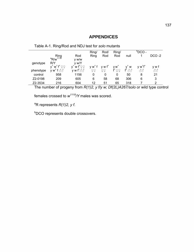
137
APPENDICES
Table A-1. Ring/Rod and NDJ test for solo mutants
Ring Rod Ring/Ring
Rod/Rod
Ring/Rod null
bDCO -1 DCO -2
genotype aR/w1118 R/Y
y w/w y w/Y
phenotype y+ w+ f+ ♀♀ y w+ f ♂♂
y+ w f+♀♀ y w f+♂♂
y w+ f ♀♀
y w f+
♀♀ y w+
f+ ♀♀ y+ w f+ ♂♂
y w+f+
♂♂ y w f ♂♂
control 958 1156 0 0 0 50 8 21 Z2-0198 209 605 6 58 68 306 6 3 Z2-3534 216 604 12 51 65 318 7 2 The number of progeny from R(1)2, y f/y w; Df(2L)A267/solo or wild type control
females crossed to w1118/Y males was scored.
aR represents R(1)2, y f.
bDCO represents double crossovers.
138
CHAPTER 4 - NOVEL ROLES OF VASA IN DROSOPHILA
MALE MEIOSIS: HOMOLOGOUS RECOMBINATION AND
CHROMOSOME SEGREGATION
139
Abstract
Drosophila males undergo meiosis without recombination under normal
conditions whereas recombination is required for female meiosis. How males
prevent exchanges between homologous chromosomes is not known. vasa
(vas) is a translation initiation factor and is involved in oogenesis and
embryogenesis. Here we report novel functions of vas in male meiosis.
Mutations of vas caused X-Y exchanges at elevated frequency in meiosis,
producing sterile progeny that exhibited position effect variegation (PEV). The
PEV and sterility of the progeny resulted from the partial loss of Y chromosome
due to X-Y exchange. Cytological analysis revealed formation of chromatin
bridges at anaphase I and II. Chromatin masses were also found in the midzone
at anaphase I and II. vas and solo double mutants showed higher nondisjunction
than either solo or vas single mutants. Additionally, vas and solo double mutant
showed precocious segregation of homologs at metaphase I in addition to
chromatin bridges at anaphase I and II. Our data thus for the first time
demonstrate that inhibition of meiotic recombination during male meiosis requires
vas function. Furthermore, interaction between vas and solo regulates
chromosome dynamics in male meiosis.
140
INTRODUCTION
Accurate chromosome segregation during meiosis is essential for proper
transmission of genetic material in sexual reproduction. Errors in meiosis are
the primary cause of miscarriages and genetic diseases in human beings
(Hassold and Hunt, 2001).
Homolog pairing and segregation at the first meiotic division (meiosis I) is
required for successful meiosis (McKee, 2004). In most eukaryotes, including
Drosophila females, tight homolog pairing requires homologous recombination
and formation of synaptonemal complexes (SCs), proteinaceous structures that
connect aligned homologs from end to end (Page and Hawley 2004). SC is
disassembled and the tight homolog pairing is lost during mid prophase I and
homologs thereafter are only connected at discrete sites by chiasmata, which are
the cytological manifestations of crossovers, the reciprocal products of meiotic
recombination (Page and Hawley, 2003; Petronczki et al., 2003). Chiasmata
hold homologs together until the onset of anaphase I when homologs segregate
and are essential for bi-orientation of homologs during late prophase I and
metaphase I (Hawley 1988).
In contrast to females, Drosophila males do not form SCs (Meyer 1960,
Rasmussen, 1973) and do not recombine and form chiasmata during meiosis
(McKee, 2004), i.e. achiasmate meiosis. However, homologs do pair and
segregate efficiently in males. Recent studies revealed that homolog pairing and
segregation during Drosophila male meiosis requires two proteins: SNM
141
(Stromalin in Meiosis), a homolog of SCC3/SA cohesion protein, and MNM
(Mod(mdg4) in meiosis), a BTB domain protein that is involved in many protein-
protein interactions (Thomas et al., 2005, Thomas and McKee, 2007). Moreover,
the genetic and cytological studies carried out by McKee and his collaborators
have revealed that X-Y chromosomes pair at specific sites: 240bp repeated
sequence within the intergenic spacers of the ribosomal RNA gene arrays (rDNA)
on both X and Y chromosomes (McKee et al., 1992; Merrill et al., 1992; McKee,
1996). The rDNA arrays are located in the middle of the heterochromatin of X
chromosome and on the short arm of Y chromosome proximal to the centromere
and consist of total 200-250 tandem copies of the genes for the 18S, 5.8S, and
28S ribosomal RNAs (rRNAs) (Ritossa 1976). Deletions of the entire rDNA
arrays disrupt X-Y chromosome pairing and cause random X-Y segregation
(McKee and Lindsley 1987). The transgenes containing either rDNA repeats or
240bp repeats can partially restore the ability of X-Y pairing (McKee and Karpen
1990; McKee et al., 1992; Merrill et al., 1992). Moreover, the 240bp repeats are
required for the pairing between mini-X chromosome and normal X-Y bivalents
(Thomas and McKee, 2007).
The frequency of spontaneous exchanges between X and Y is very low
(0.01%). When the males are treated by ionizing agent, like irradiation, the
frequency may increase up to 2% (Ashburner et al., 2004). Recently Maggert
and Golic (2005) took advantage of the presence of a sequence very similar to
the recognition site for the I-CreI restriction endonuclease in the 28S rDNA to
generate DSBs on X and Y chromosome. Heat-shock induced I-CreI expression
142
caused very high exchange (about 20%) between X and Y chromosomes. .
Mei-W68 is the Drosophila ortholog of Spo11, which generates double
strand breaks (DSBs) that are required for recombination. mei-W68 is
transcribed in testis although at low level, suggesting DSBs are probably
generated in spermatogonia and spermatocytes. The extremely low frequency of
spontaneous exchange suggests that there is a mechanism to prevent
chromosome exchanges in males. How the males prevent exchange between
homologous chromosomes and between sister chromatids is elusive. No
mutants that affect this process have been identified.
The Drosophila gene vas encodes a DEAD-box RNA helicase that is
required for pole cell development and dorsal-ventral axis specification during
oogenesis and embryogenesis (Hay et al., 1988; Lasko and Ashburner, 1988;
Liang et al., 1994; Styhler et al., 1998; Tinker et al., 1998; Tomancak et al.,
1998). VASA shares sequence similarity with the translation initiation factor
eIF4A and controls the translation of some key germline-specific mRNAs that are
critical for oocyte patterning, e.g. Oskar and Gurken (Hay et al., 1988; Lasko and
Ashburner, 1988; Liang et al., 1994; Dahanukar and Wharton, 1996; Styhler et
al., 1998; Tinker et al., 1998;). Although vas is also expressed in male germline
cells, particularly strong in spermatogonia and young spermatocytes (Hay et al.,
1988), vas males are fully fertile and no phenotypes in males have been
reported.
Here we report the novel functions of vas in male meiosis. vas mutations
cause high frequency of X-Y chromosome exchange at rDNA loci and low
143
nondisjunction (NDJ) of sex chromosomes. The majority of recombinants
between X and Y chromosomes are sterile and show position effect variegation
due to the loss of Y-chromosome heterochromatin including fertility factors. The
recovery of chromatin bridges at anaphase I and anaphase II together with the
evidence that majority of recombinants are not clusters indicate that most, if not
all, of the recombination events occur during meiosis. In addition, vas, solo
double mutants cause premature loss of linkage between homologous
chromosomes in addition to chromatin bridges at anaphase I, suggesting that
solo and vas may interact with each other in male meiosis.
144
MATERIALS AND METHODS
Drosophila stocks and strains
All flies were maintained at 23°C on standard cornmeal molasses medium.
vas alleles were obtained from M. Ashburner (Cambridge University), P. Lasko
(McGill University), D. Montell (John Hopkins University), and the Bloomington
Stock Center at the University of Indiana. All chromosomes and markers are
described in Flybase (2007). Unless otherwise specified, tested males were
crossed singly to two or three females in shell vials. Parents were removed from
the vial on day 10 and progeny were counted between day 13 and day 22.
Sex chromosome NDJ and position effect variation (PEV) assays
To measure X-Y NDJ, +/BSYy+ ; vas/Df(2L)A267 males were crossed
singly to 2-3 females carrying structurally normal X chromosomes marked with y1
and w1118. Regular progeny are + females and y+ w BS males; paternal NDJ
generates y+ w+ BS female and y w B+ male progeny. %NDJ = 100 x (y+ w+ BS
♀♀ + y w B+ ♂♂)/N. Besides normal and NDJ progeny, other kinds of
exceptional males, y+ w B+ and y w BS, were also recovered. For simplicity of
discussion, the Y chromosomes in both males were denoted hereafter as YabB+ y+
and YabB y, respectively. All of the y+ w B+ males showed strong variegation of
pigment (yvar) with patches of yellow and yellow+ pigment intermingled in the
abdomen.
145
Fertility test
y+ w B+, y w BS, and XO (from NDJ) male progeny from the above NDJ
and PEV assay were crossed to y w females. Whether there were progeny was
observed.
PEV and sterility inhibition assays
+/BSYy+; Df(2L)A267, b cn bw/vas males were crossed to YSX.YL,
In(1)EN, y B/y w females. The bar eyes are larger in females with B marker than
those in males with BS marker. When B and BS are combined, the bar eyes are
even smaller. The males with B w+ eyes, representing the males with YSX.YL,
In(1)EN, y B/YabB+ y+ were selected and crossed to y w females. The Yab
B+ y+
chromosome combined with y w X chromosome produced PEV males again.
The PEV males were crossed to y w females again to test their fertility. Three
vas alleles vas3, vas5, and vasRG53 were analyzed.
X-Y chromosome exchange test
+/BSYy+; Df(2L)A267, b cn bw/vas males were crossed to C(1)RM/YS
females, respectively. C(1)RM/YS females produced diplo-X and YS eggs at
approximately equal frequency. Among the sperm produced by the males, two
kinds of them are +/BSYy+, from NDJ, and BSYL.Xy+ w+, from X-Y
recombination. When these two kinds of sperm were fertilized by YS eggs, both
of them yielded the males with BS y+ w+ eyes that were not distinguishable. The
males with BS y+ w+ eyes were selected and crossed to y w females. If the male
146
is from NDJ, it produces 4 types of progeny: BS y+ w males, B+ y+ w+ females, B+
y w males, BS y+ w+ females. If the male is from X-Y recombination, it only
produces two types of progeny: B+ y w males, BS y+ w+ females, i.e., all BS flies
are females. Thus the X-Y exchange can be tested.
Testis Immunostaining
α-tubulin/DAPI staining of testes was carried out as described (Thomas et
al. 2005). MNM-GFP live imaging was performed according to Thomas et al.
(2005).
Microscopy and image processing
All images were collected using an Axioplan (ZEISS) microscope
equipped with an HBO 100-W mercury lamp and high-resolution CCD camera
(Roper). Image data were collected and merged using Metamorph Software
(Universal Imaging Corporation). Images were processed with Adobe Photoshop
CS2.
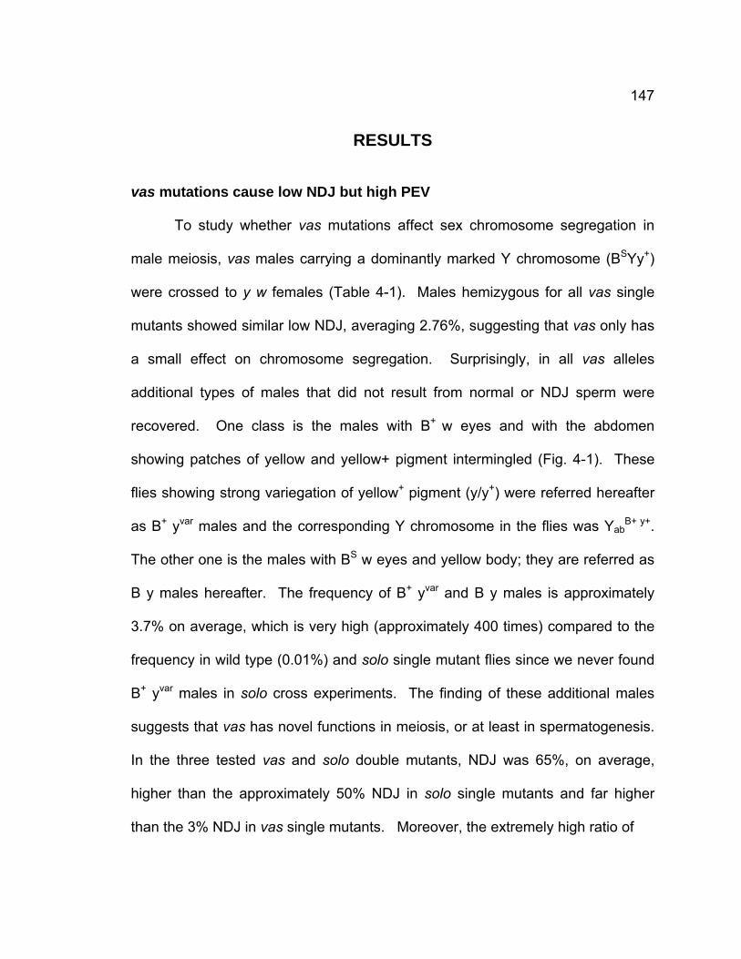
147
RESULTS
vas mutations cause low NDJ but high PEV
To study whether vas mutations affect sex chromosome segregation in
male meiosis, vas males carrying a dominantly marked Y chromosome (BSYy+)
were crossed to y w females (Table 4-1). Males hemizygous for all vas single
mutants showed similar low NDJ, averaging 2.76%, suggesting that vas only has
a small effect on chromosome segregation. Surprisingly, in all vas alleles
additional types of males that did not result from normal or NDJ sperm were
recovered. One class is the males with B+ w eyes and with the abdomen
showing patches of yellow and yellow+ pigment intermingled (Fig. 4-1). These
flies showing strong variegation of yellow+ pigment (y/y+) were referred hereafter
as B+ yvar males and the corresponding Y chromosome in the flies was YabB+ y+.
The other one is the males with BS w eyes and yellow body; they are referred as
B y males hereafter. The frequency of B+ yvar and B y males is approximately
3.7% on average, which is very high (approximately 400 times) compared to the
frequency in wild type (0.01%) and solo single mutant flies since we never found
B+ yvar males in solo cross experiments. The finding of these additional males
suggests that vas has novel functions in meiosis, or at least in spermatogenesis.
In the three tested vas and solo double mutants, NDJ was 65%, on average,
higher than the approximately 50% NDJ in solo single mutants and far higher
than the 3% NDJ in vas single mutants. Moreover, the extremely high ratio of
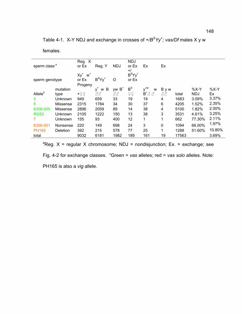
148
Table 4-1. X-Y NDJ and exchange in crosses of +/BSYy+; vas/Df males X y w
females.
sperm class a Reg. X or Ex Reg. Y NDJ
NDJ or Ex Ex Ex
sperm genotype Xy+ w+ or Ex BSYy+ O
+/ BSYy+ or Ex
Progeny
Alleleb mutation type +♀♀
y+ w B ♂♂
yw B+
♂♂ BS
♀♀ yvar w B+♂ ♂
B y w ♂♂ total
%X-Y NDJ
%X-Y Ex
3 Unknown 949 659 33 19 19 4 1683 3.09% 3.37% 5 Missense 2315 1784 34 30 37 6 4205 1.52% 2.35% 6356-005 Missense 2896 2059 89 14 38 4 5100 1.82% 2.00% RG53 Unknown 2105 1222 150 13 38 3 3531 4.61% 3.25% 7 Unknown 155 93 400 12 1 1 662 77.30% 2.11%
6356-001 Nonsense 220 149 698 24 3 0 1094 66.00% 1.97%
PH165 Deletion 392 215 578 77 25 1 1288 51.60% 10.80% total 9032 6181 1982 189 161 19 17563 3.69%
aReg. X = regular X chromosome; NDJ = nondisjunction; Ex. = exchange; see
Fig. 4-2 for exchange classes. bGreen = vas alleles; red = vas solo alleles. Note:
PH165 is also a vig allele.
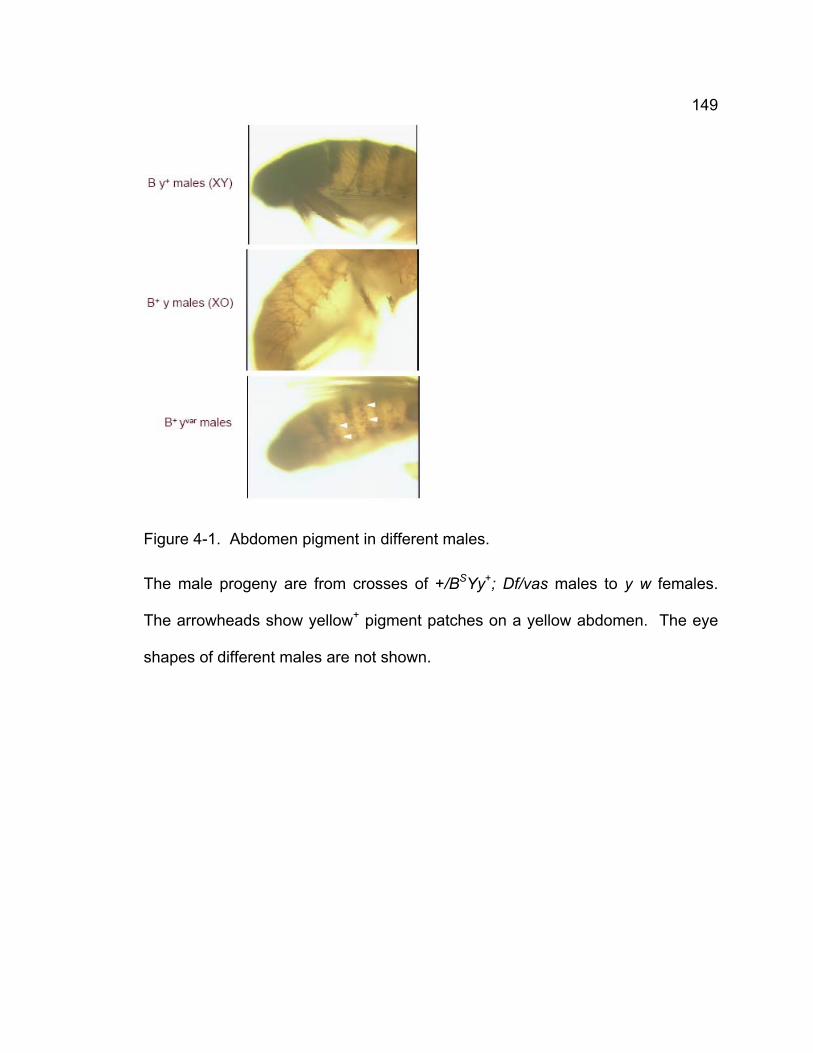
149
Figure 4-1. Abdomen pigment in different males.
The male progeny are from crosses of +/BSYy+; Df/vas males to y w females.
The arrowheads show yellow+ pigment patches on a yellow abdomen. The eye
shapes of different males are not shown.
150
XO progeny indicates that the majority of NDJ sperm in solo and vas double
mutants were null sperm, which is very unlike in vas and solo single mutants.
These results suggest that SOLO and VAS might co-cooperate in male meiosis
in addition to having their own distinct effects. Furthermore, in all vas mutants,
progeny from nullo-XY sperm were more frequent than the progeny from XY
sperm. Particularly, in vasRG53, vas7, vas6356-001, and vasPH165 mutants, the ratio
of progeny from nullo-XY sperm to that from XY sperm is about 10 or even higher.
However, we need to be very cautious to make this conclusion due to the
limitation of our cross. The BS y+ females could be either NDJ (XXY) or the
recombinant (BSYL.X,y+/yw) and could not be distinguished from their
phenotypes. It was possible therefore that there was no true NDJ in vas
mutants, i.e. there were no or almost no XXY progeny instead of recombinant.
XO progeny may come from chromosome loss (e.g. from anaphase I and
anaphase II chromatin bridges) rathter than NDJ. For similar reason, the ratios
of the XY:O bias in vas and solo double mutants were not accurate either.
Nevertheless, our data showed X/Y recovery bias among regular progeny, 1.46
on average (9032/6181). Similar observation was made in the progeny of
homeless, another DEAD/DEAH box RNA helicase (Stapleton et al., 2001). This
bias is not observed in solo and snm mutants, which are required for sister
chromatid cohesion and homolog pairing, respectively, suggesting that vas has a
distinct functions from solo and snm in meiosis, and sister chromatid cohesion
and homolog pairing are not involved in the bias.
151
The PEV males are sterile due to loss of fertility factors on Y chromosomes
The y+ variegation of B+ yvar male progeny seemed likely to be due to
heterochromatic position-effect since the y+ gene on the BSYy+ chromosome is
adjacent to heterochromatin. Considering the fact that the reduced
heterochromatic content of the genome would enhance PEV (Gowen and Gay
1934), we speculated the Y chromosome in the exceptional B+ yvar and B y males
may not be intact due to chromosome loss or recombination. In order to test this
idea, we first tested the fertility of these males. They were crossed to y w
females. Corresponding to our speculation, more than 50 tested B+ yvar males
(primarily from the crosses of three different vas alleles, vas3, vas5, vasRG53) were
all sterile, suggesting one or more fertility factors were lost along with BS marker.
This also strengthened our explanation for PEV of these sterile males because
the heterochromatin content of their Y chromosomes was reduced. In the fertility
test for B y males, which were also crossed to y w females, 16 out of a total of 18
flies were sterile while the other two were fertile. Careful examination revealed
that fertile B y males had extremely small Bar eyes (Bext y).
To further test the idea that PEV and sterility of B+ yvar males resulted from
the partial loss of the Y chromosome, +/BSYy+; vas/Df were crossed to YSX.YL,
In(1)EN, y B/y w females. The Bar eyes are larger in females with B marker than
those in males with BS marker. When B and BS are combined, the bar eyes are
even smaller. In total, 23 males with B w+ eyes (from three crosses: vas3, vas5,
vasRG53), representing the males with YSX.YL, In(1)EN, y B/YabB+ y+ were
selected and crossed to y w females. All of these males exhibited normal,
152
unvariegated expression of the y+ marker on the Y chromosome. The crosses
yielded B+ yvar males, suggesting that their sex chromosomes were YabB+ y+
chromosome and normal y w X chromosome. This suggested the PEV and
sterility were suppressed by an additional Y chromosome. All the B+ yvar males
were crossed to y w females again and proved to be sterile, further confirming
that PEV and sterility resulted from incomplete Y chromosome.
The possible exchange patterns between X and Y chromosomes
The uniform loss of fertility and dominant marker of the B+ yvar male progeny
indicates that B+ yvar and B y male progeny may be primarily arisen from X-Y
chromosome exchange, rather than random Y chromosome breakage. Four rDNA blocks
exist within the X and Y chromosomes used in the crosses (Fig. 4-2A) and several
kinds of exchanges may occur among them. The X chromosome used in the
crosses is normal in structure carrying y and w recessive mutations. For
simplicity of discussion, we refer to the region between the very short arm of X
chromosome and the rDNA region as XR, and the region distal to rDNA locus as
XL. The BSYy+ chromosome is a normal Y chromosome except that it is
appended with BS and y+ markers that were derived from X chromosome on both
the long and short arm, respectively. Besides the two markers, two additional
sex chromosome heterochromatin blocks that contain rDNA arrays were on the
long arm just proximal to BS, and on the short arm just proximal to y+ (Gatti and
Pimpinelli 1983). For simplicity of discussion, we define the region of long arm
containing fertility factors kl1, kl2, kl3, and kl5, centromere
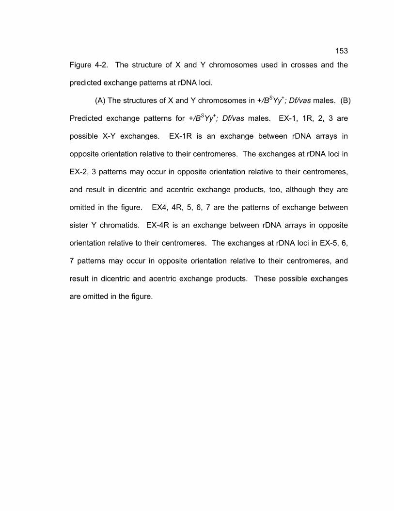
153
Figure 4-2. The structure of X and Y chromosomes used in crosses and the
predicted exchange patterns at rDNA loci.
(A) The structures of X and Y chromosomes in +/BSYy+; Df/vas males. (B)
Predicted exchange patterns for +/BSYy+; Df/vas males. EX-1, 1R, 2, 3 are
possible X-Y exchanges. EX-1R is an exchange between rDNA arrays in
opposite orientation relative to their centromeres. The exchanges at rDNA loci in
EX-2, 3 patterns may occur in opposite orientation relative to their centromeres,
and result in dicentric and acentric exchange products, too, although they are
omitted in the figure. EX4, 4R, 5, 6, 7 are the patterns of exchange between
sister Y chromatids. EX-4R is an exchange between rDNA arrays in opposite
orientation relative to their centromeres. The exchanges at rDNA loci in EX-5, 6,
7 patterns may occur in opposite orientation relative to their centromeres, and
result in dicentric and acentric exchange products. These possible exchanges
are omitted in the figure.
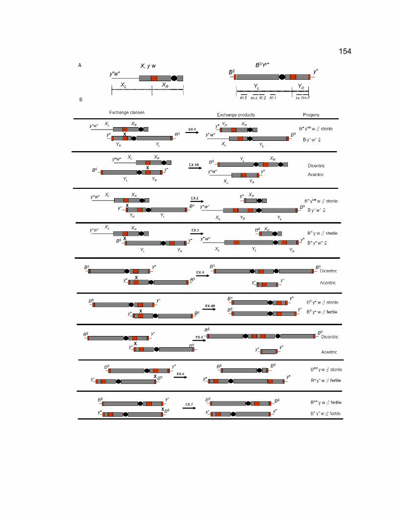
154
155
and part of the short arm heterochromatin proximal to the centromere as YL,
and the region located distal to the rDNA arrays as YR.
We considered nine types of exchanges that could generate the
exceptional progeny in the cross: three types of inter-chromosomal exchanges
between X and Y chromosomes, one type of inter-chromosomal exchange in
opposite orientation relative to their centromeres, four types of intra-
chromosomal exchanges between two Y sister chromatids, and one type of intra-
chromosomal exchange in opposite orientation relative to their centromeres (Fig.
4-2B). The three inter-chromosomal exchanges would generate B+ yvar and B y
males that are sterile, and B+ y+ w+ and B y+ w+ females that are fertile and
cannot be distinguished from regular and NDJ female progeny, respectively,
when they are fertilized with y w females. Sterile B+ yvar and B y males were
recovered in the cross, demonstrating that exchanges between X and Y
chromosomes at rDNA loci occurred as we predicted. The crosses yielded more
B+ yvar males than B y males, probably due to more rDNA repeats were involved
in type of EX-1 exchange than that of EX-3 type of exchange. The five intra-
chromosomal exchanges would generate fertile B+ y+ w and Bext y w males. One
fertile B+ y+ w and two Bext y w fertile males were recovered in the crosses of
+/BSYy+; Df/vas to y w females, suggesting that exchanges occur between sister
chromatids.
To test for the generation of XL.YL BS chromosome in the crosses of vas,
which would generate B y+ w+ female progeny and cannot be distinguished from
XXY NDJ females, we crossed +/BSYy+; vas/Df males to C(1)RM, y/YS females.
156
The C(1)RM females (an attached-X) produce the eggs with attached X and YS
eggs at approximately equal frequency. As expected, the cross generated B y+
sons, carrying the recombinant y+ w+ XL.YL BS chromosome. The B y+ sons
from the C(1)RM/YS cross were all fertile. The B y+ sons from C(1)RM/YS cross
were expected to result from two sources: XY recombinant (y+ w+ XL.YL BS/YS)
or XY NDJ males (+/BSYy+/YS). A recombinant male mated to y w females
produces only two kinds of progeny: B+ y w males and B y+ w+ females, i.e. all B
flies are females. An XY NDJ male if mated to y w females produces four types
of progeny: B y+ w males, B+ y+ w+ females, B+ y w males, B y+ w+ females. In
the tested 24 B y+ males (from three crosses: vas3, vas5, vasRG53), four were
proved to from XY NDJ while 20 were proved to carry the XY chromosome
recombinant (y+ w+ XL.YL BS), and all Bar flies in the progeny were females.
These data confirmed our prediction about the structure of X-Y chromosome
recombinant and that the sterility of the recombinant males could be rescued by
an additional YS chromosome.
We also analyzed the distribution of exchanges among singly crossed
males. In the 127 vas/Df males producing B+ yvar males, 1 male generated 5 B+
yvar males, which is almost certainly a result of a single mitotic exchange. 12 out
of 127 vas/Df males produced three B+ yvar males in the progeny. 24 out of 127
vas/Df males produced 2 B+ yvar males in the progeny, which were probably from
meiotic recombination. 90 out of 127 (71%) males produced only one B+ yvar
progeny, which were almost certainly from meiotic recombination. These data
suggest most of the recombinants were generated in meiotic stages.
157
Cytological analysis of anaphase I and II of vas mutants
The multiple phenotypes of vas mutations could be manifested at
cytological level. We didn’t try to identify the chromosome recombinant, like
XL.YL BS, since they were technically not easy to be detected at cytological level.
Instead we explored chromatin bridges that could be generated when dicentric
recombination occur between homologs and between sister chromatids (Fig. 4-
3). Spermatocytes from vas/Df were stained to visualize DNA and spindle. As
we predicted, long, thin chromatin bridges were seen at anaphase I in all tested
vas alleles (Fig. 4-3A), suggesting dicentric recombination occur between
homologs during meiosis. Some discontinuous-like chromatin bridges were also
observed. They might be connected but were not detectable at some spots.
Short chromatin bridges were observed at anaphase II in tested vas alleles (Fig.
4-3B), suggesting that exchanges between sister chromatids occurred. Although
at this point we do not know what the bridges represent and how they related to
other phenotypes of vas, we prefer the idea they are dicentric chromosomes that
result from X-Y recombination. Chromatin clumps were also observed in the
midbody of spindle of anaphase I and II (Fig. 4-3), suggesting that acentric
recombination products also occurred. Thus our cytological data confirmed that
meiotic recombination occur when vas is mutated.
vas and solo double mutations cause precocious segregation of homologs.
Interestingly, vas7, a vas and solo double mutant, showed precocious
segregation of homologs. Eight DNA clumps were apparently seen in vas7
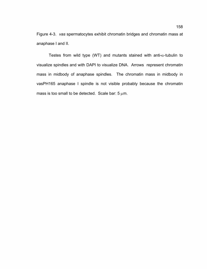
158
Figure 4-3. vas spermatocytes exhibit chromatin bridges and chromatin mass at
anaphase I and II.
Testes from wild type (WT) and mutants stained with anti-α-tubulin to
visualize spindles and with DAPI to visualize DNA. Arrows represent chromatin
mass in midbody of anaphase spindles. The chromatin mass in midbody in
vasPH165 anaphase I spindle is not visible probably because the chromatin
mass is too small to be detected. Scale bar: 5 μm.
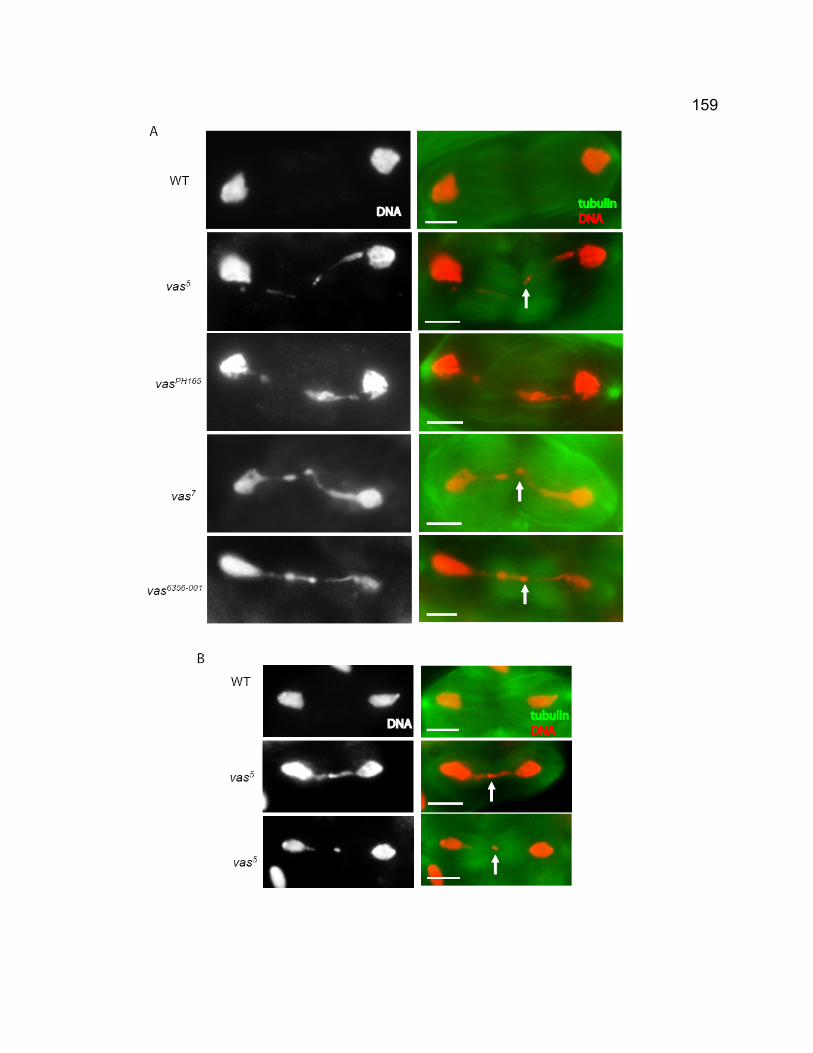
159
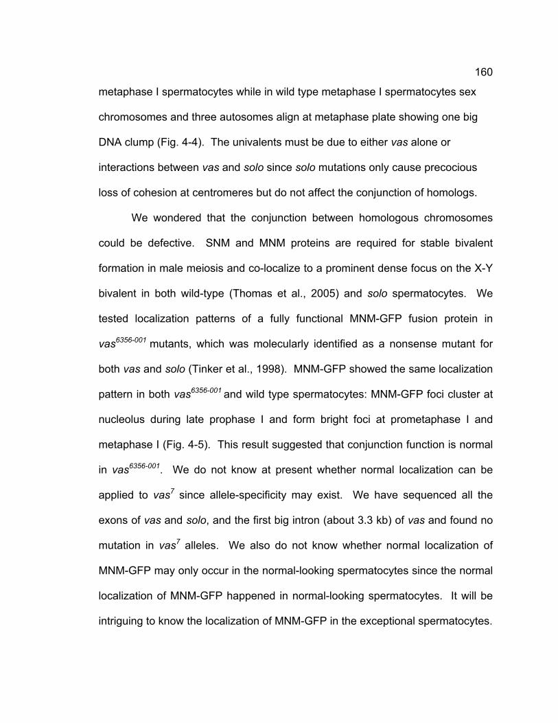
160
metaphase I spermatocytes while in wild type metaphase I spermatocytes sex
chromosomes and three autosomes align at metaphase plate showing one big
DNA clump (Fig. 4-4). The univalents must be due to either vas alone or
interactions between vas and solo since solo mutations only cause precocious
loss of cohesion at centromeres but do not affect the conjunction of homologs.
We wondered that the conjunction between homologous chromosomes
could be defective. SNM and MNM proteins are required for stable bivalent
formation in male meiosis and co-localize to a prominent dense focus on the X-Y
bivalent in both wild-type (Thomas et al., 2005) and solo spermatocytes. We
tested localization patterns of a fully functional MNM-GFP fusion protein in
vas6356-001 mutants, which was molecularly identified as a nonsense mutant for
both vas and solo (Tinker et al., 1998). MNM-GFP showed the same localization
pattern in both vas6356-001 and wild type spermatocytes: MNM-GFP foci cluster at
nucleolus during late prophase I and form bright foci at prometaphase I and
metaphase I (Fig. 4-5). This result suggested that conjunction function is normal
in vas6356-001. We do not know at present whether normal localization can be
applied to vas7 since allele-specificity may exist. We have sequenced all the
exons of vas and solo, and the first big intron (about 3.3 kb) of vas and found no
mutation in vas7 alleles. We also do not know whether normal localization of
MNM-GFP may only occur in the normal-looking spermatocytes since the normal
localization of MNM-GFP happened in normal-looking spermatocytes. It will be
intriguing to know the localization of MNM-GFP in the exceptional spermatocytes.
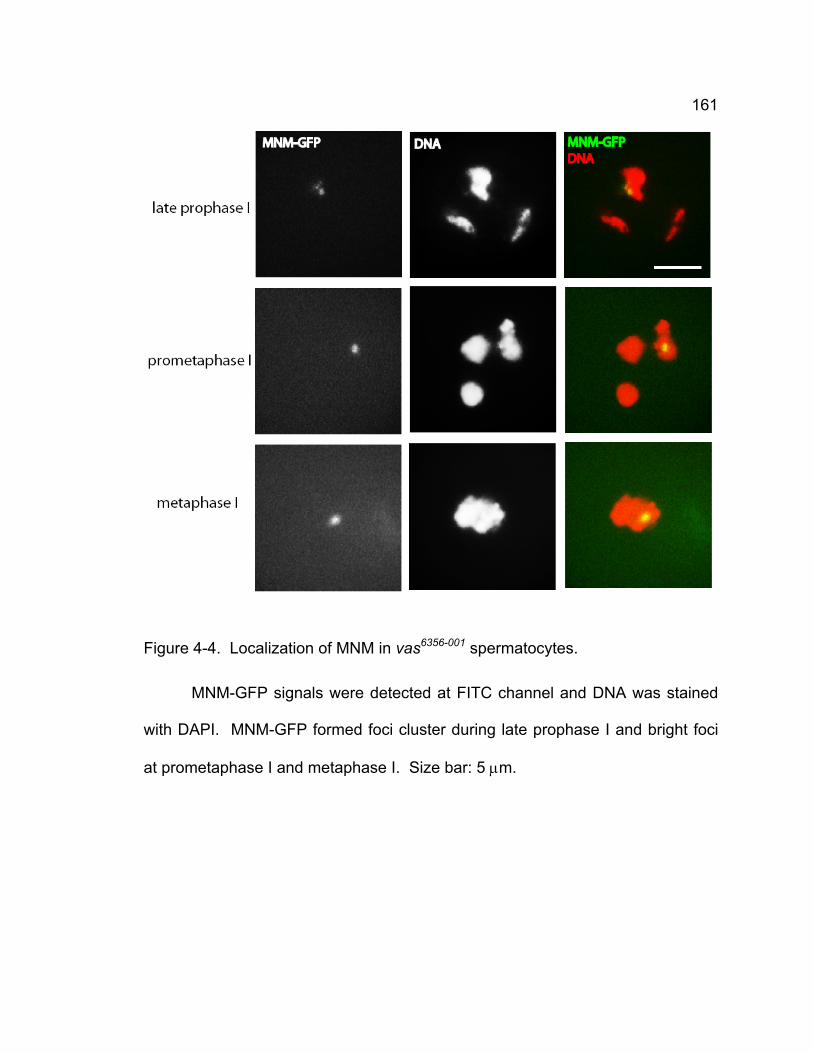
161
Figure 4-4. Localization of MNM in vas6356-001 spermatocytes.
MNM-GFP signals were detected at FITC channel and DNA was stained
with DAPI. MNM-GFP formed foci cluster during late prophase I and bright foci
at prometaphase I and metaphase I. Size bar: 5 μm.
162
Figure 4-5. Cytological phenotypes of vas7 spermatocytes.
Testes from vas7 mutant were stained with anti-α-tubulin to visualize
spindles and with DAPI to visualize DNA. vas7 spermatocytes show chromatin
bridges at Telophase I (A) and conjunction failure at metaphase I (B). Scale bar:
5 μm.
163
DISCUSSION
Meiotic recombination in Drosophila males
Recombination is normally absent during male meiosis although meiotic
recombination in Drosophila females is a prerequisite for the successful
progression of oogenesis (McKee, 2004). However, very little is known how
Drosophila spermatocytes respond to endogenous and exogenously-induced
DSBs and prevent recombination between homologous sequences during
meiosis. We demonstrated here that in a specific case Drosophila males
undergo meiosis with meiotic recombination. Our data indicate that vas
mutations cause high frequencies of recombination between sex chromosomes
compared to the very low frequency of spontaneous recombination in normal
condition (Ashburner et al., 2004). Meiotic recombination may occur only
between sex chromosomes since there is no recombination between rucuca (ru h
st th cu sr e ca) and a wild type 3rd chromosome in vas5/Df or vasD5/Df (B.D.
McKee, personal communication). Our cytological analysis further confirmed the
existence of dicentric and acentric recombinant products by revealing chromatin
bridges at anaphase I and II and the presence of chromatin clumps at midbody of
anaphase I and II. The fact that the majority of recombinants are recovered
singly rather than on clusters suggests that recombination occurs mainly during
meiosis. Mei-W68 appears in young spermatocytes (McKim and Hayashi-
Hagihara, 1998), suggesting meiotic recombination may occur in these stages.
However, the mechanism of meiotic recombination is unclear at present.
164
Although it is not known whether recombination occurs at other repetitive
sequence besides rDNA arrays (it doesn’t occur in 3rd chromosome
heterochromatin (McKee, personal communication)) and how vas-induced
recombination is related to homolog pairing and segregation during meiosis I, our
studies strongly suggest that meiotic recombination does occur when vas is
mutated. Thus our data suggest that vas may suppress the occurrence of
meiotic recombination during male meiosis. If this is true, it would provide a clue
to why Drosophila males undergo meiosis without recombination.
vas shows pleiotropic functions in Drosophila male meiosis
vas has been shown to be required for pole cell development and
embryonic axis specification in oogenesis and embryogenesis (Hay et al., 1988;
Lasko and Ashburner, 1988). No phenotypes were reported in spermatogenesis
although vas is expressed in spermatogonia and young spermatocytes. Our
studies definitely show vas functions in suppressing recombination and
chromosome segregation in Drosophila male meiosis. In all tested vas alleles
vas mutations cause high frequency of meiotic exchange between X and Y
chromosomes. vas mutations also cause NDJ of sex chromosome although the
frequency is low but it is significantly higher than that of wild type. Since in
Drosophila males sex chromosomes pair at rDNA loci and meiotic recombination
between X and Y chromosome may lead to increase or decrease of rDNA copy
number, the fidelity of pairing of X and Y chromosome at rDNA may be damaged.
This would lead to NDJ of sex chromosomes in vas mutants. Furthermore, vas7,
165
an allele of vas and solo double mutant, causes precocious segregation of
homologous chromosomes, a phenomenon that does not happen in single solo
mutants. Although vas mutations show pleiotropic phenotypes, vas may play
indirect roles in these phenotypes since vas is a translation initiation factor and
localized to nucleoplasm but not to chromatin.
Conjunction failure in vas and solo double mutant?
Homologous chromosomes are linked by a mechanism involving SNM and
MNM during male meiosis (Thomas et al., 2005). vas7, which is a double mutant
for vas and solo through genetic test but the mutation site are not identified,
revealed conjunction failure at metaphase I. Eight chromatin clumps that
apparently represent univalents were seen. We have not found any evidence
that solo single mutants cause conjunction failure, so the conjunction failure in
vas7 must result from vas alone or the interaction between vas and solo.
Interesting, solo and snm double mutants cause precocious segregation of
homolog and sister chromatids, leading to the appearance of up to 16 sister
chromatids in prometaphase I. How the sister chromatids were kept together
while homologs were separated in vas7 mutant is not known. Another vas allele
vas6356-001 that has been molecularly identified as a null mutant of solo and vas
showed chromatin bridges at anaphase I. The failure of homolog conjunction
was not observed and MNM location appeared to be normal in this mutant. Thus
it is possible that SNM or MNM still hold sister chromatids (univalents) but the
linkage between two univalents is disrupted. Further experiments need to be
166
done to learn whether SNM or MNM still exist in the separated univalents, which
would provide strong evidence of whether SNM and MNM hold two sister
chromatids in a univalent at pairing sites (e. g. rDNA in sex chromosomes) other
than centromeres that is held together by classical cohesion complex. However,
we cannot rule out the failure of homolog conjunction due to mutation complexity
of vas7 allele. Nevertheless, our finding suggests that besides SNM and MNM,
other mechanisms exist to control homolog segregation during male meiosis.
167
CHAPTER 5 – GENERAL CONCLUSIONS AND FUTURE
DIRECTIONS
168
GENERAL CONCLUSIONS
The main goal of this study is to explore the mechanism of meiosis in
Drosophila melanogaster. Plenty of studies have been carried out in yeast to
study cohesion’s function in meiosis and revealed many important principles of
meiosis (Nasmyth, 2001; Petronczki et al., 2003; Marston and Amon, 2004).
However, the mechanism in higher eukaryotes still remains elusive to large
extent and some observations that are controversial to yeast emerged (Prieto et
al., 2002; Eijpe et al, 2003; Valdeolmillos et al., 2007). Our studies have
identified a novel cohesin protein, SOLO, which is essential for all aspects of
sister chromatid cohesion in Drosophila meiosis. It is required for homologous
recombination, homolog synapsis, maintenance of chiasmata in female meiosis.
It is essential for maintaining centromeric cohesion in meiosis I and establishing
orientation of sister centromeres in meiosis I and II spindles.
SOLO is essential for homologous recombination, homolog synapsis, and
maintenance of chiasmata.
Sister chromatid cohesin is required for homologous chromosome
recombination and maintaining the resulting chiasmata, which are required for
proper homolog segregation during meiosis I (Petronczki et al., 2003). In
Drosophila females, ORD, a protein required for maintaining cohesion, is
involved in homolog recombination and maintaining chiasmata (Bickel et al.,
169
2002; Webber et al., 2004). BubR1, a checkpoint protein in mitosis, is required
for synapsis (Malmanche et al., 2007). SOLO is a novel sister chromatid
cohesion protein. solo mutations cause reduced frequency of homologous
recombination and altered distribution of crossovers. Synapsis is severely
disrupted at the early prophase I when solo is mutated. In solo females, residual
chiasmata cannot be maintained. Thus SOLO is essential for proper homolog
dynamics during meiosis I.
SOLO is essential for orientation of sister centromeres.
In solo males, sister centromeres segregate precociously before
prometaphase I. Correspondingly, the cohesin containing SMC1 is lost at
centromeres at the earliest meiotic stage. Genetic analyses show that homologs
disjoin together in male and female meiosis. However, complete separation of
sister chromatids is not observed. These data suggest that precocious
separation of sister chromatids does not occur before meiosis I although sister
centromere co-orientation is disrupted and sister centromeres orient randomly in
meiosis I when solo is mutated.
SOLO and SMC1 function together in Drosophila meiosis.
Since the Drosophila genome contains only one copy of the SMC1 and
SMC3 genes, one can imagine that SMC1 must be one of the components if a
meiotic cohesin exists. Our data show centromeric foci of SMC1 appear at the
170
earliest meiotic stage and throughout meiosis I and until anaphase II when
centromeric cohesion is degraded to allow sister chromatids segregate,
suggesting that a cohesin containing SMC1 does exist in Drosophila meiosis.
SOLO appears from the earliest meiotic stage to metaphase II and then
disappears at anaphase II, and colocalizes with SMC1 throughout. ord, a protein
required for sister chromatid cohesion, appears to bind chromatin at mid
prophase I and localize to centromeres from prometaphase I to anaphase II
(Balicky et al., 2002). Thus, SOLO becomes the first cohesion protein that
shows the exactly same localization pattern with SMC1, strongly indicating that
SOLO and SMC1 work together as partners. Moreover, the evidence that solo
mutations cause disappearance of SMC1 at centromeres from the earliest
meiotic stage further support the idea that SOLO and SMC1 are the components
of a cohesin in Drosophila, albeit it may be non-canonical.
Meiotic recombination is not suppressed in vas mutants.
Drosophila males undergo meiosis without recombination. This
phenomenon was observed long time ago (Morgan, 1912). By contrast,
Drosophila females undergo meiosis with high frequency of recombination. Why
there is no recombination in male meiosis is not known. Our findings
demonstrate that meiotic recombination occur in the case of vas mutations. vas
mutations cause about 300 fold X-Y exchanges compared to the frequency of
spontaneous X-Y exchange (Ashburner et al., 2004). Cytological analyses
171
reveal chromatin bridges that are the products of dicentric exchange at anaphase
I and anaphase II. Although we don’t know the mechanism at present, our
observations reveal that meiotic recombination in Drosophila males is inhibited
by VASA.
172
FUTURE DIRECTIONS
Our studies have been focusing on the mechanism of Drosophila meiosis.
Our finding of the novel cohesion protein SOLO and the novel functions of VASA
in meiosis would enrich knowledge of meiosis.
The studies about mutant phenotypes and localization pattern of SOLO
provide substantial support to the idea that SOLO is a component of cohesin
containing SMC1 in Drosophila meiosis. However, SOLO is not conserved
outside of Drosophila species and does not show high sequence similarity with
known cohesion proteins by current bioinformatics methods. To prove that
SOLO is a cohesin component, the most straightforward way to determine
whether SOLO interact with SMC1 or other cohesin component in vitro and in
vivo at molecular level. The current Venus-SOLO transgenic flies can be used to
perform co-immunoprecipitation experiments using antibodies against GFP,
which is successful in yeast (Cristea et al., 2005). If GFP pull-down method is
not applicable in Drosophila, classical pull-down using anti-HA or Flag can be
used, which were successfully applied in Drosophila (Heidmann et al., 2004).
Our Venus-SOLO is prepared by Gateway technology and is very easy to
change the Venus tag to HA or Flag. After pull-down experiments, western blot
and MALDI mass spectrometry can be applied to identify the potential proteins
that interact with SOLO.
Another very interesting issue is to learn whether expression of SOLO
173
during pre-meiotic S phase is required for establishment of proper cohesion in
meiosis. In yeast cohesin must be present at chromatin region during pre-
meiotic S phase to establish functional cohesion in meiosis (Watanabe et al.,
2001). SOLO is expressed in the spermatogonia in males and oogonia in
females to form foci at centromeres and thus is present during pre-meiotic stage.
solo and vas share the 5’ UTR and the first three exons, suggesting they may
share the same promoter. Both Nanos and VASA are expressed in
embryogenesis, suggesting the SOLO expression pattern induced by nanos:Gal4
is probably true, which is supported by complete rescue of UAS:Venus-SOLO
when induced by nanos:Gal4. The upstream of vas will be fused to Venus-SOLO
to express SOLO by its native promoter. If the construct can rescue the
phenotypes of solo, it indicates the cloned upstream contains the true promoter
of solo. Then the construct will be used for study whether SOLO is expressed
before or during pre-meiotic S phase. Using specific G2 (after meiotic S phase)
promoter to forcibly express SOLO in solo background, whether SOLO
expression before or during pre-meiotic S phase is required for establish proper
cohesin can be determined.
It will be very intriguing to determine the interactions between SOLO and
other cohesion proteins. As revealed by studies in other model organisms,
meiotic cohesion is probably not mediated only by classical cohesin complex,
other meiotic cohesion proteins, even some cohesion proteins that are first
thought mitotic-specific were found to be involved in meiosis (Prieto et al., 2002;
Eijpe et al, 2003). Although classical meiotic cohesin is not identified in
174
Drosophila until now, proteins required for sister chromatid cohesion have been
identified, including ORD, SOLO, SUN (McKee, personal communication).
Exploring interplays between SOLO and these meiotic cohesion proteins and
some mitotic cohesion protein, like dRAD21, etc. will be helpful to understand the
mechanism of meiotic cohesion in Drosophila.
Drosophila males undergo meiosis without recombination. vas, the
alternative splicing products of solo, is surprisingly involved in the suppression of
meiotic recombination. vas mutations cause high X-Y exchange during meiosis.
Since the homology sequences in X and Y chromosomes are primarily rDNA
repeats, it is important to further know whether recombinations occur at the rDNA
repeated sequences although we have already narrowed the recombination
region containing rDNA loci. This can be done by using bobbed X chromosome
flies, or X heterochromatin deficiency flies with rDNA transgenes. They will yield
less X-Y exchange by compared to that of the normal X chromosome. It is also
important to know the origin of chromatin bridges in anaphase I. rDNA or even
240bp repeat probes will be the first candidate probes and then other tandem
repeats will be tried if rDNA or 240bp repeats are not.

175
LIST OF REFERENCES
176
Adams, M.D., Celniker, S.E., Holt, R.A., Evans, C.A., Gocayne, J.D., Amanatides, P.G., Scherer, S.E., Li, P.W., Hoskins, R.A., Galle, R.F., et al. (2000). The genome sequence of Drosophila melanogaster. Science 287, 2185-2195.
Adams, R.R., Carmena, M., and Earnshaw, W.C. (2001). Chromosome
passengers and the (Aurora) ABCs of mitosis. Trends in Cell Biol. 11, 49-54. Ahmad, K., and Henikoff, S. (2001). Centromeres are specialized replication
domains in heterochromatin. J. Cell Biol. 153, 101-110. Alex, D., and Lee, K.A.W. (2005). RGG boxes of the EWS oncoprotein repress a
range of transcriptional activation domains. Nucl. Acids Res. 33, 1323-1331. Anderson, D.E., Losada, A., Erickson, H.P., and Hirano, T. (2002). Condensin
and cohesin display different arm conformations with characteristic hinge angles. J. Cell boil. 156, 419-424.
Anderson, L.K., Royer, S.M., Page, S.L., McKim, K.S., Lai, A., Lilly, M.A., and
Hawley, R.S. (2005). Juxtaposition of C(2)M and the transverse filament protein C(3)G within the central region of Drosophila synaptonemal complex. Proc. Natl. Acad. Sci. USA. 102, 4482-4487.
Ashburner, M., Thompson, P., Roote, J., Lasko, P.F., Grau, Y., El Messal, M.,
Roth, S., and Simpson, P. (1990). The genetics of a small autosomal region of Drosophila melanogaster containing the structural gene for alcohol dehydrogenase. Genetics 126, 679-694.
Ashburner, M., Golic, K.G., Hawley, R.S. (2004). Drosophila: A laboratory
handbook, Ed. 2. Cold Spring Harbor Laboratory Press, Cold Spring Harbor, NY.
Balicky, E.M., Endres, M.W., Lai, C., and Bickel, S.E. (2002). Meiotic cohesion
requires accumulation of ORD on chromosomes before condensation. Mol. Biol. Cell. 13, 3890-3900.
Basu, J., Bousbaa, H., Logarinho, E., Li, Z., Williams, B.C., Lopes, C., Sunkel,
C.E., and Goldberg, M.L. (1999). Mutations in the essential spindle checkpoint gene bub1 cause chromosome missegregation and fail to block apoptosis in Drosophila. J. Cell Biol. 146, 13-28.
Bergerat, A., de Massy, B., Gadelle, D., Varoutas, P.C., Nicolas, A., and Forterre,
P. (1997). An atypical topoisomerase II from archaea with implications from meiotic recombination. Nature 386, 414-417.
177
Bernard, P., Maure, J.F., and Javerzat, J.P. (2001). Fission yeast Bub1 is essential in setting up the meiotic pattern of chromosome segregation . Nature cell Biol. 3, 522-526.
Bhagat R., Manheim, E.A., Sherizen, D.E., and McKim K.S. (2004). Studies on
crossover-specific mutants and the distribution of crossing over in Drosophila females. Cytogenet. Genome Res. 107, 160-171.
Bickel, S.E., Wyman, D.W., Miyazaki, W.Y., Moore, D.P., and Orr-Weaver, T.L.
(1996). Identification of ORD, a Drosophila protein essential for sister chromatid cohesion. EMBO J. 15, 1451-1459.
Bickel, S. E., Wymann, D. W., and Orr-Weaver, T. L. (1997). Mutational analysis
of the Drosophila sister-chromatid cohesion protein ORD and Its role in the maintenance of centromeric cohesion. Genetics 146, 1319-1331.
Bickel, S.E., Orr-Weaver, T.L., and Balicky, E.M. (2002). The sister-chromatid
cohesion protein ORD is required for chiasma maintenance in Drosophila oocytes. Curr. Biol. 12, 925-929.
Blower, M.D., and Karpen, G.H. (2001). The role of Drosophila CID in
kinetochore formation, cell-cycle progression and heterochromatin interactions. Nat. Cell Biol. 3, 730-739.
Bonaccorsi, S., Giansanti, M.G., Cenci, G., and Gatti, M. (2000). Cytological
analysis of spermatocyte growth and male meiosis in Drosophila melanogaster. In Drosophila Protocols, W. Sullivan, M. Ashburner, and R. Scott Hawley, eds. (New York: Cold Spring Harbor Press).
Buonomo, S.B., Clyne, R.K., Fuchs, J., Loidl, J., Uhlmann, F., and Nasmyth, K.
(2000). Disjunction of homologous chromosomes in meiosis I depends on proteolytic cleavage of the meiotic cohesin Rec8 by separin. Cell 103, 387-398.
Cai, X., Dong, F., Edelmann, R.E., and Makaroff, C.A. (2003). The Arabidopsis
SYN1 cohesin protein is required for sister chromatid arm cohesion and homologous chromosome pairing. J. Cell Sci. 116, 2999-3007.
Carlson, P.S. (1972). The effects of inversions and C(3)G mutation on introgenic
recombination in Drosophila. Genet. Res. 19, 129-132. Carmena, M., and Earnshaw, W.C. (2003). The celllar geography of Aurora
kinases. Nat. Rev. Mol. Cell Biol. 4, 842-854.
178
Carpenter, A.T. and Sandler, L. (1974). On recombination-defective meiotic mutants in Drosophila. Genetics 76, 453-475.
Carpenter, A.T.C. (1979). Synaptonemal complex and recombination nodules in
wild-type Drosophila melanogaster females. Genetics 92, 511-541. Cenci, G., Bonaccorsi, S., Pisano, C., Verni, F., and Gatti, M. (1994). Chromatin
and microtubule organization during premeiotic, meiotic and early postmeiotic stages of Drosophila melanogaster spermatogenesis. J. Cell Sci. 107, 3521-3534.
Cheeseman, I.M, Anderson, S., Jwa, M., Green, E.M>, Kang, J., Yates, J.R. 3rd,
Chan, C.S., Drubin, D.G., and Barnes, G. (2002). Phospho-regulation of kinetochore-microtubule attachments by the Aurora kinase Ipl1p. Cell 111, 163-172.
Chelysheva, L., Diallo, S., Vezon, D., Gendrot, G., Vrielynck, N., Belcram, K.,
Rocques, N., Marquez-Lema, A., Bhatt, A.M., Horlow, C. et al. (2005). AtREC8 and AtSCC3 are essential to the monopolar orientation of kinetochores during meiosis. J. Cell Sci. 118, 4621-4632.
Clyne, R.K., Katis, V.L., Jessop, L., Benjamin, K.R., Herskowitz, I., Lichten, M.,
and Nasmyth, K. (2003). Polo-like kinase Cdc5 promotes chiasmata formation and cosegregation of sister centromeres at meiosis I. Nature Cell Biol. 5, 480-485.
Cohen-Fix, O., Peters, J.M., Kirschner, M.W., and Koshland, D. (1996).
Anaphase initiation in Saccharomyces cerevisiae is controlled by the APC-dependent degradation of the anaphase inhibitor Pds1p. Genes Dev. 10, 3081-3093.
Cristea, I.M., Williams, R., Chait, B.T., and Rout, M.P. (2005). Fluorescent
proteins as proteomic probes. Mol. Cell. Proteomics. 4, 1933-1941. Dej, K.J. and Spradling, A.C. (1999). The edocycle controls nurse cell polytene
chromosomes structure during Drosophila oogenesis. Development 126, 293-303.
Dernburg, A.F., McDonald, K., Moulder, G., Barstead, R., Dresser, M., and
Villeneuve, A.M. (1998). Meiotic recombination in C. elegans initiates by a conserved mechanism and is dispensable for homologous chromosome synapsis. Cell 94, 387-398.
179
Dewar, H., Tanaka, K., Nasmyth, K., and Tanaka, T.U. (2004). Tension between two kinetochores suffices for their bi-orientation on the mitotic spindle. Nature 428, 93-97.
Doren, M.V., Williamson, A.L., and Lehmann, R. (1998). Regulation of zygotic
gene expression in Drosophila primordial germ cells. Curr. Biol. 8, 243-246. Eijpe, M., Offenberg, H., Jessberger, R., Revenkova, E., and Heyting, C. (2003).
Meiotic cohesin REC8 marks the axial elements of rat synaptonemal complexes before cohesins SMC1beta and SMC3. J. Cell Biol. 160, 657-670.
FLYBASE (2007a). URL: http://flybase.bio.indiana.edu/. FLYBLAST, (2007b). URL: http://flybase.bio.indiana.edu/blast/ Funabiki, H. Yamano, H., Kumada, K., Nagao, K., Hunt, T., and Yanagida, M.
(1996). Cut2 proteolysis required for sister chromatid separation in fission yeast. Nature 381, 438-441.
Gatti, M., and Pimpinelli, S. (1983). Cytological and genetic analysis of the Y
chromosome of Drosophila melanogaster. Chromosoma 88, 349-373. Goldstein, L.S. (1981). Kinetochore structure and its role in chromosome
orientation during the first meiotic division in male D. melanogaster. Cell 25, 591-602.
Gowen, J.W. and Gay, E.H. (1934). Chromosome constitution and behavior in
ever-sporting and mottling in Drosophila melanogaster. Genetics 19, 189-208. Grelon, M., Vezon, D., Gendrot, G., and Pelletier, G. (2001). AtSPO11 is
necessary for efficient meiotic recombination in plants. EMBO J. 20, 589-600. Gruber, S., Arumugam, P., Katou, Y., Kuglitsch, D., Helmhart, W., Shirahige, K.,
and Nasmyth, K. (2006). Evidence that loading of cohesion onto chromosomes involves opening of its SMC hinge. Cell 127, 523-537.
Haering, C.H., Lowe, J., Hochwagen, A., and Nasmyth. (2002). Molecular
architecture of SMC proteins and the yeast cohesin complex. Mol. Cell 9, 773-788.
Hall, J.C. (1972). Chromosome segregation influenced by two alleles of the
meiotic mutant c(3)G in Drosophila melanogaster. Genetics 71, 367-400. Hassold, T., Hunt, P. (2001). To err (meiotically) is human: the genesis of human
aneuploidy. Nat. Rev. Genet. 2, 280-291.
180
Hauf, S., and Watanabe, Y. (2004). Kinetochore orientation in mitosis and meiosis. Cell 119, 317-327.
Hawley, R.S. (1988). Exchange and chromosomal segregation in eukaryotes. In
Genetic recombination, ed. R. Kucherlapati, G. Smith, pp. 497-527. Washington, DC: American Society for Microbiology.
Hawley, R.S. (2002). Meiosis: How male flies do meiosis. Curr. Biol. 12, R660-
R662. Hay, B., Jan, L.Y., and Jan, Y.N. (1988). A protein component of Drosophila
polar granules is encoded by vasa and has extensive sequence similarity to ATP-dependent helicases. Cell 55, 577-587.
Heidmann, D., Horn, S., Heidmann, S., Schleiffer, A., Nasmyth, K., and Lehner,
C.F. (2004). The Drosophila meiotic kleisin C(2)M functions before the meiotic divisions. Chromosoma 113, 177-187.
Hirano, T. (2000). Chromsome cohesion, condensation, and separation. Annu.
Rev. Biochem. 69, 115-144. Jessberger, R. (2002). The many functions of SMC proteins in chromosome
dynamics. Nat. Rev. Mol. Cell Biol. 3, 767-778. Johnson, R.D., and Jasin, M. (2000). Sister chromatid gene conversion is a
prominent double-strand break repair pathway in mammalian cells. EMBO J. 19, 3398-3407.
Kawashima, S.A., Tsukahara, T., Langegger, M., Hauf, S., Kitajima, and
Watanabe, Y. (2007). Shugoshin enables tension-generating attachment of kinetochores by loading Aurora to centromeres. Genes Dev. 21, 420-435.
Katis, V.L., Galova, M., Rabitsch, K.P., Gregan, J., and Nasmyth, K. (2004a).
Maintenance of cohesin at centromeres after meiosis I in budding yeast requires a kinetochore-associated protein related to MEI-S332. Curr. Biol. 14, 560-572.
Katis, V.L., Matos, J., Mori, S., Shirahige, K., Zachariae, W., and Nasmyth, K.
(2004b). Spo13 facilitates monopolin recruitment to kinetochores and regulates maintenance of centromeric cohesion during yeast meiosis. Curr. Biol. 14, 2183-2196.
Keeney, S., Giroux, C.N., and Kleckner, N. (1997). Meiosis-specific DNA double-
strand breaks are catalyzed by Spo11, a member of a widely conserved protein family. Cell 88, 375-384.
181
Keeney, S., Baudat, F., Angeles, M., Zhou, Z.H., Copeland, N.G., Jenkins, N.A., Manova, K., and Jasin, M. (1999). A mouse homolog of the Saccharomyces cerevisiae meiotic recombination DNA transesterase Spo11p. Genomics 61, 170-182.
Kerrebrock, A.M., Miyazaki, W.Y., Birnby, D., and Orr-Weaver, T.L. (1992). The Drosophila mei-S332 gene promotes sister-chromatid cohesion in meiosis
following kinetochore differentiation. Genetics 130, 827-841. Kerrebrock, A.M., Moore, D.P., Wu, J.S., and Orr-Weaver, T.L. (1995). Mei-
S332, a Drosophila protein required for sister-chromatid cohesion, can localize to meiotic centromere regions. Cell 83, 247-256.
Khetani, R.S. and Bickel, S.E. (2007). Regulation of meiotic cohesion and
chromosome core morphogenesis during pachytene in Drosophila oocytes. J. cell Sci. 120, 3123-3137.
Kitajima, T.S., Miyazaki, Y, Yamamoto, M., and Watanabe, Y. (2003a). Rec8
cleavage by separase is required for meiotic nuclear divisions in fission yeast. EMBO J. 22, 5643-5653.
Kitajima, T.S., Yokobayashi, S., Yamamoto, M., and Watanabe, Y. (2003b).
Distinct cohesin complexes organize meiotic chromosome domains. Science 300, 1152-1155.
Kitajima, T.S., Kawashima, S.A., and Watanabe, Y. (2004). The conserved
kinetochore protein Shugoshin protects centromeric cohesion during meiosis. Nature 427, 510-517.
Kitajima, T.S., Sakuno, t., Ishiguro, K., Lemura, S., Natsume, T., Kawashima,
S.A., and Watanabe, Y. (2006). Shugoshin collaborates with protein phosphatase 2A to protect cohesion. Nature 44, 46-52.
Klein, F., Mahr, P., Galova, M., Buonomo, S.B., Michaelis, C., Nairz, K., and
Nasmyth, K. (1999). A central role for cohesins in sister chromatid cohesion, formation of axial elements, and recombination during yeast meiosis. Cell 98, 91-103.
Koundakjian, E.J., Cowan, D.M., Hardy, R.W., and Becker, A.H. (2004). The
Zuker collection: a resource for the analysis of autosomal gene function in Drosophila melanogaster. Genetics 167, 203-206.
182
Kudo, N.R., Wassmann, K., Anger, M., Schuh, M., Wirth, K.G., Xu, H., Helmhart, W., Kudo, H., McKay, M., Maro, B., Ellenberg, J., de Boer, P., and Nasmyth, K. (2006). Resolution of chiasmata in oocytes requires separase-mediated proteolysis. Cell 126, 125-136.
Lasko, P.F. and Ashburner, M. (1988). The product of the Drosophila gene vasa
is very similar to eukaryotic initiation factor-4A. Nature 335, 611-617. Lee, B.H., and Amon, A. (2003). Role of Polo-like kinase CDC5 in programming
meiosis I chromosome segregation. Science 300, 482-486. Lee, B.H., Kiburz, B.M., and Amon, A. (2004). Spo13 maintains centromeric
cohesion and kinetochore coorientation during meiosis I. Curr. Boil. 14, 2168-2182.
Lee, J., Iwai, T., Yokota, T., and Yamashita, M. (2003). Temporally and spatially
selective loss of Rec8 protein for meiotic chromosomes during mammalian meiosis. J. Cell Sci. 116, 2781-2790.
Lee, J., Okada, K., Ogushi, S., Miyano, T., Miyake, M., and Yamashita, M.
(2006). Loss of Rec8 from chromosome arm and centromere region is required for homologous chromosome separation and sister chromatid separation, respectively, in mammalian meiosis. Cell Cycle 5, 1448-1455.
Lee, J.Y. and Orr-Weaver, T.L. (2001). The molecular basis of sister-chromatid
cohesion. Annu. Rev. Cell Dev. Biol. 17, 753-777. Liang, L., Diehl-Jone, W., and Lasko, P. (1994). Localization of vasa protein to
the Drosophila pole plasm is independent of its RNA-binding and helicase activities. Development 120, 1201-1211.
Losada, A., and Hirano, T. (2005). Dynamic molecular linkers of the genome: the
first decade of SMC proteins. Genes Dev. 19, 1269-1287. Maggert, K.A. and Golic, K.G. (2005). Highly efficient X-Y interchanges
produced by I-CreI expression in Drosophila. Genetics 171, 1103-1114. Malmanche, N., Maia, A., and Sunkel, C.E. (2006). The spindle assembly
checkpoint: preventing chromosome mis-segregation during mitosis and meiosis. FEBS Lett. 580, 2888-2895.
Malmanche, N., Owen, S., Gegick, S., Steffensen, S., Tomkiel, J.E., and Sunkel,
C.E. (2007). Drosophila BubR1 is essential for meiotic sister chromatid cohesion and maintenance of synaptonemal complex. Curr. Biol. 14, 1489-1497.
183
Manheim, E.A., McKim, K. (2003). The synaptonemal complex component C(2)M regulates meiotic crossing over in Drosophila. Curr. Biol. 13, 276-285.
Manova, K., and Jasin, M. (1999). A mouse homolog of the Saccharomyces
cerevisiae meiotic recombination DNA transesterase Spo11p. Genomics 61, 170-182.
Marston, A.L., Tham, W.H., Shah, H., and Amon, A. (2004). A genome-wide
screen identifies genes required for centromeric cohesion. Science 303, 1467-1370.
Marston, A.L. and Amon, A. (2004). Meiosis: cell-cycle controls shuffle and deal.
Nat. Rev. Mol. Cell Biol. 5, 983-997. Martini, E., Diaz, R.L, Hunter, N., and Keeney, S. (2006). Crossover homeostasis
in yeast meiosis. Cell 126, 285-295. McKee, B.D., and Lindsley, D.L. (1987). Inseparability of X-heterochromatic
functions responsible for X:Y pairing, meiotic drive, and male fertility in Drosophila melanogaster males. Genetics 116, 399-407.
McKee, B.D., and Karpen, G. H. (1990). Drosophila ribosomal RNA genes
function as an X-Y pairing site during male meiosis. Cell 61, 61-72. McKee, B.D., Habera, L., and Vrana, J.A. (1992). Evidence that intergenic spacer
repeats of Drosophila melanogaster rRNA genes function as X-Y pairing sites in male meiosis, and a general model for achiasmatic pairing. Genetics 132, 529-544.
McKee, B. D. (1996). The license to pair: identification of meiotic pairing sites in Drosophila. Chromosoma 105, 135-141. McKee, B.D. (2004). Homologous pairing and chromosome dynamics in meiosis
and mitosis. Biochim. Biophys. Acta 1677, 165–180. McKim, K.S., Dahmus, J.B., Hawley, R.S. (1996). Cloning of the Drosophila
melanogaster meiotic recombination gene mei-218: a genetic and molecular analysis of interval 15E. Genetics 144, 215-228.
McKim, K.S., Green-Marroquin, B.L., Sekelsky, J.J., Chin, G., Steinberg, C.,
Khodosh, R., and Hawley, R.S. (1998). Meiotic synapsis in the absence of recombination. Science 279, 876-878.
184
McKim, K.S., and Hayashi-Hagihara, A. (1998). Mei-W68 in Drosophila melanogaster encodes a Spo11 homolog: evidence that the mechanism for initiating meiotic recombination is conserved. Genes Dev. 12, 2932-2942.
McKim, K.S., Jang, J.K., and Manheim, E.A. (2002). Meiotic recombination and
chromosome segregation in Drosophila females. Annu. Rev. Genet. 36, 205-232.
Melby, T.E., Ciampaglio, C.N., Briscoe, G., and Erickson, H.P. (1998). The
symmetrical structure of structural maintenance of chromosomes (SMC) and Mukb proteins: long, antiparallel coiled coils, folded at a flexible hinge. J Cell boil. 142, 595-1604.
Mercier, R., Armstrong, S.J. Horlow, c., Jackson, N.P., Makaroff, C.A., Vezon, D.,
Pelletier, G., Jones, G.H., and Franklin, F.C. (2003). The meiotic protein SWI1 is required for axial element formation and recombination initiation in Arabidopsis. Development 130, 3309-3318.
Merrill, C.J., Chakravarti, D., Habera, L., Das, S., Eisenhour, L. and McKee, B.D.
(1992). Promoter-containing ribosomal DNA fragments function as X-Y meiotic pairing sites in D. melanogaster males. Dev. Genet. 13, 468-484.
Metz, C.W. (1926). Observation on spermatogenesis in Drosophila. Z. Zellforsch.
und mikroskop. Anat. 4, 1-28. Meyer, G.F. (1960). The fine structure of spermatocyte nuclei of Drosophila
melanogaster. In: AL Houwinck, BJ Spit (eds): Proceedings of the European Regional Conference on Electron Microscopy. Die Nederlandse Verening voor Electronmicroscopic Delft, Delft, 951-954.
Miyazaki, W.Y. and Orr-Weaver, T.L. (1992). Sister-chromatid misbehavior in
Drosophila ord mutants. Genetics. 132, 1047-1061. Molnar, M., Bähler, J., Sipiczki, M., and Kohli, J. (1995). The rec8 gene of
Schizosaccharomyces pombe is involved in linear element formation, chromosome pairing and sister-chromatid cohesion during meiosis. Genetics 141, 61-73.
Monje-Casa, F., Prabhu, V.R., Lee, B.H., Boselli, M., and Amon, A. (2007).
Kinetochore orientation during meiosis is controlled by Aurora B and the monopolin complex. Cell 128, 477-490.
Morgan, T.H. (1912). Complete linkage in the second chromosome of the male of
Drosophila. Science 36, 719-20.
185
Muller, H.J. (1916). The mechanism of crossing-over II. Amer. Naturalist 50, 284-305.
Nasmyth, K. (2001). Disseminating the genome: joining, resolving, and
separating sister chromatids during mitosis and meiosis. Annu. Rev. Genet. 35, 673-745.
Nasmyth, K. and Haering, C.H. (2005). The structure and function of SMC1 and
kleisin complexes. Annu. Rev. Biochem. 74, 595-648. Page, S.L., and Hawley, R.S. (2001). C(3)G encodes a Drosophila synaptonemal
complex protein. Genes Dev. 15, 3130-3143. Page, S.L., and Hawley, R.S. (2003). Chromosome choreography: the meiotic
ballet. Science 301, 785-789. Page, S.L., and Hawley, R.S. (2004). The genetics and molecular biology of
synaptonemal complex. Annu. Rev. Cell Dev. Biol. 20, 525-558. Parra, M.T., Viera, A., Gomez, R., Page, J., Benavente, R., Santos, J.L., Rufas,
J.S., and Suja, J.A. (2004). Involvement of the cohesin Rad21 and SCP3 in monopolar attachment of sister kinetochores during mouse meiosis I. J. Cell Sci. 117: 1221-1234.
Pasierbek, P., Jantsch, M., Melcher, M., Schleiffer, A., Schweizer, D., and Loidl,
J. (2001). A Caenorhabditis elegans cohesion protein with functions in meiotic chromosome pairing and disjunction. Genes Devel. 15, 1349-1360.
Pauliulis, L.V. and Nicklas, R.B. (2000). The reduction of chromosome number in
meiosis is determined by properties built into the chromosomes. J. Cell Biol. 150, 1223-1232.
Petronczki, M., Siomos, M.F., and Nasmyth, K. (2003). Un Me´nage a` Quatre:
the molecular biology of chromosome segregation in meiosis. Cell 112, 423-440.
Petronczki, M., Matos, J., Mori, S., Gregan, J., Bogdanova, A., Schwickart, M.,
Mechtler, K., Shirahige, K., Zachariae, W., and Nasmyth, K. (2006). Monopolar attachment of sister kinetochores at meiosis I requires casein kinase 1. Cell 126, 1049-1064.
Prieto, I., Suja, J.A., Pezzi, N., Kremer, L., Martinez-A, C., Rufas, J.S., and
Barbero, J.L. (2001). Mammalian STAG3 is a cohesin specific to sister chromatid arms during meiosis I. Nat. Cell Biol. 3, 761-766.
186
Prieto, I., Pezzi, N., Buesa, J.M., et al. (2002). STAG2 and Rad21 mammalian mitotic cohesins are implicated in meiosis. EMBO Rep. 3, 543-50.
Rabitsch, K.P., Petronczki, M., Javerazt, J.P., Genier, S., Chwalla, B., Schleiffer,
A., Tanaka, T.U., and Nasmyth, K. (2003). Kinetochore recruitment of two nucleolar proteins is required for homolog segregation in meiosis I. Dev. Cell 4, 535-548.
Rabitsch, K.P., Gregan, J., Schleiffer, A., Javerzat, J.P., Eisenhaber, F., and
Nasmyth, K. (2004). Two fission yeast homologs of Drosophila mei-S332 are required for chromosome segregation during meiosis I and II. Curr. Bio. 14, 287- 301.
Rasmussen, S.W. (1973). Ultrastructural studies of spermatogenesis in
Drosophila melanogaster Meigen. Z. Zellforsch. Mikrosk. Anat. 140, 125-144. Rasmussen, S.W. (1975). Ultrastructural studies of meiosis in males and females
of the c93)G mutant in Drosophila melanogaster meigen. C.R. Trac. Lab. Carlsberg 40, 163-173.
Rebollo, E. and Gonzalez, C. (2000). Visualizing the spindle checkpoint in
Drosophila spermatocytes. EMBO Rep. 1, 65-70. Resnick, T.D., Satinover, D. L., MacIsaac, F., Stukenberg, P.T., Earnshaw, W.C.,
Orr-Weaver, T.L., and Carmena, M. (2006). INCENP and Aurora B promote meiotic sister chromatid cohesion through localization of the Shugoshin MEI-S332 in Drosophila. Dev. Cell 11, 57-68.
Revenkova, E., Eijpe, M., Heyting, C., Gross, B., Jessberger, R. (2001). Novel
meiosis specific isoform of mammalian SMC1. Mol. Cell Biol. 21, 6984-6998. Revenkova, E., Eijpe, M., Heyting, C., Hodges, C.A., Hunt, P.A., Liebe, B.,
Scherthan, H., and Jessberger, R. (2004). Cohesin SMC1 beta is required for meiotic chromosome dynamics, sister chromatid cohesion and DNA recombination. Nat. Cell Biol. 6, 555-562.
Ritossa, F. (1976). The bobbed locus. In: Ashburner M, Novitski E (eds)
Genetics and biology of Drosophila melanogaster, vol 1b. Academic Press, New York, pp 801-846.
Riedel, C.G., Katis, V.L, Katou, Y., Mori, S., Itoh, T., et al. (2006). Protein
phosphatase 2A protects centromeric sister chromatids cohesion during meiosis I. Nature 44, 53-61.
187
Robinett, C. C., Straight, A., Li, G., Willhelm, C., Sudlow, G. et al. (1996). In vivo localization of DNA sequences and visualization of large-scale chromatin organization using lac operator/repressor recognition. J. Cell Biol. 135, 1685-1700.
Romanienko, P.J. and Camerini-Otero, R.D. (1999). Cloning, characterization,
and localization of mouse and human SPO11. Genomics 61, 156-69. Romanienko, P.J. and Camerini-Otero, R.D. (2000). The mouse Spo11 gene is
required for meiotic chromosome synapsis. Mol. Cell 6, 675-687. Sandler, L., Romans, P., and Figenshow, J. (1974). An effect of centromere
function on the behavior of ring-X chromosomes in Drosophila melanogaster. Genetics 77, 299-307.
Schleiffer, A., Kaitna, S., Maurer-Stroh, S., Glotzer, M., Nasmyth, K., and
Eisenhaber, F. (2003). Kleisins: a superfamily of bacterial and eukaryotic SMC protein partners. Mol. Cell 11, 571-575.
Schupbach, T., and Wieschaus, E. (1991). Female sterile mutations on the
second chromosome of Drosophila melanogaster. Genetics 129, 1119-1136. Schwacha, A. and Klener, N. (1997). Interhomolog bias during meiotic
recombination: meiotic functions promote a highly differentiated interhomolog-only pathway. Cell 90, 1123-1135.
Shonn, M.A., McCarroll, R., and Murray, A.W. (2002). Spo13 protects meiotic
cohesion at centromeres in meiosis I. Genes Dev. 16, 1659-1671. Siomos, M.F., Badrinath, A., Pasierbek, P., Livingstone, D., White, J., Glotzer,
M., and Nasmyth, K. (2001). Separase is required for chromosome segregation during meiosis I in Caenorhabditis elegans. Curr. Biol. 11, 1825-1835.
Spradling, A.C. (1993). Developmental genetics of oogenesis. In Drosophila development (ed. M. Bate and A. Martinez-Arias), pp.1-70. Cold Spring Harbor NY: Cold Spring Harbor Press. Stapleton, W., Das, S., and McKee, B.D. (2001). A role of the Drosophila
homeless gene in repression of Stellate in male meiosis. Chromosoma 110, 228-240.
Steiner, W.W., Schreckhise, R.W., and Smith, G.R. (2002). Meiotic DNA breaks
at the S. pombe recombination hot spot M26. Mol. Cell. 9, 847-855.
188
Straight, A. F., Belmont, A.S., Robinett, C.C., and Murray, A.W. (1996). GFP tagging of budding yeast chromosomes reveals that protein-protein interactions can mediate sister chromatid cohesion. Curr. Biol. 6, 1599-1608.
Styhler, S., Nakamura, A., Swan, A., Suter, B., and Lasko, P. (1998). vasa is
required for GURKEN accumulation in the oocyte, and is involved in oocytes differentiation and germline cyst development. Development 125, 1569-1578.
Sullivan, W., Ashburner, M., and Hawley, R.S. (2000). Cytological In Drosophila
Protocols. Cold Spring Harbor Press, Cold Spring Harbor, New York. Sym, M., Engebrecht, J., and Roeder, G.S. (1993). Zip1 is a synaptonemal
complex protein required for meiotic chromosome synapsis. Cell 72, 365-378. Sym, M. and Roeder, G.S. (1994). Crossover interference is abolished in the
absence of a synaptonemal complex protein. Cell 79, 283-292. Tanaka, T.U., Rachidi, N., Janke, C., Pereira, G., Galova, M., Schiebel, E., Stark,
M.J., and Nasmyth, K. (2002). Evidence that the Ipl1-Sli5 (Aurora kinase –INCENP) complex promotes chromosome bi-orientation by altering kinetochore-spindle pole connections. Cell 108, 317-329.
Tang, Z., Shu, H., Qi, W., Mahmood, M., Mumby, M., and Yu, H. (2006). PP2A is
required for centromeric localization of Sgo1 and proper chromosome segregation. Dev. Cell 10, 575-585.
Thomas, S.E., Soltani-Bejnood, M., Roth, P., Dorn, R., Logsdon Jr., J.M, and
McKee, B.D. (2005). Identification of two proteins required for conjunction and regular segregation of achiasmate homologs. Cell 123, 555-568.
Thomas, S.E., and McKee, B.D. (2007). Meiotic pairing and disjunction of mini-X
chromosomes in Drosophila is mediated by 240bp rDNA repeats and the homolog conjunction proteins SNM and MNM. Genetics 177, 785-799.
Thorne, L.W. and Byers, B. (1993). Stage-specific effects of X-irradiation on
yeast meiosis. Genetics 134, 29-42. Tinker, R., Silver, D., and Montell, D.J. (1998). Requirement for the vasa RNA
helicase in gurken mRNA localization. Dev. Biol. 199, 1-10. Tomancak, P., Guichet, A., Zavorszky, P., and Ephrussi, A. (1998). Oocyte
polarity depends on regulation of gurken by Vasa. Development 125, 1723-1732.
189
Tóth, A., Rabitsch, K.P., Gálová, M., Schleiffer, A., Buonomo, S.B., Nasmyth, K. (2000). Functional genomics identifies monopolin: a kinetochore protein required for segregation of homologs during meiosis I. Cell 103, 1156-1168.
Uhlmann, F., Wernic, D., Poupart, M.A., Koonin, E.V., and Nasmyth, K. (2000).
Cleavage of cohesion by the CD clan protease separin triggers anaphase in yeast. Cell 103, 375-386.
Uhlmann, F. (2004). The mechanism of sister chromatid cohesion. Exp Cell Res.
296, 80-85. Vagnarelli, P., and Earnshaw, W.C. (2004). Chromosomal passengers: the four-
dimensional regulation of mitotic events. Chromosoma 113, 211-222. Valdeolmillos, A., Rufas, J.S., Suja, J.A., Vass, S., Heck, M.M.S., Martinez-A, C.,
and Barbero, J.L. (2004). Drosophila cohesions DSA1 and Drad21 persist and colocalize along the centromeric heterochromatin during mitosis. Biol. Cell. 96, 457-462.
Valdeolmillos, A.M., Viera, A., Page, J., Prieto, I. et al. (2007). Sequential loading
of cohesin subunits during the first meiotic prophase of grasshoppers. PLoS Genet. 3(2), e28.
van Heemst, D. and Heyting, C. (2000). Sister chromatid cohesion and
recombination in meiosis. Chromosoma. 109, 10-26. Vass, S., Cotterill, S., Valdeolmillos, A.M., Barbero, J.L., Lin, E., Warren, W.D.,
and Heck M.M.S. (2003). Depletion of Drad21/Scc1 in Drosophila cells leads to instability of the cohesin complex and disruption of mitotic progression. Curr. Biol. 13, 208-218.
Vazquez, J., Belmont, A.S., and Sedat, J.W. (2002). The dynamics of
homologous chromosome pairing during male Drosophila meiosis. Curr. Biol. 12, 1473-1483.
Wakimoto, B.T., Lindsley, D.L., and Herrera, C. (2004). Toward a comprehensive
genetic analysis of male fertility in Drosophila melanogaster. Genetics 167, 207-216.
Warren, W.D., Lin, E., Nheu, T.V., Hime, G.R., and McKay, M.J. (2000a).
Drad21, a Drosophila rad21 expressed in S-phase cells. Gene 250, 77-84.
190
Warren, W.D., Steffensen, S., Lin, E., Coelho, P., Loupart, M., Cobbe, N., Lee, J.Y., McKay, M.J., Orr-Weaver, T., and Heck, M.M.S. (2000b). The Drosophila RAD21 cohesin persists at the centromere region in mitosis. Curr. Biol. 10, 1463-1466.
Watanabe, Y., and Nurse, P. (1999). Cohesin rec8 is required for reductional
chromosome segregation at meiosis. Nature 400, 461-464. Watanabe, Y., Yokobayashi, S., Yamamoto, M., and Nurse P. (2001). Pre-
meiotic S phase is linked to reductional chromosome segregation and recombination. Nature 409, 359-363.
Watanabe, Y. (2005a). Shugoshin: guardian spirit at the centromere. Curr. Opin.
Cell Biol. 17, 590-595. Watanabe, Y. (2005b). Sister chromatid cohesion along arms and at
centromeres. Trends in Genetics 21, 405-412. Webber, H.A., Howard, L., and Bickel, S.E. (2004). The cohesion protein ORD is
required for homologue bias during meiotic recombination. J. Cell. Biol. 164, 819-829.
Xu, H., Beasley, M.D., Warren, W.D., van der Horst, G.T., and McKay, M.J.
(2005). Absence of mouse REC8 cohesin promotes synapsis of sister chromatids in meiosis. Dev. Cell 8, 949-961.
Yokobayashi, S., Yamamot, M., and Watanabe, Y. (2003). Cohesins determine
the attachment manner of kinetochores to spindle microtubules at meiosis in fission yeast. Mol. Cell boil. 23, 3965-3973.
Yokobayashi, S. and Watanabe, Y. (2005). The kinetochore protein Moa1
enables cohesion-mediated Monopolar Attachment at Meiosis I. Cell 123, 803-817.
191
VITA
Rihui Yan was bron in Loudi City, Hunan Province, China in 1971. He attended
Wuhan University in 1989 and graduated with a Bachelor of Science in 1993. He
graduated with a Master of Sciences from Shanghai Institute of Plant Physiology,
Chinese Academy of Sciences in 1997. He then worked in FosunPharma, a
biotechnology company. He began his graduate study in 2002 at the University
of Tennessee, Knoxville. He defended his dissertation in 2007.


