Download - Biomechanics of Lumbar Spine
The SpineJ. Brent Feland, MSPT, PhD
CSCCa National Conference, Orlando, 2017
Why Understand the Back?
Do you really have your
athlete’s back?
Do you just ignore
complaints of pain?
Do you know how to
accommodate back
pain?
Understanding the
mechanics of the spine
can hopefully change
how you address back
issues in training.
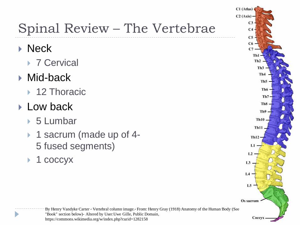
Spinal Review – The Vertebrae
Neck
7 Cervical
Mid-back
12 Thoracic
Low back
5 Lumbar
1 sacrum (made up of 4-
5 fused segments)
1 coccyx
By Henry Vandyke Carter - Vertebral column image.- From: Henry Gray (1918) Anatomy of the Human Body (See
"Book" section below)- Altered by User:Uwe Gille, Public Domain,
https://commons.wikimedia.org/w/index.php?curid=1282158
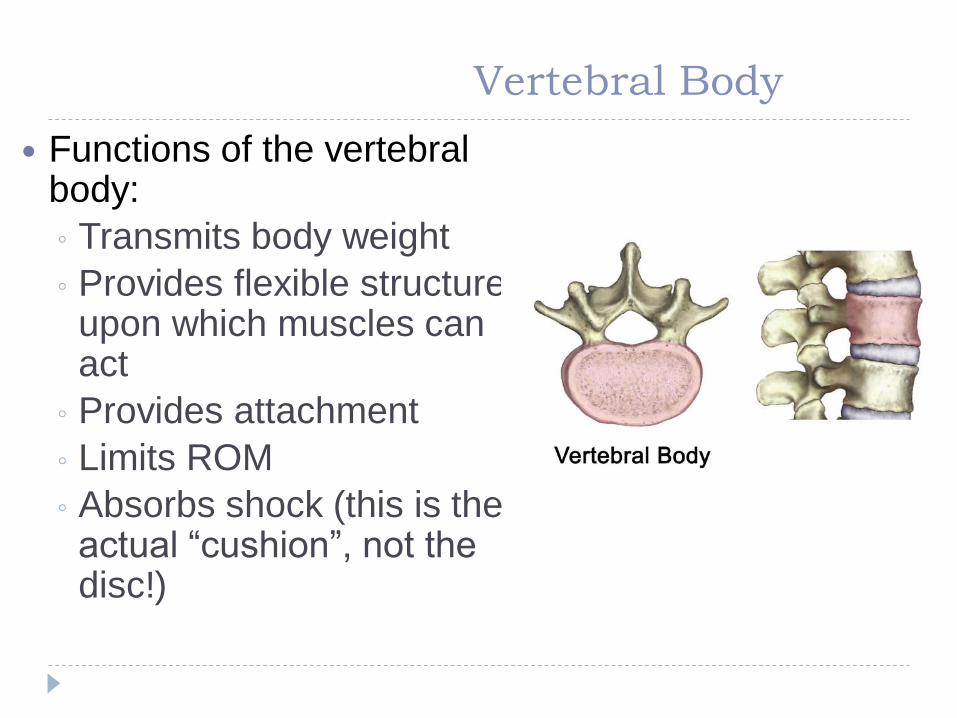
Vertebral Body
Functions of the vertebral body:
◦ Transmits body weight
◦ Provides flexible structure upon which muscles can act
◦ Provides attachment
◦ Limits ROM
◦ Absorbs shock (this is the actual “cushion”, not the disc!)
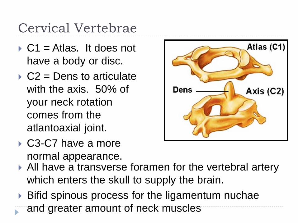
Cervical Vertebrae
C1 = Atlas. It does not
have a body or disc.
C2 = Dens to articulate
with the axis. 50% of
your neck rotation
comes from the
atlantoaxial joint.
C3-C7 have a more
normal appearance. All have a transverse foramen for the vertebral artery
which enters the skull to supply the brain.
Bifid spinous process for the ligamentum nuchae
and greater amount of neck muscles
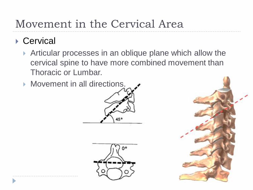
Movement in the Cervical Area
Cervical
Articular processes in an oblique plane which allow the
cervical spine to have more combined movement than
Thoracic or Lumbar.
Movement in all directions.
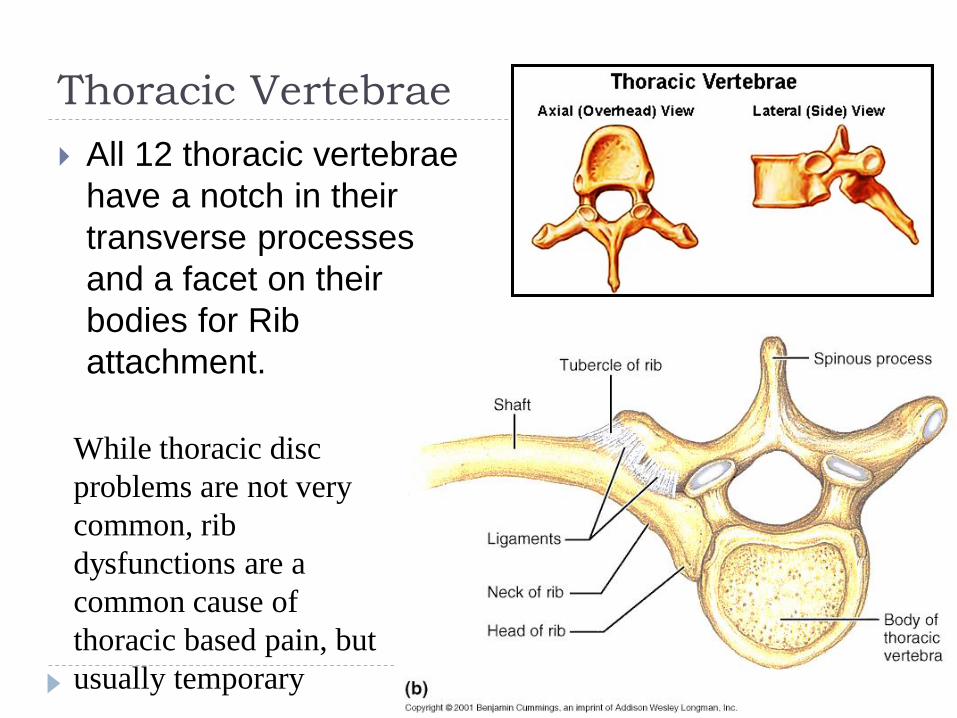
Thoracic Vertebrae
All 12 thoracic vertebrae
have a notch in their
transverse processes
and a facet on their
bodies for Rib
attachment.
While thoracic disc
problems are not very
common, rib
dysfunctions are a
common cause of
thoracic based pain, but
usually temporary
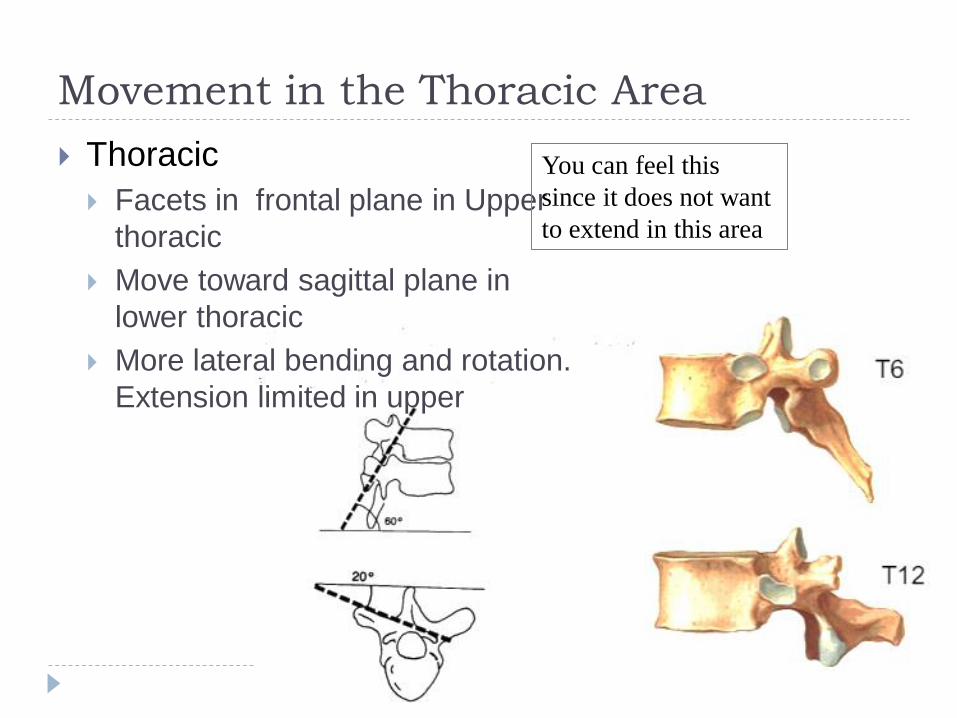
Movement in the Thoracic Area
Thoracic
Facets in frontal plane in Upper
thoracic
Move toward sagittal plane in
lower thoracic
More lateral bending and rotation.
Extension limited in upper
You can feel this
since it does not want
to extend in this area
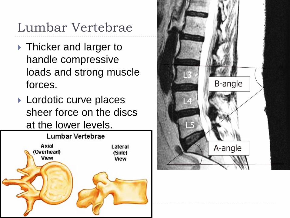
Lumbar Vertebrae
Thicker and larger to
handle compressive
loads and strong muscle
forces.
Lordotic curve places
sheer force on the discs
at the lower levels.
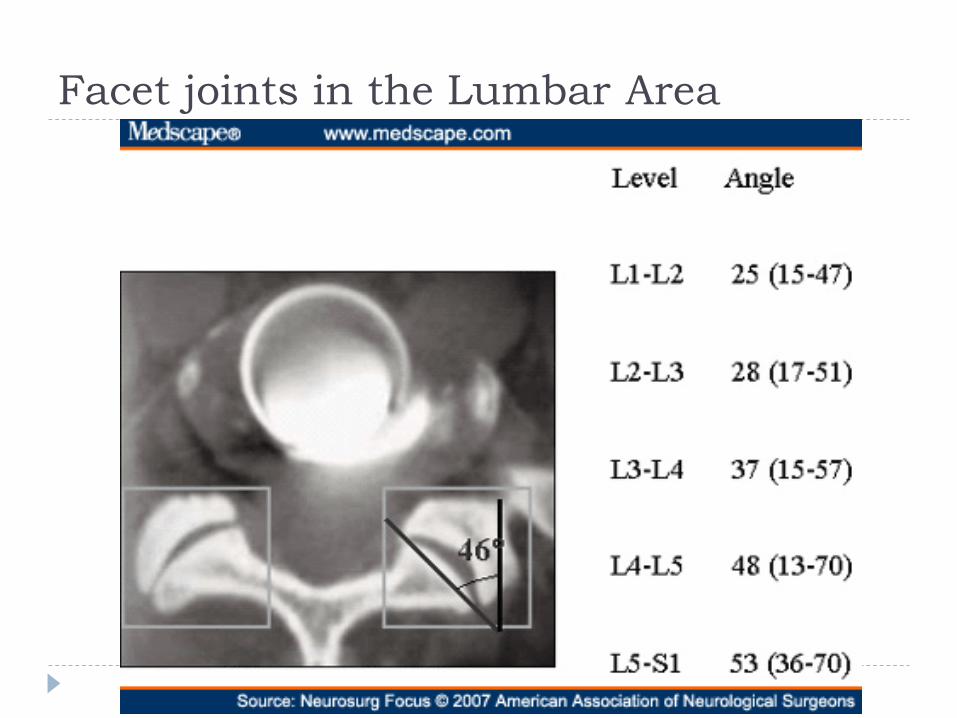
Facet joints in the Lumbar Area
Back Pain – What Kind?
There are a myriad of potential causes of back pain
depending on what level the pain is occurring. This
makes determining the actual cause quite difficult.
For this presentation I am going to focus on the 3
specific areas that I feel are the most common
issues I see.
1. Rib dysfunctions in the thoracic spine
2. Sacroiliac joint dysfunctions (SI joint)
3. Generalized low back pain
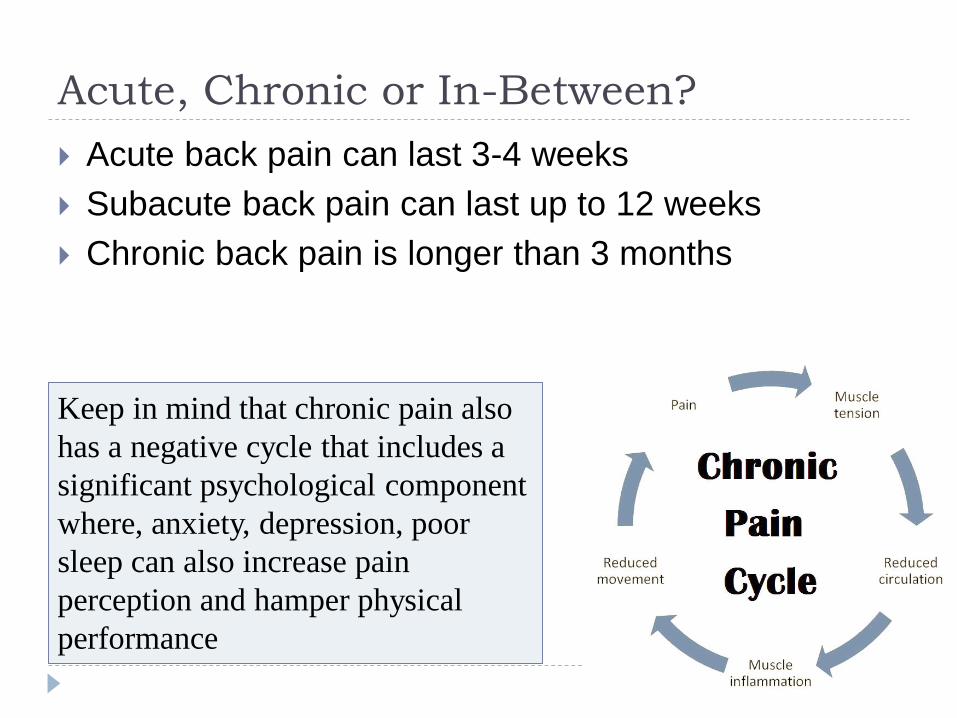
Acute, Chronic or In-Between?
Acute back pain can last 3-4 weeks
Subacute back pain can last up to 12 weeks
Chronic back pain is longer than 3 months
Keep in mind that chronic pain also
has a negative cycle that includes a
significant psychological component
where, anxiety, depression, poor
sleep can also increase pain
perception and hamper physical
performance
Potentially Serious?
A common Question I get is when should I actually
go see the Dr?
Pain and it’s characteristics vary greatly, but I feel it
is important for you to understand the difference
between what is called:
Mechanical Pain – also known as positional pain
Non Mechanical Pain – also known as non-positional pain
Mechanical Back Pain
Often Acute or sudden
onset
Damage or irritation to
Ligament
Muscle
Connective tissue
Facet joint (or bone)
Possible early Annular
damage to the disc
Usually gets worse over
the course of the day
Not directly nerve
related, but can radiate
down to the buttocks or
hips
Pain is usually cyclic
Pain is aggravated by a
specific direction or
movement (Positional)
Pain is relieved by lying
down, or a specific
movement or position
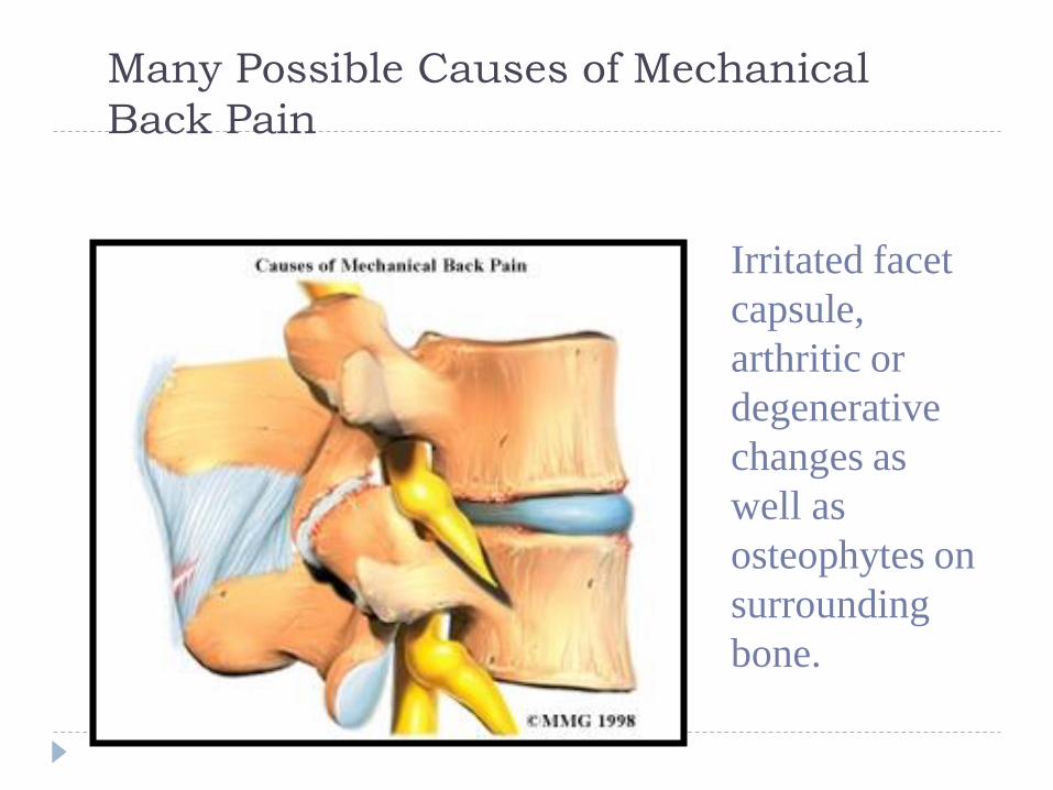
Many Possible Causes of Mechanical
Back Pain
Irritated facet
capsule,
arthritic or
degenerative
changes as
well as
osteophytes on
surrounding
bone.
Non-Mechanical Back Pain or Neurologic
Pain Often progressive and
insidious onset, but can
be acute.
Possible irritation to
Intervertebral disc
Nerve root
Internal organ
Random pain patterns
tend to worsen over
time
Sensory changes in the
saddle area or problems
with micturition should
be checked asap.
Nerve related can
radiate down to the
lower leg and foot
Pain usually
exacerbated by sitting
and better when
standing
Internal organ problem
(i.e. kidney stone)
creates vague achy
deep pain that does not
appear to have any
position that alleviates it.
(non-positional pain)

Referred Pain Patterns
By OpenStax College - Autonomic Reflexes and Homeostasis http://cnx.org/content/m46579/1.2/, CC BY
3.0, https://commons.wikimedia.org/w/index.php?curid=30017359
When do I see a Doc?
Athletes should be reporting any and all back pain
Serious issues for referring the athlete (or yourself)
include:
Radiating pain or numbness (especially if going below the
knee)
Non-positional pain (may indicate internal organ referred
pain)
Pain or numbness in the saddle area
Noticeable and explainable changes in micturition
What is a Rib Dysfunction?
Usually an acute onset of mechanical pain
More commonly found between the scapulae
Pain is usually localized in the back, but the right
kind of rib dysfunction can radiate pain around the
rib toward the sternum
Rib dysfunctions can exist without “back pain” and
be evident in respiratory restrictions.
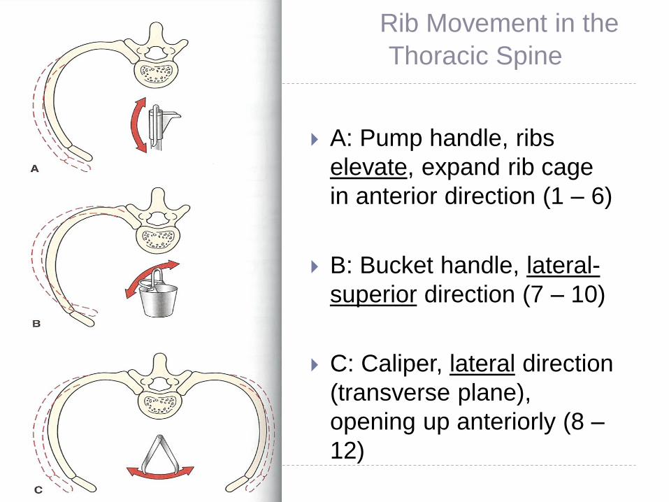
Rib Movement in the
Thoracic Spine
A: Pump handle, ribs
elevate, expand rib cage
in anterior direction (1 – 6)
B: Bucket handle, lateral-
superior direction (7 – 10)
C: Caliper, lateral direction
(transverse plane),
opening up anteriorly (8 –
12)
Rib Motions
Primary Motions: Inhalation and Exhalation
Pump handle motion (major movement in upper 6 ribs)
Bucket handle motion (major movement in below rib 6)
Caliper motion of Rib 11 and 12
Torsional movement
When T5 rotates to the right in relation to T6, the posterior
aspect of the right 6th rib turns externally and the posterior
aspect of the left 6th rib turns internally.
Greenman’s Principles of Manual Medicine, 4th ed., pg 256.
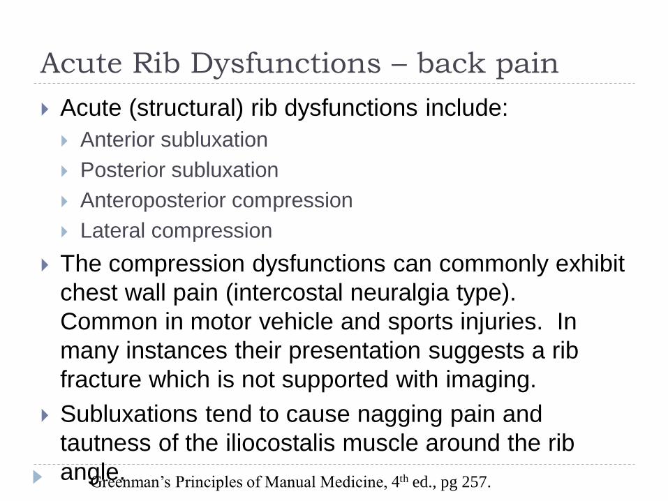
Acute Rib Dysfunctions – back pain
Acute (structural) rib dysfunctions include:
Anterior subluxation
Posterior subluxation
Anteroposterior compression
Lateral compression
The compression dysfunctions can commonly exhibit
chest wall pain (intercostal neuralgia type).
Common in motor vehicle and sports injuries. In
many instances their presentation suggests a rib
fracture which is not supported with imaging.
Subluxations tend to cause nagging pain and
tautness of the iliocostalis muscle around the rib
angle.Greenman’s Principles of Manual Medicine, 4th ed., pg 257.
Final Rib Thoughts
1st rib issues can cause pretty significant lower neck
pain.
2nd rib issues can be a pain in the neck but also risk
pain down the arm due to it’s close proximity to the
brachial plexus.
Thoracic mobility is vitally important to normal rib
function
Repeated subluxations or pain in the same rib region
point to musculoskeletal imbalance (look at scapula
motion, shoulder range of motion and posture to
identify tight vs weak muscle groups.
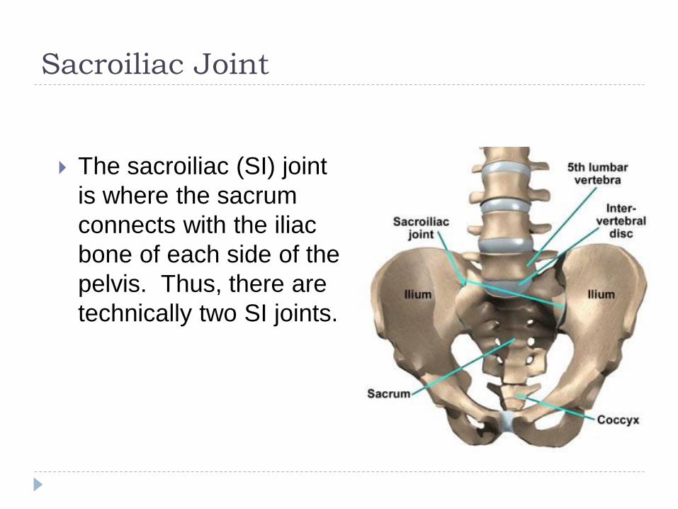
Sacroiliac Joint
The sacroiliac (SI) joint
is where the sacrum
connects with the iliac
bone of each side of the
pelvis. Thus, there are
technically two SI joints.
SI Joint continued
There can be two distinct problems with SI joint.
Sacroiliac problem– where the sacrum is not positioned or
moving correctly on the innominate (1 pelvic side or ilia
bone).
Iliosacral problem– where the innominate is not moving
properly on the sacrum
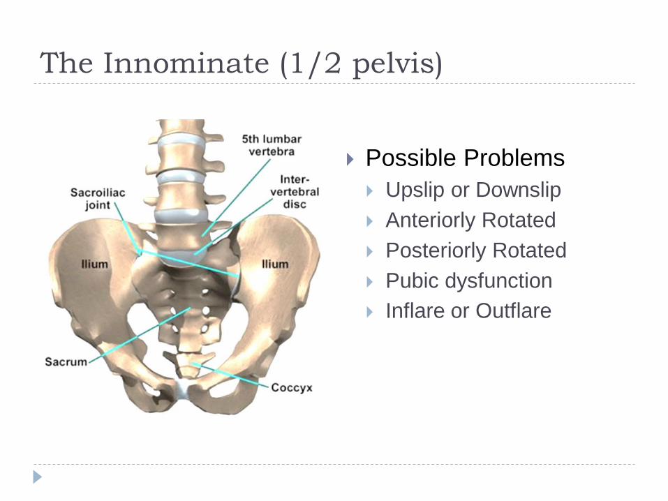
The Innominate (1/2 pelvis)
Possible Problems
Upslip or Downslip
Anteriorly Rotated
Posteriorly Rotated
Pubic dysfunction
Inflare or Outflare
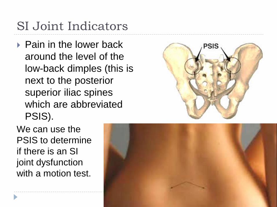
SI Joint Indicators
Pain in the lower back
around the level of the
low-back dimples (this is
next to the posterior
superior iliac spines
which are abbreviated
PSIS).
We can use the
PSIS to determine
if there is an SI
joint dysfunction
with a motion test.
SI Joint Indicators
Most SI joint problems
cause discomfort on
either the Right or Left
side, not commonly
both.
Mechanical in nature
where movement into
flexion or extension will
hurt more.
Athlete can perform
most activities but risk
of more mechanical
disruption is possible
(sacrum or spine)
Most athletic trainers
are aware of Innominate
assessment and basic
fixes, but seeing the
athlete repeatedly
means the primary
problem is not getting
fixed.
The sacrum or lower
lumbar area could be
involved and commonly
increase pain that could
be across the low back
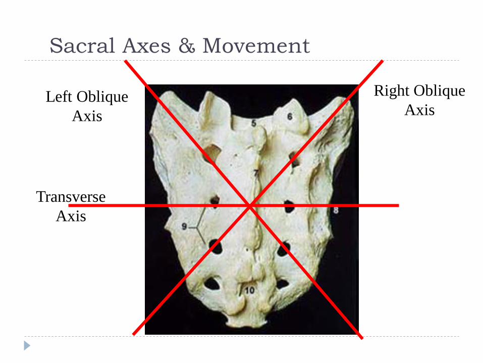
Sacral Axes & Movement
Right Oblique
AxisLeft Oblique
Axis
Transverse
Axis
Lumbar Spine vs. Sacrum
When the lumbar spine flexes, the sacrum posteriorly nutates (extends)
When the lumbar spine extends, the sacrum anteriorly nutates (flexes)
Abnormal mechanics in the lumbar spine can negatively affect sacral movement.
Abnormal sacral position directly affects the pelvis and proper pelvic motion.
A lumbar or sacral problem could produce SI joint pain and dysfunction. If so, simply fixing the innominate is not going to fix the problem. You need to be able to fix the lumbar and sacral problem.
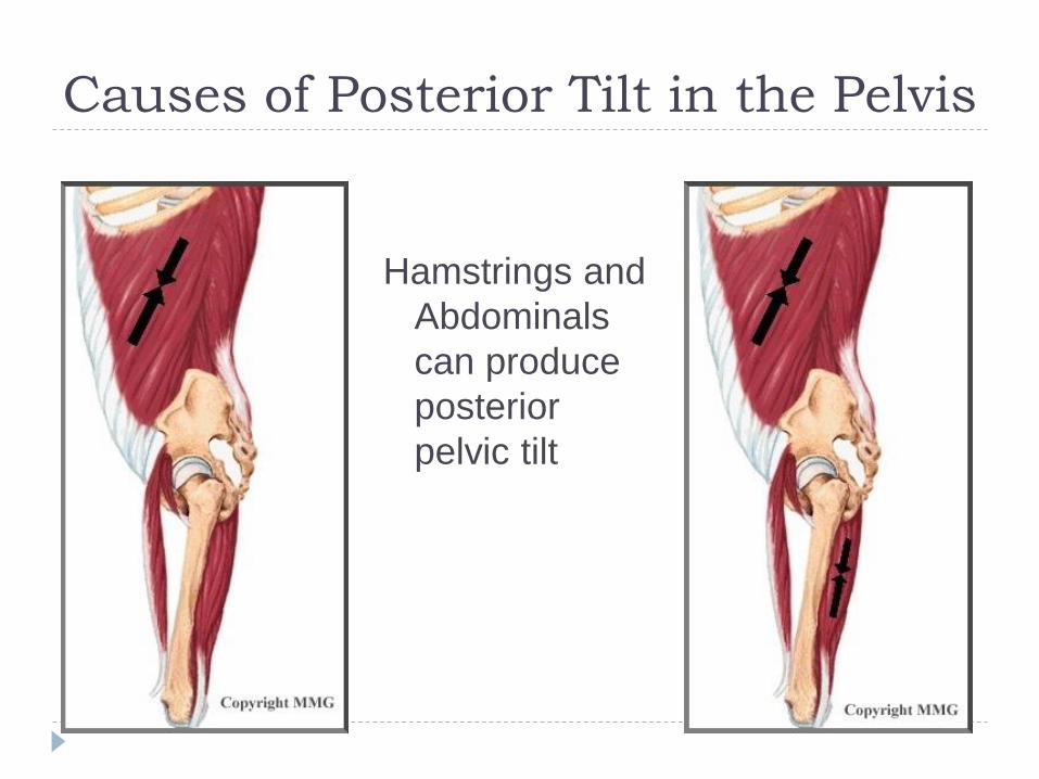
Causes of Posterior Tilt in the Pelvis
Hamstrings and
Abdominals
can produce
posterior
pelvic tilt
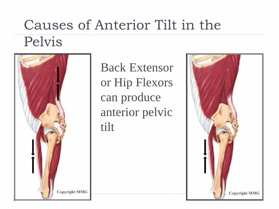
Causes of Anterior Tilt in the
Pelvis
Back Extensor
or Hip Flexors
can produce
anterior pelvic
tilt
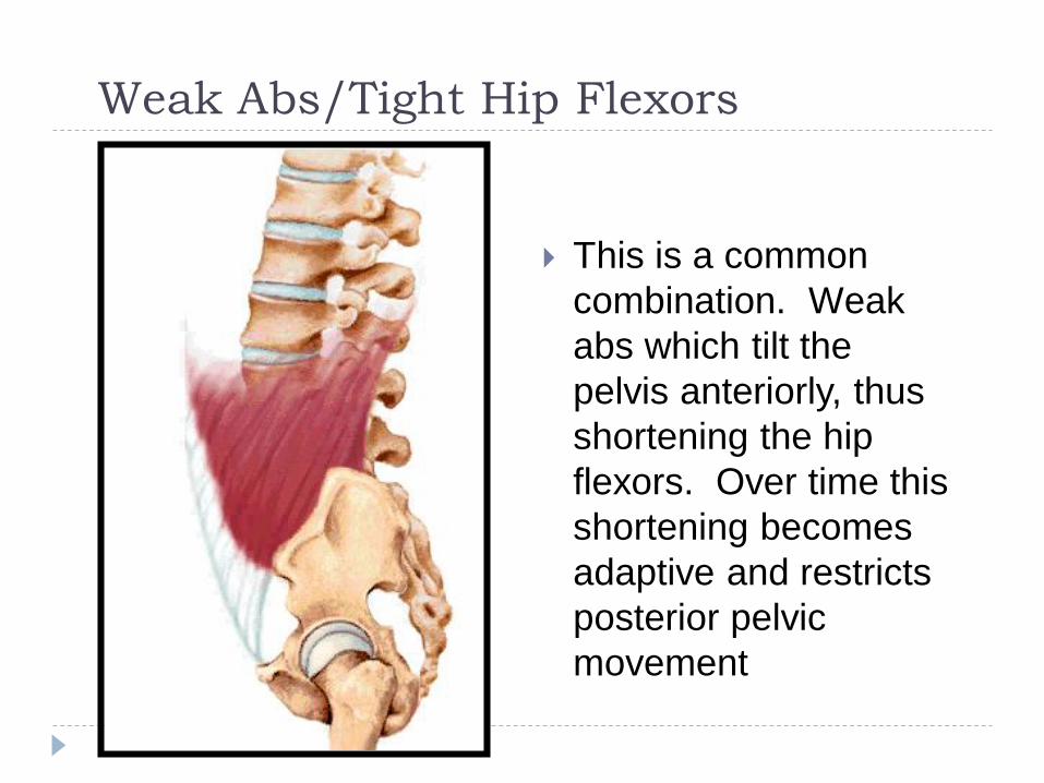
Weak Abs/Tight Hip Flexors
This is a common
combination. Weak
abs which tilt the
pelvis anteriorly, thus
shortening the hip
flexors. Over time this
shortening becomes
adaptive and restricts
posterior pelvic
movement
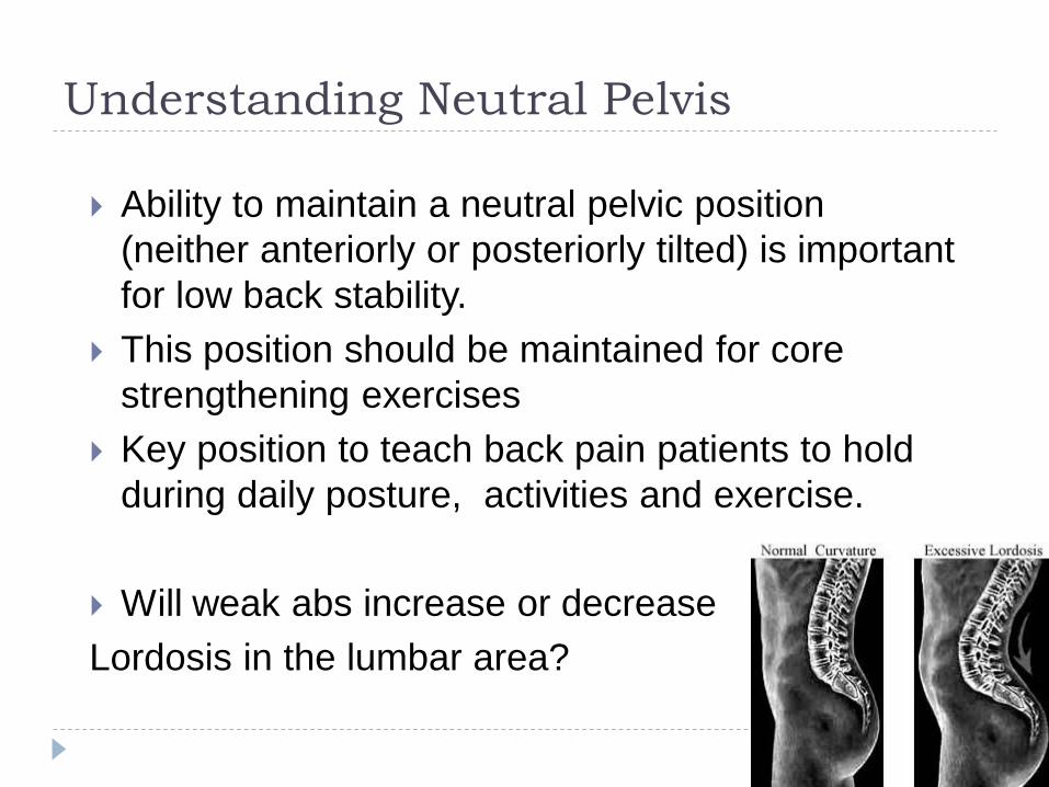
Understanding Neutral Pelvis
Ability to maintain a neutral pelvic position
(neither anteriorly or posteriorly tilted) is important
for low back stability.
This position should be maintained for core
strengthening exercises
Key position to teach back pain patients to hold
during daily posture, activities and exercise.
Will weak abs increase or decrease
Lordosis in the lumbar area?
SI Joint Exercises
These are subtle exercises to emphasize SI joint
movement without multiple joints involved which can
compensate for SI immobility.
Hip ER stretch with core hold
Supine Knee toward Chest (hold with isometric hip
extension)
Supine core hold, knees bent to 90, feet on floor, ball
squeeze with core hold 5-10 seconds
Same as above but theraband resistance for
abduction while holding core. Gradual progression
on distance into abduction.
Nuttal diagonal reaching exercise.
Low Back Pain Epidemic
Up to 80% of low back pain is considered to be
idiopathic (actual cause or origin unknown)
In the general population this is the most common
form of back pain.
According to a 2006 review (Katz), total costs
associated with LBP in the United States exceed
$100 billion per year, two-thirds of which are a result
of lost wages and reduced productivity.
Katz JN. Lumbar disc disorders and low-back pain: socioeconomic factors and consequences [review]. J
Bone Joint Surg Am. 2006;88(suppl 2): 21-24.
Low Back Pain in Athletes
Generalized back pain is still the most common form
of back pain in college athletes.
Based on a review of numerous publications (US
and international), the more common forms of
diagnosed or specific back pain is spondylolysis
(with some accompanying spondylolisthesis),
discogenic, facet impingement and SI joint.
Let’s quickly take a look at what these terms mean.
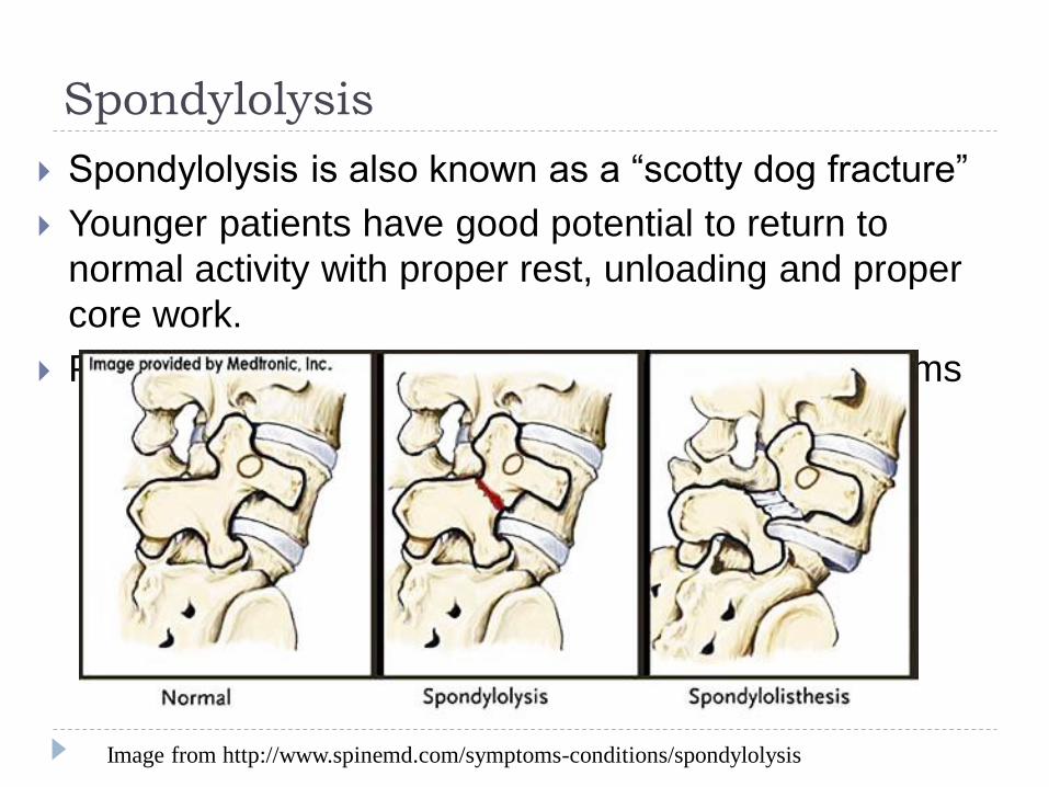
Spondylolysis
Spondylolysis is also known as a “scotty dog fracture”
Younger patients have good potential to return to
normal activity with proper rest, unloading and proper
core work.
Progression leads to disc and degenerative problems
Image from http://www.spinemd.com/symptoms-conditions/spondylolysis
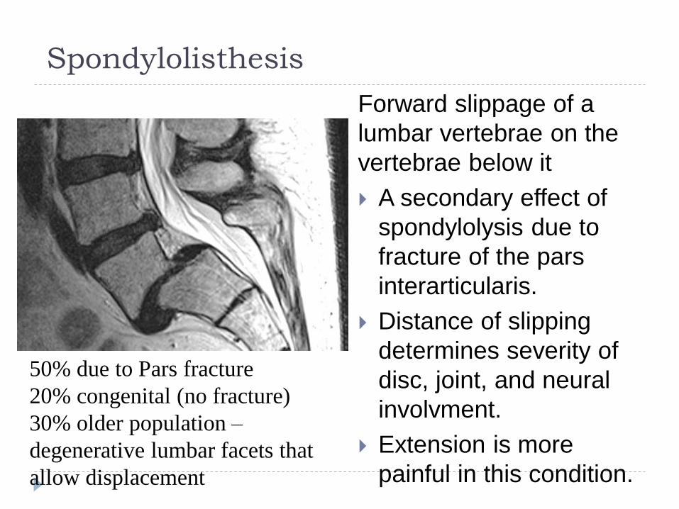
Spondylolisthesis
Forward slippage of a
lumbar vertebrae on the
vertebrae below it
A secondary effect of
spondylolysis due to
fracture of the pars
interarticularis.
Distance of slipping
determines severity of
disc, joint, and neural
involvment.
Extension is more
painful in this condition.
50% due to Pars fracture
20% congenital (no fracture)
30% older population –
degenerative lumbar facets that
allow displacement
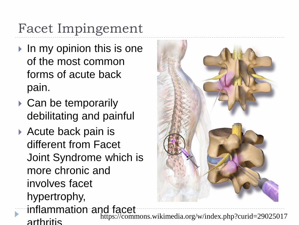
Facet Impingement
In my opinion this is one
of the most common
forms of acute back
pain.
Can be temporarily
debilitating and painful
Acute back pain is
different from Facet
Joint Syndrome which is
more chronic and
involves facet
hypertrophy,
inflammation and facet
arthritis.https://commons.wikimedia.org/w/index.php?curid=29025017
Facet Impingement
The facet joint has a
capsule that can
sometimes get
“impinged” or pinched
between the 2 articular
surfaces.
When this happens it
will get inflamed and be
painful. It will also
restrict motion at that
joint and make it difficult
to open or close the
facet joint.
https://www.youtube.co
m/watch?v=9w6NiPc8B
p8
Facet Joints
Acute pain (lasting up to
7 days) and subacute
pain (up to 3 months)
seem related to capsule
damage, and chronic
pain is probably related
to osteoarthritis.
Disc degeneration and
facet joint stress go
hand in hand
Facet Joints help guide
the proper motion for
the spinal segment
Important for helping
carry the load on the
spine and an important
resisting shear loads in
the lumbar spine.
Joint capsules have
their own nerve
innervation which adds
to pain syndromesBiomechanics of the Posterior Lumbar Articulating Elements
Hassan A. Serhan, Ph.D.1; Gus Varnavas, M.D.2; Andrew P. Dooris, Ph.D.1; Avinash Patwardhan, Ph.D.3;
Michael Tzermiadianos, M.D.3 , Neurosurg Focus. 2007;22(1)
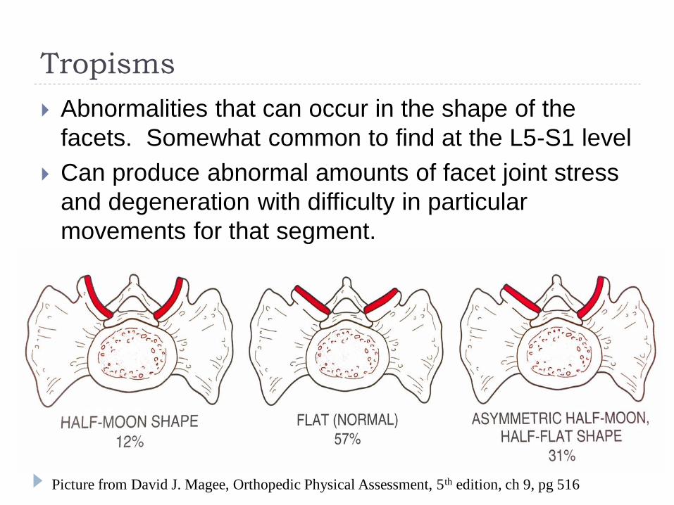
Tropisms
Abnormalities that can occur in the shape of the
facets. Somewhat common to find at the L5-S1 level
Can produce abnormal amounts of facet joint stress
and degeneration with difficulty in particular
movements for that segment.
Picture from David J. Magee, Orthopedic Physical Assessment, 5th edition, ch 9, pg 516
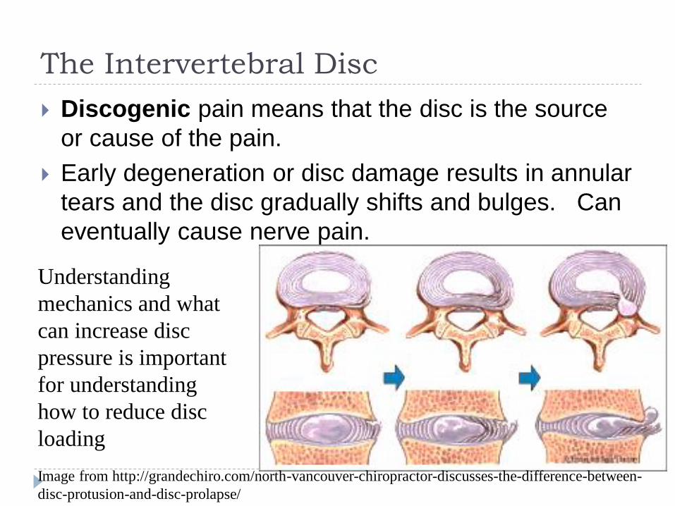
The Intervertebral Disc
Discogenic pain means that the disc is the source
or cause of the pain.
Early degeneration or disc damage results in annular
tears and the disc gradually shifts and bulges. Can
eventually cause nerve pain.
Image from http://grandechiro.com/north-vancouver-chiropractor-discusses-the-difference-between-
disc-protusion-and-disc-prolapse/
Understanding
mechanics and what
can increase disc
pressure is important
for understanding
how to reduce disc
loading
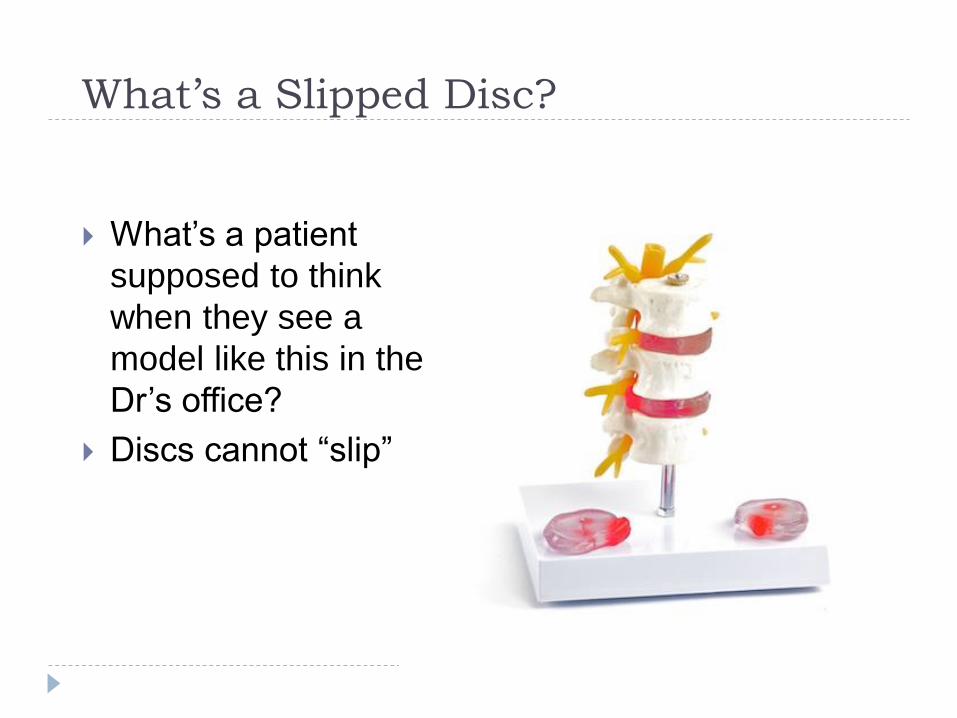
What’s a Slipped Disc?
What’s a patient
supposed to think
when they see a
model like this in the
Dr’s office?
Discs cannot “slip”
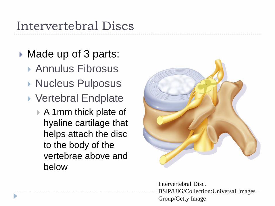
Intervertebral Discs
Made up of 3 parts:
Annulus Fibrosus
Nucleus Pulposus
Vertebral Endplate
A 1mm thick plate of
hyaline cartilage that
helps attach the disc
to the body of the
vertebrae above and
below
Intervertebral Disc.
BSIP/UIG/Collection:Universal Images
Group/Getty Image
The Intervertebral Discs
Nucleus Pulposus
- is the central portion of the disc (except L-spine; post.)
- it is a loose collagen fibril network contained within an extensive gelatinous matrix (primarily Type II collagen)
- at birth the nucleus contains a high portion of proteoglycans (made up of glyscosoaminoglycans; GAGs), decreases with age and is replaced by collagen (degeneration begins to occur after age 20)
- imbibing properties of the proteoglycans lead to nucleus’ % water: at birth 85%, 6th decade 65%
The Intervertebral Discs
Nucleus Pulposus Functions:
- Imbibition (taking up and holding fluid); if released from
confining annulus, is able to swell up 200-300% in hours!
- Transmission of force: its incompressibility is responsible
for transmitting much weight across the spinal segment
- Equalization of stress: hydrostatic property of transmitting forces equally in all directions
- Movement: provides “rocking” action to movement
Nutrition: only the periphery of the disc is vascularized, receives nourishment via diffusion from the periphery of the annulus and the vertebral endplate
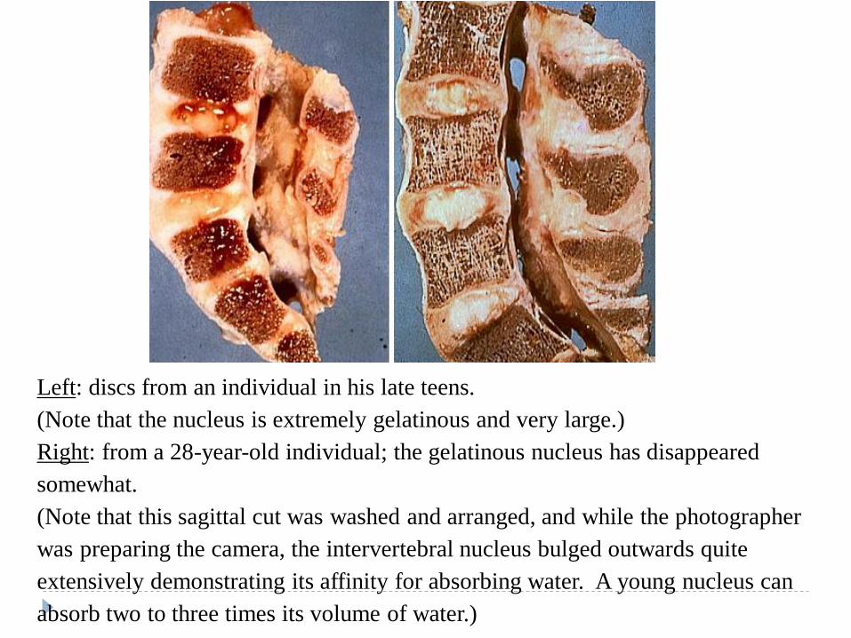
Left: discs from an individual in his late teens.
(Note that the nucleus is extremely gelatinous and very large.)
Right: from a 28-year-old individual; the gelatinous nucleus has disappeared
somewhat.
(Note that this sagittal cut was washed and arranged, and while the photographer
was preparing the camera, the intervertebral nucleus bulged outwards quite
extensively demonstrating its affinity for absorbing water. A young nucleus can
absorb two to three times its volume of water.)
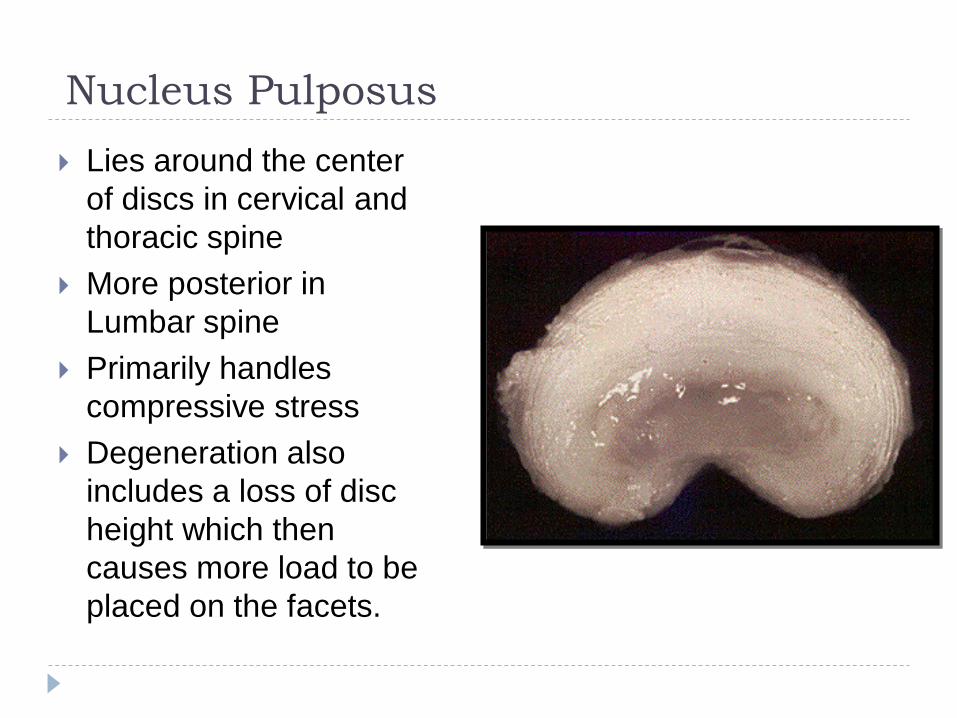
Nucleus Pulposus
Lies around the center
of discs in cervical and
thoracic spine
More posterior in
Lumbar spine
Primarily handles
compressive stress
Degeneration also
includes a loss of disc
height which then
causes more load to be
placed on the facets.
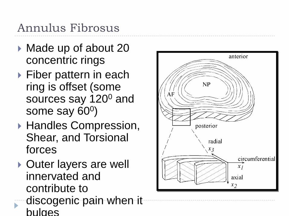
Annulus Fibrosus
Made up of about 20 concentric rings
Fiber pattern in each ring is offset (some sources say 1200 and some say 600)
Handles Compression, Shear, and Torsional forces
Outer layers are well innervated and contribute to discogenic pain when it bulges
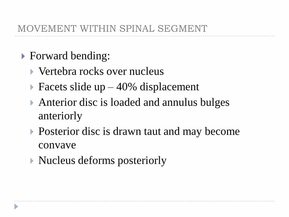
MOVEMENT WITHIN SPINAL SEGMENT
Forward bending:
Vertebra rocks over nucleus
Facets slide up – 40% displacement
Anterior disc is loaded and annulus bulges
anteriorly
Posterior disc is drawn taut and may become
convave
Nucleus deforms posteriorly
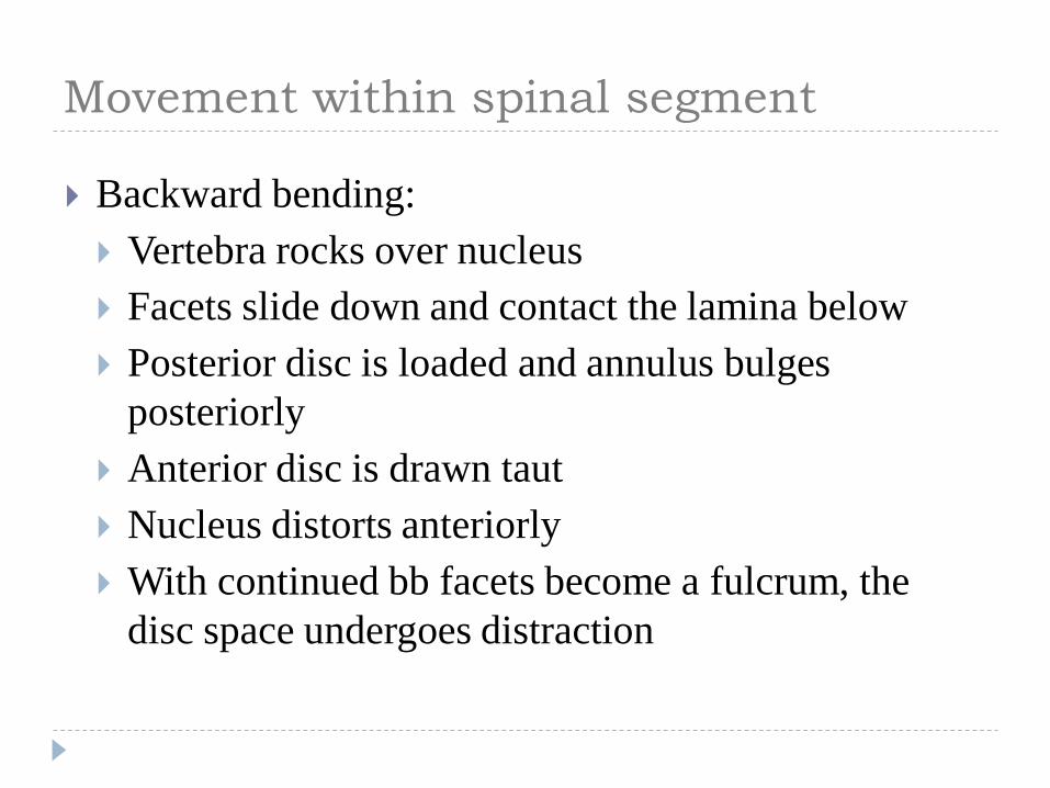
Movement within spinal segment
Backward bending:
Vertebra rocks over nucleus
Facets slide down and contact the lamina below
Posterior disc is loaded and annulus bulges
posteriorly
Anterior disc is drawn taut
Nucleus distorts anteriorly
With continued bb facets become a fulcrum, the
disc space undergoes distraction
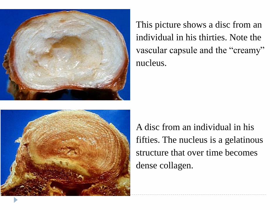
This picture shows a disc from an
individual in his thirties. Note the
vascular capsule and the “creamy”
nucleus.
A disc from an individual in his
fifties. The nucleus is a gelatinous
structure that over time becomes
dense collagen.
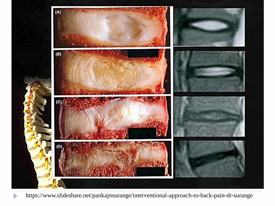
https://www.slideshare.net/pankajnsurange/interventional-approach-to-back-pain-dr-surange
Questions
What is the most detrimental movement for the
disc?
Functions of the nucleus pulposus
What “feature” makes the nucleus
incompressible?
What pathology could be associated with a
fracture of the vertebral endplate
(superior/inferior direction)?
Lumbar pain
Lumbar pain presents in the bulk of the paraspinal or
low back musculature. It is often unilateral but under
certain circumstances can be bilateral.
A decent amount of flexion and extension available
as well as rotation.
Sidebending better at top portion and limited in lower
Clinically I use sidebending as quick screen for
lumbar dysfunction (especially when I am looking at
an SI joint problem). Normally sidebending will
produce a nice C or reverse C shape (depending on
which side you lean), but lumbar issues will normally
cause this to decrease and the lumbar area will stay
straight.
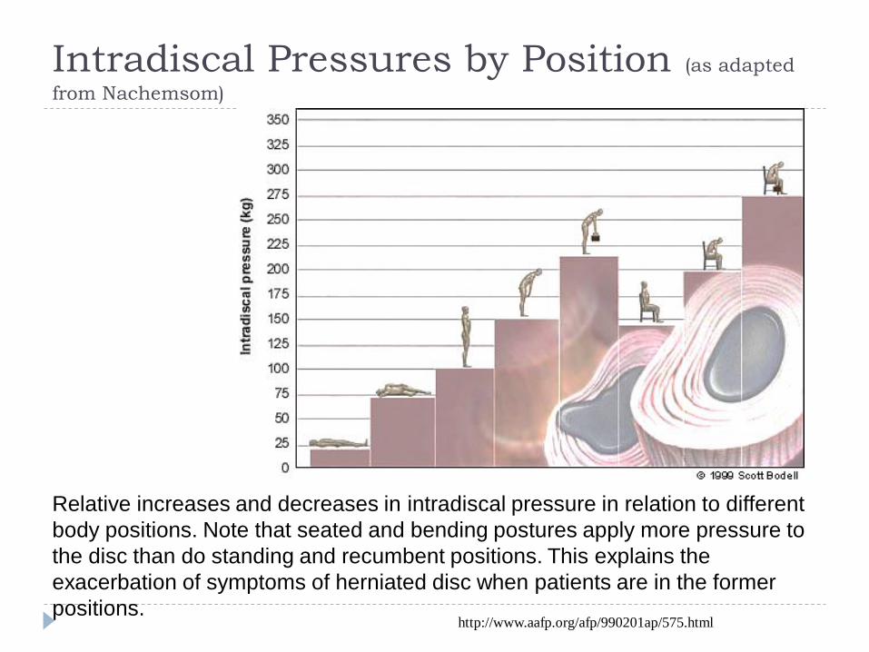
Intradiscal Pressures by Position (as adapted
from Nachemsom)
Relative increases and decreases in intradiscal pressure in relation to different
body positions. Note that seated and bending postures apply more pressure to
the disc than do standing and recumbent positions. This explains the
exacerbation of symptoms of herniated disc when patients are in the former
positions.http://www.aafp.org/afp/990201ap/575.html
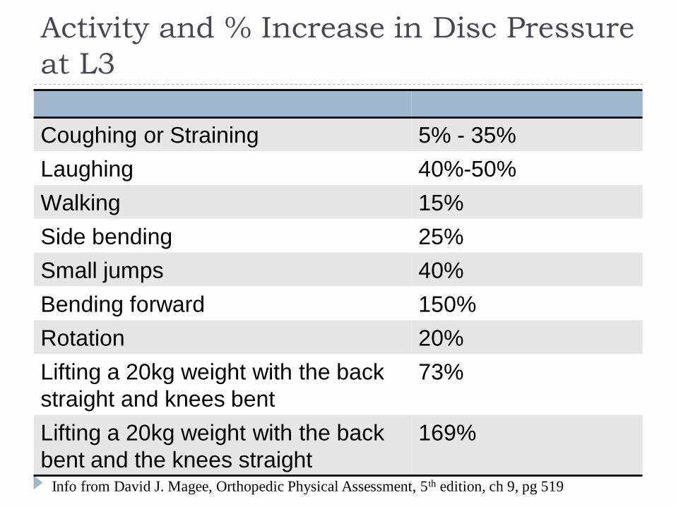
Activity and % Increase in Disc Pressure
at L3
Coughing or Straining 5% - 35%
Laughing 40%-50%
Walking 15%
Side bending 25%
Small jumps 40%
Bending forward 150%
Rotation 20%
Lifting a 20kg weight with the back
straight and knees bent
73%
Lifting a 20kg weight with the back
bent and the knees straight
169%
Info from David J. Magee, Orthopedic Physical Assessment, 5th edition, ch 9, pg 519
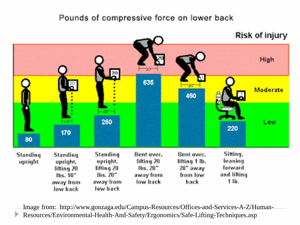
Image from: http://www.gonzaga.edu/Campus-Resources/Offices-and-Services-A-Z/Human-
Resources/Environmental-Health-And-Safety/Ergonomics/Safe-Lifting-Techniques.asp
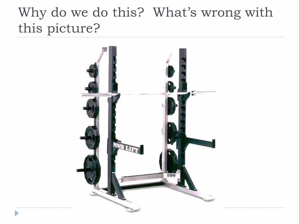
Why do we do this? What’s wrong with
this picture?
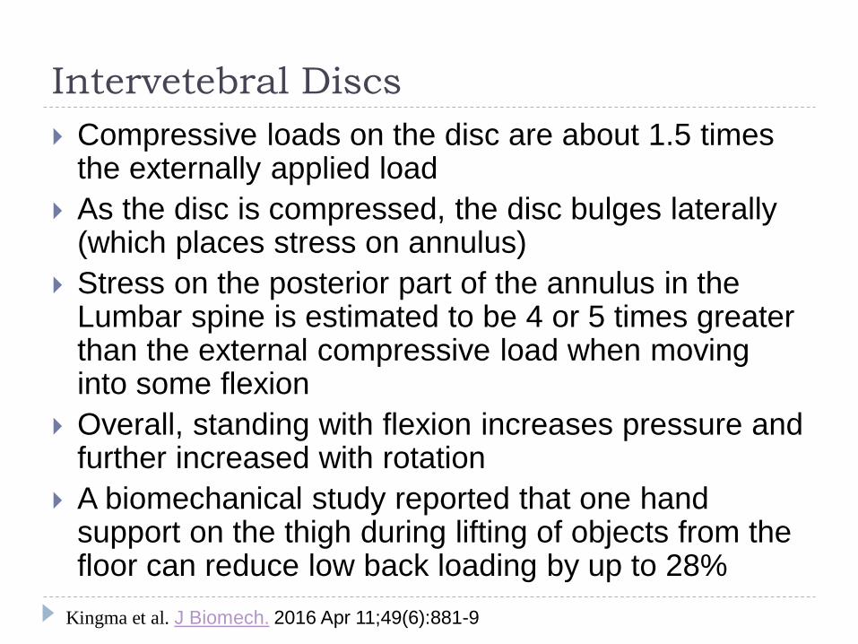
Intervetebral Discs
Compressive loads on the disc are about 1.5 times the externally applied load
As the disc is compressed, the disc bulges laterally (which places stress on annulus)
Stress on the posterior part of the annulus in the Lumbar spine is estimated to be 4 or 5 times greater than the external compressive load when moving into some flexion
Overall, standing with flexion increases pressure and further increased with rotation
A biomechanical study reported that one hand support on the thigh during lifting of objects from the floor can reduce low back loading by up to 28%
Kingma et al. J Biomech. 2016 Apr 11;49(6):881-9
4 Factors That Increase Load on
the Spine- when carrying an object
1. Position of the object relative to the center of
load on spine
2. Size, shape, weight, density of object
3. Degree of flexion and rotation of spine
4. Rate of loading
The Importance of Abs
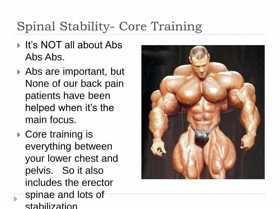
Spinal Stability- Core Training
It’s NOT all about Abs
Abs Abs.
Abs are important, but
None of our back pain
patients have been
helped when it’s the
main focus.
Core training is
everything between
your lower chest and
pelvis. So it also
includes the erector
spinae and lots of
stabilization
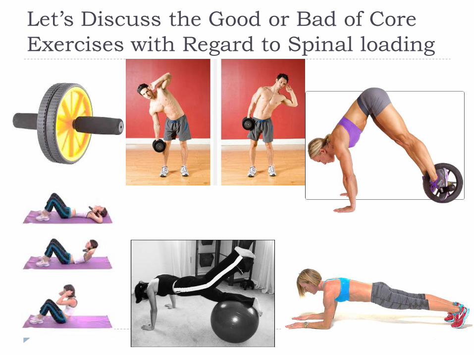
Let’s Discuss the Good or Bad of Core
Exercises with Regard to Spinal loading
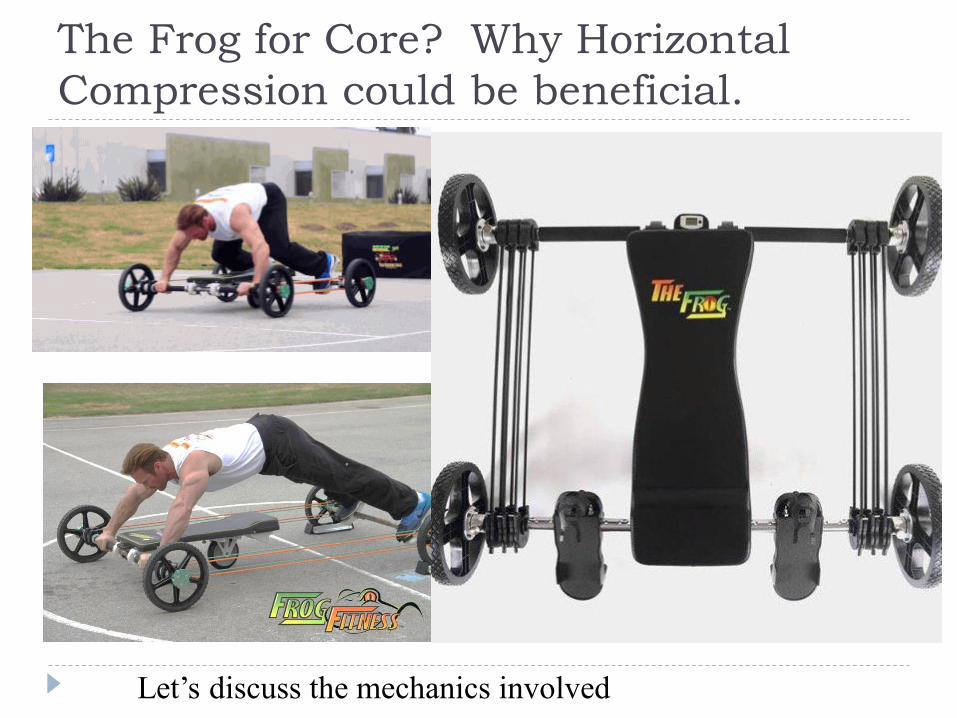
The Frog for Core? Why Horizontal
Compression could be beneficial.
Let’s discuss the mechanics involved
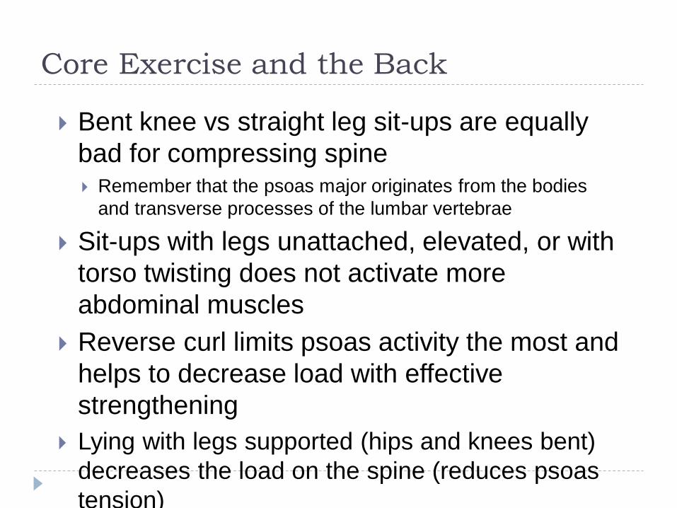
Core Exercise and the Back
Bent knee vs straight leg sit-ups are equally
bad for compressing spine Remember that the psoas major originates from the bodies
and transverse processes of the lumbar vertebrae
Sit-ups with legs unattached, elevated, or with
torso twisting does not activate more
abdominal muscles
Reverse curl limits psoas activity the most and
helps to decrease load with effective
strengthening
Lying with legs supported (hips and knees bent)
decreases the load on the spine (reduces psoas
tension)
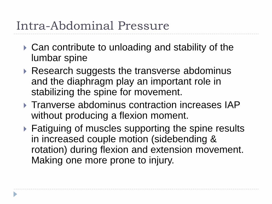
Intra-Abdominal Pressure
Can contribute to unloading and stability of the lumbar spine
Research suggests the transverse abdominus and the diaphragm play an important role in stabilizing the spine for movement.
Tranverse abdominus contraction increases IAP without producing a flexion moment.
Fatiguing of muscles supporting the spine results in increased couple motion (sidebending & rotation) during flexion and extension movement. Making one more prone to injury.
Exercises I think are problematic
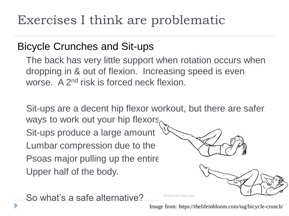
Bicycle Crunches and Sit-ups
The back has very little support when rotation occurs when
dropping in & out of flexion. Increasing speed is even
worse. A 2nd risk is forced neck flexion.
Sit-ups are a decent hip flexor workout, but there are safer
ways to work out your hip flexors!
Sit-ups produce a large amount
Lumbar compression due to the
Psoas major pulling up the entire
Upper half of the body.
So what’s a safe alternative?Image from: https://thelifeinbloom.com/tag/bicycle-crunch/
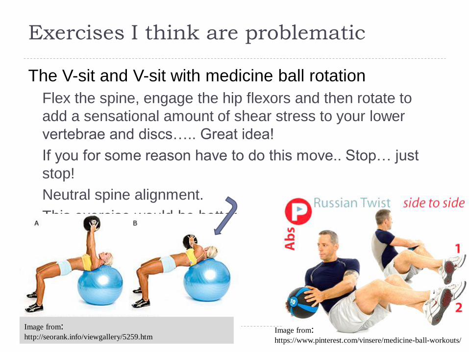
Exercises I think are problematic
The V-sit and V-sit with medicine ball rotation
Flex the spine, engage the hip flexors and then rotate to
add a sensational amount of shear stress to your lower
vertebrae and discs….. Great idea!
If you for some reason have to do this move.. Stop… just
stop!
Neutral spine alignment.
This exercise would be better
Image from: https://www.pinterest.com/vinsere/medicine-ball-workouts/
Image from: http://seorank.info/viewgallery/5259.htm
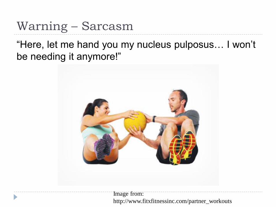
Warning – Sarcasm
“Here, let me hand you my nucleus pulposus… I won’t
be needing it anymore!”
Image from:
http://www.fitxfitnessinc.com/partner_workouts
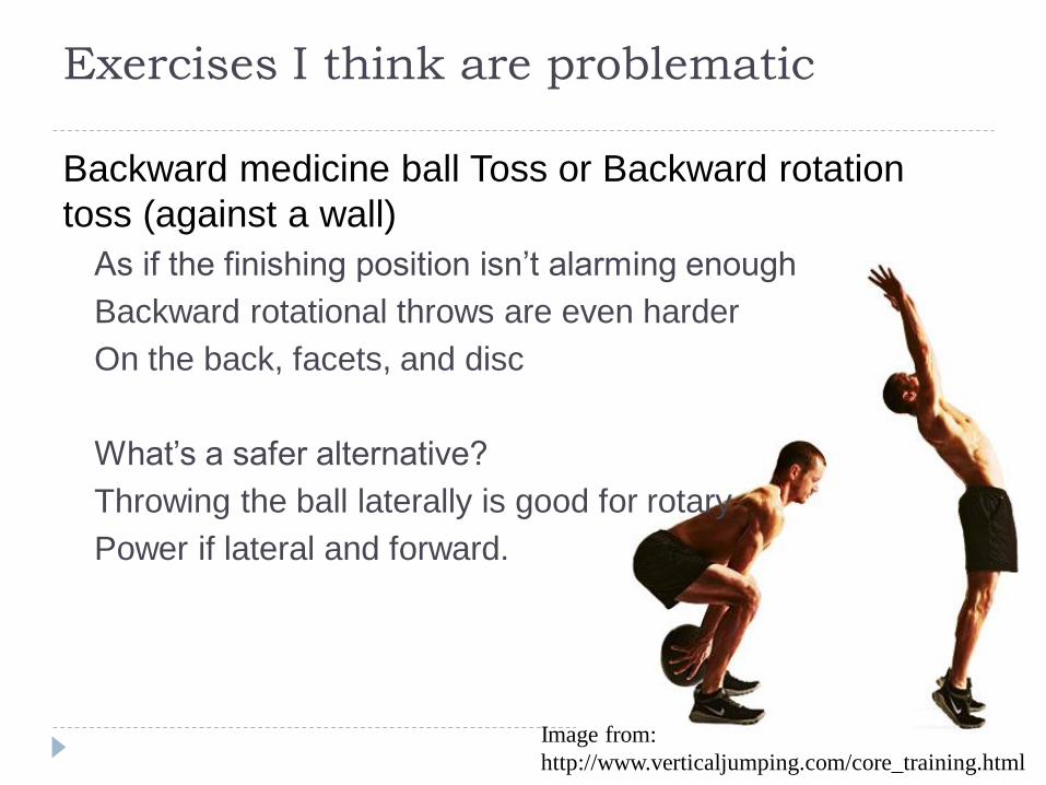
Exercises I think are problematic
Backward medicine ball Toss or Backward rotation
toss (against a wall)
As if the finishing position isn’t alarming enough
Backward rotational throws are even harder
On the back, facets, and disc
What’s a safer alternative?
Throwing the ball laterally is good for rotary
Power if lateral and forward.
Image from:
http://www.verticaljumping.com/core_training.html
Exercises I think are problematic
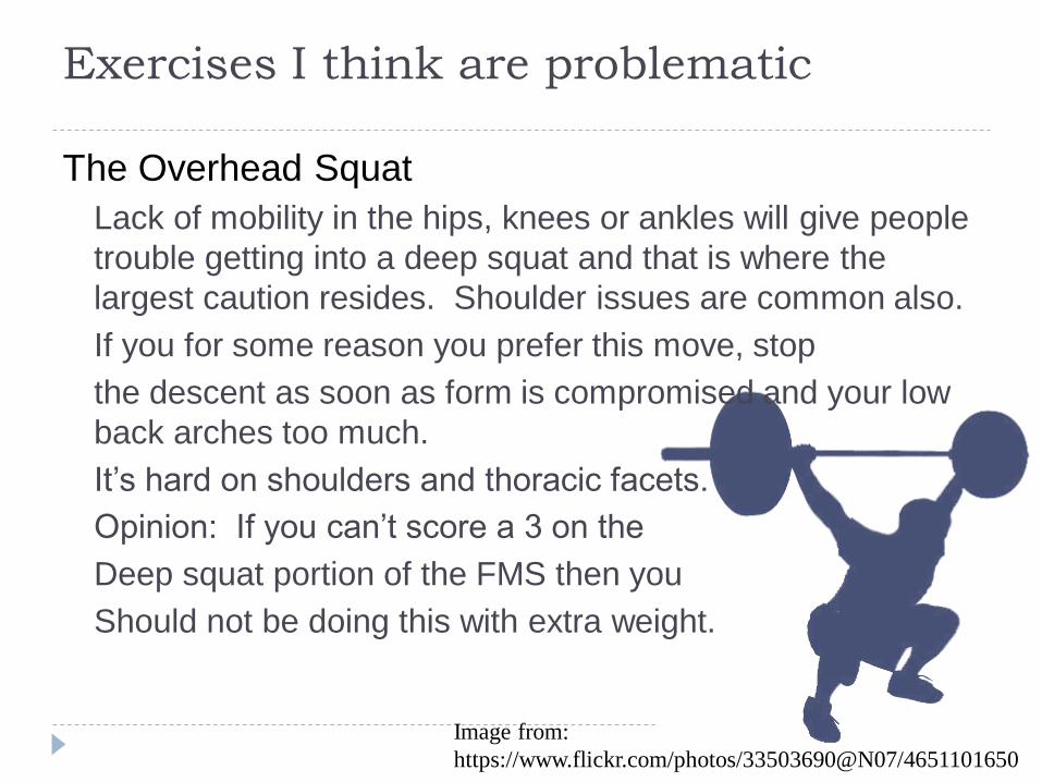
The Overhead Squat
Lack of mobility in the hips, knees or ankles will give people
trouble getting into a deep squat and that is where the
largest caution resides. Shoulder issues are common also.
If you for some reason you prefer this move, stop
the descent as soon as form is compromised and your low
back arches too much.
It’s hard on shoulders and thoracic facets.
Opinion: If you can’t score a 3 on the
Deep squat portion of the FMS then you
Should not be doing this with extra weight.
Image from:
https://www.flickr.com/photos/33503690@N07/4651101650
Exercises I think are problematic
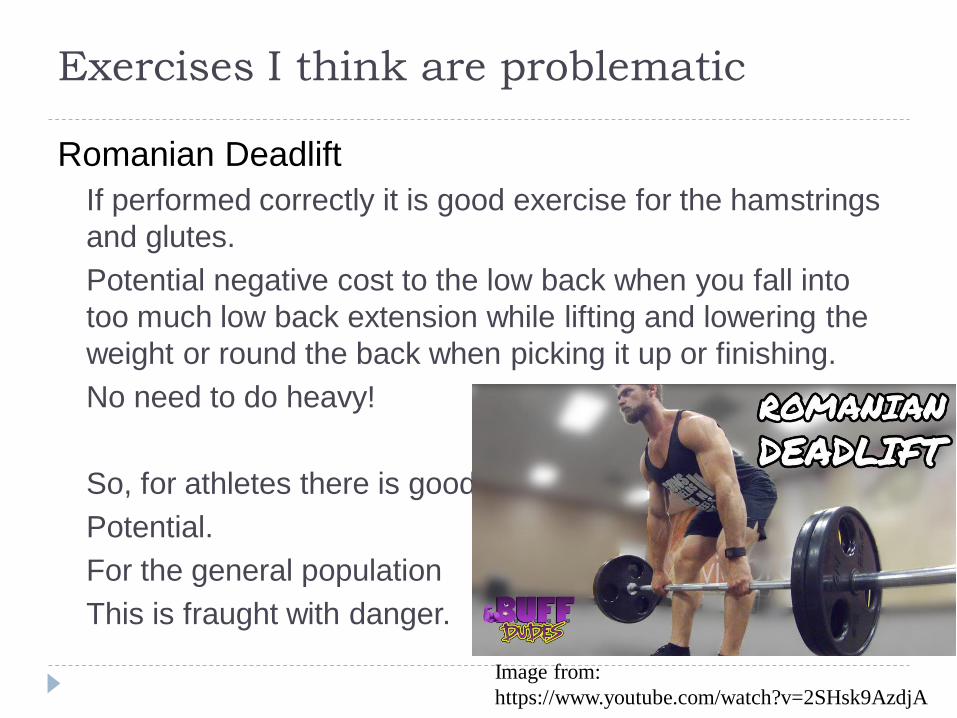
Romanian Deadlift
If performed correctly it is good exercise for the hamstrings
and glutes.
Potential negative cost to the low back when you fall into
too much low back extension while lifting and lowering the
weight or round the back when picking it up or finishing.
No need to do heavy!
So, for athletes there is good
Potential.
For the general population
This is fraught with danger.
Image from:
https://www.youtube.com/watch?v=2SHsk9AzdjA
Better Alternatives (especially if you
have any form of low back pain)
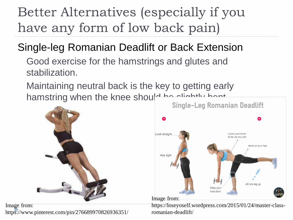
Single-leg Romanian Deadlift or Back Extension
Good exercise for the hamstrings and glutes and
stabilization.
Maintaining neutral back is the key to getting early
hamstring when the knee should be slightly bent
Image from:
https://www.pinterest.com/pin/276689970826936351/
Image from:
https://loseyoself.wordpress.com/2015/01/24/master-class-
romanian-deadlift/


