Download - AMERICAN NEUROTOLOGY SOCIETY - Combined
PROGRAM and ABSTRACTS
of the
AMERICAN NEUROTOLOGY SOCIETY
56th Annual Spring Meeting
Saturday, April 10, 2021Virtual Meeting
Table of Contents (ANS 2021 Program Book)
Page 3
Page 4
Page 5
Page 5
Page 6
Page 7
Pages 11-18
Pages 19-49
Pages 50-91
Pages 92-100
Pages 101-105
Pages 106
Page 107
Pages 108-126
Page 127
ANS Executive Council
ANS Mission Statement
ANS Diversity and Inclusion Statement
Disclosure Information/Publication/Submission Statement
Recognition of the 2021 Program Planning Members
COSM links/ANS Reception/Upcoming ANS meetings/Administrative Office
ANS Program Objectives
ANS Scientific Program
ANS Oral Abstracts
ANS Posters Abstracts
Recipients of Awards & Named Lecturers
ANS Research Grant/ 2020 Grant Recipient Progress Reports
ANS Past Presidents
ANS Past Secretary-Treasurers
ANS 2020-2021 Membership Roster
In Memoriam
Pages 8-10
2
AMERICAN NEUROTOLOGY SOCIETY 2020-2021 EXECUTIVE COUNCIL
President Bradley W. Kesser, MD
Charlottesville, VA
President-Elect Craig A. Buchman, MD
St. Louis, MO
Secretary-Treasurer Elizabeth H. Toh, MD, MBA
Burlington, MA
Immediate Past President Nikolas H. Blevins, MD
Stanford, CA
Education Director Howard W. Francis, MD, MBA
Durham, NC
ANS Diversity/Inclusion Chair Stephanie Moody Antonio, MD
Norfolk, VA
Members at Large David S. Haynes, MD, MMHC
Nashville, TN
Maura K. Cosetti, MD New York, NY
Hussam K El-Kashlan, MD Ann Arbor, MI
3
American Neurotology Society Mission Statement Purpose The American Neurotology Society (ANS) is committed to improving public health care through the provision of high-quality continuing medical education (CME) to our members. The overall goal of the ANS Continuing medical Education program is to provide CME activities that will address the knowledge gaps and enhance the clinical competence of the participants. The ANS is dedicated to improving public health care through the development, dialogue and dissemination of advances in evidence-based diagnosis and management of neurotologic and related skull base disorders. The focus on the scientific advances in these combined fields is translated into approaches to quality care that are consistent with ACGME/ABMS general competency areas and the Institute of Medicine recommendations.
Target Audience The primary target audience includes members of both the American Neurotology Society and our sister Society, the American Otological Society as well as healthcare professionals in the fields of otology, otolaryngology neurotology and skull base research and healthcare. The members served include physicians, otologists, neurotologists, residents, fellows, researchers, nurses, occupational and speech therapists and other healthcare professionals who are involved in the care of patients with otologic and neurotologic conditions.
Types of Activities Provided In order to accomplish the goals of the ANS CME program, the Education committee will offer a range of activities with specific educational outcomes in mind. Current offerings include:
• Scientific symposia, delivered twice per year at national venues, showcasing the latestresearch in the field and featuring national and international experts on related clinical topics.
• Study groups & mini-seminars offered at the annual meeting of the American Academy ofOtolaryngology-Head and Neck Surgery.
• Facilitation of manuscript submission on presented materials for publication in apeer reviewed journal (Otology & Neurotology).
• The Otology & Neurotology Journal provides an additional vehicle for further collaborationand dissemination of new information, science and standards of care.
Content The content of the ANS CME program centers on clinical issues related to Neurotology and disorders of the skull base. The ANS also strives to respond to our members’ educational needs that are not being met by other organizations, and therefore also offers activities in the areas of risk management, patient safety, physician-patient communications, coding, HIPAA compliance, and other regulatory issues as they relate to Neurotology. The educational efforts will also highlight the ACGME/ABMS general competencies within the context of this field and relate the significance of communication, professionalism, patient safety and systems-based practice within these workplace environments.
Expected Results The CME program of the ANS strives to enhance the participants’ knowledge and clinical competence in subject areas relevant to the field of Neurotology. The other expected outcome from this CME program is continued development of new evidence-based science, dissemination of ongoing research in the clinical area of Neurotology.
4
Resolution on Diversity of Meeting Presenters and Participation for the American Otological Society and the American Neurotology Society
• Whereas, the councils of the American Neurotology Society and American Otological Society desire topromote inclusivity within the membership of both organizations.
• Whereas it is recognized that diverse leadership and diversity of presenters allows for cross pollinationof knowledge, perspective and experiences enabling a stronger and more robust educational experiencefor our members.
• Whereas the Councils of the organizations recognize the importance of acknowledging diversity amongour patients, our trainees and our colleagues.
• Whereas, the purpose of the education programs of both organizations is to disseminate informationdesigned to improve physician knowledge, patient care and outcomes, and advance the respectivespecialties.
• Whereas, valuable scientific contributions to Otology and Neurotology by colleagues (regardless ofgender, race, or other attributes) should be presented at the society’s respective meetings.
• Be it resolved that the Scientific Program Committees of the American Neurotology Society andAmerican Otological Society will select speakers and panel members endeavoring to balanceeducational goals while promoting the diversity of our respective Societies’ memberships andeducational offerings.
• Be it resolved the Executive Councils of the ANS and AOS will select participation at all levels of theorganizations endeavoring to reflect diversity of our respective Societies’ memberships.
Disclosure Information
Please see the COSM website for all program COI and Disclosure information for the 2021 program.
https://cosm.md/cme/
PUBLICATION /SUBMISSION STATEMENT
The material in this abstract, has not been submitted for publication, published, nor presented previously at another national or international meeting and is not under any consideration for presentation at another national or international meeting.
The penalty for duplicate presentation/publication is prohibition of the author and co-authors from presenting at a COSM society meeting for a period of three years. Submitting Author’s Signature (required All authors were advised that the submitted paper becomes the property of Otology & Neurotology and cannot be reprinted without permission of the Journal.
Duplicate abstract submission to more than one Society will result in the abstract being disqualified and it will not be considered for presentation on either the ANS or AOS program.
5
THE AMERICAN NEUROTOLOGY SOCIETY WOULD LIKE TO THANK THE FOLLOWING MEMBERS FOR THEIR CONTRIBUTION TO THE 2021 ANS SCIENTIFIC PROGRAM
Scientific Program Committee Bradley W. Kesser, MD, ANS President
Howard W. Francis, MD – ANS Education Director Meredith E. Adams, MD*
Jennifer Alyono, MD Eduardo Corrales, MD Christine T. Dinh, MD Justin S. Golub, MD*
Douglas M. Hildrew, MD Candace E. Hobson, MD Andrew A. McCall, MD
Theodore R. McRackan, MD Brian D. Nicholas, MD*
Pamela C. Roehm, MD, PhD J. Thomas Roland, MD*Emily Z. Stucken, MD*
Sean Wise, MD Erika A. Woodson, MD
ANS Education Committee Howard W. Francis, MD
Yuri Agrawal, MD Wade W. Chien, MD
Ana Hae-Ok Kim, MD Jennifer Maw, MD* Mia E. Miller, MD
Stephanie Moody Antonio, MD Brian P. Perry, MD
Jeffrey D. Sharon, MD* Esther X. Vivas, MD*
Soha N. Ghossaini, MD Brandon Isaacson, MD
Selena Briggs, MD Marc Eisen, MD
Laura Brainard, MDMatthew L. Bush, MD, PhD
(Socio-Economic Committee Chair)Ronna P. Hertzano, MD, PhD*
(Research Committee Chair)Sarah E. Mowry, MD
(ANS Young Member representative)John P. Leonetti, MD
(Coordinator-Facial Nerve Study Group)Ravi N. Samy, MD*
(*ANS Program Moderator)
Poster Judges Laura Brainard, MD Selena Briggs, MD Marc Eisen, MD
Brandon Isaacson, MD
6
VIRTUAL COSM ANS PROGRAM LINK - AVAILABLE FOR REGISTERED ATTENDEEShttps://www.eventscribe.net//2021/COSM/index.asp
COSM REGISTRATION LINKhttps://cosm.md/registration-information/
ANS 56th Annual President's Reception Friday, April 9, 2021 via ZOOM
Open to all members and presentersSee website/newsletter for details.
UPCOMING MEETINGS ANS “Super Saturday” is scheduled October 2, 2021 Details TBDCall for Papers for Study groups begins May 15, 2021.Submission details will be posted on the ANS website in May
57th ANS Spring Meeting (in conjunction with COSM) April 29–May 1, 2022 Hyatt Regency Dallas Dallas, Texas
The Abstract deadline for the ANS 57th Annual Spring meeting is Friday, October 15, 2021. Abstract Instructions and submission form will be available on website August 15-October 15, 2021. Website - www.americanneurtologysociety.com
All primary and contributing authors are required to complete a disclosure/conflict of interest statement at time of abstract submission in order for the abstract to be considered by the Scientific Program Committee.
Journal Requirements/Instructions to Primary Authors Manuscripts are required of ALL presenters. Manuscripts must be submitted online a minimum of four weeks prior to the annual meeting, via the journal’s website. Instructions for registering, submitting a manuscript, and the author guidelines can be found on the Editorial Manager site: https://www.editorialmanager.com/on/
The journal of OTOLOGY & NEUROTOLOGY does not accept paper manuscripts. Manuscripts will be peer reviewed prior to the Annual meeting for conflict of interest review and resolution if necessary.
Failure to comply with the guidelines & requirements of the American Neurotology Society and the O&N Journal will result in the disqualification of your presentation.
For Society business, please forward all inquiries to the ANS Administrative Office.
Kristen Bordignon, Administrative TeamAshley EikenberryANS Administrative Office 5830 1st St. N. Petersburg, FL 33703 Ph: 217-638-0801 Fax: 727-800-9428 Email: [email protected] Website: www.americanneurtologysociety.com
7
What are the practice or patient care issues being addressed by the ANS Program?
Our Spring program, entitled, "Neurotology Across the Lifespan" features three panels that address common and rare neurotologic problems among pediatric, middle age, and geriatric patients.
Our pediatric panel will be a fast-paced series of case presentations. moderated by Robert O'Reilly, MD posing challenging pediatric clinical problems such as cochlear implantation in the malformed cochlea, amplification options for unilateral hearing loss in children, and evaluation and management of the pediatric dizzy patient.
Our "middle-age" panel will turn to the COVID-19 pandemic. Moderated by Sarah Mowry, MD, panelists will discuss lessons learned from the pandemic including those in training programs, options for Telehealth during the pandemic, and what the future will hold. This panel also hopes to draw out healthcare disparities in this country laid bare by the pandemic, and how we as ear surgeons can address these disparities.
The third, "geriatric" panel moderated by Justin Golub, MD, will separate fact from fiction in the relationship between hearing loss and dementia. Panelists will include an audiologist, an epidemiologist, and a neurologist invested in identifying correlations between hearing loss and dementia. The relationship is correlative at best, and the panel seeks to elucidate the correlations as well as debunk the false claims that seek to prey on consumers.
The program also highlights outstanding research done in the areas of cochlear implantation, vestibular schwannoma management, tinnitus, and dizziness and imbalance through both oral and poster abstract presentations.
Why do these issues exist? Is there a deficit in provider's knowledge or skill? Is there a deficit in health care system process or outcomes?
Clinicians and researchers alike do not know the ideal management of vestibular schwannoma. Options include observation, radiation, and microsurgery. These issues exist because we also do not know the natural history of vestibular schwannoma - why do some grow and others do not? Why are some more aggressive than others? Which tumors are ideal to operate and which to radiate or simply observe? This activity seeks to close knowledge gaps among neurotology practitioners in the best practices of vestibular schwannoma management.
The COVID-19 pandemic has clearly laid bare healthcare disparities in this country. Why do these disparities exist? Do they exist in the field of neurotology? How can we practitioners optimize hearing healthcare for all and not just the privileged few? Our panel hopes to address these questions in our health system and offer opportunities for all practitioners to close healthcare disparity gaps in our field.
As specialists, we often come to rely on expensive diagnostic imaging modalities such as magnetic resonance imaging (MRI) to rule in or rule out specific diagnoses (e.g., vestibular schwannoma, stroke in the dizzy patient). Dr David Zee, Professor of Neurology at Johns Hopkins University, is our William House lecturer and will be speaking on, "Vestibular Diagnosis in a Pandemic: Return of the House Call." This lecture promises to be illustrative in the physical exam of the dizzy patient and sorting out peripheral versus central (i.e., stroke) etiologies of dizziness.
LEARNING OBJECTIVES
8
Optimal rehabilitation of patients with severe-profound sensorineural hearing loss with cochlear implantation remains an important and evolving issue for neurotologists. The latest research regarding cochlear implant (CI) candidacy, hearing preservation cochlear implant surgery, expectations regarding second side cochlear implantation, and the implant technology itself - type, position, and coating - will be addressed in a section of CI abstracts.
A section on research abstracts covering tinnitus and dizziness, two patient complaints that can be confusing and frustrating, will be presented. Participants will learn the latest research in the evaluation and treatment of pulsatile, pulse-synchronous tinnitus as well as a novel agent for the treatment of nonpulsatile tinnitus. Learners will also evaluate whether machine learning can predict common causes of dizziness and how electrocochleography can predict outcomes in superior semicircular plugging either via a trans mastoid or middle fossa approach.
How will this program improve the learners' competence, performance and/or patient outcomes?
As noted above, the educational activity will improve learners' competence and patient outcomes by presenting challenging pediatric cases for discussion, by improving learners' ability to perform a focused physical exam for the detection of central vs. peripheral causes of dizziness, by bringing to light healthcare disparities in neurotology and offering ways to address these disparities, by presenting the latest research in vestibular schwannoma management, cochlear implantation, tinnitus, dizziness, and the possible mitigating effects of atorvastatin in cisplatin-related hearing loss among head and neck cancer patients, our 2021 William Hitselberger lecture given by Lisa Cunningham, PhD, Senior Investigator at the NIDCD.
How do we anticipate this activity improving health care systems?
Our panel, "Lessons Learned: Neurotology in the Covid-19 Pandemic," will address telehealth in the time of the pandemic and how we can carry forward the lessons learned practicing telemedicine in the non-pandemic world. Panelists will also discuss how the training of residents and fellows during the pandemic has changed how we will continue to train residents and fellows, and how we as individual practitioners can effect positive change in addressing healthcare disparities.
Our educational program also tackles opioid and non-opioid use after skull base and otological surgery and will offer alternatives to opioids for postoperative pain management.
Several abstracts in the research sections of our activity address reducing or optimizing healthcare resource utilization, including, "Diagnostic Yield and Utility of Radiographic Imaging in the Evaluation of Pulsatile Tinnitus: A Systematic Review," "Comparison of Outcomes of Surgical Repair of Spontaneous Temporal Bone CSF Leaks and Encephaloceles Using Bone Cement and Autologous Material," and "Higher Readmission Rates after Hip Fracture among Patients with Vestibular Disorders."
9
How do we anticipate this activity impacting the health of patients and their communities?
Exploring the correlation between dementia and hearing loss, as addressed in our geriatric panel, will empower our learners with the evidence to discuss the relationship with their patients and family members. False claims of "If you don't purchase a hearing aid, you will become demented" abound, and this panel seeks truth over fiction and falsehood. The panel will enlighten learners on this relationship to help our practitioners and their patients make informed decisions about amplification, cochlear implant surgery, and the decision not to amplify.
The COVID-19 panel will address hearing healthcare disparities and help our clinicians become advocates for closing gaps in hearing healthcare.
Learning objectives for this CME Activity.
1) Identify disparities in healthcare and hearing healthcare access related to non-COVID and COVIDrelated factors
2) List 2 ways telehealth/virtual visits can be utilized in delivery of high quality neurotology care
3) Identify a new method for educational opportunities to utilize in your practice/program.
4) Elaborate on the current evidence level explaining the relationship between hearing loss andcognition
5) Identify and utilize appropriate (and identify inappropriate) counseling of patients based on thecurrent evidence
6) Discuss the limitations in understanding causality
7) Perform a focused physical examination of the dizzy patient with emphasis on deciphering centralversus peripheral causes of dizziness8) Assess the use of Atorvastatin in mitigating cisplatin-induced hearing loss in head and neck cancerpatients
9) Incorporate into your practice the research work being done in the evaluation and management ofvestibular schwannoma and the evolving treatment paradigms leaning toward observation of thesetumors
10) Assist patients considering cochlear implantation for severe-profound sensorineural hearing loss intheir expectations and in their hearing outcomes; evaluate electrode technology, position in the cochlea,and future electrode modalities
11) Evaluate and treat pediatric patients with complex neurotologic problems including dizziness andbalance disorders and hearing loss, both acquired and congenital
10
AMERICAN NEUROTOLOGY SOCIETY 56th Annual Spring Meeting
VIRTUAL PROGRAM ALL TIMES listed are Central Time
SATURDAY, APRIL 10, 2021
SCIENTIFIC PROGRAM - “NEUROTOLOGY ACROSS THE LIFE SPAN”
9:00 WELCOME AND OPENING REMARKS & POSTER WINNERS ANNOUNCED Bradley W. Kesser, MD
9:05 PRESIDENTIAL CITATIONS - Bradley W. Kesser, MD Paul R. Lambert, MD George T. Hashisaki, MD Paul A. Levine, MD Robert A. Jahrsdoerfer, MD
9:15 2ND ANNUAL NOEL L. COHEN AWARD FOR SIGNIFICANT CONTRIBUTIONS TO OTOLOGY AND NEUROTOLOGY Bradley W. Kesser, MD
9:22 INTRODUCTION OF HOUSE LECTURE Bradley W. Kesser, MD
9:25 WILLIAM F. HOUSE MEMORIAL LECTURE “Vestibular Diagnosis in a Pandemic: Return of the House Call” David S. Zee, MD Professor of Neurology Joint Appointments in Ophthalmology, Otolaryngology, Head & Neck Surgery, and Neuroscience The Wilmer Eye Institute The Johns Hopkins School of Medicine, Baltimore, MD
9:50 DISCUSSION
10:00 PANEL - "Pediatric Potpourri: Challenging Cases to Stump the Stars" Robert C. O’Reilly, MD, Moderator Stephanie A. Moody Antonio, MD Sharon Cushing, MD, MSc Tina C. Huang, MD John L. Dornhoffer, MD
11:05 INTRODUCTION OF COCHLEAR IMPLANTATION ABSTRACT PRESENTATIONS Jennifer L. Maw, MD & J. Thomas Roland Jr, MD, Moderators
11
11:06 Exploring Factors Responsible for Delay in Pediatric Cochlear Implantation Jacquelyn K. DeVries, BS Yin Ren, MD, PhD
Julie Purdy, PhD, CCC-A, Daniela Carvalho, MD, MMM, Elina Kari, MD
11:12 Outcomes in Patients Meeting Cochlear Implant Criteria in Noise but not in Quiet Anthony Thai, BA Emma Tran, BS
Austin Swanson, AuD Matthew B. Fitzgerald, PhD Nikolas H. Blevins, MD Jennifer C. Alyono, MD
11:18 ANS TRAINEE AWARD Zwitterionic Coating of Cochlear Implants Reduces Friction and Force of Insertion Douglas M. Bennion, MD, PhD Ryan Horne
Adreann Peel C. Allan Guymon, PhD Marlan R. Hansen, MD
11:25 NEUROTOLOGY FELLOW AWARD The Impact of Age on Noise Sensitivity in Cochlear Implant Recipients Matthew A. Shew, MD Craig A. Buchman, MD
Dorina Kallogjeri, MD Stephanie Chen, MD Cameron C. Wick, MD Nedim Durakovic, MD Jacques A. Herzog, MD CI532 Study Group
11:31 The Influence of Cochlear Implant Electrode Type and Position on Hearing Preservation Elizabeth L. Perkins, MD Matthew O’Malley, MD
Marc Bennett, MD David S. Haynes, MD Jack H. Noble, PhD Robert F. Labadie, MD, PhD René Gifford, PhD
11:37 Role of Pre-Implant Patient Expectations in Adult Cochlear Implant Outcomes Theodore R. McRackan, MD, MSCR Mark S. Costello, MD
Priyanka Reddy, BS Judy R. Dubno, PhD
11:43 Time-to-Peak Speech Perception Score after Cochlear Implantation in Single-sided Deafness Ashley M. Nassiri, MD, MBA John P. Marinelli, MD
Katherine P. Wallerius, MD
12
Christine M. Lohse, MS Colin L. W. Driscoll, MD Brian A. Neff, MD Aniket A. Saoji, PhD Matthew L. Carlson, MD
11:49 Identification of Factors Associated with Second-Side Cochlear Implant Speech Recognition
Outcomes in Adults James R. Dornhoffer, MD*
Yuan F. Liu, MD* Elise E. Zhao, BS Elizabeth L. Camposeo, AuD Ted A. Meyer, MD, PhD Theodore R. McRackan, MD, MSCR *Authors contributed equally to this work
11:55 Characterizing Cochlear Implant Magnet-Related MRI Artifact and Visualization of Indicated
Structures Nathan D. Cass, MD Douglas J. Totten, BA
Elizabeth L. Perkins, MD John D. Ross, MD Matthew R. O’Malley, MD
12:01 DISSCUSSION (10 min) - Jennifer L. Maw, MD & J. Thomas Roland Jr, MD 12:11 LUNCH BREAK 1:00 INTRODUCTION OF VESTIBULAR SCHWANNOMA ABSTRACT PRESENTATIONS
Ravi N. Samy, MD & Esther X. Vivas, MD, Moderators 1:01 Natural History of Growing Vestibular Schwannomas During Observation: An International
Multi-Institutional Study of 593 Growing Tumors John P. Marinelli, MD Matthew L. Carlson, MD
Jacob B. Hunter, MD Ashley M. Nassiri, MD, MBA Martin Reznitsky, MD Sven-Eric Stangerup, MD, DMSc Per Caye-Thomasen, MD, DMSc
1:07 Effect of AR42 on Tumor Growth and Hearing Loss In Vivo and on Primary Vestibular
Schwannoma Cells Carly Misztal, BS Olena Bracho, BS
Michael Estivill, BS Cristina Fernandez Valle, PhD Fred Telischi, MD Xue-Zhong Liu, MD, PhD Christine T. Dinh, MD
13
1:13 Cost-effectiveness of Microsurgery, Radiosurgery, and Observation in the Management of Small and Medium-sized Sporadic Vestibular Schwannoma
Robert J. Macielak, MD Viengneesee Thao, PhD, MS
Bijan J. Borah, PhD James P. Moriarty, MS Jamie J. Van Gompel, MD Matthew L. Carlson, MD
1:19 NEUROTOLOGY FELLOW AWARD
Complications after Surgical Salvage for Vestibular Schwannoma following Failed Stereotactic Radiosurgery
Alexander L. Luryi, MD Seilesh Babu, MD
John F. Kveton, MD Dennis I. Bojrab, MD Elias M. Michaelides, MD Christopher A. Schutt, MD
1:25 Delayed Facial Nerve Palsy following Resection of Vestibular Schwannoma: Clinical and Surgical
Characteristics Bridget MacDonald, BA Yin Ren, MD, PhD
Bita Shahrvini, BS Kareem Tawfik, MD Omid Moshtaghi, MD Marc Schwartz, MD Rick Friedman, MD, PhD
1:31 Subset of Intracanalicular Vestibular Schwannomas Demonstrate Minimal Growth over
a 10 Year Period Matthew J. Wu, BS Renata M. Knoll, MD
Michael J. McKenna, MD Elliott D. Kozin, MD* David H. Jung, MD, PhD*
1:37 Evaluating the Impact of Frailty and Advanced Age on Morbidity following Vestibular Schwannoma
Surgery Alvin DeTorres, MD Gentry Carter, BS
Alvin Kwok, MD, MPH Christian Bowers, MD Neil S. Patel, MD Richard K. Gurgel, MD, MSCI
1:43 The Influence of Extent of Resection and Tumor Morphology on Facial Nerve Outcomes
following Acoustic Neuroma Surgery Elizabeth L. Perkins, MD Nauman F. Manzoor, MD
Douglas J. Totten, BA Alexander D. Sherry, MD Matthew O’Malley, MD
14
Marc Bennett MD, MHCC David S. Haynes, MD, MHCC
1:49 DISCUSSION (10 min) - Ravi N. Samy, MD & Esther X. Vivas, MD
2:02 PANEL - “Lessons Learned: Neurotology in the Covid-19 Pandemic” (1 hour) Sarah E. Mowry, MD, Moderator Jennifer B. Nuzzo, DrPH Courtney C. J. Voelker, MD, PhD Sonya Malekzadeh, MD Shivesh Maharaj, MBBCH, FCORL, MMED
3:02 BREAK
3:14 INTRODUCTION OF HITSELBERGER LECTURER Ronna Hertzano, MD, PhD
3:16 WILLIAM E. HITSELBERGER MEMORIAL LECTURE “Atorvastatin Is Associated with Reduced Cisplatin-Induced Hearing Loss in Patients with Head and Neck Cancer” Lisa L. Cunningham, PhD Senior Investigator Laboratory of Hearing Biology and Therapeutics National Institute on Deafness and Other Communication Disorders National Institutes of Health, Bethesda, MD
3:41 DISCUSSION (5 min)
3:47 INTRODUCTION OF 2019 ANS RESEARCH GRANT RECIPIENT
3:48 ANS 2019 RESEARCH GRANT (9 min) Targeting Epigenetic Modifying Enzymes for Hair Cell Regeneration Dunia E. Abdul-Aziz, MD Massachusetts Eye and Ear
4:00 INTRODUCTION OF ABSTRACT PRESENTATIONS – TINNITUS AND DIZZINESS Meredith E. Adams, MD & Jeffrey Sharon, MD, Moderators
4:01 Diagnostic Yield and Utility of Radiographic Imaging in the Evaluation of Pulsatile Tinnitus: A Systematic Review Austin C. Cao, BA Caitlin Cavarocchi, BA Tiffany P. Hwa, MD Steven J. Eliades, MD PhD Michael J. Ruckenstein, MD Douglas C. Bigelow, MD Jason A. Brant, MD
4:07 A Phase 1/2 Study of OTO-313 Given as a Single Intratympanic Injection in Patients with Moderate to Severe, Persistent Tinnitus Jeffery J. Anderson, PhD Kenneth S. Maxwell, MD (Presenter) James M. Robinson, MS
15
Ines Hoffman, PhD Gordon T. McMurry, MD Grant D. Searchfield, PhD David M. Baguley, PhD
4:13 Transtemporal Sigmoid Sinus Decompression: A Novel Surgical Procedure for the Treatment of Idiopathic Pulsatile Tinnitus Patrick W. Slater, MD Bailey H. Duhon, BS Neha Korla, MDS
4:19 Supervised Machine Learning Models for Predicting Common Causes of Dizziness Eric J. Formeister, MD, MS Jeffrey D. Sharon, MD
4:25 Head Roll-Tilt Subjective Visual Vertical Test in the Diagnosis of Persistent Postural-Perceptual Dizziness (PPPD) Chihiro Yagi, MD Yuka Morita, MD, PhD Meiko Kitazawa, MD Kuniyuki Takahashi, MD, PhD Yoshiro Wada, MD, PhD Tadashi Kitahara, MD, PhD Arata Horii, MD, PhD
4:31 ANS TRAINEE AWARD Intraoperative Electrocochleography Predicts Outcomes in Transmastoid and Middle Cranial Fossa SSCD Repair Susan E. Ellsperman, MD Steven A. Telian, MD Paul R. Kileny, PhD Christopher M. Welch, MD, PhD
4:37 DISCUSSION (10 min) - Meredith E. Adams, MD & Jeffrey Sharon, MD
4:47 Break
5:00 INTRODUCTION OF ABSTRACT PRESENTATIONS – POTPOURRI Brian D. Nicholas, MD & Emily Z. Stucken, MD, Moderators
5:01 Comparison of Outcomes of Surgical Repair of Spontaneous Temporal Bone CSF Leaks and Encephaloceles Using Bone Cement and Autologous Material Vir Patel, MD Tiffany Peng Hwa, MD Steven J. Eliades, MD, PhD Jason A. Brant, MD Douglas C. Bigelow, MD Michael J. Ruckenstein, MD
5:07 Perineural Invasion of the Intratemporal Facial Nerve: How Far Proximally Do We Chase the Positive Margin? Joshua Cody Page, MD Marc-Elie Nader, MD, FRCSC
16
Diana Bell, MD Paul W. Gidley, MD
5:13 The Laterality of Early Age-Related Hearing Loss and Brain Beta-Amyloid Alexandria L. Irace, BA Brady Q. Rippon, MS
Adam M. Brickman, PhD José A. Luchsinger, MD, MPH Justin S. Golub, MD, MS
5:19 NICHOLAS TOROK VESTIBULAR AWARD
Higher Readmission Rates after Hip Fracture among Patients with Vestibular Disorders Steven D. Curry, MD, MPH Alessandro Carotenuto, MD
Devin A. DeLuna, BS Dennis J. Maar II, BA Ye Huang, BA Justin C. Siebler, MD Jonathan L. Hatch, MD
5:25 Diameter-Based Volumetric Models May Inaccurately Calculate Jugular Paraganglioma Volume
following Sub-Total Resection Douglas J. Totten, BA Nauman F. Manzoor MD
Elizabeth L. Perkins MD Nathan D. Cass MD, Mohamed H. Khattab MD David S. Haynes MD MMHC Joseph M. Aulino MD
5:31 Opioid and Non-Opioid Usage in the Post-operative Period following Otologic Surgery Neal Rajan Godse, MD Rahilla A. Tarfa, PhD
Philip Perez, MD Barry E. Hirsch, MD Andrew A. McCall, MD
5:37 Opioid Prescribing Patterns after Skull Base Surgery for Vestibular Schwannoma Yin Ren, MD, PhD Pasha Mehranpour, BS
Omid Moshtaghi, MD Marc S. Schwartz, MD Rick A. Friedman, MD, PhD
5:43 DISCUSSION (10 min) - Brian D. Nicholas, MD & Emily Z. Stucken, MD 5:55 PANEL - “Hearing Loss and Dementia: Separating Fact from Fiction” (45 min)
Justin S. Golub, MD, Moderator Katharine K. Brewster, MD Carrie L. Nieman, MD, MPH Jennifer A. Deal, PhD M. Kathleen Pichora-Fuller, PhD
17
6:42 OTOLOGY & NEUROTOLOGY OPEN (ONO) ANNOUNCEMENT Michael E. Hoffer, MD, ONO Editor-in-Chief 6:45 INTRODUCTION OF INCOMING ANS PRESIDENT - Craig A. Buchman, MD Bradley W. Kesser, MD 6:47 CLOSING REMARKS Bradley W. Kesser, MD 6:50 ADJOURN
18
SELECTED ABSTRACTS
ORAL
PRESENTATIONS
56thAnnual Virtual Spring Meeting
AMERICAN NEUROTOLOGY SOCIETY
LIVE! Saturday, April 10, 2021
Also Available ON DEMAND
19
Exploring Factors Responsible for Delay in Pediatric Cochlear Implantation
Jacquelyn DeVries, BS, Yin Ren, MD, PhD Julie Purdy, PhD, CCC-A, Daniela Carvalho, MD, MMM, Elina Kari, MD
Objective: To identify and characterize demographic and socioeconomic factors associated with delays in cochlear implantation (CI) in children
Study Design: Retrospective
Setting: Tertiary pediatric CI referral center
Patients: All CI recipients under 18 years of age receiving CI between March 2018 and February 2020.
Interventions: CI
Main Outcome Measures: Primary outcome measures included age at implantation and time from candidacy evaluation to CI.
Results: Seventy-two patients were identified (44% female, average age at implantation 4.87 years). Age at implantation was later in patients with public, rather than private, insurance (5.98±0.78yr vs. 3.13±0.66yr, p=.007) and those from low-income areas (8.58±7.6y vs. 2.35±3.00y, p=0.007). Time between identification as a CI candidate and implantation was longer in publicly insured patients (721±107d vs. 291±64d, p=.001) and in bilingual children (888±160d) compared to those who spoke solely Spanish (473±101d, p=0.036) or English (400± 95d, p=.022). Latinx children were more often publicly insured whereas white children were more often privately insured, (p<.05). Publicly insured patients had delays in each step of the pre-CI workup, including vestibular evaluation (621±132d vs. 197±67d, p=.007), developmental evaluation, (517±106d vs. 150±56d, p=.003), speech evaluation (482±107dvs. 163±65d, p=.013), and Children’s Implant Profile (ChIP) assessment (572±107d vs. 184±59d, p=.002). On ChIP evaluation, concerns regarding education were higher in Spanish-speaking children (p= 0.024; p =2.6x10⁻⁴ ) and children with public insurance (p=0.016; p=.002). Income and language spoken were found to predict age at implantation (p=0.006;p=0.019) while race and language spoken predicted delay from candidacy identification to implantation (p=0.18; p=0.007).
Conclusions: Disparities in access to cochlear implanation continue to affect timing of implantation.
Define Professional Practice Gap & Educational Need: 1. Lack of understanding regarding persistent disparities in timing of pediatric cochlear implanation as based on type of insurance, ethnicity, and language(s) spoken in the home.
Learning Objective: 1. To identify determine factors associated with delay in pediatric cochlear implantation
Desired Result: 1. Attendees will have a better understanding of demographic factors associated with delays in pediatric cochlear implanation. 2. Attendees will have knowledge when in the pre-implantation process delays are likely to occur in order to target areas of improvement.
Level of Evidence – Level IV – Historical cohort or case-control studies
Indicate IRB or IACUC : Rady Children’s Hospital IRB # 190779. Approved 12/11/2019. All data was collected after IRB approval.
20
Outcomes in Patients Meeting Cochlear Implant Criteria in Noise but not in Quiet
Anthony Thai, BA; Emma Tran, BS; Austin Swanson, AuD; Matthew B. Fitzgerald, PhD Nikolas H. Blevins, MD; Jennifer C. Alyono, MD
Objective: Evaluate outcomes in cochlear implant (CI) recipients qualifying based on AzBio in noise but not in quiet
Study Design: Retrospective cohort study
Setting: Tertiary otology/neurotology clinic
Patients: After excluding device failures, this study included 216 implanted ears (mean age 65.0±18.7 years, 59.6% male). The cohort group comprised 23 ears with preoperative AzBio scores ≥40% in quiet and ≤40% in either +10 or +5 speech-to-noise ratio (SNR). The control group included 193 ears with preoperative AzBio scores <40% in quiet. Age and gender were similar between the two groups.
Interventions: Cochlear implantation
Main Outcome Measures: 1-year post-operative AzBio score in quiet and noise
Results: Cohort group AzBio scores improved in +10 SNR (pre-operative: 25.4%, post-operative: 51.4%, p<0.001) but not quiet (pre-operative: 62.0%, post-operative: 71.0%, p=0.16). In contrast, controls improved in AzBio +10 SNR (preoperative: 8.0%, postoperative: 55.7%, p<0.001) and quiet (preoperative: 11.8%, postoperative: 66.8%, p<0.001). Both groups had similar postoperative AzBio quiet (p=0.47) and +10 SNR (p=0.50). Compared to controls, the cohort had fewer ears with significant within-subject improvement in AzBio quiet (≥15% improvement; control: 89.9%, cohort: 41.1%, p<0.001). Ears displaying within-subject improvements in AzBio quiet were more likely to have lower baseline scores in AzBio quiet (p<0.001) and CNC words (p=0.004), but not baseline AzBio +10 SNR, aided pure tone average and unaided word recognition scores (p>0.05).
Conclusions: Patients qualifying for CI candidacy because of performance in noise display significant post-implantation benefit in noise. However, these patients are less likely to show significant individual improvement in quiet.
*Define Professional Practice Gap & Educational Need: Major insurance companies define CI criteria based on sentencerecognition scores without specifying whether such testing should be performed in quiet or in noise. Our study presents alarger group of patients with longer follow-up than exists in prior literature, and confirms that patients meeting CI candidacysolely in noise still benefit from implantation, although to a lower extent than patients qualifying in quiet.
*Learning Objective:Cochlear implant candidacy criteria do not specify the level of background noise that should be employed for sentencerecognition testing.Patients meeting CI criteria in noise but not in quiet display significantly improved AzBio scores in noise post-implantation.Patients with low baseline AzBio scores in quiet and/or in noise are most likely to have clinical benefit from CI.
*Desired Result:Cochlear implantation should be considered in patients meeting cochlear implant criteria solely in noise. Patients with lowerbaseline scores are more likely to derive significant objective benefit.
Level of Evidence - IV
Indicate IRB: IRB 50573, Stanford University
21
- Understand the effect of zwitterionic coating in reducing the coefficient of friction between various biomaterials andbiologic surfaces- Appreciate the impact of thin-film coating on human cochlear implant arrays in reducing insertional forces in a cochlearimplant in explanted human cadaveric cochleae.
Desired Result: Improved understanding of the potential for zwitterionic thin-film coatings at reducing friction and force of cochlear implant insertion
Level of Evidence: N/A
Indicate IRB or IACUC: Exempt.
ANS TRAINEE AWARD
Zwitterionic Coating of Cochlear Implants Reduces Friction and Force of Insertion
Douglas M. Bennion, MD, PhD; Ryan Horne; Adreann PeelC. Allan Guymon, PhD; Marlan Hansen, MD
Background: Strategies to minimize intracochlear trauma during implantation of an electrode array are critical to optimize outcomes including hearing preservation. To this end, bioengineering advances in application of thin-film zwitterionic hydrogels to relevant biomaterials provide a promising avenue.
Methods: Using a recently designed one-step process, thin-film coatings containing zwitterionic sulfobetaine methacrylate (SBMA) were polymerized and photografted to the surface of polydimethylsiloxane (PDMS, silastic) samples and also to cochlear implant (CI) arrays from two manufacturers. Methylene and fluorescein staining and scanning electron microscopy with energy-dispersive X-ray spectroscopy verified and characterized the coatings. Tribometry was used to measure the co-efficient of friction between uncoated and coated PDMS and biologic tissues. Force transducer measurements were obtained during manual insertion and robotic motorized insertion of uncoated (n=9) and coated CI electrode arrays (n=9) into human cadaveric cochleae.
Results: Image analysis confirmed uniform coating of the PDMS samples and the CI electrode arrays with SBMA polymer films. SBMA thin-film coating of PDMS resulted in >90% reduction in frictional co-efficients across various biologic tissues (subdermis, trachea, aorta, bladder, dura, p<0.001). During insertion of electrode arrays into human cadaveric cochleae, SBMA coatings reduced maximum force by more than 40% during both manual insertion (p<0.005) and micromotorized insertion (p<0.005).
Conclusion: Thin-film SBMA coatings of PDMS and electrode arrays significantly reduce frictional co-efficients and insertional forces in cadaveric cochleae. These encouraging findings support thin-film zwitterionic coatings of CI electrode arrays as a method for reducing insertional trauma and thereby promoting hearing preservation.
Define Professional Practice Gap & Educational Need: Hearing preservation in cochlear implantation has become an important priority in cochlear implantation and bioengineering strategies designed to prevent intracochlear trauma by decreasing friction and insertional forces are discussed.
Learning Objective: - Become familiar with the biochemistry of photografting of zwitterionic hydrogels
22
The Impact of Age on Noise Sensitivity in Cochlear Implant Recipients
Matthew Shew, MD; Craig Buchman, MD; Dorina Kallogjeri, MD; Stephanie Chen, MDCameron Wick, MD; Nedim Durakovic, MD; Jacques Herzog, MD
and CI532 Study Group
Objective: To evaluate the impact of noise on speech perception testing in adult cochlear implant (CI) recipients above and below 65 years.
Study Design and Setting: Multi-institution, prospective, non-randomized, single-subject repeated measures design.
Patients: 96 adults ≥18 years old with post-lingual bilateral sensorineural hearing loss.
Intervention(s): Participants received a CI532 in one ear. Speech perception measures were evaluated before and 6-months after activation.
Main outcome measure(s): Subjects completed consonant-nucleus-constant (CNC) words in quiet and AzBio sentences in noise using +10dB and +5dBSNR, and Montreal Cognitive Assessment (MOCA).
Results: 96 adult patients were enrolled (n=70 older (≥65years), n=26 younger (<65years)). There was no significant difference in CNC scores (CI alone 58.4 vs 67.5, p=0.0857; best aided 66.7 vs 76.1, p=0.3357). Older adults performed worse on AzBio+10dBSNR compared to younger patients (CI alone 37.4 vs 56.9, p=0.0006; best aided 51.4 vs 68.2; p=0.01), and in AzBio+5dBSNR (CI alone 7.7 vs 11.2, p=0.0002; best aided 15.3 vs 22.3, p= 0.0005). The magnitude of change in AzBio+10dBSNR was significantly less in older adults in CI alone (15.3 vs 22.3; p= 0.0005) but not best aided (21.5vs31.3; p=0.105), and was drastically worse in AzBio +5dBSNR (CI alone 6.7vs22.1, p=0.0014; best aided 9.5vs21.5; p=0.0142). There was no significant difference in MOCA between the two age groups.
Conclusions: While both older and younger patients have similar outcomes with respect to CNC word scores, the addition of noise disproportionally impacts older patients. Caution should be exercised when adding noise to candidacy testing in the elderly.
*Define Professional Practice Gap & Educational Need:- There are varying CI candidacy criteria used by Medicare and third-party payers. Additionally, the use of sentence
recognition test to be utilized and the addition to background noise is not specified. The current study prospectivelyevaluates the impact of different open set sentence speech recognition tests in quiet, +10dB SNR, +5db SNR inolder adults (≥65 years) compared to their younger counterparts.
- With an increasing number of older adults impacted by hearing loss, understanding the role of CI candidacy testingin quiet and noise in the elderly is an essential component as we move forward with creating consensus guidelinesfor CI candidacy.
*Learning Objective:- Evaluate baseline and change in different speech recognition tests between younger and older adult CI recipients- Understand the impact of background noise to CI candidacy testing and performance in younger and older adult CI
recipients.
*Desired Result: I- Similar magnitude of improvement in speech recognition scores between younger and older CI recipients- The addition of background noise to speech recognition testing affects both younger and older CI recipients equally.
*Level of Evidence – Level III
Indicate IRB or IACUC : Registered on clinicaltrials.gov (NCT03007472), approved by each institutions’ respective IRB.
NEUROTOLOGY FELLOW AWARD
23
The Influence of Cochlear Implant Electrode Type and Position on Hearing Preservation
Elizabeth L. Perkins, MD; Matthew O’Malley, MD; Marc Bennett, MD; David S. Haynes, MD Jack H. Noble, PhD; Robert F. Labadie, MD, PhD; René Gifford, PhD
Objective: : To analyze the influence of electrode type and position on hearing preservation longevity following cochlear implantation
Study Design: Retrospective chart review
Setting: Tertiary referral center
Patients: Adult cochlear implant recipients between 2013-2019 with hearing preserved post-operatively and post-operative CT scans
Interventions: CT scan analysis of electrode position. Stepwise regression to determine influence of electrode position, electrode type, and patient demographics on post-operative low frequency hearing.
Main Outcome Measures: Low frequency pure tone average (LFPTA), LFPTA shift, angular insertion depth (AID), base insertion depth (BID), scalar position, mean perimodiolar distance
Results: Sixty (49.6%) were implanted with straight versus 32 (26.4%) implanted with a pre-curved electrode, and 29 patients (24.0%) with a pre-curved, nonstyletted electrode. Mean length of surgery date to last follow up was 28.6 months (range 1-103). There was no significant difference in activation, 6- and 12-month, and last follow up LPFTA shift when the cohort was separated by electrode type (straight p=0.3020, pre-curved, styletted p= 0.5226, pre-curved, non styletted p= 0.7651). Pre-operative LFPTA and age of implantation was a significant predictor of LFPTA shift at activation, accounting for 30.8% of variance (F(2, 113) = 26.603, p < 0.0001). LFPTA shift at activation, scalar position, and base insertion depth were significant predictors of variability and accounted for 39.1% of variance in LFTA shift at 6 months (F(3,87) = 20.269, p < 0.0001).
Conclusions: Patients had excellent long-term residual hearing regardless of electrode type. Age, pre-operative acoustic hearing, and BID may influence short and long-term hearing preservation.
*Define Professional Practice Gap & Educational Need: The relationship of electrode type and position with speechoutcomes has been establish for conventional cochlear implantation, yet the impact and stability of residual low frequencyhearing remains to be investigated.
*Learning Objective: To understand the potential influence of cochlear implant electrode type and position on short andlong-term hearing preservation
*Desired Result: For practitioners to gain knowledge of the potential influences of patient demographics (age, residuallow-frequency hearing) and electrode type on hearing preservation.
Level of Evidence - IV
Indicate IRB or IACUC : Exempt
24
Role of Pre-Implant Patient Expectations in Adult Cochlear Implant Outcomes
Theodore R. McRackan, MD, MSCR, Mark S. Costello, MDPriyanka Reddy, BS; Judy R. Dubno, PhD
Objective: Pre-operative expectations affect patient outcomes in many health conditions, but expectations are rarely assessed in adult cochlear implant (CI) users. This study is a first step in assessing the contribution of pre-operative expectations to post-operative CI outcomes, including speech recognition, CI quality of life (CIQOL), and CI satisfaction.
Study Design: Cross-sectional study
Setting: Tertiary medical center
Patients: 41 adult CI patients
Interventions/Main Outcome Measures: Pre-operative expectation questionnaire results, pre-and post-operative speech recognition (CNC and AzBio) scores, post-operative CIQOL domain and global scores and CI satisfaction scores using a visual analog scale (VAS). Cohen’s d was used to express effect size.
Results: Overall, patients with lower pre-operative CI performance expectations showed higher post-operative QOL. This effect was large for the emotional, entertainment, and social domains (d=0.85-1.02) of the CIQOL-35 and medium for the communication, listening effort domains, and the Global score (d=0.55-0.63). Pre-operative performance expectations showed minimal associations with pre-operative vs. post-operative change in CNC (d=-0.26; -0.69-0.18) or AzBio scores (d=-0.28; -0.72-0.15). Determining the extent to which pre-operative expectations played in role in post-operative satisfaction with CIs was limited by the clustering of satisfaction scores in the upper range of the scale (VAS mean 81.1).
Conclusions: This study provides preliminary evidence that patients’ expectations prior to cochlear implantation may influence their post-operative quality of life and other outcomes, but not speech recognition. This suggests that an increased emphasis should be placed on measuring and counseling expectations in CI candidates. This assumption needs to be confirmed with additional research with larger sample sizes, more sensitive satisfaction measures, and a prospective design.
Define Professional Practice Gap & Educational Need: Despite being extensively investigated, the patient and audiological factors that are routinely evaluated account for only a small degree of variation in CI outcomes (QOL and speech recognition ability). Patient expectation has been shown to have a substantial impact on outcomes and directly contribute to patient satisfaction in many health conditions. However, understanding patient pre-CI expectation and its impact on patient outcomes remains a major research gap in adult cochlear implantation.
Learning Objective: Determine the potential impact of patient pre-CI expectations on QOL, speech recognition and satisfaction outcomes
Desired Result: Practitioners and researchers will understand that pre-CI expectations may have a substantial impact on post-operative CIQOL. As such, this area may be a modifiable factor that could be addressed more completely in the pre-operative setting and investigated in controlled prospective trials.
Level of Evidence – Level IV
Indicate IRB or IACUC : Medical University of South Carolina; Pro00073019
25
Time-to-Peak Speech Perception Score after Cochlear Implantation in Single-sided Deafness
Ashley M. Nassiri, MD, MBA; John P. Marinelli, MD; Katherine P. Wallerius, MD Christine M. Lohse, MS; Colin L. W. Driscoll, MD; Brian A. Neff, MD
Aniket A. Saoji, PhD; Matthew L. Carlson, MD
Objectives: 1) Characterize speech perception scores over time and 2) determine time-to-peak speech perception scores in patients with single-sided deafness (SSD) who underwent cochlear implantation (CI).
Study Design: Retrospective case review
Setting: Tertiary academic medical center
Patients: Adult patients with SSD who underwent CI from 2014-2019
Interventions: CI, speech perception testing
Main Outcome Measure: Time-to-peak speech perception score
Results: Thirty-six patients met inclusion criteria. Median age at implantation was 52.5 years (IQR 38-60.5), while median duration of deafness was 2.0 years (IQR (0.9-4.4). Median CNC scores at 1, 3, 6, and 12 months postoperatively were 54%, 46%, 50% and 55% respectively, while AzBio sentences in quiet scores were 77.5%, 78%, 68.5% and 72%, respectively. A study participant was considered to reach peak scores when CNC reached 48% and AzBio reached 56%, defined as 80% of mean peak scores of 60% CNC and 70% AzBio for SSD patients reported in prior studies. In total, 24 patients reached peak CNC score at a median of 3 months (IQR 1-6) and 32 reached peak AzBio score at a median of 3 months (IQR 1-12). Duration of deafness was negatively correlated with CNC scores (correlation coefficient -0.13; p=0.51) and AzBio scores (correlation coefficient -0.14; p=0.46) at last follow-up, but these associations were not statistically significant.
Conclusions: Patients with SSD who undergo CI may experience a shorter time-to-peak speech perception score when compared to previously reported rates in traditional CI candidates. This may reflect the benefit of auditory input from a normal hearing contralateral ear.
*Define Professional Practice Gap & Educational Need: Single-sided deafness is a relatively new indication forcochlear implantation. Consequently, outcomes data for this population is limited compared to those of traditionalcochlear implant candidates. Outcomes data is important both for postoperative care guidelines and expectations and forpatient counseling.
*Learning Objective: For the single-sided deafness with cochlear implant population: 1) understand median speechperception scores over time postoperatively, and 2) understand trends in time-to-peak speech perception scores.
*Desired Result: Physicians and audiologists will have additional knowledge about the postoperative speech perceptionoutcomes and trends for cochlear implantation in the single-sided deafness population. This can potentially be used inpatient counseling.
Level of Evidence - Level V
Indicate IRB or IACUC: Mayo Clinic IRB Approved #16-006130
26
Identification of Factors Associated with Second-Side Cochlear Implant Speech Recognition Outcomes in Adults
James R. Dornhoffer, MD*; Yuan F. Liu, MD*; Elise E. Zhao BS; Elizabeth L. Camposeo, AuDTed A. Meyer, MD, PhD; Theodore R. McRackan, MD, MSCR
*Authors contributed equally to this work
Objective: Assess the relationship between patient, hearing, and cochlear implant (CI)-related factors and second sided CI speech recognition outcomes in bilaterally implanted adults.
Study Design: Retrospective review of a prospectively maintained CI database.
Setting: Tertiary academic center
Patients: 102 adults receiving bilateral sequential or simultaneous CIs
Interventions/Main Outcome Measures: Post-implantation Consonant-Nucleus-Consonant (CNC) word and AzBio sentence scores at ≥12 months.
Results: Of patient, hearing, and CI-specific factors examined, only post-implantation speech recognition scores of the first CI were independently associated with speech recognition performance of the second CI on multivariable regression analysis (CNC: ß=0.471[0.298, 0.644]; AzBio: ß=0.602[0.417, 0.769]). First-side postoperative CNC scores explained 24.3% of variation in second CI postoperative CNC scores, while improvement in first CI AzBio scores explained 40.3% of variation in second CI AzBio scores. Based on established 95% confidence intervals, 75.2%(CNC) and 65.9%(AzBio) of patients score equivalent or better with their second CI compared to first CI performance. Age at implantation, duration of hearing loss, receiving simultaneous vs. sequential CIs, and pre-operative residual hearing (measured by pure-tone average and aided speech recognition scores) were not associated with 12-month speech recognition scores.
Conclusions: The degree of improvement in speech recognition from first CI may predict speech recognition with a second CI. This provides preliminary evidence-based expectations for patients considering a second CI. Counseling should be guarded given the remaining unexplained variability in outcomes. Nonetheless, these data may assist decision making when considering a second CI versus continued use of a hearing aid for an unimplanted ear.
Define Professional Practice Gap & Educational Need: There is little evidence to help guide the decision between second CI and bimodal amplification (CI in one ear with hearing aid in the other) in patients with bilateral SNHL who have undergone initial unilateral CI.
Learning Objective: To explore demographic and audiologic factors that may be associated with second CI speech recognition performance.
Desired Result: Practitioners and researchers will recognize that the postoperative performance in speech recognition with one CI significantly correlated with performance on the second CI for patient undergoing bilateral implantation. As such, clinicians may offer limited evidenced-based guidance for patients pursuing a second CI vs. bimodal amplification with a hearing aid.
Level of Evidence – Level IV: Historical cohort or case-controlled studies.
Indicate IRB or IACUC : Pro00071518
27
Characterizing Cochlear Implant Magnet-Related MRI Artifact and Visualization of Indicated Structures
Nathan D. Cass MD; Douglas J. Totten, BA; Elizabeth L. Perkins, MD John D. Ross MD; Matthew R. O’Malley, MD
Objective: Characterize the magnetic resonance imaging (MRI) artifact from cochlear implant (CI) magnets and assess ability to identify and monitor indicated structures.
Study Design: Retrospective case series.
Setting: Tertiary referral center.
Patients: Patients undergoing MRI following CI placement from 2010-2019.
Main Outcome Measures: CI magnet-related artifact size and ability to visualize the indicated structure of interest on MRI.
Results: 20 cochlear implantees underwent 54 MRIs with retained magnet between 2010 and 2019. Median age at implantation of the patients was 58.8 (IQR: 50.4-66.7). MED-EL devices were implanted in 17 patients (85%) and Cochlear devices in 3 patients (15%). One patient was diagnosed with neurofibromatosis type 2 (NF2). Non-NF2 vestibular schwannoma was the most common indication for MRI (33%) followed by NF2 (19%). Magnet-related artifact size ranged from 4.6–5.9 cm, measured in radii at image level of maximum signal loss, with differences between spin and gradient echo pulse sequences, and additional ring artifacts in fat saturated sequences. Structure of interest was visualized in 33 (61%) of 54 MRIs; 9 (100%) with Cochlear devices and 24 (53%) with MED-EL devices.
Conclusions: While MRI-compatible CIs enable radiological follow-up of important structures after implantation, artifact from the implant can severely limit the ability to visualize and monitor these structures. Devices create varying levels of MRI artifact, which should be considered by the surgeon and patient prior to implantation, particularly in the setting of known intracranial disease. When possible, CI receiver-stimulator placement may also be altered to facilitate visualization of structures of interest.
Define Professional Practice Gap & Educational Need: New MRI-compatible CIs herald increased head and neck imaging in implantees; currently there is a lack of characterization and reporting of CI magnet-related artifact and the situations in which it limits ability to visualize and monitor structures of interest on MRI.
Learning Objective: Characterize CI magnet-related MRI artifact and determine how often structures of interest were able to be visualized and monitored on MRI following CI placement.
Desired Result: This study can provide context for discussion regarding artifact-related decisions including implant choice and device location placement in patients with high likelihood of needing post-implantation MRIs.
Level of Evidence - IV
Indicate IRB or IACUC: IRB Approved (192331, Vanderbilt University Medical Center)
28
Natural History of Growing Vestibular Schwannomas During Observation: An International Multi-Institutional Study of 593 Growing Tumors
John P. Marinelli, MD; Matthew L. Carlson, MD; Jacob B. Hunter, MD; Ashley M. Nassiri, MD, MBA Martin Reznitsky, MD; Sven-Eric Stangerup, MD, DMSc; Per Caye-Thomasen, MD, DMSc
Objective: To characterize the natural history of growing sporadic vestibular schwannoma (VS) during observation in an international multi-institutional cohort.
Study Design: Cohort study.
Setting: Four tertiary referral centers across the United States and Denmark.
Patients: Patients with two prior MRI scans demonstrating growth that continued observational management.
Intervention: Observation with serial imaging.
Main Outcome Measure: Subsequent linear growth-free survival (i.e., an additional ≥2mm of growth) following initial growth of ≥2mm from tumor size at diagnosis.
Results: Five hundred ninety-three patients met inclusion criteria. Median age at initial growth was 66 years (IQR 59-73) for intracanalicular tumors (N=65) and 62 years (IQR 54-70) for tumors with cerebellopontine angle extension (N=528). The median number of MRIs from diagnosis to last follow up was 5 (IQR 4-7) for intracanalicular tumors and 5 (IQR 3-6) for cerebellopontine angle tumors. The median duration of MRI surveillance following initial detection of tumor growth was 5.2 years (IQR 2.4-6.9) for intracanalicular tumors and 1.0 year (IQR 1.0-3.3) for cerebellopontine angle tumors. For intracanalicular tumors, subsequent growth-free survival rates (95% CI; number still at risk) at 1, 2, 3, 4, and 5 years following the initial MRI that demonstrated growth were 77% (67-88; 49), 53% (42-67; 31), 46% (35-60; 23), 34% (24-49; 17), and 32% (22-47; 13), respectively. For cerebellopontine angle tumors, subsequent growth-free survival rates were 72% (68-76; 451), 47% (42-52; 259), 33% (28-38; 140), 26% (22-31; 82), and 23% (18-28; 57), respectively.
Conclusions: Growth detected during observation does not necessarily portend future growth. Toleration of some growth during observation is justifiable in appropriately selected cases.
Define Professional Practice Gap & Educational Need: Tumor growth during observation is often assumed to foreshadow future growth. In this setting, patients are typically recommended to undergo definitive treatment with either microsurgery or radiosurgery. However, if not all tumors continue to grow after detection of initial growth, then continued observation with serial imaging may be appropriate in select cases (e.g., vestibular schwannoma in an only-hearing ear, advanced age with a slowly growing tumor, significant medical comorbidities). Given the widespread existing treatment paradigm surrounding treatment of growing tumors during observation, little data currently characterizes the natural history of growing vestibular schwannoma.
Learning Objective: Describe the natural history of sporadic vestibular schwannoma that has already met criteria for tumor growth during observation.
Desired Result: Physicians would consider toleration of some growth during observation in appropriately selected cases.
Level of Evidence: III
Indicate IRB or IACUC: We performed this research with approval from the required Institutional Review Boards (IRB 15-008224, 112016-040, 181440).
29
Effect of AR42 on Tumor Growth and Hearing Loss In Vivo and on Primary Vestibular Schwannoma Cells
Carly Misztal, BS; Olena Bracho, BS; Michael Estivill, BS; Cristina Fernandez Valle, PhD Fred F. Telischi, MD; Xue-Zhong Liu, MD, PhD; Christine T. Dinh, MD
Hypothesis: AR42, a histone deacetylase (HDAC) inhibitor, reduces the viability of primary vestibular schwannoma (VS) cells and delays the progression of tumor growth and hearing loss (HL) in a xenograft model of VS.
Background: AR42 showed promising results when treating meningiomas and schwannomas in vivo; however, the effectiveness of AR42 in preventing tumor progression and HL with VS is unknown.
Methods: Pharmacokinetic studies for AR42 were performed in Fischer rats using mass spectrometry. Merlin-deficient Schwann cells were grafted onto cochleovestibular nerves of immunodeficient rats and treated with vehicle (n=7) or AR42 (25mg/kg/day for 4 weeks; n=12). Auditory brainstem response, rotarod, and tumor bioluminescence imaging were performed to 6 weeks. At the study endpoint, tumor weight and toxicities were measured. Primary human VS cells from 7 patients were cultured with AR42 (0-3.0µM) for 72 hours and viability assays were performed. Immunohistochemistry for HDAC was also conducted.
Results: AR42 reached peak concentrations in nerve ~24 hours after oral administration. AR42 delayed the progression of HL from 2 to 4 weeks at 4 and 32 kHz. When compared to control, AR42 did not affect tumor weight, auditory hair viability, and histology of liver and kidney. Overall, AR42 caused dose-dependent reductions in viability of VS cell-lines (p<0.05); however, some cell-lines responded better than others.
Conclusions: AR42 delayed the progression of HL temporarily but did not prevent tumor growth in an animal model of VS. A subset of VS cell lines demonstrated good response to AR42. Further investigations are warranted to evaluate whether AR42 would be effective in NF2 patients.
Define Professional Practice Gap & Educational Need: AR42 is a HDAC inhibitor that has shown benefit in vivo for meningiomas and schwannomas and may be beneficial in treating vestibular schwannomas in patients with Neurofibromatosis Type 2; however, the effectiveness of AR42 in controlling tumor growth in vestibular schwannoma is not well studied.
Learning Objective: Understand the effects of AR42 on tumor growth and hearing loss in an in vivo model of vestibular schwannoma and on viability of primary human vestibular schwannoma cells in vitro.
Desired Result: Understand that there is a need for novel therapies for Neurofibromatosis Type 2 (NF2) and that AR42, a HDAC inhibitor, may be a potential candidate in the treatment of patients with NF2-associated vestibular schwannoma.
Level of Evidence: N/A
Indicate IRB or IACUC: University of Miami IRB #20150637, approved 03/04/2019
30
Cost-effectiveness of Microsurgery, Radiosurgery, and Observation in the Management of Small-and Medium-sized Sporadic Vestibular Schwannoma
Robert J. Macielak, MD; Viengneesee Thao, PhD, MS; Bijan J. Borah, PhD James P. Moriarty, MS; Jamie J. Van Gompel, MD; Matthew L. Carlson, MD
Background: The management of small- and medium-sized sporadic vestibular schwannoma (VS) remains controversial. Despite increasing emphasis on costs within healthcare, literature on this subject in the realm of VS care remains sparse.
Objective: To determine the most cost-effective VS management strategy.
Methods: A Markov model was created to determine the most cost-effective management algorithm for patients diagnosed with a sporadic <1.5 cm VS in both lifetime cost and quality-adjusted life-years (QALY). Treatment regimens included upfront microsurgery (MS), upfront radiosurgery (RS), observation with microsurgery strictly reserved for observed tumor growth (OMS), and observation with radiosurgery strictly reserved for observed tumor growth (ORS). Tumor growth and recurrence rates, MRI surveillance schedule, treatment outcomes, and health-related quality of life (HRQoL) values were derived from previously published data. Cost estimates were based on CMS Fee Schedule reimbursement rates.
Results: Across all ages, ORS was the most cost-effective management algorithm while upfront MS was the least cost-effective. When presented with a hypothetical 50-year-old patient, the most cost-effective strategy was ORS ($18,889, 14.17 QALY), followed by OMS ($21,189, 14.14 QALY), RS ($32,456, 14.03 QALY), and MS ($44,552, 13.58 QALY). Sensitivity analyses varying mortality rates, estimated costs, and HRQoL values noted largely similar results.
Conclusions: When diagnosed with a small- to medium-sized sporadic VS, observation provides the most cost-effective management at any age, with RS being the most cost-effective adjunct if growth is noted. Upfront MS is the least-cost effective management strategy.
*Define Professional Practice Gap & Educational Need: Despite the increasing emphasis on health-care costs, fewstudies have compared the cost and cost-effectiveness of the available VS management strategies.
*Learning Objective: Learners should be able to identify the most cost-effective management strategy when presentedwith a small- to medium-sized VS to allow for cost-conscious decision making.
*Desired Result: To provide practitioners with an additional factor to consider when determining the best course ofmanagement when all management strategies are available.
Level of Evidence – N/A
Indicate IRB or IACUC: Exempt
31
*Define Professional Practice Gap & Educational Need: Prior reports on outcome after surgical salvage for vestibularschwannoma are lacking and conflicting. Further data on this important topic are needed.
*Learning Objective: To establish the complication profile of salvage surgery for vestibular schwannoma and reviewexisting literature on the topic.
*Desired Result: Participants will understand the increased risk of complications associated with salvage surgery forvestibular schwannoma when compared with primary surgery.
Level of Evidence - IV
Indicate IRB or IACUC : IRB approved; Yale University School of Medicine #2000023466
NEUROTOLOGY FELLOW AWARD
Complications after Surgical Salvage for Vestibular Schwannoma following Failed Stereotactic Radiosurgery
Alexander L. Luryi, MD; Seilesh Babu, MD; John F. Kveton, MD Dennis I. Bojrab, MD; Elias M. Michaelides, MD; Christopher A. Schutt, MD
Objective: To assess complication rates following surgery for vestibular schwannoma after failed stereotactic radiosurgery (SRS).
Study Design: Retrospective chart review.
Setting: Two tertiary otology and neurotology centers.
Patients and Interventions: Patients undergoing their first surgery for vestibular schwannoma between 2007 and 2018.
Main Outcome Measures: Post-operative complications.
Results: Five hundred seventy patients met inclusion criteria, 16 of whom (2.8%) had undergone previous SRS. Patients who had previously undergone SRS were older (average age 59.6 vs. 52.7, p = 0.04) but were otherwise similar to those who had not. Patients who had previously undergone SRS had a higher likelihood of post-operative cerebrospinal fluid (CSF) leak (25.0% vs. 8.1%, p = 0.05), any post-operative complication (43.8% vs. 17.5%, p = 0.007), and need for unplanned revision surgery (31.3% vs. 8.1%, p = 0.001). Multivariate analysis confirmed an association between previous SRS and CSF leak (OR 4.20, p = 0.02), any post-operative complication (OR 3.42, p = 0.02), and need for unplanned revision surgery (OR 4.63, p = 0.009), independent of age, tumor volume, body mass index, gender, and surgical approach. There were no significant associations between previous SRS and facial nerve functional outcomes (p > 0.05).
Conclusions: Lateral skull base surgery for vestibular schwannoma in the setting of previous SRS is associated with an increased risk of complications. Patients undergoing such surgeries or deciding between SRS and alternative management should be counseled of this increased risk.
32
Delayed Facial Nerve Palsy following Resection of Vestibular Schwannoma: Clinical and Surgical Characteristics
Bridget MacDonald, BA; Yin Ren, MD, PhD; Bita Shahrvini, BS; Kareem Tawfik, MD Omid Moshtaghi, MD; Marc Schwartz, MD*; Rick Friedman, MD, PhD*
(*Equal senior authorship)
Objective: Analyze delayed facial nerve palsy (DFNP) following resection of vestibular schwannoma (VS) to describe distinct characteristics and facial nerve (FN) functional course.
Study Design: Prospective cohort with retrospective review.
Setting: Academic medical center.
Patients: Consecutive patients undergoing VS resection 11/2017-08/2020. Exclusion criteria: preoperative House-Brackmann (HB) ≥III, postoperative HB ≥III without delayed palsy, <30 days follow-up.
Interventions: VS resection with intraoperative electromyographic (EMG) monitoring.
Main Outcome Measures: FN outcomes utilizing the HB scale; comparison between patients with DFNP (deterioration greater than one HB grade 24 hours to 30 days postoperatively) vs. those with HBI-II throughout.
Results: 288 patients met criteria: mean age 47.6 years, 36.1% male; 24.0% middle cranial fossa, 28.5% retrosigmoid, 47.6% translabyrinthine. DFNP occurred in 31 (10.8%) patients with average time to onset of 8.1 days. Of these, 22 (71.0%) recovered HBI-II and 3 (9.7%) recovered HBIII. Patients who experienced DFNP, on average, had larger maximum tumor diameter (23.4 vs. 18.7mm, p=0.014), lower rate of retrosigmoid approach (9.7% vs. 30.7%, p=0.014), higher rate of translabyrinthine approach (67.7% vs. 45.1%, p=0.017), lower rate of gross-total resection (54.8% vs. 75.5%, p=0.014), and lower rate of ≥100µV FN response to 0.05mA stimulus intraoperatively (80.6% vs. 94.9%, p=0.002). In multivariable logistic regression, patients with FN response ≥100µV to 0.05mA stimulus were 72.0% less likely to develop DFNP (p=0.021).
Conclusions: Intraoperative EMG facial nerve response, tumor size, surgical approach, and extent of resection may play a role in development of DFNP following resection of VS. Most patients who develop DFNP recover near-normal function.
Define Professional Practice Gap & Educational Need: There exists a need to understand the nature of delayed facial nerve palsy following resection for vestibular schwannoma such that appropriate pre- and postoperative prognostication can take place and best practices can be instituted to avoid this morbidity.
Learning Objective: To describe the prevalence and clinical course of delayed facial nerve palsy and to identify clinical and surgical characteristics that may be associated with its development.
Desired Result: Attendees will be better able to understand the risk of DFNP following resection of vestibular schwannoma, identify clinical and surgical factors that may play a role in its development, and finally, gain a better understanding of the functional course for these patients.
Level of Evidence: III – Cohort and case-control studies
Indicate IRB or IACUC: Approval was obtained from the UCSD Institutional Review Board, #180978XL.
33
Subset of Intracanalicular Vestibular Schwannomas Demonstrate Minimal Growth over a 10 Year Period
Matthew J. Wu, BS; Renata M. Knoll, MD Michael J. McKenna, MDElliott D. Kozin, MD; David H. Jung, MD, PhD
Objective: Characterize growth rates of intracanalicular vestibular schwannomas (VS) over a 10-year period.
Study Design: Retrospective chart review.
Setting: Tertiary care referral center.
Patients: Patients diagnosed with tumors earlier than 2012 with VS originating in the internal auditory canal (IAC) without neurofibromatosis 2 and available magnetic resonance imaging (MRI).
Main Outcome Measures: Primary outcomes included tumor growth rate (GR) and tertile location within the IAC (fundus, midpoint, porus) of untreated tumors. GR was evaluated at 5- and 10-years following diagnosis. Tumors arising from a single tertile were defined as Group A and those encompassing multiple tertiles were defined as Group B.
Results: We identified 57 intracanalicular VS (25 received treatment and 32 were untreated within 5 years of diagnosis). For untreated tumors, 14 were in Group A and 18 in Group B. The mean age of diagnosis and follow-up time were 52.0±14.1 and 6.4±2.4 years, respectively. Mean tumor size at the baseline MRI for Groups A and B was 4.3±1.7 and 10.8±3.8 mm, respectively. Overall, untreated intracanalicular VS exhibited little growth at 10-year follow-up (0.05mm/year). GR between Groups A and B at 5-year and 10-year follow-up periods were similar (p=0.40 and p=0.57, respectively). VS that originated in the fundus had no growth at 10-year follow-up whereas those that originated at the IAC midpoint grew 0.22mm/year (p=0.03).
Conclusions: In this longitudinal study examining intracanalicular VS over a 10-year period, over 50% of tumors required no treatment and exhibited no significant growth. Tumors originating in the fundus demonstrated the least rate of growth.
*Define Professional Practice Gap & Educational Need:The management of vestibular schwannomas (VS) has changed over the past few decades. Tumors are increasingly beingobserved prior to treatment with radiation or surgery. Few studies have examined the long-term rate of growth ofintracanalicular VS.
*Learning Objective: Understand the rate of growth of intracanalicular vestibular schwannomas based on location withinthe internal auditory canal.
*Desired Result: For intracanalicular VS, initial presentation within the IAC (near fundus, midpoint, or porus) may helpguide decision making regarding treatment and prolonged surveillance.
Level of Evidence – Level IV
Indicate IRB or IACUC: Exempt.
34
Evaluating the Impact of Frailty and Advanced Age on Morbidity following Vestibular Schwannoma Surgery
Alvin DeTorres, MD; Gentry Carter, BS; Alvin Kwok, MD, MPH Christian Bowers, MD; Neil S. Patel, MD; Richard Gurgel, MD, MSCI
Objective: Correlate frailty and advanced age with morbidity in vestibular schwannoma surgery.
Study Design: Retrospective cohort study using a national database.
Setting: The National Surgical Quality Improvement Program (NSQIP) datasets 2008-2018.
Patients: All patients in the NSQIP database during 2008-2018 diagnosed with benign neoplasm of cranial nerves, 225.1 (ICD-9) or D33.3 (ICD-10), who underwent surgical resection determined by current procedural terminology codes 61520, 61526, 61590, 61591, 61595, or 61569.
Interventions: Surgical resection
Main Outcome Measures: Frailty, using the five-factor modified frailty index (mFI-5), was correlated with 30-day post-operative morbidity using linear regression models. Morbidity rates and frailty were compared in age groups 65-69, 70-74, 75-79, 80+.
Results: Data from 1856 patients was captured from the database. Univariate linear model showed mFI-5 to be a statistically significant predictor of morbidity (p=0.0005). Multivariate linear regression identified age as a predictor of morbidity (p=0.025) while mFI-5 was not (p=0.069). Patients age 65 years and older who underwent surgery (n=292) were generally robust with a mean mFI-5 of 0.83±0.72 and had a morbidity rate of 26.7%. When categorized by age (65-69, 70-74, 75-79, and 80+), there was no statistically significant difference in morbidity among the different groups (p=0.47). In these older patients, morbidity did not correlate with either increasing age (p=0.46) or frailty (p=0.69).
Conclusions: Using a multivariate model, both age and frailty are important predictors of morbidity after vestibular schwannoma surgery. Advanced age alone should not be considered a contraindication for surgery without also considering frailty. The NSQIP demonstrates that older but robust patients have similar complication rates to their younger cohorts.
*Define Professional Practice Gap & Educational Need: Frailty has been used as a predictor of morbidity after surgeryin other surgical specialties, however, the impact of frailty and advanced age on vestibular schwannoma surgery has onlyrecently been studied and is not well understood.
*Learning Objective: To understand how frailty and age over 65 affects vestibular schwannoma post-surgical morbidity.
*Desired Result: Attendees can use this data to counsel patients on how age or comorbid conditions may impact theirpost-operative course. Attendees may consider how frailty and/or advanced age may affect complications in othersurgeries. Interest in additional research on these factors and their impact on outcomes for other otologic, neurotologic, orneurosurgical procedures will be generated.
Level of Evidence - III
Indicate IRB or IACUC : Exempt
35
The Influence of Extent of Resection and Tumor Morphology on Facial Nerve Outcomes following Acoustic Neuroma Surgery
Elizabeth L. Perkins, MD; Nauman F. Manzoor, MD; Douglas J. Totten, BAAlexander D. Sherry, MD; Matthew O’ Malley, MD
Marc Bennett ,MD, MHCC; David S. Haynes, MD, MHCC
Objective: To determine the influence extent of resection (EOR), patient demographics, and tumor characteristics on facial nerve (FN) outcomes following microsurgical resection of acoustic neuromas (AN).
Study Design: Retrospective chart review
Setting: Tertiary referral center
Patients: 385 patients who underwent AN microsurgical resection
Interventions: microsurgical resection of AN, post-operative evaluation of FN function
Main Outcome Measures: House-Brackmann (HB) scores post-operatively. Poor FN function was defined as HB score 3-6 and good FN function was defined as HB score 1-2. Propensity-score matching was used in subset analysis to balance tumor volume between the surgical cohorts followed by multivariable analysis.
Results: Seventy-one patients (18%) underwent STR while 314 patients (82%) underwent GTR. 214 patients (63%) had good FN function at 2-3 weeks post-operatively, while 80% had good FN function at 1 year. In single predictor analysis, STR didn’t influence FN function at 2-3 weeks (OR 0.88, 95% CI 0.49-1.55, p=0.65). In propensity-score matched subset analysis (N=178), patients with STR were less likely to have poor FN function at 2-3 weeks(OR 0.43, 95% CI 0.21-0.88, p=0.02) independent of tumor volume (OR 1.07, 95% CI 1.02-1.11, p=0.004), while there was no correlation between STR and FN function at 1 year (p= 0.09). Ventral extension of tumor to IAC line was associated with poor FN outcomes at 2-3 weeks (OR 1.20, 95% CI 1.11-1.30, P0.0001) and 1 year post-op (OR 1.16 95% CI 1.05-1.27, p=0.002).
Conclusions: When accounting for tumor volume, STR is protective in immediate pre-operative FN function compared to GTR. Ventral extension of the tumor correlated with long-term FN outcomes, but not extent of resection.
*Define Professional Practice Gap & Educational Need: Over the last decade there has been a trend towardssubtotal (STR) versus gross total (GTR) acoustic neuroma resection in favor of preserving facial nerve (FN)function. Beyond the approach, other tumor related factors, such as size, ventral extension, and cysticappearance, can potentially influence FN outcomes.
*Learning Objective: To understand the potential influence of tumor morphology and extent of resection on short andlong-term facial nerve function.
*Desired Result: For practitioners to consider that sub-total resection may be protective in the short-term recovery, whileventral extension is associated with worse short and long term facial nerve outcomes.
Level of Evidence - IV
Indicate IRB or IACUC :181440
36
Diagnostic Yield and Utility of Radiographic Imaging in the Evaluation of Pulsatile Tinnitus: A Systematic Review
Austin C. Cao, BA; Caitlin Cavarocchi, BA; Tiffany P. Hwa, MD; Steven J. Eliades, MD PhDMichael J. Ruckenstein, MD; Douglas C. Bigelow, MD; Jason A. Brant, MD
Objective: Assess diagnostic yield of imaging modalities used to evaluate patients presenting with pulsatile tinnitus(PT).
Data sources: PubMed, Embase, and Scopus was queried using the search terms “pulsatile tinnitus,” “pulse-synchronous tinnitus,” and “pulse synchronous tinnitus” with no date or language limitations.
Study selection: Studies that reported diagnostic imaging for patients presenting with pulsatile tinnitus were included.
Data extraction: Sample size, gender, age, imaging study, indications, and diagnoses. The primary outcome measure from aggregated data was the yield of positive diagnoses made with each imaging modality. The quality of evidence was assessed using the Cochrane risk-of-bias tool.
Data synthesis: From an initial search of 412 articles, 18 manuscripts met inclusion criteria, of which 14 studies evaluated individual imaging modalities. 950 patients were included, of which 73.2% were female and mean age was 56.5. 40.6% of patients received CT temporal bone, primarily for suspected venous pathology. 24.1% of patients received carotid duplex sonography (CDS), primarily for suspected arterial pathology. The diagnostic yield varied between modalities: CDS (21.0%, range: 18.1-30%, n=229), CT temporal bone (64.0%, range: 21.4-81.1%, n=386), CTA (85.5%, range: 43.8-97.2%, n=152), MRI (71.8%, 68.5-79.2%, n=78), MRA (58.4%, range: 40.7-81.2%, n=137), and multimodal (78.4%, range: 67.6-91.0%, n=464).
Conclusions: We present an evidence-based diagnostic algorithm for the workup of undifferentiated PT. Clinical findings that distinguish between arterial and venous origin should be used to determine the appropriate imaging modality. Studies varied widely on inclusion criteria, demonstrating that positive diagnostic yield for imaging can approach 90% if strict judicious indications are followed.
Define Professional Practice Gap & Educational Need: No widely accepted guidelines exist for the diagnostic workup of pulsatile tinnitus. Due to a lack of familiarity with navigating the wide differential diagnosis that exists for this symptom, providers often order a multitude of imaging studies to capture all possible pathologies. This likely leads to an inefficient utilization of facility resources, without demonstrated marginal benefits for each individual study. The lack of large-scale studies comparing imaging modalities limits the clinical applicability of published studies.
Learning Objective: 1) Review the differential diagnosis for pulsatile tinnitus.2) Determine the clinical indications and the diagnostic value for different imaging studies.3) Describe a diagnostic algorithm for evaluating undifferentiated pulsatile tinnitus.
Desired Result: Attendees should be able to determine the appropriate imaging modality for different clinical presentations of pulsatile tinnitus.
Level of Evidence – Level II
Indicate IRB or IACUC: This project was exempt from IRB approval.
37
A Phase 1/2 Study of OTO-313 Given as a Single Intratympanic Injection in Patients with Moderate to Severe, Persistent Tinnitus
James M. Robinson, MS; Kenneth S. Maxwell, MD,(presenter) Ines Hoffman, PhDGordon T. McMurry, MD; Grant D. Searchfield, PhD
David M. Baguley, PhD, Jeffery J. Anderson, PhD
Objective: To evaluate the safety and exploratory efficacy of intratympanic OTO-313 in patients with tinnitus.
Study Design: Randomized, double-blind, placebo-controlled study.
Setting: Tertiary referral centers.
Patients: Patients with unilateral tinnitus of moderate to severe intensity (score of ≥ 25 on the Tinnitus Functional Index [TFI]) and a duration of tinnitus of 1 to 6 months.
Interventions: A single intratympanic injection of OTO-313 (0.32 mg/0.2 mL) or placebo (0.2 mL vehicle).
Main Outcome Measures: Change from baseline in the TFI, daily ratings of tinnitus loudness and annoyance, and PGIC.
Results: OTO-313 was well-tolerated with a lower incidence of adverse events than placebo. The mean TFI reduction from baseline trended in favor of OTO-313 at Day 15, 29, and 57. A clinically meaningful, 13-point improvement on the TFI was observed in 43% (6/14) of OTO-313 patients at both Day 29 and Day 57 versus 13% (2/16) of placebo patients (adhoc p-value < 0.05). The higher responder rate for OTO-313 was maintained for all TFI improvement levels of 15, 20, 25 and30 points. Treatment with OTO-313 led to reduction in the daily ratings of tinnitus loudness and annoyance as well asimproved PGIC scores.
Conclusions: OTO-313 was well-tolerated and demonstrated a higher proportion of responders than placebo based on a clinically meaningful reduction in TFI scores across consecutive study visits (Day 29 and 57). Reductions in TFI scores strongly correlated with improvements in tinnitus loudness and annoyance, and PGIC. These findings support further clinical development of OTO-313 for the treatment of tinnitus.
*Define Professional Practice Gap & Educational Need: There are no approved pharmacological treatments for tinnitusand current management focuses on sound therapy and behavioral approaches to modify the patient’s attention and responseto the sensation.
*Learning Objective: To learn about the early clinical results of OTO-313 in the treatment of tinnitus.
*Desired Result: Increase the physician’s knowledge of the safety and exploratory efficacy of OTO-313 in the treatmentof tinnitus.
Level of Evidence - Level II
Indicate IRB or IACUC: IRB approved on March 18, 2019 by IntegReview #4007596
38
Transtemporal Sigmoid Sinus Decompression: A Novel Surgical Procedure for the Treatment of Idiopathic Pulsatile Tinnitus
Patrick W. Slater, MD; Bailey H. Duhon, BS; Neha Korla, MDS
Objective: Idiopathic pulsatile tinnitus (IPT) is associated with high patient morbidity although treatment methods remain unsatisfactory. In the present study, the novel transtemporal sigmoid sinus decompression is used in the treatment of idiopathic pulsatile tinnitus.
Study Design: Retrospective case study
Setting: Tertiary referral center
Patients: From 2005 to 2020, 287 patients presented with a complaint of pulsatile tinnitus. After exclusion criteria, 25 patients were diagnosed with IPT. Those patients underwent treatment and were included in a retrospective study.
Interventions: Following failed conservative therapies, the primary author performed a transtemporal sigmoid sinus decompression surgery on the patients under general anesthesia.
Main Outcome Measures: Long-term resolution of IPT was measured using the Tinnitus Handicap Inventory (THI). Outcome measurements were taken preoperatively, immediately postoperatively, three months postoperatively, and the status of all 25 patients is known at the time of this study.
Results: Transtemporal sigmoid sinus decompression was performed on 25 patients (mean age: 51.7 years, 80.0% female). Out of the 25 patients, 23 (92.0%) patients experienced complete resolution of their IPT. Statistically significant differences based on preoperative THI (mean THI: 4.19) were evident immediately after surgery (mean THI: 1.31; p<0.001), at three months postoperatively (mean THI: 1.19; p<0.001), and over a mean follow-up time of 68.7 months (range, 3-168 months)(mean THI: 1.38; p<0.001). Out of the two patients considered unsuccessful, one patient (case 21) experienced a partial resolution. No major postoperative complications occurred.
Conclusions: Transtemporal sigmoid sinus decompression is a safe and effective surgical procedure demonstrated to significantly decrease pulsatile tinnitus and provide long-term relief in patients diagnosed with IPT.
*Define Professional Practice Gap & Educational Need: Treatment modalities for patients diagnosed with IPT areoften ineffective. Part of the difficulty in the treatment of IPT is the lack of a differential diagnosis strategy. Medicaltherapies are often inadequate, and the surgical treatments to this point have primarily focused on treating anatomicalanomalies found in and around the sigmoid sinus. There is little research concerning the use of sigmoid sinusdecompression alone as a treatment for IPT. This study intends to bridge that gap.
*Learning Objective: To better understand the clinical presentation, exclusion criteria for diagnosis, and the surgicaltreatment using transtemporal sigmoid sinus decompression of patients with IPT.
*Desired Result: We seek to increase clinician’s ability to identify and treat patients with IPT.
Level of Evidence: Level V
IRB Approval: IntegReview IRB 02-003657, 10/13/2020.
39
Supervised Machine Learning Models for Predicting Common Causes of Dizziness
Eric J. Formeister MD, MS; Jeffrey D. Sharon, MD
Background: Machine learning (ML) is a type of artificial intelligence in which a computer learns patterns between variables in order to correctly predict outcomes in large datasets. Its previously demonstrated utility in numerous other industries shows promise for use in the field of otolaryngology.
Objective: The objective of this study was to use a ML platform and a national population-based dataset to identify factors important in predicting vestibular migraine (VM) and other common types of dizziness.
Methods: Based on established clinical criteria and available subject responses from the 2008 National Health Interview Survey (n=21,781), we generated case definitions for VM, benign paroxysmal positional vertigo, Ménière’s disease, persistent postural-perceptual dizziness, superior canal dehiscence, and bilateral vestibular hypofunction. Fifty-seven variables consisting of sociodemographic characteristics and medical comorbidities were used to develop supervised ML decision tree models to predict these common types of dizziness.
Results: The one-year prevalence of dizziness in the U.S. was 11.9% (2,490 respondents). VM was highly prevalent, with 2.7% (584 respondents) classified as having VM. ML decision tree models were able to correctly predict VM with high accuracy (sensitivity=86%; specificity=83%). The most important factors identified by the model included age, alcohol use, amount of sleep, and the number of healthcare encounters.
Conclusions: In a large population-based dataset of U.S. adults, supervised ML models accurately predicted dizziness subtypes based on responses to questions that do not pertain to dizziness symptoms alone. Additional analyses using ML models will further explore the complex interaction between comorbid medical conditions, lifestyle behaviors, and dizziness.
*Define Professional Practice Gap & Educational Need: Machine learning, a type of artificial intelligence, showsgreat promise for investigating better ways to diagnose and treat neurotologic disease, especially in the setting of largedatabases. Correctly classifying patients into dizziness subtypes is crucial for organizing efficient care delivery to improvequality of life, and this classification could become more efficient through the use of machine learning models.
*Learning Objective: The objective of this presentation is to describe a supervised machine learning model to helpcorrectly predict (classify) dizziness subtypes based on presenting sociodemographic characteristics and medicalcomorbidities in a large, nationwide database.
*Desired Result: The participant will learn about a common method of artificial intelligence/machine learning calleddecision trees to help correctly identify dizziness subtypes. Additionally, they will appreciate the relative prevalence ofthe most common types of dizziness in the U.S. adult population.
Level of Evidence – III.
Indicate IRB or IACUC : Exempt due to the lack of protected health information or identifying respondent data in the publicly available dataset.
40
Head Roll-Tilt Subjective Visual Vertical Test in the Diagnosis of Persistent Postural-Perceptual Dizziness (PPPD)
Chihiro Yagi, MD; Yuka Morita, MD, PhD; Meiko Kitazawa, MD; Kuniyuki Takahashi, MD, PhD Yoshiro Wada, MD, PhD; Tadashi Kitahara, MD, PhD; Arata Horii, MD, PhD
Objective: To examine a role of head roll-tilt subjective visual vertical (HT-SVV) test in the diagnosis of persistent postural-perceptual dizziness (PPPD).
Study Design: Retrospective chart review.
Setting: Tertiary referral center.
Patients: 75 PPPD, 21 unilateral vestibular hypofunction (UVH), and 37 psychogenic dizziness (PD) patients showing chronic vestibular symptoms (> 3 months).
Main Outcome Measures: In addition to the conventional vestibular tests, upright SVV and mean perceptual gain during head roll-tilt (perceived/actual head tilt angle) were measured, which was termed the head-tilt perception gain (HTPG). One-sample t-tests against previously reported mean values in healthy subjects and multiple comparison analysis for comparison between the disease groups were performed. Receiver operating characteristic (ROC) curve to predict PPPD by HTPG was created.
Results: In patients with PPPD, SVV, HTPG and the Romberg ratio on foam, a marker for visual dependency of postural control, were significantly higher than normals (p<0.01), while bithermal caloric test, cervical- and ocular-vestibular evoked myogenic potentials, and video head impulse test were normal. HTPG was significantly higher in the PPPD group than the PD group. There was no significant difference in posturography between the disease groups. The area under the curve of the ROC curve was 0.712 and the HTPG value of 1.202 had sensitivity and specificity of 44.3% and 90.5%, respectively for diagnosing PPPD.
Conclusions: While PPPD showed no obvious abnormalities of the semicircular canal and otolith functions, high HTPG, an excessive perception of head tilt, can be a specific marker in discriminating PPPD from other chronic vestibular diseases.
*Define Professional Practice Gap & Educational Need: 1. Lack of coherent findings regarding the conventionalvestibular tests in patients with PPPD. 2. Lack of objective tests to diagnose patients with PPPD.
*Learning Objective: 1. To increase knowledge of findings regarding the conventional vestibular tests in patients withPPPD. 2. To understand the usefulness of head roll-tilt subjective visual vertical test in the diagnosis of PPPD.
*Desired Result: A better understanding of the pathogenesis of PPPD and the ability to accurately diagnose patients withPPPD in a group of patients with chronic vertigo.
Level of Evidence - Level Ⅳ - Historical cohort or case-control studies
Indicate IRB or IACUC : This study was approved by the IRB of Niigata University Medical and Dental Hospital on January 21,2019. (#2018-0345)
41
*Define Professional Practice Gap & Educational Need: It is not certain whether outcomes differ between the twoadopted approaches for SSCD repair
*Learning Objective: To highlight the reliability and utility of intraoperative ECoG and demonstrate its use in predictingsymptom improvement for TM and MCF approaches to SSCD repair
*Desired Result: To report subjective and objective outcomes following SSCD repair and encourage adoption ofintraoperative ECoG monitoring
Level of Evidence - Level V
Indicate IRB or IACUC: IRB review considers this study exempt (HUM00169949)
ANS TRAINEE AWARD
Intraoperative Electrocochleography Predicts Outcomes in Transmastoid and Middle Cranial Fossa SSCD Repair
Susan Ellsperman, MD; Steven A. Telian, MD; Paul Kileny, PhD; Christopher Welch, MD, PhD
Objective: To determine whether electrocochleography (ECoG) predicts audiologic and vestibular outcomes after repair of superior semicircular canal dehiscence (SSCD) via transmastoid (TM) and middle cranial fossa (MCF) approaches
Study Design: Retrospective review
Setting: Academic tertiary referral center
Patients: Adults with SSCD who underwent repair between 2005 and 2019.
Hypothesis: Intraoperative ECoG will predict SSCD repair outcomes, which may differ between TM and MCF approaches
Main Outcome Measures: Patient-reported vestibular and audiologic symptoms; pre-, intra-, and post-operative ECoG measures, dizziness handicap index (DHI) scores
Results: Forty-seven patients underwent SSCD repair (40 unilateral, 7 bilateral) between 2005 and 2019, including 25 MCF and 29 TM approaches. There were no differences in preoperative, intraoperative, or post-repair ECoG SP/AP values between the MCF and TM groups (p 0.16, 0.56, 0.58). Patients had subjective improvement in vestibular symptoms (or stable symptoms in patients who underwent the procedure for predominately audiologic manifestations) with both approaches (MCF: 88%; TM: 90%; p 0.65) which was predicted by intraoperative SP/AP ratio normalization (p 0.0005). Similarly, DHI scores returned to baseline postoperatively with both approaches in those with the most significant preoperative vestibular dysfunction (p 0.58, p 0.52). Reported vestibular symptoms persisted more often in patients with migraine (56% vs. 31%, p 0.04), with more persistently elevated ECoG measures, though not significant (38% vs. 15%, p 0.09). Patients had subjective improvement or stability in audiologic symptoms with both approaches (MCF: 96%; TM: 100%; p 0.62) predicted by SP/AP ratio normalization (p 0.0004).
Conclusions: Abnormal preoperative ECoG reliably predicts the presence of a physiologically significant SSCD and normalization correlates with patient symptom improvement after SSCD repair; no significant differences in post-operative outcomes were noted between patients undergoing TM versus MCF repair. Circumspection regarding the likelihood of an ideal outcome after SSCD repair should be exercised when counseling patients with concomitant migraine.
42
Comparison of Outcomes of Surgical Repair of Spontaneous Temporal Bone CSF Leaks and Encephaloceles Using Bone Cement and Autologous Material
Vir Patel, MD; Tiffany Peng Hwa, MD; Steven Eliades, MD PhDJason Brant, MD; Douglas Bigelow, MD; Michael Ruckenstein, MD
Objective: To compare success rates of transmastoid repair of spontaneous temporal bone cerebrospinal fluid(CSF) leak and encephalocele using only bone cement(BC) versus only autologous material(AM) versus combined repair(CR) with both bone cement and autologous material.
Study Design: Retrospective Chart Review
Setting: Tertiary Care Hospital
Patients: 43 adult patients undergoing transmastoid repair of spontaneous temporal CSF leak and/or encephalocele between 2014 and 2020(BC:12, AM:15, CR:16).
Interventions: Cortical mastoidectomy with identification of defect and repair with either BC (Cranios® hydroxyapatite), AM (local bone, fascia, fat, and/or cartilage), or CR (Cranios® combined with autologous materials).
Main Outcome Measures: Successful repair sustained without recurrent CSF leak or encephalocele throughout follow up.
Results: Of patients undergoing repair, 51% were female (BC:42%, AM:60%, CR:50%). Mean age at repair was 58.6 years (SD 10.9; BC:57.3, AM:57.6, CR:60.5). Mean BMI at repair was 35.4 (SD 7.6; BC:36.3, AM:36.5, CR:33.6). Forty-one (95.3%) patients had successful repair without known recurrent CSF leak or encephalocele since surgery (BC:11, 91.6%; AM:14, 93.3%; CR:16, 100%; p=0.49). Mean length of follow up was 41.6 months (BC: 29.9, AM: 51.2, CR: 41.5). The difference in mean operative time amongst the groups was statistically significant (BC: 100.2 minutes, AM: 182.8, CR: 133.2; P<.00001).
Conclusions: BC, AM, and CR techniques each demonstrate effective and sustained means of repair for temporal CSF leak and encephalocele. Use of isolated BC offers a a significant decrease in operative time with a noninferior outcome.
*Define Professional Practice Gap & Educational Need: Spontaneous CSF leaks and encephaloceles are an importantcause of hearing loss and middle ear effusion and can also lead to more serious complications like meningitis. Reportedsurgical techniques encompass an array of approaches and use of varying materials for repair. However, no study to datehas specifically compared success rates of repair using bone cement to repair with autologous material. Thus, furtherelucidation of the success rates of these surgical techniques can clarify which repair approaches can serve as effective anddurable options for patients with this pathology.
*Learning Objective: After participation in this poster or oral presentation, learners will be able to:1) Discern the signs and symptoms of spontaneous temporal bone CSF leak and encephalocele2) Understand the initial work up and testing for suspected temporal bone CSF leak or encephalocele3) Appreciate temporal bone anatomy in its relation to potential locations of CSF leaks and encephaloceles4) Identify patient comorbidities that may be associated with spontaneous temporal bone CSF leak and encephaloceles5) Describe multiple methods of surgical intervention for temporal bone CSF leak and encephalocele, along with
potential advantages and disadvantages of using varying repair materials
*Desired Result: Learners will improve competency in initial evaluation, diagnostic work up, and surgical management ofspontaneous temporal bone CSF leak and encephalocele, including an expanded understanding of the various repairmaterials available and how this may affect success rate and operative time.
Level of Evidence – Level III
Indicate IRB or IACUC: Hospital of the University of Pennsylvania IRB, protocol number 843735.
43
Perineural Invasion of the Intratemporal Facial Nerve: How Far Proximally Do We Chase the Positive Margin?
Joshua Cody Page, MD, Marc-Elie Nader, MD, FRCSC, Diana Bell, MD, Paul W. Gidley, MD
Objective: To determine recurrence patterns in patients with head and neck cancers requiring facial nerve sacrifice and to determine optimal management of the proximal positive facial nerve margin.
Study Design: Case series with chart review.
Setting: Tertiary Care Center
Patients: 65 patients with head and neck malignancies who underwent sacrifice of the intratemporal facial nerve (ITFN) between August 1, 2002, and November 30, 2015. Demographics, preoperative facial nerve function, prior oncologic treatment, histology, operative details and recurrence patterns were reviewed.
Main Outcome Measures: Recurrence rates and recurrence location were of primary interest.
Results: Histopathologic evidence of perineural invasion (PNI) was found in 33.8% (n=22) of cases. Of these, 5 had positive proximal margins on final pathology. Three of the 5 (60%) experienced recurrence of disease following initial treatment which included radiation in each case. None of the disease recurrence occurred proximally along the facial nerve. Segments of the facial nerve biopsied included: at the stylomastoid foramen (n=45), mastoid segment (37), tympanic (6), geniculate (2) and labyrinthine (2). Patient follow-up was greater than 5 years.
Conclusions: The data suggests that a conservative approach to chasing the proximal facial nerve margin may be optimal with respect to operative planning, patient morbidity and recurrence pattern. Recurrence proximally along the facial nerve is an exceedingly rare event and the necessity of biopsy proximal to the geniculate ganglion is called into question.
*Define Professional Practice Gap & Educational Need: 1) Lack of understanding how to best manage positive proximalmargins of the facial nerve. 2) Lack of understanding of recurrence patterns in tumors involving the facial nerve with positive proximal margins.
*Learning Objective: 1) To demonstrate that conservative resection may be best for managing positive proximal marginsof the facial nerve. 2) To discuss recurrence patterns in patients with tumors involving or in close proximity to the facialnerve.
*Desired Result: 1) Attendees will better understand arguments for conservative management with respect to facial nervemargins that are positive proximally. 2) Attendees will be able to more effectively establish preoperative surgical planningwhen the facial nerve is presumed to be involved with tumor.
Level of Evidence - Level IV
Indicate IRB or IACUC : UT MD Anderson Cancer Center, IRB# DR08-0802
44
The Laterality of Early Age-Related Hearing Loss and Brain Beta-Amyloid
Alexandria L. Irace, BA; Brady Q. Rippon, MS; Adam M. Brickman, PhD José A. Luchsinger, MD, MPH; Justin S. Golub, MD, MS
Objective: We previously noted an association between hearing and brain Beta-amyloid, a marker of Alzheimer’s pathology. The objective was to determine if a stronger association exists in the left versus right ear.
Study Design: Cross-sectional analysis of a prospective study
Setting: Community-based cohort in New York City
Patients: n=98
Interventions: None
Main Outcome Measures: The outcome was brain Beta-amyloid standardized uptake value ratio (SUVR) on positron emission tomography. Linear regression was performed to analyze the association between Beta-amyloid and hearing in each ear, adjusting for age, sex, education, cardiovascular disease, and hearing aid use.
Results: Mean age was 64.3±3.5 years. Mean pure-tone average was 24.0±10.5 dB in the right ear and 22.9±11.6 dB in the left ear. Adjusting for confounders, a 10 dB worsening in pure-tone average in the left ear was associated with a significant increase of Beta-amyloid SUVR in the left frontal lobe (coefficient=0.028, 95% confidence interval=0.006-0.049), left cingulate (0.029, 0.003-0.055), right cingulate (0.029, 0.003-0.054), right temporal (0.019, 0.002-0.037), and right frontal lobe (0.024, 0.002-0.047). For every 10% decrease in word recognition score for the left ear, significantly increased Beta-amyloid SUVR was observed in these same regions-of-interest, the whole brain, and both parietal lobes. No significant associations were observed between Beta-amyloid SUVR and hearing in the right ear.
Conclusions: Worsening hearing in the left, but not right ear was associated with higher Beta-amyloid levels. Understanding lateralized differences in central auditory pathways and related neural networks may offer new insight into the relationship between hearing loss and dementia.
Define Professional Practice Gap & Educational Need: Age-related hearing loss is severely undertreated, as only a minority of patients regularly wear hearing aids. The relationship between age-related hearing loss and cognitive decline requires further characterization to facilitate effective counseling and management of this condition. Additionally, understanding the effects of lateralized differences in central auditory pathways may provide key insight into the mechanistic underpinnings of this relationship.
Learning Objective: After this presentation, the learner will be able to describe the relationship between age-related hearing loss and Beta-amyloid, summarize how these findings may be related to clinical dementia, and hypothesize about why the left versus right ear may contribute differently to this relationship.
Desired Result: Hearing specialists will better understand the complex relationship between age-related hearing loss and cognitive decline and the role of Beta-amyloid as a possible mediator. They will utilize this knowledge to counsel patients on the potential long-term sequelae of untreated age-related hearing loss.
Level of Evidence – Level III
Indicate IRB or IACUC: New York-Presbyterian Hospital/Columbia University Irving Medical Center, New York, NY; IRB # AAAR5012; Approved 8/9/2017
45
NICHOLAS TOROK VESTIBULAR AWARD
Higher Readmission Rates after Hip Fracture among Patients with Vestibular Disorders
Steven D. Curry, MD, MPH; Alessandro Carotenuto, MD; Devin A. DeLuna, BSDennis J. Maar II, BA; Ye Huang, BA; Justin C. Siebler, MD; Jonathan L. Hatch, MD
Objective: Falls in older adults are associated with high injury severity and mortality. Patients with vestibular disorders may have increased risk. The purpose of this study was to examine the outcomes among patients with underlying vestibular disorders who have hip fractures and identify predictors of increased morbidity and mortality.
Study Design: Retrospective cohort study.
Setting: Tertiary care academic medical center.
Patients: 201 adults diagnosed with a vestibular disorder prior to treatment for hip fracture due to a ground-level fall, compared to 327 age- and sex-matched controls who had hip fracture due to a ground-level fall without a vestibular diagnosis. Patients were treated between 2013-2019.
Main Outcome Measures: Length of hospital stay during admission for hip fracture, 30-day readmission rate, and 30-day mortality rate.
Results: 30-day readmission rate after hip fracture was significantly increased in patients with vestibular disorders compared to matched controls (p<0.0001), odds ratio 3.12 (95% CI 1.84-5.39). No significant difference was found for length of hospital stay (p=0.507) or 30-day mortality rate (p=0.986). Reasons for readmission in the vestibular patient group included higher rates of repeat fall, infection, and recurrent vestibular symptoms. Use of medication classes associated with falls or hip fractures was not significantly different between groups, except for higher rates of antihypertensive use at the time of fracture in the control group (p=0.002). No significant differences were found between groups for age, sex, race, rate of surgical treatment for hip fracture, or disposition at discharge.
Conclusions: Patients with vestibular disorders are at a significantly higher risk of hospital re-admission within 30 days after discharge for treatment for hip fracture.
Define Professional Practice Gap & Educational Need: While falls and hip fractures are common in the elderly, less is known about the increased risk in patients with underlying vestibular disorders.
Learning Objective: To understand the heightened risk in the care after hip fracture among patients with vestibular disorders and reasons for readmission within 30 days after discharge.
Desired Result: Better understanding of the increased morbidity of hip fracture in patients with underlying vestibular disorders.
Level of Evidence - III.
Indicate IRB or IACUC: Approved 8/19/2019, IRB #412-19-EX.
46
Diameter-Based Volumetric Models May Inaccurately Calculate Jugular Paraganglioma Volume following Sub-Total Resection
Douglas J. Totten, BA; Nauman F. Manzoor, MD; Elizabeth L. Perkins, MDNathan D. Cass, MD; Mohamed H. Khattab, MD
David S. Haynes, MD, MMHC; Joseph M. Aulino, MD
Objective: To assess validity of commonly used diameter-based models to measure postoperative jugular paraganglioma (JP) tumor volume after subtotal resection (STR) in determining residual tumor growth as compared to gold-standard slice-by-slice segmentation.
Study Design: Retrospective case series.
Setting: Tertiary referral center.
Patients: Patients with jugular paragangliomas who underwent STR from 2007-2019.
Main Outcome Measures: Pre- and postoperative tumor volumes as measured by three commonly used diameter-based models (cuboidal, ellipsoidal, and spherical) were compared to slice-by-slice segmentation volume as measured manually by a senior neuroradiologist. Models with absolute percent error (APE) > 20% compared to segmentation were considered unsatisfactory based on published criteria.
Results: 21 patients were included. Median postoperative APE exceeded the established 20% threshold for each of the volumetric models as cuboidal, ellipsoidal, and spherical model APE were 63%, 28%, and 27%, respectively. The postoperative cuboidal model had significant systematic bias overestimating volume (p=0.002) whereas the postoperative ellipsoidal and spherical models lacked systematic bias (p=0.11 and p=0.82). The postoperative cuboidal, ellipsoidal, and spherical model biases were, respectively, 39% (95% Limits of Agreement [LOA] -47% to 125%), -22% (95% LOA -107% to 62%), and -7% (95% LOA -75% to 62%).
Conclusions: Cuboidal, ellipsoidal, and spherical models do not provide accurate assessments of postoperative JP tumor volume and may result in salvage therapies that are unnecessary or inappropriately withheld due to inaccurate assessment of residual tumor growth. While more time-consuming, contouring by an experienced neuroradiologist provides a substantially more accurate and precise measurement of tumor volume that may optimize clinical management.
Define Professional Practice Gap & Educational Need: Lack of reliability between measurements may result in over- and undertreatment of jugular paraganglioma patients following subtotal resection.
Learning Objective: Assess accuracy of commonly used diameter-based models in measuring jugular paraganglioma tumor volume following subtotal resection in order to determine reliability of models in identifying residual tumor growth.
Desired Result: This study can guide postoperative patient management and may prompt further investigation into how to more effectively measure tumor volume postoperatively.
Level of Evidence - IV
Indicate IRB or IACUC: IRB Approved (201632, Vanderbilt University Medical Center)
47
Opioid and Non-Opioid Usage in the Post-operative Period following Otologic Surgery
Neal R. Godse, MD; Rahilla A. Tarfa, PhD; Philip Perez, MD Barry E. Hirsch, MD; Andrew A. McCall, MD
Objective: To prospectively analyze post-operative pain and medical management following otologic surgery stratified by surgical approach.
Study Design: Cohort study using prospective data logs tracking pain level and pain management following otologic surgery.
Setting: Tertiary academic hospital.
Patients: 48 adults undergoing outpatient otologic surgeries.
Interventions: Surveys detailing post-operative pain levels and treatment with prescription opioid and over the counter (OTC) analgesics.
Main Outcome Measures: Self-reported pain scores, use of OTC medications, and use of opioid medications. Outcomes were compared to potential predictive independent factors including surgical approach, age, gender, alcohol use, tobacco use, and comorbid anxiety/depression.
Results: 56.3% of patients had surgery with a postauricular (PA) approach while 43.7% had surgery with a transcanal (TC) approach. Patients used opioids a majority of the time for pain scores were ≥ 6 and OTC medications for pain scores ≤ 5. Compared to TC approach, the PA approach was associated with significantly higher average pain scores on POD1 (TC: 2.7 ± 0.5 vs. PA: 5.1 ± 0.5; p = 0.0018) and POD5 (TC: 0.4 ± 0.2 vs. PA: 2.2 ± 0.5; p = 0.0015), and a higher average milligram morphine equivalent (MME) use on POD5 (TC: 0 vs. PA: 3.4 ± 1.2; p = 0.01). Multivariate linear regression demonstrated a significant negative correlation between age and total MME use, and a significant positive correlation between the PA approach and total MME use.
Conclusions: Postauricular approach is associated with increased pain levels and opioid use following otologic surgery. Patient- and approach-specific opioid prescribing is feasible following otologic surgery.
Define Professional Practice Gap & Educational Need: I. There are no set guidelines on opioid prescription following otologic surgeries. II. There is also a lack of understanding of the pain levels associated with various otologic surgical approaches, duration and intensity of post-operative pain, and the necessary amount of opioid and OTCs needed to control post-operative pain following surgery.
Learning Objective: Attendees will get a better appreciation of the pain levels associated with various otologic surgical approaches, the duration of this pain, and noted trends of opioid and OTC use following surgery among this cohort of patients.
Desired Result: Attendees will be able to discuss steps towards creating a patient- and surgery- specific opioid prescription regimen for otologic surgery.
Level of Evidence – Level III – Cohort and case-control studies
Indicate IRB or IACUC: Approved through the University of Pittsburgh Medical Center Quality Improvement Center, project 2129.
48
Opioid Prescribing Patterns after Skull Base Surgery for Vestibular Schwannoma
Yin Ren, MD, PhD; Pasha Mehranpour, BS; Omid Moshtaghi, MD Marc S. Schwartz, MD; Rick A. Friedman, MD, PhD
Objective: Excessive opioid prescription is a source of prescription diversion and could contribute to chronic opioid abuse. This study describes the opioid prescribing patterns and risk factors for additional opioid prescription after surgical resection of vestibular schwannoma (VS).
Study Design: Retrospective chart review
Setting: A single tertiary referral center
Patients: Adult patients undergoing surgical resection of VS between May 2019 and March 2020.
Interventions: Opioid use postoperatively and up to one year following surgery were characterized from medical records and by querying the state-wide Controlled Substance Utilization Review and Evaluation System.
Main Outcome Measures: The presence of additional opioid prescriptions within 60 days of surgery.
Results: A total of 109 patients (mean age 50 years, 65.5% female) were prescribed an average of 138.2±117.8 mg of morphine equivalents (MME). Twenty-two (20.9%) required additional prescriptions of 163.2±103.2 MME. Age, gender, tumor size, or the choice of surgical approach (translabyrinthine, retrosigmoid, versus middle fossa) were not associated with additional prescriptions. Patients with additional prescriptions had higher body-mass index (BMI 28.8 vs. 25.8 kg/m2, p=0.015) and required more opioid medications during the hospital stay (51.8 vs. 29.1 MME, p=0.002). On multivariate logistic regression, higher BMI (odds ratio [OR]=1.32; p=0.001), history of headaches (OR=11.9, p=0.011) or opioid use (OR=29.3, p=0.008) were associated with additional prescription.
Conclusions: Additional opioid prescriptions may be necessary in a portion of VS patients undergoing surgery. The choice of surgical approach is not associated with excess opioid requirements. Patients with higher BMI, pre-existing headaches or opioid use may require additional prescriptions.
*Define Professional Practice Gap & Educational Need: Excessive opioid prescription practices after otologic andneurotologic surgery could contribute to the ongoing national opioid crisis. While recent reports have attempted tocharacterize the opioid use patterns after otologic surgery, there is a vastly unmet need to understand the prescriptionpatterns and identify patient risk factors for excess opioid requirements after resection of vestibular schwannomas viavarious surgical approaches.
*Learning Objective: To characterize and understand the opioid prescription patterns for patients up to one year afterundergoing craniotomy (including translabyrinthine, middle fossa, and retrosigmoid approaches) for resection of vestibularschwannomas.
*Desired Result: Healthcare providers including neurotologists and skull-base neurosurgeons will understand factorsassociated with excess opioid requirements, better counsel patients regarding postoperative pain, provide appropriateamounts of opioid medications after surgery.
Level of Evidence – Level IV
Indicate IRB or IACUC : IRB approved - University of California San Diego IRB # 180978XL, 10/25/2018.
49
POSTER PRESENTATIONS
56thAnnual Virtual Spring Meeting
AMERICAN NEUROTOLOGY SOCIETY
SELECTED ABSTRACTS
April 7-11, 2021
50
ANS001
Vestibular Schwannoma Tumor Volume by Location Correlated with Postoperative Facial Nerve Weakness
Jacob Kahane, MD; Madelinn R. Fink, MS; Jacob L. Seicshnaydre, MSMAJ Isaac D. Erbele, MD Moisés A. Arriaga, MBA, MD
Objective: Determine where vestibular schwannoma tumor volume might be a factor in postoperative facial nerve weakness
Study design: Retrospective review
Setting: Two tertiary care centers between January 2015 to December 2019
Patients: Sporadic vestibular schwannoma patients undergoing surgery, excluding patients with previous surgery for their tumor.
Intervention: Vestibular schwannoma tumor volume of the entire tumor, the cerebellopontine angle, and the internal auditory canal were correlated with facial nerve outcomes as measured by House-Brackmann (HB) scores.
Main Outcome Measures: Facial nerve weakness up to one year postoperatively
Results: A total of 53 surgical vestibular schwannoma patients had imaging available to analyze. The median House-Brackmann score was II/VI at both the three and 12 month follow up. The median tumor size was 1.5 cm3 (interquartile range (IQR) 0.5-4.7 cm3). The median tumor volume within the cerebellopontine angle (CPA) was 1.2 cm3 (IQR 0.2-4.3 cm3), while the median tumor within the internal auditory canal (IAC) and median bony canal IAC were both 0.3 cm3 (IQR 0.2-0.4 cm3). There was a statistically significant moderate correlation between 12 month follow up HB scores and both IAC tumor volume (rho = 0.51, p = 0.007) and IAC bony canal volume (rho = 0.44, p = 0.03).
Conclusions: A vestibular schwannoma filling and widening the IAC portends worse long-term facial nerve outcomes.
*Professional Practice Gap & Education Need: Identify and prevent causes of postoperative facialweakness after vestibular schwannoma surgery
*Learning Objective: Identify patients at high risk for facial nerve weakness after surgery
*Desired Result: Aid in safely dissecting vestibular schwannomas from the facial nerve to preventpostoperative weakness
Level of Evidence: IV
IRB: Approved (OLOL-9419)
51
ANS002
Multi-Disciplinary Skull Base Conference and its Effects on Patient Management
Scott B. Shapiro MD, Nathan Kemper MD, Joseph Breen, MD Michael Hazenfield MD Mario Zuccarello MD, Jonathan Forbes MD, Ravi N. Samy MD
Objective: Examine the effects of a multi-disciplinary skull base conference (MDSBC) on the management of patients seen for skull base pathology in a neurotology clinic.
Study Design: Retrospective case review
Setting: Tertiary care academic medical center.
Patients: Patients who were seen in a neurotology clinic for pathology of the lateral skull base and were discussed at an MDSBC between July 2019 and February 2020.
Intervention(s): Discussion at MDSBC.
Main Outcome Measure(s): Percentage of patients for which management changed as a result of discussion at an MDSBC.
Results: 82 patients with pathology of the lateral skull base were discussed at a MDSBC during the 8-month study period. 54 (65.9%) had a mass in the internal auditory canal and/or cerebellopontine angle while 28 (34.1%) had other pathology of the lateral skull base. Forty-nine (59.8%) were new patients and 33 (40.2%) were established. The management plan changed in 11 (13.4%) patients as a result of the skull base conference discussion. The planned management changed from some form of treatment to observation in 4 patients, and changed from observation to some form of treatment in 4 patients. For 3 patients who underwent surgery, the planned approach was altered.
Conclusions: For a significant proportion of patients with pathology of the lateral skull base, the management plan changed as a result of discussion at an MDSBC. Although participants of a MDSBC would agree of its importance, it is unclear how an MDSBC affects patient outcomes.
*Define Professional Practice Gap & Educational Need: Though multidisciplinary skull base conferences are probablycommonplace, their actual effects on patient management are unknown.
*Learning Objective: Participants will understand the effects of a multidisciplinary skull base conference on patientmanagement.
*Desired Result: Improve outcomes in patients with skull base pathology, as well as improve satisfaction of patients andtreating physicians.
Level of Evidence - Level IV
Indicate IRB or IACUC : IRB protocol 8258
52
ANS003
Diagnostic Imaging of Pulsatile Tinnitus: A Retrospective Review
Patrick Lynch, BA; Mark Newcomer, MD; Tanner Mitton, BSDaniel Killeen, MD; Walter Kutz, MD
Objective: To ascertain the frequency of different etiologies of pulsatile tinnitus and determine the diagnostic accuracy of different head and neck (H&N) imaging studies in the evaluation of pulsatile tinnitus.
Study Design: Retrospective Review
Setting: Tertiary Academic Center
Patients: All patients with a diagnosis of pulsatile tinnitus who received a H&N imaging study or a lumbar puncture to evaluate the etiology of their pulsatile tinnitus at our institution.
Intervention: Diagnostic imaging study or lumbar puncture
Main Outcome Measures: Definitive diagnosis for etiology of pulsatile tinnitus
Results: Of 220 charts reviewed, 37% of patients met the inclusion criteria with common reasons for exclusion including: no physician diagnosis of pulsatile tinnitus (15%), did not obtain imaging (5%), did not present at our institution (36%), and imaging was not for pulsatile tinnitus (6%). The most common etiologies of pulsatile tinnitus were idiopathic (52%), paragangliomas (7%), atherosclerotic disease (8%), and SSCD (6%). The most commonly obtained imaging studies included MRI with contrast, MRA, MRV, CT without contrast, and CTA. An MRI with contrast most effectively diagnosed paragangliomas and neuromas, while MRA and CTA most effectively diagnosed atherosclerosis & aneurysms. CT without contrast most effectively diagnosed SSCD. Lumbar punctures most effectively diagnosed IIH. Male gender and subjective ipsilateral hearing loss were associated with a significantly higher likelihood of a non-idiopathic etiology of pulsatile tinnitus.
Conclusions: Most patients with pulsatile tinnitus have an idiopathic cause. No single imaging study can effectively diagnose all non-idiopathic etiologies of pulsatile tinnitus. A provider’s choice of imaging study must incorporate the patient’s demographics, medical history, concomitant symptoms, and their physical exam.
*Define Professional Practice Gap & Educational Need: There are several potential causes of pulsatile tinnitus withmultiple different imaging modalities that have varied efficacy in diagnosing specific etiologies of pulsatile tinnitus, andthere is still uncertainty with regard to the most efficacious imaging study for the diagnosis of pulsatile tinnitus.
*Learning Objective: Overview the different etiologies of pulsatile tinnitus, the diagnostic imaging studies available, andan algorithm for investigating the cause of a patient’s pulsatile tinnitus
*Desired Result: To provide a framework for approaching the patient with pulsatile tinnitus
Level of Evidence – Level IV
Indicate IRB or IACUC: IRB #STU-2020-0651, UT Southwestern. Approval obtained July 8th, 2020.
53
ANS004
Matched Cohort Analysis of the Effect of the Facial Recess Approach on Cerebrospinal Fluid Leak after Translabyrinthine Surgery for Schwannoma
Laura H. Christopher, MD; Gregory P. Lekovic, MD, PhD; William H. Slattery, MDGautam U. Mehta, MD; Mia E. Miller, MD
Objective: Translabyrinthine surgery for vestibular schwannoma (VS) has been described with and without a facial recess approach, which can be used to further expose the Eustachian tube (ET) for packing. We sought to determine the effect of including this technique on the development of postoperative nasopharyngeal cerebrospinal fluid (CSF) leaks.
Study Design: Retrospective chart review.
Setting: Private practice.
Patients: Two cohorts of patients with VS underwent translabyrinthine surgery with and without a facial recess approach.
Interventions: Translabyrinthine surgery for tumor resection.
Main Outcome Measures: Postoperative nasopharyngeal CSF leaks were recorded and utilized as the primary outcome measure.
Results: Using an exact matching protocol based on tumor size, 102 patients were included in each group (204 total, 111 female, 93 male). Overall, 9 patients (4.4%) demonstrated a postoperative nasopharyngeal CSF leak. Postoperative CSF rhinorrhea was noted in 3.9% of the group who underwent a facial recess approach for packing of the ET and 4.9% of the group who did not undergo a facial recess approach. This rate was not significantly different between groups (P=0.99, Odds ratio: 0.79, 95% CI: 0.15 to 3.8). Secondary variables including age, tumor size, diagnosis of NF2, and packing material used were not significant predictors of nasopharyngeal CSF leaks.
Conclusions: CSF rhinorrhea is infrequent after translabyrinthine surgery. The incidence of this complication is not affected by whether or not a facial recess approach is performed during surgery to pack the ET. Based on these data, use of this technique should be based on surgeon preference.
Define Professional Practice Gap & Educational Need: Practice patterns vary among neurotologists regarding whether a facial recess is performed during translabyrinthine surgery for vestibular schwannoma removal. This study attempts to provide evidence and support regarding whether a facial recess approach changes outcomes.
Learning Objective: To provide the reader with information regarding the effect of a facial recess approach on the development of postoperative nasopharyngeal cerebrospinal fluid (CSF) leaks during translabyrinthine surgery.
Desired Result: To provide surgeons with evidence-based outcomes pertaining to the facial recess approach in translabyrinthine surgery for vestibular schwannoma removal to help in during surgical decision making and prediction of potential postoperative complications.
Level of Evidence - IV
Indicate IRB or IACUC : Exempt
54
ANS005
Outcomes of Endolymphatic Sac Surgery for Meniere’s Disease with and without Comorbid Migraine
Norman A. Orabi, MD; Brian M. Kellermeyer, MD; Christopher A. Roberts, MD Stephen J. Wetmore, MD; Adam M. Cassis, MD
Objective: To explore outcomes of endolymphatic sac surgery (ESS) for patients with Meniere’s disease with and without the co-morbid condition of migraine.
Study Design: A retrospective chart review of adult patients undergoing ESS at a single tertiary care center from 1987 to 2019 was performed.
Setting: Single tertiary care center
Patients: Adult patients who underwent ESS were included. Exclusion criteria were prior ESS, contralateral ESS within 2 years, or lack of a minimum postoperative follow-up time of 6 months.
Interventions: Endolymphatic sac surgery
Main Outcome Measures: Major vertigo episodes, 1995 AAO vertigo class, and functional level scale (FLS) score were compared preoperatively and postoperatively following ESS. Subsequent surgical intervention was tracked as well.
Results: Of 60 patients, 49 patients had good control we defined as 1995 AAO vertigo classes A-B and 11 patients had poor control with classes C-F. Migraine was associated in 18.4% vs 27.3% of the good and poor control groups respectively (p=0.677, N=12). Good control was associated with higher preoperative vertigo frequency of 19.3 vs 5.9 per month (p=0.001). Overall, ESS improved major vertigo frequency and FLS with 16.8 vs 2.8 per month (p<0.001) and 4.2 vs 2.7 (p<0.001) respectively after intervention. Revision surgery was required in 54.5% vs 24.5% of the poor and good control groups respectively.
Conclusions: ESS is an effective surgical intervention for intractable Meniere’s disease. Co-morbid migraine should be routinely assessed.
Define Professional Practice Gap & Educational Need: The comorbid condition of migraine is often overlooked and its effect on ESS outcomes is unknown.
Learning Objective: Demonstrate the importance of assessing for comorbid migraine in patients who may be considered for ESS.
Desired Result: Otolaryngologists should assess comorbid migraine as part of routine preoperative factors when selecting patients for ESS.
Level of Evidence - Level IV - Historical cohort or case-control studies
Indicate IRB or IACUC: IRB 2004966591. West Virginia University
55
ANS006
Quantitative Analysis of the Lateral Skull Base in Search for Predictors of Tegmen Demineralization
Sean P. Holmes, MD; Atefeh Geimadi MD; Ahmed Mamilly, MD; Mickie Hamiter, MD Hugo Cuellar MD, PhD DABR; Gauri Mankekar, MD, PhD
Objective: Develop a novel refined measurement algorithm for quantification of bone mineral density (BMD) of the lateral skull base. Compare bone density between obese and non-obese patient groups. Identify predictors of tegmen bone mineral density.
Study design: Blinded retrospective case-control series
Setting: Tertiary referral center
Patients: Patients placed into obese group if BMI>30, sequential matched controls with BMI<30 were selected for the control group.
Intervention(s): CT scans from each patient were analyzed by three blinded reviewers, inter-rater reliability was assessed. Average Tegmen BMDs were compared between obese and non-obese (control) group.
Main outcome measure(s): Differences in tegmen BMD between obese and non-obese patients, relationship between medications for hypertension and tegmen BMD, patient demographics.
Results: 23 patients in obese group, 27 matched controls in non-obese group. Inter-rater reliability was “strong” to “near complete” (k=0.75-0.86). No differences in tegmen BMD were found between groups (P= .64). Number of active blood pressure medications correlated positively with lateral skull base BMD.
Conclusions: Prior studies have reported lateral skull base thinning in relation to obesity, however the majority of these were qualitative. Here, we have developed and validated a novel refined quantitative measurement algorithm for assessment of lateral skull base BMD. Obesity did not significantly affect tegmen BMD. We propose that other underlying processes affect tegmen demineralization independently of obesity, possibly congenital predispositions which become uncovered by a chronic tegmen insult.
*Define Professional Practice Gap & Educational Need: We are submitting this manuscript because we would like tocontribute to the foundation of quantitative CT-imaging analysis. Our novel measurement algorithm has been employed toanalyze Tegmen and lateral skull base bone mineral density in this current study. Certain clinical factors have beenreportedly linked to lateral skull base thinning, possibly predisposing to spontaneous CSF leak. Our measurementalgorithm analysis hopes to improve the understanding of factors that may contribute to spontaneous CSF leak throughlateral skull base demineralization or erosion.
*Learning Objective: We hope for our readers to learn more about quantitative lateral skull base analysis as it applies toTegmen thinning in patients who are at risk.
*Desired Result: That all practitioners will be able to employ our measurement algorithm if they’d like at any time toasses for Tegmen demineralization in patients whom they think may be at risk
Level of Evidence – level IV
IRB: Approved by LSU HSC Shreveport IRB, approval number: STUDY00001399
56
ANS007
Prevalence of Macrophages within the Cochlear Vessels following Cochlear Implantation in the Human
Tadao Okayasu, MD, PhD; Jennifer T. O’Malley, BA; Joseph B. Nadol Jr., MD
Objective: To exam the prevalence of monocyte-derived macrophages in the cochlea following cochlear implantation.
Background: Recently, we reported an increase in the number of Iba1-positive macrophages in selected cochlear sites such as the osseous spiral lamina and Rosenthal’s canal following cochlear implantation. Since activation of the immune system induces the recruitment of monocyte-derived macrophages, the prevalence of monocyte-derived macrophage in the cochlear vessels may increase following cochlear implantation. However, the delivery system of macrophages to the human cochlea is incompletely understood.
Study Design: Otopathology study.
Setting: Otopathology laboratory.
Methods: The prevalence of monocyte-derived macrophages within cochlear blood vessels in 10 human subjects who had undergone unilateral cochlear implantation was studied by light microscopy using anti-Iba1 immunostaining. The densities of Iba1-positve monocytes within vessels in the sections near the round window in implanted ears were compared with the contralateral unimplanted ears.
Results: The prevalence of Iba1-positive monocytes in vessels near the round window in implanted ear was significantly greater than in the opposite unimplanted ear (p<0.01). The density of Iba1-positive monocytes per area of lumen of cochlear vessels in implanted ears tended to be greater than that in unimplanted ears (p=0.06). The density of Iba1-positive macrophages within the cochlear vessels was significantly correlated with duration but not in the unimplanted ear.
Conclusions: An increase in prevalence of monocyte-derived macrophages in cochlear blood vessels after cochlear implantation was demonstrated. These findings suggest the presence of a delivery system of Iba1-positive macrophages in cochlear vessels in human following cochlear implantation.
*Define Professional Practice Gap & Educational Need: There is a little evidence on the delivery system ofmacrophages to the human cochlea following cochlear implantation.
*Learning Objective:Learners will be better able to understand immune-system of human cochlea following cochlear implantation.
*Desired Result:The prevalence of Iba1-positive monocytes within cochlear vessels is expected to be significantly increase followingcochlear implantation.
Level of Evidence - Level V Case series.
Indicate IRB or IACUC : The study was approved by the institutional review board of the Massachusetts Eye and Ear (exemption #4). Principal Investigator: Joseph B. Nadol Jr., MD
57
ANS008
Multiple Audiometric Analysis in the Screening of Vestibular Schwannoma
Erika Celis-Aguilar, MD, M.Ed., Alejandra Obeso-Pereda, MD, Karla Castro-Bórquez, MD Heloisa Coutinho-De Toledo, MD, Alfredo Vega-Alarcón, MD, Blanca Nuñez-Millan MD
Objective: To identify the audiometric pattern that would serve as a predictor of vestibular schwannoma compared to the diagnostic gold standard of gadolinium-enhanced nuclear magnetic resonance imaging (MRI) in patients with asymmetric hearing loss.
Study Design: Cross sectional study.
Setting: secondary and tertiary care center
Patients: patients with asymmetric hearing loss
Interventions: Clinical, audiometric and imaging (MRI with contrast) variables were collected. Asymmetric hearing loss was defined as a difference of 15 dB in one or more frequencies between both ears.
Main Outcome Measures: sensitivity, specificity, positive predictive value (PPV), negative predictive value (NPV) and accuracy of different audiometric patterns were analyzed.
Results: : A total of 107 patients were studied and divided into two groups: group 1 without vestibular schwannoma (n=98); and group 2 with vestibular schwannoma (n=9). The audiometric pattern with the best results was a difference >20 dB in the 4000 Hz frequency, with sensitivity of 77.78%, specificity of 30.61%, PPV of 8.33%, NPV of 93.75% and accuracy of 34.50%.
Conclusions: The audiometric pattern with the best results was a difference >20 dB in the 4000 Hz frequency range; however, patients with asymmetric hearing loss could not be differentiated from patients with retrocochlear lesions based only on audiometry. Asymmetrical hearing loss must be studied with MRI.
*Define Professional Practice Gap & Educational Need: Asymmetric hearing loss evaluation
*Learning Objective: Identify multiple audiometric patterns for asymmetric hearing loss, determine their value.
*Desired Result: Emphasize on the multiple audiometric patterns, review their sensitivity and specificity, understand thereis no audiometric pattern that can obviate the use of MRI.
Level of Evidence – Level IV
Indicate IRB or IACUC : Approved by the INNN research committee.
58
ANS009
Natural History of Cystic Vestibular Schwannomas
Noor-E-Seher Ali, MD; Zahra N. Sayyid, MD, PhD; Jennifer C. Alyono, MD
Objective: To determine the growth rate of cystic vestibular schwannomas (VS)
Study Design: Retrospective cohort
Setting: Single tertiary academic hospital
Patients: Adults diagnosed with cystic VS who had at least two MRIs performed at least six months apart between 2008 and 2016 with no intervening treatment.
Main Outcome Measures: Volumetric growth rates of both the entire tumor and the cystic components of each tumor were measured. In addition, linear growth rate of the entire tumor was assessed using the largest diameter parallel to the petrous face at the cerebellopontine angle (CPA).
Results: Twenty-four patients met inclusion criteria. The average volumetric growth rate of the tumor was 1.2 ± 1.9 (range: -1.2 – 7.8) cm3/year, while the average growth rate of the cystic component was 0.7 ± 1.5 (range: -0.5 – 5.25) cm3/year.The CPA diameter of the tumor demonstrated an average linear growth rate of 1.3 ± 4.3 (range: -8 – 13.5) mm/year. Withregards to tumor diameter, 8/24 (33%) remained stable, 4/24 (17%) decreased in size, while 12/24 (50%) increased in size.Linear growth rates amongst growing tumors was 4.1 ± 3.1 (0.7 – 13.5) mm/year.
Conclusions: Compared to literature meta-analyses of solid VS, cystic tumors may demonstrate wider variability in growth rate. Larger, multi-center studies will be required to further clarify this relationship.
REQUIRED: Define Professional Practice Gap & Educational Need: Growth rate of cystic VS has not been studied extensively. Multiple case reports have shown cystic VS to behave differently than solid VS. Hence, there is a need to understand the natural history of cystic VS in a larger study.
Learning Objective: To determine the growth rate of cystic VS
Desired Result: Physicians will be able to counsel cystic VS patients on risk of enlargement and growth rate of their tumor.
Level of Evidence: V
Indicate IRB or IACUC: IRB approved – 40717 (03/08/18)
59
ANS010
Evaluation of Contralateral Routing of Signal (CROS) Hearing Aid Use after Translabyrinthine Resection of Vestibular Schwannoma
Jacob Kahane, MD; MAJ Isaac D. Erbele, MD; Moisés A. Arriaga, MBA, MD
Objective: Evaluate the differences between those patients using CROS hearing aids and those not using CROS hearing aids after translabyrinthine craniotomy for resection of vestibular schwannoma.
Study design: Cross Sectional Analysis
Setting: Two tertiary care centers between January 2015 to December 2019
Patients: Sporadic vestibular schwannoma patients undergoing translabyrinthine craniotomy, excluding patients without information on hearing rehabilitation methods.
Intervention: The cohort of patients using a CROS hearing aid at any time in the postoperative period was comparted to those patients not using CROS aids.
Main Outcome Measures: The use of a CROS hearing aid at any point in the postoperative period within a 5 year follow up after translabyrinthine resection of vestibular schwannoma
Results: A total of 101 translabyrinthine vestibular schwannoma patients had data as to their CROS hearing aid use. A total of 35 patients (35%) used a CROS aid, while 66 patients (65%) did not. The median age in years of CROS users and non-users was 48 and 63 respectively (p<.001). The median preoperative speech discrimination in the operated ear for users and non-users was 36% and 4% respectively (p=.01). The median postoperative speech recognition thresholds in the non-surgical ear for users and non-users was 30dB and 60dB respectively (p=.026. There was no statistically significant difference between postoperative House-Brackmann scores, complications, and postoperative dizziness handicap inventory scores for CROS users and non-users.
Conclusions: Within this sample, users of CROS hearing aids after translabyrinthine surgery were more likely to be younger and have better contralateral hearing and speech discrimination when compared to non-users.
*Professional Practice Gap & Education Need: Improve postoperative hearing rehabilitation after translabyrinthine vestibular schwannoma surgery.
*Learning Objective: Identify patients who would likely benefit from CROS hearing aids after translabyrinthine surgery.
*Desired Result: Improve patient selection for CROS hearing aid use as an auditory rehabilitation method after translabyrinthine craniotomy.
Level of Evidence: IV
IRB: Approved (OLOL-9419)
60
ANS011
Analysis of Hearing Preservation in Middle Cranial Fossa Resection of Vestibular Schwannoma
Olivia A. La Monte, BS; Kareem O. Tawfik, MD Usman Khan, MD, PhD; Rick Friedman, MD, PhD
Objective: Describe the effect of preoperative hearing on the likelihood of hearing preservation after middle cranial fossa (MCF) resection of vestibular schwannoma (VS) and the effect of hearing preservation on postoperative quality of life (QOL).
Study Design: Retrospective chart review.
Setting: Academic tertiary care skull base surgery program.
Patients: Adult (18 years or older) patients with preoperative word recognition score (WRS) ≥ 50% who underwent MCF resection of VS between 2017 and 2020.
Interventions: All patients underwent MCF VS resection with attempted hearing preservation.
Main Outcome Measures: Hearing preservation (postoperative WRS ≥ 50%).
Results: 63 patients with mean age 47.4(±9.6) years and tumor size 11.5(±0.5) millimeters were analyzed. Hearing was preserved (+HP) and lost (-HP) in 37 (58.7%) and 26 (41.3%) patients, respectively. Preoperatively, pure tone average was significantly lower among the +HP group (20.0 dB) vs. -HP (31.0 dB, p<.003) and AAO-HNS Class A hearing and WRS higher among +HP vs. -HP (94% vs. 84%, respectively; p<.002). Linear regression showed that intra- vs extrameatal tumor status, sudden hearing loss history, fundal fluid cap thickness, and tumor size had no relationship with HP outcomes.
When evaluating postoperative QOL data (n=37) hearing-related PANQOL score differed between +HP and -HP (t35=2.458, P=.0191) groups.
Conclusions: In this cohort of patients undergoing MCF resection of VS, rates of HP were higher for patients with excellent preoperative hearing. Postoperatively, +HP patients reported improved hearing PANQOL scores vs. -HP.
Define Professional Practice Gap & Educational Need: The success of hearing preservation in middle cranial fossa (MCF) approaches for resection of VS is variable across practitioners and institutions. Here we provide a single institution retrospective cohort analysis of hearing preservation using MCF. Hearing preservation was analyzed in a general linear model (GLM) with PANQOL measures, suggesting benefits to several key quality of life measures. These findings favor surgical resection, even in the absence of hearing preservation, to preserve quality of life in patients with VS.
Learning Objective: To learn the utility of the MCF in hearing preservation for VS resection. To identify the benefits that resection, even in the absence of hearing preservation, may have on quality of life in VS patients.
Desired Result: To provide evidence that supports resection as the dominant strategy to treat VS when hearing preservation is desired. To provide evidence that surgical resection provides substantial benefits to quality of life.
Level of Evidence - III
Indicate IRB or IACUC : Exempt
61
ANS012
The Parietal Notch (Brammer’s Pointer): Accuracy of a Surface Landmark for Temporal Bone Surgery
Michael S. Castle, MD; Orrin B. Myers, PhD; Bradley P. Pickett, MD
Hypothesis: The parietal notch is a reliable surface landmark of the sigmoid sinus at the sinodural angle.
Background: Currently no surface landmark approximates the anterior boarder of the sigmoid sinus. Additionally, the temporal line may not accurately identify the tegmen near the sinodural angle. This study examines the reliability of the parietal notch as a surface landmark of the sigmoid sinus at the sinodural angle.
Methods: Forty-seven cadaveric temporal bones were used to identify the parietal notch by two independent observers. The parietal notch and sinodural angle were labeled with radiopaque markers, mounted on foam, and CT imaged in the axial plane. The horizontal and vertical distances between the labeled landmarks were measured using PACS software.
Results: The parietal notch location was identified in 43/47 (91%) of specimens by both observers. The notch was posterior to the sinodural angle in 90.6% and superior to the sinodural angle in 65% of the specimens. The average horizontal and vertical distance between the two landmarks was 6.1 mm (SD=5.4) and 0.8 mm (SD=8.7) respectively. In 60% of the specimens the parietal notch was within 6 mm of the sinodural angle in the horizontal dimension.
Conclusions: The parietal notch is identified in most temporal bones. It also approximates the anterior boarder of the sigmoid sinus and level of the tegmen due to its proximity to the sinodural angle. Combined with other landmarks, the parietal notch helps to define the posterior and superior margins of a mastoid dissection.
Define Professional Practice Gap & Educational Need: Temporal bone anatomy is complex and the challenge of learning the surgical anatomy is compounded by anatomic variation of important structures. Having reliable anatomic landmarks during temporal bone surgery improves both efficiency and safety. Currently no surface landmark has been used for approximating the anterior boarder of the sigmoid sinus and this study proposes that the use of the parietal notch can help define the location of this vascular structure.
Learning Objective: Otologic surgeons will be able describe the potential benefits of using the parietal notch as an anatomic landmark to define the posterior and superior aspects of a mastoid dissection.
Desired Result: Use of the parietal notch will serve as a reliable surface landmark in temporal bone surgery for experienced and novice surgeons improving surgical efficiency and safety.
Level of Evidence – N/A – basic science study utilizing cadavers to observe anatomic relationships and measurements.
Indicate IRB or IACUC: University of New Mexico (IRB No. 18-377)
62
ANS013
Surgical Outcomes after Conservative Resection of Vestibular Schwannoma in the Elderly
Alexander L. Luryi MD, Seilesh Babu MD, John F. Kveton, MD, Dennis I. Bojrab MD, Elias M. Michaelides MD, Christopher A. Schutt MD
Objective: To assess outcomes after surgery for vestibular schwannoma in patients over 70 years of age.
Study Design: Retrospective chart review.
Setting: Two tertiary otology and neurotology centers.
Patients and Interventions: Patients undergoing their first surgery for vestibular schwannoma between 2007 and 2018.
Main Outcome Measures: Post-operative complications and surgical outcomes.
Results: A total of 452 patients met inclusion criteria, 31 of whom (6.9%) were over 70 years of age. Age ranged from 18 to 90 years with a mean of 53 years. Patients over 70 years old were more likely to have pre-existing hypertension (58.1% vs. 34.0%, p = 0.007) and diabetes mellitus (19.4% vs. 7.4%, p = 0.02). Elderly patients were less likely to achieve a gross total resection of their tumor (35.5% vs. 60.6%, p = 0.05) although they were not statistically significantly more likely to undergo a subtotal (<95%) resection (25.8% vs. 14.7%, p > 0.05). Patients over 70 years of age and were less likely to undergo a second stage procedure (0% vs. 9.5%, p = 0.05). There were no significant differences between patients over 70 and under 70 years of age in the rates of any complications, ultimate facial nerve function, or duration of surgery. No patients over 70 years of age expired within 1 year of surgery.
Conclusions: Conservative surgery for vestibular schwannoma in appropriately selected elderly patients is appropriate and safe, given adequate consideration is given to risk-benefit analysis and goals of care.
*Define Professional Practice Gap & Educational Need: Management of vestibular schwannoma is controversial,particularly in older patients in whom surgery is ostensibly riskier. However, some elderly patients are candidates for surgery due to rapid tumor growth or debilitating vestibular symptoms. More information about surgical outcomes for these patientsis needed.
*Learning Objective: To understand the decisions involved in choosing a treatment modality in elderly patients withvestibular schwannoma, and to understand expected outcomes for surgery.
*Desired Result: Participants will learn about treatment strategies for vestibular schwannoma in the elderly.
Level of Evidence - IV
Indicate IRB or IACUC : IRB approved; Yale University School of Medicine #2000023466
63
ANS014
The “Clinical History” of Neurotology-Otology Fellowship Training in the United States
Geoffrey C. Casazza, MD; Bradley W. Kesser, MD; Hilary C. McCrary, MDClough Shelton, MD; Richard K. Gurgel, MD
Objective: Determine patterns in neurotology-otology fellowship training in the United States (USA)
Study Design: National database review
Setting: USA
Subjects: Active neurotologic surgeons
Interventions: The American Neurotology Society and ENThealth.org membership databases were used to identify neurotologic surgeons within the USA. Only providers practicing independently as of June 2020 were included. Fellowship-training institution and practice affiliation (academic vs. non-academic) were identified. The practitioner’s H-index was collected from Scopus.com as a proxy-measurement of academic productivity.
Main Outcome Measures: Providers were compared based on fellowship location and practice affiliation.
Results: 482 neurotologic surgeons were identified with 408 training at one of the 23 accredited fellowship programs or former academic-affiliated fellowships (academic-affiliated), 10 at international fellowships, 57 at non-accredited, non-academic-affiliated fellowships, and 7 with no formal fellowship training. Of those training at an academic-affiliated fellowship, the mean generational-gap to the House Ear Clinic is 1.3 ±0.8 (range 0-3) generations. Those training in academic-affiliated fellowships are more likely to be currently employed at an academic institution (63% vs. 36.5%; p <0.0001; OR 2.9, 1.74-4.85) and have a higher H-index (12.9 ±10.5 vs. 8.9 ±9.8; p =0.0003). For those training at an academic-affiliated fellowship, H-index was associated with fellowship training location (F =3.88; p <0.0001).
Conclusions: Fellowship training in neurotology-otology is associated with future clinical practice patterns and academic productivity. As neurotology-otology fellowship training continues to grow, these factors may be an important consideration for future applicants to the specialty.
*Define Professional Practice Gap & Educational Need: Fellowship training and clinical practice in neurotology-otology
*Learning Objective: To better understand patterns in neurotologic fellowship training and its potential effect on clinicalpractice in the United States
*Desired Result: Improved understanding of patterns in neurotology-otology fellowship training practice patterns
Level of Evidence – Level IV
Indicate IRB or IACUC : Exempt
64
ANS015
Comparing Outcomes and Operating Room Costs of Middle Cranial Fossa and Transmastoid Approaches for Otogenic Encephalocele and Cerebrospinal Fluid Leak Repair
Tirth R. Patel, MD; Ali Z. Piracha, BS; Alexa S. Roy, BS; Richard Byrne, MD Miral Jhaveri, MD, MBA; Elias Michaelides, MD; R. Mark Wiet, MD
Objective: Comparison of outcomes and operating room materials costs of patients treated at our institution using transmastoid (TM), middle cranial fossa (MCF), and combined approaches for repair of otogenic cerebrospinal fluid (CSF) leaks and encephaloceles.
Study Design: Retrospective cohort review
Setting: Tertiary-care hospital
Patients: Seventy-three patients with 79 ears who underwent repair of an otogenic encephalocele or CSF leak between January 2010 and March 2020.
Interventions: Surgical repair of an otogenic encephalocele or CSF leak using either a TM, MCF, or combined approach.
Main Outcome Measures: Failure of repair, length of operation, cost of operating room materials, post-operative need for intensive care, post-operative length of stay
Results: Forty-two repairs (53%) were performed by TM approach, 27 (34%) by MCF, and 10 (13%) by combined TM/MCF approach. Mean procedure length was longer for TM (170 minutes) compared to MCF (142 minutes; p=0.006). However, the TM group included 24 patients who had cholesteatoma that was simultaneously addressed with mastoidectomy. The mean cost of operating room materials charged to the patient was significantly greater in the MCF group ($10,988) than the TM group ($6191; p=0.002). All MCF patients were admitted to intensive care compared to 3/42 (7%) TM patients. Mean length of stay was significantly shorter in TM patients (2.0 days) than MCF (3.3 days; p<0.001). On follow-up, CSF leak recurred in 4/79 (5%) cases: 3/27 (11%) MCF, 1/42 (2%) TM, and 0/10 combined TM/MCF patients.
Conclusions: In carefully selected patients, the TM approach is an effective and less costly alternative to MCF approaches for repair of otogenic cerebrospinal fluid (CSF) leaks and encephaloceles.
*Define Professional Practice Gap & Educational Need: The TM and MCF approaches are both commonly used forrepair of otogenic CSF leaks and encephaloceles. The success rate of the repair must be weighed against the financial costsof either approach to deliver the most efficient care.
*Learning Objective: TM approaches for CSF leak or encephalocele repair are highly effective. The TM approach offersa shorter length of stay, and quicker return to work after surgery. When utilizing the TM approach, patients should becounseled that the MCF approach may be necessary should the TM approach fail. In our experience, the majority of otogenicCSF leaks can be repaired with the TM approach.
*Desired Result: For surgeons to consider the greater costs associated with the MCF approach compared to the TMapproach for CSF leak and encephalocele repair.
Level of Evidence – Level IV
Indicate IRB or IACUC : Rush University; ORA #: 20031701-IRB01
65
ANS016
Initial Observation versus Upfront Microsurgical Resection Yields Comparable Outcomes for Small to Medium-sized Vestibular Schwannoma
Ankita Patro, MD MS; Elizabeth Perkins, MD; Douglas Totten, BA; Alexander Sherry, MD; Marc L. Bennett, MD; Matthew R. O’Malley, MD; David S. Haynes, MD
Objective: To assess postoperative outcomes and predictive factors of patients observed prior to surgery and those undergoing upfront resection for small and medium-sized vestibular schwannoma (VS).
Study Design: Retrospective cohort.
Setting: Tertiary referral center.
Patients: VS patients who had surgery from 2003 to 2018 for tumors up to 2.5 cm.
Main Outcome Measures: Postoperative facial nerve function and interventions, complications, extent of resection, and salvage therapy.
Results: Of 220 patients, 118 were initially observed, and 102 pursued upfront surgery. There was no significant association between initial observation and upfront surgery for postoperative facial nerve function at 2-3 weeks (p=0.14) or 12 months (p=0.5), facial nerve intervention (p=0.4), major/minor complications (p=0.4/0.7), recurrence (p=0.8), subtotal resection (p=0.6), or salvage therapy (p=0.9). Time from initial consultation to surgery did not significantly impact outcomes. Intrameatal tumors were more likely to be observed (OR 3.08; 95% CI 1.61-5.92; p<0.001). Patients with larger tumor volume (OR 0.50; 95% CI 0.36-0.70; p<0.0001), brainstem compression (OR 0.29; 95% CI 0.09-0.94; p=0.04), or higher pure tone averages (PTA) were less likely to undergo observation (OR 0.99; 95% CI 0.97-0.998; p=0.02). On multivariable analysis, predictive factors for observation were smaller tumor volume (OR 0.52; 95% CI 0.37-0.73; p<0.001), lower PTA (OR 0.99; 95% CI 0.98-1.00; p=0.04), and diabetes (OR 2.69, 95% CI 0.996-7.27, p=0.051).
Conclusions: Patients with worse hearing, larger tumor volume, and brainstem compression were more likely to pursue upfront surgery. A watchful waiting period does not appear to worsen outcomes and can be considered for patients with better hearing and smaller tumors without brainstem compression.
Define Professional Practice Gap & Educational Need: Lack of awareness regarding the outcomes of observing prior to surgery for small and medium-sized vestibular schwannoma.
Learning Objective: Evaluate the postoperative outcomes and predictive factors for observation prior to resection versus immediate surgery for vestibular schwannomas up to 2.5 cm.
Desired Result: This information can guide clinicians in management of small and medium-sized tumors and improve patient counseling with regards to treatment selection.
Level of Evidence: Level IV – Historical cohort or case-control studies.
Indicate IRB or IACUC: IRB Approved (151481, Vanderbilt University).
66
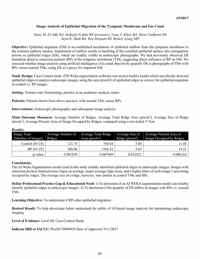
ANS017
Image Analysis of Epithelial Migration of the Tympanic Membrane and Ear Canal
Hany M. El-Adle BA, Sudeepti Vedula BS (presenter), Lena A. Kheir BA, Shree Nadkarni BSSejal K. Shah BA, Ron Despain BS, Robert Jyung MD
Objective: Epithelial migration (EM) is an established mechanism of epithelial outflow from the tympanic membrane to the external auditory meatus. Impairment of outflow results in buckling of the cornified epithelial surface into corrugations known as epithelial ridges (ER), which are readily visible in endoscopic photographs. We had previously observed ER formation distal to retraction pockets (RP) of the tympanic membrane (TM), suggesting direct influence of RP on EM. We assessed whether image analysis using artificial intelligence (AI) could objectively quantify ER in photographs of TMs with RPs versus normal TMs, using ER as a proxy for impaired EM.
Study Design: Case-Control study. FIJI Weka segmentation software was used to build a model which specifically detected epithelial ridges to analyze endoscopic images, using the area (pixels2) of epithelial ridges as a proxy for epithelial migration in control vs. RP images.
Setting: Tertiary-care Neurotology practice at an academic medical center.
Patients: Patients drawn from above practice, with normal TMs versus RPs.
Interventions: Endoscopic photography and subsequent image analysis.
Main Outcome Measures: Average Number of Ridges, Average Total Ridge Area (pixels2), Average Size of Ridge (pixels2), Average Percent Area of Image Occupied by Ridges, compared using a two-tailed T-Test.
Results: Image Type (Number of Images)
Average Number of Ridges
Average Total Ridge Area (pixels2)
Average Size of Ridge (pixels2)
Average Percent Area of Image Occupied by Ridges
Control (N=25) 121.76 704.64 5.80 11.60 RP (N=25) 206.00 1166.52 5.65 19.21
p-value 0.001858 0.007049 0.652921 0.006162
Conclusions: The AI Weka Segmentation model used in this study reliably identified epithelial ridges in endoscopic images. Images with retraction pockets featured more ridges on average, larger average ridge areas, and a higher share of each image’s area being occupied by ridges. The average size of a ridge, however, was similar in control TMs and RPs.
Define Professional Practice Gap & Educational Need: 1) To determine if an AI WEKA segmentation model can reliably identify epithelial ridges in endoscopic images. 2) To determine if the quantity of ER differs in images with RPs vs. normal TMs.
Learning Objective: To understand if RPs alter epithelial migration.
Desired Result: To help physicians better understand the utility of AI-based image analysis for interpreting endoscopic imaging.
Level of Evidence: Level III, Case-Control Study
Indicate IRB or IACUC: Pro20170000936 Date of Approval: 9/11/2017
67
ANS018
Favorable Cochlear Implant Performance in an Adult Patient with Prior Neonatal Hyperbilirubinemia
Jeffrey P. Aldinger, MD; Peter G. Volsky, MD
Objective: To describe initial observations following cochlear implantation with hearing preservation in a young adult with hearing loss attributed to neonatal hyperbilirubinemia.
Study Design: Case report
Setting: Tertiary care
Patients: Single female patient, with speech and movement disorder, normal Mini-Mental Status Exam score, and bilateral moderate to severe sensorineural hearing loss satisfying cochlear implant criteria. She had absent distortion-product otoacoustic emissions and absent brainstem auditory evoked potentials.
Interventions: Bilateral, sequential cochlear implantation (CI 522, Cochlear Ltd.) by the senior author
Main Outcome Measures: AzBio scores and pure-tone thresholds
Results: Four months after implantation of the right ear, she preferred the CI to her left hearing aid, and scored 25% (AzBio) and 30% (CNC words), improved from 12% and 20%, respectively, before surgery. The left ear was implanted 6 months after the right. Two months later, using left CI alone, she scored 51% (AzBio), improved from 26% before surgery. Interestingly, neural response telemetry (NRT) post-implantation was detectable. Low-tone (125, 250, and 500 Hz) pure tone averages (PTA) bilaterally were 48 dB before implantation and 52-55 dB postoperatively. Late-term follow-up to be reported as it becomes available.
Conclusions: In young adults with hearing loss from neonatal hyperbilirubinemia, cochlear implantation can be achieved with hearing preservation and provides benefit to the patient.
*Define Professional Practice Gap & Educational Need: Because this pathology is also associated with auditoryneuropathy/dys-synchrony, and outcomes data is scant for this population of youth and adults, these observations valuableto audiologists and surgeons, who may remain guarded when counseling patients. Good results are achievable in spite ofpreoperative evidence of central auditory dysfunction.
*Learning Objective: Hearing loss in the setting of chronic bilirubin encephalopathy is caused by pathology of the cochlea, brainstem nuclei, and central nervous system. We report the case of a patient with bilateral moderate to severe sensorineuralhearing loss, absent otoacoustic emissions, absent brainstem auditory evoked responses, who experienced benefit fromcochlear implantation with hearing preservation.
*Desired Result: Inform surgeons and audiologists in their counseling of patients with moderate to severe hearing lossattributed to chronic bilirubin encephalopathy.
Level of Evidence - Level V
Indicate IRB or IACUC : Exempt
68
ANS019
Migraine Features in Patients with Migraine-Related Aural Fullness
Adwight Risbud, BS; Mehdi Abouzari, MD, PhD; Ariel Lee, BS Sina Soltanzadeh-Zarandi, BS; Ethan Muhonen, MD; Elaine Martin, MD; Hamid R. Djalilian, MD
Objective: To evaluate the presence of migraine features between patients with migraine-related aural fullness (AF) who meet the International Classification of Headache Disorders (ICHD) 3rd edition criteria for migraine headache and those who do not.
Study Design: Retrospective cohort.
Setting: Tertiary-care neurotology clinic.
Patients: Seventy-seven patients diagnosed with migraine-related AF (defined as prolonged aural fullness concurrent with migraine features and other etiologies ruled out with exam, audiometry, CT, and MRI) between 2014-2020, with a mean age of 56 ± 15 years.
Interventions: Patients were evaluated for meeting the ICHD 3rd edition criteria for migraine headache.
Main Outcome Measures: We compared the prevalence of migraine features in patients who met the full ICHD 3rd edition criteria for migraine headache to those who did not.
Results: There were 55 females (71%) and 22 male patients (29%). Eleven patients (14%) fulfilled the full ICHD criteria for migraine headache (migraine group). Of the patients who did not meet the full criteria (non-migraine), 17 (22%) met 4/5 criteria, and 32 (42%) met 3/5 criteria, for a total of 49 (64%) patients. The migraine and non-migraine groups were only different in 5 of 20 migraine features, including family history of migraine (p=0.007), sound sensitivity (p<0.001), mental fogginess (p=0.008), visual motion sensitivity (p=0.008), and light sensitivity (p<0.001).
Conclusions: The lack of meaningful differences in migraine features between patients in our cohort with migraine-related AF who fulfilled the ICHD migraine criteria and those who do not suggests that the diagnostic criteria for migraine may be too strict and unnecessarily exclude many patients from receiving migraine treatment.
Define Professional Practice Gap & Educational Need: Recent literature described the clinical entity of isolated, prolonged aural fullness as a potential symptom of otologic migraine, with many patients experiencing symptomatic and quality of life improvement following treatment with migraine prophylaxis and lifestyle modification. However, many patients with migraine-related AF do not meet the ICHD criteria for migraine and thus may not be treated as migraine patients by clinicians. In order to account for these patients, it will be important to re-examine the ICHD migraine criteria and to determine whether there exist meaningful differences in the prevalence of migraine features and symptomatology in these patients compared to those who meet the ICHD migraine criteria. Our study further supports the need to evaluate patients presenting with isolated aural fullness for possible migraine disorder when other causes have been ruled out.
Learning Objective: To educate ANS members on a series of patients with aural fullness and further characterize migraine-related AF and identify limitations in the diagnostic criteria for migraine that may prevent patients from receiving appropriate treatment.
Desired Result: Increased awareness and consideration of migraine-related AF in the differential diagnosis by clinicians and expansion of the ICHD criteria for migraine headache may help expand the pool of patients who benefit from migraine therapy.
Level of Evidence - IV
Indicate IRB or IACUC: The study has IRB approval from the UC Irvine review board under the PI name of Hamid R. Djalilian.
69
ANS020
Distribution of Neurotology-Otology Practitioners Across the United States
Geoffrey C. Casazza, MD; Bradley W. Kesser, MD; Clough Shelton, MD Richard K. Gurgel, MD; George T. Hashisaki, MD
Objective: Determine the geographic and per capita distribution of neurotologic surgeons within the United States (USA).
Study Design: National database review
Setting: USA
Subjects: Active neurotologic surgeons
Interventions: The American Neurotology Society and ENThealth.org membership databases were used to identify neurotology-otology practitioners within the USA. Only providers practicing independently as of June 2020 were included. Providers were divided by regional national census areas, state, and by largest population statistical area based on 2019 USA Census population estimates.
Main Outcome Measures: Number of neurotology-otology practitioners per one million populations determined. Correlation between number of practitioners and population size calculated.
Results: 482 practitioners were identified, representing 49 states. The East-North-Central (Wisconsin, Michigan, Illinois, Indiana, Ohio) was the most concentrated region (1.89 per million) whereas the West-South-Central (Texas, Oklahoma, Arkansas, Louisiana) was the least concentrated region (0.9 per million). The highest concentration of practitioners by state are within the District of Columbia (4.25), Vermont (3.21), North Dakota (2.62), Massachusetts (2.61), and New York (2.21) (per million), whereas Mississippi (0.67), Georgia (0.66), New Mexico (0.48), Nevada (0.15), and Wyoming (0.0) (per million) were the least concentrated. Increasing number of practitioners was significantly correlated to largest census designation (r =0.94; p <0.0001), state (r =0.9; p <0.0001), and regional national census area (r =0.92; p =0.0005).
Conclusions: Neurotology-otology practitioners are distributed disproportionately across the USA, where higher population areas show higher numbers per capita of practitioners. Some regions are significantly under-represented. Variability in clinical practice may account for increasing under-representation not captured in this analysis.
*Define Professional Practice Gap & Educational Need: distribution of neurotologic practitioners across the UnitedStates
*Learning Objective: to better understand the national distribution of neurotologic practitioners with identification ofunder- and over-served regions, and the effect of various regional population size on number of providers
*Desired Result: improved understanding of the national distribution of neurotologic surgeons and identification ofunder-served regions
Level of Evidence – Level IV
Indicate IRB or IACUC: Exempt
70
ANS021
Post-Traumatic Superior Semicircular Canal Dehiscence: A Newly Described Phenomenon
Hilary McCrary MD, MPH; Eric Babajanian MD; Neil Patel MDMatthew L. Carlson MD; Richard K. Gurgel MD, MSCI
Objective: To evaluate patients who become symptomatic for superior semicircular canal dehiscence (SSCD) immediately following trauma.
Study Design: Retrospective case series.
Setting: Two academic medical centers.
Patients: 11 patients diagnosed with SSCD following trauma.
Interventions: Imaging, VNG/VEMP testing, audiometric assessment, and surgical repair.
Main Outcome Measures: 1) Description of audio-vestibular symptoms, 2) Mean pre- and post-operative pure tone average (PTA), word recognition score (WRS), and air bone gap (ABG)
Results: Among patients included in the study, 82% were male. A majority of patients suffered a fall (68%) as the mechanism of trauma. Approximately 36% were found to have bilateral SSCD on imaging, with 65% of patients pursuing surgical management of their SSCD. The most common presenting symptoms were: pulsatile tinnitus (91%), hearing loss (64%), vertigo (55%), autophony (46%%), and somatosounds (27%). 54% of patients underwent VNG/VEMP testing, with 83.3% of those demonstrating abnormal results. The mean audiometric findings on the symptomatic side were the following: PTA=36.6 dB, WRS=79%, and ABG=17.4 dB. Among patients that underwent surgery, there was no statistically significant change in the PTA (preoperative=45.2 dB versus postoperative=45.6 dB; p=0.89) or WRS (preoperative=73% versus postoperative=60%; p=0.53). However, there was a reduction in the ABG (preoperative=25 dB versus postoperative=15 dB; p=0.04).
Conclusions: A subset of SSCD patients become symptomatic following trauma, most commonly presenting with pulsatile tinnitus and hearing loss. A proposed mechanism is an abrupt elevation in labyrinthine or intracranial pressure that results in a critical "break" of an already thin or minimally dehiscent superior semicircular canal.
Define Professional Practice Gap & Educational Need: Currently, there are only sparce case reports describing the potential effects of trauma on patients with SSCD. Given concern for the potential effects of trauma on this subset of patients, a case series is presented to further describe this unique patient population.
Learning Objective: To understand the presenting symptoms and objective assessments of patients presenting with SSCD after a trauma.
Desired Result: Increased awareness of a unique subset of patients presenting with SSCD after trauma.
Level of Evidence – Level V: Case series, studies with no controls
Indicate IRB or IACUC: Approved: University of Utah IRB #00045048
71
ANS022
Endolymphatic Sac Surgery in Intractable Meniere’s Disease: A Single-Center Retrospective Cohort Study
Oliver Y. Chin, MD; Doron Sagiv, MD; Hilary A. Brodie, MD, PhD; Rodney C. Diaz, MD
Objective: To demonstrate the impact that endolymphatic sac surgery (ELS) has in patients with Ménière's disease as measured by vertigo control and hearing results.
Study Design: Retrospective cohort review.
Setting: Tertiary care referral center.
Patients: 26 adult patients who underwent ELS for Ménière's disease were included. All patients suffered from debilitating episodic vertigo despite maximal medical therapy.
Interventions: Endolymphatic sac surgery was performed with either decompression alone or shunting. Techniques differed between 2 surgeons.
Main Outcome Measures: Vertigo symptom control and hearing outcomes
Results: Improvement in vertigo symptom control was reported in 15 patients (57.7%), while hearing was improved or stable in 25 patients (96.1%). Only one patient endorsed hearing loss thought to be unrelated to surgical trauma. Drop attacks were reported in 2 patients and complete control was achieved in both. Four patients were able to return to work or play. Only 1 patient required labyrinthectomy for continued debilitating vertigo. There was a median change in 4-tone average of -0.5 dB (95% CI -6.5 to 10.8) and a median change in word-recognition score average of 0% (95% CI -20.9 to 6.9).
Conclusions: Endolymphatic sac surgery remains a highly effective surgical option for Ménière's disease in patients with incapacitating vertigo while preserving hearing.
*Define Professional Practice Gap & Educational Need: Provide further evidence that endolymphatic sac surgery is safeand efficacious in the algorithm for treatment of debilitating episodic vertigo of Ménière's disease for patients who failmaximal medical therapy
*Learning Objective: Review endolymphatic sac surgery as a safe and efficacious treatment option for Ménière's diseaseto address debilitating vertigo low hearing morbidity and potential subjective and audiometric improvement of sac surgery.
*Desired Result: To convince the audience to alter their practice and incorporate endolymphatic sac surgery in escalationtherapy.
Level of Evidence - Level IV
Indicate IRB or IACUC: Approved, #1610645-1 UC Davis IRB Administration
72
ANS023
The Laterality of Age-Related Hearing Loss and Depression
Alexander Chern, MD; Alexandria L. Irace, BA; Justin S. Golub MD, MS
Objective: There is a known association between hearing loss (HL) and depressive symptoms. The objective was to establish if there is a stronger association with the left or right ear.
Study Design: Cross-sectional analysis of prospective epidemiologic cohort study
Setting: Hispanic Community Health Study (US, multicentered)
Patients: 5,499 adults ≥50 years old
Interventions: none
Main Outcome Measures: The main outcome was depressive symptoms, measured by the 10-Item Center for Epidemiologic Studies Depression Scale-10 (CESD-10) and defined continuously and binarily. Subjects with CESD-10≥10 were categorized as having clinically significant depressive symptoms (CSDS). Linear and logistic regressions were performed to assess the association between depressive symptoms and hearing in each ear, adjusting for age, sex, education, cardiovascular disease, and hearing aid use.
Results: Mean age was 58.5±6.3 years. Mean pure-tone average (PTA) was 20.3±11.7 dB (range=0-125) in the right ear and 20.3±12.4 dB (range=-2.5-120) in the left. Multivariable regression adjusting for covariates demonstrated significant associations between depressive symptoms and HL in both the left and right ear. For every 10-dB worsening in right ear PTA, there was 0.89-point increase in CESD-10 (95% confidence interval=0.59-1.2), and odds of CSDS increased 1.31 times (1.17-1.46). For every 10-dB worsening in left ear PTA, there was a 0.85-point increase in CESD-10 (0.55-1.14), and odds of CSDS increased 1.34 times (1.2-1.49).
Conclusions: Worsening hearing in both right and left ears was associated with increased depressive symptoms and odds of CSDS. No ear laterality was demonstrated. This will help inform understanding of the laterality of central auditory connections.
REQUIRED: Define Professional Practice Gap & Educational Need: Age-related hearing loss is a highly prevalent and severely undertreated disease. Studies have established an independent association between hearing loss and neuropsychiatric disorders of the elderly (e.g., depression). However, there is minimal literature examining the laterality of this association (i.e., if hearing loss of one side contributes to this association more than the other side) and existing studies demonstrate conflicting results. Understanding laterality differences in central auditory pathways may help elucidate mechanisms explaining the relationship between age-related hearing loss and depression. This will help inform healthcare personnel of hearing loss as a targetable modifiable risk factor (i.e., with hearing aids) for prevention of neuropsychiatric disorders of the elderly.
Learning Objective: After this presentation, the learner will be able to describe the relationship between age-related hearing loss and depressive symptoms and posit why hearing loss has been identified as a modifiable risk factor for prevention of neuropsychiatric disorders of the elderly.
Desired Result: Otolaryngologists will better understand the complex relationship between age-related hearing loss and neuropsychiatric diseases of the elderly, such as depression.
Level of Evidence – Level III
Indicate IRB or IACUC: AAAQ9546(M00Y01): Designated Not Human Subjects Research Under 45 CFR 46
73
ANS024
Traumatic Perilymphatic Fistula: A Systematic review
Rebecca A. Compton, MD; Julian F. Oviedo, BA; Jonathon S. Sillman, MD
Objective: To identify cases of traumatic perilymphatic fistula reported in the literature in order to review the optimal management of this condition.
Study Design: Systematic review
Setting: Case reports of traumatic perilymphatic fistula identified between 1968 to present
Patients: 60 patients who presented with cochleovestibular symptoms following blunt or penetrating trauma, with evi-dence of perilymphatic fistula on examination, audiogram, or imaging
Interventions: None
Main Outcome Measures: Resolution of vertigo and improvement in hearing loss on audiogram after either conservative or surgical therapy
Results: A total of 60 cases were identified during the study time frame, including 38 cases of blunt trauma and 22 cases of pene-trating trauma. Perilymphatic fistula was suspected based upon a positive fistula test, abnormal audiovestibular testing, and/or pneumolabyrinth on computed tomography. Treatment information was available in 55 cases; 7 patients were man-aged conservatively, with improvement in vertigo in all cases and improved hearing in 4 cases. For the remaining 48 cases, middle ear exploration was performed and this was typically in delayed fashion when such information was re-ported (average 3 months after injury). There was improvement in vertigo symptoms in 92% (44/48) and in hearing loss in 63% (30/48).
Conclusions: Vertigo and hearing loss after traumatic perilymphatic fistula may resolve with conservative measures. Middle ear exploration can be offered in delayed fashion for those cases that do not.
*Define Professional Practice Gap & Educational Need: The optimal management of traumatic perilymphatic fistularemains unclear, and there is a need for consensus statements on this condition.
*Learning Objective: To identify cases of traumatic perilymphatic fistula reported in the literature in order to review theoptimal management of this condition.
*Desired Result: To find and collate greater than 50 cases of traumatic perilymphatic fistula in order to investigate themanagement of this condition.
Level of Evidence: Level V
Indicate IRB or IACUC: Exempt
74
ANS025
Intravenous Sodium Fluorescein and Vestibular Schwannoma Surgery
C. Scott Brown, MD; Ashish H. Shah, MD; Michael Ivan, MD; Christine Dinh, MD
Objective: Describe the operative experience using intravenous (IV) sodium fluorescein (SF) during vestibular schwannoma (VS) surgery
Study Design: Case report
Setting: Tertiary Care Hospital
Patients: A 39-year-old male with right-sided hearing loss was diagnosed with a large tumor involving the right internal auditory canal (IAC) and cerebellopontine angle (CPA) with brainstem compression. The IAC was severely dilated and without a fundal fluid cap.
Interventions: Informed consent was obtained and IV SF (2 mg/kg) was administered shortly after anesthesia induction. A retrosigmoid craniotomy with IAC drilling was performed. Once the CPA was exposed, the FL560 fluorescence module was applied to the operative microscope (~160 minutes after SF administration). Intraoperative video and photography were performed.
Main Outcome Measures: Ability of surgeons to visualize tumor and distinguish from adjacent cranial nerves
Results: When the FL560 module was applied, surgeons were able to distinguish tumor from adjacent vessels and cranial nerves, by observing the tumor’s green fluorescence. As time progressed, the tumor’s green fluorescence diminished and the tumor-tissue interfaces were difficult to distinguish. A sliver of tumor was left in the IAC, where it was encasing the blood supply to the facial nerve. The facial and cochlear nerves were grossly preserved and the facial nerve stimulated at 0.05 mA. Pathology confirmed a diagnosis of VS.
Conclusions: In this patient, the VS demonstrated preferential uptake of IV SF, which helped surgeons distinguish tumor from normal intracranial structures under fluorescent conditions. Future studies are needed to determine optimal dosing strategies and the utility of fluorescein guidance in challenging VS surgeries.
*Define Professional Practice Gap & Educational Need:Although preferential sodium fluorescein uptake by vestibular schwannoma has been shown in vitro, in vivo, and in isolatedcase reports, no specific protocols or guidelines exist for fluorescein-guided vestibular schwannoma surgery.
*Learning Objective:Recognize the potential of sodium fluorescein for delineating tumor from adjacent vessels and cranial nerves duringchallenging vestibular schwannoma surgery.
*Desired Result:Understanding that fluorescein guidance may potentially improve tumor visualization during vestibular schwannomasurgery and clinical trials to determine optimal dosing strategies are warranted.
Level of Evidence - V
Indicate IRB or IACUC: Exempt
75
ANS026
Primary Vestibular Schwannoma Cells Activate RAD51-Associated DNA Repair Following Radiation-Induced DNA Damage
Torin P. Thielhelm, BS; Stefania Goncalves, MD; Scott Welford, PhD Eric Mellon, MD, PhD; Michael E. Ivan, MD; Fred Telischi, MD; Christine T. Dinh, MD
Hypothesis: Vestibular Schwannoma (VS) can avoid cell death following radiation injury by entering cell cycle arrest and activating RAD51-related DNA repair.
Background: Although the radiobiology of various cancers is well-studied, the biological effects of radiation on VS are poorly understood. In this study, we describe how VS cells enter cell cycle arrest (through p21 and phospho-Rb expression), activate DNA repair mechanisms (through RAD51 upregulation), and avoid cell death, in response to radiation-induced double-strand breaks (DSB) in DNA (as measured by gamma-H2AX).
Methods: Primary human VS cells were cultured on 96-well plates and 16-well culture slides at 10,000 cells/well and exposed to either 0 or 18 Gray of radiation. Viability assays were performed at 96 hours in vitro and compared between cell lines. Immunofluorescence for gamma-H2AX, RAD51, p21, and phospho-Rb were performed at 6 hours.
Results: Radiation (18 Gray) induced the expression of gamma-H2AX in cultured VS, suggesting that radiation initiated DSBs in DNA. Cell cycle proteins, p21 and phospho-Rb, were also upregulated in irradiated cells, indicating that VS cells enter into cell cycle arrest following radiation injury. While in cell cycle arrest, VS initiated RAD51 expression in an effort to repair radiation-induced DNA damage to evade cell death, as demonstrated by viability assays.
Conclusions: In response to radiation-induced DNA damage, primary VS cells can enter cell cycle arrest and express RAD51 DNA repair mechanisms to avoid cell death. Further investigation into the expression of DNA repair proteins and cell cycle checkpoints in VS may provide important insight on radiation resistance.
Define Professional Practice Gap & Educational Need: The biological mechanisms responsible for radiation resistance in vestibular schwannoma are unknown. Understanding the mechanisms behind radiation-induced DNA damage and the activation of DNA repair pathways in vestibular schwannoma will provide important insight into ways of overcoming radiation resistance in the future.
Learning Objective: Recognize that ionizing radiation can cause DNA damage in vestibular schwannoma and that tumors can activate mechanisms to evade radiation-induced cell death.
Desired Result: Understand that radiation resistance in vestibular schwannoma may be related to the ability of tumor cells to enter into cell cycle arrest to repair DNA damage.
Level of Evidence: N/A
Indicate IRB or IACUC: Approved 9/26/2017. University of Miami, IRB #20150637
76
ANS027
Middle Ear Exploration and Application of Steroid Soaked Gelfoam in Treatment of Meniere’s Disease
Luis P. Roldan, MD, Meghana Kalaver, MS (presenter), Lilliana Ein MDCaralin Schneider MS, Michael Hoffer MD
Objective: To evaluate vertigo control with application of steroid-soaked gelfoam to the middle ear in patients with Meniere’s Disease (MD)
Study Design: Retrospective review
Setting: Single institution review at University of Miami Hospital
Patients: Adult patients with MD undergoing labryinthotomy with steroid application to the middle ear between 1/12014 - 7/1/2019
Interventions: Middle ear exploration (MEE) with direct placement of dexamethasone soaked gelfoam to the round and oval window
Main Outcome Measures: Primary outcome measures included patient reported frequency of vertigo attacks and subjective vertigo response post intervention. Secondary outcome measures included subjective responses to hearing, tinnitus, and aural fullness.
Results: 30 adult patients were included in final analysis. Frequency of pretreatment vertigo occurred at an average of 8.84 vertigo attacks per month. Post treatment vertigo at one month follow up occurred at average of 1.76 attacks per month with similar findings observed at 1-3 months (average 0.067 vertigo attacks/month) and 3-6 months (1.667 attacks/month). 23/30 (76%) reported subjective improvement in vertigo symptoms following intervention.
Conclusions: Operative placement of steroid soaked gelfoam to the oval and round window yields favorable results in patient with MD with respect to vertigo control
Define Professional Practice Gap & Educational Need: Transtympanic steroid therapy is an alternative method for treatment of MD associated vertigo. The ideal way to deliver steroids to the inner ear has not yet been proven.
Learning Objective: Our study describes an alternative method for delivering steroid therapy to the inner ear that involves middle ear exploration and placement of steroid soaked gelfoam to the oval and round windows. This allows for sustained steroid delivery to the inner ear which may improve vertigo symptoms in patients with MD.
Desired Result:This review yielded favorable preliminary data in highlighting the proposed intervention as an effective means of vertigo control in MD
Level of Evidence Level V
Indicate IRB or IACUC : IRB 20180646 at University of Miami
77
ANS028
Remote Intraoperative Neural Response Telemetry: Technique and Results in Cochlear Implant Surgery
Ali Kouhi, MD; Austin Swanson, PhD; Matthew Fitzgerald, PhD; Nikolas Blevins, MD
Objective: Present results with remote intraoperative neural reponse telemetry (NRT) during cochlear implantation (CI) and its utility in overcoming the inefficiency of in-person NRT.
Study Design: Case series
Setting: Tertiary academic otology practice
Patients: All patients undergoing primary or revision CI, both adult and pediatric, were enrolled.
Interventions: Remote intraoperative NRT performed by audiologists using a desktop computer to control a laptop in the operating room. Testing was performed over the hospital network using commercially available software. A single system was used to test all 3 FDA-approved manufacturers’ devices.
Main Outcome Measures: Success rate and time savings of remote NRT.
Results: Out of 254 procedures, 252 (99.2%) underwent successful remote NRT. In 2 procedures (0.7%), remote testing was unsuccessful, and required in-person testing to address technical issues. Both failed attempts were due to hardware failure (OR laptop or headpiece problems). There was no relation between success of the procedure and patient/surgical factors such as difficult anatomy, or the approach used for inner ear access. The audiologist time saved using this approach was considerable when compared with in-person testing.
Conclusions: Remote intraoperative NRT testing during cochlear implantation can be performed effectively using standard hardware and remote-control software. Especially important during the Covid-19 pandemic, such a procedure can reduce in-person contacts, and limit the number of individuals in the operating room. Remote testing can provide additional flexibility and efficiency in audiologist schedules.
*Define Professional Practice Gap & Educational Need: In-person NRT can be time consuming and requiresconsiderable resources, especially in centers with high physical distance between the OR and the audiology department.Remote testing can be easily and effectively implemented to address these concerns.
*Learning Objective: benefits and feasibility of remote NRT in CI surgery
*Desired Result: Remote NRT can be reliably and successfully performed
Level of Evidence - IV
Indicate IRB or IACUC : Stanford University IRB (Protocol number: 48035)
78
ANS029
Primary Melanoma of the Middle Ear and Petrous Temporal Bone Treated with Radiation Therapy and Single-Agent Nivolumab
Kevin J. Carlson, MD; Peter G. Volsky, MD
Objective: We present a case of primary melanoma of the middle ear and petrous temporal bone with remission achieved following radiation and single-agent nivolumab.
Study Design: Single Case Report
Setting: Tertiary Care
Description of Case: A 74-year-old male developed sudden right sided unilateral deafness, disequilibrium, and otalgia with CN V and VII weakness. Imaging demonstrated an osteolytic lesion involving the right middle ear and anterior petrous apex with encasement of the carotid artery. Middle ear biopsy demonstrated melanoma. Initial treatment consisted of radiation therapy (30 Gy, 10 Fractions) followed by induction nivolumab. Following 5 cycles every 2 weeks of nivolumab, cranial neuropathies resolved, and PET/CT imaging demonstrated complete response. Following maintenance therapy (6 cycles, q3w), an FDG-avid retroperitoneal lymph node responded to continued treatment. Remission was achieved after completion of one year of nivolumab and stable remission was demonstrated after four months of active surveillance. Nine months following cessation of treatment, imaging demonstrated a progressive right parafalcine frontal lobe lesion, treated with Gamma Knife radiosurgery. The patient later underwent craniotomy and biopsy of the frontal lesion, confirming melanoma. The patient expired from cardiac complications shortly after surgery, 2 years and 8 months from diagnosis.
Conclusions: Primary mucosal melanoma of the middle ear and petrous temporal bone is exceedingly rare, with less than 25 reported cases. Management is case specific and often involves surgical resection. In this case, an unresectable tumor demonstrated complete response and reversal of cranial nerve neuropathies following radiation and anti-PDL1 therapy, suggesting a non-surgical option for similar lesions.
*Define Professional Practice Gap & Educational Need: The rarity of primary mucosal melanoma of the middle earmakes treatment case specific. There is a paucity of literature describing non-surgical options for these patients.
*Learning Objective: To describe a case of unresectable mucosal melanoma of the middle ear and petrous apex in whichcranial nerve deficits were reversed and remission achieved after treatment with radiation and nivolumab.
*Desired Result: Appreciate nivolumab as potential treatment for non-resectable mucosal melanoma of the middle ear andpetrous apex.
Level of Evidence - Level V
Indicate IRB or IACUC : Exempt.
79
ANS030
Endoscopic Medial Reepithelization for Inflammatory Canal Stenosis
Sonia M. Scaria; Aaron D. Tward, MD, PhD
Objective: Inflammatory External Auditory Canal (EAC) Stenosis arises from infiltration of inflammatory cells, edema and eventual sclerosing of the medial EAC, leading to complete obstruction and conductive hearing loss. Current treatment includes surgical resection of the affected area with widening and reepithelization of the EAC via post-auricular incision, but the condition is reported to recur with high frequency. Our aim was to assess the feasibility of endoscopic treatment as an alternative to open surgery and understand its effect on recurrence rates.
Study Design: Retrospective case review
Setting: Tertiary referral center
Patients: 4 patients were included who had conductive hearing loss and inflammatory canal stenosis all with gross thickening of the tympanic membrane.
Interventions: Patients underwent endoscopic removal of obstructive tissue and reepithelization with split-thickness skin grafting.
Main Outcome Measures: Post-operative air-bone gap (ABG), lack of recurrence, subjective reporting of hearing improvement, and lack of drainage.
Results: 8 out of 8 ears (n = 4 patients) had significant improvement in hearing. No recurrence has been observed in any of the patients over a mean follow-up time of 46 months (range =31-65). Average reduction in ABG was 14 dB (SD = 7) with a statistically significant difference between the pre-operative and post-operative ABG at 512 Hz (p =.0014)
Conclusions: Endoscopic treatment of Inflammatory EAC Stenosis obviates the need for post-auricular incision and results in clinical improvement with a favorable recurrence rate.
REQUIRED: Define Professional Practice Gap & Educational Need: Currently, Inflammatory EAC Stenosis is treated via a post-auricular approach, and there is a high rate of recurrence.
Learning Objective: Review the current practice in regard to treatment of inflammatory EAC stenosis; Analyze the utility of an endoscopic treatment for EAC Stenosis; Observe the change in patient outcomes using the Endoscopic Medial Reepithelization
Desired Result: The patients will report significantly better hearing loss post-surgery, and the air-bone gap will be decreased substantially using an endoscopic medial reepithelization, while also reducing recurrence rates. Physicians can then see the value in potentially adopting a non-invasive treatment for Inflammatory EAC stenosis.
Level of Evidence – V – case series
Indicate IRB or IACUC : IRB
80
ANS031
Auricular Cartilage Resection for Treatment-Refractory Chronic Chondritis: A Case Series
Steven A. Gordon, MD, MPH; Ashley M. Nassiri, MD, MBA; Colin L. Driscoll, MD Matthew L. Carlson, MD; Neil S. Patel, MD
Objective: Describe a series of cases of chronic auricular chondritis refractory to antibiotics and steroids treated successfully with surgery
Study Design: Case Series
Setting: Two tertiary academic medical centers
Patients: We analyzed four patients diagnosed with chronic auricular deformity, pain, and drainage for a period of 1-5 years who had failed prolonged treatment consisting of antibiotics, corticosteroids, and incision and drainage. All four patients were smokers, three were diabetic. One patient suffered a gastrointestinal bleed related to prolonged corticosteroid therapy during her medical treatment.
Interventions: Operative subcutaneous partial auriculectomy (removal of diseased cartilage and excess skin) was performed.
Main Outcome Measures: Resolution of pain and drainage, need for additional procedures, and reduction in narcotics required for pain control were analyzed.
Results: Two of the four patients were given an immediate post-operative course of doxycycline and ciprofloxacin. With a minimum of 6 weeks’ follow-up, all four patients had complete resolution of pain and recurrent drainage post-operatively. One patient requiring multi-daily narcotic medication for pain and benzodiazepine for sleep preoperatively no longer required prescription medication. All specimens revealed chronic dermal and cartilage inflammation. Three of four cases had polymicrobial infection. One case had only skin contaminant growth on culture following multiple oral and parental antibiotic regimens.
Conclusions: Surgical excision of diseased cartilage as a result of chronic chondritis is an effective treatment in those cases refractory to antibiotics and incision and drainage, and should be considered early in the treatment algorithm for similar patients to avoid morbidity related to prolonged medical treatment.
*Define Professional Practice Gap & Educational Need: Chronic chondritis refractory to antibiotics and incision anddrainage presents a treatment challenge. There exists little data to support treatment decision-making. This case seriesdescribes a promising surgical approach to treatment with excellent results and resolution of presenting symptoms andshould serve as a resource for surgeons who encounter similar cases.
*Learning Objective: Understanding the effect of surgical management in the treatment of patients with refractorychronic auricular chondritis.
*Desired Result: To offer an additional tool in the armamentarium for treatment of refractory chronic auricularchondritis.
Level of Evidence - Level V
Indicate IRB or IACUC: Exempt.
81
ANS032
Transmastoid Repair of Encephaloceles and Cerebrospinal Fluid Leaks in Patients with Canal Wall Down Mastoidectomy
Tirth R. Patel, MD; Alexa S. Roy, BS; Ali Z. Piracha, BSElias Michaelides, MD; R. Mark Wiet, MD
Objective: To describe the efficacy of single-stage transmastoid repair of encephaloceles or cerebrospinal fluid (CSF) leaks in patients requiring canal wall down mastoidectomy (CWD).
Study Design: Case series
Setting: Tertiary-care hospital
Patients: A total of 14 patients were included in the study. Three patients had a CWD mastoidectomy performed in a separate operation prior to repair of the encephalocele or CSF leak. The remaining 11 patients had an encephalocele repaired concurrently with CWD mastoidectomy.
Interventions: Surgical repair of an otogenic encephalocele or CSF leak using a CWD transmastoid approach or in patients with a prior CWD cavity.
Main Outcome Measures: Failure of repair, size of defect repaired, materials used for repair, use of lumbar drain.
Results: Eight patients had cholesteatoma noted at the time of surgery. Mean size of the encephalocele repaired was 0.91 cm. Three patients had mastoidectomy cavities obliterated with fat graft and closure of the external auditory canal. A lumbar drain was used for one patient. No patient required repeat surgery for encephalocele repair or CSF leak.
Conclusions: In appropriately selected cases single-stage transmastoid repair of encephaloceles or CSF leaks in patients with pre-existing CWD cavities or patients undergoing concurrent CWD mastoidectomy, without the addition of a simultaneous middle fossa craniotomy, is effective and safe.
*Define Professional Practice Gap & Educational Need: While transmastoid repair of these encephaloceles has beenreported previously, there have been very few reports of the efficacy of single-stage transmastoid repair of encephalocelesin patients requiring CWD mastoidectomy or with a prior CWD cavity.
*Learning Objective: A single-stage transmastoid approach to CSF leak or encephalocele repair for patients requiringCWD or with a prior CWD cavity may be effective and reduces the morbidity associated with multi-stage or middle cranialfossa craniotomy procedures.
*Desired Result: For surgeons to consider using a single-stage transmastoid approach to CSF leak or encephalocele repairfor patients requiring CWD or with a prior CWD cavity.
Level of Evidence – Level V
Indicate IRB or IACUC: Rush University; ORA #: 20031701-IRB01
82
ANS033
Surgical Management of Temporal Bone Osteoradionecrosis
Peter L. Nguy, MD; Christine Stuart, BS; Kelly Moyer, MD; Peter Ahn, MDK. William Harter, MD; Bruce Davidson, MD; H. Jeffrey Kim, MD
Objective: To review the relevant literature and discuss the surgical management of osteoradionecrosis of the temporal bone
Study Design: Retrospective chart review
Setting: Tertiary academic medical center
Patients: Patients diagnosed with and surgically managed for temporal bone osteoradionecrosis from 2009 to 2019
Interventions: Surgical management of temporal bone osteoradionecrosis
Main Outcome Measures: Incidence and factors influencing surgical management, surgical techniques
Results: In 24 patients (58.3% male) diagnosed with temporal bone osteoradionecrosis, 7 (29.2%) patients ultimately received surgical management. Women were diagnosed significantly earlier than men, at 4.2 years compared to 9.7 years (p=0.0197). After conservative management of all patients via aural toilet, progression of osteoradionecrosis or recurrent otorrhea prompted surgical intervention in seven patients after a mean of 22 months. The procedures performed on these ears included lateral temporal bone resection with mastoid obliteration (n=1), ear canal reconstruction with autograft (n=4), and vascularized pedicled flaps (n=5). 2 patients received more than 1 operation for de novo areas of osteoradionecrosis. Hyperbaric oxygen was used in 3 cases as a preoperative measure prior to surgery for osteoradionecrosis. Parotid (n=45.8%) and nasopharynx (25.0%) primary subsites represented the majority of the sample, with a higher predominance of parotid primaries (n=86%) among those who underwent surgery.
Conclusions: A variety of surgical techniques have been described previously for temporal bone osteoradionecrosis. Vascularized pedicled flaps are a viable option for reconstructing defect of the external auditory canal after unsuccessful conservative measures.
*Define Professional Practice Gap & Educational Need: Osteoradionecrosis of the temporal bone is an extremely rareoccurance and surgical management is controversial, but should be considered in cases with intractable pain.
*Learning Objective: The objective of this study is to better acquaint providers with the surgical management options forosteoradionecrosis of the temporal bone
*Desired Result: Providers will have better knowledge and competence for the surgical management options for temporalbone osteoradionecrosis
Level of Evidence – Level IV
Indicate IRB or IACUC : Georgetown University IRB STUDY00001290
83

ANS034
Sound Distortion with Loud Sound: A Possible Otologic Migraine Phenomenon
Adwight Risbud, BS; Mehdi Abouzari, MD, PhD; Ariel Lee, BS Ethan Muhonen, MD; Elaine Martin, MD; Harrison W. Lin, MD; Hamid R. Djalilian, MD
Objective: To describe a novel series of cases of noise-induced sound distortion in patients with migraine-related tinnitus and hyperacusis.
Study Design: Retrospective cohort.
Setting: Tertiary-care neurotology clinic.
Patients: Four patients with a diagnosis of migraine-related tinnitus and symptoms of sound distortion and ear crackling when exposed to loud noises (at the end of the sounds).
Interventions: All patients were counseled on lifestyle and dietary modification with supplementation with vitamin B2 200 mg bid and magnesium 400 mg bid and if not improved, patients were prescribed migraine prophylactic medications per protocol. MRI and CT of the temporal bones were obtained on all patients and were normal.
Main Outcome Measures: The main outcome measure was reduction or resolution of sound distortion/ear crackling, migraine, and tinnitus symptoms at 4-month follow up.
Results: Three out of four patients had unilateral tinnitus with ipsilateral symptoms of ear crackling heard at the end of loud sounds. All four patients presented with hyperacusis and reported additional migraine features including headache, ocular pain, otalgia, and aural fullness. All patients were treated with lifestyle and diet changes and two patients required migraine prophylactic therapy. All four patients had significant improvement (>50% decrease) in frequency of tinnitus, sound distortion, and migraine symptoms within a 4-month follow-up period. Two of the patients had complete resolution of the sound-induced distortion/crackling.
Conclusions: We report the first cases of sound distortion with loud sound in patients with atypical migraine who responded positively to migraine therapy. Our report adds further support to recent studies proposing an association between migraine disorder and various otologic symptoms.
Define Professional Practice Gap & Educational Need: Until recently, symptoms such as tinnitus and hyperacusis were thought to result exclusively from primary otologic disorders. Furthermore, researchers have attempted to explore the relationship between these audiological symptoms and other disorders, particularly migraine, given the high prevalence of these symptoms observed in migraine cohorts. At present, many patients present with only a minority of the classic migraine symptoms and may not fulfill the current International Classification of Headache Disorders criteria for migraine. However, with a complete history and exam, we may uncover additional features suggestive of migraine disorder and potentially expand the number of patients who may benefit from migraine therapy. This would help further elucidate the pathophysiology of migraine, through a better understanding of the less common otologic manifestations that may contribute to the underdiagnosis and undertreatment of this disabling condition.
Learning Objective: To inform clinicians on a series of patients experiencing loud noise-induced sound distortion and ear crackling and to further characterize the potential otologic manifestations of migraine and migraine-related tinnitus/hyperacusis.
Desired Result: Increased awareness and consideration of sound distortion and ear crackling as migraine symptoms and expanded use of migraine prophylaxis and lifestyle management in patients with these atypical features.
Level of Evidence - IV
Indicate IRB or IACUC: The study has IRB approval from the UC Irvine review board under the PI name of Hamid R. Djalilian.
84

ANS035
Prevalence of Polypharmacy in Patients with Vestibular and Balance Complaints: a Single-Center retrospective review
Tatianna Timor, PharmD Candidate; Adrienne Busch, PharmD Candidate; James Sterrett, PharmD; Habib Rizk, MD, MSc
WITHDRAWN
BY AUTHOR
85
ANS036
Comparison of Cranioplasty Techniques following Translabyrinthine Surgery: Effect on Postoperative Pain and Reduced Opioid Requirements
Pedrom C. Sioshansi, MD; Mulin Xiong, BA; Nathan Tu, MD; Robert S. Hong, MDChristopher A. Schutt, MD; Dennis I Bojrab, MD; Seilesh C. Babu, MD
Objective: To assess differences in postoperative pain, opioid usage, and surgical outcomes between cranioplasty using abdominal fat graft (AFG) versus hydroxyapatite cement (HAC) following translabyrinthine surgery.
Study Design: Retrospective case control
Setting: Tertiary referral center
Patients: Thirty translabyrinthine procedures was evaluated, including 15 consecutive HAC patients and 15 matched AFG patients. Patients were matched by age, gender, body mass index, and tumor size.
Intervention: Cranioplasty using HAC or AFG following translabyrinthine resection of vestibular schwannoma.
Main Outcome Measures: Postoperative patient pain ratings, narcotic usage, inpatient length of stay, and complication rates.
Results: Patients that underwent HAC cranioplasty had lower postoperative pain scores on several measures (p<0.05) and less postoperative narcotic usage (mean 55.3 morphine equivalents, p=0.0031) when compared to those that received AFG closure. There was no difference in length of stay. Postoperative CSF leaks in both groups and skin reactions in AFG closure patients were infrequent.
Conclusion: HAC cranioplasty is a safe technique comparable to AFG closure following translabyrinthine surgery which can decrease postoperative pain and narcotic usage.
*Define Professional Practice Gap & Educational Need: Assess differences between cranioplasty techniques on surgicaloutcomes and narcotic utilization rates.
*Learning Objective: Compare patient rated subjective outcomes and objective differences in opioid requirements between cranioplasty techniques.
*Desired Result: Assess differences in postoperative pain scores and opioid use following translabyrinthine surgery.
Level of Evidence: III
Indicate IRB or IACUC : Ascension Providence IRB approval 1534095-1
86
ANS037
Management of Superior Semicircular Canal Dehiscence Syndrome: Impact of Disease Severity on Management and Outcomes
with Tragal Cartilage Cap Resurfacing Technique
Rohan Basu ,BS; Tiffany P. Hwa, MD; Adam C. Kaufman, MD, PhD Jason A. Brant, MD; Steven J. Eliades, MD, PhD; Michael J. Ruckenstein, MD
Objective: To report outcomes after surgical intervention with tragal cartilage cap resurfacing for patients with superior semicircular canal dehiscence (SSCD)
Study: Retrospective chart review
Setting: Academic tertiary referral center
Patients: Thirty-one consecutive patients (17 operative) from a single clinician with SSCD from 2012-2019
Interventions: Transmastoid tragal cartilage cap resurfacing technique
Primary Outcome Measure: Patient-reported symptom severity in vestibular, otologic, and global disease severity
Results: Thirty-one adult patients, including 18 males and 13 females, were evaluated for SSCD. Seventeen patients chose surgical intervention, while the remainder opted for observation. Mean age and gender distribution were similar between the treatment and observation groups, but patients opting for surgery reported significantly more severe vestibular symptoms on a Likert scale (treatment 2.68 vs. observation 1.32, p<0.01). For patients undergoing surgery, mean operative time was 78.1 minutes, with a mean postoperative stay of 1.11 days. Significant postoperative improvements were seen in subjective ratings for balance (2.68 vs. 1.38, p<0.01), hearing (3.26 vs. 2.20, p<0.01), and global symptom severity (3.50 vs.2.17, p<0.01). One patient experienced persistent vertigo requiring admission with subsequent resolution, and one patient required revision surgery, which resulted in improvement in all symptom domains. There were no other complications and no cases of postoperative sensorineural hearing loss.
Conclusions: For patients electing to undergo surgery, transmastoid tragal cartilage occlusion is an effective technique in the management of symptomatic SSCD syndrome. Benefits include surgical familiarity for the neurotologist, avoidance of a craniotomy, decreased operative time, and noninferior symptomatic outcomes without increased postoperative complications.
Define Professional Practice Gap & Educational Need: To report outcomes on the conservative surgical intervention of tragal cartilage resurfacing; To report patient decision-making for elective surgical intervention based on a self-reported symptom severity index
Learning Objective: To identify and characterize surgical outcomes after tragal cartilage cap resurfacing for superior semicircular canal dehiscence
Desired Result: Improvement in symptomatology for vestibular, otologic, and global disease severity
Level of Evidence – Level IV
Indicate IRB or IACUC: Approved by the University of Pennsylvania Institutional Review Board. Approval#833914. Date of Approval 8/12/2019.
87
ANS038
Endoscope-assisted Superior Semicircular Canal Dehiscence Repair: Single Institution Outcomes
Douglas J. Totten, BA; Miriam R. Smetak, MD; Elizabeth L. Perkins, MDNathan D. Cass MD; David S. Haynes, MD, MMHCAlejandro Rivas, MD; Marc L. Bennett, MD, MMHC
Objective: To determine efficacy of endoscope-assisted middle cranial fossa (MCF) repair of superior semicircular canal dehiscence (SSCD) compared to microscopic MCF repair.
Study Design: Retrospective cohort.
Setting: Tertiary medical center neurotology practice
Patients: SSCD patients who underwent surgical repair via MCF with or without endoscope assistance from 2010-2019.
Main Outcome Measures: Pre- and post-operative changes in symptom burden, as calculated from eight patient-reported symptoms. Pre- and post-operative changes in symptom burden were assessed using paired t-tests. Single-predictor binary logistic regression was used to compare final reported symptoms between cohorts. Linear regression was performed to assess air-bone gap (ABG) changes postoperatively between cohorts.
Results: 46 patients received surgical management for SSCD. Of these, 27 (59%) were male and 19 (41%) were female. Bilateral SSCD was present in 14 cases (29%), of which three underwent surgical management bilaterally, for a total of 49 surgical ears. Surgery was performed on the right ear in 19 cases (39%) and on the left in 30 cases (61%). 40 ears (82%) underwent microscopic repair while 9 (18%) underwent endoscope-assisted repair. Microscopic and endoscope-assisted MCF repair both demonstrated significantly improved symptom burden postoperatively (p < 0.001 for each). There was no significant difference in change in ABG between the two cohorts. On average, patient-reported symptoms and audiometrically-tested hearing improved postoperatively in both groups.
Conclusions: Endoscope-assisted MCF repair yields similar results and is equivalent to microscopic MCF repair while possibly providing better visualization of medial and downslope defects.
Define Professional Practice Gap & Educational Need: Lack of understanding regarding outcomes of endoscope-assisted repair of SSCD.
Learning Objective: Evaluate outcomes of endoscope-assisted SSCD repair compared to repair via the traditional microscopic MCF approach.
Desired Result: This study can identify preliminary results of endoscope-assisted SSCD repair and may guide clinical and surgical management as well as future studies.
Level of Evidence - IV
Indicate IRB or IACUC: IRB Approved (201632, Vanderbilt University Medical Center)
88
ANS039
Cost Analysis following Microsurgical Resection of Vestibular Schwannomas: Does Extent of Resection matter?
Elizabeth L. Perkins, MD; Douglas J. Totten, BA; Nathan D. Cass, MDAlexander D. Sherry, MD; Ankita Patro, MD, David S. Haynes, MD, MMHC
Marc L. Bennett, MD, MMHC
Objective: Compare costs of surgery and complications, cost and number of postoperative interventions, and facial nerve outcomes following subtotal resection (STR) or near-total resection (NTR) and gross total resection (GTR) of vestibular schwannomas (VS).
Study Design: Retrospective case series.
Setting: Tertiary referral center.
Patients: 117 Patients who underwent microsurgical resection of VS between 2014-2018.
Main Outcome Measures: Initial cost of surgery and postoperative care, number and cost of postoperative magnetic resonance images (MRIs) and interventions, surgical or otherwise, and facial nerve function one-year postoperatively.
Results: STR/NTR was not associated with significantly greater cost of admission vs. GTR (average: $47,211 vs $44,252, respectively) (p=0.21) or length of stay (4.2 vs 4.0 days, respectively) (p=0.73). Average House-Brackmann (HB) score for facial nerve function at one year was 1.5 for the STR/NTR cohort compared to 1.6 for GTR (p=0.81). STR/NTR was associated with significantly greater number of post-intervention MRIs compared to GTR (3.0 MRIs vs 2.5 MRIs) (p=0.039). Average length of follow-up was 18 months for STR and 41 months for GTR. There was no significant difference between groups regarding readmissions or facial nerve interventions (p=0.45 and 0.50, respectively).
Conclusions: Within our cohort, STR/NTR was not associated with significantly greater initial costs of admission but was associated with a greater number of postoperative MRIs of compared to GTR. There was no significant difference in one-year facial nerve outcomes between the STR/NTR and GTR cohorts.
Define Professional Practice Gap & Educational Need: Lack of understanding differences in costs of care and overall outcomes between gross total resection and subtotal resection of vestibular schwannomas.
Learning Objective: Demonstrate costs of care and clinical outcomes, particularly regarding facial nerve function associated with subtotal resection compared to those associated with gross total resection.
Desired Result: This preliminary study can provide context for decision-making regarding extent of tumor resection.
Level of Evidence - IV
Indicate IRB or IACUC: IRB Approved (201632, Vanderbilt University Medical Center)
89
The Fallopian Bridge Technique for Access to a Giant Infratemporal Fossa Cholesteatoma: A Case Report
Amir Mohammadzadeh, BS; Joshua A. Stramiello, MDRick A. Friedman, MD, PhD; Jeffrey P. Harris, MD, PhD
Objective: To report management of a giant infratemporal cholesteatoma using the fallopian bridge technique and blind sac closure of the external auditory canal.
Methods: Case report.
Results: We present the case of a patient with an acquired cholesteatoma secondary to an iatrogenic inferior bony canal wall defect. An infratemporal approach was employed for this cholesteatoma that partially exposed the vertical segment of the carotid artery, the dura, and the jugular bulb. Rather than transpose the facial nerve, we performed a fallopian bridge technique to attain appropriate surgical access to these critical structures. The patient had a House-Brackmann 1 throughout his postoperative course.
Conclusion: Giant cholesteatomas that erode the critical structures of the skull base require wide access that can result in transposing the facial nerve. The fallopian bridge technique allowed excellent exposure and access for management of this extensive cholesteatoma without increasing risk of facial nerve injury.
*Define Professional Practice Gap & Educational Need: Cholesteatoma has a recidivism rate as high as 70% and requiressignificant exploration to ensure all disease is removed. While the use of the fallopian bridge technique has been well-described for jugular foramen tumors and exploration of the retrotympanum, this technique has proven utility in theextirpation of a giant cholesteatoma of the temporal bone that invades the infratemporal fossa.
*Learning Objective: In the setting of a giant cholesteatoma with hypotympanum and infratemporal fossa extension, thefallopian bridge technique proved to increase surgical access without increasing risk of facial nerve injury.
*Desired Result: The fallopian bridge technique remains a good option in the surgeon’s armamentarium to manage anumber of pathologies including glomus jugulare and jugular foramen tumors, as well as extensivecholesteatomas. Increasing awareness of its utility may improve patient facial nerve outcomes for practicingneurotologists.
Level of Evidence - V
Indicate IRB or IACUC: Exempt
ANS040
90
Preoperative Vestibular Schwannoma Functional Testing as a Correlate of Size, Volume, and Location
MAJ Isaac D. Erbele, MD; Jacob L. Seicshnaydre, MSMadelinn R. Fink, MS; Moisés A. Arriaga, MBA, MD
Objective: Evaluate preoperative functional vestibular schwannoma as correlated with linear size, total tumor volume, and tumor volume within the internal auditory canal (IAC) and cerebellopontine angle (CPA)
Study Design: Retrospective review
Setting: One tertiary care center over five years
Patients: Surgical sporadic vestibular schwannoma patients, excluding revision surgery
Interventions: Preoperative tumor linear size, total volume, IAC volume, and CPA volume. Volumes measured using contrast enhanced T1 MRI with radiation mapping software.
Main Outcome Measures: Preoperative caloric testing, cervical vestibular evoked myogenic potentials (cVEMP), electroneurography, audiogram thresholds, and word recognition scores (WRS)
Results: Fifty-eight patients were included. Statistically significant correlation was found with caloric testing and total tumor volume (r=0.42, p=0.002), CPA tumor volume (r=0.39, p=0.004), and linear size (r=0.49, p=0.0001). Similarly, cVEMP was correlated with total tumor volume (r=0.41, p=0.003), CPA tumor volume (r=0.39, p=0.004), and linear size (r=0.49, p=0.0001). WRS were also correlated with total tumor volume (r=-0.30, p=0.03), CPA tumor volume (r=-0.30, p=0.03), and linear size (r=-0.33, p=0.01). Linear size alone was correlated with hearing thresholds at 0.25kHz (r=0.27, p=0.04). No statistically significant correlations were found with IAC tumor volumes.
Conclusions: Total tumor volume, CPA tumor volume, and linear size were correlated with elements of audiovestibular testing, but IAC tumor volumes were not. Volumes were not clearly favored over linear size in predicting functional loss. While measuring vestibular schwannoma tumor volumes may have a role for other clinical purposes, its role for evaluating functional loss may be of greater academic than clinical interest at this time.
*Define Professional Practice Gap & Educational Need: Determine role of vestibular schwannoma volume in evaluatingand managing functional audiovestibular loss
*Learning Objective: Choose appropriate tumor measure for managing functional loss in vestibular schwannomas
*Desired Result: Assess how tumor dimensions influence audiovestibular loss in vestibular schwannoma
Level of Evidence - IV
Indicate IRB or IACUC : IRB Approved (OLOL – 9419)
ANS041
91
RECIPIENTS OF AWARDS & NAMED LECTURERS
In honor of the 50th anniversary of the American Neurotology Society, 1965 - 2015, the House/Hitselberger Lifetime Achievement Award was established to honor the legacy of two giants in the field of neurotology, Dr. William F. House and Dr. William E. Hitselberger. The award recognizes those individuals who have demonstrated superb surgical skills and patient care, a commitment toward education and cumulative scientific contributions that have profoundly impacted the field of neurotology. At the 50th Annual Fall meeting in Dallas, TX on September 26, 2015, the first awards were presented to nine neurotologists from the USA and Europe.
HOUSE/HITSELBERGER LIFETIME ACHIEVEMENT AWARD
Derald E. Brackmann, MD House Ear Clinic - Los Angeles, CA
Prof. Ugo Fisch, MD Fisch International Microsurgery Foundation Zurich, Switzerland
Emilio García-Ibáñez, MD Instituto De Otologia Garcia-Ibanez - Barcelona, Spain
Michael E. Glasscock, III, MD The Otology Group, Nashville, TN The Glasscock Hearing Center - Houston, TX
Malcolm D. Graham, MD Emory University - Atlanta, GA
David A. Moffat, PhD, FRCS Addenbrooks Hospital - Cambridge, UK
Joseph B. Nadol, Jr., MD Massachusetts Eye & Ear Infirmary - Boston, MA
Prof. Mario Sanna, MD Gruppo Otologico, Piacenza-Rome, Italy
Prof. Jean-Marc Sterkers, MD Paris, France
92
WILLIAM F. HOUSE MEMORIAL LECTURE
William F. House, MD - 1988, Palm Beach, CA
Michael E. Glasscock III, MD - 1989, San Francisco, CA
Prof. Ugo Fisch, MD - 1990, Palm Beach, FL
Harold F. Schuknecht, MD - 1991, Hawaii, HI
Frederick H. Linthicum Jr., MD - 1992, Palm Desert, CA
William W. Montgomery, MD - 1993, Los Angeles, CA
Robert J. Keim, MD - 1994, Palm Beach, FL
Derald E. Brackmann, MD - 1995, Palm Desert, CA
Antonio De La Cruz, MD - 1996, Orlando, FL
Malcolm D. Graham, MD - 1997, Scottsdale, AZ
Brian F. McCabe, MD - 1998, Palm Beach, FL
William Lo, MD - 1999, Palm Desert, CA
Jens Thomsen, MD - 2000, Orlando, FL
Mansfield Smith, MD - 2001, Palm Desert, CA
Bruce J. Gantz, MD - 2002, Boca Raton, FL
John W. House, MD - 2004, New York, NY
Professor Richard Ramsden - 2005, Boca Raton, FL
John K. Niparko, MD - 2006, Chicago, IL
Robert K. Jackler, MD - 2007, San Diego, CA
Richard A. Chole, MD, PhD - 2008, Orlando, FL
Lloyd B. Minor, MD - 2009, Phoenix, AZ
Jeffrey P. Harris, MD, PhD - 2010, Las Vegas, NV
Debara L. Tucci, MD - 2011, Chicago, IL
Paul R. Lambert, MD - 2012, San Diego, CA
D. Bradley Welling, MD, PhD - 2013, Orlando, FL
Yehoash Raphael, PhD - 2014, Las Vegas, NV
Noel L. Cohen, MD - 2015, Boston, MA
Per Cayé-Thomasen, MD, DMSc - 2016, Chicago, IL
Professor Gerard M. O'Donoghue, FRCS 2017, San Diego, CA
Robert F. Labadie, MD, PhD, MMHC – 2018, National Harbor, MD
Nancy M. Young, MD – 2019, Austin, TX
Paul Van de Heyning, MD, PhD – 2020 - Virtual Meeting
David S. Zee, MD - 2021, Virtual Meeting
93
WILLIAM E. HITSELBERGER MEMORIAL LECTURE
William E. Hitselberger, MD - l999, Palm Desert, CA
Peter Dallos, PhD - 2000, Orlando, FL
James Battey, MD, PhD - 2001, Palm Desert, CA
David Fabry, PhD - 2002, Boca Raton, FL
Amin B. Kassam, MD - 2004, New York, NY
William W. M. Lo, MD - 2005, Los Angeles, CA
G. Michael Halmagyi, MD - 2006, Toronto, Canada
Takanori Fukushima, MD, DMSc - 2007, Wash DC
D. Bradley Welling, MD, PhD - 2008, Chicago, IL
Philip H. Gutin, MD - 2009, San Diego, CA
David A. Moffat, MD - 2010, Boston, MA
George T. Hashisaki, MD - 2011, San Francisco, CA
Karen I. Berliner, PhD - 2013, Orlando, FL
Dennis S. Poe, MD - 2014, Las Vegas, NV
Jeffrey W. Kysar, PhD - 2015, Boston, MA
Ali R. Zomorodi, MD - 2015, Dallas, TX
Marcus Atlas, MBBS, FRACS – 2017, San Diego, CA
Robert K. Jackler, MD - 2018, National Harbor, MD
Bruce J. Gantz, MD – 2019, Austin, TX
Lisa L. Cunningham, PhD - 2021, Virtual Meeting
94
FRANKLIN M. RIZER MEMORIAL LECTURE
Stefan Heller, PhD - 2004, New York
Philip Theodosopoulos, MD -2006, Toronto, Canada
Charley C. Della Santina, MD, PhD - 2007, Wash. DC
Conrad Wall III, PhD - 2007, Wash. DC
Ebenezer Yamoah, PhD - 2008, Chicago, IL
Gerard O’Donoghue, MD - 2009, San Diego, CA
Saumil N. Merchant, MD - 2010, Boston, MA
Richard L. Goode, MD - 2012, Washington, DC
Richard A. Chole, MD, PhD - 2013, Vancouver, BC
Karen B. Avraham, PhD - 2014, Orlando, FL
Professor Mario Sanna - 2015, Dallas, TX
Thomas Lenarz, Prof. Dr.med - 2016, Chicago, IL
Jennifer J. Lentz, PhD – 2017, San Diego, CA
Craig A. Buchman, MD – 2018, National Harbor, MD
Michael J. McKenna, MD – 2019, Austin, TX
Jeffrey R. Holt, PhD – 2020, Virtual Meeting
95
NEUROTOLOGY FELLOWSHIP AWARD
Colin L.W. Driscoll, MD - 1998, Palm Beach, FL
Robert M. Owens, MD - 1999, Palm Desert, CA
Katrinia R. Stidham, MD - 2000, Orlando, FL
Zoran Becvarovski, MBBS - 2001, Palm Desert, CA
John S. Oghalai, MD - 2002, Boca Raton, FL
Anthony O. Owa, MD - 2002, Boca Raton, FL
Richard J. Kennedy, MD - 2003, Nashville, TN
Ana H. Kim, MD - 2006, Chicago, IL
Marc D. Eisen, MD - 2007, San Diego, CA
Benjamin T. Crane, MD, PhD - 2008, Orlando, FL
R. Mark Wiet, MD - 2008, Orlando, FL
Kevin D. Brown, MD, PhD - 2009, Phoenix, AZ
Jerry W. Lin, MD, PhD - 2009, Phoenix, AZ
John C. Goddard, MD - 2010, Las Vegas, NV
Matthew L. Bush, MD - 2011, Chicago, IL
Felipe Santos, MD - 2011, Chicago, IL
Alicia Quesnel, MD - 2012, San Diego, CA
Mia Miller, MD - 2013, Orlando, FL
Peter L. Santa Maria, MBBS, PhD -2014, Las Vegas, NV
Christine T. Dinh, MD - 2015, Boston, MA
Seth E. Pross, MD - 2016, Chicago, IL
Michael S. Harris, MD – 2017, San Diego, CA
Kathryn Y. Noonan, MD – 2018, National Harbor, MD
Enrique Perez, MD – 2018, National Harbor, MD
Ksenia A. Aaron, MD – 2019, Austin, TX
James G. Naples, MD – 2019, Austin, TX
Matthew G. Crowson, MD, MPA – 2020, Virtual Meeting
Kenny F. Lin, MD – 2020, Virtual Meeting
96
ANS TRAINEE AWARD
Thomas R. Pasic, MD - 1990, Palm Beach, CA University of Washington, Seattle, WA
Charles A. Syms III, MD - 1991, Hawaii, HI USAF Medical Center, Lackland AFB, TX
Eric Tallan, MD - 1992, Palm Desert, CA Mayo Clinic, Rochester, MN
Mark E. Reiber, MD - 1993, Los Angeles, CA Vanderbilt University Medical Center, Nashville, TN
Gary B. Coleman, MD - 1994, Palm Beach, FL University of Michigan, Ann Arbor, MI
Donald D. Robertson, MD - l995, Palm Desert, CA University of Manitoba, Winnipeg, Manitoba Canada
Greg A. Krempl, MD - l997, Scottsdale, AZ University of Texas, San Antonio, TX
Bac H. Nguyen, MD - 1998, Palm Beach, FL University of Minnesota, Minneapolis, MN
Jennifer L. Maw, MD - 1999, Palm Desert, CA Hearing Institute for Children & Adults, San Jose, CA
Wayne E. Berryhill, MD - 2000, Orlando, FL University of Minnesota, Minneapolis, MN
Dmitriy Niyazov - 2001, Palm Desert, CA Medical Student, Los Angeles, CA
Stacey L. Halum, MD - 2003, Nashville, TN Medical College of Wisconsin
Norman N. Ge, MD - 2004, Phoenix, AZ Davis Medical Center, Sacramento, CA
Ritvik P. Mehta, MD - 2005, Boca Raton, FL Massachusetts Eye & Ear; Harvard Medical School
Wade Chien, MD - 2006, Chicago, IL Massachusetts Eye & Ear, Harvard Medical School
Hideko Heidi Nakajima, MD, PhD - 2009, Phoenix, AZ Massachusetts Eye & Ear; Harvard Medical School
Yuri Agrawal, MD - 2012, San Diego, CA Johns Hopkins University, Baltimore, MD
Samuel A. Spear - 2013, Orlando, FL The Ohio State University, Columbus, OH
Christine T. Dinh, MD - 2014, Las Vegas, NV University of Miami, Miami, FL
James Naples, MD - 2015, Boston, MA University of Connecticut, Farmington, CT
Jacob B. Hunter, MD - 2016, Chicago, IL Vanderbilt University, Nashville, TN
Yarah M. Haidar, MD – 2017, San Diego, CA University of California at Irvine, Orange, CA
Ashley M. Nassiri, MD - 2018, National Harbor, MD Vanderbilt University Medical Center, Nashville, TN
Matthew Shew, MD – 2019, Austin, TX Washington University, St Louis, MO
Armine Kocharyan, MD - 2020, Virtual MeetingCase Western Reserve University
John P. Marinelli, MD – 2020, Virtual Meeting Mayo Clinic
97
NICHOLAS TOROK VESTIBULAR AWARD
Stephen P. Cass, MD - l990, Palm Beach, FL Michigan Ear Institute, Farmington Hills, MI
P. Ashley Wackym, MD - 1992, Palm Desert, CAUniversity of Iowa Hospitals and Clinics, Iowa City, IA
Robert P. Muckle, MD - 1993, Los Angeles University of Minnesota, Minneapolis, MN
Thomas A. Salzer, MD - 1994, Palm Beach Baylor College of Medicine, Houston, TX
Akira Ishiyama, MD - 1995, Palm Desert UCLA School of Medicine, Los Angeles, CA
Anil K. Lalwani, MD - 1998, Palm Beach, CA University of California, San Francisco, CA
Lloyd B. Minor, MD - 1999, Palm Desert, FL Johns Hopkins University, Baltimore, MD
Vincent B. Ostrowski, MD - 2000, Orlando, FL Northwestern University Medical School, Chicago, IL
D. Bradley Welling, MD, PhD - 2001, Palm Desert, CAThe Ohio State University, Columbus, OH
John P. Carey, MD - 2003, Nashville, TN Johns Hopkins University, Baltimore, MD
John C. Li, MD - 2005, Boca Raton, FL Loyola University Medical Center, Chicago, IL
Judith A. White, MD, PhD - 2006, Chicago, IL The Cleveland Clinic, Cleveland, OH
Abraham Jacob, MD - 2007, San Diego, CA The Ohio State University - Columbus, OH
Rahul Mehta, MD - 2014, Las Vegas, NV Louisiana State University - New Orleans, LA
Benjamin T. Crane, MD, PhD - 2015, Boston, MA University of Rochester Medical Center - Rochester, NY
Jeffrey D. Sharon, MD - 2016, Chicago, IL Johns Hopkins University - Baltimore, MD
Anne K. Maxwell, MD – 2017, San Diego, CA University of Colorado Hospital – Aurora, CO
Renee M. Banakis Hartl, MD – 2018, National Harbor, MD University of Colorado Hospital – Aurora, CO
Tiffany P. Hwa, MD – 2020, Virtual Meeting University of Pennsylvania- Philadelphia, PA
98
RECIPIENTS OF THE SILVERSTEIN AWARD
ANS/AAO-HNS Otology/Neurotology Research Award
Funding provided by Dr. Herbert Silverstein/ANS/AAO
Lawrence R. Lustig, MD - 7/1999 Johns Hopkins University
David R. Friedland, MD - 7/00-6/02 Johns Hopkins University Rose Mary Stocks, MD - 7/02-6/04 University of Tennessee
Clifford R. Hume, MD, PhD - 7/03-6/05 University of Washington
Alan G. Micco, MD - 7/04-6/06 Northwestern University
Romaine Johnson, MD - 7/05-6/07 Children’s Hospital Cincinnati
Joseph P. Roche, MD - 7/08-6/10 University of North Carolina
Alan Cheng, MD - 07/10 - 06/12 Stanford University
Yuri Agrawal, MD - 07/10 - 06/12 Johns Hopkins University
Nathan Schularick, MD - 07/12 - 06/14 The University of Iowa
Dylan Chan, MD, PhD - 07/14 - 06/16 University of California-SF
David H. Jung, MD, PhD – 07/16 - 06/18 Massachusetts Eye and Ear Infirmary/Harvard Medical School
Elliott D. Kozin, MD – 07/18 - 06/20 Massachusetts Eye and Ear Infirmary/Harvard Medical School
99

RECIPIENTS OF THE NOEL L. COHEN AWARD FOR SIGNIFICANT CONTRIBUTIONS TO
OTOLOGY AND NEUROTOLOGY
Thomas J. Balkany, MD – 2020 – Miami, FL University of Miami Miller School of Medicine
100
RECIPIENTS OF THE ANS RESEARCH AWARD $25,000 annual award established in 2014/15
Funding provided by the American Neurotology Society
Christine T. Dinh, MD - 2015 "Cochlear Irradiation and Dosimetry: Apoptosis, Necrosis, and Hearing Loss" University of Miami, Miami, FL
Harrison Lin, MD - 2016 “Chronic Implantation of the Facial Nerve for Selective Facial Muscle Contraction” University of California-Irvine, Orange, CA
Michael S. Harris, MD -2017 “Verbal Memory as Outcome Predictor in Adults Receiving Cochlear Implants” Medical College of Wisconsin - Milwaukee, WI
Ksenia A. Aaron, MD - 2018 “Modelling and Restoring Hearing and Vestibular Deficit of Non-Syndromic Deafness” University of California-Los Angeles, CA
Dunia Abdul-Aziz, MD - 2019 “Targeting Epigenetic Modifying Enzymes for Hair Cell Regeneration” Massachusetts Eye & Ear - Boston, MA
Tatiana Correa, MD, MPH – 2020 “Comparison of Surgical Routes for Localized Inner Ear Viral Vector-Mediated Gene Therapy in the Guinea Pig Using Helper-Dependent Adenovirus Type 5” University of Iowa – Iowa City, IA
Courtney C.J. Voelker, MD, PhD – 2020 "In Vivo Neuronal Mapping of the Auditory Pathway in Pediatric Patients with Congenital Unilateral Sensorineural Hearing Loss and those with Normal Hearing” University of Southern California and Children’s Hospital of Los Angeles - Los Angeles, CA
Douglas Bennion, MD and Megan Foggia Jensen, MD – 2020 “Durable Zwitterionic Thin Film Coatings for Cochlear Implant Biomaterials” University of Iowa – Iowa City, IA
GRANT SUBMISSION INFORMATION
The purpose of the American Neurotology Society (ANS) Research Grant is to encourage and support academic research in sciences related to the investigation of otology and neurotology. Appropriate areas of research include diagnosis, management, and pathogenesis of diseases of the ear and/or skull base. Grants that focus on addressing clinical gaps are especially encouraged. Grants may involve cell/molecular studies, animal research, or human subjects research.
The maximum award request is $25,000 per year (US dollars) and is annually renewable on a competitive basis. ANS may distribute up to three $25,000 grants each finding cycle. Indirect costs (overhead) are not allowed. Grants are available to physician investigators in the United States and Canada only. We particularly encourage those individuals without a history of K08, R03, R21, or R01 funding to apply.
If you would like to submit a grant application, the deadline is March 1st.
Applications should be sent via email to Dr. Ronna Hertzano, [email protected] Chair of the ANS Research Committee. Full instructions and the application form can be found at the ANS website.
101
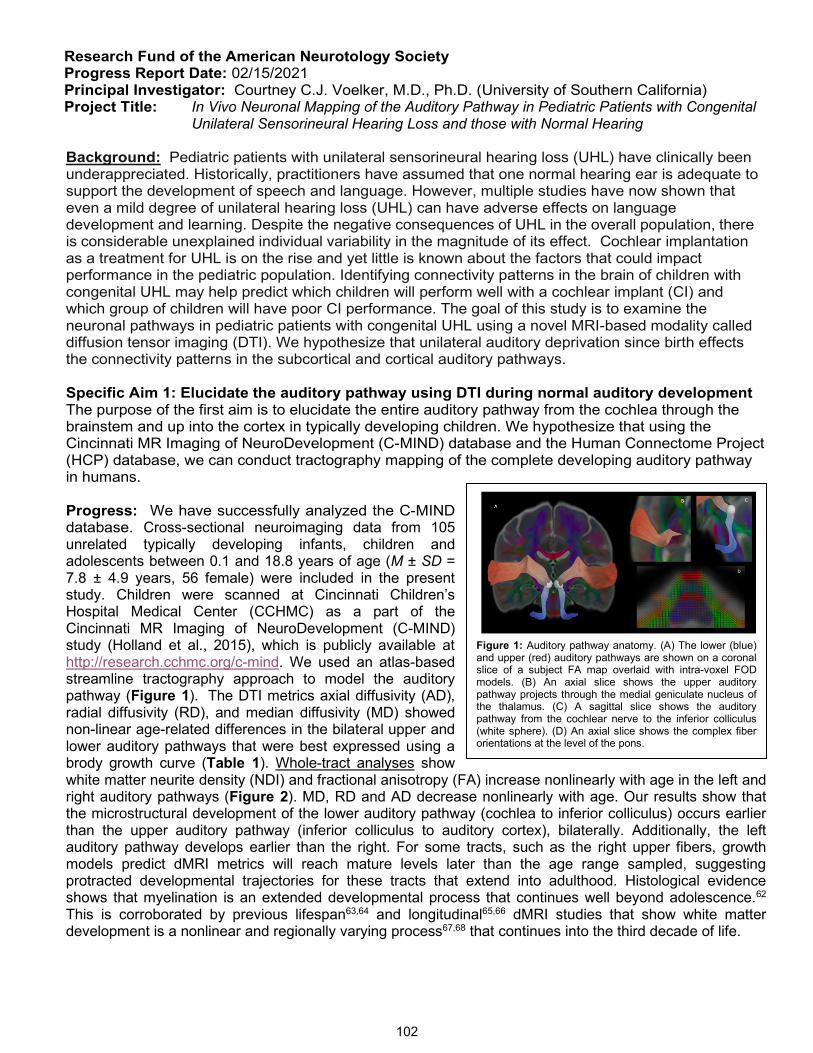
Research Fund of the American Neurotology Society Progress Report Date: 02/15/2021 Principal Investigator: Courtney C.J. Voelker, M.D., Ph.D. (University of Southern California) Project Title: In Vivo Neuronal Mapping of the Auditory Pathway in Pediatric Patients with Congenital
Unilateral Sensorineural Hearing Loss and those with Normal Hearing
Background: Pediatric patients with unilateral sensorineural hearing loss (UHL) have clinically been underappreciated. Historically, practitioners have assumed that one normal hearing ear is adequate to support the development of speech and language. However, multiple studies have now shown that even a mild degree of unilateral hearing loss (UHL) can have adverse effects on language development and learning. Despite the negative consequences of UHL in the overall population, there is considerable unexplained individual variability in the magnitude of its effect. Cochlear implantation as a treatment for UHL is on the rise and yet little is known about the factors that could impact performance in the pediatric population. Identifying connectivity patterns in the brain of children with congenital UHL may help predict which children will perform well with a cochlear implant (CI) and which group of children will have poor CI performance. The goal of this study is to examine the neuronal pathways in pediatric patients with congenital UHL using a novel MRI-based modality called diffusion tensor imaging (DTI). We hypothesize that unilateral auditory deprivation since birth effects the connectivity patterns in the subcortical and cortical auditory pathways.
Specific Aim 1: Elucidate the auditory pathway using DTI during normal auditory development The purpose of the first aim is to elucidate the entire auditory pathway from the cochlea through the brainstem and up into the cortex in typically developing children. We hypothesize that using the Cincinnati MR Imaging of NeuroDevelopment (C-MIND) database and the Human Connectome Project (HCP) database, we can conduct tractography mapping of the complete developing auditory pathway in humans.
Progress: We have successfully analyzed the C-MIND database. Cross-sectional neuroimaging data from 105 unrelated typically developing infants, children and adolescents between 0.1 and 18.8 years of age (M ± SD = 7.8 ± 4.9 years, 56 female) were included in the present study. Children were scanned at Cincinnati Children’s Hospital Medical Center (CCHMC) as a part of the Cincinnati MR Imaging of NeuroDevelopment (C-MIND) study (Holland et al., 2015), which is publicly available at http://research.cchmc.org/c-mind. We used an atlas-based streamline tractography approach to model the auditory pathway (Figure 1). The DTI metrics axial diffusivity (AD), radial diffusivity (RD), and median diffusivity (MD) showed non-linear age-related differences in the bilateral upper and lower auditory pathways that were best expressed using a brody growth curve (Table 1). Whole-tract analyses show white matter neurite density (NDI) and fractional anisotropy (FA) increase nonlinearly with age in the left and right auditory pathways (Figure 2). MD, RD and AD decrease nonlinearly with age. Our results show that the microstructural development of the lower auditory pathway (cochlea to inferior colliculus) occurs earlier than the upper auditory pathway (inferior colliculus to auditory cortex), bilaterally. Additionally, the left auditory pathway develops earlier than the right. For some tracts, such as the right upper fibers, growth models predict dMRI metrics will reach mature levels later than the age range sampled, suggesting protracted developmental trajectories for these tracts that extend into adulthood. Histological evidence shows that myelination is an extended developmental process that continues well beyond adolescence.62 This is corroborated by previous lifespan63,64 and longitudinal65,66 dMRI studies that show white matter development is a nonlinear and regionally varying process67,68 that continues into the third decade of life.
Figure 1: Auditory pathway anatomy. (A) The lower (blue) and upper (red) auditory pathways are shown on a coronal slice of a subject FA map overlaid with intra-voxel FOD models. (B) An axial slice shows the upper auditory pathway projects through the medial geniculate nucleus of the thalamus. (C) A sagittal slice shows the auditory pathway from the cochlear nerve to the inferior colliculus (white sphere). (D) An axial slice shows the complex fiber orientations at the level of the pons.
102
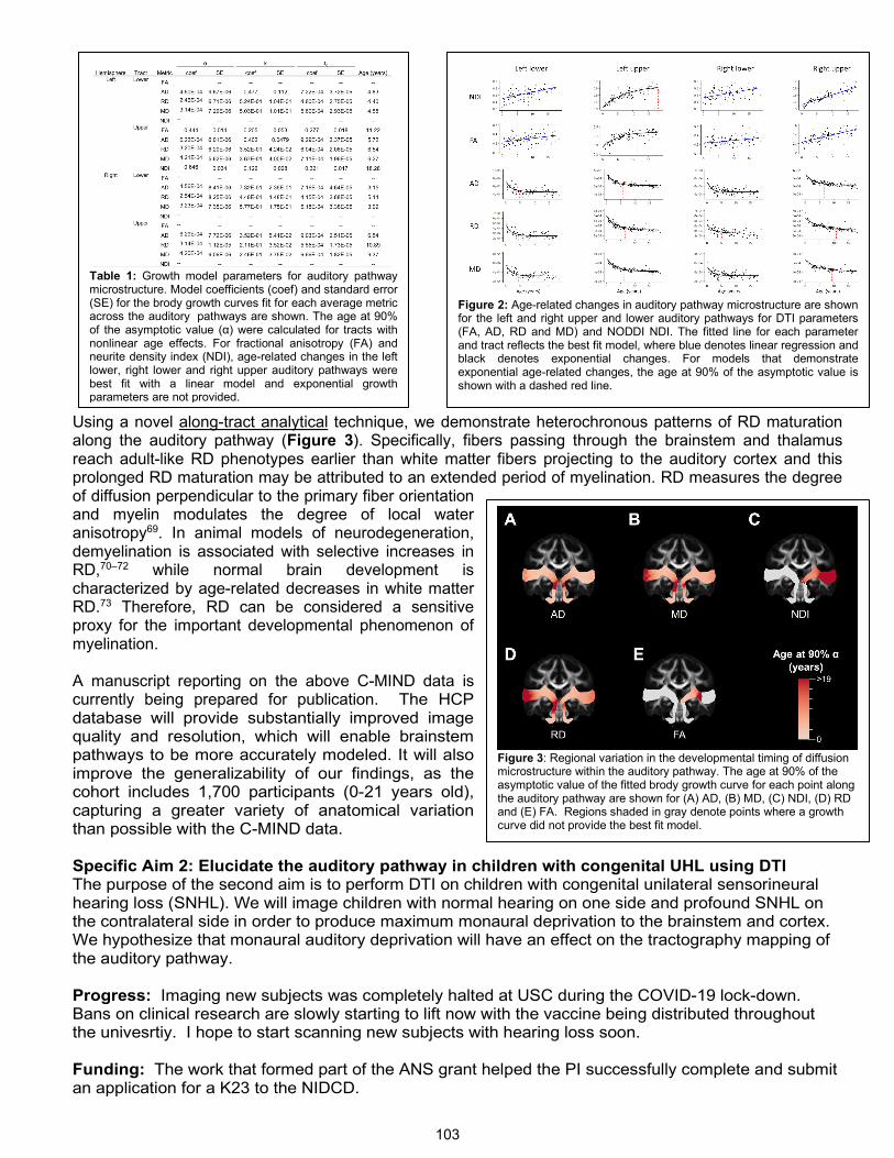
Using a novel along-tract analytical technique, we demonstrate heterochronous patterns of RD maturation along the auditory pathway (Figure 3). Specifically, fibers passing through the brainstem and thalamus reach adult-like RD phenotypes earlier than white matter fibers projecting to the auditory cortex and this prolonged RD maturation may be attributed to an extended period of myelination. RD measures the degree of diffusion perpendicular to the primary fiber orientation and myelin modulates the degree of local water anisotropy69. In animal models of neurodegeneration, demyelination is associated with selective increases in RD,70–72 while normal brain development is characterized by age-related decreases in white matter RD.73 Therefore, RD can be considered a sensitive proxy for the important developmental phenomenon of myelination.
A manuscript reporting on the above C-MIND data is currently being prepared for publication. The HCP database will provide substantially improved image quality and resolution, which will enable brainstem pathways to be more accurately modeled. It will also improve the generalizability of our findings, as the cohort includes 1,700 participants (0-21 years old), capturing a greater variety of anatomical variation than possible with the C-MIND data.
Specific Aim 2: Elucidate the auditory pathway in children with congenital UHL using DTI The purpose of the second aim is to perform DTI on children with congenital unilateral sensorineural hearing loss (SNHL). We will image children with normal hearing on one side and profound SNHL on the contralateral side in order to produce maximum monaural deprivation to the brainstem and cortex. We hypothesize that monaural auditory deprivation will have an effect on the tractography mapping of the auditory pathway.
Progress: Imaging new subjects was completely halted at USC during the COVID-19 lock-down. Bans on clinical research are slowly starting to lift now with the vaccine being distributed throughout the univesrtiy. I hope to start scanning new subjects with hearing loss soon.
Funding: The work that formed part of the ANS grant helped the PI successfully complete and submit an application for a K23 to the NIDCD.
Table 1: Growth model parameters for auditory pathway microstructure. Model coefficients (coef) and standard error (SE) for the brody growth curves fit for each average metric across the auditory pathways are shown. The age at 90% of the asymptotic value (α) were calculated for tracts with nonlinear age effects. For fractional anisotropy (FA) and neurite density index (NDI), age-related changes in the left lower, right lower and right upper auditory pathways were best fit with a linear model and exponential growth parameters are not provided.
Figure 2: Age-related changes in auditory pathway microstructure are shown for the left and right upper and lower auditory pathways for DTI parameters (FA, AD, RD and MD) and NODDI NDI. The fitted line for each parameter and tract reflects the best fit model, where blue denotes linear regression and black denotes exponential changes. For models that demonstrate exponential age-related changes, the age at 90% of the asymptotic value is shown with a dashed red line.
Figure 3: Regional variation in the developmental timing of diffusion microstructure within the auditory pathway. The age at 90% of the asymptotic value of the fitted brody growth curve for each point along the auditory pathway are shown for (A) AD, (B) MD, (C) NDI, (D) RD and (E) FA. Regions shaded in gray denote points where a growth curve did not provide the best fit model.
103
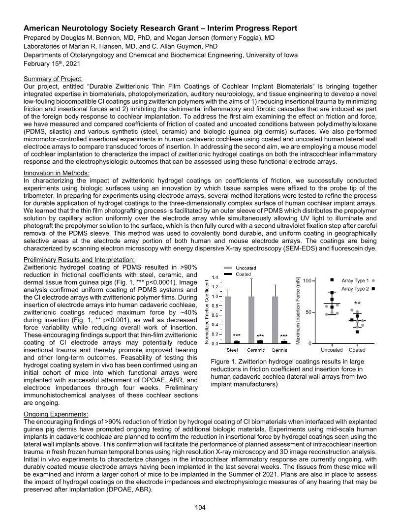
American Neurotology Society Research Grant – Interim Progress Report Prepared by Douglas M. Bennion, MD, PhD, and Megan Jensen (formerly Foggia), MD Laboratories of Marlan R. Hansen, MD, and C. Allan Guymon, PhD Departments of Otolaryngology and Chemical and Biochemical Engineering, University of Iowa February 15th, 2021
Summary of Project: Our project, entitled “Durable Zwitterionic Thin Film Coatings of Cochlear Implant Biomaterials” is bringing together integrated expertise in biomaterials, photopolymerization, auditory neurobiology, and tissue engineering to develop a novel low-fouling biocompatible CI coatings using zwitterion polymers with the aims of 1) reducing insertional trauma by minimizing friction and insertional forces and 2) inhibiting the detrimental inflammatory and fibrotic cascades that are induced as part of the foreign body response to cochlear implantation. To address the first aim examining the effect on friction and force, we have measured and compared coefficients of friction of coated and uncoated conditions between polydimethylsiloxane (PDMS, silastic) and various synthetic (steel, ceramic) and biologic (guinea pig dermis) surfaces. We also performed micromotor-controlled insertional experiments in human cadaveric cochleae using coated and uncoated human lateral wall electrode arrays to compare transduced forces of insertion. In addressing the second aim, we are employing a mouse model of cochlear implantation to characterize the impact of zwitterionic hydrogel coatings on both the intracochlear inflammatory response and the electrophysiologic outcomes that can be assessed using these functional electrode arrays.
Innovation in Methods: In characterizing the impact of zwitterionic hydrogel coatings on coefficients of friction, we successfully conducted experiments using biologic surfaces using an innovation by which tissue samples were affixed to the probe tip of the tribometer. In preparing for experiments using electrode arrays, several method iterations were tested to refine the process for durable application of hydrogel coatings to the three-dimensionally complex surface of human cochlear implant arrays. We learned that the thin film photografting process is facilitated by an outer sleeve of PDMS which distributes the prepolymer solution by capillary action uniformly over the electrode array while simultaneously allowing UV light to illuminate and photograft the prepolymer solution to the surface, which is then fully cured with a second ultraviolet fixation step after careful removal of the PDMS sleeve. This method was used to covalently bond durable, and uniform coating in geographically selective areas at the electrode array portion of both human and mouse electrode arrays. The coatings are being characterized by scanning electron microscopy with energy dispersive X-ray spectroscopy (SEM-EDS) and fluorescein dye.
Preliminary Results and Interpretation: Zwitterionic hydrogel coating of PDMS resulted in >90% reduction in frictional coefficients with steel, ceramic, and dermal tissue from guinea pigs (Fig. 1, *** p<0.0001). Image analysis confirmed uniform coating of PDMS systems and the CI electrode arrays with zwitterionic polymer films. During insertion of electrode arrays into human cadaveric cochleae, zwitterionic coatings reduced maximum force by ~40% during insertion (Fig. 1, ** p<0.001), as well as decreased force variability while reducing overall work of insertion. These encouraging findings support that thin-film zwitterionic coating of CI electrode arrays may potentially reduce insertional trauma and thereby promote improved hearing and other long-term outcomes. Feasability of testing this hydrogel coating system in vivo has been confirmed using an initial cohort of mice into which functional arrays were implanted with successful attainment of DPOAE, ABR, and electrode impedances through four weeks. Preliminary immunohistochemical analyses of these cochlear sections are ongoing.
Ongoing Experiments: The encouraging findings of >90% reduction of friction by hydrogel coating of CI biomaterials when interfaced with explanted guinea pig dermis have prompted ongoing testing of additional biologic materials. Experiments using mid-scala human implants in cadaveric cochleae are planned to confirm the reduction in insertional force by hydrogel coatings seen using the lateral wall implants above. This confirmation will facilitate the performance of planned assessment of intracochlear insertion trauma in fresh frozen human temporal bones using high resolution X-ray microscopy and 3D image reconstruction analysis. Initial in vivo experiments to characterize changes in the intracochlear inflammatory response are currently ongoing, with durably coated mouse electrode arrays having been implanted in the last several weeks. The tissues from these mice will be examined and inform a larger cohort of mice to be implanted in the Summer of 2021. Plans are also in place to assess the impact of hydrogel coatings on the electrode impedances and electrophysiologic measures of any hearing that may be preserved after implantation (DPOAE, ABR).
Figure 1. Zwitterion hydrogel coatings results in large reductions in friction coefficient and insertion force in human cadaveric cochlea (lateral wall arrays from two implant manufacturers)
104
American Neurotology Society 2020 Research Grant- Interim Progress Report Comparison of Surgical Routes for Localized Inner Ear Viral Vector-Mediated Gene Therapy in the Guinea Pig Using Helper-Dependent Adenovirus Type 5 Tatiana Correa February 8, 2021
In recent years, viral vector-mediated gene therapy has emerged as a potential avenue for new biologics to address a number of diseases including sensorineural hearing loss (SNHL). Treatment of SNHL using viral vector-mediated gene therapy will require vectors that are non-toxic, have large carrying capacities, and are modifiable to target multiple types of cells in the inner ear. Up until now, most research looking at viral vector-mediated gene therapy for SNHL has focused on adeno-associated virus (AAV) vectors, which are non-toxic and modifiable but lack the carrying capacity needed to address most cases of SNHL. Helper-dependent adenovirus (HdAd) is a potential vector for gene therapy that is non-toxic and has the large carrying capacity needed for novel SNHL gene therapies. We set out to begin an assessment of HdAd as a potential vector for inner ear gene therapy by assessing HdAd ability to access different compartments of the inner and the effect of delivery route on accessibility of tissues.
Specific Aim 1: Characterize HdAd transduction via clinically translatable surgical routes in the adult guinea pig. To eliminate confounding effects of vector serotype and promoter from our analysis, HdAd vectors utilizing an adenovirus capsid protein with a known receptor for infection and ubiquitous CMV promoter were used. Progress: We have delivered HdAd vectors with an mClover reporter expression cassette into the inner ear of guinea pigs using three approaches: round window, round window with posterior semicircular canal fenestration and endolymphatic sac. Animals have been sacrificed seven days after initial delivery and confocal imaging of whole mount preparations with immunofluorescent staining has been completed. Less than two surgeries remain for each approach to complete the study cohort. Both round window and round window with canal fenestration delivery approaches have resulted in transduction of cells within the organ of Corti suggesting HdAd particle size is not a barrier to crossing the basilar membrane. Endolymphatic sac delivery did not result in transduction in the cochlea but it did reveal transduction of vestibular hair cells, which was not seen with the other two methods.
Impact: Understanding of HdAd as a potential vector for inner ear gene therapy is limited. The work completed in this study helps us characterize which parts of the inner ear can be transduced by delivering HdAd through various approaches and is foundational to future studies where HdAd serotype and promoter elements are modified to manipulate tropism.
105
AMERICAN NEUROTOLOGY SOCIETY PAST PRESIDENTS
1965-69 Fred Harbert, MD
1969-70 Richard E. Marcus, MD
1970-71 Wallace Rubin, MD
1971-72 Malcolm H. Stroud, MD
1972-73 Martin Spector, MD
1973-74 Nicholas Torok, MD
1974-75 Cecil W. Hart, MD
1975-76 Sidney N. Busis, MD
1976-77 Brian F. McCabe, MD
1977-78 Bruce Proctor, MD
1978-79 David A. Dolowitz, MD
1979-80 Fred H. Linthicum Jr., MD
1980-81 Harold Schuknecht, MD
1981-82 Hugh Barber, MD
1982-83 Kenneth H. Brookler, MD
1983-84 Richard Gacek, MD
1984-85 Derald Brackmann, MD
1985-86 Robert J. Keim, MD
1986-87 Jack D. Clemis, MD
1987-88 Malcolm Graham, MD
1988-89 Robert A. Jahrsdoerfer, MD
1989-91 Shokri Radpour, MD
1992-92 Antonio De La Cruz, MD
1992-93 Fredric W. Pullen II, MD
1993-94 Charles M. Luetje II, MD
1994-95 Sam E. Kinney, MD
1995-96 Joseph DiBartolomeo, MD
1996-97 Jack M. Kartush, MD
1997-98 Bruce J. Gantz, MD
1998-99 John W. House, MD
1999-00 Richard J. Wiet, MD
2000-01 Richard T. Miyamoto, MD
2001-02 Stephen G. Harner, MD
2002-03 Newton J. Coker, MD
2003-04 Paul R. Lambert, MD
2004-05 Robert K. Jackler, MD
2005-06 Debara L. Tucci, MD
2006-07 Joel A. Goebel, MD
2007-08 D. Bradley Welling, MD, PhD
2008-09 Karen J. Doyle, MD, PhD
2009-10 Samuel H. Selesnick, MD
2010-11 J. Douglas Green Jr., MD
2011-12 Jeffrey T. Vrabec, MD
2012-13 Clough Shelton, MD
2013-14 Hilary A. Brodie, MD, PhD
2014-15 Anil K. Lalwani, MD
2015-16 John T. McElveen, Jr., MD
2016-17 Lawrence R. Lustig, MD
2017-18 Moisés A. Arriaga, MD, MBA
2018-19 Barry E. Hirsch, MD
2019-20 Nikolas H. Blevins, MD
2020-21 Bradley W. Kesser, MD
106
AMERICAN NEUROTOLOGY SOCIETY PAST SECRETARY-TREASURERS
1965-68 Richard E. Marcus, MD
1968-70 Bruce Proctor, MD
1970-71 F. Blair Simmons, MD
1971-72 Cecil Hart, MD
1972-74 Sidney Busis, MD
1974-76 Jack Pulec, MD
1976-79 Michael Glasscock III, MD
1979-85 Robert Keim, MD
1985-88 Shokri Radpour, MD
1988-92 Charles M. Luetje II, MD
1992-95 Jack M. Kartush, MD
1995-98 Richard J. Wiet, MD
1998-01 Newton J. Coker, MD
2001-04 Debara L. Tucci, MD
2004-07 Karen J. Doyle, MD, PhD
2007-10 Jeffrey T. Vrabec, MD
2010-13 Anil K. Lalwani, MD
2013-16 Moisés A. Arriaga, MD, MBA
2016-19 Bradley W. Kesser, MD
2019-present Elizabeth H. Toh, MD, MBA
107
AMERICAN NEUROTOLOGY SOCIETY
2020-2021 Membership Roster (includes 27 new members
inducted at 2021 Spring meeting)
Ksenia A. Aaron, MD Stanford, CA Trainee
Meredith E. Adams, MD Minneapolis, MN 2011 Fellow
Oliver F. Adunka, MD Columbus, OH 2010 Fellow
Yuri Agrawal, MD Baltimore, MD 2013 Fellow
Sameer Ahmed, MD Downey, CA 2019 Fellow
Syed F. Ahsan, MD Irvine, CA 2012 Fellow
Mohammad Al Saadi, MD Brussels, Belgium 2020 Trainee
Pedro Luiz Albernaz, MD, PhD Sao Paulo, Brazil 1976 Senior Fellow
Thomas Hanson Alexander, MD La Jolla, CA 2017 Fellow
George Alexiades, MD New York, NY 2015 Fellow
Kyle P. Allen, MD Tampa, FL 2014 Fellow
Ahmed Al-Sayed, MD Halifax, NS Canada 2019 Trainee
Sean R. Althaus, MD Georgetown, TX 1976 Emeritus
Jennifer Alyono, MD Stanford, CA 2019 Associate
Ronald G. Amedee, MD New Orleans, LA 1990 Fellow
James Andrews, MD Manhattan Beach, CA 1996 Fellow
Simon I. Angeli, MD Miami, FL 2013 Fellow
Philip F. Anthony, MD Fort Worth, TX 1980 Senior Fellow
Patrick J. Antonelli, MD Gainesville, FL 1995 Fellow
Charles L. Anzalone, MD Little Rock, AR 2020 Trainee
Eric N. Appelbaum, MD Marietta, GA 2021 Associate
Irving Arenberg, MD Centennial, CO 1977 Emeritus
Moises A. Arriaga, MD Metairie, LA 1993 Fellow
H. Alexander Arts, MD Ann Arbor, MI1993 Fellow
Gregory J. Artz, MD Grand Rapids, MI 2020 Fellow
Peter E Ashman, Jersey City, NJ 2020 Trainee
James S. Atkins, Jr., MD Celebration, FL 1988 Fellow
Gregory A. Ator, MD Kansas City, KS 1994 Senior Associate
Michael Patrick Avillion, MD Waipahu, HI 2020 Trainee
John W. Ayugi, ChB Nairobi, Kenya 2014 Associate
Seilesh C. Babu, MD Farmington Hills, MI 2004 Fellow
Douglas D. Backous, MD Edmonds, WA 2005 Fellow
R. Stanley Baker, MD Oklahoma City, OK1996 Fellow
Robert L. Baldwin, MD Birmingham, AL 1990 Senior Fellow
Thomas J. Balkany, MD Long Key, FL 1982 Senior Fellow
Ben J. Balough, MD Sacramento, CA 2015 Fellow
Manohar L. Bance, MD Cambridge, United Kingdom 2017 Fellow
108
David M. Barrs, MD Phoenix, AZ 1984 Senior Fellow
Loren J. Bartels, MD Tampa, FL 1984 Fellow
Richard M. Bass, MD Springfield, IL 1988 Senior Fellow
Marc K. Bassim, MD Beirut, YT Lebanon 2020 Fellow
Gregory Joseph Basura, MD, PhD Ann Arbor, MI 2018 Fellow
Alex S. Battaglia, MD, PhD San Rafael, CA 2007 Fellow
Robert A. Battista, MD Hinsdale, IL 1995 Fellow
Carol A. Bauer, MD Springfield, IL 1996 Fellow
David D. Beal, MD Anchorage, AK 1990 Senior Fellow
Charles W. Beatty, MD Rochester, MN 1989 Emeritus
James E. Benecke, MD Scottsdale, AZ 1985 Senior Fellow
Jaime Benitez, MD Farmington Hills, MI 1970 Senior Fellow
Marc L. Bennett, MD Nashville, TN 2019 Fellow
Brent J. Benscoter, MD Sacramento, CA 2015 Fellow
Aaron G. Benson, MD Greenfield, WI 2021 Fellow
Karen I. Berliner, PhD Marina Del Rey, CA 1990 Associate
Jason A. Beyea, MD, PhD Kingston, ON Canada 2016 Associate
Sanjay Bhansali, MD Atlanta, GA 1994 Fellow
Alexander G. Bien, MD Oklahoma City, OK 2011 Fellow
Douglas C. Bigelow, MD Philadelphia, PA 1992 Fellow
Brian W. Blakley, MD, PhD Winnipeg, MB Canada 1994 Senior Fellow
Nikolas H. Blevins, MD Stanford, CA 2004 Fellow
Dennis I. Bojrab, MD Farmington Hills, MI 1987 Fellow
Dennis I. Bojrab II, MD Bloomfield Hills, MI 2021 Associate
Jean-Michel Bourque, MD Lévis, QC Canada 2019 Trainee
K. Paul Boyev, MD Tampa, FL 2002 Fellow
Derald E. Brackmann, MD Los Angeles, CA 1975 Senior Fellow
Laura Brainard, MD Detroit, MI 2013 Fellow
Thomas G. Brammeier, MD Belton, TX 2003 Fellow
Robert E. Brammer, MD St Clr Shores, MI 1988 Fellow
Jason A. Brant, MD Wallingford, PA 2017 Fellow
Joseph T. Breen, MD Montgomery, OH 2017 Fellow
Arnold K. Brenman, MD Jenkintown, PA 1973 Emeritus
Robert J. S. Briggs, MD Kooyong, Australia 1996 Fellow
Selena E. Briggs, MD, MBA Washington, DC 2013 Fellow
B. Hill Britton, MDSan Antonio, TX1973 Emeritus
Hilary A. Brodie, MD, PhD Sacramento, CA 1999 Fellow
Gerald B. Brookes, MD London, 1994 Fellow
Kenneth H. Brookler, MD Norwalk, CT 1972 Emeritus
109
Morgan Brosnan, MD Ontario Canada 1983 Senior Fellow
C. Scott Brown, MDMiami, FL 2017 Trainee
Jeffrey J. Brown, MD, PhD Portland, OR 1988 Emeritus
William Colby Brown, MD Cleveland Hts, OH 2016 Trainee
Kevin D. Brown, MD Chapel Hill, NC 2012 Fellow
J. Dale Browne, MD Winston Salem, NC 1995 Fellow
Patrick Cody Buchanan, DO Tulsa, OK 2020 Associate
Craig A. Buchman, MD St. Louis, MO 1998 Fellow
Cameron L. Budenz, MD Sleepy Hollow, NY 2015 Fellow
Hana T. Bui, MD Fullerton, CA 1995 Associate
Don L. Burgio, MD Scottsdale, AZ 1995 Fellow
Matthew L. Bush, MD, PhD Lexington, KY 2012 Fellow
Audrey P. Calzada, MD Carlsbad, CA 2015 Fellow
Robert W. Cantrell, MD Charlottesville, VA 1976 Emeritus
John P. Carey, MD Baltimore, MD 2004 Fellow
Matthew J. Carfrae, MD Clive, IA 2010 Fellow
Matthew L. Carlson, MD Rochester, MN 2015 Fellow
Stephen P. Cass, MD, MPH Aurora, CO 1991 Fellow
Nathan D. Cass, MDNashville, TN 2019 Trainee
Ryan M. Casserly, MD Monterey, CA 2021 Associate
Adam M. Cassis, MD Chandler, AZ 2014 Fellow
Ned Chalat, MD Grosse Pointe, MI 1981 Senior Fellow
Eleanor Y. Chan, MD Farmington Hills, MI 2020 Fellow
Sujana S. Chandrasekhar, MD New York, NY 1995 Fellow
C. Y. Joseph Chang, MD Houston, TX 1996 Fellow
Guyan A. Channer, MD Kingston , Jamaica, FL 2013 Associate
Divya A. Chari, MDBoston, MA 2018 Trainee
Brian S. Chen, MD Tripler, HI 2017 Fellow
Douglas A. Chen, MD Wexford, PA 1988 Fellow
Joseph M. Chen, MD Toronto, ON Canada 2007 Fellow
Alexander Chern, MDNew York, NY 2019 Trainee
Steven W. Cheung, MD San Francisco, CA 2006 Fellow
Wade W. Chien, MD Potomac, MD 2014 Fellow
Rebecca C. Chiffer, MD Philadelphia, PA 2021 Associate
Edgar L. Chiossone, MD Miami, FL 1983 Senior Fellow
Edward I. Cho, MD Los Angeles, CA 2014 Associate
Won-Taek Choe, MD New York, NY 2008 Fellow
Richard A. Chole, MD, PhD Saint Louis, MO 1994 Emeritus
Laura H. Christopher, MDLos Angeles, CA 2018 Trainee
110
Jack Clemis, MD Chicago, IL 1968 Senior Fellow
Francois Cloutier, MD Longueuil, QC Canada 2016 Associate
Daniel H. Coelho, MD Richmond, VA 2008 Fellow
Erin R. Cohen, MD Miami, FL 2018 Trainee
Burton J. Cohen, MD Louisville, KY 1986 Senior Fellow
Newton J. Coker, MD Santa Fe, NM 1984 Senior Fellow
Candice Colby, MD Midland, MI 2016 Fellow
George H. Conner, MD Lebanon, PA 1976 Emeritus
Timothy Cooper, MD, FRCS Edmonton, AB Canada 2021 Associate
Mario Augusto Corona Ruiz, MD San Juan, Puerto Rico 2019 Trainee
C. Eduardo Corrales, MD Boston, MA 2015 Fellow
Maura K. Cosetti, MD New York, NY 2012 Fellow
Justin Cottrell, MD Toronto, ON Canada 2020 Trainee
Matthew D. Cox, MD Winter Park, FL 2018 Associate
Benjamin T. Crane, MD, PhD Rochester, NY 2011 Fellow
James V. Crawford, MD Boise, ID 2011 Fellow
Francis X. Creighton, MD Baltimore, MD 2021 Associate
Matthew G. Crowson, MD, MPA Boston, MA 2021 Associate
Roberto A. Cueva, MD San Diego, CA 1991 Fellow
Robert D. Cullen, MD Kansas City, MO 2008 Fellow
Calhoun D. Cunningham III, MD Raleigh, NC 2005 Fellow
Steven D. Curry, MD Omaha, NE 2020 Trainee
Frank S. Curto, Jr., MD Bethesda, MD 1996 Senior Fellow
Julien Dallaire, MDSherbrooke, QC Canada 2020 Trainee
Robert L. Daniels, MD Grand Rapids, MI 2007 Fellow
Christopher J. Danner, MD Tampa, FL 2007 Fellow
D. Spencer Darley, MD Provo, UT2013 Associate
C. Phillip Daspit, MD Paradise Valley, AZ 1973 Emeritus
Christopher De Souza, MD Bombay, India 1998 Fellow
Nicholas L. Deep, MD Phoenix, AZ 2021 Associate
Charles Della Santina, MD, PhD Baltimore, MD 2017 Fellow
M. Jennifer Derebery, MD Los Angeles, CA1991 Fellow
Nicholas A. Dewyer, MD Tucson, AZ 2020 Associate
Joseph Di Bartolomeo, MD Santa Barbara, CA 1983 Senior Fellow
Rodney C. Diaz, MD Sacramento, CA 2014 Fellow
John R.E. Dickins, MD Fayetteville, AR 1989 Emeritus
Elizabeth A. Dinces, MD Scarsdale, NY 2014 Fellow
Christine T. Dinh, MD Miami, FL 2017 Fellow
Michael J. Disher, MD Fort Wayne, IN 1994 Fellow
111
Peter R Dixon, MD Toronto, ON Canada 2020 Trainee
Hamilton S. Dixon, MD East Ellijay, GA 1972 Emeritus
Hamid R. Djalilian, MD Irvine, CA 2005 Fellow
Edward Dodson, MD Dublin, OH 1997 Fellow
Karl W. Doerfer, MDLivonia, MI 2020 Trainee
Joni K. Doherty, MD, PhD Los Angeles, CA 2008 Fellow
Katsumi Doi, MD, PhD Osaka- Sayama, Japan 2020 Associate
James R. Dornhoffer, MD Charleston, SC 2020 Trainee
John L. Dornhoffer, MD Little Rock, AR 2002 Fellow
Karen J. Doyle-Enright, MD, PhD Alameda, CA 1994 Fellow
David A. Drachman, MD Worcester, MA 1974 Emeritus
Colin L. W. Driscoll, MD Rochester, MN 2002 Fellow
Larry Duckert, MD, PhD Seattle, WA 1984 Senior Fellow
Brian E. Duff, MD E Greenwich, RI 2005 Fellow
Paul Dutcher, MD Rochester, NY 1988 Senior Fellow
Thomas L. Eby, MD Jackson, MS 1995 Fellow
Marc D. Eisen, MD, PhD Farmington, CT 2013 Fellow
David J. Eisenman, MD Baltimore, MD 2016 Fellow
Hussam K. El-Kashlan, MD Ann Arbor, MI 1999 Fellow
John R. Emmett, MD Memphis, TN 1981 Fellow
Susan D. Emmett, MD Durham, NC 2019 Associate
Isaac Erbele, MD San Antonio, TX 2021 Associate
Adrien A. Eshraghi, MD Weston, FL 2007 Fellow
Abraham Eviatar, MD Scarsdale, NY 1975 Senior Fellow
George W. Facer, MD Bonita Springs, FL 1975 Emeritus
Jay B. Farrior, MD Tampa, FL 1983 Fellow
Jose N. Fayad, MD Dhahran, Saudi Arabia 2007 Fellow
Linnea Fechtner, MDBuffalo, NY 2020 Trainee
Robert S. Feehs, MD Englewood, CO 1997 Fellow
Joseph G. Feghali, MD Bronx, NY 1991 Fellow
Bruce A. Feldman, MD Potomac, MD 1987 Emeritus
Bruce L. Fetterman, MD Germantown, TN 1997 Fellow
Terry D. Fife, MD Phoenix, AZ 2006 Fellow
Dennis C. Fitzgerald, MD Philadelphia, PA 1984 Senior Fellow
Eric J. Formeister, MD Baltimore, MD 2020 Trainee
Michael F. Foster, DO Ada, MI 2018 Associate
David Foyt, MD Albany, NY 2007 Fellow
Howard W. Francis, MD, MBA Durham, NC 2008 Fellow
Daniel J. Franklin, MD Houston, TX 1998 Fellow
112
Douglas W. Frerichs, MD Flagstaff, AZ 1984 Senior Fellow
David R. Friedland, MD, PhD Milwaukee, WI 2008 Fellow
Rick A. Friedman, MD, PhD La Jolla, CA 1996 Fellow
David R. Friedmann, MD, MSC New York, NY 2017 Fellow
Michael H. Fritsch, MD Indianapolis, IN 1987 Fellow
Michael J. Fucci, MD Chandler, AZ 1997 Fellow
Richard R. Gacek, MD Worcester, MA 1970 Emeritus
Deepa Galaiya, MD Baltimore, MD 2021 Associate
Michele M. Gandolfi, MD Winston-Salem, NC 2018 Fellow
Bruce J. Gantz, MD Iowa City, IA 1983 Fellow
Juan M. Garcia, MD Miami, FL 1998 Fellow
L. Gale Gardner, MD Shreveport, LA 1976 Senior Fellow
George A. Gates, MD Boerne, TX 1970 Senior Associate
Bechara Y. Ghorayeb, MD Houston, TX 1990 Fellow
Soha N. Ghossaini, MD Astoria, NY 2011 Fellow
Gerard J. Gianoli, MD Covington, LA 2007 Fellow
William P. R. Gibson, MD Birchgrove, Australia 1989 Senior Fellow
Neil A. Giddings, MD Spokane, WA 1992 Fellow
Paul W. Gidley, MD Houston, TX 2007 Fellow
Martin Gizzi, MD, PhD Hackensack, NJ 2007 Fellow
Michael B. Gluth, MD Chicago, IL 2011 Fellow
John C. Goddard, MD Clackamas, OR 2012 Fellow
Joel A. Goebel, MD Saint Louis, MO 1987 Emeritus
Robert A. Goldenberg, MD Dayton, OH 1983 Emeritus
Elliot Goldofsky, MD Great Neck, NY 1994 Associate
M. Miles Goldsmith, MD Savannah, GA 2007 Fellow
Hernan Goldsztein, MD La Jolla, CA 2014 Fellow
Justin S. Golub, MD, MSC New York, NY 2016 Fellow
Stefania Goncalves, MD Miami, FL 2019 Trainee
Quinton Gopen, MD Los Angeles, CA 2018 Fellow
Michael A. Gordon, MD West Hempstead, NY 1997 Senior Fellow
Malcolm Graham, MD Atlanta, GA 1972 Emeritus
J. Douglas Green, Jr., MD Jacksonville, FL 1993 Fellow
Andrew J. Griffith, MD, PhD Bethesda, MD 2014 Associate
Lawrence R. Grobman, MD Miami, FL 1989 Fellow
Samuel P. Gubbels, MD Aurora, CO 2009 Fellow
A. Julianna Gulya, MD Locust Grove, VA 1985 Senior Fellow
Sachin Gupta, MD Portland, OR 2018 Fellow
Richard K. Gurgel, MD Salt Lake City, UT 2013 Fellow
113
Thomas J. Haberkamp, MD Cleveland, OH 1988 Senior Fellow
Rex S. Haberman, MD Gainesville, FL 1996 Fellow
Kevin S. Hadley, MD Aiea, HI 2014 Fellow
Yoav Hahn, MD Dallas, TX 2015 Fellow
G. Michael Halmagyi, MD Sydney, Australia2006 Honorary
Paul E. Hammerschlag, MD New York, NY 1983 Senior Fellow
Marlan R. Hansen, MD Iowa City, IA 2007 Fellow
Matthew B. Hanson, MD Brooklyn, NY 2002 Fellow
Lee Harker, MD Omaha, NE 1974 Emeritus
Stephen G. Harner, MD Rochester, MN 1988 Senior Fellow
Jeffrey P. Harris, MD, PhD San Diego, CA 1984 Senior Fellow
Michael S. Harris, MD Milwaukee, WI 2018 Fellow
Cecil W Hart, MD Palm Springs, CA 1968 Emeritus
Steven A. Harvey, MD Milwaukee, WI 1996 Fellow
George T. Hashisaki, MD Charlottesville, VA 1990 Fellow
Jonathan Hatch, MD Omaha, NE 2019 Fellow
David S. Haynes, MD Nashville, TN 1996 Fellow
Katherine Do Heidenreich, MD Ann Arbor, MI 2012 Associate
Edward Hendershot, MD Lodi, OH 1976 Senior Fellow
Ronna Hertzano, MD, PhD Baltimore, MD 2015 Fellow
Jacques A. Herzog, MD Chesterfield, MO 1987 Fellow
Thomas Oma Hester, MD Charleston, SC 1999 Fellow
George Hicks, MD Indianapolis, IN 1981 Fellow
Douglas M. Hildrew, MD New Haven, CT 2018 Fellow
Todd A. Hillman, MD Wexford, PA 2004 Fellow
Christopher W. Hilton, MD St. Paul, MN 2011 Fellow
Barry E. Hirsch, MD Pittsburgh, PA 1985 Fellow
Michael Hoa, MD Washington, DC 2015 Fellow
Candace E. Hobson, MD Atlanta, GA 2018 Fellow
Sarah E. Hodge, MD Los Angeles, CA 2019 Trainee
Michael E. Hoffer, MD Miami, FL 2001 Fellow
Ronald A. Hoffman, MD New York, NY 1983 Senior Fellow
Dick L. Hoistad, MD Seattle, WA 2011 Fellow
Sean P. Holmes, MD Shreveport, LA 2021 Trainee
James J. Holt, MD Marshfield, WI 1996 Senior Fellow
Robert S. Hong, MD, PhD Farmington Hills, MI 2013 Fellow
Vicente Honrubia, MD Los Angeles, CA 1972 Senior Fellow
Arata Horii, MD Niigata, Japan 2008 Fellow
Karl L. Horn, MD Santa Fe, NM 1986 Senior Fellow
114
Melton J. Horwitz, MD Houston, TX 1983 Senior Fellow
John W. House, MD Los Angeles, CA 1976 Senior Fellow
James R. House, III, MD Jackson, MS 2000 Fellow
May Y. Huang, MD Seattle, WA 1998 Fellow
Tina C. Huang, MD Minneapolis, MN 2015 Fellow
Dominic W. Hughes, PhD West Linn, OR 1984 Senior Associate
Timothy E. Hullar, MD Portland, OR 2006 Fellow
Jacob B. Hunter, MD Dallas, TX 2017 Fellow
Tiffany Hwa, MD Philadelphia, PA 2020 Trainee
Makoto Igarashi, MD Tokyo, Japan 1968 Senior Associate
Takao Imai, MD, PhD Suita-City, Japan 2013 Fellow
Terence E. Imbery, MD Chicago, IL 2020 Fellow
Brandon Isaacson, MD Dallas, TX 2005 Fellow
Jon E. Isaacson, MD Hershey, PA 2007 Fellow
Akira Ishiyama, MD Los Angeles, CA 2015 Fellow
Huseyin Isildak, MD Hershey, PA 2014 Associate
Robert K. Jackler, MD Stanford, CA 1987 Fellow
Neal M. Jackson, MD New Orleans, LA 2019 Associate
Carol Jackson, MD Newport Beach, CA 1985 Fellow
Lance E. Jackson, MD San Antonio, TX 2002 Fellow
Abraham Jacob, MD Tucson, AZ 2006 Fellow
Taha A. Jan, MD San Francisco, CA 2021 Associate
Herman A. Jenkins, MD Aurora, CO 1982 Fellow
Daniel Jethanamest, MD New York, NY 2014 Fellow
Nicole Tin-Lok Jiam, MDSan Francisco, CA 2020 Trainee
Alan J. Johnson, MD, MPH Temple, TX 1994 Fellow
Raleigh O. Jones, MD Lexington, KY 1990 Fellow
Timothy T. K. Jung, MD, PhD Riverside, CA 1990 Fellow
David H. Jung, MD, PhD Boston, MA 2015 Fellow
Jacob B. Kahane, MD Baton Rouge, LA 2020 Trainee
Donald B. Kamerer, MD Pittsburgh, PA 1974 Emeritus
Romain E. Kania, MD, PhD Paris, France 2014 Associate
Howard M. Kaplan, MD Plantation, FL 1900 Senior Fellow
Elina Kari, MD La Jolla, CA 2014 Fellow
Alexandre Karkas, MD, PhD St Priest en Jarez, France 2020 Fellow
Jack Kartush, MD Bloomfield Hills, MI 1985 Senior Fellow
Rustin Ghamsarian Kashani, MD Menlo Park, CA 2021 Trainee
Athanasios Katsarkas, MD Montreal, Canada 1978 Emeritus
Adam C. Kaufman, MD Palo Alto, CA 2019 Trainee
115
David M. Kaylie, MD Durham, NC 2007 Fellow
Emily Kay-Rivest, MD Montreal, QC Canada 2020 Trainee
Brian Kellermeyer, MD Morgantown, WV 2020 Associate
Robert Kellman, MD Syracuse, NY 1984 Senior Fellow
Elizabeth A. Kelly, MD Elkhorn, NE 2018 Fellow
David C. Kelsall, MD Englewood, CO 1995 Associate
Bradley W. Kesser, MD Charlottesville, VA 2000 Fellow
Jeffrey Keyser, MD Providence, UT 1999 Associate
Paul Kileny, PhD Ann Arbor, MI 1999 Associate
Daniel Killeen, MD Dallas, TX 2015 Trainee
Hung Jeffrey Kim, MD Washington, DC 1998 Fellow
Ana H. Kim, MD New York, NY 2012 Fellow
Harold H. Kim, MD Portland, OR 2008 Fellow
Susan Marenda King, MD San Antonio, TX 1998 Fellow
Sam E. Kinney, MD Moreland Hills, OH 1979 Senior Fellow
Matthew L. Kircher, MD Maywood, IL 2014 Fellow
Ruwan Kiringoda, MD Fremont, CA 2018 Fellow
Tadashi Kitahara, MD, PhD Kashihara-city, Japan 2008 Fellow
Glenn W. Knox, MD Jacksonville, FL 2007 Fellow
Pelin Kocdor, MD Goztepe/Istanbul, Turkey 2018 Associate
Darius Kohan, MD New York, NY 1994 Fellow
Gavriel D. Kohlberg, MD Seattle, WA 2020 Fellow
Robert Kohut, MD Woodleaf, NC 1975 Emeritus
Horst R. Konrad, MD Springfield, IL 1974 Senior Fellow
Richard D. Kopke, MD Oklahoma City, OK 2005 Senior Fellow
Harold W. Korol, MD Palo Alto, CA 1984 Senior Fellow
Ali Kouhi, MD Tehran, Iran 2020 Associate
Elliott D. Kozin, MD Boston, MA 2021 Associate
Wesley W.O. Krueger, MD San Antonio, TX 1987 Senior Fellow
Thomas C. Kryzer, MD Wichita, KS 1995 Associate
Jeffery J. Kuhn, MD Virginia Beach, VA 1999 Fellow
Arvind Kumar, MD Hinsdale, IL 1991 Senior Fellow
Brian Kung, MD Bellevue, WA 2020 Fellow
J. Walter Kutz, Jr., MD Dallas, TX 2008 Fellow
John Kveton, MD New Haven, CT 1984 Fellow
Jed Kwartler, MBA||MD South Orange, NJ 1996 Fellow
Robert F. Labadie, MD, PhD Nashville, TN 2009 Fellow
Anil K. Lalwani, MD New York, NY 1999 Fellow
Paul R. Lambert, MD Charleston, SC 1985 Fellow
116
Lukas D. Landegger, MD Vienna, Austria 2016 Trainee
Alan W. Langman, MD Seattle, WA 1991 Fellow
Michael J. LaRouere, MD Northville, MI 1990 Senior Fellow
John M. Lasak, MD Wichita, KS 2001 Fellow
Lorenz F. Lassen, MD Suffolk, VA 1996 Fellow
Daniel J. Lee, MD Boston, MA 2015 Fellow
Alice D. Lee, MD Riverside, CA 2011 Associate
Joel F. Lehrer, MD Teaneck, NJ 1976 Senior Fellow
John P. Leonetti, MD Maywood, IL 1988 Fellow
S. George Lesinski, MD Cincinnati, OH 1976 Emeritus
Samuel C. Levine, MD Eden Prairie, MN 1988 Senior Fellow
John C. Li, MD Jupiter, FL 1996 Fellow
Daqing Li, MD Philadelphia, PA 1992 Fellow
Charles J. Limb, MD San Francisco, CA 2005 Fellow
Harrison W. Lin, MD Irvine, CA 2015 Associate
Brian Lin, MD Baltimore, MD 2019 Trainee
Kenny F. Lin, MD Houston, TX 2021 Associate
Vincent Y.W. Lin, MD Toronto, ON Canada 2021 Fellow
James Lin, MD Kansas City, 2009 Fellow
Roger Lindeman, MD Seattle, WA 1984 Senior Fellow
Nathan R. Lindquist, MDHouston, TX 2018 Trainee
Alan F. Lipkin, MD Denver, CO 1986 Fellow
Philip D. Littlefield, MD San Diego, CA 2008 Fellow
Brenda L. Lonsbury-Martin, PhD Palm Springs, CA 1997 Senior Associate
Charles M. Luetje, MD Olathe, KS 2006 Senior Fellow
Larry B. Lundy, MD Ponte Vedra Beach, FL 1991 Fellow
Michal Luntz Kaminski, MD Tel Aviv, Israel 1998 Associate
J. Eric Lupo, MD Englewood, CO2016 Fellow
Lawrence R. Lustig, MD New York, NY 2005 Fellow
William Luxford, MD Los Angeles, CA 1985 Fellow
John D. Macias, MD Phoenix, AZ 1998 Fellow
Robert James Macielak, MD Rochester, MN 2020 Trainee
Hossein Mahboubi, MDLos Angeles, CA 2019 Trainee
Tomoko Makishima, MD, PhD Galveston, TX 2015 Associate
Matthew Maksimoski, MD Chicago, IL Trainee
Bulent Mamikoglu, MD Peru, IL 2009 Fellow
Charles A. Mangham, Jr., MD Hailey, ID 1982 Emeritus
Wolf J. Mann, MD, PhD 55137 Mainz, Germany 1999 Senior Associate
RaviSankar Manogaran, MD Lucknow, India 2020 Associate
117

Nauman F. Manzoor, MD Avon, OH 2021 Associate
John P. Marinelli, MD San Antonio, TX 2020 Trainee
Robert Marlan, MD Dupont, WA 1995 Senior Associate
Michael A. Marsh, MD Fort Smith, AR 2004 Fellow
Sam J. Marzo, MD Maywood, IL 2007 Fellow
Theodore P. Mason, MD Springfield, MA 2013 Fellow
Kenneth Mattucci, MD Orient, NY 1987 Senior Fellow
Jennifer Maw, MD San Jose, CA 1998 Fellow
Anne K. Maxwell, MD New Orleans, LA 2021 Associate
John May, MD Winston Salem, NC 1993 Fellow
Jacob Seth McAfee, MD Neptune City, NJ 2018 Fellow
Andrew A. McCall, MD Pittsburgh, PA 2013 Fellow
Don E. McCleve, MD Monte Sereno, CA 1996 Senior Fellow
John T. McElveen, MD Raleigh, NC 1985 Fellow
William J. McFeely Jr, MD Huntsville, AL 1999 Fellow
Benjamin M. McGrew, MD Birmingham, AL 2004 Fellow
Larry D. McIntire, DO Joplin, MO 1996 Senior Associate
Michael J. McKenna, MD Boston, MA 1995 Fellow
Kevin X. McKennan, MD Sacramento, CA 1990 Fellow
Brian J. McKinnon, MBA, MD, MPH Galveston, TX 2006 Fellow
Sean McMenomey, MD New York, NY 1994 Fellow
Gorden T. McMurry, MD Louisville, KY 1984 Senior Fellow
Beth N. McNulty, MD Lexington, KY 2017 Fellow
Robert D. McQuiston, MD Indianapolis, IN 1976 Emeritus
Theodore R. McRackan, MD Charleston, SC 2016 Fellow
Cliff A. Megerian, MD Cleveland, OH 2005 Fellow
Rahul Mehta, MD New Orleans, LA 2016 Associate
Lawrence Z. Meiteles, MD Yorktown Heights, NY 1993 Fellow
Thomas Meyer, PhD Basel, Switzerland 2015 Affiliate
Ted A. Meyer, MD, PhD Charleston, SC 2021 Fellow
Alan G. Micco, MD Chicago, IL 1999 Fellow
Elias M. Michaelides, MD Elmhurst, IL 1999 Fellow
Josef M. Miller, PhD Ann Arbor, MI 1994 Senior Associate
Mia E. Miller, MD Los Angeles, CA 2014 Fellow
Lloyd B. Minor, MD Stanford, CA 1994 Fellow
Richard T. Miyamoto, MD Indianapolis, IN 1979 Senior Fellow
Aaron C. Moberly, MD Columbus, OH 2014 Fellow
Aage R. Moller, MD Dallas, TX 1990 Senior Fellow
Timothy B. Molony, MD New Orleans, LA 1990 Fellow
118
Ashkan Monfared, MD Washington, DC 2011 Fellow
Edwin Monsell, MD, PhD Detroit, MI 1988 Senior Fellow
Stephanie A. Moody Antonio, MD Norfolk, VA 2003 Fellow
Lindsay Scott Moore, MD Birmingham, AL 2020 Trainee
Gary F. Moore, MD Omaha, NE 1990 Senior Fellow
Dennis M. Moore, MD Maywood, IL 1990 Associate
William Moretz, MD Augusta, GA 1999 Senior Fellow
William Morgan, MD Charleston, WV 1973 Emeritus
Howard S. Moskowitz, MD, PhD Bronx, NY 2014 Fellow
Sarah Mowry, MD Beachwood, OH 2013 Fellow
Robert Muckle, MD Englewood, CO 2006 Fellow
Terrence P. Murphy, MD Atlanta, GA 1988 Fellow
Euan Murugasu, MD, PhD Clementi Park, Singapore 2000 Associate
Marc-Elie Nader, MD, MSC Houston, TX 2017 Associate
Joseph B. Nadol, MD Boston, MA 1983 Senior Fellow
James G. Naples, MD Needham, MA 2020 Associate
Ashley M Nassiri, MDRochester, MN 2018 Trainee
Ilka C. Naumann, MD, PhD Farmington Hills, MI 2018 Fellow
Julian M. Nedzelski, MD Toronto, Canada 1982 Senior Fellow
Brian A. Neff, MD Rochester, MN 2004 Fellow
Rick F. Nelson, MD, PhD Indianapolis, IN 2015 Fellow
Erik G. Nelson, MD Lake Forest, IL 1991 Fellow
James Nelson, MD La Jolla, CA 1976 Emeritus
Ralph Nelson, MD Manchester, WA 1984 Senior Fellow
Matthew Ng, MD Las Vegas, NV 2015 Fellow
Anh T. Nguyen-Huynh, MD, PhD Shaker Heights, OH 2015 Fellow
Brian D. Nicholas, MD Syracuse, NY 2014 Fellow
Carrie L. Nieman, MD, MPH Baltimore, MD 2021 Associate
Alan J. Nissen, MD Lincoln, NE 1988 Senior Fellow
Yasuya Nomura, MD Tokyo, Japan 1993 Honorary
Kathryn Y. Noonan, MD Boston, MA 2020 Associate
Michael A. Novak, MD, MB Champaign, IL 1987 Fellow
Brendan O'Connell, MD Carrboro, NC 2018 Fellow
Lars Odkvist, MD, PhD Linkoping, Sweden 1995 Senior Associate
John S. Oghalai, MD Los Angeles, CA 2004 Fellow
Michael J. Olds, MD Spokane, WA 2003 Associate
Dennis P. O'Leary, PhD Pasadena, CA 1984 Senior Associate
Eric R. Oliver, MD Roanoke, VA 2012 Fellow
Robert C. O'Reilly, MD Philadelphia, PA 2004 Fellow
119
Vincent B. Ostrowski, MD Indianapolis, IN 2004 Fellow
Robert M. Owens, MD Plano, TX 2018 Fellow
Levent N. Ozluoglu, MD Ankara, Turkey 2005 Fellow
Joshua Cody Page, MD Missouri City, TX 2020 Trainee
Michael M. Paparella, MD Minneapolis, MN 1976 Senior Associate
James J. Pappas, MD Little Rock, AR 1977 Senior Fellow
Dennis G. Pappas, MD Birmingham, AL 1974 Senior Fellow
Dennis G Pappas, Jr., MD Birmingham, AL 1996 Fellow
Simon C. Parisier, MD New York, NY 1987 Senior Fellow
James L. Parkin, MD Salt Lake City, UT 1996 Senior Fellow
Lorne S. Parnes, MD London, ON Canada 1989 Fellow
Steven M. Parnes, MD Albany, NY 1982 Fellow
Neil S. Patel, MD Salt Lake City, UT 2021 Associate
Stanley Pelosi, MD New Hyde Park, NY 2020 Fellow
Angela S.Y. Peng, MD Houston, TX 2014 Fellow
Kevin A. Peng, MD Los Angeles, CA 2018 Fellow
Myles L. Pensak, MD Cincinnati, OH 1986 Fellow
Enrique Ramon Perez, MD, MBANew York City, NY 2020 Associate
Philip L. Perez, MD Pittsburgh, PA 2018 Trainee
Rodney Perkins, MD Woodside, CA 1976 Senior Associate
Elizabeth L. Perkins, MD Nashville, TN 2018 Trainee
Brian P. Perry, MD San Antonio, TX 2000 Fellow
Brian R. Peters, MD Dallas, TX 2008 Fellow
Bradley P. Pickett, MD Albuquerque, NM 1995 Fellow
Harold C. Pillsbury, MD Chapel Hill, NC 1991 Senior Fellow
Dennis S. Poe, MD Boston, MA 1988 Fellow
Jacob Pogson, MD Baltimore, MD 2020 Trainee
Ryan G. Porter, MD Urbana, IL 2013 Fellow
W. Hugh Powers, MD Simi Valley, CA1978 Senior Fellow
Sanjay Prasad, MD Rockville, MD 1995 Fellow
Leonard R. Proctor, MD Baltimore, MD 1975 Emeritus
Seth E. Pross, MD Mountain View, CA 2018 Fellow
James C. Prueter, DO Dayton, OH 2020 Associate
Fredric W. Pullen, MD Wellington, FL 1974 Emeritus
G. Mark Pyle, MD Madison, WI 2001 Senior Fellow
Alicia M. Quesnel, MD Boston, MA 2016 Fellow
Mitchell J. Ramsey, MD Kalispell, MT 2004 Fellow
Steven D. Rauch, MD Boston, MA 2012 Fellow
Mallory Raymond, MD Charleston, SC 2019 Trainee
120
Miriam I. Redleaf, MD Albuquerque, NM 2004 Fellow
Aaron K. Remenschneider, MD, MPH Boston, MA 2017 Fellow
Bradford D. Ress, MD Bigfork, MT 1999 Senior Fellow
William J. Rice, MD Grosse Pointe, MI 1978 Emeritus
Alejandro Rivas, MD Cleveland, OH 2013 Fellow
Jose Antonio Rivas, MD Bogota, Colombia 1977 Emeritus
Arnaldo Luis Rivera, MD Columbia, MO 2018 Fellow
Joseph B. Roberson, MD E. Palo Alto, CA2007 Fellow
Daniel S. Roberts, MD Farmington, CT 2018 Fellow
Mendell Robinson, MD Rehoboth, MA 1974 Emeritus
Joseph Roche, MD Middleton, WI 2017 Fellow
Grayson Rodgers, MD Birmingham, AL 1994 Fellow
Brian Rodgers, MD Dallas, TX 2018 Fellow
Pamela C. Roehm, MD, PhD Philadelphia, PA 2008 Fellow
Peter S. Roland, MD Eden UT 1986 Senior Fellow
J. Thomas Roland, Jr., MD New York, NY1995 Fellow
Max L. Ronis, MD Philadelphia, PA 1996 Senior Fellow
Seth I. Rosenberg, MD Sarasota, FL 1991 Fellow
Steven D. Rowley, MD Lehi, UT 1988 Senior Fellow
Robert J. Ruben, MD New York, NY 1969 Emeritus
Allan M. Rubin, MD, PhD Perrysburg, OH 1990 Senior Fellow
Jay T. Rubinstein, MD, PhD Seattle, WA 1997 Fellow
Michael J. Ruckenstein, MD, MSc Philadelphia, PA 1996 Fellow
Douglas S. Ruhl, MD DuPont, WA 2018 Fellow
Christina Lee Runge, PhD Milwaukee, WI 2020 Affiliate
Leonard P. Rybak, MD, PhD Springfield, IL 1982 Emeritus
Hamed Sajjadi, MD San Jose, CA 1996 Fellow
Masafumi Sakagami, MD, PhD Hyogo, Japan 2007 Fellow
Ravi N. Samy, MD Cincinnati, OH 2007 Fellow
Peter L. Santa Maria, MD, PhD Emerald Hills, CA 2020 Fellow
Felipe Santos, MD Boston, MA 2021 Fellow
Joshua M. Sappington, MD Saint Louis, MO 2018 Fellow
Eric W. Sargent, MD Farmington Hills, MI 2005 Fellow
Robert Sataloff, MD Philadelphia, PA 1982 Fellow
James E. Saunders, MD Lebanon, NH 2003 Fellow
David G. Schall, MD, MPH Colorado Springs, CO 1995 Associate
William R. Schmitt, MD Spokane, WA 2021 Associate
David R. Schramm, MD Ottawa, Ontario, Canada 2010 Fellow
Arnold G. Schuring, MD Warren, OH 1986 Senior Fellow
121
Christopher A. Schutt, MD Farmington Hills, MI 2019 Associate
Mitchell K. Schwaber, MD Nashville, TN 1984 Senior Fellow
Nofrat Schwartz, MD New Haven, CT 2021 Associate
Seth R. Schwartz, MD, MPH Seattle, WA 2015 Fellow
Dan A. Sdrulla, MD, PhD Castle Rock, CO 2016 Associate
Michael D. Seidman, MD Celebration, FL 1994 Fellow
Samuel H. Selesnick, MD New York, NY 1993 Fellow
Maroun T. Semaan, MD Pepper Pike, OH 2020 Fellow
Levent Sennaroglu, MD Sihhiye 06100, Turkey 1998 Fellow
Mark A. Severtson, MD Louisville, KY 2004 Fellow
Alexander B.G. Sevy, MD Union City, CA 2019 Fellow
Mohammad Seyyedi, MD Augusta, GA 2017 Associate
Fred T. Shaia, MD Richmond, VA 1975 Emeritus
Wayne T. Shaia, MD Henrico, VA 2014 Fellow
Weiru Shao, MD, PhD Auburndale, MA 2014 Fellow
Scott B. Shapiro, MD Cincinnati, OH 2016 Trainee
Jeffrey D. Sharon, MD San Francisco, CA 2017 Fellow
Edward F. Shaver, Jr., MD Charlotte, NC 1976 Senior Fellow
M. Coyle Shea, MD Memphis, TN 1976 Emeritus
Paul F. Shea, MD Memphis, TN 2009 Fellow
John J. Shea, III, MD Memphis, TN 1988 Fellow
Clough Shelton, MD Salt Lake City, UT 1988 Fellow
Neil T. Shepard, PhD Missoula, MT 1990 Emeritus
Matthew Shew, MD St. Louis, MO 2017 Trainee
Lucy Shih, MD Pasadena, CA 1990 Senior Fellow
Michael J. Shinners, MD Fargo, ND 2009 Fellow
Jack A. Shohet, MD Newport Beach, CA 1998 Fellow
Nael Shoman, MD Halifax, NS Canada 2021 Fellow
Abraham Shulman, MD Hollis Hills, NY 1974 Senior Fellow
Jonathan Sillman, MD Brookline, MA 2005 Fellow
Herbert Silverstein, MD Sarasota, FL 1970 Senior Fellow
Jonathan C. Simmonds, MD Boston, MA 2017 Trainee
L. Clark Simpson, MD Birmingham, AL 1991 Fellow
George T. Singleton, MD Gainesville, FL 1974 Emeritus
Pedrom C. Sioshansi, MD Northville, MI 2018 Trainee
Aristides Sismanis, MD Richmond, VA 1987 Senior Fellow
Henryk Skarzynski, MD, PhD Warsaw, Poland 2015 Associate
Piotr H. Skarzynski, MD, PhD Warsaw, Poland 2018 Associate
Patrick W. Slater, MD Austin, TX 1999 Fellow
122
Eric L. Slattery, MD Salt Lake City, UT 2016 Fellow
William H. Slattery III, MD Los Angeles, CA 1995 Fellow
Tine Smets, MD Nieuwerkerken, Belgium 2021 Trainee
Peter G. Smith, MD, PhD Grover, MO 1985 Senior Fellow
Sam Smith, MD Memphis, TN 2019 Trainee
Eric E. Smouha, MD New York, NY 1990 Fellow
James B. Snow, Jr., MD West Grove, PA 1968 Emeritus
Yohan Song, MD Boston, MA 2019 Trainee
Alexander Sorin, MD New Hyde Park, NY 2007 Associate
Samuel A. Spear, MD JBSA Fort Sam Houston, TX 2016 Fellow
Gershon J. Spector, MD St. Louis, MO 1976 Emeritus
Neil M. Sperling, MD New York, NY 1995 Fellow
Jeffrey P. Staab, MD Rochester, MN 2006 Associate
Hinrich Staecker, MD, PhD Kansas City, KS 2011 Fellow
Konstantina M. Stankovich, MD Boston, MA 2011 Fellow
Ronald Steenerson, MD Atlanta, GA 1984 Senior Fellow
Ted N. Steffen, MD Louisville, KY 1991 Senior Fellow
Shawn M. Stevens, MD Phoenix, AZ 2018 Fellow
C. Matthew Stewart, MD, PhDBaltimore, MD2019 Fellow
Katrina R. Stidham, MD Tuckahoe, NY 2003 Associate
Ian S. Storper, MD New York, NY 1996 Fellow
Barry Strasnick, MD Norfolk, VA 1994 Fellow
Emily Z. Stucken, MD Ann Arbor, MI 2016 Fellow
Daniel Q. Sun, MD Baltimore, MD 2019 Associate
Jun-Ichi Suzuki, MD Tokyo, Japan 1978 Emeritus
Maja Svrakic, MD New Hyde Park, NY 2016 Fellow
Alex D. Sweeney, MD Houston, TX 2016 Fellow
Charles A. Syms, MD, MBA San Antonio, TX 1996 Fellow
Mark J. Syms, MD Phoenix, AZ 2003 Fellow
Kareem O. Tawfik, MDSan Diego, CA 2017 Trainee
Michael T. Teixido, MD Newark, DE 1995 Fellow
Steven A. Telian, MD Ann Arbor, MI 1988 Senior Fellow
Fred F. Telischi, MD Miami, FL 1994 Fellow
Nirmal Thapa, MD MIami, FL 2020 Trainee
Britt A. Thedinger, MD Omaha, NE 1981 Fellow
Bradley S. Thedinger, MD Kansas City, MO 1984 Senior Fellow
Scott W. Thompson, MD Columbia, SC 1999 Fellow
Jens Thomsen, MD, PhD Hellerup, Denmark 1999 Senior Associate
Elizabeth H. Toh, MD, MBA Boston, MA 2004 Fellow
123
Tetsuya Tono, MD Miyazaki, Japan 2020 Associate
B. Joseph Touma, MD Huntington, WV2004 Associate
Joseph B. Touma, MD Huntington, WV 1983 Senior Associate
Betty Tsai Do, MD Danville, CA 2013 Fellow
Nathan Chin-yau Tu, MD Beverly Hills, MI 2019 Trainee
Debara L. Tucci, MD, MS, MBA Durham, NC 1993 Fellow
Aaron Tward, MD San Francisco, CA 2018 Fellow
Joseph A. Ursick, MD Kansas City, MO 2012 Fellow
Carla V. Valenzuela, MDSt Louis, MO 2019 Trainee
Galdino E. Valvassori, MD Wilmette, IL 1968 Senior Associate
Andrea Vambutas, MD New Hyde Park, NY 2010 Fellow
Mark J. Van Ess, DO Springfield, MO 2012 Associate
Varun V. Varadarajan, MDHilliard, OH Trainee
Jonathon Walter Vargo, MD Lakewood, OH 2019 Trainee
David M. Vernick, MD West Roxbury, MA 1984 Fellow
Eloy Villasuso III, MD Weston, FL 2007 Fellow
Christophe G. Vincent, MD, PhD Lille, France2015 Associate
Esther X. Vivas, MD ATLANTA, GA 2015 Fellow
Courtney C. J. Voelker, MD, PhD Los Angeles, CA 2015 Fellow
Peter G. Volsky, MD Norfolk, VA 2016 Fellow
Peter G. Von Doersten, MD Missoula, MT 1997 Fellow
Richard Voorhees, MD Seattle, WA 1978 Senior Fellow
Nopawan Vorasubin, MD Los Angeles, CA 2021 Fellow
Jeffrey T. Vrabec, MD Houston, TX 1995 Fellow
P. Ashley Wackym, MDNew Brunswick, NJ1992 Fellow
David D. Walker, MD Little Rock, AR 2020 Associate
Erika M. Walsh, MD Birmingham, AL 2020 Associate
Hayes H. Wanamaker, MD Syracuse, NY 1994 Fellow
George B. Wanna, MD New York, NY 2011 Fellow
Bryan K. Ward, MD Baltimore, MD 2019 Fellow
Frank M. Warren III, MD Portland, OR 2008 Fellow
Theodore A. Watson, MD Anderson, SC 1984 Senior Fellow
John W. Wayman, MD Rochester, NY 1994 Associate
Jack J. Wazen, MD Sarasota, FL 1985 Fellow
Michael M Weber, MD Brooklyn, NY 2021 Trainee
Peter Weber, MD, MBA Boston, MA 1995 Fellow
Roger E. Wehrs, MD Tulsa, OK 1982 Senior Fellow
Heather M. Weinreich, MD Wilmette, IL 2016 Fellow
Alfred Weiss, MD Meadville, PA 1968 Senior Fellow
124
Peter A. Weisskopf, MD Phoenix, AZ 2008 Fellow
Christopher M. Welch, MD, PhD Ann Arbor, MI 2021 Associate
D. Bradley Welling, MD, PhD Boston, MA 1989 Fellow
Louis W. Welsh, MD Huntingdon Vy, PA 1983 Senior Fellow
Brian D. Westerberg, MD Vancouver, BC Canada 2020 Fellow
Stephen J. Wetmore, MD Morgantown, WV 1988 Emeritus
Mark E. Whitaker, MD Hershey, PA 2006 Fellow
David W. White, MD Tulsa, OK 1995 Fellow
Thomas White, MD Oakland, CA 1983 Associate
Helena Wichova, MD Los Angeles, CA 2018 Trainee
Cameron C. Wick, MD St. Louis, MO 2018 Fellow
Mark H. Widick, MD Boca Raton, FL 1995 Fellow
Richard J. Wiet, MD Sawyer, MI 1983 Senior Fellow
R. Mark Wiet, MD Chicago, IL 2015 Fellow
Brent J. Wilkerson, MD Farmington Hills, MI 2021 Fellow
Eric P. Wilkinson, MD Boise, ID 2009 Fellow
Thomas O. Willcox, MD Philadelphia, PA 1997 Fellow
Robert A. Williamson, MD Austin, TX 2011 Fellow
Mark L. Winter, MD Lubbock, TX 1987 Senior Fellow
Sean R. Wise, MD Lyme, NH 2014 Fellow
Robert J. Wolfson, MD Philadelphia, PA 1968 Emeritus
Matthew Wong, MD Medina, WA 1982 Fellow
Marc Wong, MD Honolulu, HI 1994 Senior Associate
Charles I. Woods, MD Syracuse, NY 1989 Fellow
Erika A. Woodson, MD Cleveland, OH 2011 Fellow
Benjamin J. Wycherly, MD Farmington, CT 2012 Associate
Takao Yabe, MD, PhD Tokyo, Japan 1997 Associate
Kristen L. Yancey, MD Nashville, TN 2020 Trainee
Charles W. Yates, MD Indianapolis, IN 2011 Fellow
Robert J. Yawn, MD Germantown, TN 2020 Associate
Yu-Lan Mary Ying, MD Millburn, NJ 2013 Fellow
Noriko Yoshikawa, MD Oakland, CA 2019 Fellow
Nancy M. Young, MD Chicago, IL 1989 Fellow
John W. Youngblood, MD Fredericksburg, TX 1983 Senior Fellow
Heng-Wai Yuen, MD Singapore, Singapore 2009 Associate
John J. Zappia, MD Farmington Hills, MI 1994 Fellow
Daniel M. Zeitler, MD Seattle, WA 2012 Fellow
Kevin Yizhe Zhan, MD Columbus, OH 2020 Trainee
Sheng Zhou, MD Pasadena, CA 2020 Trainee
125

Michael Zoller, MD Savannah, GA 1986 Senior Fellow
Steven A. Zuniga, MD Minneapolis, MN 2020 Trainee
Maria Geraldine Zuniga, MD Hannover, Germany 2020 Trainee
126
(in alphabetical order)
The ANS Administrative office was notified of the following members death since our last in person meeting in the Fall of 2019.
Please take a moment of silence to remember these outstanding colleagues & friends.
Prof Ugo Fisch, MD
Frederick H. Linthicum, Jr., MD
Travis J. Pfannenstiel, MD
127


