figure 6-1 - veterinary pathology · figure 6-1 innate immunity = natural, native 1. epithelial...
TRANSCRIPT

Figure 6-1
Innate Immunity = natural, native1. Epithelial barrier2. Phagocytic cells (neutrophils, MØ) - recognize mannose residues on microbes - recognize N-formyl methionine-containing peptides - recognize Toll-like receptors (TLRs)3. NK cells4. Plasma proteins - complement via the alternative and lectin pathway - mannose-binding lectin– coat microbe for phagocytosis and C activation - C-reactive protein – coat microbe for phagocytosis and C activation - lung surfactant
Phagocyte recognizes microbial surface structure TLRs signal by a common pathway activation of transcription factors (NF-κB) stimulatesproduction of cytokines and proteins responsible for the microbicidal activities of phagocytes phagocytes internalize microbes into vesicles microbes destroyed by reactive oxygen and nitrogen intermediates and hydrolytic enzymes
Adaptive Immunity = acquired, specific1, Cell mediated immunity - T cells – from the thymus - account for 60-70% of lymphocytes - most T cells (95%) have αβTCR that recognize peptide Ag displayed by MHC on APC - T cells cannot be activated by soluble Ag - small numbers of T cells have γδTCR that recognize peptides, lipids, and small molecules WITHOUT display on an MHC2. Humoral immunity - B cells

Box 6-1
Toll-like Receptors (TLRs)1. Membrane proteins that recognize a variety of microbe-derived molecules and stimulate innate immune responses2. All receptors contain leucine-rich repeats flanked by cysteine-rich motifs in the extracellular region and a conserved cytoplasmic region that is the same as that of IL-1 and IL-183. TLRs are expressed on many different cell types - MØ - dendritic cells - neutrophils - NK cells - mucosal epithelial cells - endothelial cells4. TLRs respond to a variety of molecules found on microbes only - LPS - Gram + bacterial peptidoglycan - bacterial lipoproteins - bacterial flagellar protein (flagellin) - HSP 60 - unmethylated CpG DNA motifs (found in many bacteria) - dsRNA (found in RNA viruses)
LPS binds to LPS-binding protein (LBP) this binds to CD14 and then LBP is released LPS-CD14 associates with TLR4 extracellularaccessory protein MD2 binds to this complex ligand binding to TLR4 recruits intracellular adapter protein MyD88 and IL-1 receptor associatedkinase (IRAK) IRAK autoP and disassociates from MyD88 activates TNF-R associated factor-6 (TRAF-6) this activates the I-κB kinase cascade leading to activation of NF-κB transcription factor
Some TLRs engage other signaling pathways (MAP kinase cascade), leading to activation of AP-1 transcription factor
Genes that are expressed in response to TLR signaling:1. Inflammatory cytokines (TNF, IL-1, IL-12)2. Endothelial adhesion molecules (E-selectin)3. Proteins (nitric oxide synthase)

Figure 6-2 Adaptive Immunity
Processed AgSoluble AgActivated by
CD2, CD3, CD4, CD8, CD28
Naïve T cells, NK-T cellsB cellsCell type
Cellular ImmunityHumoral Immunity
TCRs1. αβTCR - 95% T cells have these - require MHC presentation - CD4—MHC II - CD8—MHC I2. γδTCR - 5% of T cells have these - does not require MHC3. diversity is generated by somatic rearrangments of the genes that encode the TCR chains

Figure 6-4 T cell Receptor (TCR)1. Most (95%) of T cells have the αβTCR - require peptide Ag to be presented by MHC on an APC - CD4 = MHC II - CD8 = MHC I - consists of a disulfide-linked heterodimer made up of α and β polypeptide chain - each TCR is noncovalently linked to 5 polypeptide chains - 3 of which form the CD3 complex - 2 are the dimer of the ζ (zeta) chain - the CD3 and ζ proteins are INVARIANT, do NOT bind Ag but are involved in transduction of signals after the TCR has bound the Ag - TCR diversity is generated by somatic rearrangement of genes that encode the TCR chain - TCR gene rearrangements are markers of T-lineage cells2. Few (5%) of T cells have the γδTCR - recognize peptide, lipids, and small molecules - these do NOT require the Ag to be presented by MHC - these cells aggregate at the epithelial cell surface - precise function is unknown3. A small subset of T cells express markers that are found on NK cells = NK-T cells - recognize glycolipids displayed by the MHC-like molecule (CD1) - have very limited diversity of TCRs
T cell accessory molecules1. CD32. ζ dimer3. CD44. CD85. CD26. Integrins7. CD28
**T cells need TWO signals for activation:1. TCR – MHC bound Ag CD4/CD8 – MHC molecule
2. CD28 – B7-1 (CD80) or B7-2 (CD86)
effector T cells
T cell activation secrete IL-2 T cell proliferation
memory T cells
CD4+ T cell = master regulator of T cells, B cells, MØ, NK cellsTh1 = synthesize and secrete IL-2, INF-γ but NOT IL-4, IL-5 - INF-γ = causes DTH, MØ activation, synthesis of opsonizing and C-fixing Ab (IgG2a)Th2 = synthesize and secrete IL-4, IL-5, IL-13 but NOT IL-2, INF-γ - IL4, IL-13 = aides in synthesis of IgE - IL-5 = activation of eosinophils

Figure 6-5
B cells and B-cell Receptor (BCR)1. Develop from immature precursors in BM2. Recognize protein and nonprotein Ag via the B cell receptor complex3. IgM and IgD are present on ALL naïve B cells; constitute the Ag-binding component of the B-cell receptor complex4. BCR has unique Ag specificity - derived from somatic rearrangements of Ig genes - used as a molecular marker of B-lineage cells5. BCR contains a heterodimer (Igα and Igβ) - similar to CD3, these do NOT bind Ag but are essential for signal transduction6. B cell responses to Ag require CD4+ T cells - T cells activate B cells through interaction with CD40 (a member of the TNF-receptor family) and secretion of cytokines - different cytokines stimulate B cells to produce different Ab
T cell CD40 ligand binds to CD40 on the B cell B cell activation differentiation into plasma cells (IgG, IgA, IgE) reside in lymphoid organs and mucosal tissues may migrate to BM
B cell accessory molecules1. Igα2. Igβ3. Complement receptors - C receptor (CD21) is the receptor for Epstein-Barr virus (EBV)4. Fc receptors5. CD40

Figure 6-6
Macrophages1. Important role in both the induction and effector phase of immune responses2. Phagocytize microbes and protein Ag process the Ag present peptide fragments to T cells (CMI)3. Activated by IFN-γ produced by Th1 subset of CD4+ T cells - enhances microbicidal properties - augments their ability to kill tumor cells4. Important in effector phase of humoral immunity - phagocytose microbes that are opsonized by IgG or C3b
Dendritic cells1. The most important APC for initiating the primary immune responses against protein Ag2. Two types: Interdigitating dendritic cells and follicular dendritic cells
Interdigitating dendritic cells (in skin – Langhans cells)1. Located under epithelia and in the interstitia of all tissues2. Express many receptors for capturing and responding to microbes and other Ag - TLRs - mannose receptors3. In response to microbes, they express the same cytokine receptors as do naïve T cells and are in T-cell zones of LN4. Express high levels of MHC II, B7-1, B7-2 and therefore have the machinery to present Ag to CD4+ T cells
Follicular dendritic cells1. Located in germinal centers of lymphoid follicles in spleen, LN2. Have Fc receptors for IgG and C3b3. Present Ag to B cells4. Select B cells with the highest affinity for the Ag

Figure 6-8 Natural Killer Cells (Large granular lymphocytes)1. Make up 10-15% of peripheral blood lymphocytes2. Do NOT bear TCR or surface Ig3. Contain azurophilic granules4. Innate ability to kill without previous sensitization - tumor cells - virally infected cells - some normal cells5. Part of the innate immune system; first line of defense against - viral infections - some tumors6. Do NOT rearrange T cell receptor genes and are CD3 (-)7. Cell surface markers (CD16, CD56) - CD16 = Fc receptor for IgG; allows NKs to kill IgG coated cells (ADCC)8. NK cell regulation - activating receptors = stimulate NK killing - NKG2D family = recognize stress-induced proteins which are increased in virus infected cells and neoplastic transformation - Ig-like receptors - killer inhibitory receptors = inhibit NK through recognition of self-MHC I - MHC I is usually decreased in virally infected and tumor cells9 NKs secrete INF-γ, TNF, GM-SCF - INF-γ = activates MØ (defense a/g intracellular microbial infections) promotes differentiation of naïve Tcells Th1 cells (DTH) (opsonins)
IL-2, IL-15 = stimulate proliferation of NK cellsIL-12 = activates killing and secretion of INF-γ

Cytokines: Messenger molecules of the Immune System1. Mediate innate immunity - IL-1 – promote leukocyte recruitment and acute inflammation - IL-6 - TNF – promote leukocyte recruitment and acute inflammation - type 1 interferons – protect against viral infections - IL-12 – involved in both innate and adaptive immunity - INF-γ – involved in both innate and adaptive immunity
2. Regulate lymphocyte growth, activation, differentiation - IL-2 – growth factor for T cells - IL-4 – stimulates differentiation to the Th2 pathway; acts on B cells - IL12 – stimulates differentation to the Th1 pathway - IL-15 – stimulates growth and activity of NK cells - IL-10 – down regulates the immune system - TGF-β – down regulates the immune system
3. Activate inflammatory cells - IFN-γ – activates MØ - IL-5 – activates eosinophils - TNF – induces acute inflammation; acts on neutrophils and endothelial cells - TNF-β (lymphotoxin) – induces acute inflammation; acts on neutrophils and endothelial cells
4. Affect leukocyte movement (Chemokines) - C-X-C – produced by activated MØ and tissue cells (endothelium) - C-C – produced by T cells - normally produced in tissue - responsible for anatomic localization of T and B cells in LN
5. Stimulate hematopoiesis - GM-CSF – act on committed progenitor cells - G-CSF – act on committed progenitor cells - stem cell factor (c-kit ligand) – act on pluripotent stem cells

Figure 6-9
Structure and Function of MHC1. Principle function = bind peptide fragments of foreign proteins for presentation to Ag specific T cells2. Three classes of MHC - MHC I genes = encode cell surface glycoproteins involved in Ag presentation - MHC II genes = encode cell surface glycoproteins involved in Ag presentation - MHC III genes = encode component of Complement system3. MHC I - expressed on all nucleated cells and platelets - heterdimer – polymorphic α (heavy chain) nonpolymorphic peptide (β2-microglobulin); not encoded w/in the MHC - bind and display peptides that are derived from proteins synthesized within the cell4. MHC II - noncovalently associated α chain and β chain - present exogenous antigens (extracellular microbes and proteins) - restricted to APC (MØ, dendritic cells, B cells); but can be induced on endothelial cells and fibroblasts by IFN-γ5. MHC regulate T-cell mediated immunity by - variable inheritance of MHC II specificity for various Ag - the type of MHC molecules a T cell encounters during development affects their reactivity6. Diseases that show association with HLA locus include: - inflammatory diseases - inherited errors of metabolism - autoimmune diseases
Figure 6-10

Perivascular cellular infiltrates; edema; cell destruction; granuloma formation
Activated T lymphocytes → i) release of cytokines and macrophage activation; ii) T cell-mediated cytotoxicity
Contact dermatitis; multiple sclerosis; type I, diabetes; transplant rejection; tuberculosis
Cell-mediated (type IV) hypersensitivity
Necrotizing vasculitis (fibrinoid necrosis); inflammation
Deposition of antigen-antibody complexes → complement activation → recruitment of leukocytes by complement products and Fc receptors → release of enzymes and other toxic molecules
Systemic lupus erythematosus; some forms of glomerulonephritis; serum sickness; Arthus reaction
Immune complex- mediated (type III) hypersensitivity
Cell lysis; inflammationProduction of IgG, IgM → binds to antigen on target cell or tissue → phagocytosis or lysis of target cell by activated complement or Fc receptors; recruitment of leukocytes
Autoimmune hemolytic anemia; Goodpasture syndrome
Antibody-mediated (type II) hypersensitivity
Vascular dilation, edema, smooth muscle contraction, mucus production, inflammation
Production of IgE antibody → immediate release of vasoactive amines and other mediators from mast cells; recruitment of inflammatory cells (late-phase reaction)
Anaphylaxis; allergies; bronchial asthma (atopic forms)
Immediate (type I) hypersensitivity
Pathologic LesionsImmune MechanismsPrototype DisorderType
Disorders of the Immune System:1. Hypersensitivity reactions2. Autoimmune diseases3. Immunologic deficiency syndromes4. Amyloidosis
Table 6-2 Mechanisms of Immunologically Mediated Diseases
Type I = release of vasoactive and spasmogenic substances that act on vessels and smooth muscle release cytokines that recruit inflammatory cellsType II = secreted Ab directly injuring cells by promoting phagocytosis or lysis injury to cells by inducing inflammationType III = Ab bind Ag and then induce inflammation directly or by activating Complement recruited neutrophils and monocytes produce tissue damage by release of lysosomal enzymes and generation of toxic free radicalsType IV = sensitized T cells are the cause of cellular and tissue injury
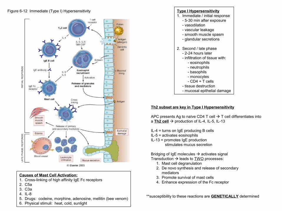
Figure 6-12 Immediate (Type I) Hypersensitivity Type I Hypersensitivity1. Immediate / initial response - 5-30 min after exposure - vasodilation - vascular leakage - smooth muscle spasm - glandular secretions
2. Second / late phase - 2-24 hours later - infiltration of tissue with: - eosinophils - neutrophils - basophils - monocytes - CD4 + T cells - tissue destruction - mucosal epithelial damage
Causes of Mast Cell Activation:1. Cross-linking of high affinity IgE Fc receptors2. C5a3. C3a4. IL-85. Drugs: codeine, morphine, adenosine, mellitin (bee venom)6. Physical stimuli: heat, cold, sunlight
Th2 subset are key in Type I Hypersensitivity
APC presents Ag to naïve CD4 T cell T cell differentiates into a Th2 cell production of IL-4, IL-5, IL-13
IL-4 = turns on IgE producing B cellsIL-5 = activates eosinophilsIL-13 = promotes IgE production stimulates mucus secretion
Bridging of IgE molecules activates signal Transduction leads to TWO processes: 1. Mast cell degranulation 2. De novo synthesis and release of secondary mediators 3. Promote survival of mast cells 4. Enhance expression of the Fc receptor
**susceptibility to these reactions are GENETICALLY determined
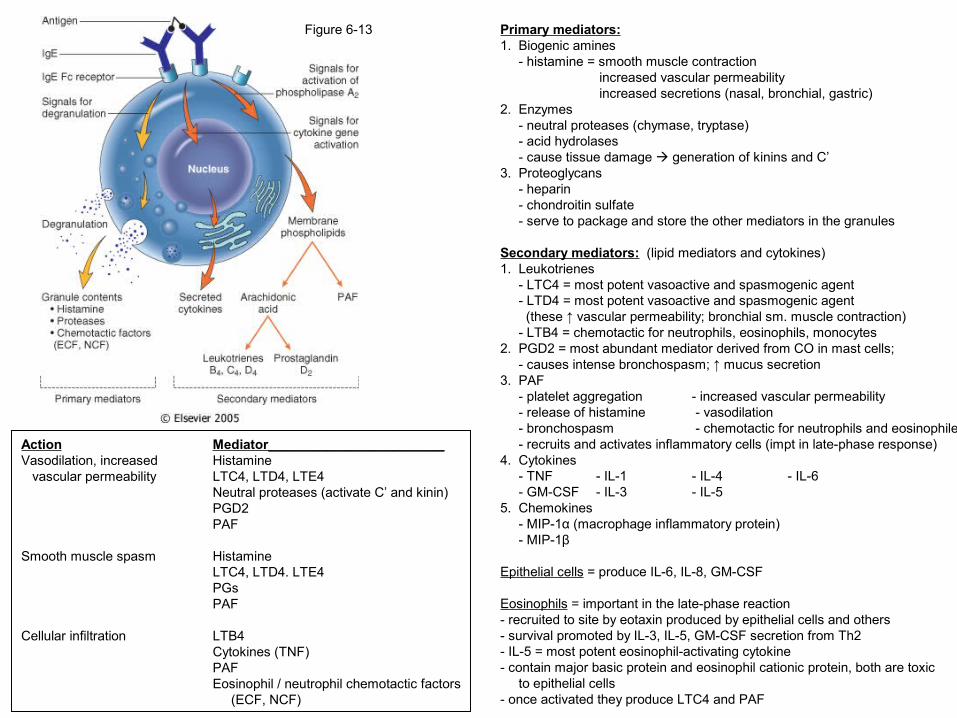
Primary mediators:1. Biogenic amines - histamine = smooth muscle contraction increased vascular permeability increased secretions (nasal, bronchial, gastric)2. Enzymes - neutral proteases (chymase, tryptase) - acid hydrolases - cause tissue damage generation of kinins and C’3. Proteoglycans - heparin - chondroitin sulfate - serve to package and store the other mediators in the granules
Secondary mediators: (lipid mediators and cytokines)1. Leukotrienes - LTC4 = most potent vasoactive and spasmogenic agent - LTD4 = most potent vasoactive and spasmogenic agent (these ↑ vascular permeability; bronchial sm. muscle contraction) - LTB4 = chemotactic for neutrophils, eosinophils, monocytes2. PGD2 = most abundant mediator derived from CO in mast cells; - causes intense bronchospasm; ↑ mucus secretion3. PAF - platelet aggregation - increased vascular permeability - release of histamine - vasodilation - bronchospasm - chemotactic for neutrophils and eosinophiles - recruits and activates inflammatory cells (impt in late-phase response) 4. Cytokines - TNF - IL-1 - IL-4 - IL-6 - GM-CSF - IL-3 - IL-55. Chemokines - MIP-1α (macrophage inflammatory protein) - MIP-1β
Epithelial cells = produce IL-6, IL-8, GM-CSF
Eosinophils = important in the late-phase reaction- recruited to site by eotaxin produced by epithelial cells and others- survival promoted by IL-3, IL-5, GM-CSF secretion from Th2- IL-5 = most potent eosinophil-activating cytokine- contain major basic protein and eosinophil cationic protein, both are toxic to epithelial cells- once activated they produce LTC4 and PAF
Figure 6-13
Action Mediator________________________Vasodilation, increased Histamine vascular permeability LTC4, LTD4, LTE4
Neutral proteases (activate C’ and kinin)PGD2PAF
Smooth muscle spasm HistamineLTC4, LTD4. LTE4PGsPAF
Cellular infiltration LTB4Cytokines (TNF)PAFEosinophil / neutrophil chemotactic factors (ECF, NCF)

Figure 6-14 Antibody-Mediated (Type II) HypersensitivityAntibody-Mediated (Type II) Hypersensitivity1. Mediated by Ab directed toward Ag present on cell surfaces or extracellular matrix - Ag may be intrinsic to the cell membrane or matrix - Ag may be exogenous but absorbed on a cell wall2. Results in binding of Ab to normal or altered cell-surface Ag3. There are three Ab dependent mechanisms - Opsonization and C’ and Fc receptor mediated phagocytosis - C’ and Fc receptor-mediated inflammation - Ab mediated cellular dysfunction
Opsonization and C’ and Fc Receptor-Mediated Phagocytosis1. IgG and IgM deposited on the surface of target cells and activate C’2. C’ activation results in C3b and C4b which are deposited on the surfaces of cells recognized by MØ phagocytosis cell death3. C’ activation leads to MAC cell destruction4. Cells opsonized by IgG are recognized by Fc receptors5. ADCC: cells are coated with low levels IgG binds to Fc receptor on monocytes, neutrophils, eosinophils, NK cells cell lysis proceeds without phagocytosis via porforins6. Examples: - transfusion rxn - erythroblastosis fetalis - autoimmune hemolytic anemia, agranulocytosis, thrombocytopenia - certain drug rxn
Complement and Fc Receptor-Mediated Inflammation1. Ab deposit on extracellular tissue (BM and matrix) activate C’ generation of C5a recruit neutrophils, monocytes leukocytes are activated release enzymes, O2 intermediates damage to tissue2. Examples: - glomerulonephritis - vascular rejection
Antibody-Mediated Cellular Dysfunction1. Ab directed against cell surface receptors impair or dysregulate function w/o causing cell injury or inflammation2. Example: - myasthenia gravis (block receptor muscle weakness) - pemphigus vulgaris - graves disease (stimulate cells hyperthyroidism)

Figure 6-15 Immune Complex-Mediated (Type III) Hypersensitivity1. Ag-Ab complexes produce tissue damage mainly by eliciting inflammation at sites of deposition2. Ab binds Ag in circulation circulating immune complexes complexes deposit in vessel walls or extracellularly if Ag was deposited previously 3, The formation of Ag-Ab complexes in circulation does NOT equal disease4. Two general types of Ag cause immune-complex mediated injury - exogenous Ag (foreign protein, bacteria, virus) - endogenous Ag (Ab against self-components)5. Disease can be generalized or localized
Systemic immune-complex disease has three phases:1. Formation of Ag-Ab complexes in circulation - introduction of Ag (protein) interaction with immune competent cells Ab formation formation of Ag-Ab complexes2. Deposition of immune complexes in various tissues - Ag-Ab complex deposition depends on: a) size of immune complexes - large complexes formed in Ab excess rapidly removed - small or intermediate size formed in slight Ag excess = most pathogenic b) functional status of the mononuclear phagocyte system c) charge of immune complexes d) valency of the Ag e) avidity of the Ab f) affinity of the Ag to various tissues g) three-dimensional structure of complex h) hemodynamic factors - sites of immune complex deposition a) renal glomeruli b) joints c) skin d) heart e) serosal surfaces f) small blood vessels3. Inflammatory reaction is caused by two mechanisms - activation of the complement cascade production of chemotactic factors (C5a) and release of anaphylatoxins (C3a and C5a) - activation of neutrophils and MØ through their Fc receptors or C3b receptors release/generation of pro-inflammatory substances - PGs - vasodilator peptides - chemotactic substances - lysosomal enzymes

Figure 6-16 Immune Complex-Mediated (Type III) Hypersensitivity
Chemotactic factors C5aAnaphylatoxins C3a, C5aLysosomal enzymes proteases that degrade BM, collagen, elastin, cartilage
End result = vascular compromise -- vasculitis (in vessels), glomerulonephritis (in kidney), arthritis (in joints)

Figure 6-18 Cell-Mediated (Type IV) Hypersensitivity
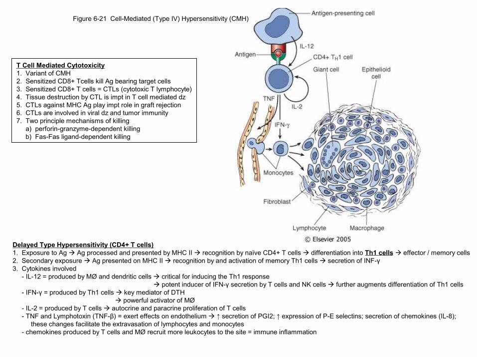
Delayed Type Hypersensitivity (CD4+ T cells)1. Exposure to Ag Ag processed and presented by MHC II recognition by naïve CD4+ T cells differentiation into Th1 cells effector / memory cells2. Secondary exposure Ag presented on MHC II recognition by and activation of memory Th1 cells secretion of INF-γ3. Cytokines involved - IL-12 = produced by MØ and dendritic cells critical for inducing the Th1 response potent inducer of IFN-γ secretion by T cells and NK cells further augments differentiation of Th1 cells - IFN-γ = produced by Th1 cells key mediator of DTH powerful activator of MØ - IL-2 = produced by T cells autocrine and paracrine proliferation of T cells - TNF and Lymphotoxin (TNF-β) = exert effects on endothelium ↑ secretion of PGI2; ↑ expression of P-E selectins; secretion of chemokines (IL-8); these changes facilitate the extravasation of lymphocytes and monocytes - chemokines produced by T cells and MØ recruit more leukocytes to the site = immune inflammation
Direct Cell Cytotoxicity (CD8+ T cells)1. Sensitized CD8+ T cells (cytotoxic T lymphocytes- CTLs) kill Ag bearing target cells2. CTLs play an important roll in graft rejection and resistance to viruses and tumor immunity3. Infection with virus viral peptides associated with MHC I recognized by TCR of cytotoxic CD8+ T cells lysis of infected cell by two mechanisms - perforin-granzyme-dependent killing = porforin perforates the plasma membrane granzyme delivered to target cell activate caspases apoptosis; perforin pores also allow water into the cell osmotic lysis - Fas-Fas ligand-dependent killing = induces apoptosis via the Fas ligand-receptor pathway
Characteristics of Activated Macrophages:1. augmented phagocytosis2. augmented killing of microorganisms3. increased express of MHC II4. secretion of - PDGF = ↑ fibroblast proliferation; collagen syn. - TNF = promote inflammation - IL-1 = promote inflammation - IL-12 =amplify Th1 response
Cell-Mediated (Type IV) Hypersensitivity1. Initiated by Ag-activated (sensitized) T cells and includes: - delayed type hypersensitivity – CD4+ T cells - direct cell cytotoxicity – CD8+ T cells2. Immunologic response to intracellular bacteria, viruses, fungi, protozoa, parasites
Delayed Type Hypersensitivity (CD4+ T cells)1. Exposure to Ag Ag processed and presented by MHC II recognition by naïve CD4+ T cells differentiation into Th1 cells effector / memory cells2. Secondary exposure Ag presented on MHC II recognition by and activation of memory Th1 cells secretion of INF-γ3. Cytokines involved - IL-12 = produced by MØ and dendritic cells critical for inducing the Th1 response potent inducer of IFN-γ secretion by T cells and NK cells further augments differentiation of Th1 cells - IFN-γ = produced by Th1 cells key mediator of DTH powerful activator of MØ - IL-2 = produced by T cells autocrine and paracrine proliferation of T cells - TNF and Lymphotoxin (TNF-β) = exert effects on endothelium ↑ secretion of PGI2; ↑ expression of P-E selectins; secretion of chemokines (IL-8); these changes facilitate the extravasation of lymphocytes and monocytes - chemokines produced by T cells and MØ recruit more leukocytes to the site = immune inflammation
Figure 6-21 Cell-Mediated (Type IV) Hypersensitivity (CMH)
T Cell Mediated Cytotoxicity1. Variant of CMH2. Sensitized CD8+ Tcells kill Ag bearing target cells3. Sensitized CD8+ T cells = CTLs (cytotoxic T lymphocyte)4. Tissue destruction by CTL is impt in T cell mediated dz5. CTLs against MHC Ag play impt role in graft rejection6. CTLs are involved in viral dz and tumor immunity7. Two principle mechanisms of killing a) perforin-granzyme-dependent killing b) Fas-Fas ligand-dependent killing

Figure 6-23 Events leading to destruction of grafts
Mechanisms of graft rejection1. HLA genes are highly polymorphic2. Rejection is complex and is due to both CMI and circulating Ab
T Cell-Mediated Rejection (cellular rejection)1. Mediated by two mechanisms a) destruction of graft cells by CD8+ CTLs b) DTH triggered by CD4+ Th2. Recipent’s T cells recognize Ag in the graft by a) direct pathway -major route in acute graft rejection -T cells of recipient recognize allogenic (donor) MHC molecules on APCs in the graft -dendritic cells in donor are the most impt immunogens -express both MHC I and MHC II -have costimulatory molecules (B7-1, B7-2) -both CD4+ and CD8+ T cells are involved -CD8+ cells recognize MHC I Ag on donor and cells mature into CTLs -CD4+ cells recognize MHC II Ag on donor and cells develop into Th1 DTH b) indirect pathway -major route in chronic graft rejection -recipient T cells recognize Ag of the graft after they are presented by the recipient’s own APCs -CD4+ T cells are generated and they enter the graft and then recognize graft Ag displayed by host APCs -results in a DTH -CD8+ CTLs may be generated in this pathway but they can’t recognize/kill graft cells because these CTLs recognize graft Ag presented by host APCs -principle mechanism of rejection is T cell cytokine production and DTH
Antibody-Mediated Rejection (Humoral Rejection)1. Ab evoked against alloantigens can also mediate rejection2. Two forms: a) Hyperacute = preformed antidonor Ab are present in the circulation of the recipient - circulating Ab react with and deposit rapidly on the vascular endothelium of the graft C’ fixation thrombosis b) Acute = not previously sensitized; Ab will form to HLA I and HLA II Ag - C’ dependent cytotoxicity, inflammation, ADCC - initial target = graft vasculature rejection vasculitis

Three Requirements of Pathologic Autoimmunity1. presence of autoimmune rxn2. rxn is not secondary to tissue damage3. absence of another well-defined dz
Figure 6-27 Mechanisms of central and peripheral tolerance
Immunologic tolerance = individual is incapable of developing an immune response to a specific Ag
Self tolerance = lack of responsiveness to individual’sown Ag- two mechanisms: -central tolerance -peripheral tolerance
Central Tolerance1. Death/deletion of self-reactive T and B cell clones during their maturation in the thymus and BM2. AIRE (autoimmune regulator) stimulates expression of many peripheral self-Ag in the thymus and is critical for deletion of immature self-reactive T cells3. Developing T cells that express high-affinity receptors for self-Ag are negatively selected/deleted by apoptosis
Peripheral Tolerance (Several back up mechanisms) 1. Anergy = prolonged/irreversible functional inactivation of lymphocytes, induced by encounter with Ag under certain conditions (lack of 2o signal)2. Suppression by regulator T cells = CD4+ CD25+ Tcell; Foxp3 required for development/func; IL-10 and TGF-β inhibit lymphocyte activation3. Clonal deletion by activation-induced cell death = Fas- Fas ligand system4. Antigen sequestration = brain, eye, testis are immune-privileged sites
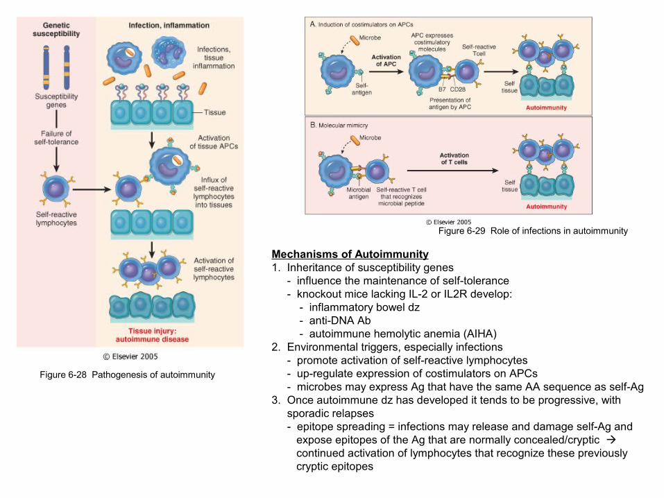
Figure 6-28 Pathogenesis of autoimmunity
Figure 6-29 Role of infections in autoimmunity
Mechanisms of Autoimmunity1. Inheritance of susceptibility genes - influence the maintenance of self-tolerance - knockout mice lacking IL-2 or IL2R develop: - inflammatory bowel dz - anti-DNA Ab - autoimmune hemolytic anemia (AIHA)2. Environmental triggers, especially infections - promote activation of self-reactive lymphocytes - up-regulate expression of costimulators on APCs - microbes may express Ag that have the same AA sequence as self-Ag3. Once autoimmune dz has developed it tends to be progressive, with sporadic relapses - epitope spreading = infections may release and damage self-Ag and expose epitopes of the Ag that are normally concealed/cryptic continued activation of lymphocytes that recognize these previously cryptic epitopes

Figure 6-30 Systemic Lupus Erythematosus1. Vast array of autoAb, especially antinuclear Ab (ANAs)2. Mechanism = failure of mechanisms that maintain self-tolerance3. Chronic, remitting, relapsing, febrile illness4. Injury primarily to: - skin - joints - kidney - serosal membranes5. ANA are directed against several nuclear Ag: - DNA - histones - nonhistone proteins bound to RNA - nucleolar Ag6. Ab to dsDNA and the Smith Ag are diagnostic of SLE7. Knockout mice lacking C4 or certain C’ receptors are prone to developing lupus-like autoimmunity
Key to pathogenesis = T helper cells
Morphology1. Deposition of immune complexes: - blood vessels - kidneys - connective tissue - skin
EM = subendothelial deposits; between the endothelium and the BM
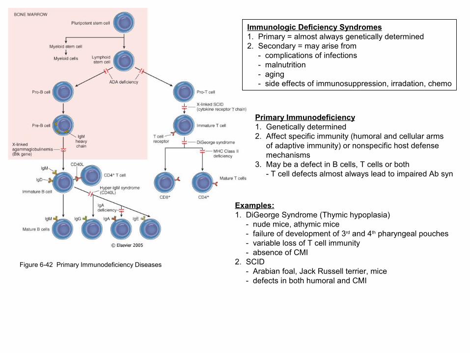
Figure 6-42 Primary Immunodeficiency Diseases
Immunologic Deficiency Syndromes1. Primary = almost always genetically determined2. Secondary = may arise from - complications of infections - malnutrition - aging - side effects of immunosuppression, irradation, chemo
Primary Immunodeficiency1. Genetically determined2. Affect specific immunity (humoral and cellular arms of adaptive immunity) or nonspecific host defense mechanisms3. May be a defect in B cells, T cells or both - T cell defects almost always lead to impaired Ab syn
Examples:1. DiGeorge Syndrome (Thymic hypoplasia) - nude mice, athymic mice - failure of development of 3rd and 4th pharyngeal pouches - variable loss of T cell immunity - absence of CMI2. SCID - Arabian foal, Jack Russell terrier, mice - defects in both humoral and CMI

Figure 6-43
Figure 6-46
Figure 6-45
Acquired Immunodeficiency Syndromes
Comparative Pathology1. Immunosuppressive Lentiviruses (infect lymphocytes only) - HIV - BIV - SIV - FIV2. Immunostimulatory Lentiviruses (infect MØ and lymphocytes) - CAEV - Ovine lentivirus (OPP, Meadi-Visna) - EIA
- CD4 molecule = high affinity receptor for HIV- HIV gp120 first binds to CD4 conformational change- HIV gp 120 then binds to coreceptors CCR5 and CXCR4- M-tropic strains can infect MØ, monocytes, and T cells (CCR5)- T-tropic strains can infect only T cells (CXCR4)

Figure 6-44
HIV1. Virus core contains: - major capsid protein p24—target for Ab used in dx - nucleocapsid protein p7/p9 - two copies of genomic RNA - three viral enzymes (protease, reverse transcriptase, integrase)2. Viral matrix protein, p173. On the viral envelope are two viral glycoproteins, gp120, gp41 - critical for HIV infection of cells4. HIV-1 RNA genome contains gag, pol, env genes - accessory genes = tat, rev, vif, nef, vpr, vpu = regulate syn and assembly
HALLMARK of AIDS = profound immunosuppression primarily CMI is affected
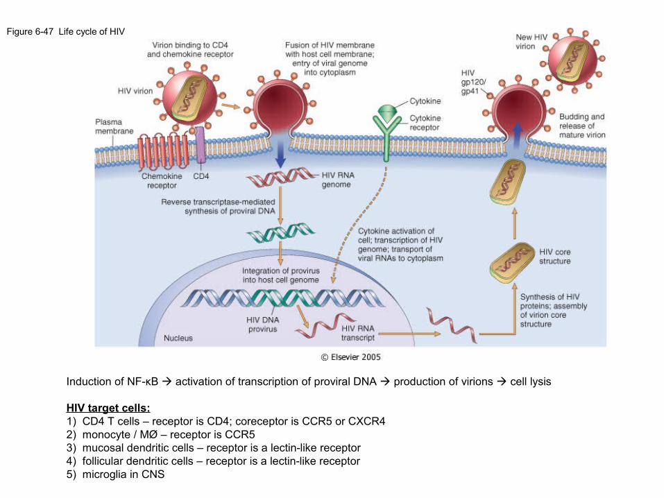
Figure 6-47 Life cycle of HIV
Induction of NF-κB activation of transcription of proviral DNA production of virions cell lysis
HIV target cells:1) CD4 T cells – receptor is CD4; coreceptor is CCR5 or CXCR42) monocyte / MØ – receptor is CCR53) mucosal dendritic cells – receptor is a lectin-like receptor4) follicular dendritic cells – receptor is a lectin-like receptor5) microglia in CNS

- Productive infection of T cells and viral replication in infected cells is the MAJOR mechanism by which HIV causes lysis of CD4+ T cells
- Other mechanisms include: 1) HIV colonizes lymphoid organs resevoir progressive destruction 2) activation induced cell death = activation of uninfected cells to respond to the virus apoptosis 3) loss of immature precursors of CD4+ T cells 4) fusion of infected and uninfected cells syncytia 5) apoptosis of uninfected CD4+ T cells by binding of soluble gp120 and CD4
Figure 6-48 Mechanisms of CD4 cell loss in HIV
Figure 6-50 Typical course of HIV
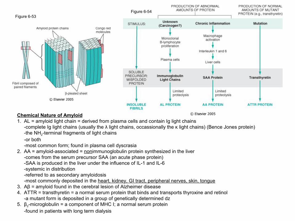
Figure 6-53Figure 6-54
Chemical Nature of Amyloid1. AL = amyloid light chain = derived from plasma cells and contain Ig light chains -complete Ig light chains (usually the λ light chains, occassionally the κ light chains) (Bence Jones protein) -the NH2-terminal fragments of light chains -or both -most common form; found in plasma cell dyscrasia2. AA = amyloid-associated = nonimmunoglobulin protein synthesized in the liver -comes from the serum precursor SAA (an acute phase protein) -SAA is produced in the liver under the influence of IL-1 and IL-6 -systemic in distribution -referred to as secondary amyloidosis -most commonly deposited in the heart, kidney, GI tract, peripheral nerves, skin, tongue3. Aβ = amyloid found in the cerebral lesion of Alzheimer disease4. ATTR = transthyretin = a normal serum protein that binds and transports thyroxine and retinol -a mutant form is deposited in a group of genetically determined dz5. β2-microglobulin = a component of MHC I; a normal serum protein -found in patients with long term dialysis