fibroepithelial lesions of the breast - houston society of ... - hscp 2016.pdf · 3 core biopsy...
TRANSCRIPT
04/04/2016
1
Fibroepithelial lesions of the breast
Dr Puay Hoon Tan
Department of Pathology
Singapore General HospitalSGH
Pathology
Fibroepithelial breast lesions are
biphasic tumors composed of both
epithelial and stromal components,
and include the common
fibroadenoma and the rarer
phyllodes tumor.
Scope
Fibroadenoma:
Core biopsy diagnosis.
Insights into tumorigenesis.
Relationship with phyllodes tumor.
Phyllodes tumor:
Grading, prediction of biological behavior.
Molecular highlights.

04/04/2016
2
Fibroadenoma
Common benign biphasic tumor.
Circumscribed breast neoplasm arising from the
terminal-duct lobular unit (TDLU).
Features a proliferation of both epithelial and stromal
elements.
Occurs most frequently in women of childbearing
age, especially those aged < 30 years, although it
may be encountered at any age.
Estimated 10% of women have fibroadenomas.
Gross anatomy of
fibroadenoma
Bosselated outlines Myxoid surface
Fibrous homogenous appearanceLobulated cut suface
Ossified fibroadenomaGiant fibroadenoma
Microscopic anatomy of
fibroadenomaCellular fibroadenoma
Intracanalicular pattern
Pericanalicular pattern
Juvenile fibroadenoma
Complex fibroadenoma

04/04/2016
3
Core biopsy diagnosis of
fibroadenoma Core biopsies represent the standard of care in
preoperative diagnosis of breast lesions discovered
clinically & radiologically.
No further treatment is needed for a diagnosis of
fibroadenoma, vs excision biopsy for a conclusion of
phyllodes tumor on core biopsy.
How reliable is a core biopsy diagnosis of
unambiguous fibroadenoma?
Do we need to be concerned about underdiagnosing
a phyllodes tumor?
Phyllodes Tumor Subsequent to a
Diagnosis of Fibroadenoma on
Breast Core Needle Biopsy:
Frequency and Characteristics
Timothy W Jacobs1, Yunn-Yi Chen2, Donald G Guinee1, Peter R
Eby1, Aye Aye Thike3, Poonam Vohra2, Puay Hoon Tan3
1. Virginia Mason Medical Center, Seattle, WA
2. UCSF, San Francisco, CA
3. Singapore General Hospital, Singapore
Courtesy of Dr Timothy Jacobs, platform presentation at USCAP 2014, San Diego California
Conclusions
The incidence of PT subsequent to a diagnosis of FA on
CNB is extremely low (0.38%, 16 out of 4163 cases).
Most PT were categorized as benign (14 benign, 2
borderline).
PT heterogeneity (e.g. FA-like areas) likely contributed to
CNB-excision discrepancies.
No pathologic features on CNB appeared to be prospectively
predictive of PT at excision.
Suspicious imaging features at time of CNB or on follow-up
should prompt consideration for surgical excision.
Diagnosing FA on CNB is reliable and safe, provided there is
adequate imaging correlation and follow-up.
Courtesy of Dr Timothy Jacobs, platform presentation at USCAP 2014, San Diego California

04/04/2016
4
Core biopsy diagnosis of cellular fibroepithelial
lesions – prediction of phyllodes tumor
Author Reference Key findings predicting phyllodes tumor
Jacobs et al Am J Clin Pathol 2005;
124: 342-354Marked stromal cellularity, mitoses in moderate
stromal cellularity, Ki67 & topoisomerase IIα
indices
Lee et al Histopathology 2007;
51: 336-344Stromal cellularity ≥ 50% stroma, stromal
overgrowth, fragmentation, adipose within
stroma
Resetkova et
al
Breast J 2010; 16:573-
80. No predictive value of clinical, radiologic or
pathologic data
Suggested follow-up alone for a patient subset
Jara-Lazaro
et al
Histopathology 2010;
57: 220-232Marked stromal cellularity/atypia, stromal
overgrowth, mitoses ≥ 2 per 10 hpf, ill-defined
lesional borders, Ki67 & topoisomerase IIα
indices ≥ 5%, reduced CD34 staining
Yasir et al Am J Clin Pathol 2014;
142: 362-369 Mitoses, stromal overgrowth, fragmentation,
adipose infiltration, heterogeneity, subepithelial
condensation, nuclear pleomorphism
21 cases of fibroepithelial lesions that were challenging to classify as
cellular fibroadenoma or phyllodes tumor.
One to 2 representative slides of each case along with patient age were
sent to 10 breast pathologists.
WHO criteria for phyllodes tumors and a diagnosis form were included with
the study set.
Only 2 cases had uniform agreement as to whether the tumor was a
fibroadenoma or phyllodes tumor.
If diagnoses of fibroadenoma and benign phyllodes tumor were combined
and separated from borderline and malignant phyllodes tumors, there was
100% agreement in 53% of cases and 90% agreement in 79% of cases.
Molecular genetics of fibroadenomas
Cytogenetic abnormalities in 20% to 30% of
fibroadenomas, usually translocations.
No consistent pattern of specific chromosomal
alterations.
Both epithelial and stromal components are
polyclonal. (Noguchi et al. Cancer Res1993; 53: 4071-4072)
Possible evolution into phyllodes tumors.
(Noguchi et al. Cancer1995; 76: 1779-1785)
Low levels of LOH (0% to 1.5%).
(Wang et al. Breast Cancer Res Treat 2006; 97: 301-309)
No evidence of recurrent genetic alterations characteristic of
fibroadenomas. Brogi E, in Rosen’s Breast Pathology 4th edition 2014

04/04/2016
5
P
Key findings:• Exome sequencing of 8 fibroadenomas with matching whole blood samples revealed
recurrent somatic mutations solely in MED12 (encodes a Mediator complex subunit).
• Targeted sequencing of an additional 90 fibroadenomas confirmed highly frequent
MED12 exon 2 mutations (58/98, 59%) that are probably somatic, with 71% of
mutations occurring in codon 44.
• Using laser capture microdissection, it was confirmed that MED12 fibroadenoma
mutations are present in stromal but not epithelial mammary cells.
MED12 mutations in breast fibroadenoma
MED12 is located on the X chromosome.
Frequent MED12 exon 2 somatic mutations have
been found previously only in uterine leiomyoma (UL).
MED12 mutation spectrum observed in
fibroadenomas was nearly identical to that of UL in
both exon location and variant codon preference.
Possibility that MED12 exon 2 mutations could be
associated with hormonal expression.
MED12 in phyllodes tumors.
Phyllodes tumor

04/04/2016
6
Phyllodes tumor
Uncommon fibroepithelial neoplasm
with proliferation of both epithelial and
stromal components.
“Phyllodes”
- Derived from the Greek word “phyllon”
meaning leaf, and “eidos” meaning form.
Phyllodes tumor: fibroepithelial neoplasm resembling
intracanalicular fibroadenoma, but with exaggerated
fronded pattern and stromal hypercellularity
0.3-1% of all primary
breast tumors.
Affects middle aged
women (40-50 years).
Higher incidence in
Asian women.
Graded according to
histological
characteristics.
Tendency to recur if
incompletely excised.Benign phyllodes tumor
Large tumor stretching skin
Circumscribed bulging mass,
mucoid, fleshy, whorled

04/04/2016
7
Ultrasound scan
Excision biopsy
Mastectomy
Historical perspectives
1838
Cystosarcoma
phyllodesJohannes Müller
1774
Ochme described a
rapidly benign growing
tumor (about 4kg) of a
young woman
1960
Tumor
phyllodes(Lomonaco, Tumori)
1824
Chelius singled out
a ‘cystic hydatid’ of
the mammary gland
1829
Cooper termed it
‘cellular hydatid’,
described tumors
later deemed as
phyllodes
1941
Owens & Adams - term
cystosarcoma should be
avoided as the lesion is
usually not sarcomatous.
Proposed ‘giant
intracanalicular
fibroadenoma of the breast’
1839-1930
Numerous terms and
descriptions devoted to
the same tumor, eg
intracanalicular
fibroma,
pseudosarcoma,
serocystic tumor etc.
Virchow,1867, opined its
‘limited malignancy, but
capacity to metastasize’
Fiks A. Cystosarcoma Phyllodes of the Mammary Gland – Müller’s
Tumour: For the 108th Birthday of Johannes Müller. Virchows Archiv
1981; 392:1-6
Evolving concepts on classification
Treves and Sunderland (1951) reported 18 of 77 tumours could not fit in either category
“…the disease is perfectly innocent” Müller (1838)
Benign
• Lee and Pack (1931) reported 5 recurrences in 91 cases with available clinical outcome
• White (1940) reported recurrence and subsequent metastasis of a case
Cooper and Ackerman (1943) proposed
Benign Malignant
Treves and Sunderland (1951) proposed
Benign Borderline Malignant

04/04/2016
8
Histological assessment
Norris and Taylor (1967) – 94 cases
Proposed criteria:
1) Contours (margins) and size of tumor
2) Degree of mitotic activity 3) Cellular atypism
Oberman; Hart et al (1965;1978)
Suggested stromal overgrowth as an additional adverse prognostic factor
Ward and Evans (1986)
Specified objective for stromal overgrowth, assessment of cellularity and atypia in semi-quantitative way
Tan et al (2011)
Quantitative weightage of risk based on stromal atypia, mitotic rate, overgrowth and surgical margins
Phyllodes tumor
WHO Classifications 1981, 2003, 2012
WHO classification of breast tumours 2012

04/04/2016
9
Tumor borders
circumscribed permeative
Stromal cellularity
mild moderate marked
Stromal atypia
mild
moderate
marked

04/04/2016
10
Mitotic activity
Benign
< 5
mitoses/10hpf
Borderline
5 to 9
mitoses/10hpf
Malignant
≥ 10
mitoses/10hpf
Rosen’s Breast Pathology 4th edition 2014 classifies phyllodes tumors with >2
mitoses/10 hpf as borderline, page 237
Stromal overgrowth
Absent Present
Phyllodes tumors Histologic features have been helpful to
some extent in predicting biologic behavior.
Specific parameters that can define the likelihood for recurrence are not universally accepted. Benign: local recurrence.
Borderline: local recurrence, and rarely metastases.
Malignant: local recurrence and metastases.
SGH
Pathology

04/04/2016
11
Phyllodes tumors Limitations of morphologic
classification: Subjectivity
Continuum of microscopic features
Five histological parameters
Each parameter with 2 to 3 categories ≈ 108 permutations
Certainty at extreme ends of classification.
Ambiguity in between.
Phyllodes tumors:
predicting clinical behavior
Phyllodes tumors:
prediction of biological behavior Grade correlates with behavior.
Grade assignment is imperfect:
Stromal hypercellularity, atypia, mitoses, overgrowth,
borders.
Questions:
Does each histological parameter have equal importance?
Can we determine if some parameters have a greater
weightage in predicting behavior?
Is there an objective scoring system that can define
behavior?

04/04/2016
12
Phyllodes tumors:
prediction of biological behavior 605 women with phyllodes tumors diagnosed
at SGH Pathology between 1992 and 2010.
Histological parameters assessed.
Clinical follow-up to determine recurrent
disease.
Statistical model focusing on parameters
with impact on recurrence.
J Clin Pathol. 2012 Jan;65(1):69-76.
Phyllodes tumors: 605 cases
Benign 440 (72.7%)
Borderline 111 (18.4%)
Malignant 54 (8.9%)
Median follow-up 56.9 months; range
3.3 to 229.2 months.
8

04/04/2016
13
Recurrences
Mean and median times to recurrence 29.8 and 24.6 months
respectively.
Phyllodes tumors:
prediction of biologic behavior
Statistical method:
Reverse step modelling.
Assessment of multiple variables and their
inter-relationship.
Derivation of a set of variables with least
overlap.
Nomogram tested with bootstrapped
sample set.
A : Atypia
M: Mitoses
O: Overgrowth
S : Surgical margin

04/04/2016
14
http://phyllodes.com/
Validation of the SGH nomogram Japanese cohort of 45 patients with phyllodes tumors (2 excluded due to
death from other causes), Shikoku Cancer Center, Matsuyama Japan.
Median age 45 years; follow-up 4.7 to 309.9 months (median 129 months);
median time to recurrence 113.3 months.
Factor No of events/
No of patients
HR (95% CI) p-value
Mitotic activity 6/43 0.89 (0.53, 1.50) 0.665
Stromal overgrowth
Absent
Present
4/38
2/5
Reference
3.60 (0.66, 19.79) 0.115
Surgical margins
Negative
Positive
0/25
6/18
Reference
- 0.0006
Stromal atypia
Mild
Moderate
6/37
0/6
Reference
- 0.287
Nishimura et al. J Clin Pathol. 2014 Aug;67(8):748-50.

04/04/2016
15
Validation of the SGH nomogram
Japanese cohort.
HR (95% CI) P-value Concordance
index
Nomogram 1.11 (1.02, 1.20) 0.0005 0.904
High concordance index indicates the ability of the SGH nomogram to
accurately predict the recurrence likelihood of the Japanese cohort of patients.
Nishimura et al. J Clin Pathol. 2014 Aug;67(8):748-50.
Phyllodes tumor:
relationship with fibroadenoma
Relationship between fibroadenoma
& phyllodes tumor (I)
3 fibroadenomas progressing to phyllodes tumors.
Noguchi et al. Cancer 1995; 76: 1779-85
Clonality analysis of fibroadenomas:
One 'simple' fibroadenoma and one complex fibroadenoma with
monoclonality of stroma.
Monoclonal 'simple' fibroadenoma was histologically described to
contain a phyllodes component which also demonstrated stromal
monoclonality. Kasami et al. Breast Cancer Res Treat 1998; 50: 185-91
Monoclonality in areas of apparent stromal expansion within
fibroadenomas, suggesting stromal progression of
fibroadenoma to phyllodes tumor. Kuijper et al. J Pathol 2002; 197: 575-81

04/04/2016
16
Relationship between fibroadenoma
& phyllodes tumor (II)
Identical loss at the same microsatellite locus in a synchronous
fibroadenoma and phyllodes tumor of the same breast, while allelic losses
at TP53 and another microsatellite locus were observed in the phyllodes
tumor but not in the synchronous fibroadenoma; implicating TP53 in
progression of fibroadenoma to phyllodes tumor. Hodges KB et al. Appl Immunohistochem Mol Morphol 2009; 17: 345-50
36 malignant phyllodes tumors:
11 associated with previous fibroadenomas (which could be interpreted as
progression of fibroadenoma to phyllodes tumors).
Better clinical outcome in malignant phyllodes tumors preceded by
fibroadenomas than in those diagnosed de novo.
Abe et al. Breast Cancer 2011; 18: 268-72
Relationship between fibroadenoma
& phyllodes tumor (III)
Fibroadenoma areas are seen in phyllodes tumors in variable
frequency:
Phyllodes tumor is in close juxtaposition to a pre-existing hyalinized
fibroadenoma.
Phyllodes tumor is arising from the fibroadenoma.
Fibroadenoma-like areas in phyllodes tumors reflect heterogeneity of
phyllodes tumors.
Molecular genetics of
phyllodes tumors:
an update
04/04/2016
17
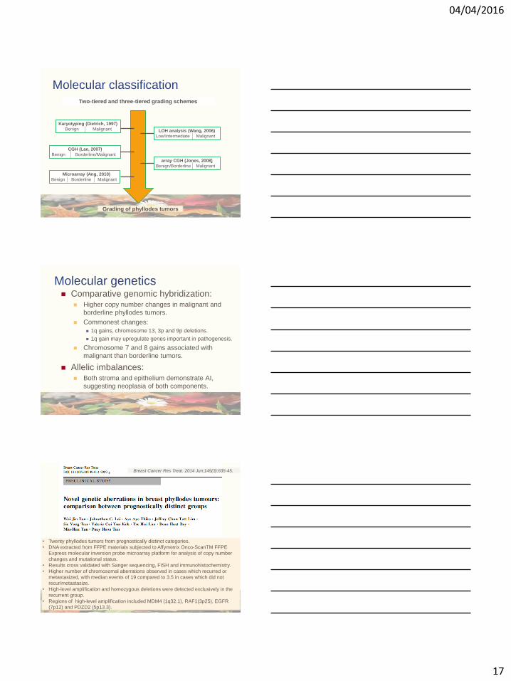
Molecular classification
Karyotyping (Dietrich, 1997)
Benign Malignant LOH analysis (Wang, 2006)
Low/Intermediate Malignant
CGH (Lae, 2007)
Benign Borderline/Malignant
array CGH (Jones, 2008)
Benign/Borderline Malignant
Two-tiered and three-tiered grading schemes
Microarray (Ang, 2010)
Benign Borderline Malignant
Grading of phyllodes tumors
Molecular genetics Comparative genomic hybridization:
Higher copy number changes in malignant and
borderline phyllodes tumors.
Commonest changes:
1q gains, chromosome 13, 3p and 9p deletions.
1q gain may upregulate genes important in pathogenesis.
Chromosome 7 and 8 gains associated with
malignant than borderline tumors.
Allelic imbalances: Both stroma and epithelium demonstrate AI,
suggesting neoplasia of both components.
• Twenty phyllodes tumors from prognostically distinct categories.
• DNA extracted from FFPE materials subjected to Affymetrix Onco-ScanTM FFPE
Express molecular inversion probe microarray platform for analysis of copy number
changes and mutational status.
• Results cross validated with Sanger sequencing, FISH and immunohistochemistry.
• Higher number of chromosomal aberrations observed in cases which recurred or
metastasized, with median events of 19 compared to 3.5 in cases which did not
recur/metastasize.
• High-level amplification and homozygous deletions were detected exclusively in the
recurrent group.
• Regions of high-level amplification included MDM4 (1q32.1), RAF1(3p25), EGFR
(7p12) and PDZD2 (5p13.3).
Breast Cancer Res Treat. 2014 Jun;145(3):635-45.

04/04/2016
18
Genomic landscapes of breast fibroepithelial tumors
Tan J et al. Nat Genet. 2015 Nov;47(11):1341-5.
Proposed model of the genomic progression of breast fibroepithelial tumors
Tan J et al. Nat Genet. 2015 Nov;47(11):1341-5.
MED12 mutations in breast fibroepithelial lesions

04/04/2016
19
MED12 mutations in breast fibroepithelial lesions
What’s the clinical relevance? Genomics based classification of breast
fibroepithelial lesions, enhancing
diagnostic accuracy.
Discovery of candidate therapeutic
targets in borderline/malignant PT:
PIK3CA activating mutations
EGFR amplifications
MED12 and RARA mutations linked to
hormone receptor signaling.

04/04/2016
20
Summary
Phyllodes tumors present distinct
challenges relating to their diagnosis,
classification, predicted behavior, and
clinical management.
Practical recommendations I
Grading of phyllodes tumors should aim to achieve
accuracy and consistency at the benign and
malignant ends of the spectrum.
Definitive distinction between cellular fibroadenomas
and benign phyllodes tumors may not be crucial, in
light of similar reported recurrence rates. The term
benign fibroepithelial lesion/neoplasm may be
recommended for cases where clear diagnostic
distinction cannot be made, although this should be
used sparingly.
Practical recommendations II
Malignant phyllodes tumors are diagnosed when
there are marked stromal hypercellularity, atypia,
increased mitoses of ≥10/10 HPFs, permeative
tumor borders, and stromal overgrowth. The
presence of a malignant heterologous component
places the tumor into the malignant category
regardless of other histological features.
A conservative approach can be accorded to benign
phyllodes tumors that have been initially enucleated
without margins.

04/04/2016
21
Practical recommendations III
Excision with negative margins should be achieved
for recurrent and malignant phyllodes tumors. Most
would recommend that borderline tumors should
also be completely excised. Although the literature
often refers to a margin width of at least 10 mm, a
robust evidence base to support this approach is
lacking. Therefore an ideal margin width remains to
be determined, and may need to be considered in
relation to factors such as tumor size and cosmesis.
Practical recommendations IV
From a diagnostic and management perspective, it
is important to accurately recognize malignant
phyllodes tumors, which should be surgically
eradicated and effectively treated at diagnosis, as
these tumors have a well-established but relatively
infrequent risk of metastasis and death.
The role of adjuvant radiation therapy in borderline
and malignant tumors remains to be defined.
Routine axillary dissection is not recommended.
Acknowledgements
Pathology SGHBreast research team• Dr Aye Aye Thike
• Ms Wai Jin Tan
• Mr Jeffrey Lim
• Ms Valerie Koh
• Ms Jane Tan
• Ms Nur Diyana Bte Md Nasir
Breast service team
• Dr Angela Chong
• Dr Inny Busmanis
• Dr Jabed Iqbal
• Dr Syed Salahuddin
• Dr Benjamin Yongcheng Tan
Clinical Research SGH
• Dr HuiHua Li
Duke-NUS• Dr Bin-Tean Teh
• Dr Patrick Tan
• Dr Steve Rosen
• Dr Jing Tan
• Dr Cedric Ng
• Dr Choon Kiat Ong
• Dr Weng Khong Lim
• Ms Swe Swe Myint
Breast Surgical Oncology• Dr Kong Wee Ong
• Dr Benita TanAnatomy, Yong Loo Lin
School of Medicine, NUS• Dr Boon Huat Bay

04/04/2016
22
Thank you