fibroblast growth factor 10 induces proliferation and differentiation of human primary cultured...
TRANSCRIPT

Fibroblast Growth Factor 10 Induces Proliferation andDifferentiation of Human Primary Cultured Keratinocytes
Cinzia Marchese,* Alessandra Felici,* Vincenzo Visco,* Giuseppe Lucania,* Makoto Igarashi,²Mauro Picardo,³ Luigi Frati,*§ and Maria Rosaria Torrisi*³*Dipartimento di Medicina Sperimentale e Patologia, UniversitaÁ di Roma ``La Sapienza'', Roma, Italy; ²Mount Sinai School of Medicine, New York,
U.S.A.; ³Istituto Dermatologico San Gallicano, Roma, Italy; §Istituto Neurologico Mediterraneo ``Neuromed'', Pozzilli, Italy
Fibroblast growth factor 10 is a novel member of the®broblast growth factor family, which is involved inmorphogenesis and epithelial proliferation. It ishighly homologous to the keratinocyte growth factor(or ®broblast growth factor 7), a key mediator ofkeratinocyte growth and differentiation. Both ®bro-blast growth factor 10 and keratinocyte growth fac-tor bind with high af®nity to the tyrosine kinasekeratinocyte growth factor receptor. Here we ana-lyzed the effect of ®broblast growth factor 10 on pri-mary cultures of human keratinocytes, grown inchemically de®ned medium, and we compared theproliferative and differentiative cell responses to®broblast growth factor 10 with those induced bykeratinocyte growth factor and epidermal growthfactor. Cell counting, 5-bromo-2¢-deoxyuridine
incorporation, and western blot analysis showed that®broblast growth factor 10, similarly to keratinocytegrowth factor, not only is a potent mitogen forhuman keratinocytes, but also promotes the expres-sion of both early differentiation markers K1 andK10 and late differentiation marker ®laggrin inresponse to the Ca2+ signal, and seems to sustain theproliferative activity in suprabasal strati®ed cells.Immunoprecipitation/western blot analysis revealedthat ®broblast growth factor 10, similarly to kerati-nocyte growth factor, is able to induce tyrosinephosphorylation of keratinocyte growth factor recep-tor and of cellular substrates such as PLCg. Keywords: ®broblast growth factor 10/human keratinocytes/keratinocyte growth factor. J Invest Dermatol 116:623±628,2001
The ®broblast growth factors (FGF) constitute a familyof at least 19 members and are responsible for differentactivities on cell proliferation and differentiation,embryonic development, angiogenesis, and woundhealing (for a review, see Mason, 1994). FGF-10 is a
novel member of the FGF family, whose biologic activities remainto be elucidated (Yamasaki et al, 1996; Emoto et al, 1997). It hasrecently been reported that FGF-10 plays a key role in controllingdirectional outgrowth and inducing epithelial buds in lungdevelopment (Bellusci et al, 1997; Park et al, 1998). Moreover,FGF-10, together with other members of the FGF family, appearsto be essential for signaling induction during limb development(Xu et al, 1998). Finally, generation of FGF-10 de®cient mice leadsto lack of both limb and lung formation (Min et al, 1998; Sekine etal, 1999).
When compared to the other members of the FGF family, boththe amino acid sequence and the tissue expression of FGF-10 showhigh similarity to those of the keratinocyte growth factor (KGF/FGF-7). Unlike the other known FGF, KGF acts speci®cally onepithelial cells (Finch et al, 1989), modulating their growth anddifferentiation. In fact, KGF not only is a mitogen for murine
(Rubin et al, 1989; Duglosz et al, 1994) and human culturedkeratinocytes (Marchese et al, 1990), but also promotes their earlydifferentiation program (Marchese et al, 1990; 1997) and inhibitstheir terminal differentiation and apoptosis (Hines and Allen-Hoffman, 1996). In addition, KGF seems to play a key role inreepithelialization during normal human and experimental woundhealing (Werner et al, 1992, 1994; Staiano-Coico et al, 1993;Marchese et al, 1995). Similarly to KGF, FGF-10 seems to exertmitogenic activity on mouse and rat keratinocyte cell lines (Emotoet al, 1997; Igarashi et al, 1998). Expression of FGF-10 does notseem to be modulated, however, during experimental woundhealing (Beer et al, 1997), although the topical addition of thegrowth factor promotes wound repair in the same model (Jimenezand Rampy, 1999).
Both FGF-10 and KGF bind only and with high af®nity (Igarashiet al, 1998) to the KGF receptor (KGFR), a splicing variant of FGFreceptor 2 (FGFR2), expressed exclusively on epithelial cells (Mikiet al, 1991, 1992). The two growth factors show a differentsensitivity to heparin in their mitogenic activity, however, implyingthat the extracellular matrix may play a role in regulating thecellular response to FGF-10 and KGF (Igarashi et al, 1998).
We have recently shown that KGFR is up-modulated in vitroduring differentiation from basal to suprabasal keratinocytes(Marchese et al, 1997), suggesting that receptor expression maycontrol the proliferative±differentiative cell program, and consistentwith the ability of KGF, unlike the epidermal growth factor (EGF),to induce both cell proliferation and expression of differentiationmarkers in response to the Ca2+ signal (Marchese et al, 1990, 1997).To ascertain if FGF-10 is able to exert activities similar to KGF on
0022-202X/01/$15.00 ´ Copyright # 2001 by The Society for Investigative Dermatology, Inc.
623
Manuscript received July 7, 1999; revised August 8, 2000; accepted forpublication December 7, 2000.
Reprint requests to: Dr. Maria Rosaria Torrisi, Dip. MedicinaSperimentale e Patologia, Viale Regina Elena 324, 00161 Roma, Italy.Email: [email protected]
Abbreviations: FGF, ®broblast growth factor; FGFR, ®broblast growthfactor receptor; KGF, keratinocyte growth factor; KGFR, keratinocytegrowth factor receptor.

primary cultures of human keratinocytes, we compared here theproliferative and differentiative cell response to FGF-10, KGF, andEGF. Our results show that FGF-10 is a potent mitogen for humankeratinocytes. In addition, similarly to KGF and differently fromEGF, FGF-10 allows the expression of the early suprabasaldifferentiation markers K1 and K10 and of the late differentiationmarker ®laggrin, promoting therefore the initiation and progressionof the keratinocyte maturation program.
MATERIALS AND METHODS
Human keratinocyte culture Primary cultures of humankeratinocytes derived from neonatal foreskin as previously described(Rheinwald and Green, 1977) were maintained in MCDB 153 medium(Clonetics, San Diego, CA), supplemented with 0.5 mg per mlhydrocortisone, 5 mg per ml insulin, 5 mg per ml gentamycin, and 70 mgper ml whole bovine pituitary extract. Ca concentration in the mediumwas maintained at 0.15 mM (Boyce and Ham, 1983). Cell adhesion wasachieved by precoating the tissue culture 24-well plates used for theproliferation assay with 1 mg per ml of ®bronectin (Sigma Chemical, St.Louis, MO). Cultures were supplemented with recombinant humanFGF-10 (Igarashi et al, 1998) (0.05±5 mM, 1±100 ng per ml), with EGF(0.05±5 mM, 3±300 ng per ml; Collaborative Research, Lexington,MA), or with KGF (0.05±5 mM, 1.4±140 ng per ml; UpstateBiotechnology, Lake Placid, NY). To measure cell number, cultureswere harvested by incubation in 0.5% trypsin, 0.2% ethylenediaminetetraacetic acid (EDTA) for 15 min at 37°C, and the cell counts wereperformed in triplicate using a hemocytometer.
5-Bromo-2¢-deoxyuridine (BrdU) incorporation and immuno-¯uorescence Cells, grown on tissue culture chamber slides (Nunc,Naperville, IL), were treated for 3 d with each growth factor at aconcentration of 20 ng per ml. At the end of treatment, 100 mM BrdU(Sigma) was added to the medium and the cultures were incubated for24 h at 37°C to allow BrdU incorporation. Cells were then ®xed in 4%formaldehyde in phosphate-buffered saline (PBS) for 30 min at 25°C,
followed by treatment with 0.1 M glycin for 20 min at 25°C and with0.5% HCl, 0.1% Triton X-100 for an additional 45 min at 25°C toallow permeabilization. After extensive washing in PBS, cells werebuffered with 0.1 M Na2B4O7 and incubated with anti-BrdUmonoclonal antibody (Sigma) (1:50 in blocking buffer: PBS 0.5% bovineserum albumin, 0.5% Tween 20) for 30 min at 25°C in a humiditychamber, followed by goat antimouse IgG-Texas Red (1:50 in blockingbuffer) (Jackson Immuno Research Laboratories, PA). For doubleimmuno¯uorescence, cells were incubated with anti-BrdU as above andwith anti-K1 polyclonal antibodies (Berkeley Antibody, Richmond, CA)(1:100 in PBS) visualized with goat antimouse IgG-¯uoresceinisothiocyanate (1:10 in PBS) (Cappel Research Products, Durham, NC)and goat antirabbit IgG-Texas Red (1:50 in PBS) (Jackson ImmunoResearch Laboratories) after appropriate washing with PBS.Colocalization of the two ¯uorescence signals was analyzed by recordingand merging single stained images using a cooled CCD color digitalcamera SPOT-2 (Diagnostic Instruments, MI) and FISH 2000/H1software (Delta Sistemi, Roma, Italy).
Western blot analysis Cells cultured for 6 d in medium containing1 mM Ca2+ concentration and supplemented with 20 ng per ml FGF-10, KGF, or EGF were lyzed in RIPA buffer [10 mM Tris, pH 7.4;50 mM NaCl; 1 mM EDTA; 10 mM KCl; 1% Nonidet P-40; 0.1%sodium dodecyl sulfate (SDS); 0.05% Tween 20] supplemented withprotease inhibitors [10 mg per ml aprotinin, 1 mM phenylmethylsulfonyl¯uoride (PMSF), 10 mg per ml leupeptin]. Twenty-®ve micrograms oftotal proteins were resolved under reducing conditions by 10% SDSpolyacrylamide gel electrophoresis (PAGE) and transferred to reinforcednitrocellulose (PROTRAN, Schleider & Schuell, Keene, NH). Themembrane was blocked with 3% nonfat dry milk in PBS with 0.05%Tween 20 for 4 h at room temperature. After washing for 20 min inPBS 0.05% Tween 20, the membrane was incubated with anti-K1polyclonal antibody diluted 1:500 (Berkeley Antibody), with anti-K10/13 mouse monoclonal antibody diluted 1:200 (Santa CruzBiotechnology, Santa Cruz, CA), and with anti®laggrin monoclonalantibody diluted 1:500 (Biomedical Technologies, Stoughton, MA) for1 h at 25°C, followed by enhanced chemiluminescence (ECL) detection(Amersham, Arlington Heights, IL).
Figure 1. Comparison of the proliferative activities of FGF-10 (in the presence or absence of 0.3 mg per ml heparin), KGF, and EGF onhuman primary cultured keratinocytes grown in standard low Ca2+ medium. Treatments and cell counts were performed as described inMaterials and Methods. The results represent the mean values from three different experiments.
624 MARCHESE ET AL THE JOURNAL OF INVESTIGATIVE DERMATOLOGY

Immunoprecipitation and western blot analysis Subcon¯uentcultures of NIH3T3 KGFR transfectants were lyzed in RIPA buffer(10 mM Tris pH 7.4; 50 mM NaCl; 1 mM EDTA; 10 mM KCl; 1%Nonidet P-40; 0.1% SDS; 0.05% Tween 20) supplemented with proteaseinhibitors (10 mg per ml aprotinin, 1 mM PMSF, 10 mg per mlleupeptin) and phosphatase inhibitors (25 mM sodium orthovanadate,20 mM sodium pyrophosphate, 0.5 M NaF). Fifteen micrograms of totalproteins were resolved under reducing conditions by 7% SDS-PAGE andtransferred to reinforced nitrocellulose (BA-S 83, Schleider & Schuell).The membrane was blocked with 5% nonfat dry milk in PBS with 0.1%Tween 20 for 4 h at room temperature. After washing for 20 min inPBS 0.1% Tween 20 the membrane was incubated alternatively withrabbit anti-Bek polyclonal antibodies, diluted 1:200 (C-17, Santa CruzBiotechnology), or with mouse anti-Bek monoclonal antibody, diluted1:200 (C-8, Santa Cruz Biotechnology), both antibodies raised againstthe carboxy terminal region of the intracellular domain of the FGFR2/KGFR, for 1 h at 25°C, followed by ECL detection (Amersham). Forthe detection of KGFR tyrosine phosphorylation, NIH3T3 KGFR cellswere serum starved for 12 h, treated with 100 ng per ml KGF 0.3 MNaCl or with 100 ng per ml FGF-10 for 10 min at 37°C, lyzed, andprocessed as above for immunoblotting with antiphosphotyrosinemonoclonal antibody diluted 1:1000 (Upstate Biotechnology). Forimmunoprecipitation/immunoblotting experiments, cell were lyzed in1% Triton X100, 50 mM HEPES buffer containing 150 mM NaCl, 1%glycerol, 1.5 mM MgCl2, 5 mM ethyleneglycol-bis(b-aminoethyl ether)-N,N,N¢,N¢-tetraacetic acid, and protease and phosphatase inhibitors asabove. A hundred micrograms of total protein were immunoprecipitatedwith 4 mg per ml anti-Bek antibodies. Immunocomplexes aggregatedwith 50 ml of g bind Protein-G Sepharose (Pharmacia, Uppsala, Sweden)were washed four times with 0.6 ml of buffer. The pellets were boiled
in Laemmli buffer for 5 min and proteins were resolved under reducingconditions by 10% SDS-PAGE and transferred to reinforcednitrocellulose (PROTRAN, Schleider & Schuell). The membrane wasblocked with 3% bovine serum albumin in PBS (0.05% Tween 20)overnight and probed for 1 h with antiphosphotyrosine antibody,followed by ECL detection.
For the detection of phospholipase Cg (PLCg), the nitrocellulose usedfor the experiment of immunoprecipitation/immunoblotting describedabove was stripped for 30 min at 55°C with 100 mM mercaptoethanol,2% SDS, and 62.5 mM Tris pH 6.7, washed for 15 min in PBS 0.1%Tween 20 at room temperature, and blocked with 5% nonfat dry milk inPBS/Tween for 1 h. The membrane was then incubated with mono-clonal antibody anti-PLCg 2 mg per ml (Upstate Biotechnology) for 1 hat room temperature followed by ECL detection.
RESULTS
Proliferation of human keratinocytes in response to FGF-10,KGF, and EGF To analyze the possible mitogenic effect ofFGF-10 on primary cultures of human keratinocytes and tocompare the proliferative response induced by FGF-10 with that ofKGF and EGF, we plated the cells at a density of 2 3 105 per welland grew them for 6 d in standard medium supplemented withvarying concentrations of the growth factors. As it has recentlybeen shown that the biologic activity of FGF-10 on a mousekeratinocyte cell line is highly dependent on heparin and that theoptimal concentration of heparin required for maximal DNAsynthesis is 0.3 mg per ml (Igarashi et al, 1998), we used this heparinconcentration in our dose±response curve of FGF-10 proliferation
Figure 2. BrdU incorporation in keratinocytecultures grown in standard low Ca2+
medium and supplemented with FGF-10,KGF, EGF, or kept in medium alone.Immuno¯uorescence with anti-BrdU antibodiesreveals that in FGF-10-treated and in KGF-treatedcultures most of the cell nuclei were positivelystained, compared with a lower number in EGF-treated cells and the minority in control untreatedcells. In both FGF-10- (A, B) and KGF-supplemented (C, D) cultures, but not in EGF-treated (E, F) or untreated (G, H) cultures, BrdUstaining was not con®ned to basal cell nuclei butappeared also on suprabasal cells (A, C, arrows), asshown in parallel phase contrast images (B, D).Scale bar: 10 mm.
VOL. 116, NO. 4 APRIL 2001 FGF-10 AND HUMAN PRIMARY CULTURED KERATINOCYTES 625

(Fig 1). Addition of FGF-10 was also performed in the absence ofheparin. A signi®cant increase in cell number (approximately 5-fold) was already detectable at low concentration of the growthfactor (0.05 nM) and the maximal mitogenic activity (7-foldincrease) was observed at a concentration of 0.5 nM FGF-10.Comparison of the increases in cell number obtained in response tothe growth factors revealed that FGF-10 induced the greatestproliferation in our culture conditions and con®rmed the heparinrequirement for optimal mitogenic activity (Fig 1). Thus FGF-10appears to represent a novel potent mitogen for primary cultures ofhuman keratinocytes.
To con®rm the ability of FGF-10 to stimulate DNA synthesis inprimary cultures of human keratinocytes and to identify theproliferating cells in the growing colonies kept at a standard lowconcentration of Ca2+ (0.15 mM), we performed a BrdU incorp-oration assay and observed the labeled nuclei at immuno¯uores-cence. Cells, treated with each growth factor for 3 d, wereincubated with BrdU for 24 h at 37°C and ®xed in formaldehyde;incorporated BrdU was then visualized with anti-BrdU antibodyfollowed by Texas Red-conjugated secondary antibodies. Inuntreated control keratinocytes, grown in standard medium andplated at low cell density, only a minority of the cells (approxi-mately 20%) showed nuclei positively stained for BrdU (Fig 2G).Parallel phase contrast microscopy revealed colonies small in sizeand in cell number and composed mostly of basal cells (Fig 2H).When keratinocyte cultures were exposed to FGF-10 (Fig 2A, B),KGF (Fig 2C, D), or EGF (Fig 2E, F) before BrdU incorporation,most of the cells (approximately 80% of FGF-10-treated cells, 70%of KGF-treated cells, and 50% of EGF-treated cells) showed intenseBrdU staining in the nuclei. Parallel phase contrast observation ofcultures treated with FGF-10 and KGF showed large coloniescharacterized by an evident increase in size and in cell numbercompared to the untreated cultures; in addition, in FGF-10- andKGF-treated cultures, the colonies were composed not only ofbasal but also of suprabasal cells (Fig 2B, D). Moreover, in FGF-10-treated and KGF-treated cultures, BrdU staining was notcon®ned to basal cell nuclei but appeared also on suprabasalstrati®ed cells, superimposed on the basal ones (Fig 2A, C, arrows).The differentiated nature of the suprabasal cells positive for BrdUwas unequivocally demonstrated by double immuno¯uorescencewith anti-K1 polyclonal antibodies (Fig 3). In control experiments,performed by omission of BrdU from the culture medium during
the 24 h incubation, the cell nuclei were all negative. Thus, FGF-10 and KGF, but not EGF, appear to promote the initialstrati®cation of keratinocytes in vitro and to sustain theirproliferative activity also in cells located suprabasally.
Effect of FGF-10 on the expression of K1, K10, and ®laggrindifferentiation markers Basal keratinocytes committed to thedifferentiation program are known to become suprabasal and toexpress the early differentiation markers K1 and K10 and then thelate differentiation marker ®laggrin (for a review see Fuchs, 1990).EGF blocks the in vitro expression of K1 and ®laggrin induced byhigh concentrations of Ca2+, whereas KGF seems to promote suchexpression (Marchese et al, 1990, 1997). To analyze the effect ofFGF-10, compared with KGF and EGF, on keratinocytedifferentiation, we exposed keratinocyte cultures to the Ca2+
differentiation signal (1.0 mM Ca2+) in the presence of the growthfactors. Western blot analysis with anti-K1, anti-K10, andanti®laggrin antibodies showed that both the K1 and K10 earlyproteins and the late differentiation protein ®laggrin were expressedin FGF-10 supplemented cultures as well as in KGF-treated and incontrol untreated cultures; in contrast, the expression of the twokeratins and of ®laggrin was highly inhibited by treatment withEGF, as expected (Fig 4) (Marchese et al, 1990, 1997). Thus,FGF-10, similarly to KGF, promotes the expression of both earlyand late differentiation markers in response to the Ca2+ signal.
Tyrosine phosphorylation and activation of KGFR inducedby FGF-10 binding To analyze the effects of FGF-10,compared to KGF, on the phosphorylation of KGFRs, wetreated KGFR transfected NIH3T3 cells with FGF-10 (100 ngper ml) or with KGF (100 ng per ml) for 10 min at 37°C. Celllysates were immunoprecipitated with anti-Bek polyclonalantibodies, which speci®cally recognize the intracellular domainshared by the two splicing isoforms, FGFR2 and KGFR, followedby immunoblotting with antiphosphotyrosine monoclonalantibody. FGF-10 binding to KGFR induced phosphorylation ofa protein species reacting with anti-Bek antibodies andcorresponding to the molecular weight of the KGFR (Fig 5), asexpected (Marchese et al, 1998), and no reactivity was detected incontrol nontransfected NIH3T3 cells (data not shown). Similartyrosine phosphorylation of KGFR was induced by treatment withKGF; a weak tyrosine phosphorylation of KGFR was detected alsoin untreated cells (Fig 5), most likely as a consequence of autocrine
Figure 3. BrdU incorporation and K1expression in human keratinocytes culturedin standard low Ca2+ medium andsupplemented with FGF-10, KGF, or EGF,or kept in medium alone. Doubleimmuno¯uorescence with anti-BrdU (green) andanti-K1 (red) antibodies show codistribution ofthe nuclear BrdU staining and the cytosolic K1signal by overlapping single images in FGF-10-(A) or KGF-supplemented (B) cultures, but not inEGF-stimulated (C) or untreated control (D)cultures. Arrows in (A) and (B) point to suprabasalcells positive for BrdU and expressing K1. Scalebar: 40 mm.
626 MARCHESE ET AL THE JOURNAL OF INVESTIGATIVE DERMATOLOGY
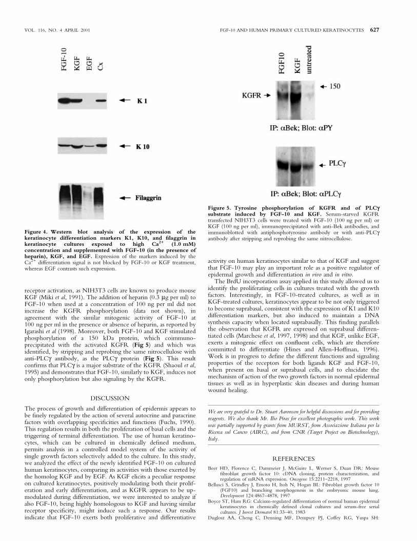
receptor activation, as NIH3T3 cells are known to produce mouseKGF (Miki et al, 1991). The addition of heparin (0.3 mg per ml) toFGF-10 when used at a concentration of 100 ng per ml did notincrease the KGFR phosphorylation (data not shown), inagreement with the similar mitogenic activity of FGF-10 at100 ng per ml in the presence or absence of heparin, as reported byIgarashi et al (1998). Moreover, both FGF-10 and KGF stimulatedphosphorylation of a 150 kDa protein, which coimmuno-precipitated with the activated KGFR (Fig 5) and which wasidenti®ed, by stripping and reprobing the same nitrocellulose withanti-PLCg antibody, as the PLCg protein (Fig 5). This resultcon®rms that PLCg is a major substrate of the KGFR (Shaoul et al,1995) and demonstrates that FGF-10, similarly to KGF, induces notonly phosphorylation but also signaling by the KGFR.
DISCUSSION
The process of growth and differentiation of epidermis appears tobe ®nely regulated by the action of several autocrine and paracrinefactors with overlapping speci®cities and functions (Fuchs, 1990).This regulation results in both the proliferation of basal cells and thetriggering of terminal differentiation. The use of human keratino-cytes, which can be cultured in chemically de®ned medium,permits analysis in a controlled model system of the activity ofsingle growth factors selectively added to the culture. In this study,we analyzed the effect of the newly identi®ed FGF-10 on culturedhuman keratinocytes, comparing its activities with those exerted bythe homolog KGF and by EGF. As KGF elicits a peculiar responseon cultured keratinocytes, positively modulating both their prolif-eration and early differentiation, and as KGFR appears to be up-modulated during differentiation, we were interested to analyze ifalso FGF-10, being highly homologous to KGF and having similarreceptor speci®city, might induce such a response. Our resultsindicate that FGF-10 exerts both proliferative and differentiative
activity on human keratinocytes similar to that of KGF and suggestthat FGF-10 may play an important role as a positive regulator ofepidermal growth and differentiation in vivo and in vitro.
The BrdU incorporation assay applied in this study allowed us toidentify the proliferating cells in cultures treated with the growthfactors. Interestingly, in FGF-10-treated cultures, as well as inKGF-treated cultures, keratinocytes appear to be not only triggeredto become suprabasal, consistent with the expression of K1 and K10differentiation markers, but also induced to maintain a DNAsynthesis capacity when located suprabasally. This ®nding parallelsthe observation that KGFR are expressed on suprabasal differen-tiated cells (Marchese et al, 1997, 1998) and that KGF, unlike EGF,exerts a mitogenic effect on con¯uent cells, which are thereforecommitted to differentiate (Hines and Allen-Hoffman, 1996).Work is in progress to de®ne the different functions and signalingproperties of the receptors for both ligands KGF and FGF-10,when present on basal or suprabasal cells, and to elucidate themechanism of action of the two growth factors in normal epidermaltissues as well as in hyperplastic skin diseases and during humanwound healing.
We are very grateful to Dr. Stuart Aaronson for helpful discussions and for providing
reagents. We also thank Mr. Ilio Piras for excellent photographic work. This work
was partially supported by grants from MURST, from Associazione Italiana per la
Ricerca sul Cancro (AIRC), and from CNR (Target Project on Biotechnology),
Italy.
REFERENCES
Beer HD, Florence C, Dammeier J, McGuire L, Werner S, Duan DR: Mouse®broblast growth factor 10: cDNA cloning, protein characterization, andregulation of mRNA expression. Oncogene 15:2211±2218, 1997
Bellusci S, Grindley J, Emoto H, Itoh N, Hogan BL: Fibroblast growth factor 10(FGF10) and branching morphogenesis in the embryonic mouse lung.Development 124:4867±4878, 1997
Boyce ST, Ham RG: Calcium-regulated differentiation of normal human epidermalkeratinocytes in chemically de®ned clonal cultures and serum-free serialcultures. J Invest Dermatol 81:33±40, 1983
Duglosz AA, Cheng C, Denning MF, Dempsey PJ, Coffey RG, Yuspa SH:
Figure 4. Western blot analysis of the expression of thekeratinocyte differentiation markers K1, K10, and ®laggrin inkeratinocyte cultures exposed to high Ca2+ (1.0 mM)concentration and supplemented with FGF-10 (in the presence ofheparin), KGF, and EGF. Expression of the markers induced by theCa2+ differentiation signal is not blocked by FGF-10 or KGF treatment,whereas EGF contrasts such expression.
Figure 5. Tyrosine phosphorylation of KGFR and of PLCgsubstrate induced by FGF-10 and KGF. Serum-starved KGFRtransfected NIH3T3 cells were treated with FGF-10 (100 ng per ml) orKGF (100 ng per ml), immunoprecipitated with anti-Bek antibodies, andimmunoblotted with antiphosphotyrosine antibody or with anti-PLCgantibody after stripping and reprobing the same nitrocellulose.
VOL. 116, NO. 4 APRIL 2001 FGF-10 AND HUMAN PRIMARY CULTURED KERATINOCYTES 627

Keratinocyte growth factor receptor ligands induce transforming growth factora expression and activate the epidermal growth factor receptor signalingpathway in cultured epidermal keratinocytes. Cell Growth Differ 5:1283±1292,1994
Emoto H, Tagashira S, Mattei MG, et al: Structure and expression of human®broblast growth factor 10. J Biol Chem 272:23191±23194, 1997
Finch PW, Rubin JS, Miki T, Ron D, Aaronson SA: Human KGF is FGF-relatedwith properties of a paracrine effector of epithelial cell growth. Science (WashDC) 245:752±755, 1989
Fuchs E: Epidermal differentiation: the bare essential. J Cell Biol 111:2807±2814,1990
Hines MD, Allen-Hoffman BL: Keratinocyte growth factor inhibits cross-linkedenvelope formation and nucleosomal fragmentation in cultured humankeratinocytes. J Biol Chem 271:6245±6251, 1996
Igarashi M, Finch PW, Aaronson SA: Characterization of recombinant human®broblast growth factor (FGF)-10 reveals functional similarities withkeratinocyte growth factor (FGF)-7. J Biol Chem 273:13230±13235, 1998
Jimenez PA, Rampy MA: Keratinocyte growth factor-2 accelerates wound healing inincisional wounds. J Surg Res 81:238±242, 1999
Marchese C, Rubin JS, Ron D, et al: Human keratinocyte growth factor activity onproliferation and differentiation of human keratinocytes: differentiationresponse distinguishes KGF from EGF family. J Cell Physiol 144:326±333, 1990
Marchese C, Chedid M, Dirsch OR, et al: Modulation of keratinocyte growth factorand its receptor in reepithelializing human skin. J Exp Med 182:1369±1376,1995
Marchese C, Sorice M, De Stefano C, Frati L, Torrisi MR: Modulation ofkeratinocyte growth factor receptor expression in human culturedkeratinocytes. Cell Growth Differ 8:989±997, 1997
Marchese C, Mancini P, Belleudi F, et al: Receptor-mediated endocytosis ofkeratinocyte growth factor. J Cell Sci 111:3517±3527, 1998
Mason IJ: The ins and outs of ®broblast growth factor. Cell 78:547±552, 1994Miki T, Fleming TP, Bottaro DP, Rubin JS, Ron D, Aaronson SA: Expression DNA
cloning of the KGF receptor by creation of a transforming autocrine loop.Science (Wash DC) 251:72±75, 1991
Miki T, Bottaro DP, Fleming TP, Smith CL, Burgess WH, Chan AML, Aaronson
SA: Determination of ligand binding speci®city by alternative splicing: twodistinct growth factor receptors encoded by single gene. Proc Natl Acad Sci USA89:246±250, 1992
Min H, Danilenko DM, Scully SA, et al: FGF-10 is required for both limb and lungdevelopment and exhibits striking functional similarity to Drosophilabranchless. Genes Dev 12:3156±3161, 1998
Park WY, Miranda B, Lebeche D, Hashimoto G, Cardoso WV: FGF-10 is achemotactic factor for distal epithelial buds during lung development. Dev Biol201:125±134, 1998
Rheinwald JG, Green H: Epidermal growth factor and the multiplication of culturedhuman epidermal keratinocytes. Nature 265:421±424, 1977
Rubin JS, Osada H, Finch PW, Taylor WG, Rudikoff S, Aaronson SA: Puri®cationand characterization of newly identi®ed growth factor speci®c for epithelialcells. Proc Natl Acad Sci USA 86:802±806, 1989
Sekine K, Ohuchi H, Fujiwara M, et al: FGF10 is essential for limb and lungformation. Nature Gen 21:138±141, 1999
Shaoul E, Reich-Slotky R, Berman B, Ron D: Fibroblast growth factor receptorsdisplay both common and distinct signaling pathways. Oncogene 10:1553±1561,1995
Staiano-Coico L, Krueger GJ, Rubin JS, et al: Human keratinocyte growth factoreffects in a porcine model of epidermal wound healing. J Exp Med 178:865±878, 1993
Werner S, Peters KG, Longaker MT, Fuller-Pace F, Banda MJ, Williams LT: Largeinduction of keratinocyte growth factor expression in the dermis during woundhealing. Proc Natl Acad Sci USA 89:6896±6900, 1992
Werner S, Smola HM, Xiang L, Longaker MT, Krieg T, Hofschneider PH, WilliamsLT: The function of KGF in morphogenesis of epithelium andreepithelialization of wounds. Science 266:819±822, 1994
Xu X, Weinstein M, Cuiling Li, et al: Fibroblast growth factor receptor 2 (FGFR2)-mediated reciprocal regulation loop between FGF8 and FGF10 is essential forlimb induction. Development 125:753±765, 1998
Yamasaki M, Miyake A, Tagashira S, Itoh N: Structure and expression of the ratmRNA encoding a novel member of the ®broblast growth factor family. J BiolChem 271:15918±15921, 1996
628 MARCHESE ET AL THE JOURNAL OF INVESTIGATIVE DERMATOLOGY