fibrinogen-derived fibrinostatin inhibits tumor growth ... · fibrinogen-derived fibrinostatin...
TRANSCRIPT
Fibrinogen-derived fibrinostatin inhibits tumorgrowth through anti-angiogenesisChuanke Zhao,1,4 Yahui Su,1,4 Jianzhi Zhang,2,4 Qin Feng,1 Like Qu,1 Lixin Wang,1 Caiyun Liu,1 Beihai Jiang,3 Lin Meng1
and Chengchao Shou1
1Department of Biochemistry and Molecular Biology; 2Thoracic Surgery; 3Minimally Invasive Gastrointestinal Surgery, Key Laboratory of Carcinogenesis andTranslational Research (Ministry of Education), Peking University Cancer Hospital and Institute, Beijing, China
Key words
Anti-angiogenesis, fibrinogen, fibrinostatin, peptide, tu-mor inhibition
Correspondence
Chengchao Shou, Department of Biochemistry and Molec-ular Biology, Peking University Cancer Hospital and Insti-tute, 52 Fucheng Road, Beijing 100142, China.Tel: +0086-10-88196766; Fax: +0086-10-88122437;E-mail: [email protected]
4These authors contributed equally to this work.
Funding InformationThis research was supported by the Major Project onDrug Research and Development for the 12th Five-YearPlan of China (2011ZX09102-001-16), the National 973Program of China (2015CB553906), the National NaturalScience Foundation of China (81301966, 30672418) andthe New Teacher’s Fund from Chinese Ministry of Educa-tion (20130001120120).
Received April 17, 2015; Revised August 10, 2015;Accepted August 19, 2015
Cancer Sci 106 (2015) 1596–1606
doi: 10.1111/cas.12797
Angiogenesis is a prerequisite of tumor growth and metastasis and, thus, anti-an-
giogenesis treatment has become an important part of cancer therapy. A 15-
amino acid peptide of the fibrinogen a chain, fibrinostatin, was previously found
in serum samples of gastric cancer patients. Herein we demonstrated that fibrino-
statin has anti-angiogenesis activity in several angiogenesis models and it
reduces tumor growth in mouse xenografts and allografts. Increased tumor
necrosis and reduced microvessel density in tumors were observed in mouse
xenograft models. Fibrinostatin inhibited proliferation and induced apoptosis in
HUVEC, but not in cancer cells. In addition, fibrinostatin specifically entered
HUVEC. Fibrinostatin also prevented migration, adhesion and tubule formation of
HUVEC in vitro. A single-dose acute toxicity testing and a repeated-dose chronic
toxicity study in the mouse, rat and monkey indicated that fibrinostatin had a
wide margin of safety. Taken together, fibrinostatin shows promise as a poten-
tial anti-angiogenesis therapeutic agent.
A ngiogenesis involves the degradation of components ofthe extracellular matrix and then migration, proliferation
and differentiation of endothelial cells from pre-existing bloodvessels to form sprouts and eventually new capillaries,(1–4)
whereas hemostasis maintains the blood flow by regulatingplatelet adherence and fibrin deposition. Both systems nor-mally appear quiescent, but become activated in response tosuch injury as the leaky blood vessels in solid tumors.(5)
Cancer has long been recognized to be associated withhypercoagulation as a result of imbalanced coagulation (fibrindeposition) and fibrinolysis (fibrin degradation). Fibrinogen, amultimeric molecule containing two of three non-identicalchains, the a, b and c chains, is the soluble circulating precur-sor of fibrin. It accumulates around leaky blood vessels insolid tumors and at the host–tumor interface, which contributesto the tumor growth and spreading.(6,7) Fibrinogen can becleaved by plasmin and thrombin and its proteolytic productscan either promote or inhibit angiogenesis. Plasmin cleavagegenerates the carboxyl termini of the paired a, b and c chainsof fibrinogen and the central domain of fibrinogen (fibrinogenE-fragment [FgnE]). FgnE is a potent anti-angiogenic factorcapable of inhibiting endothelial cell migration and tubule for-mation.(8,9) When cleaved by thrombin, fibrinogen is converted
into fibrin monomers after the removal of the amino termini ofboth a and b chains, yielding fibrinopeptides A (FpA) and B(FpB), respectively. The fibrin-E fragment (FnE) of fibrindegraded by plasmin was reported to promote angiogenesis.The main difference between FnE and FgnE lies in its loss ofFpA.We previously found a 15-amino acid (aa) fragment peptide
in the sera of gastric cancer patients by surface-enhanced laserdesorption ⁄ ionization and time-of-flight mass spectrometry(SELDI TOF-MS). This 15-mer peptide (DSGEGD-FLAEGGGVR) was identified as the amino terminal fragment(2-16aa) of the fibrinogen a chain, which was named as fibri-nostatin.(10) Compared with FpA, fibrinostatin losses the firstalanine from the amino terminus of FpA, which could be aresult of the cleavage by exoprotease, because differentialexoprotease activities acting on the ex vivo coagulation andcomplement–degradation pathways could generate cancer type-specific serum peptides.(11) We and other groups have foundthat fibrinostatin is differentially present in the sera of patientsin a variety of cancers: higher in gastric, ovarian, hepatocellu-lar and urothelial cancers,(12–15) lower in prostate, bladder andthyroid cancers,(11,16) but no difference in breast cancerpatients when compared with healthy controls.(11) Based on
Cancer Sci | November 2015 | vol. 106 | no. 11 | 1596–1606 © 2015 The Authors. Cancer Science published by Wiley Publishing Asia Pty Ltdon behalf of Japanese Cancer Association.This is an open access article under the terms of the Creative CommonsAttribution-NonCommercial License, which permits use, distribution andreproduction in any medium, provided the original work is properly cited and isnot used for commercial purposes.
these findings, we sought to evaluate the role of fibrinostatinin tumor development. Our results support the notion that fibri-nostatin is a potential antagonist of tumor angiogenesis.
Materials and Methods
Cells and animals. Human umbilical vein endothelial cellswere isolated and cultured as previously described.(17) Humantumor cell lines of BGC-823 (gastric carcinoma), PG (lungcarcinoma) and HT-29 (colon carcinoma) were cultured inRPMI-1640 with 10% FBS in 5% CO2 at 37°C. HEK-293cells, fibroblasts (WI-38) cells and human microvascularendothelial cells were maintained in DMEM (10% FBS) at37°C in 5% CO2.BALB ⁄ c nude mice were obtained from the Animal Center
of the Chinese Academy of Medical Science (Beijing, China).Fertilized eggs of white Leghorn chickens were supplied bythe Chicken Center of the Chinese Agriculture University (Bei-jing, China). Wistar rats and SD rats were obtained from VitalRiver Laboratories (Beijing, China).
Synthetic peptides and reagents. Fibrinostatin and truncatedpeptides, FITC-conjugated fibrinostatin peptide and FITC-con-jugated TAT peptide were synthesized by ChinaTech Peptide(Suzhou, China). The amino acid sequences are: DSGEGD-FLAEGGGVR (fibrinostatin) and GRKKRRQRRRGY(TAT).(18)
Xenograft and H22 allograft models. For the present study,2 9 106 tumor cells resuspended in PBS were injected subcu-taneously into the backs of mice. When tumors reached0.1 cm3, the mice were intravenously injected with PBS con-trol or 1, 2.5 or 5 mg ⁄kg fibrinostatin every day for 16 days.H22 allograft model was generated as previously described.(19)
The mice were randomly divided into 10 groups and adminis-tered with different treatments for 7 days. Mice were sacrificedunder sodium pentobarbital anesthesia, and tumor weightswere measured. Tumor volume was calculated according to theequation: tumor volume = width2 9 length ⁄2.
Immunohistochemistry and microvascular density analy-
sis. Paraffin-embedded sections were dewaxed using standardhistological procedures. To stain the microvasculature, the sec-tions were incubated with an antibody against CD31 (ab59251;Abcam, Cambridge, UK) overnight at 4°C in a humidifiedchamber and bound antibody was detected using the EnVisionKit (DAKO, Carpinteria, CA, USA). In each group, differencesin vascularity and number of CD31-positive structures permicroscopic field were determined for each section at 9200magnification in a blinded fashion.(20) Mean � SD MVD ⁄fieldwas calculated based on four fields per section from six xeno-grafts.
Chick chorioallantoic membrane assay. Fertilized eggs ofwhite Leghorn chickens were incubated at 37°C in 65–70%humidity. The 6-day-old embryos with intact yolks wereplaced in a bowl and incubated at 37°C.(21) A filter disk of 6-mm diameter containing 10 lL PBS or fibrinostatin (2, 5 or10 lg ⁄lL) was placed onto the chick chorioallantoic mem-brane (CAM) of the embryos. After 48 h of incubation, vesselformation was observed under a stereomicroscope.
Zebrafish embryos assay. Transgenic zebrafish assessmentwas performed by Hunter Biotechnology Corporation (Hang-zhou, China). Drugs were injected into the yolk sac of zebra-fish 2 days after fertilization by a microinjection method. Afterbeing incubated for another 72 h, 10 zebrafish of each groupwere randomly selected, and fluorescence of the subintestinalvein vessels (SIV) was detected and photographed using a
multi-purpose zoom microscope system (AZ 100, Nikon).Image analysis was performed by calculating the SIV areausing Nikon NIS-Elements D 3.10 Advanced image processingsoftware.
Cell proliferation assay. The MTT (3-[4,5-Dimethylthiazol-2-yl]-2,5-diphenyltetrazolium bromide) assay kit from Promega(Madison, WI, USA) was used to assess proliferation ofHUVEC and cancer cells, as described previously.(22)
Flow cytometry analysis. Cells were treated with PBS or25 lg ⁄mL fibrinostatin for 48 h, then washed twice and fixedin 0.04% methanol at �20°C for 16 h. After staining with thepropidium iodide (PI) solution, samples were then analyzedusing a FACSAria flow cytometry system (BD Biosciences,San Jose, CA, USA).
DNA ladder assay. Cells were treated with PBS or fibrinos-tatin (25 lg ⁄mL) for 72 h, then washed twice with ice-coldTBS (pH 7.6) and lysed with lysis buffer (10 mM Tris-HClpH 8.0, 5 mM EDTA, 1% Triton-100, 10 mM NaCl and100 lg ⁄mL proteinase K) for 10 min. Then 300 lL of satu-rated hydroxybenzene and chloroform was added for genomicDNA extraction by centrifugation of 12 000 g for 10 min, andthe supernatant was collected and two volumes of ethanol with10% 3 mol ⁄L NaAc were added to precipitate DNA. 20 lLH2O with 1 mg ⁄mL RNase A was used to dissolve DNA, andthe DNA Ladder were analyzed in 1.2% agarose gel.
TUNEL assay. Cells were seeded on glass coverslips in 24-well plates. After treatment with fibrinistatin (25 lg ⁄mL) for72 h, TUNEL assay was performed with a kit from Rocheaccording to the manufacturer’s instructions and observedunder a TCS-SP2 confocal microscopy (Leica Microsystems,Heidelberg, Germany).
Fluorescence visualization for entry of peptides. To assay theentries of FITC-labeled peptides, HUVEC, BGC-823, PG andHT-29 cells were incubated with FITC-conjugated fibrinostatin(25 lg ⁄mL) or TAT (25 lg ⁄mL) for 3 h. The cells were thenfixed with 4% paraformldehyde and permeabilized with 0.1%NP-40 in PBS. The dye 40,6-diamidino-2-phenylindole (DAPI,5 ng ⁄mL) was added to stain nuclei, washed three times withPBS, and observed under a TCS-SP2 confocal microscopy (Le-ica Microsystems, Heidelberg, Germany).
Cell migration assay. HUVEC (5 9 104 cells) pretreated withPBS control or 25 lg ⁄mL fibrinostatin for 24 h were plated inthe upper chamber of Transwell inserts with 8-lm pores, andthe lower chamber contained RPMI-1640, 10% FBS (to controlthe HUVEC growth) with PBS or fibrinostatin (25 lg ⁄mL).The cells were allowed to migrate for 24 h at 37°C. The meannumber of cells per field that migrated across the filter wascalculated based on the cell number counted in four fields at9200 magnification per insert. Each condition was performedin triplicate.
Cell adhesion assay. Six-well plates were coated with200 lL ⁄well of Matrigel (1 mg ⁄mL; BD Biosciences) over-night. The plates were blocked with 500 lL ⁄well of 100 lg⁄mL BSA for 2 h. Then 1 mL of the HUVEC suspension(2 9 105 cells ⁄mL) pretreated with PBS control or 25 lg ⁄mLfibrinostatin for 24 h, was added to each well and incubatedfor 2 h at 37°C. After washing twice with PBS, the number ofattached cells was counted after methylene blue staining at9200 magnification. Mean � SD attached cells was calculatedbased on four fields per insert. Each condition was performedin triplicate.
Tubule formation assay. A total of 200 lL Matrigel (1 mg⁄mL) was added to each well of a 24-well plate and allowed topolymerized for 20 min at 37°C. A suspension of HUVEC
Cancer Sci | November 2015 | vol. 106 | no. 11 | 1597 © 2015 The Authors. Cancer Science published by Wiley Publishing Asia Pty Ltdon behalf of Japanese Cancer Association.
Original Articlewww.wileyonlinelibrary.com/journal/cas Zhao et al.
(5 9 104 cells) was plated on the Matrigel-coated wells andthen treated with PBS or fibrinostatin (10, 20 and 50 lg ⁄mL)in RPMI-1640 with 10% FBS and 10 ng ⁄mL VEGF for 18 hin 5% CO2 at 37°C. The network structure of tubules wasobserved using a CK2 Olympus Microscope (Olympus Corpo-ration, Tokyo, Japan). The assays were performed in triplicate.
Rat aortic ring assay. The rat aortic ring assay was performedas described previously, with slight modifications.(23) Sixweek-old Sprague–Dawley rats were sacrificed by CO2 inhala-tion and their thoracic aortas were dissected and sliced into 1-mm thick rings. The rings were placed on a 24-well plate andembedded in 100 lL Matrigel followed by 30 min incubationat 37°C. The wells were then overlaid with 300 lL of RPMI-1640 containing 10% FBS with PBS control or fibrinostatin of10, 20 and 50 lg ⁄mL. The rings were incubated for 5 days at37°C and observed using a CK2 Olympus microscope (9100magnification).
Western blot. Cells lysed in RIPA buffer were subjected towestern blot analysis following the standard procedure. Pri-mary antibodies anti-Bax mAb (sc-7480; Santa Cruz, CA,USA), anti-p53 mAb (sc-126; Santa Cruz), anti-VEGFR1 mAb(#1303-1; Epitomics, Burlingame, CA, USA), anti-phospho-VEGFR1 (Tyr1213) rabbit antibody (#07-758; Millipore, Bill-erica, MA, USA), anti-VEGFR2 mAb (#2479; Cell SignalingTechnology), anti-phospho-VEGFR2 (#2478; Cell Signalingtechnology, Danvers, MA, USA), anti-phospho-ERK1 ⁄2 mAb(sc-7383; Santa Cruz) and anti-Phospho-Akt (Ser473) (#4060;Cell Signaling Technology) were used here.
Statistical analysis. Statistical analysis software packageSPSS 12.0 (SPSS, Chicago, IL) was used to perform an ANOVA
test and the unpaired Student’s t-test. P-values < 0.05 wereconsidered statistically significant.
Results
Fibrinostatin inhibits in vivo tumor growth but does no harm
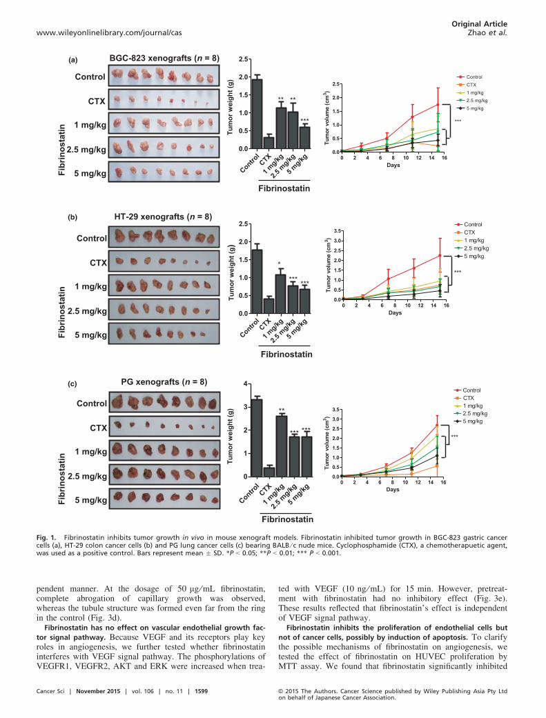
to the hosts. Fibrinostatin was first found in the serum of gas-tric cancer patients in our previous study by SELDI TOF-MSanalysis.(10) To investigate the function(s) of fibrinostatinin vivo, we synthesized the 15-amino acid peptide (fibrinos-tatin) and utilized three tumor xenograft models (gastric can-cer, lung cancer and colorectal cancer). Chemotherapeuticagent cyclophosphamide (CTX) was used as a positive control.We found that fibrinostatin significantly inhibited xenografttumor growth in mice (Fig. 1a–c). For the gastric cancer BGC-823 in mice, the mean tumor volume and weight were0.515 � 0.228 cm3 and 0.61 � 0.24 g in the fibrinostatin-trea-ted group, compared with 1.932 � 0.652 cm3 and1.92 � 0.40 g in the control group (P < 0.05). The maximalinhibition rates of tumor growth and tumor volume by fibrinos-tatin were 68.3% and 36%, respectively. The inhibition of fib-rinostatin acts in a dose-dependent manner (Fig. 1a). Similarinhibition was observed in the colon (HT-29) and lung (PG)cancer in mice (Fig. 1b,c). We also conducted a toxicologystudy in pre-clinical animal models according to Good Labora-tory Practice (GLP) standards and found no observable sideeffects (data not shown).
Fibrinostatin induces necrosis and decreases microvascular den-
sity in tumors. To explore the effects of fibrinostatin on tumorgrowth and tumor angiogenesis, we dissected and analyzed allthe xenografts from the mice by histological H&E staining.We observed necrotic areas in the fibrinostatin-treated tumors,in which more lymphocyte and erythrocyte exudation andsome necrotic tumor cells were identified, with significantly
less areas of necrosis in the control tumors (Fig. 2a). We thenassessed the microvascular density (MVD) by immunostainingendothelial cells with an anti-CD31 antibody and calculatedMVD as a percentage of tumor section area using the Chalkleygrid counting method.(24) We found that the MVD in the fibri-nostatin-treated group was significantly decreased comparedwith the control group (P < 0.05) (Fig. 2b). These results sug-gest that the fibrinostatin’s anti-tumor activity is associatedwith inhibition of tumor angiogenesis.
Fibrinostatin inhibits vessel formation in vivo. To test thedirect effect of fibrinostatin on angiogenesis, we applied thechick chorioallantoic membrane assay for vessel formation.We found that the 4-day treatment of fibrinostatin dramaticallyinhibited microvessel formation in a dose-dependent manner,whereas the control group had normal vascularization(Fig. 2c).
Effects of fibrinostatin on angiogenesis in zebrafish. The anti-angiogenic effect of fibrinostatin was further confirmed using atransgenic zebrafish model; zebrafish express enhanced greenfluorescent protein (EGFP) in their endothelial cells. Observa-tions demonstrated that both fibrinostatin and Endostar (as apositive control) inhibited the development of subintestinalvein vessels and had no tocixity to zebrafish. Fibrinostatin wasadministrated at 333.5, 1110 and 3335 ng and the inhibitionrates of angiogenesis were 17.6 � 3.0%, 23.9 � 3.1% and33.7 � 5.0%, respectively (Fig. 2d left and middle panel).When micro-injected with 3335 ng of fibrinostatin, the numberof subintestinal vascular vessels was decreased to 3–4, whichwas less than that of the control group (7–8; Fig. 2d rightpanel).
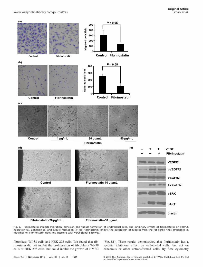
Fibrinostatin inhibits endothelial cell migration, adhesion and
tubule formation. The above results suggest that fibrinostatinmay act on endothelial cells. To investigate this possibility, weisolated HUVEC from the vein of human umbilical cords andtested the effect of fibrinostatin on HUVEC. Migration andadhesion of vascular endothelial cells are critical steps forangiogenesis. We first determined the effect of fibrinostatin onthe migration of HUVEC using a transwell chamber assay. Wefound that the migration of HUVEC treated with fibrinostatinwas significantly reduced compared with that of the controlcells (mean 89 � 36 cells ⁄ chamber vs mean 265 � 82 cells⁄ chamber, P < 0.05; Fig. 3a).We then coated the 24-well plate with Matrigel and exam-
ined the adhesion of HUVEC with or without treatment of fib-rinostatin by counting the number of cells attached on theMatrigel membrane. Our data showed an approximate 50%reduction of HUVEC adhesion by fibrinostatin treatment com-pared with that of the control cells (mean 96 � 39 vs mean186 � 72, P < 0.05; Fig. 3b).Matrigel tubule formation assay was used as an in vitro
model to study the effect of fibrinostatin. HUVEC were pre-treated with fibrinostatin and angiogenesis was assessed afteran 18-h incubation period on Matrigel. Untreated cells formeda branching and anastomosing network of capillary-like tubuleswith multi-centric junctions, whereas the fibrinostatin-treatedcells had reduced branching points, tubule number and length.HUVEC treated with 50 lg ⁄mL of fibrinostatin grew intocomplete single cells and no capillary-like tubule formed(Fig. 3c).The fibrinostatin-induced inhibition of tubule formation was
further confirmed by the rat aortic ring assay. After culturingthe aortic rings for 5 days, the tubules grown from the ringswere observed in the control group, but the tubule formationwas inhibited in the fibrinostatin-treated group in a dose-de-
© 2015 The Authors. Cancer Science published by Wiley Publishing Asia Pty Ltdon behalf of Japanese Cancer Association.
Cancer Sci | November 2015 | vol. 106 | no. 11 | 1598
Original ArticleEndogenous peptide inhibits angiogenesis www.wileyonlinelibrary.com/journal/cas
pendent manner. At the dosage of 50 lg ⁄mL fibrinostatin,complete abrogation of capillary growth was observed,whereas the tubule structure was formed even far from the ringin the control (Fig. 3d).
Fibrinostatin has no effect on vascular endothelial growth fac-
tor signal pathway. Because VEGF and its receptors play keyroles in angiogenesis, we further tested whether fibrinostatininterferes with VEGF signal pathway. The phosphorylations ofVEGFR1, VEGFR2, AKT and ERK were increased when trea-
ted with VEGF (10 ng ⁄mL) for 15 min. However, pretreat-ment with fibrinostatin had no inhibitory effect (Fig. 3e).These results reflected that fibrinostatin’s effect is independentof VEGF signal pathway.
Fibrinostatin inhibits the proliferation of endothelial cells but
not of cancer cells, possibly by induction of apoptosis. To clarifythe possible mechanisms of fibrinostatin on angiogenesis, wetested the effect of fibrinostatin on HUVEC proliferation byMTT assay. We found that fibrinostatin significantly inhibited
Control
1 mg/kg
CTX
2.5 mg/kg
5 mg/kg
HT-29 xenografts (n = 8)Tu
mor
wei
ght (
g)(b)
Control
1 mg/kg
CTX
2.5 mg/kg
5 mg/kg
PG xenografts (n = 8)
Tum
or w
eigh
t (g)
(c)
Control
1 mg/kg
CTX
2.5 mg/kg
5 mg/kgFibr
inos
tatin
Fibr
inos
tatin
Fibr
inos
tatin
BGC-823 xenografts (n = 8)
Fibrinostatin
Tum
or w
eigh
t (g)
(a)
ControlCTX
1 mg/kg
2.5 m
g/kg
5 mg/kg
0.0
0.5
1.0
1.5
2.0
2.5
** **
***
ControlCTX
1 mg/kg
2.5 m
g/kg
5 mg/kg
0.0
0.5
1.0
1.5
2.0
2.5
*
*** ***
Fibrinostatin
ControlCTX
1 mg/kg
2.5 m
g/kg
5 mg/kg
0
1
2
3
4
**
*** ***
Fibrinostatin
0 2 4 6 8 10 12 14 160.0
0.5
1.0
1.5
2.0
2.5ControlCTX1 mg/kg2.5 mg/kg5 mg/kg
***
Days
mc(e
mulovro
muT3 )
0 2 4 6 8 10 12 14 160.0
0.5
1.0
1.5
2.0
2.5
3.0
3.5ControlCTX1 mg/kg2.5 mg/kg5 mg/kg
***
Days
mc(e
mulovro
muT3 )
0 2 4 6 8 10 12 14 160.0
0.5
1.0
1.5
2.0
2.5
3.0
3.5
ControlCTX1 mg/kg2.5 mg/kg5 mg/kg
***
Days
mc(e
mulovro
mu T3 )
Fig. 1. Fibrinostatin inhibits tumor growth in vivo in mouse xenograft models. Fibrinostatin inhibited tumor growth in BGC-823 gastric cancercells (a), HT-29 colon cancer cells (b) and PG lung cancer cells (c) bearing BALB ⁄ c nude mice. Cyclophosphamide (CTX), a chemotherapuetic agent,was used as a positive control. Bars represent mean � SD. *P < 0.05; **P < 0.01; *** P < 0.001.
Cancer Sci | November 2015 | vol. 106 | no. 11 | 1599 © 2015 The Authors. Cancer Science published by Wiley Publishing Asia Pty Ltdon behalf of Japanese Cancer Association.
Original Articlewww.wileyonlinelibrary.com/journal/cas Zhao et al.

HUVEC proliferation in a concentration-dependent manner(10–50 lg ⁄mL). At 20 lg ⁄mL concentration, fibrinostatinalmost reached the plateau of inhibition (P < 0.05) (Fig. 4a).
In contrast, fibrinostatin had no distinguishable effects onproliferations of BGC-823, PG and HT-29 cancer cells. Wealso tested the inhibitory effect of fibrinostatin on HMEC,
(c)
(b)
0
5
10
15
MVD
(ves
sels
/fiel
d)
P < 0.05
Control Control
FibrinostatinFibrinostatin
(d)
(a)
Control Fibrinostatin
Control 5 µg/mL2 µg/mL 10 µg/mL
Fibrinostatin
ControlFibrinostatin 333.5 ng
Fibrinostatin 1110 ng
Fibrinostatin 3335 ng
Vehicle
Endostar 250 ng
Control
Endostar 250 ng
Fibrinostatin 3335 ng
***
*******
Fig. 2. Fibrinostatin inhibits angiogenesis in vivo. (a) Tumor sections from BGC-823 xenografts were immunostained with anti-CD31 anti-body. Black arrowheads point to the stained vessels. White arrowheads point to tumor cells (left) and necrotic tumor cells (right). Scalebar = 50 lm (b) Tumor sections from BGC-823 xenografts immunostained with anti-CD31 antibody were used to examine the vessel number.(9200 magnification, left). Boxed regions show CD31-positive structures magnified at 9400 in the insets. Mean � SD microvascular density(MVD) per field was calculated (right). P < 0.05 (unpaired Student’s t-test). Scale bar = 100 lm. (c) Fibrinostatin inhibited angiogenesis inchick chorioallantoic membrane (CAM). (d) Fibrinostatin inhibited angiogenesis in transgenic zebrafish. *** P < 0.001; **P < 0.01; (Student’st-test).
© 2015 The Authors. Cancer Science published by Wiley Publishing Asia Pty Ltdon behalf of Japanese Cancer Association.
Cancer Sci | November 2015 | vol. 106 | no. 11 | 1600
Original ArticleEndogenous peptide inhibits angiogenesis www.wileyonlinelibrary.com/journal/cas
fibroblasts WI-38 cells and HEK-293 cells. We found that fib-rinostatin did not inhibit the proliferation of fibroblasts WI-38cells or HEK-293 cells, but could inhibit the growth of HMEC
(Fig. S1). These results demonstrated that fibrinostatin has aspecific inhibitory effect on endothelial cells, but not oncancerous or other untransformed cells. By flow cytometry
0
100
200
300
400
ControlFibrinostatinControl
Adh
ered
cel
ls/fi
eld
P < 0.05
Control 20 μg/mL1 μg/mL 50 μg/mLFibrinostatin
Control Fibrinostatin-10 μg/mL
(a)
(b)
(c)
Fibrinostatin-20 μg/mL Fibrinostatin-50 μg/mL
0
100
200
300
400
500
Fibrinosta nControl
Mig
rate
d ce
lls/fi
eld
(d)
P < 0.05
(e)
VEGFR1
pVEGFR1
VEGFR2
pVEGFR2
pERK
pAKT
β-actin
– + + VEGF– – + Fibrinostatin
Fibrinosta n
Fibrinosta nControl
Fig. 3. Fibrinostatin inhibits migration, adhesion and tubule formation of endothelial cells. The inhibitory effects of fibrinostatin on HUVECmigration (a), adhesion (b) and tubule formation (c). (d) Fibrinostatin inhibits the outgrowth of tubules from the rat aortic rings embedded inMatrigel. (e) Fibrinostatin does not interfere with VEGF signal pathway.
Cancer Sci | November 2015 | vol. 106 | no. 11 | 1601 © 2015 The Authors. Cancer Science published by Wiley Publishing Asia Pty Ltdon behalf of Japanese Cancer Association.
Original Articlewww.wileyonlinelibrary.com/journal/cas Zhao et al.

(c)
PG HT-29
+ ++ +
Control Fibrinosta nHUVEC
Control Fibrinosta nBGC-823
Control Fibrinosta nPG
Control Fibrinosta nHT-29
BGC-823HUVEC
+ ++ +
ControlFibrinostatin
ControlFibrinostatin
0
0.2
0.4
0.6
0.8
1
1.2
0 24 48 72 96
PG
#
##
##
0
0.5
1
1.5
0 24 48 72 96
OD
540
nm
BGC-823#
#
##
#
0.0
0.2
0.4
0.6
0.8
1.0
0 24 48 72 96
HT-29
##
#
OD
540
nm
#
#
0
0.2
0.4
0.6
0.8
0 24 48 72 96
Control
5 μg/mL
10 μg/mL
20 μg/mL
50 μg/mL
Control
5 μg/mL
10 μg/mL
20 μg/mL
50 μg/mL
Control
5 μg/mL
10 μg/mL
20 μg/mL
50 μg/mL
Control
5 μg/mL
10 μg/mL
20 μg/mL
50 μg/mL
OD
540
nmO
D54
0 nm
HUVEC
*
#
##
(h) (h)
(h)(h)
*(a)
(b)
Fig. 4. Fibrinostatin inhibits proliferation and induces apoptosis of endothelial cells specifically. (a) The effects of fibrinostatin on the prolifera-tion of HUVEC, BGC-823, PG and HT-29 cells were determined with an MTT assay. *P < 0.05. #P > 0.05, ANOVA. (b) Flow cytometry analysis of cellstreated with PBS or fibrinostatin and stained with propidium iodide. The percentage of the sub-G1 cells is presented. (c) DNA ladder analysis ofcells treated with PBS or 25 lg ⁄mL fibrinostatin.
© 2015 The Authors. Cancer Science published by Wiley Publishing Asia Pty Ltdon behalf of Japanese Cancer Association.
Cancer Sci | November 2015 | vol. 106 | no. 11 | 1602
Original ArticleEndogenous peptide inhibits angiogenesis www.wileyonlinelibrary.com/journal/cas
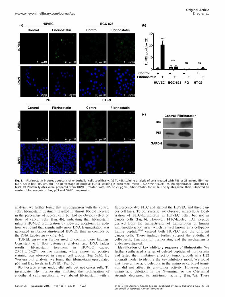
analysis, we further found that in comparison with the controlcells, fibrinostatin treatment resulted in almost 10-fold increasein the percentage of sub-G1 cell, but had no obvious effect onthose of cancer cells (Fig. 4b), indicating that fibrinostatininhibits HUVEC proliferation by inducing apoptosis. In addi-tion, we found that significantly more DNA fragmentation wasgenerated in fibrinostatin-treated HUVEC than in controls bythe DNA Ladder assay (Fig. 4c).TUNEL assay was further used to confirm these findings.
Consistent with flow cytometry analysis and DNA ladderresults, fibrinostatin treatment in HUVEC caused20.51 � 6.62% positive staining, while almost no positivestaining was observed in cancer cell groups (Fig. 5a,b). ByWestern blot analysis, we found that fibrinostatin upregulatedp53 and Bax levels in HUVEC (Fig. 5c).
Fibrinostatin enters endothelial cells but not cancer cells. Toinvestigate why fibrinostatin inhibited the proliferation ofendothelial cells specifically, we labeled fibrinostatin with a
fluorescence dye FITC and stained the HUVEC and three can-cer cell lines. To our surprise, we observed intracellular local-ization of FITC-fibrinostatin in HUVEC cells, but not incancer cells (Fig. 6). However, FITC-labeled TAT peptidederived from the transactivator of transcription of humanimmunodeficiency virus, which is well known as a cell-pene-trating peptide,(25) entered both HUVEC and the differentcancer cells. These findings further support the endothelialcell-specific functions of fibrinostatin, and the mechanism isunder investigated.
Identification of key inhibitory sequence of fibrinostatin. Wefurther synthesized a series of deleted peptides of fibrinostatinand tested their inhibitory effect on tumor growth in a H22allograft model to identify the key inhibitory motif. We foundthat three amino acid deletions in the amino or carboxyl termi-nus did not affect its anti-tumor activity. However, moreamino acid deletions in the N-terminal or the C-terminalstrongly decreased its anti-tumor activity (Fig. 7a). These
IPAD
LEN
UT
Control Fibrinostatin
HUVEC
0
10
20
30
)%(
evitisopLE
NUT
HUVEC BGC-823 PG HT-29
ControlFibrinostatin
+ + + ++ + + +
Control Fibrinostatin
BGC-823IPA
DLE
NUT
Control Fibrinostatin
PG
Control Fibrinostatin
HT-29
Bax
P53
GAPDH
Control Fibrinostatin
(a) (b)
(c)
nsns ns
***
Fig. 5. Fibrinostatin induces apoptosis of endothelial cells specifically. (a) TUNEL staining analysis of cells treated with PBS or 25 lg ⁄mL fibrinos-tatin. Scale bar, 100 lm. (b) The percentage of positive TUNEL staining is presented; mean � SD ***P < 0.001; ns, no significance (Student’s t-test). (c) Protein lysates were prepared from HUVEC treated with PBS or 25 lg ⁄mL fibrinostatin for 48 h. The lysates were then subjected towestern blot analysis of Bax, p53 and GAPDH expression.
Cancer Sci | November 2015 | vol. 106 | no. 11 | 1603 © 2015 The Authors. Cancer Science published by Wiley Publishing Asia Pty Ltdon behalf of Japanese Cancer Association.
Original Articlewww.wileyonlinelibrary.com/journal/cas Zhao et al.

results suggested that the six amino acids (DFLAEG) of fibri-nostatin comprised the key inhibitory sequence. In addition,we performed tubule formation and migration assays to definethe effect of these deleted peptides. Consistent with in vivoexperiment results, peptides C3d, N3d, N6d and N5C4d alsoinhibit tubule formation and migration of HUVEC in vitro(Fig. 7b,c).
Discussion
The hemostatic system has long been shown to regulate angio-genesis involving various hemostatic precursor proteins andcleavage of these proteins by proteolysis, which can generatenovel cryptic fragments as either positive or negative factorsto control the rate of angiogenesis.(5,26,27) Some cryptic frag-ments can antagonize angiogenesis, such as Domain 5 ofkininogen, fragment-1 and -2 of prothrombin and cleaved AT-III (serpin antithrombin).(28–30) Fibrinostatin detected inthe serum is the amino terminal fragment (2–16 aa) derivedfrom the precusor fibrinogen a chain,(10) differing from FpA inits loss of the first alanine at the amino terminus. Previousstudies showed that a linear form of FpA was inactive in vitro,but the first 24 amino terminal amino acids (ADSGEGD-FLAEGGGVRGPRVVERH) of the fibrinogen a chain(named alphastatin) tested as a synthetic peptide showed anti-angiogenesis activity.(31) Neither the FpA portion (1-16aa)(ADSGEGDFLAEGGGVR) nor the 17–24 amino acids(GPRVVERH) of alphastatin could mimic the inhibitory effectof alphastatin.(31) In alphastatin, a fragment of 11 amino acids(DFLAEGGGVRG) was identified to be a key sequence con-taining the activity (termed AHN419).(32) In our study, we syn-thesized a series of deleted peptides of fibrinostatin and testedtheir inhibition of tumor growth in mice. We found that the
six amino acids (DFLAEG) of fibrinostatin (DSGEGD-FLAEGGGVR) comprised the key inhibitory sequence offibrinostatin, which was included in AHN419. These resultssuggest that the addition of extra amino acids at either theamino or the carboxyl terminus of the key sequence(DFLAEG) may contribute to the conformation or stability ofthe core sequence that subsequently contributes to the biologi-cal activity of the key sequence. We speculate that a properstructure may be required for the interaction and stabilizationof binding of fibrinostatin with its cellular target protein.VEGF and its receptors play important roles in tumor
angiogenesis.(33) However, we did not find fibrinostatin tointerfere with the VEGF signal pathway. Together with fluo-rescence visualization results, we propose that fibrinostatinbinds to its target protein in cytoplasm and induces apoptosis.Increased p53 and Bax expressions were observed when trea-ted with fibrinostatin, but the mechanism underlying thesetwo molecules’ upregulation and their contributions to fibri-nostatin-induced apoptosis remain to be determined. Thetarget of fibrinostatin and the mechanism of fibrinostatin’sspecific effect on endothelial cells also deserve further inves-tigation.Taken together, our study provides evidence for develop-
ment of a new therapeutic candidate, fibrinostatin, as an anti-angiogenesis inhibitor targeting endothelial cells. Fibrinostatinblocks angiogenesis by inhibiting endothelial cell proliferation,adhesion, migration and tubule formation. We have alsodemonstrated the anti-tumor effects of fibrinostatin on threecancer types (gastric cancer, lung cancer and colon cancer) inmouse xenograft models, supporting the notion that fibrinos-tatin may have a broad spectrum of anti-tumor ⁄ anti-angiogene-sis activities. The toxicity studies demonstrated the good safetyprofiles in rats and monkeys. These findings support further
FITC
-TAT
HUVEC BGC-823 PG HT-29FI
TC-fi
brin
osta
tin
25 μm 25 μm 25 μm 25 μm
Fig. 6. Fibrinostatin enters endothelial cell specifically. Cells were incubated with 25 lg ⁄mL FITC-conjugated fibrinostatin (green) or TAT (green,positive control), fixed, stained with DAPI (blue), and observed under a confocal microscope. Scale bar = 25 lm.
© 2015 The Authors. Cancer Science published by Wiley Publishing Asia Pty Ltdon behalf of Japanese Cancer Association.
Cancer Sci | November 2015 | vol. 106 | no. 11 | 1604
Original ArticleEndogenous peptide inhibits angiogenesis www.wileyonlinelibrary.com/journal/cas

H22 allografts (n=10) Inhibition % Treatment Sequence of deletion peptide
Control
89.9 ± 4.3% CTX
65.4 ± 10.8% Fibrinostatin D S G E G D F L A E G G G V R
63.3 ± 15.1% N3d E G D F L A E G G G V R
57.6 ± 12.9% C3d D S G E G D F L A E G G
54.9 ± 7.4% N5C4d D F L A E G
51.3 ± 18.7% N6d F L A E G G G V R
34.5 ± 22.3% C9d D S G E G D
34.5 ± 20.8% N9d E G G G V R
32.3 ± 29.5% C6d D S G E G D F L A
0
100
200
300
400
***
*
******
*** ***
Con
C3d C6d C9d N5C4d
N3d N6d N9d Fibrinostatin
(a)
(b)
(c)
ns ns
Mig
rate
d ce
lls/fi
eld
Control
Fig. 7. Identification of key sequence of fibrinastatin. (a) The inhibitory effects of truncated peptides on tumor growth were assessed in murineH22 allografts. Cyclophosphamide (CTX) was used as a positive control. Inhibition rates were calculated compared with the control group. Theinhibitory effects of fibrinostatin deleted peptides on HUVEC tubule formation (b) and migration (c) are presented; mean � SD *P < 0.05;**P < 0.01; ***P < 0.001 compared with the control group (Student’s t-test).
Cancer Sci | November 2015 | vol. 106 | no. 11 | 1605 © 2015 The Authors. Cancer Science published by Wiley Publishing Asia Pty Ltdon behalf of Japanese Cancer Association.
Original Articlewww.wileyonlinelibrary.com/journal/cas Zhao et al.
investigation of fibrinostatin in preclinical and clinical studiesas a drug candidate for treatment of a variety of cancers.
Acknowledgments
We thank Dr Ullrich Schwertschlag (Tufts University), Dr XiaojiaChang (Attogen Biocompany) and Dr Sonya Wei Song (Capital Medi-cal Science University) for their critical comments on the manuscript.This research was supported by the Major Project on Drug Research
and Development for the 12th Five-Year Plan of China(2011ZX09102-001-16), the National 973 Program of China(2015CB553906), the National Natural Science Foundation of China(81301966, 30672418) and the New Teacher’s Fund from the ChineseMinistry of Education (20130001120120).
Disclosure Statement
The authors declare no competing financial interests.
References
1 Folkman J. Angiogenesis in cancer, vascular, rheumatoid and other disease.Nat Med 1995; 1: 27–31.
2 Carmeliet P. Mechanisms of angiogenesis and arteriogenesis. Nat Med 2000;6: 389–95.
3 Hanahan D, Folkman J. Patterns and emerging mechanisms of the angio-genic switch during tumorigenesis. Cell 1996; 86: 353–64.
4 Folkman J. Angiogenesis. Annu Rev Med 2006; 57: 1–18.5 Browder T, Folkman J, Pirie-Shepherd S. The hemostatic system as a regula-
tor of angiogenesis. J Biol Chem 2000; 275: 1521–4.6 Costantini V, Zacharski LR, Memoli VA, Kisiel W, Kudryk BJ, Rousseau
SM. Fibrinogen deposition without thrombin generation in primary humanbreast cancer tissue. Cancer Res 1991; 51: 349–53.
7 Zacharski LR, Memoli VA, Rousseau SM. Coagulation-cancer interactionin situ in renal cell carcinoma. Blood 1986; 68: 394–9.
8 Bootle-Wilbraham CA, Tazzyman S, Marshall JM, Lewis CE. FibrinogenE-fragment inhibits the migration and tubule formation of human der-mal microvascular endothelial cells in vitro. Cancer Res 2000; 60: 4719–24.
9 Brown NJ, Staton CA, Rodgers GR, Corke KP, Underwood JC, Lewis CE.Fibrinogen E fragment selectively disrupts the vasculature and inhibits thegrowth of tumours in a syngeneic murine model. Br J Cancer 2002; 86:1813–6.
10 Su Y, Shen J, Qian H et al. Diagnosis of gastric cancer using decision treeclassification of mass spectral data. Cancer Sci 2007; 98: 37–43.
11 Villanueva J, Shaffer DR, Philip J et al. Differential exoprotease activitiesconfer tumor-specific serum peptidome patterns. J Clin Invest 2006; 116:271–84.
12 Ebert MP, Niemeyer D, Deininger SO et al. Identification and confirmationof increased fibrinopeptide a serum protein levels in gastric cancer sera bymagnet bead assisted MALDI-TOF mass spectrometry. J Proteome Res2006; 5: 2152–8.
13 Bergen HR 3rd, Vasmatzis G, Cliby WA, Johnson KL, Oberg AL, Muddi-man DC. Discovery of ovarian cancer biomarkers in serum using NanoLCelectrospray ionization TOF and FT-ICR mass spectrometry. Dis Markers2003; 19: 239–49.
14 Orvisky E, Drake SK, Martin BM et al. Enrichment of low molecular weightfraction of serum for MS analysis of peptides associated with hepatocellularcarcinoma. Proteomics 2006; 6: 2895–902.
15 Theodorescu D, Wittke S, Ross MM et al. Discovery and validation of newprotein biomarkers for urothelial cancer: a prospective analysis. LancetOncol 2006; 7: 230–40.
16 Villanueva J, Martorella AJ, Lawlor K et al. Serum peptidome pat-terns that distinguish metastatic thyroid carcinoma from cancer-freecontrols are unbiased by gender and age. Mol Cell Proteomics 2006; 5:1840–52.
17 Jaffe EA, Nachman RL, Becker CG, Minick CR. Culture of human endothe-lial cells derived from umbilical veins. Identification by morphologic andimmunologic criteria. J Clin Invest 1973; 52: 2745–56.
18 Snyder EL, Dowdy SF. Cell penetrating peptides in drug delivery. PharmRes 2004; 21: 389–93.
19 Gao Y, Su Y, Qu L et al. Mitochondrial apoptosis contributes to the anti-cancer effect of Smilax glabra Roxb. Toxicol Lett 2011; 207: 112–20.
20 Vermeulen PB, Gasparini G, Fox SB et al. Second international consensuson the methodology and criteria of evaluation of angiogenesis quantificationin solid human tumours. Eur J Cancer 2002; 38: 1564–79.
21 An P, Lei H, Zhang J et al. Suppression of tumor growth and metastasis bya VEGFR-1 antagonizing peptide identified from a phage display library. IntJ Cancer 2004; 111: 165–73.
22 Liu J, Kolath J, Anderson J et al. Positive interaction between 5-FU andFdUMP[10] in the inhibition of human colorectal tumor cell proliferation.Antisense Nucleic Acid Drug Dev 1999; 9: 481–6.
23 Kogan NM, Blazquez C, Alvarez L et al. A cannabinoid quinone inhibitsangiogenesis by targeting vascular endothelial cells. Mol Pharmacol 2006;70: 51–9.
24 Tozer GM, Bhujwalla ZM, Griffiths JR, Maxwell RJ. Phosphorus-31 mag-netic resonance spectroscopy and blood perfusion of the RIF-1 tumor follow-ing X-irradiation. Int J Radiat Oncol Biol Phys 1989; 16: 155–64.
25 Toro A, Grunebaum E. TAT-mediated intracellular delivery of purine nucle-oside phosphorylase corrects its deficiency in mice. J Clin Invest 2006; 116:2717–26.
26 Zacharski LR. Basis for selection of anticoagulant drugs for therapeutic trialsin human malignancy. Haemostasis 1986; 16: 300–20.
27 Zacharski LR, Memoli VA, Costantini V, Wojtukiewicz MZ, Ornstein DL.Clotting factors in tumour tissue: implications for cancer therapy. BloodCoagul Fibrinolysis 1990; 1: 71–8.
28 Scott CF, Brandwein H, Whitbread J, Colman RW. Lack of clinically signifi-cant contact system activation during platelet concentrate filtration by leuko-cyte removal filters. Blood 1998; 92: 616–22.
29 Rhim TY, Park CS, Kim E, Kim SS. Human prothrombin fragment 1 and 2inhibit bFGF-induced BCE cell growth. Biochem Biophys Res Commun1998; 252: 513–6.
30 O’Reilly MS, Pirie-Shepherd S, Lane WS, Folkman J. Antiangiogenic activ-ity of the cleaved conformation of the serpin antithrombin. Science 1999;285: 1926–8.
31 Staton CA, Brown NJ, Rodgers GR et al. Alphastatin, a 24-amino acid frag-ment of human fibrinogen, is a potent new inhibitor of activated endothelialcells in vitro and in vivo. Blood 2004; 103: 601–6.
32 Staton CA, Stribbling SM, Garcia-Echeverria C et al. Identification of keyresidues involved in mediating the in vivo anti-tumor ⁄ anti-endothelial activ-ity of Alphastatin. J Thromb Haemost 2007; 5: 846–54.
33 Ellis LM, Hicklin DJ. VEGF-targeted therapy: mechanisms of anti-tumouractivity. Nat Rev Cancer 2008; 8: 579–91.
Supporting Information
Additional supporting information may be found in the online version of this article:
Fig. S1. Fibrinostatin inhibits proliferation of HMEC, but not WI-38 or HEK-293.
© 2015 The Authors. Cancer Science published by Wiley Publishing Asia Pty Ltdon behalf of Japanese Cancer Association.
Cancer Sci | November 2015 | vol. 106 | no. 11 | 1606
Original ArticleEndogenous peptide inhibits angiogenesis www.wileyonlinelibrary.com/journal/cas