fibrin ii induces endothelial cell capillary tube formation
TRANSCRIPT

Fibrin II Induces Endothelial Cell Capillary Tube Formation Diana G. Chalupowicz, Zinnat A. Chowdhury, Tami L. Bach, Carl Barsigian, and Jose Martinez
Cardeza Foundation for Hematologic Research and Division of Hematology, Department of Medicine, Jefferson Medical College of Thomas Jefferson University, Philadelphia, Pennsylvania 19107
Abstract. We studied the formation of capillary tubes by endothelial cells which were sandwiched between two fibrin gels under serum-free conditions. After for- mation of the overlying fibrin gel, the endothelial cell monolayer rearranged into an extensive net of capillary tubes. Tube formation was apparent at 5 h and was fully developed by 24 h. The capillary tubes were vacu- olated, and both intracellular and intercellular lumina were present. Maximal tube formation was observed with fibrin II (which lacks both fibrinopeptide A and B), minimal tube formation with fibrin I (which lacks only fibrinopeptide A), and complete absence of tube formation with fibrin 325 (which lacks the NH2-termi- nal 1315-42 sequence, in addition to fibrinopeptides A and B). The inability of fibrin 325 to stimulate capillary
tube formation supports the idea that 1315-42 plays an important role in this process, and its importance was confirmed by the finding that exogenous soluble 1315-42 inhibited fibrin II-induced capillary tube formation. This effect was specific for fibrin, since 1315-42 did not inhibit tube formation by endothelial cells sandwiched between collagen gels. The interaction of the apical sur- face of the endothelial cell with the overlying fibrin II gel, as opposed to the underlying fibrin gel upon which the cells were seeded, was necessary for capillary tube formation. These studies suggest that the 1315-42 se- quence of fibrin interacts with a component of the api- cal cell surface and that this interaction plays a funda- mental role in the induction of endothelial capillary tube formation.
NCIO6ENESIS, which is the formation of new blood vessels, plays a fundamental role in many physio- logical and pathological processes including wound
healing, tumor growth, and tissue repair following inflam- mation (2, 12, 15, 17). Because of the broad context in which angiogenesis is involved, a variety of in vitro assays have been developed that attempt to approximate condi- tions which occur in vivo. These models involve the cul- ture of endothelial cells on purified matrix proteins such as collagen (16, 29, 34, 35), fibrin (18, 30, 34, 35), fibronectin (22, 26), or on complex multimolecular matrices such as Matrigel (20, 23, 35). Under the appropriate conditions, endothelial cells migrate into the protein matrix gel and undergo a shape change, resulting in the formation of a net-like array of capillary-like tubes which may contain lu- mina (16, 35). Depending on the model used, this process may require cell proliferation and migration into the gel through controlled proteolysis of the matrix proteins (30, 37, 43).
Several specific regulatory factors, such as fibroblast growth factor, platelet-derived growth factor, and vascular endothelial growth factor, seem to be involved in the in- duction as well as the propagation of angiogenesis (4, 14,
Address all correspondence to Jose Martinez, M.D., Cardeza Foundation for Hematologic Research, 1015 Walnut Street, Curtis Building, Room 804, Philadelphia, PA 19107-5099. Tel.: (215) 955-7790. Fax: (215) 923- 3836.
17, 26, 31). The interplay of these angiogenic growth fac- tors with factors supporting the propagation of angiogene- sis, most notably specific molecular components of the extraceUular matrix (1, 9-11), appears to be crucial for morphogenesis of endothelial cells into capillary-like tubes (16, 21, 27). In many angiogenic assays, capillary tube formation requires several days to weeks to occur, while in other systems using specific matrix proteins and experimental conditions, tube formation can sometimes be observed after I to 2 d (3, 18, 22, 26). Thus, the type of ex- perimental model used profoundly affects the regulation of angiogenesis under these in vitro conditions.
Fibrin plays a prominent role in wound healing and tu- mor growth (2, 12, 15), and has, therefore, frequently been used as a matrix model system designed to test the effect of angiogenesis-inducing compounds (18, 30, 35). In this report, we describe a novel serum-flee system where hu- man umbilical vein endothelial cells (HUVEC) 1 are sand- wiched between two fibrin gels. When the second fibrin gel is formed overlying the endothelial cell monolayer, the cells rapidly differentiate into capillary tubes. This system allows for analyses of the effects of the overlying versus underlying fibrin gels and also of the molecular structure of fibrin which is active in the induction of capillary tube
1. Abbreviat ions used in this paper. HUVEC, human umbilical vein endo- thelial cells; RGDS, Arg-Gly-Asp-Ser.
© The Rockefeller University Press, 0021-9525/95/07/207/9 $2.00 The Journal of Cell Biology, Volume 130, Number 1, July 1995 207-215 207
on April 3, 2019jcb.rupress.org Downloaded from http://doi.org/10.1083/jcb.130.1.207Published Online: 1 July, 1995 | Supp Info:

formation. The results indicate that it is the overlying gel which mediates the formation of endothelial cell capillary tubes and that the 1315-42 sequence located at the NH2 terminus of the fibrin 13 chain is responsible for the induc- tion of capillary tube formation.
Materials and Methods
Reagents Endothelial cell basal medium was from Clonetics Corp. (San Diego, CA); basic fibroblast growth factor human recombinant and rat tail collagen (type I) were from Collaborative Biomedical Products (Bedford, MA); and Gibco BRL media supplement general (GMS-G) supplement (100X) con- taining sodium selenite (0.00067 g/liter), insulin (1.00 g/liter), and transferrin (0,55 g/liter) was from GIBCO BRL (Gaithersburg, MD). Lyophilized hu- man thrombin and Atroxin (Bothrops atrox venom) were from Sigma Chemical Co. (St. Louis, MO). Carrier-free lzsI (NaI in NaOH) was from Amersham Corp. (Arlington Heights, IL). Reagents for electron micros- copy were from Polysciences, Inc. (Warrington, PA). Transparent culture plate inserts ([2-mm diameter) were from Mlllipore Corp. (Bedford, MA). Crotalus atrox protease III was donated by Dr. Andrei Budzynski (Temple University, Philadelphia, PA) and Dr. Perumal Thiargarajan (University of Texas, Health Science Center at Houston, Houston, TX). Monoclonal anti- body 7E3 was a gift from Dr. Barry Coller (Mt. Sinai Medical Center, New York). Purified human yon Willehrand factor was a gift from Dr. Luigi De Marco (Centro Transfusionale C. R. O., Aviano, Pordenone, Italy). Human arterial smooth muscle cells were donated by Dr. Kerri Pratt (Jefferson Medical College of Thomas Jefferson University, Philadelphia, PA). All other reagents were purchased from Fisher Scientific Co. (Pittsburgh, PA), Sigma Chemical Co., and GIBCO B R L
Endothelial Cells Primary cultures of HUVEC were isolated from umbilical cords and maintained in culture as described (28). HUVEC (passages 1-4) were de- tached by incubation for 5 min in 0.05% trypsin]0.53 mM EDTA at room temperature. The cells were centrifuged at 60 g for 5 min and resuspended in serum-free endothelial cell basal medium containing bovine serum al- bumin (1% wt/vol), basic fibroblast growth factor (2-5 ng/ml), insulin (0.01 mg/mi), transferrin (5.5 p,g/ml), and sodium selenite (6.7 ng/ml).
Synthetic Peptides Peptide 1315-42 was synthesized by the Fmoc strategy of solid-phase syn- thesis using a peptide synthesizer (430A; Applied Biosystems, Inc., Foster City, CA) and purified by an Applied Biosystems semipreparative system equipped with a reverse-phase colunm (C18; Rainin Instrument Co. Inc., Woburn, MA). Synthesis of the peptide was carried out at the Jefferson Cancer Institute (Philadelphia, PA). The GHRP peptide (1315-18) from the NH 2 terminus of the fibrin 13 chain was purchased from Sigma Chemi- cal Co.
Capillary Tube Formation by Endothelial Cells Sandwiched between Fibrin Gels Fibrinogen was purified from human plasma as previously described (28). For some experiments, the purified fibrinogen was chromatographed on DEAE-Sepharose (Sigma Chemical Co.) (33) to remove contaminating plasminogen, yon Willebrand factor, fibronectin, and factor XIII. The pu- rified fibrinogen was free of contaminating yon Willebrand factor and fi- bronectin, as demonstrated by Western blot analysis, and of factor XIII, as demonstrated by the absence of cross-linking of the ~ chain following clot formation induced by thrombin.
Preparation of Fibrin H Gels. Fibrinogen (1-3 mg/mi) was dialyzed into 0.05 M Tris/0.15 M NaC1. To make the underlying fibrin II gel, 225 ~l of fi- brinogen solution was placed into each well of a 24-well culture plate, and human thrombin (10-30 U/ml in 10× minimum essential medium) was added to a final concentration of i U of thrombin/mg of fibrinogen. After the gels were polymerized (at least 5 rain at 37°C), 1-ml aliquots of endo- thelial cells (250,000 cells/ml) suspended in serum-free endothelial cell basal medium, supplemented as indicated above, were seeded onto each fibrin II gel. After 24 h the cells had spread to form a confluent mono- layer. After aspiration of the culture medium, the same procedure was
used to generate a second fibrin II gel overlying the apical surface of the cells. This fibrin II gel was allowed to polymerize for N5 min at 37°C, and then 1-mi aliquots of fresh, supplemented, serum-free endothelial cell basal medium were added to each well. In some experiments, overlying preformed fibrin or collagen gels, prepared on the reverse side of trans- parent culture plate inserts (Millipore Corp.), were gently placed, gel side down, on the surface of the cell monolayers. Fresh media were then added to the interior of the plastic inserts, as well as to the wells mto which the inserts had been placed.
Preparation ofFibrinlGels. Fibrin I gels were prepared in the same manner as fibrin II gels except that Atroxin (10-30 Ixg/ml in sterile dis- tilled water) was added in place of thrombin to a final concentration of 1 ~g/mg of fibrinogen. In experiments where fibrin I was generated on top of monolayers established on fibrin II made with thrombin, residual thrombin was neutralized with hirudin (1 U/ml) for 30 min at 37°C.
Preparation of Fibrin-325 Gels. Fibrinogen 325, which lacks the B131-42 amino-terminal sequence, was prepared by digestion of fibrinogen with protease III from Crotalus atrox at an enzyme-to-fibrinogen ratio of 1:1,000 (36). The digestion was carried out in 50 mM ammonium bicar- bonate (pH 8.0) containing 2 mM EDTA for 3 h at 37°C. The action of the protease was terminated by addition of phenylmethylsulfonyl fluoride to a concentration of 1 raM. The fibrinogen 325 was dialyzed extensively against 50 mM Tris (pH 7.5) and stored at -20°C. Underlying and overly- ing fibrin gels were prepared as described above for fibrin II with the fol- lowing exceptions. Fibrinogen 325 was in 50 mM Tris (pH 7.5) to which were added calcium chloride (final concentration 1 raM) and human fac- tor XIII (final concentration 10 Ixg/ml). Thrombin (dissolved in 50 mM Tris) was added to a final concentration of 10 U/ml, and the fibrin was al- lowed to gel for 1-2 h. Control fibrin II and fibrin I gels for these experi- ments were prepared in 50 mM Tris with added calcium chloride and fac- tor XIII and either thrombin (10 U/ml) or Atroxin (1 Ixg/mi), respectively.
Capillary Tube Formation by Endothelial Cells Sandwiched between Collagen Gels Type I collagen from rat tail was diluted to a concentration of 1 mg/ml, and the pH was neutralized by adding one-tenth of the volume of 10 × MEM. 250-pA aliquots were immediately added to each well of 24-well cul- ture plates and incubated at 37°C until gelation occurred. Endothelial cells were seeded on the collagen-coated wells and incubated for 24 h. After as- piration of unattached cells, overlying collagen gels were generated. In some experiments, preformed collagen gels were placed on top of the monolayers as described above for fibrin II.
Inhibition of Capillary Tube Formation by Peptides or Antibodies To study inhibition by the peptides 1315-42 or 1315-18, endothelial cells were seeded on either fibrin II gels prepared from fibrinogen (0.5 mg/ml) or on collagen gels (1 mg/ml) prepared as described above. After 24 h the media were replaced by fresh media containing 1 mM 1315-42 or 1315-18. After 1 h of incubation, the media were removed, and preformed fibrin or collagen gels on plastic culture plate inserts were gently placed on top of the cells. Fresh media containing the same concentration of peptides was added to the interior of the plastic inserts, as well as to the wells into which the inserts had been placed. Controls were treated similarly, but no peptide was present in the culture media. To study inhibition by Arg-Gly- Asp-Set (RGDS) or 7E3, endothelial cells were seeded on fibrin If gels as indicated above. After 24 h the media were replaced by fresh media con- taining the appropriate concentrations of RGDS or 7E3 and incubated for at least 30 rain. Media were then removed and fibrin II gels, prepared from ribrinogen solutions containing RGDS or 7E3, were generated on top of the cells. Fresh media containing the agents were then added to the wells.
Microscopic Analysis of Capillary Tubes Endothelial cell monolayers and capillary tubes were assessed by phase contrast microscopy. Capillary tubes were defined as straight cellular ex- tensions joining two cell masses or branch points. The measurement of the number, length, and width of capillary tubes was done by digitalized com- puter microscopy (Power Macintosh 7100/66 computer and Nikon Micro- phot SA microscope with Sony CCD video camera, using NIH Image, Version 1.55, software) of cells that had been fixed and stained with Leu- kostate TM Stainkit (Fisher Scientific). In some experiments, photographs were taken of 0.67-ram 2 fields which manifested high visual clarity (18).
The Journal of Cell Biology, Volume 130, 1995 208
From the photographs, the number of capillary tubes was determined, and the length and width of the tubes were measured using a calibrated ruler. Statistical analysis was done using the Students t test. For electron micro- scopic analysis, wells were washed with PBS, and the endothelial cell- fibrin sandwiches containing reorganized capillary tubes were fixed for 24 h at 4°C in a 2.5% glutaraldehyde/2% paraformaldehyde mixture in 0.1 M sodium cacodylate buffer (pH 7.4). The fixed gels were washed with 0.1 M sodium cacodylate buffer (pH 7.4) and postfixed in 1% osmium tetroxide in 0.1 M sodium cacodylate buffer (pH 7.4) for 1 h at room temperature. The samples were dehydrated in a graded series of acetone and embedded in Spurr's low-viscosity embedding media. Thin sections were cut with a diamond knife and counterstained with uranyl acetate and lead citrate. The stained sections were examined, and photomicrographs were obtained using a transmission electron microscope (7000; Hitachi Ltd., Tokyo, Japan).
SDS-Polyacrylamide Gel Electrophoresis Fibrinogen was solubilized, under reducing conditions, and run using a 4% polyacrylamide stacking gel and a 10% resolving gel (28). The pro- teins in the gel were visualized by staining with Coomassie brilliant blue. Some gels were transferred to nitrocellulose paper, and Western blots were performed using polyclonal antisera against plasminogen, yon Wille- brand factor, or fibronectin. Protein bands were visualized using second- ary antibody conjugated with horseradish peroxidase. The polypeptide chain composition of fibrin II, fibrin I, and fibrin 325 was assessed by 7% SDS-PAGE after disulfide bond reduction (41).
Results
HUVEC Monolayers Sandwiched between Fibrin H Gels Rapidly Form Capillary Tubes
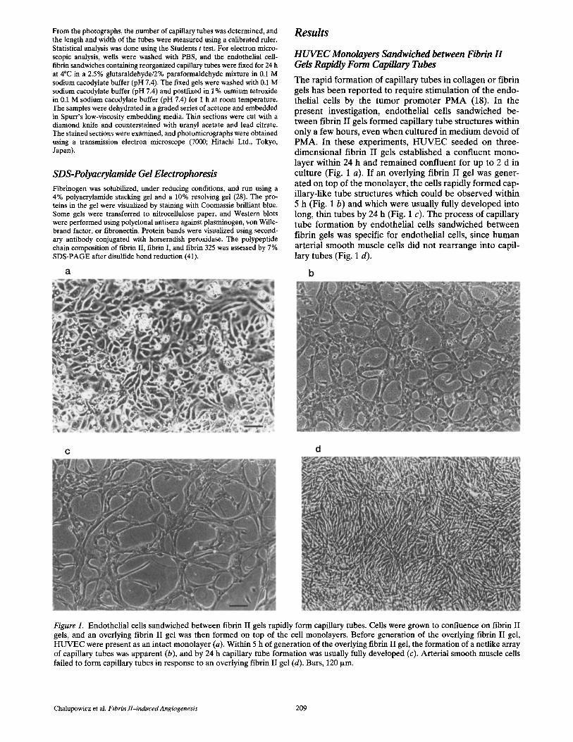
The rapid formation of capillary tubes in collagen or fibrin gels has been reported to require stimulation of the endo- thelial cells by the tumor promoter PMA (18). In the present investigation, endothelial cells sandwiched be- tween fibrin II gels formed capillary tube structures within only a few hours, even when cultured in medium devoid of PMA. In these experiments, HUVEC seeded on three- dimensional fibrin II gels established a confluent mono- layer within 24 h and remained confluent for up to 2 d in culture (Fig. 1 a). If an overlying fibrin II gel was gener- ated on top of the monolayer, the cells rapidly formed cap- illary-like tube structures which could be observed within 5 h (Fig. 1 b) and which were usually fully developed into long, thin tubes by 24 h (Fig. 1 c). The process of capillary tube formation by endothelial cells sandwiched between fibrin gels was specific for endothelial cells, since human arterial smooth muscle cells did not rearrange into capil- lary tubes (Fig. 1 d).
Figure 1. Endothel ial cells sandwiched between fibrin II gels rapidly form capillary tubes. Cells were grown to confluence on fibrin II gels, and an overlying fibrin II gel was then formed on top of the cell monolayers. Before generation of the overlying fibrin II gel, H U V E C were present as an intact monolayer (a). Within 5 h of generat ion of the overlying fibrin II gel, the formation of a netlike array of capillary tubes was apparent (b), and by 24 h capillary tube formation was usually fully developed (c). Arter ial smooth muscle cells failed to form capillary tubes in response to an overlying fibrin II gel (d). Bars, 120 ixm.
Chalupowicz et al. Ftbrin ll-induced Angiogenesis 209

Figure 2. Capillary tubes formed be- tween fibrin II gels manifest intercel- lular and intracellular lumina. Speci- mens were fixed for 24 h at 4°C in a 2.5% glutaraldehyde/2% paraform- aldehyde solution, postfixed in 1% osmium tetroxide, dehydrated in ace- tone, and embedded in Spurr's low- viscosity embedding media. Thin sec- tions were cut and counterstained with uranyl acetate and lead citrate. In this figure, an endothelial capillary tube has been sectioned along its lon- gitudinal axis to reveal both an inter- cellular lumen (**) formed by the in- terdigitation of several endothelial cell processes as well as a putative in- tracellular lumen (*) present within the cytoplasm of a single cell. Note that the intracellular lumen is lined by a cell membrane. Both lumina are filled with cellular debris (D). The in- tercellular junctions between individ- ual endothelial cells can be seen (ar- rows). The underlying and overlying fibrin gels (F) are shown. The under- lying fibrin gel appears much denser. ×10,000. Bar, 6 i~m,
By light microscopy, the capillary tubes were observed to contain vacuoles and channels and were very similar in overall appearance to endothelial cell capillary tubes ob- served by other investigators (16, 18). By electron micros- copy, debris-filled intercellular lumina, formed by the in- terdigitation of several endothelial cells, as well as distinct intracellular lumina could be observed (Fig. 2). Proteolysis of the fibrin was not required for tube formation, since aprotinin did not inhibit tube formation and the fibrin gels demonstrated intact a, [3, and ~/chains in SDS-polyacryl- amide gels (not shown). Finally, tube formation was not due to contaminating proteins such as fibronectin, factor XIII , or von Willebrand factor, since depletion of these proteins f rom fibrinogen was without effect on tube for- mation (not shown).
Since thrombin itself has been reported to cause endo- thelial cell retraction (24), experiments were performed to delineate the potential effect of thrombin from that of the fibrin II gel. When thrombin was added to medium overly- ing an intact endothelial cell monolayer established on a fibrin II gel, rearrangement of the cells was not observed and the cells remained as a monolayer (not shown), indi- cating that thrombin was not responsible for the formation of capillary tubes induced by fibrin II gels. Furthermore, endothelial cells sandwiched between preformed fibrin II gels that had been previously exposed to hirudin (1 U/ml for 30 min), to neutralize the thrombin, formed normal- appearing capillary tubes (not shown).
The avfl3 Integrin Receptor Is Not Essential for the Formation of Capillary Tubes by Sandwiched HUVEC
The txvl33 integrin, which is the major integrin receptor of endothelial cells currently known to interact with fibrino- gen (1, 9-11), has been shown to play a role in regulating
angiogenesis under some experimental conditions (6, 18). We therefore analyzed the role of the av[~3 integrin recep- tor under our experimental conditions in which capillary tube formation is induced by sandwiching the endothelial cells between two fibrin gels in a serum-free system that is devoid of phorbol esters. When H U V E C were sandwiched between fibrin II gels and incubated in medium without or with R G D S or the monoclonal antibody 7E3 directed against e~vl~3, the formation of capillary tubes was unal- tered (Table I). Increasing the concentration of 7E3 to 30 ixg/ml had an apparently slight enhancing effect on tube formation similar to that reported by other investigators (18). Overall, our data indicate that disruption of etvl33 in- tegrin interaction with fibrin did not inhibit capillary tube formation by cells sandwiched between two fibrin II gels.
HUVEC Monolayers Sandwiched between Fibrin I Gels Fail to Form Capillary Tubes
Fibrin II is formed from fibrinogen by thrombin cleavage of fibrinopeptide A from the A s chain and fibrinopeptide B from the Bfl chain, resulting in the generation of the
Table L Effects of RGDS or 7E3 on Capillary Tube Formation
Capillary tube parameters*
Treatment Numbed Length Width
p/n /un Control 26 ± 7 122 - 24 25 _ 6 RGDS150pma 25 ± 7 117 ± 19 26 ± 7 RGDS500p~m 23 _ 9 102 ± 21 22 ± 3 7E310p.g/ml 27 ± 7 124 ± 17 25 ± 6
*Values are means + SD of at least 10 expenments for control and 4-7 experiments for treatments. *Number of tubes in 0.3 mm 2.
The Journal of Cell Biology, Volume 130, 1995 210

Figure 3. Electrophoretic behavior of structurally distinct forms of fi- brin. Fibrin II (FII) and fibrin (F/) were prepared by treatment of fi- brinogen (fg) with thrombin or Atroxin, respectively. Fibrinogen 325 was prepared by treatment of fibrinogen with protease III. Sam- ples were run on a 7% polyacryla- mide gel and stained with Coo- massie blue. In fibrin II and fibrin I, the cx chain migrates faster than the Act chain of fibrinogen due to cleavage of fibrinopeptide A. In fi- brin II, the 13 chain also migrates faster that the B[~ chain of fibrin- ogen due to cleavage of fibrino-
peptide B. The [3 chain of fibrin 325 migrates just above the chain due to the absence of B131--42.
and 13 chains of fibrin II, respectively (25, 39). Thus in fi- brin II, new NH2 termini are generated for both the et and
chains, relative to fibrinogen. In contrast, fibrin I can be formed from fibrinogen by Atroxin cleavage of only fi- brinopeptide A; thus, the B[3 chain of fibrin I is the same as that in fibrinogen. These biochemical differences, which can be assessed by SDS-polyacrylamide gel electrophore- sis (Fig. 3), account for structural differences between po- lymerized fibrin I and fibrin II (32), and may provide clues as to the structural features of fibrin that are important for capillary tube formation.
As reported by other investigators (7), endothelial cells seeded onto fibrin I gels remained rounded after 2 h of in- cubation (not shown), but by 24 h a confluent monolayer of endothelial cells was established on fibrin I gels (Fig. 4 a), indicating that fibrin I is capable of providing sufficient support for the eventual establishment of an intact cellular monolayer. Cell spreading on fibrin I gels was integrin me- diated, since it was markedly inhibited by RGDS (Fig. 4 b) or 7E3 (not shown). Whether the cell spreading on fibrin I in the absence of antiintegrins was supported by the re-
lease of yon Willebrand factor from the cells or was di- rectly supported by the fibrin I molecule itself is not known. Nevertheless, the spreading of endothelial cells on the fibrin I gels allowed the study of potential capillary tube formation using the fibrin-gel sandwich technique.
Endothelial cell monolayers sandwiched between fibrin I gels manifested a variable range of responses from one experiment to the next. The extremes ranged from no change in the confluent cobblestone appearance of the monolayer (not shown) to the formation of attenuated tubelike structures (Fig. 5) that were very transient. For- mation of these quasi tube structures was not a result of yon Willebrand factor secreted by the cells, since the addi- tion of purified von Willebrand factor to the fibrinogen used to generate the fibrin I gels did not result in capillary tube formation (not shown). Thus, there must be struc- tural features in fibrin II, not present in fibrin I, which play an important role in the formation of endothelial cell cap- illary tubes.
Because it was possible that the Atroxin used to gener- ate fibrin I gels inhibited the formation of capillary tubes, control experiments were performed in which fibrin II gels were made using a mixture of thrombin plus Atroxin. These fibrin II gels, which had the same structure as fibrin II gels made with thrombin alone, induced capillary tube formation that was indistinguishable from tubes formed in fibrin II gels in the absence of Atroxin (not shown), indi- cating that Atroxin was not inhibitory to capillary tube formation.
The Overlying Fibrin II Gel Induces the Formation of Capillary Tubes
To determine whether the morphogenesis of the endothe- lial cells into capillary tube structures was triggered by the interaction of the cells with the underlying fibrin gel used for seeding or with the overlying fibrin gel formed on top of the cells, experiments were done using fibrin II as the underlying gel and either fibrin II or fibrin I as the overly- ing gel. Under these conditions, the interaction of the cells with the overlying gels could be assumed to be responsible
Figure 4. Endothelial cell spreading on a fibrin I gel is mediated by cellular integrins. HUVEC were seeded onto a fibrin I gel in the ab- sence (a) or presence (b) of 500 tzm RGDS. After 24 h of culture, control cells established a confluent monolayer, while RGDS com- pletely prevented cell spreading. Bar, 120 ~m.
Chalupowicz et al. Fibrin lI-induced Angiogenesis 211
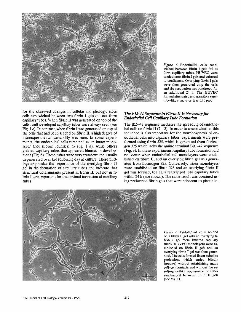
Figure 5. Endothelial cells sand- wiched between fibrin I gels fail to form capillary tubes. HUVEC were seeded onto fibrin I gels and cultured to confluence. Overlying fibrin I gels were then generated atop the cells and the incubation was continued for an additional 24 h. The HUVEC formed attenuated and transitory semi- tube-like structures. Bar, 120 ~m.
for the observed changes in cellular morphology, since cells sandwiched between two fibrin I gels did not form capillary tubes. When fibrin II was generated on top of the cells, well-developed capillary tubes were always seen (see Fig. 1 c). In contrast, when fibrin I was generated on top of the cells that had been seeded on fibrin II, a high degree of interexperimental variability was seen. In some experi- ments, the endothelial cells remained as an intact mono- layer (not shown; identical to Fig. 1 a), while others yielded capillary tubes that appeared blunted in develop- ment (Fig. 6). These tubes were very transient and usually degenerated over the following day in culture. These find- ings emphasize the importance of the overlying fibrin I I gel in the format ion of capillary tubes and indicate that structural determinants present in fibrin II, but not in fi- brin I, are important for the optimal formation of capillary tubes.
The ~15-42 Sequence in Fibrin H Is Necessary for Endothelial Cell Capillary Tube Formation
The 1315--42 sequence mediates the spreading of endothe- lial cells on fibrin I I (7, 13). In order to assess whether this sequence is also important for the morphogenesis of en- dothelial cells into capillary tubes, experiments were per- formed using fibrin 325, which is generated f rom fibrino- gen 325 which lacks the amino terminal B131--42 sequence (Fig. 3). In these experiments, capillary tube formation did not occur when endothelial cell monolayers were estab- lished on fibrin II, and an overlying fibrin gel was gener- ated from fibrinogen 325. Conversely, when monolayers were established on fibrin 325 and an overlying fibrin I I gel was formed, the cells rearranged into capillary tubes within 24 h (not shown). The same result was obtained us- ing pre formed fibrin gels that were adherent to plastic in-
Figure 6. Endothelial cells seeded on a fibrin II gel with an overlying fi- brin I gel form blunted capillary tubes. HUVEC monolayers were es- tabfished on fibrin II gels and an overlying fibrin I gel was then gener- ated. The cells formed linear tubelike projections which ended blindly (arrows) without establishing many cell-cell contacts and without the re- sulting netlike appearance of tubes sandwiched between fibrin II gels (see Fig. 1).
The Journal of Cell Biology, Volume 130, 1995 212

Figure 7. Interaction of the overlying fibrin II gel with the apical surface of the cell monolayer optimally promotes capillary tube forma- tion. HUVEC monolayers (a) were established on fibrin II gels and overlaid with preformed fibrin-325 gels or (b) where established on fibrin-325 gels and overlaid with preformed fibrin II gels. With fibrin-325 on top, the ceils remained as a monolayer (a), and with fibrin II on top the cells formed capillary tubes (b). Bar, 120 Ixm.
serts and then placed on top of the endothelial cell mono- layer. When endothelial monolayers were established on fibrin II and a preformed fibrin-325 gel placed on top, the monolayer remained intact (Fig. 7 a). In contrast, when monolayers were established on fibrin 325 and a pre- formed fibrin II gel was placed on top, the cells reorga- nized into capillary tubes (Fig. 7 b).
The importance of the [315--42 sequence within fibrin II in the induction of capillary tube formation was directly demonstrated by the ability of excess soluble [315-42 pep- tide to inhibit capillary tube formation induced by fibrin II. The addition of 1 mM [315--42 to the culture media be- fore the addition of a prepolymerized overlying fibrin II gel significantly reduced the number and length of the tubes, while significantly increasing tube diameter (Table II). The tetrapeptide GHRP ([315-18) did not induce simi- lar alterations, nor did [315-42 alter capillary tube forma- tion by endothelial cells sandwiched between collagen gels (not shown), indicating the specificity of the fibrin I I - endothelial cell interaction.
In addition to establishing the importance of the [315-42 sequence in fibrin II-induced endothelial cell capillary tube formation, the above results also indicate that throm- bin was not responsible for tube formation since the pre- formed fibrin-325 gels contained active thrombin, yet did not induce tube formation. They also corroborate the ob- servation that it is the overlying fibrin II gel interacting with the apical surface of the endothelial cell monolayer
Table II. Inhibition of Fibrin lI-induced Capillary Tube Formation by Soluble fl15-42
Capillary tube parameters*
Treatment Numbed Length Width
p/n Control 12.7 ± 4.3 101.0 ± 24.4 7 .6 ± 1.6 1315-42 7 __- 1.4 § 66.7 ___ 14.8 § 12.8 - 2.43
*Values are means -.+ SD of at least four experiments. ~:Number of tubes in 0.3 mm 2. § Significantly different from control; P < 0.05.
which is responsible for induction of the morphogenesis process.
Discussion
In this work, we demonstrate that HUVEC, when sand- wiched between two fibrin II gels, promptly rearrange into an extensive network of capillary-like tubes. These structures were clearly discernible as early as 5 h after for- mation of the overlying fibrin II gel and remained intact for up to 3 d. Consistent with the features of capillary tubes studied by others (16, 18), light microscopy revealed the presence of vacuoles and channels within the tubes that, by electron microscopy, were seen to contain both intercellular and intraceUular lumina which were partially filled with cellular debris. The formation of these struc- tures was directly supported by the fibrin II molecule it- self, since the depletion of contaminating proteins, such as fibronectin, von Willebrand factor, factor XIII, and plas- minogen, from the purified fibrinogen used to generate the fibrin II did not hinder capillary tube formation. More- over, %[33 interaction with von Willebrand factor, which may have been secreted by the cells in response to fibrin II (38), did not appear to be responsible for tube forma- tion since the addition of exogenous von Willebrand fac- tor to the fibrinogen used to form the fibrin I gels did not promote tube formation.
A unique and important finding of this study is that gen- eration of capillary tubes was optimally supported by a specific form of fibrin, namely, fibrin II. Tube formation was only minimally supported by fibrin I and never ob- served with fibrin 325. In vivo, fibrin II formation predom- inates over fibrin I formation, although both forms have been demonstrated in vascular lesions (5). The newly formed fibrin monomers are susceptible to the action of plasmin, which cleaves [315--42 (42) generating fibrin 325, which manifests markedly delayed polymerization (40). Fibrin 325 can also be generated experimentally from fi- brinogen 325, a fibrinogen derivative prepared by treat- ment of fibrinogen with protease III from Crotalus atrox,
Chalupowicz et al. Fibrin ll-inducedAngiogenesis 213

which releases the B131-42 sequence from the NH2 termi- nus of each fibrinogen B13 chain thereby reducing the mo- lecular mass to 325 kD (36). In addition to its role in fibrin polymerization, the 1315-42 sequence has also been shown to inhibit fibrinogen binding to platelets via a mechanism which may involve its binding to fibrinogen, as opposed to the platelet fibrinogen receptor GPIIb/IIIa (8). It also me- diates fibrin II-induced release of yon Willebrand factor from endothelial cells (13, 38), and accounts, at least in part, for the ability of fibrin II to support spreading of en- dothelial cells (7, 13). Our demonstration of the inability of fibrin I or fibrin 325 to trigger endothelial cell capillary tube formation and the ability of soluble 1315-42 to inhibit tube formation induced by fibrin II, but not by collagen, indicates the importance of the 1315-42 sequence as a structural determinant of fibrin II that may account for this activity.
Our observation that endothelial cells fail to spread on fibrin I within 2 h is in agreement with findings of other in- vestigators (7). However, our demonstration of the capac- ity of endothelial cells to spread on fibrin I after 24 h al- lowed us to study the formation of capillary tubes by endothelial cell monolayers sandwiched between fibrin I gels. Our results showing that RGDS and the monoclonal antibody 7E3 disorganize endothelial cell monolayers plated on fibrin I, but not on fibrin II, indicate that the txv133 inte- grin plays a major role in maintaining the integrity of endothelial cell monolayers plated on fibrin I, but not on fibrin II. These data further indicate that other determi- nants within fibrin II, notably the 1315-42 sequence, are ca- pable of supporting endothelial cell spreading as previ- ously reported (7, 13).
The high level of expression of av133 in granulation tissue indicates that this integrin is involved in angiogenesis (6). Indeed, in the chick chorioaUantoic membrane, antibodies a g a i n s t o%133 have a potent inhibitory effect on the forma- tion of new blood vessels (6). In contrast to this inhibitory effect, PMA-induced formation of capillary tubes on col- lagen or fibrin gels is enhanced by antibodies to the a2131 integrin or the av133 integrin, respectively (18). Thus, the role of Otv133 in angiogenesis is complex and the observed effect on capillary tube formation may depend, in part, on the experimental conditions. Indeed, in our system, nei- ther RGDS nor the antibody 7E3 against the 133 integrin receptor complex had any noticeable effect on the forma- tion of capillary tubes. The different results which we ob- tained in comparison with other investigators (6, 18) may possibly be due to the absence of PMA in our serum-free system utilizing cells sandwiched between fibrin gels. It may be that the simplicity of this system removes un- known factors which may be important for etv133-mediated capillary tube formation.
Angiogenesis assays frequently make use of complex bi- ologically synthesized matrices such as Matrigel, in which laminin seems to play a predominant role in formation via the expression of three sequences within its structure (23). An Arg-Gly-Asp sequence in the A chain supports cell at- tachment, while the YIGSR sequence, of the B1 chain in- duces capillary tube formation. The latter effect may be mediated by a specific interaction with a 67/32-kD endo- thelial cell surface protein (23). A third region of laminin, the SIKVAV sequence located in the A chain, promotes
migration, invasion of Matrigel, and growth and branching of capillary tubes (19). Thus interactions between matrix proteins and cell surface receptors may underlie the for- mation of capillary tubes (21). In this regard, it is most in- teresting that the t315-42 sequence of fibrin has been shown to bind to a 130-kD glycoprotein expressed at the endothelial cell surface (13), and it is conceivable that fi- brin II interaction with this receptor may initiate capillary tube formation.
In our experiments, proteolysis of matrix proteins was not required for the formation of capillary tubes, presum- ably because the monolayers are instantaneously sand- wiched between two fibrin gels and do not need to migrate into the gel in order to become surrounded by the matrix. Most importantly, our studies demonstrate that contact of fibrin II with the apical aspect of the endothelial cell monolayer is crucial for the induction of morphogenetic changes. This finding is consistent with other studies that have demonstrated that the interaction of collagen with the apical surface of endothelial cells also induces the for- mation of capillary tubes (35). However, we observed that exogenous soluble 1315-42 peptide inhibited fibrin I I - induced tube formation by 45%, while collagen-induced tube formation was unaffected. This finding indicates the specificity of interaction of fibrin with the endothelial cell surface.
In summary, we describe a new in vitro assay for the study of capillary tube formation by endothelial cells sand- wiched between two fibrin gels in serum-free medium. The induction of capillary morphogenesis is dependent on the structure of fibrin, with optimal tube formation occurring with fibrin II, minimum tube formation with fibrin I, and complete absence of tube formation with fibrin 325. Ex- cess soluble [315-42 peptide was able to inhibit fibrin II- induced capillary tube formation, suggesting that interac- tion of the NH2 terminus of the fibrin II 13 chain with the apical surface of the endothelial cells plays a principal role in promoting tube formation.
The authors thank Dr. John Farber and Mr. Tim Schneider from the De- partment of Pathology and Cell Biology at Jefferson Medical College of Thomas Jefferson University for the electron micrographs, Andrew Lik- ens for the illustrations, and Barbara Bolitsky for preparing the manu- script.
This work was supported by research grant HL-20092 (to J. Martinez) from the National Institutes of Health, National Heart, Lung, and Blood Institute; and by an American Heart Association, Southeastern Pennsyl- vania Affiliate Grant-in-Aid (to C. Barsigian).
Received for publication 2 September 1994 and in revised form 21 De- cember 1994.
References
1. Albelda, S. M., M. Daise, E. M. Levine, and C. A. Buck. 1989. Identifica- tion and characterization of cell-substratum adhesion receptors on cul- tured human endothelial cells. J. Clin. Invest. 83:1992-2002.
2. Arnold, F., and D. C. West. 1991. Angiogenesis m wound healing. Pharrna- col. & Ther. 52:407-422.
3. Auerbach, R., W. Auerbach, and I. Polakowski. 1991. Assays for angiogen- esis: a review. Pharmacol. & Ther. 51:1-11.
4. Battegay, E. J., J. Rupp, L. Iruela-Arispe, E. H. Sage, and M. Pech. 1994. PDGF-BB modulates endothelial proliferation and angiogenesis in vitro via PDGF ]5-receptors. J. Cell Biol. 125:917-928.
5. Bini, A., J. Fenoglio, Jr., J. Sobel, J. Owen, M. Velgl, and K. L. Kaplan. 1987. Immunochemical characterization of fibr~nogen, fibrin I, and fibrin II in human thrombi and atherosclerotic lesions. Blood. 69:1038-1045.
6. Brooks, P. C., g. A. F. Clark, and D. A. Cheresh. 1994. Requirement of
The Journal of Cell Biology, Volume 130, 1995 214

vascular integrin etv133 for angiogenesis. Science (Wash. DC). 264:569-571. 7. Bunce, L. A., L. A. Sporn, and C. W. Francis. 1992. Endothelial cell spread-
ing on fibrin requires fibrinopeptide B cleavage and amino acid residues 15--42 of the 13 chain. J. Clin. Invest. 89:842-850.
8. Chen, C. S., S.-H. Chou, and P. Thiagarajan. 1988. Fibrin(ogen) peptide B13 15-42 inhibits platelet aggregation and fibrinogen binding to activated platelets. Biochemistry. 27:6121-6126.
9. Cheresh, D. A., S. A. Berliner, V. Vicente, and Z. M. Ruggeri. 1989. Rec- ognition of distinct adhesive sites on fibrinogen by related integrins on platelets and endothelial ceils. Cell. 58:945-953.
10. Dejana, E., S. ColeUa, L. R. Languino, G. Balconi, G. C. Corbascio, and P. C. Marchisio. 1987. Fibrinogen induces adhesion, spreading, and mi- crofilament organization of human endothelial cells in vitro. Z Cell Biol, 104:1403-1411.
11. Dejana, E., M. G. Lampugnani, M. Giorgi, M. Gaboli, and P. C. Marchisio. 1990. Fibrinogen induces endothelial cell adhesion and spreading via the release of endogenous matrix proteins and the recruitment of more than one integrin receptor. Blood. 75:1509-1517.
12. Dvorak, H. F. 1986. Tumors: wounds that do not heal. Similarities between tumor stroma generation and wound healing. N. Engl. J. Med. 315:1650- 1659.
13. Erban, J. K., and D. D. Wagner. 1992. A 130-kDa protein on endothelial ceils binds to amino acids 15-42 of the B13 chain of fibrinogen. J. Biol. Chem. 267:2451-2458.
14. Ferrara, N. 1993. Vascular endothelial growth factor. Trends Cardtovasc. Med. 3:244-250.
15. Folkman, J. 1994. Angiogenesis and breast cancer. J. Clin. Oncol. 12:441- 443.
16. Folkman, J., and C. Haudenschild. 1980. Angiogenesis in wtro. Nature (Lond.) 288:551-556.
17. Folkman, J., and Y. Shing. 1992. Angiogenesis. J. BioL Chem. 267:10931- 10934.
18. Gamble, J. R., L. J. Matthias, G. Meyer, P. Kaur, G. Russ, R. Faull, M. C. Berndt, and M. A. Vadas. 1993. Regulation of in vitro capillary tube for- mation by anti-integrin antibodies. J. Cell Biol. 121:931-943,
19. Grant, D. S., J. L. Kinsella, R. Fridman, R. Auerbach, B. A. Piasecki, Y. Yamada, M. Zain, and H. K, Klemman. 1992. Interaction of endothelial cells with a laminm A chain peptide (SIKVAV) in vitro and induction of angiogenic behavior in vivo. J. Cell. Physiol. 153:614-625.
20. Grant, D. S., K.-I. Tashiro, B. Segui-Real, Y. Yamada, G. R. Martin, and H. K. Kleinman. 1989. Two different laminin domains mediate the differ- entiation of human endothelial cells into capillary-like structures in vitro. Cell. 58:933-943.
21. Ingber, D. 1991. Extracellular matrix and cell shape: potential control points for inhibition of angiogenesis. J. Cell. Biochem, 47:236-241.
22. Ingber, D. E., and J. Folkman. 1989. Mechanochemical switching between growth and differentiation during fibroblast growth factor-stimulated an- giogenesis in vitro: role of extracellular matrix. J. Cell Biol. 109:317-330.
23. Kubota, Y., H. K. Kleinman, G. R. Martin, and T. J. Lawley. 1988. Role of laminm and basement membrane in the morphological differentiation of human endothelial ceils into capillary-like structures. J. Cell Biol. 107: 1589-1598.
24. Laposata, M., D. K. Dovnarsky, and H. S. Shin. 1983. Thrombin-induced gap formation in confluent endothelial cell monolayers in vitro. Blood. 62:549-556.
25. Laudano, A. P., and R. F. Doolittle. 1978. Synthetic peptide derivatives
that bind to fibrinogen and prevent the polymerization of fibrin mono- mers. Proc. Natl. Acad. Sci. USA, 75:3085-3089.
26. Maciag, T., J. Kadish, L. Wilkins, M. B. Stemerman, and R. Weinstein. 1982. Organizational behavior of human umbilical vein endothelial cells. J. Cell Biol. 94:511-520.
27. Madri, J. A., and S. K. Williams. 1983. Capillary endothelial cell cultures: phenotypic modulation by matrix components. J. Cell Biol. 97:153-165.
28. Martmez, J., E. Rich, and C. Barsigian. 1989. Transglutaminase-mediated cross-linking of fibrinogen by human umbilical vein endothelial cells. J. Biol. Chem. 264:20502-20508.
29. Montesano, R,, L. Orci, and P. Vassalli. 1983. In xatro rapid organization of endothelial cells into capillary-like networks is promoted by collagen ma- trices. J. Cell B~oL 97:1648-1652.
30. Montesano, R., M. S. Pepper, J.-D. Vassalli, and L. Orci. 1987. Phorbol es- ter induces cultured endothelial cells to invade a fibrin matrix in the pres- ence of fibrino|ytic inhibitors. Z Cell. Physiol. 132:509-516.
31. Moscatelli, D.. M. Presta, and D. B. Rifkin. 1986. Purification of a factor from human placenta that stimulates capillary endothelial cell protease production, DNA synthesis, and migration. Proc. NatL Acad. Sci. USA. 83:2091-2095.
32. Mosesson, M. W. 1992. The roles of fibrinogen and fibrin in hemostasis and thrombosis. Semin. HematoL 29:177-188.
33. Mosesson, M, W., and J. S. Finlayson. 1963. Subfractions of human fibrino- gen. J, Lab. Clin. Med. 62:663-673.
34. Nicosia, R. F., and A. Ottinetti. 1990. Growth of microvessels in serum-free matrix culture of rat aorta. Lab. Invest. 63:115-122.
35. Nicosia, R. F., and A. Ottinetti. 1990. Modulation of microvascular growth and morphogenesis by reconstituted basement membrane gel in three- dimensional cultures of rat aorta: a comparative study of angiogenesis in matrigel, collagen, fibrin, and plasma clot. In Vitro Cell. & Dev. Biol. 26: 119-128.
36. Pandya, B. V., C. S. Cierniewski, and A. Z. Budzynski. 1985. Conservation of human fibrinogen conformation after cleavage of B13 chain NH2 termi- nus. J. BioL Chem. 260:2994-3000.
37. Pepper, M. S., D. Belin, R. Montesano, L. Orcl, and J.-D. Vassalli. 1990. Transforming growth factor-beta 1 modulates basic fibroblast growth fac- tor-induced proteolytic and angiogenic properties of endothelial cells in vitro. Z Cell BtoL 111:743-755.
38. Ribes, J. A., C. W. Francis, and D. D. Wagner. 1987. Fibrin induces release of yon Willebrand factor from endothehal ceils. J. Clin. Invest. 79:117- 123.
39. Shainoff, J. R., and B. N. Dardik. 1979. Fibrinopeptide B and aggregation of fibrinogen. Science (Wash. DC). 200:200-202.
40. Siebenlist, K. R., J. P. DiOrio, A. Z. Budzynski, and M. W. Mosesson. 1990. The polymerization and thrombin-binding properties of des-(B131-42)- fibrin. J. Biol. Chem. 265:18650-18655.
41. Weber, K., and M. Osborn. 1969. The reliability of molecular wetght deter- minations by dodecyl sulfate-polyacrylamide gel electrophoresis. J. Btol. Chem. 244:4406--4412.
42. Weitz, J. I., J. A. Koehn, R. E. Canfield, S. L. Landman, and R. Friedman. 1986. Development of a radio~mmunoassay for the fibrinogen-denved peptide B131-42. Blood. 67:1014-1022.
43. Yasunaga, C., Y. Nakashima, and K. Sueishi. 1989. A role of fibrinolytic ac- tivity in angiogenesis. Quantitative assay using in vitro method. Lab. In- vest. 61:698--700.
Chalupowicz et al. Fibrin ll-induced Angiogenesis 215