fever without a source clinical guideline...fever without a source clinical guideline - october 25,...
TRANSCRIPT

FEVER WITHOUT A SOURCE CLINICAL GUIDELINE - OCTOBER 25, 2017 1
Fever Without a Source Clinical Guideline
October 25, 2017

FEVER WITHOUT A SOURCE CLINICAL GUIDELINE - OCTOBER 25, 2017 2
Definition For the purpose of this pathway, Fever Without a Source (FWS) is defined as an acute febrile illness with temperature of 38°C (100.4°F) or greater taken rectally and no identifiable source of infection following a thorough history and physical examination in patients under 6 months of age. Patients with serious and/or life‐threatening infection, especially young infants, may present with hypothermia (below 36°C or 96.8°F) and may be treated using this pathway. Approximately 12% of infants under 30 days of age and 9% of infants 30‐90 days of age will have a serious bacterial infection (SBI), such as bacteremia, meningitis, or urinary tract infection (UTI). Because the clinical exam alone is unable to reliably predict serious bacterial illness in young infants, providers must rely on a combination of history, exam, diagnostic tests, and risk factors to reduce morbidity and mortality in this patient population.
Epidemiology The most common cause of fever without localizing signs is a viral infection. The key point of evaluation is distinguishing which young infants have a serious bacterial infection and using a standardized assessment to stratify risks for these infections in young infants.
Most studies used to stratify risk for serious bacterial infection in neonates have defined a fever as a rectal temperature of 38 C (100.4 F) or greater. In our recommendations we use a cutoff of 38 C for evaluation of infants < 3 months for fever and a cutoff of 39 C (102.2 F) for older children.
While viral infections are the most common cause of fever in young infants, neonates less than 28 days have a particularly higher risk of invasive bacterial infection (up to 14%)1,2 This document aims to provide a risk‐stratified method of distinguishing low risk vs high risk of invasive bacterial infection based on age, clinical appearance, and specific risk factors for certain bacterial infections. These pathways should not be used for the ill appearing young infant who by definition is considered higher risk for invasive bacterial infection.3,4
Etiology Neonates are most commonly infected via perinatal vertical transmission or postnatal exposure to organisms. Perinatal vertical transmission usually manifests within 48 to 72 hours after birth. Early‐onset sepsis is defined as occurring within the first week of life and late‐onset sepsis occurs beyond 7 days of age. Group B Streptococcus used to be the predominant pathogen in neonatal sepsis in the 1970s but with GBS screening and intrapartum antibiotic prophylaxis, there has been an approximate 80% reduction in Group B Streptococcal infection rates. Now, gram‐negative pathogens are the cause of infection in about 80% of young infants. Escherichia coli and Klebsiella pneumoniae are noted to be the most common gram‐negative pathogens and Staphylococcus aureus, Group B Streptococcus, and Enterococcus spp. as the most common gram‐positive pathogens. The majority of bacterial infections in this patient population are identified as urinary tract infections.

FEVER WITHOUT A SOURCE CLINICAL GUIDELINE - OCTOBER 25, 2017 3
Guideline Eligibility Criteria
Neonates and Infants without underlying conditions Rectal temp ≥100.4°F (38°C) OR reported temp (axillary or rectal) of ≥ 100.4°F (38°C) in the home setting Hypothermia as defined as rectal temp < 96.8°F (36°C)
Guideline Exclusion Criteria
Toxic/Septic Appearance Currently receiving antibiotic treatment Infants with a history of prematurity Underlying conditions that affect immunity or may otherwise have increased risk of SBO (e.g. VP shunt, central venous catheters). With an identified focus of infection (e.g. cellulitis, acute otitis media in infants >28 days old)
Differential Diagnosis Fever in the young infant most often raises the concern for underlying infection. Other causes of fever, such as environmental or toxin exposure should be sought in the history.
Etiologic causes of infection in the infant less than 90 d of age is a dynamic subject. Changes in pediatric medical practice over the past 20 years such as the use of new immunizations have had an impact on the epidemiology of various infections. These include the routine use of rotavirus vaccine, influenza vaccination of mothers, pneumococcal, Haemophilus influenza and varicella vaccines. In addition, widespread Group B streptococcal screening and intrapartum maternal antibiotic therapy has had an impact on the prevalence of Group B streptococcal infections. Ages, appearance, comorbidities, prematurity < 37 weeks gestation, height of fever, history of specific exposures to antibiotics are all risk factors for the presence of infection. 7,8
The etiologies for infectious causes of the febrile infant less than 90 days old include:
Viral Infections
‐ These infections are the most common cause of fever in young infants. Studies of febrile young infants, including neonates, support an identifiable viral etiology in 17‐35% of patients. 5,6
‐ Acquisition may be vertical from the mother in utero, during the birth process or exposure after birth to close family members and community
‐ Viurses can cause increased morbidity in young infants due to specific deficiencies in their functional immune system.
‐ Viruses that are important agents include HSV, Enterovirus, CMV, Varicella, RSV, Influenza, and Adenovirus.
FEVER WITHOUT A SOURCE CLINICAL GUIDELINE - OCTOBER 25, 2017 4
Bacterial Infections
‐ Invasive and serious bacterial infections in infants include urinary tract infections, blood stream infections, pneumonia, meningitis, omphalitis, skin and soft tissues infections, bone and joint infections, and gastroenteritis.
‐ These agents account for 10‐14% of infections in the young febrile infant. 5,6 ‐ Invasive bacterial infection can be caused by Gram negatives such as E coli, Enterobacter, Klebsiella,
Salmonella and Gram positives such as Group B streptococci, S. aureus, S. epidermidis, Listeria, Enterococcus.
9,10 ‐ E coli is the most common bacterial infection in the young febrile infant and is the primary cause of UTI in this
age group. 9,10
The prevalence of GBS is decreasing with the advent of widespread maternal screening and intrapartum prophylaxis for this infection. S. aureus is important in skin and soft tissue infection; S epidermidis may play a role in preterm infants. 9,10
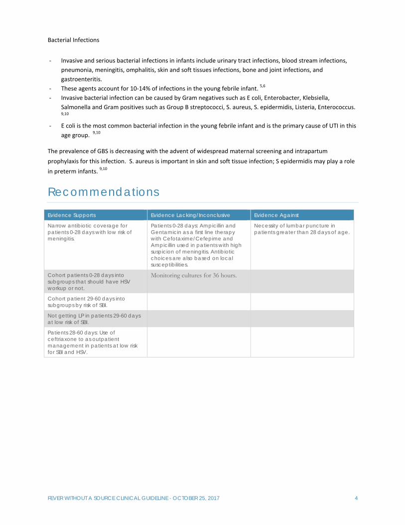
Recommendations
Evidence Supports Evidence Lacking/Inconclusive Evidence Against
Narrow antibiotic coverage for patients 0-28 days with low risk of meningitis.
Patients 0-28 days: Ampicillin and Gentamicin as a first line therapy with Cefotaxime/Cefepime and Ampicillin used in patients with high suspicion of meningitis. Antibiotic choices are also based on local susceptibilities.
Necessity of lumbar puncture in patients greater than 28 days of age.
Cohort patients 0-28 days into subgroups that should have HSV workup or not.
Monitoring cultures for 36 hours.
Cohort patient 29-60 days into subgroups by risk of SBI.
Not getting LP in patients 29-60 days at low risk of SBI.
Patients 28-60 days: Use of ceftriaxone to as outpatient management in patients at low risk for SBI and HSV.

For questions concerning this pathway,Click Here
Last Updated October 25, 2017
Fever Without a SourceAge: 0-28 Day Pathway - Emergency Department
Evidence Based Outcome Center
EXCLUSION CRITERIA
Toxic appearingNo fever Born < 37 weeks gestational age
!ALERT
Patient Toxic/Septic Appearance
Full Sepsis Workup & treat as appropriate.(LINK TO SEPSIS PATHWAY/GUIDELINE)
Severe i l lness / Hypothermia / Lethargy
Seizures
Hepatosplenomegaly
Postnatal HSV contact
Vesicular rash
Conjunctivitis
Interstitial pneumonitis
Thrombocytopenia
CSF pleocytosis
without clear bacterial infection
Transaminitis
Consider HSV work up and empiric ED
treatment for patients with any of the
following conditions:
Historical and Clinical Features
Laboratory Findings
INCLUSION CRITERIA
Non-toxic with temperature > 38°C (100.4°F) OR < 36°C (96.5°F) measured in Emergency Department OR reported measurement at home.
Order labs:Complete Blood Count with differentialBlood Culture
Complete Metabolic PanelUrinalysis with Micro
Urine Culture: Catheter or SuprapubicCerebrospinal Fluid (Hold Tube # 4)
Gram stain
CultureCell count with differential
GlucoseProtein
Stool culture & Stool WBC(If patient has diarrhea)
Focal infection
NO
Manage OFF-PATHWAYYES
ADMIT to Inpatient Management Pathway
ADD antiviral treatment:Acyclovir
CSF Pleocytosis and suspicion of meningitis OR
CSF Gram stain positive
NO
Start empiric antibiotic treatment:Ampicillin x1 + Gentamicin x1
Order labs:HSV DNA PCR of BloodMeningitis/Encephalitis PCR Panel of CSF
Swab/scraping of skin or mucous membrane lesions for HSV DFA AND HSV cultureSurface HSV cultures in viral transport media tube
ConjunctivaThroat
NasopharynxRectum
Skin vesicle (if present)
YES
Contraindications for Ceftriaxone in patients < 28 days of age:
Patient expected to or receiving calcium containing IV products.Total Bilirubin > 10 (See risk factors for hyperbilirubinemia) 1
Herpes Simplex Virus (HSV) work-up indicated
NO
YES
Patient Age: 0-7 Days
Patient Age: 8-28 Days
Change antibiotic treatment:
① Confirm meningitic dose of Ampicillin (Redose if needed)② Add Cefotaxime (Use Cefepime if supply unavailable)③ Consider HSV testing and Acyclovir therapy
Change antibiotic treatment:
① Confirm meningitic dose of Ampicillin (Redose if needed)② Add Ceftriaxone | Confirm meningitic dosing
(Use Cefepime if contraindicated ❶)③ Consider HSV workup and Acyclovir therapy
0-20 WBC/mm3
Protein 0 - 30 days: < 100 mg/dL
Normal Gram Stain
Normal CSF Values
For questions concerning this pathway,Click Here
Last Updated October 25, 2017
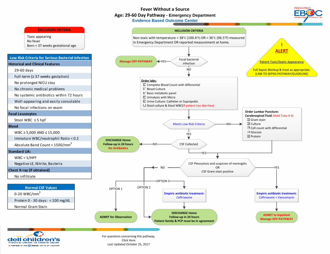
Fever Without a SourceAge: 29-60 Day Pathway - Emergency Department
Evidence Based Outcome Center
Order labs:Complete Blood Count with differentialBlood CultureBasic metabolic panelUrinalysis with MicroUrine Culture: Catheter or SuprapubicStool culture & Stool WBC(If patient has diarrhea)
Focal bacterial infection
NO
Manage OFF-PATHWAY YES
Meets Low Risk Criteria
CSF CollectedDISCHARGE Home
Follow-up in 24 hoursNo Antibiotics
ADMIT to InpatientManage OFF-PATHWAY
!ALERT
Patient Toxic/Septic Appearance
Full Sepsis Workup & treat as appropriate.(LINK TO SEPSIS PATHWAY/GUIDELINE)
YES
NO
Order Lumbar Puncture:Cerebrospinal Fluid (Hold Tube # 4)
Gram stainCultureCell count with differentialGlucoseProtein
NO
YES
Empiric antibiotic treatment: Ceftriaxone + Vancomycin
YES
EXCLUSION CRITERIA
Toxic appearingNo fever Born < 37 weeks gestational age
CSF Pleocytosis and suspicion of meningitis OR
CSF Gram stain positive
INCLUSION CRITERIA
Non-toxic with temperature > 38°C (100.4°F) OR < 36°C (96.5°F) measured in Emergency Department OR reported measurement at home.
ADMIT for ObservationDISCHARGE Home
Follow-up in 24 hoursPatient family & PCP must be in agreement
Empiric antibiotic treatment: Ceftriaxone
OPTION 1 OPTION 2
OPTION 3
NO
29-60 days
Full-term (≥ 37 weeks gestation)
No prolonged NICU stay
No chronic medical problems
No systemic antibiotics within 72 hours
Well-appearing and easi ly consolable
No focal infections on exam
Stool WBC ≤ 5 hpf
WBC ≥ 5,000 AND ≤ 15,000
Immature WBC/neutrophil Ratio < 0.2
Absolute Band Count < 1500/mm3
WBC < 5/HPF
Negative LE, Nitrite, Bacteria
No infiltrate
Low Risk Criteria for Serious Bacterial Infection
Historical and Clinical Features
Blood
Standard UA:
Chest X-ray (if obtained)
Fecal Leucocytes
0-20 WBC/mm3
Protein 0 - 30 days: < 100 mg/dL
Normal Gram Stain
Normal CSF Values
For questions concerning this pathway,Click Here
Last Updated October 25, 2017
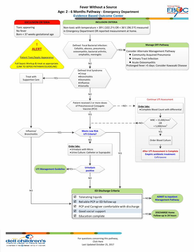
Fever Without a SourceAge: 2 - 6 Months Pathway - Emergency Department
Evidence Based Outcome Center
Defined Focal Bacterial Infection:Cellulitis, abscess, pneumonia,
osteomyelitis, bacterial arthritis, omphalitis, meningitis
Manage OFF-Pathway
Consider Alternate Management Pathway
Community Acquired Pneumonia
Urinary Tract Infection
Acute OsteomyelitisProlonged fever >5 days: Consider Kawasaki Disease
YES
NO
!ALERT
Patient Toxic/Septic Appearance
Full Sepsis Workup & treat as appropriate.(LINK TO SEPSIS PATHWAY/GUIDELINE)
Defined Viral SyndromeCroupBronchiolitisStomatitisInfluenzaVaricella
Treat with Supportive Care
YES
Patient received 2 or more doses of Pneumococcal Conjugate
Vaccine (PCV)
Influenza/Bronchiolitis
Meets Low Risk UTI Criteria?
Order labs:Urinalysis with MicroUrine Culture: Catheter or Suprapubic
NO
Urinalysis positive
UTI Management Guideline
DISCHARGE HomeFollow-up in 24 hours
YES
NO
ED Discharge Criteria
NO
YES
NO
NO
EXCLUSION CRITERIA
Toxic appearingNo fever Born < 37 weeks gestational age
INCLUSION CRITERIA
Non-toxic with temperature > 39°C (102.2°F) OR < 36°C (96.5°F) measured in Emergency Department OR reported measurement at home.
Tolerating l iquids
Reliable PCP or ED follow-up
PCP and Caregiver comfortable with discharge
Good social support
Education complete
ADMIT to Inpatient Management Pathway
NO
YES
YES
Continue UTI Assessment
Order labs:Complete Blood Count with differential
WBC > 15,000/mm3
OR < 5,000/mm3
YES
After UTI Assessment is CompleteEmpiric antibiotic treatment:
Ceftriaxone
Order Blood Culture
NO
YES
For questions concerning this pathway,Click Here
Last Updated October 25, 2017
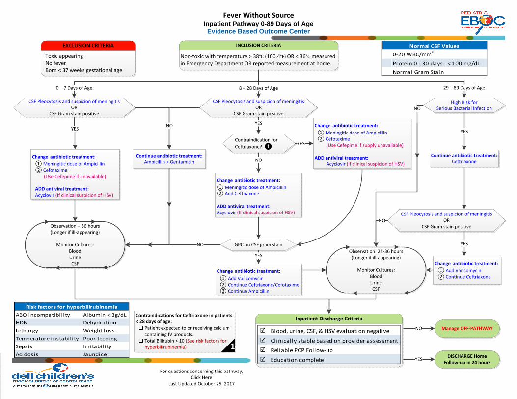
INCLUSION CRITERIA
Non-toxic with temperature > 38°C (100.4°F) OR < 36°C measured in Emergency Department OR reported measurement at home.
CSF Pleocytosis and suspicion of meningitisOR
CSF Gram stain positive
CSF Pleocytosis and suspicion of meningitis OR
CSF Gram stain positive
High Risk for Serious Bacterial Infection
0 – 7 Days of Age 8 – 28 Days of Age 29 – 89 Days of Age
YES
Continue antibiotic treatment:Ampicillin + Gentamicin
NO
Contraindication for
Ceftriaxone? ❶
YES
YES
GPC on CSF gram stain
Continue antibiotic treatment: Ceftriaxone
YES
CSF Pleocytosis and suspicion of meningitis OR
CSF Gram stain positive
YES
DISCHARGE HomeFollow-up in 24 hours
EXCLUSION CRITERIA
Toxic appearingNo fever Born < 37 weeks gestational age
Fever Without SourceInpatient Pathway 0-89 Days of AgeEvidence Based Outcome Center
NO
Manage OFF-PATHWAYNO
YES
NO
NO
Change antibiotic treatment:
① Meningitic dose of Ampicillin② Cefotaxime
(Use Cefepime if unavailable)
ADD antiviral treatment:Acyclovir (If clinical suspicion of HSV)
Change antibiotic treatment:
① Meningitic dose of Ampicillin② Cefotaxime
(Use Cefepime if supply unavailable)
ADD antiviral treatment:Acyclovir (If clinical suspicion of HSV)
Change antibiotic treatment:
① Meningitic dose of Ampicillin② Add Ceftriaxone
ADD antiviral treatment:Acyclovir (If clinical suspicion of HSV)
Change antibiotic treatment:
① Add Vancomycin② Continue Ceftriaxone/Cefotaxime③ Continue Ampicillin
Change antibiotic treatment:
① Add Vancomycin② Continue Ceftriaxone
Observation: 24-36 hours(Longer if ill-appearing)
Monitor Cultures:BloodUrineCSF
Observation – 36 hours(Longer if ill-appearing)
Monitor Cultures:BloodUrineCSF
Inpatient Discharge CriteriaContraindications for Ceftriaxone in patients < 28 days of age:
Patient expected to or receiving calcium containing IV products.Total Bilirubin > 10 (See risk factors for hyperbilirubinemia) 1
NO
YES
ABO incompatibi l ity Albumin < 3g/dL
HDN Dehydration
Lethargy Weight loss
Temperature instability Poor feeding
Sepsis Irritability
Acidosis Jaundice
Risk factors for hyperbilirubinemia
Blood, urine, CSF, & HSV evaluation negative
Clinically stable based on provider assessment
Reliable PCP Follow-up
Education complete
0-20 WBC/mm3
Protein 0 - 30 days: < 100 mg/dL
Normal Gram Stain
Normal CSF Values
For questions concerning this pathway,Click Here
Last Updated October 25, 2017
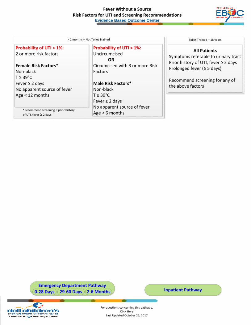
Fever Without a SourceRisk Factors for UTI and Screening Recommendations
Evidence Based Outcome Center
Inpatient PathwayEmergency Department Pathway
0-28 Days 29-60 Days 2-6 Months
Toilet Trained – 18 years> 2 months – Not Toilet Trained
Probability of UTI > 1%:2 or more risk factors
Female Risk Factors*Non-blackT ≥ 39°CFever ≥ 2 daysNo apparent source of feverAge < 12 months
Probability of UTI > 1%:Uncircumcised
ORCircumcised with 3 or more Risk Factors
Male Risk Factors*Non-blackT ≥ 39°CFever ≥ 2 daysNo apparent source of feverAge < 6 months
All PatientsSymptoms referable to urinary tractPrior history of UTI, fever ≥ 2 daysProlonged fever (≥ 5 days)
Recommend screening for any of the above factors
*Recommend screening if prior history
of UTI, fever ≥ 2 days

For questions concerning this pathway,Click Here
Last Updated October 25, 2017
Fever Without a SourceSerious Bacterial Infection
Evidence Based Outcome Center
Inpatient PathwayEmergency Department Pathway
0-28 Days 29-60 Days 2-6 Months
29-60 days
Full-term (≥ 37 weeks gestation)
No prolonged NICU stay
No chronic medical problems
No systemic antibiotics within 72 hours
Well-appearing and easily consolable
No focal infections on exam
Stool WBC ≤ 5 hpf
WBC ≥ 5,000 AND ≤ 15,000
Immature WBC/neutrophil Ratio < 0.2
Absolute Band Count < 1500/mm3
WBC < 5/HPF
Negative LE, Nitrite, Bacteria
No infi ltrate
Low Risk Criteria for Serious Bacterial Infection
Historical and Clinical Features
Blood
Standard UA:
Chest X-ray (if obtained)
Fecal Leucocytes

For questions concerning this pathway,Click Here
Last Updated October 25, 2017
Herpes Simplex Virus Workup Consists of the following labs:
HSV DNA PCR of BloodMeningitis/Encephalitis PCR Panel of CSFSwab/scraping of skin or mucous membrane lesions for HSV DFA AND HSV cultureSurface HSV cultures in viral transport media tube
ConjunctivaThroatNasopharynxRectumSkin vesicle (if present)
Inpatient PathwayEmergency Department Pathway
0-28 Days 29-60 Days 2-6 Months
Fever Without a SourceHerpes Simplex Virus
Evidence Based Outcome Center
Severe illness / Hypothermia / Lethargy
Seizures
Hepatosplenomegaly
Postnatal HSV contact
Vesicular rash
Conjunctivitis
Interstitial pneumonitis
Thrombocytopenia
CSF pleocytosis
without clear bacterial infection
Transaminitis
Patiens with any of the following conditions should be considered for
a Herpes Simplex Virus work up and empiric treatment:
Historical and Clinical Features
Laboratory Findings

For questions concerning this pathway,Click Here
Last Updated October 25, 2017
DCMC Positive Urinalysis (UA) Definition: The presence of Leukocyte Esterase OR Nitrites OR microscopic analysis results positive for leukocytes or bacteria is suggestive of an active UTI. When more than one of these findings is present at the same time, the sensitivity and specificity increase significantly.
Dell Children’s and Seton Family of Hospitals does not currently perform an enhanced urinalysis on urine specimens routinely. The following criteria are guide in diagnosing a UTI in young children using the standard method of collection and processing.
Fever Without a SourceUTI Definition and Urinalysis
Evidence Based Outcome Center
Inpatient PathwayEmergency Department Pathway
0-28 Days 29-60 Days 2-6 Months
Diagnostic Interpretation
Nitrites Poor sensitivity: Conversion of nitrates to nitrites by bacteria takes approximately 4 hours and not all bacteria reduce nitrate levels combined with frequency of infants voiding.
Helpful when positive. Few false positives and high specificity.
Leukocyte Esterase Positive leukocyte esterase is suggestive of a UTI. However, children may have WBC present in their urine in conditions other than a UTI (e.g. Kawasaki Disease)
White Blood Cells (WBC) - Pyuria
Positive if:
5 WBC per HBF via standard method Pyuria is absent in approximately 10% of children with a UTI
Bacteriuria Presence of bacteriuria alone in the absence of other findings does not define a UTI.
Culture
Method Definite* Indeterminant† Contaminant
Suprapubic Any growth Growth of non-pathogens, Mixed culture
Catheter 50,000 CFU/ML
10,000 CFU/ML
Growth of non-pathogens, Mixed culture, < 10,000 CFU/ml
* If also with presence of pyuria or bacteriuria
† Consider obtaining repeat specimen Mixed Culture = uropathogen + non-pathogen or two uropathogens Bag UA specimens should never be sent for urine culture. Only catheter or suprapubic methods are appropriate for culture collection in this age. Uropathogens
Gram Negative Escherichia coli (~80%) Klebsiella Proteus Enterobacter Citrobacter
Gram Positive Staphylococcus saprophyticus Enterococcus Staphylococcus aureus
Non-pathogens Lactobacillus Coagulase-negative Staph Corynebacterium
For questions concerning this pathway,Click Here
Last Updated October 25, 2017
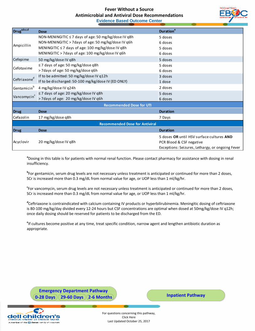
Fever Without a SourceAntimicrobial and Antiviral Dose Recommendations
Evidence Based Outcome Center
aDosing in this table is for patients with normal renal function. Please contact pharmacy for assistance with dosing in renal insufficiency.
bFor gentamicin, serum drug levels are not necessary unless treatment is anticipated or continued for more than 2 doses, SCr is increased more than 0.3 mg/dL from normal value for age, or UOP less than 1 ml/kg/hr.
cFor vancomycin, serum drug levels are not necessary unless treatment is anticipated or continued for more than 2 doses, SCr is increased more than 0.3 mg/dL from normal value for age, or UOP less than 1 ml/kg/hr.
dCeftriaxone is contraindicated with calcium containing IV products or hyperbilirubinemia. Meningitic dosing of ceftriaxone is 80-100 mg/kg/day divided every 12-24 hours but CSF concentrations are optimal when dosed at 50mg/kg/dose IV q12h; once daily dosing should be reserved for patients to be discharged from the ED.
eIf cultures become positive at any time, treat specific condition, narrow agent and lengthen antibiotic duration as appropriate.
Inpatient PathwayEmergency Department Pathway
0-28 Days 29-60 Days 2-6 Months
Druga,b,c,d
Dose Duratione
NON-MENINGITIC ≤ 7 days of age: 50 mg/kg/dose IV q8h
NON-MENINGITIC > 7days of age: 50 mg/kg/dose IV q6h5 doses
6 doses
MENINGITIC ≤ 7 days of age: 100 mg/kg/dose IV q8h
MENINGITIC > 7days of age: 100 mg/kg/dose IV q6h 5 doses
6 doses
Cefepime 50 mg/kg/dose IV q8h 5 doses
Cefotaxime≤ 7 days of age: 50 mg/kg/dose q8h
> 7days of age: 50 mg/kg/dose q6h5 doses
6 doses
Ceftriaxoned If to be admitted: 50 mg/kg/dose IV q12h
If to be discharged: 50-100 mg/kg/dose IV (ED ONLY)3 doses
1 dose
Gentamicinb 4 mg/kg/dose IV q24h 2 doses
Vancomycinc ≤ 7 days of age: 20 mg/kg/dose IV q8h
> 7days of age: 20 mg/kg/dose IV q6h5 doses
6 doses
Drug Dose Duration
Cefazolin 17 mg/kg/dose q8h 7 Days
Drug Dose Duration
Acyclovir 20 mg/kg/dose IV q8h5 doses OR until HSV surface cultures AND
PCR Blood & CSF negative
Exceptions: Seizures, Lethargy, or ongoing Fever
Ampicill in
Recommended Dose for UTI
Recommended Dose for Antiviral
FEVER WITHOUT A SOURCE CLINICAL GUIDELINE - OCTOBER 25, 2017 6
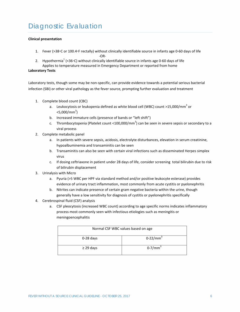
Diagnostic Evaluation Clinical presentation
1. Fever (>38◦C or 100.4◦F rectally) without clinically identifiable source in infants age 0‐60 days of life ‐OR‐
2. Hypothermia1 (<36◦C) without clinically identifiable source in infants age 0‐60 days of life Applies to temperature measured in Emergency Department or reported from home
Laboratory Tests
Laboratory tests, though some may be non‐specific, can provide evidence towards a potential serious bacterial infection (SBI) or other viral pathology as the fever source, prompting further evaluation and treatment
1. Complete blood count (CBC) a. Leukocytosis or leukopenia defined as white blood cell (WBC) count >15,000/mm3 or
<5,000/mm3) b. Increased immature cells (presence of bands or “left shift”) c. Thrombocytopenia (Platelet count <100,000/mm3) can be seen in severe sepsis or secondary to a
viral process 2. Complete metabolic panel
a. In patients with severe sepsis, acidosis, electrolyte disturbances, elevation in serum creatinine, hypoalbuminemia and transaminitis can be seen
b. Transaminitis can also be seen with certain viral infections such as disseminated Herpes simplex virus
c. If dosing ceftriaxone in patient under 28 days of life, consider screening total bilirubin due to risk of bilirubin displacement
3. Urinalysis with Micro a. Pyuria (>5 WBC per HPF via standard method and/or positive leukocyte esterase) provides
evidence of urinary tract inflammation, most commonly from acute cystitis or pyelonephritis b. Nitrites can indicate presence of certain gram negative bacteria within the urine, though
generally have a low sensitivity for diagnosis of cystitis or pyelonephritis specifically 4. Cerebrospinal fluid (CSF) analysis
a. CSF pleocytosis (increased WBC count) according to age specific norms indicates inflammatory process most commonly seen with infectious etiologies such as meningitis or meningoencephalitis
Normal CSF WBC values based on age
0‐28 days 0‐22/mm3
≥ 29 days 0‐7/mm3

FEVER WITHOUT A SOURCE CLINICAL GUIDELINE - OCTOBER 25, 2017 7
b. Increased protein can be seen in the setting of meningitis or meningoencephalitis
Normal CSF protein values based on age
0‐30 days <100 mg/dL
> 30 days 15‐45 mg/dL
c. Glucose can be decreased in acute bacterial meningitis
Normal CSF Glucose values based on age
0‐28 days 34‐119 mg/dL
≥ 29 days 40‐80 mg/dL
d. Gram stain can provide evidence of bacterial pathogens present in CNS 5. Cultures
a. Cultures of blood, urine and CSF should be obtained to rule out presence of bacterial pathogen b. Stool culture can be considered in patient where significant diarrhea is present to rule out
bacterial pathogen. Fecal WBCs can be seen in significant colitis as well as other non‐infectious sources.
6. Molecular diagnostics a. Herpes simplex virus – if concerned for acute HSV disease, following workup should be obtained
for complete evaluation i. HSV PCR blood
ii. HSV PCR CSF (can be included in Biofire – see section d.) iii. HSV surface cultures
b. Enterovirus PCR in CSF can provide etiology of pleocytosis in the absence of positive bacterial culture (can be included in Biofire – see section d.)
c. Rapid viral testing for Influenza and RSV, when taken in context of correlating clinical symptoms and community prevalence can provide evidence of a fever source in the absence of suspected SBI.
d. PCR panels (Respiratory pathogen panel, Biofire of CSF) provide rapid PCR testing for a variety of bacterial and viral pathogens and can be helpful in identifying fever source in cases where positive results would affect clinical management and potential outcomes such as
i. Antibiotic pretreatment where bacterial culture may not be reliable ii. Initiation of antimicrobials (HSV encephalitis, mycoplasma pneumonia, pertussis, etc)
Imaging Chest X‐Ray can be considered if concerned for an acute lower respiratory tract infection based on clinical symptoms.
Methods Existing External Guidelines/Clinical Pathways

FEVER WITHOUT A SOURCE CLINICAL GUIDELINE - OCTOBER 25, 2017 8
Existing External Guideline/Clinical Pathway Organization and Author Last Update
Fever Without Localizing Signs Texas Children’s Hospital 2009
Neonatal Fever Pathway Seattle Children’s 2017
Febrile Infant Clinical Pathway Children’s Hospital of Philadelphia 2015 Any published clinical guidelines have been evaluated for this review using the AGREE II criteria. The comparisons of these guidelines are found at the end of this document. AGREE II criteria include evaluation of: Guideline Scope and Purpose, Stakeholder Involvement, Rigor of Development, Clarity of Presentation, Applicability, and Editorial Independence.
Review of Relevant Evidence: Search Strategies and Databases Reviewed
Search Strategies Document Strategies Used
Search Terms Used:
Infant, neonate, less than 7 days of age, 28 days of age, risk of serious bacterial infections, herpes simplex virus, risk stratification, blood stream infection, enterovirus, antibiotic course, septic workup, sepsis, positive urine analysis, lumbar puncture, hospital admission, antibiotic management
Years Searched ‐ All Questions
2007 ‐ 2017
Language English
Age of Subjects 0 – 6 Months of age
Search Engines PubMed, Cochrane, Google
Government/State Agencies
National Guideline Clearinghouse
Evidence Found with Searches
Check Type of Evidence Found
Summary of Evidence – All Questions Number of Articles Obtained
☐ Systematic Reviews
☒ Meta‐analysis articles 1
☒ Randomized Controlled Trials 2
☒ Non‐randomized studies 27
☐ Review articles
☐ Government/State agency regulations
☐ Professional organization guidelines, white papers, ect.
☐ Other:
Evaluating the Quality of the Evidence
FEVER WITHOUT A SOURCE CLINICAL GUIDELINE - OCTOBER 25, 2017 9
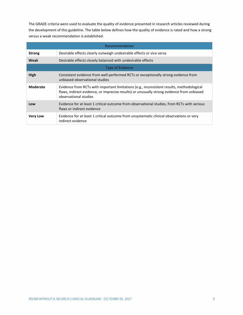
The GRADE criteria were used to evaluate the quality of evidence presented in research articles reviewed during the development of this guideline. The table below defines how the quality of evidence is rated and how a strong versus a weak recommendation is established.
Recommendation
Strong Desirable effects clearly outweigh undesirable effects or vice versa
Weak Desirable effects closely balanced with undesirable effects
Type of Evidence
High Consistent evidence from well‐performed RCTs or exceptionally strong evidence from unbiased observational studies
Moderate Evidence from RCTs with important limitations (e.g., inconsistent results, methodological flaws, indirect evidence, or imprecise results) or unusually strong evidence from unbiased observational studies
Low Evidence for at least 1 critical outcome from observational studies, from RCTs with serious flaws or indirect evidence
Very Low Evidence for at least 1 critical outcome from unsystematic clinical observations or very indirect evidence

FEVER WITHOUT A SOURCE CLINICAL GUIDELINE - OCTOBER 25, 2017 10
Executive Summary Clinical Standards Preparation
This clinical standard was prepared by the Evidence‐Based Outcomes Center (EBOC) team. DCMC EBOC Team Ronda Machen, PharmD Sujit Iyer, MD Nidhi Vaidya, MD Marisol Fernandez, MD Sarmistha Hauger, MD Wade Mincher, MD Lynn Thoreson, DO Kathryn Merkel, PharmD Katherine Simon, MD Patrick Boswell Approved by EBOC Committee: Sarmistha Hauger, MD Terry Stanley, DNP, RN, NE‐BC Deb Brown, RN Sujit Iyer, MD Tory Meyer, MD Nilda Garcia, MD Meena Iyer, MD Michael Auth, DO Jorge Ganem, MD
Recommendations Practice recommendations were directed by the existing evidence and consensus amongst the content experts. Patient and family preferences were included when possible.
Approval Process
EBOC guidelines are reviewed by DCMC content experts, the EBOC committee, and are subject to a hospital wide review prior to implementation. Recommendations are reviewed and adjusted based on local expertise.
Evaluating the Quality of the Evidence
Published clinical guidelines were evaluated for this review using the AGREE II criteria. The summary of these guidelines are included in the literature appraisal. AGREE II criteria evaluate Guideline Scope and Purpose, Stakeholder Involvement, Rigor of Development, Clarity and Presentation, Applicability, and Editorial Independence using a 4‐point Likert scale. This clinical standard specifically summarizes the evidence in support of or against specific interventions and identifies where evidence is lacking/inconclusive. The following categories describe how research findings provide support for treatment interventions. “Evidence Supports” provides evidence to support an intervention “Evidence Against” provides evidence against an intervention. “Evidence Lacking/Inconclusive” indicates there is insufficient evidence to support or refute an intervention and no conclusion can be drawn from the evidence.
Disclaimer LEGAL DISCLAIMER: The information provided by Dell Children’s Medical Center of Texas (DCMCT), including but not limited to Clinical Pathways and Guidelines, protocols and outcome data, (collectively the "Information") is presented for the purpose of educating patients and providers on various medical treatment and management. The Information should not be relied upon as complete or accurate; nor should it be relied on to suggest a course of treatment for a particular patient. The Clinical Pathways and Guidelines are intended to assist physicians and other health care providers in clinical decision‐making by describing a range of generally acceptable approaches for the diagnosis, management, or prevention of specific diseases or conditions. These guidelines should not be considered inclusive of all proper methods of care or exclusive of other methods of care reasonably directed at obtaining the same results. The ultimate judgment regarding care of a particular patient must be made by the physician in light of the individual circumstances presented by the patient. DCMCT shall not be liable for direct, indirect, special, incidental or consequential damages related to the user's decision to use this information contained herein.

FEVER WITHOUT A SOURCE CLINICAL GUIDELINE - OCTOBER 25, 2017 11
References 1. Byington CL, Enriquez FR, Hoff C, et al. Serious bacterial infections in febrile infants 1 to 90 days old with
and without viral infections. Pediatrics 2004;113(6):1662–6. 2. Caviness AC, Demmler GJ, Almendarez Y, Selwyn BJ. The prevalence of neonatal herpes simplex virus
infection compared with serious bacterial illness in hospitalized neonates. J Pediatr 2008;153(2):164–9. 3. Hui C, Neto G, Tsertsvadze A, et al. Diagnosis and management of febrile infants (0‐3 months). Evid
ReportTechnology Assess 2012;(205):1–297. 4. Gomez B, Mintegi S, Bressan S, et al. Validation of the “Step‐by‐Step” Approach in the Management of
Young Febrile Infants. Pediatrics 2016;138(2). 5. Caviness, AC et al The prevalence of neonatal herpes simplex virus infection compared with serious
bacterial illness in hospitalized neonates. J Pediatric. 2008;153(2):164 6. Byington CL, et al. Serious bacterial infections in febrile infants 1 to 90 days old with and without viral
infections. Pediatrics. 2004;113(6):1662 7. Pantell RH et al, Management and outcomes of care of fever in early infancy. JAMA. 2004;291(10):1203 8. Hui C, Neto G, Tsertsvadze A, et al. Diagnosis and Management of Febrile Infants (0‐3 months). Evidence
Report/Technology Assessment No. 205 (Prepared by the University of Ottawa: Evidence‐based Practice Center under Contract No. HHSA 290‐2007‐10059‐I). AHRQ Publication No. 12‐E004‐EF. Rockville, MD: Agency for Healthcare Research and Quality. March 2012. Available at http://www.ahrq.gov/research/findings/evidence‐based‐reports/febrinftp.html (Accessed August 3, 2015).
9. Sadow KB et al, Bacterial infections in infants 60 days and younger: epidemiology, resistance, and implications for treatment. Arch Pediatr Adolesc Med. 1999;153(6):611
10. Biondi E et al, Epidemiology of bacteremia in febrile infants in the United States Pediatrics. 2013 Dec; 132(6):990‐6. Epub 2013 Nov 11.
11. Xiao‐Qing Lv, Ling‐He Qian, Tai Wu, and Tian‐Ming, Yuan. Enterovirus infection in febrile neonates: A hospital‐based prospective cohort study
12. A. Martinez Planas; C. Munoz Almagro. Clinical Microbiology and Infection 2012; 18: 856‐861. Low prevalence of invasive bacterial infection in febrile infants under 3 months of age with enterovirus infection
13. Mohammad Javad Ghabouli Shahroodi, Kiarash Ghazvini, et al. Enteroviral in Neonates and Children of Mashhad, Iran. J. Microbiol. 2016 May; 9(5)
14. Calvo Cristina et al. Enterovirus neurological disease and bacterial coinfection in very young infants with fever. Journal of Clical Virology 85 (2016) 37‐39
15. Schroeder AR, Chang PW, Shen MW, Biondi EA, Greenhow TL. Diagnostic Accuracy of the Urinalysis for Urinary Tract Infection in Infants <3 Months of Age. Pediatrics. 2015 Jun; 135(6): 965‐71.
16. Lin DS, Huang SH, Lin CC, Tung YC, Chiu NC. Urinary Tract Infection in Febrile Infants Younger Than Eight Weeks of Age. Pediatrics. 2000 Feb; 105(2) e20.
17. Shaw KN, McGowan KL, Gorelick MH, Schwartz JS. Screening for Urinary Tract Infection in Infants in the Emergency Department: Which Test is Best? Pediatrics. 1998 Jun; 101(6) e1.
18. Hoberman A, Wald ER, Penchansky L, Reynolds EA, Young S. Enhanced Urinalysis as a Screening Test for Urinary Tract Infection. Pediatrics. 1993 Jun; 91(6) 1196‐1199.
19. Crain EF, Gershel JC. Urinary Tract Infections in Febrile Infants Younger Than 8 weeks of Age. Pediatrics. 1990 Sep; 86(3) 363‐367.
20. Nigrovic LE, Malley R, Kuppermann N. Meta‐analysis of bacterial meningitis score validation studies. Arch Dis Child. 2012 Sept;97(9):799‐805.
21. Nigrovic LE, Kuppermann N, Macias CG, Cannavino CR. Clinical prediction rule for identifying children with cerebrospinal fluid pleocytosis at very low risk for bacterial meningitis. JAMA. 2007 Jan 3;297(1);52‐60.
22. Ashkenazi‐Hoffnung L, Livni G, Amir J, et al. Serious bacterial infections in hospitalized febrile infants aged 90 days or younger: The traditional combination of ampicillin and gentamicin is still appropriate. Scandinavian Journal of Infectious Disease 2011; 43: 489‐94.
23. Byington CL, Reynold CC, Korgenski K, et al. Costs and infant outcomes after implementation of a care process model for febrile infants. Pediatrics 2012; 130: 1‐S18.

FEVER WITHOUT A SOURCE CLINICAL GUIDELINE - OCTOBER 25, 2017 12
24. Byington CL, Rittichier KK, Bassett KE, et al. Serious bacterial infections in febrile infants younger than 90 days of age: the importance of ampicillin‐resistant pathogens. Pediatr. 2003; 111:964‐968
25. Cantey JB, Lopez‐Medina E, Nguyen S, et al. Empiric antibiotics for serious bacterial infection in young infants. Pediatr Emer Care 2015; 31: 568‐71.
26. Greenhow TL, HungYY, HerzAM. Changing epidemiology of bacteremia in infants aged 1 week to 3 months. Pediatrics. 2012;129:e590–e596.
27. Harvey D, Holt DE, Bedford H. Bacterial meningitis in the newborn: A prospective study of mortality and morbidity. Seminars in Perinatology 1999; 23: 218‐25.
28. Hasoon A, Stankovic C, Rogers A, et al. Listeria and enterococcal infections in neonates 28 days of age and younger. Pediatr Emer Care 2014; 30: 240‐3.
29. Hon KL, Ting JY, Chow CM, et al. Microbiologic agents in parent‐reported neonatal fever. Journal of Tropical Pediatrics 2015; 61, 448‐54.
30. Marom R, Sakran W, Antonelli J, et al. Quick identification of febrile neonates with low risk for serious bacterial infection: an observational study. Arch Dis Child Fetal Neonatal Ed. 2007;92:F15‐18.
31. Watt K, Waddle E, Jhaveri R. Changing epidemiology of serious bacterial infections in febrile infants without localizing signs. PLoS One. 2010;5:e12448.
32. Zaidi AK, Tikmani SS, Warraich HJ, et al. Community‐based treatment of serious bacterial infections in newborns and young infants: a randomized controlled trial assessing three antibiotic regimens. Pediatr Infect Dis J. 2012;31:667–672.
33. Downie L, Armiento R, Subhi R, et al. Community‐acquired neonatal and infant sepsis in developing countries: efficacy of WHO's currently recommended antibiotics—systematic review and meta‐analysis. Arch Dis Child. 2013;98:146–154 (not included above).
34. Tessin I, Trollfors B, Thiringer K, et al. Concentrations of ceftazidime, tobramycin, and ampicillin in the cerebrospinal fluid of newborn infants. European Journal of Pediatrics 1989; 148: 679‐81.
35. Garcia‐Prats JA, Cooper TR, Schneider VF, et al. Rapid detection of microorganisms in blood cultures of newborn infants utilizing an automated blood culture system. Pediatrics 2000; 105: 523‐7.
36. Guerti K, Devos H, Ieven MM, et al. Time to positivity of neonatal blood cultures: fast and furious? J Med Microbiol 2011; 60: 446‐53.
37. Jardine L, Davies MW, Faoagali J, et al. Incubation time required for neonatal blood cultures to become positive. Journal of Pediatrics and Child Health; 2006: 797‐802.
38. Kaplan RL, Harper MB, Baskin MN, et al. Time to detection of positive cultures in 28‐ to 90‐day‐old febrile infants. Pediatrics 2000;106:1‐4.
39. Kurlat I, Stoll BJ, McGowan JE. Time to positivity for detection of bacteremia in neonates. J Clin Microbiol 1989; 27: 1068‐71.
40. Kumar Y, Qunibi M, Neal TJ, et al. Time to positivity of neonatal blood cultures. Arch Dis Child Fetal Neonatal Ed 2001; 85: F182‐6.
41. Janjindamai W, Phetpisal S. Time to positivity of blood culture in newborn infants. Southeast Asian J Trop med Public Health 2006; 37: 171‐6.
42. Evans RC, Fine BR. Time to detection of bacterial cx in infants aged 0 to 90 days. Hosp Pediatr 2013; 3 97‐102.
43. Seattle Children’s Hospital, Bishop J, Ackley H, Beardsley E, Davis J, Goldenberg C, Kronman M, Leu M, May A, Pak D, Ringer C, 2013 August. Rheumatology New Diagnosis Pathway. Available from: http://www.seattlechildrens.org/pdf/neonatal‐fever‐pathway.pdf
44. Cincinnati Children’s Hospital Medical Center. “Fever of Uncertain Source”. 2010 45. Diagnosis and management of febrile infants (0‐3 months). http://www.ahrq.gov/clinic/tp/febrinftp.htm;.
Updated 2012.