fetal medicine - malformation · 2019-08-06 · fetal medicine - malformation f1-01...
TRANSCRIPT

Fetal Medicine - Malformation
F1-01Hypochondroplasia in a family with FGFR3
gene mutations: antenatal ultrasound findings. F1-12Paternal uniparental disomy of chromosome 16 resulting in hemoglobin Bart’s hydrops fetalis.
F1-02Multiple pregnancy in a primigravida with
uncorrected pentalogy of fallot. F1-13Analysis of the outcome of 200 cases of fetal isolated nasal abnormality at 11-13+6 weeks.
F1-03Fetal intracranial teratoma: a case report.
Pregnancy with thalassemia β /hemoglobin e
disease.F1-14
The outcome of cystic hygroma with aneuploidy and structural abnormalities vs euploidy in Women's hospital in Qatar.
F1-04Pregnancy with thalassemia β /hemoglobin e
disease. F1-15Intrauterine transfusion for fetal anemia: indications and implications.
F1-05Thanatophoric dysplasia type 1 with
myelomeningocele: case report. F1-16 Limb body wall complex: a case report.
F1-06Arnold-Chiari type II malformation with
myelomeningocele: a case report. F1-17The hemolytic disease of the fetus and newborn due to alloanti-M: three Chinese cases report and review of the literature.
F1-07Difficulties in the antenatal diagnosis of
ectopic kidney: A case report. F1-18Transillumination role as diagnostic tool of hydranencephally in limited facilities of rural hospital.
F1-08An autopsy case of Potter syndrome: bilateral
renal agenesis with club foot. F1-19Prenatal diagnosis of four cases of Cri-du-chat syndrome.
F1-09The characteristics of congenital anomalies in
a tertiary teaching hospital: Sanglah Birth
Defect Integrated Center (SIDIC) Program,
Sanglah Hospital Denpasar, Bali, Indonesia.
F1-20The value of ultrasonography in prenatal diagnosis of criss-cross heart.
F1-10A comprehensive management of
gastroschisis at Sanglah General Hospital
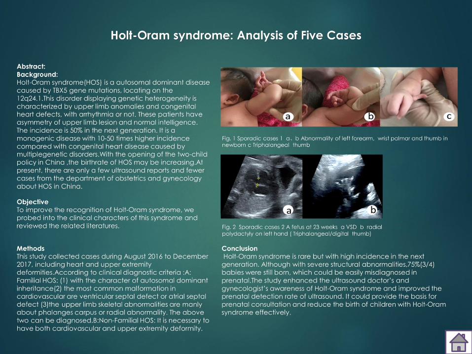
Bali.F1-21 Holt-Oram syndrome: Analysis of Five Cases
F1-11 Acephalous fetus: a rare case.
Hypochondroplasia in a family with FGFR3 gene mutations: antenatal ultrasound findings.
WC Tan, DKL Chan, & HK Tan.Department of Obstetrics and Gynaecology, Singapore General Hospital, Singapore.
CASE REPORT
• Patient was a 30 year old female, gravida 1 para 0. She had no family or medical history of note. She was of short statue, measuring 1.28m. She presented at our department at 29 weeks. Ultrasound scans were performed: (i) 29+2 weeks revealed short limbs; femur and humerus were below 3rd percentile. Right kidney contained multiple cysts. Female foetus . Amniotic fluid was normal, estimated foetal weight was 1028g, 5.3 centile. (ii) 31+4 weeks. Long bones were below 5th centile. Unable to demonstrate right kidney and right renal artery. A 1.5 cm cyst was demonstrated in right renal fossa. (iii) 34+1 weeks. Head circumference and abdominal circumference were within normal limits and Dopplers were normal. All long bones were below 5th centile with some frontal bossing, likely constitutional in view of maternal skeletal dysplasia. Right kidney was not visualised. A 2cm cyst was seen in right renal fossa - likely isolated right renal dysplasia. Postnatal examination of child revealed total length 44cm: upper segment 29cm, lower segment 15cm, arm span 40cm , flat nasal bridge, left clinodactaly, normal female genitalia, normal nose/mouth/ears. A diagnosis of hypo-chondroplasia, based on the FGFR3 test was done by pediatric geneticist.
• Two years later, patient had her second pregnancy. Ultrasound scan at 19 weeks noted all long bones at 5th percentile. No other fetal anomaly was detected, normal female genetalia. Postnatal examination revealed Upper segment: lower segment ratio 1.7, arm span 45cm, length <3rd centile. Clinically, posture was hypotonic with protubent abdomen; facial appearance looked similar to sister who was diagnosed with hypochondroplasia, frontal bossing; cranial ultrasound was normal. FGFR3 testing confirmed hypochondroplasia.
CONCLUSION: Although mutation in FGFR3 gene is a known cause of hypochondroplasia, researchers suspect that mutations in other genes are involved, although they have not been identified.

Multiple pregnancy in a primigravida with uncorrected pentalogy of fallot.P Partana, JKH Tan, EL Tan, JL Tan, & LK Tan.
Department of Obstetrics and Gynecology, Singapore General Hospital, Singapore.
Pentalogy of Fallot is a cyanotic congenital heart disease that has guarded prognosis without surgical intervention in infancy. This condition refers to the co-existence of an atrial septal defect (ASD) and Tetralogy of Fallot, the latter of which comprises an over-riding aorta, obstruction of the right ventricular outflow tract (RVOT), right ventricular hypertrophy (RVH) and a ventricular septal defect (VSD). Despite this additional anatomical defect, the underlying pathophysiology of PoF and ToF remains similar. Women with uncorrected defects rarely survive into childbearing age and pregnancy in this group is associated with a high rate of perinatal loss. Physiological cardiovascular changes in pregnancy can lead to maternal haemodynamic instability with subsequent adverse cardiac sequelae with or without fetal decompensation. Optimum management and pregnancy outcomes in mother with uncorrected Pentalogy of Fallot and twin pregnancy have not been described in the literature. We describe a successful case of monochorionic diamniotic twin pregnancy in an affected woman who has not undergone surgical repair. Her pregnancy progressed without any adverse cardiopulmonary complications. Her caesarean delivery and postpartum recovery were favourable, with successful birth of two healthy babies at 35.7 weeks. This case emphasises the importance of a multidisciplinary team, especially of obstetricians with expertise in high-risk pregnancies, adult congenital heart disease cardiologists and anaesthesiologist.

Fetal intracranial teratoma: a case report.DENSAK PONGROJPAW MD.
Maternal Fetal Medicine Unit, Department of Obstetrics and Gynecology, Faculty of Medicine,
Thammasat University,Pathumthani,Thailand.
OBJECTIVEThe author report a case of fetal intracranial teratoma diagnosed at 26 weeks of gestation after prior normal sonographicexamination.
MATERIALS & METHODCongenital intracranial tumors are rare and only account for 0.5-1.5% of all pediatric brain tumors. The diagnosis is usually made by ultrasonography in fetal life, as diagnosis of an intracranial mass, disrupting normal architecture with or without hydrocephalus. Teratoma is the most common tumor in neonatal period making up 30-50 percent of tumors.
RESULTA 37-year-old woman, G1P0 , 26 weeks of gestation was refered to Maternal Fetal Medicine Unit,Thammasat university hospital because of enlarged fetal head. Her antenatal record was uneventful.Ultrasonography revealed a 4.43 cm x 5.54 cms, heterogeneous intracranial fetal mass at the pituitary gland area .The enlargement of circle of Willis and macrocephaly were seen by colour Doppler and 3-D ultrasonography respectively (Figure 1-3). A postmortem histological examination showed an immature teratoma.
CONCLUSIONMassive congenital intracranial teratoma is an extremely rare neoplasm with a poor prognosis.They grow rapidly and cause extensive destruction in the brain.

BACKGROUNDThalassemias are group of inherited autosomalrecessive hematologic disorders that cause hemolyticanemia. Beta thalassemia is the result of deficient orabsent synthesis of beta globin chains. Betathalassemia trait (minor) occur if there is one genedefect on chromosome 11, which is usuallyasymptomatic. However, people who inheritcombination of globin structure disorder such ashemoglobin E with thalassemia β or α, may have aserious hemoglobin disorder and cause severe anemiaduring pregnancy. Worldwide, patients withthalassemia beta/hemoglobin E (β-thal/HbE) represent50 % of severe beta thalassemia. The highestfrequencies are observed in India and throughoutSoutheast Asia including Indonesia. In one study bySuchaya et al.(2008), 0.2% of all pregnant women wereaffected by β-thal/HbE.METHODCase ReportCASEPatient 28 years old came first time at Obstetric Polyclinic SanglahHospital with diagnose first pregnancy 27-28 weeks + moderateanemia(Hb:6.2g/dL)+Thalassemia(β-thal/HbE).
During pregnancy, to improve fetal outcome she got three times bloodtransfusions. Fetal scanning already done at 34-35 weeks with result no majorcongenital was found in baby. Finally, at 37-38 weeks she delivered 2500 g malebaby in vigorous condition. To determine types of thalassemia, already performedthalassemia DNA examination, which revealed her husband has normal DNA, herfather carrier of HbE disease, whereas her mother carrier of minor β-thal.Therefore could be understand why she inherited β-thal/HbE disease.DISCUSSIONPregnancies affected by β-thal/HbE disease may be associated with a high rate ofobstetric complications, especially fetal growth restriction, preterm labor and lowbirth weight. In this case, patient delivered healthy baby with no congenitalanomaly, even though she suffered moderate anemia during pregnancy. DNAexamination revealed that patient inherit β-thal/HbE disease from her parents.KEYWORDSGlobin, Hemoglobin E, Thalassemia β.
REFFERENCES1. Pignatti, C. B., Galanello, R. 2014. Thalassemia and Related Disorders:
Quantitative Disorders of Hemoglobin Synthesis. In : Greer, J.P., Arber, D. A.,Glader, B., List, A.F., Means, R.T., Paraskevas, F, Rodgers, G.M. Wintrobe’sClinical Hematology. 13th edition. Lippincott Williams& Wilkins.
2. Fucharoen, S., Weatherall, D.J. 2012. The Hemoglobin E Thalassemias. ColdSpring Harbor Perspective in Medicine, 2:a011734.
3. Old, J. 2013. Hemoglobinopathies and Thalassemias. In: Rimoin, D.L., Pyeritz,R.E., Korf, I. Emery and Rimoin’s Essential Medical Genetics. Elsevier.

THANATOPHORIC DYSPLASIA TYPE 1
WITH MYELOMENINGOCELE : CASE
REPORT

Arnold-Chiari type II malformation with myelomeningocele: a case report.KP Bautista, & AR Teotico.
Department of Obstetrics and Gynecology, Manila Central University, Philippines.
• It is essential that every second-trimester fetal anatomic survey includes a complete evaluation of the spine. Spinal and cranial lesions may be identified and diagnosed early with the use of ultrasound screening examinations. Such is the case with the Arnold Chiari II malformation, a rare congenital deformity characterized by displacement of parts of the cerebellum, fourth ventricle, pons and medulla oblongata through the foramen magnum into the spinal canal.
• We report a case of a 33 year old multigravida, whose first baby was affected with anencephaly. In this present pregnancy, the fetus was prenatally diagnosed with the fetal congenital anomaly of Arnold Chiari II malformation, open spina bifida and myelomeningocele.
• The patient underwent fetal congenital anomaly scan, revealing a vertebral defect at the level of L3-S4, and a protruding mass over it, containing linear echogenic areas, surrounded by anechoic fluid, suggestive of myelomeningocele. Prenatal detection of myelomeningocele by fetal ultrasound was greatly improved by the detection of the Arnold Chiari malformation, which was more easily appreciated than directly visualizing the spinal defect. A thorough evaluation of the entire fetus was then done in order to determine associated conditions, which may influence outcome of pregnancy, route of delivery, and the need for postnatal surgery. The patient delivered term via normal spontaneous vaginal delivery to a live baby girl weighing 3000 grams. Meningocele repair and closure of rachischisis was performed on the second day of life, followed by a shunting procedure to address the hydrocephalus.
• This case shows that prenatal diagnosis of Arnold Chiari II malformation, spina bifida and myelomeningocele is of great importance since it allows time for comprehensive discussions, counseling and emotional support for the parents of affected infants. Early detection and evaluation enables timely anticipation of antepartum and intrapartum management and facilitates appropriate postnatal surgical care.

Difficulties in the antenatal diagnosis of ectopic kidney: A case report.LSL Lee, TWC Tan, & DKL Chan.
Department of Obstetrics and Gynaecology, Singapore General Hospital Singapore.
• Introduction: Prenatal diagnosis of abnormal or abnormally located kidneys may prove to be challenging. They may present as abdominal or even thoracic masses. The application of colourDoppler to trace its vascular supply and perfusion may not provide confirmatory results. We present a case of ectopic kidney which was diagnosed antenatally as isolated renal agenesis.
• Case report: Patient was a 21 year old Vietnamese, gravida 1 para 0. She was referred from private clinic at 25+1 weeks gestation with history of CMV infection in Vietnam. Screening scan done showed a male fetus. Right kidney and right renal vessels were not seen on scan. A provisional diagnosis of absent right kidney was made. Estimated fetal weight at 27.4th centile.
• Fetal anomaly scan done at 26+4 weeks gestation showed normal amniotic fluid index. Right kidney was not demonstrated and right renal vessels were not identified on colour Doppler. There was no structure resembling right kidney noted in the fetal abdomen, thus suggesting isolated right renal agenesis. Left kidney, left renal artery and bladder are seen well. There was no other fetal anomaly of note. Suggestion was made for referral to neonatology after delivery to exclude pelvic kidney.
• Growth scan at 34 weeks gestation could not demonstrate right kidney on scan again.
• Postnatal ultrasound showed the right kidney is small in size with a bipolar diameter of 3.3cm. It lies in a lower position than normal. The left kidney is normal in size with a bipolar diameter of 4.2cm. Renal cortical echogenicities of both kidneys are within normal limits. No focal lesion seen in the bladder.
An autopsy case of Potter syndrome: bilateral renal agenesis with club foot.AR Lee, SM Kim, SE Park, JY Kim, YS Han, & IC Jung.
Department of Gynecology & Obstetrics Daejeon, St. Mary's Hospital, Catholic University, South Korea.
IntroductionPotter's syndrome is a rare condition occurring at a frequency of 1:2000 to 1:5000 fetuses. Males are affected more commonly. Here we present a case of Potter syndrome with bilateral renal agenesis with low-set abnormal ears, right ventricular hypertrophy with club foot diagnosed on autopsy.
Case reportA 28 year-old primipara woman was referred from local clinic at pregnancy 20weeks and 6days for decreased amniotic fluid index. She had no underlying disease or previous operation history except laparoscopic ovarian cystectomy done three years ago. She had never smoked cigarettes and exposed to drinking alcohols during pregnancy. Her BMI was less than 30 during pregnancy.
Under transabdominal sonography, the amniotic fluid was nearly absent. On the day of arrival, amniocentesis and 300cc of amnioinfusion was done. The amniotic fluid normalized after amniotic infusion. Chromosomal study was performed by using Giemsa-Trypsin-Leishman Banding technique, it is confirmed to have no numerical or structural chromosome abnormalities under the microscope of 550 resolving power. The patient told her amniotic fluid was low since 18 weeks.
We performed target sonography. Estimated fetal body weight was approximately 323g at the top 20 percentile on the graph. Under doppler sonography, bladder filling and renal arteries were not visible. Lying down adrenal sign with empty renal fossa was seen and right ventricular hypertrophy was seen. The chest was 19weeks sized suggesting pulmonary hypoplasia. For religious reason, she decided to continue pregnancy. One week later, she admitted via emergency department due to labor pain with amniotic membrane rupture. A 350gram weighted stillborn male baby was delivered by spontaneous vaginal delivery.
Autopsy finding
There was no kidney, ureter, bladder. The stillborn baby showed low-set ears, suppressed mandible and club foot.
Amniotic fluid chromosomal analysis : normal 46,XY
Six months later, the patient got pregnant twin males naturally. And she had caesarean section due to preterm labor on 34+2weeks. The babies were 2.2kg, 2.3kg without anomaly.
DiscussionWhen the antenatal sonography shows severely decreased amniotic fluid with intact amnion, we have to suspect urinary anomaly. If severe oligohydramnios, non-visualization of the bladder and empty renal fossa is seen, it reflects bilateral renal agenesis. But poor sonographic resolution of severe oligohydramnios makes it difficult to diagnose the disease.


1. Friedman AM, Ananth CV, Siddiq Z, et al. Gastroschisis: epidemiology and mode of
delivery, 2005-2013. Am J Obstet Gynecol 2016; 215:348.e1.
2. Wilson RD, Johnson MP. Congenital abdominal wall defects: an update. Fetal Diagn
Ther 2004;19:385–98.
3. Kuleva M, Dunlop N, Dumez Y. Is compleX gastroschisis predictable by prenatal
ultrasound. 2012;119:102-109.
4. Feldkamp ML, Carey JC, Sadler TW. Development of gastroschisis: review of
hypotheses, a novel hypothesis, and implications for research. Am J Med Genet A
2007; 143A:639.
5. Mac Bird T, Robbins JM, Druschel C, et al. Demographic and environmental risk
factors for gastroschisis and omphalocele in the National Birth Defects Prevention
Study. J Pediatr Surg 2009; 44:1546.
6. Werler MM. Teratogen update: pseudoephedrine. Birth Defects Res A Clin Mol Teratol
2006; 76:445.
7. Fillingham A, Rankin J. Prevalence, prenatal diagnosis and survival of gastroschisis.
Prenat Diagn 2008; 28:1232.
8. Houben C, Davenport M, Ade-Ajayi N, et al. Closing gastroschisis: diagnosis,
management, and outcomes. J Pediatr Surg 2009; 44:343.
9. Geslin D, Clermidi P, Gatibelza ME, et al. What prenatal ultrasound features are
predictable of complex or vanishing gastroschisis? A retrospective study. Prenat
Diagn 2017; 37:168.
10.Nicholas SS, Stamilio DM, Dicke JM, et al. Predicting adverse neonatal outcomes in
fetuses with abdominal wall defects using prenatal risk factors. Am J Obstet Gynecol
2009; 201:383.e1.
11.Baud D, Lausman A, Alfaraj MA, et al. Expectant management compared with elective
delivery at 37 weeks for gastroschisis. Obstet Gynecol 2013; 121:990.
12.Sparks TN, Shaffer BL, Page J, Caughey AB. Gastroschisis: mortality risks with each
additional week of expectant management. Am J Obstet Gynecol 2017; 216:66.e1.
13.Segel SY, Marder SJ, Parry S, Macones GA. Fetal abdominal wall defects and mode of

α-Thalassemia is inherited as an autosomal recessive disorder characterised by a microcytic hypochro
-mic anaemia. It is one of the most prevalent genetic diseases in the world and is especially frequent
in tropical regions, including South China. An estimated carrier rate of 3.16%–11.72% for a-thalasse
-mia has been reported in Guangdong Province of China. Universal prenatal screening for thalassemia
by mean corpuscular volume (MCV) and hemoglobin electrophoresis is an integral part of prenatal
care in Guangdong Province. Here we report an unusual case ot Hb Bart’s disease.
◆ A 36-year-old, G4P1, Chinese woman had a low
MCV and HbA2(2.37%) in prenatal screening. Multiplex
gap PCR for α-thalassemia – SEA , –α 3.7 , –α 4.2 , –α
THAI and –α FIL deletions showed that the woman had
normal α-globin genotype and her husband was
heterozygous for α-thalassaemia-1 (– SEA ) deletion. The
first trimester NT was 2.2mm. NIPT results for trisomy
21, 18, 13 were low risk. Ultrasound examination at 27+4
weeks’ gestation showed fetal cardiomegaly
(cardiothoracic ratio was 0.48), pericardial effusion and
raised middle cerebral artery peak systolic velocity(MCA-
PSV) of 67.3 cm/s (1.9 MoM). Cordocentesis showed
fetal anaemia with an Hb of 7.8 g/dl and Hb Bart’s
disease. Multiplex gap PCR showed that the fetal was
homozygous for α-thalassaemia-1 (– SEA ) deletion.
◆ Investigations into the cause of Hb Bart’s
disease with multiplex fluorescent PCR using
10 STR markers spanning the whole Chromo
-some 16 showed paternal uniparental disomy.
◆ Our case suggests that Hb Bart’s hydrops
fetalis with homozygous mutation in a fetus of a
couple in whom only one parent is heterozygote
of the alpha1 globin gene deletion should be
further evaluated for possible UPD(16), initially
by looking for cardiothoracic ratio and MCA-
PSV through ultrasonography . With fetal
Cardiome -galy or fetal anemia, obstetricians
should remain alert for fetal Hb Bart’s disease.
Paternal uniparental disomy of chromosome 16
resulting in hemoglobin Bart’ s hydrops fetalisLai Baoling, Zhang Quanfu, Niu Jianmin ( Shenzhen, China)

Analysis of the outcome of 200 cases of fetal isolated nasal abnormality at 11-13+6 weeks.
M Zhuo, XY Lin, YF Zhu, R Zhang, HL Wang, & KL Xiao.???Ultrasound Section, China.
Objective: To evaluate the true positive status of fetal isolated nasal abnormality at 11-13+6 weeks by ultrasound.
Methods: A retrospective analysis of the follow-up results of 200 cases of fetal solitary nasal bone abnormalities at 11-13+6 weeks was conducted. The development of the nasal bone in the middle pregnancy was compared with that in the early pregnancy. The 200 fetuses were divided into two groups. The true positive rate of nasal bone dysplasia between the two groups, the fetal crown-rump length <56.2mm group (Group A) and the fetal crown-rump length >=56.2mm group (Group B), was compared.
Results: 178 out of the 200 cases were normal nasal bone cases (89%). 22 cases of nasal bone dysplasia were confirmed in 200 patients with 20-24 weeks ultrasonic examination; the true positive rate was 11% (22/200). There were 5 cases of nasal bone dysplasia in the group A; whereas there were 17 cases of nasal bone dysplasia in the group B. The true positive rate for the Group A and the Group B was 4.39% (5/114) and 19.77% (17/69) respectively. The true positive rate of the two groups was statistically significant (P=0.0006).
Conclusion: There is still a big chance that the fetal nasal bone will develop completely in 20 weeks even if the 11-13+6 week ultrasound diagnosis shows the isolated nasal bone is abnormal. However, the positive rate of abnormal nasal bone is increasing as the increase of the examination of the gestational weeks.
The outcome of cystic hygroma with aneuploidy and structural abnormalities vs euploidy in Women's hospital in Qatar.
A Al-Ibrahim, & AMH -Baloushi. Department of Obstetrics and Gynecology, Women's hospital-Hamad Medical Corporation, Qatar.
Introduction: Cystic hygroma (CH) incidence is approximately 1/6000 live births. 70–80% occur in the neck, remainder 20–30% occurs in the axilla, superior mediastinum, chest wall, mesentery, retro-peritoneal region, pelvis and lower limbs. CH is abnormal fluid accumulation in fetal neck, sometimes associated with aneuploidy, ultrasound screening increased CH detection. As noticed in some published articles early cessation of gestation might affect intrauterine anomalies detection and possible outcome in viable fetuses, due to possible religious or cultural reasons we have good exposure to continuation in viable pregnancy with CH in consanguineous marriage. Objective: To study perinatal screening of fetal CH, the associated aneuploidy and outcome in consanguineous marriage. Material and Methods: Retrospective study conducted from reviewing computerized medical records of fetal CH, diagnosed by Fetomaternal medicine physicians in Hamad medical corporation, Qatar, between Jan 2015 and Jun 2017. Fetal sonographic examination and Karyotyping were performed. pregnancy and pediatric outcome data were collected from medical records. Analyzed by Wizard software with p value 0,05 as cut of for significance.
Results: 114 cases were included. Mean maternal age was 31.6±1.4, 39.5% were nationals. Consanguinity present in 50%. CH discovered at mean gestational age of 12.6±0.6 weeks, 60.5% were septated., 68.4% had prenatal karyotype; of them 48.6% were normal. 12 cases traveled abroad, of the remaining 102 patients, 64 patients were missed miscarriage at mean gestational age of 15.8±0.6 weeks, 2 spontaneous miscarriage and 9 cases were IUFD at median gestational age of 26.9. Remaining 27 cases continued the pregnancy, 25 delivered, and 2 cases terminated for scar dehiscence and sever structural abnormalities.
Conclusion: CH has poor outcome, but couples can be encouraged to complete pregnancy especially if fetus is euploid and structurally normal with future risks of neuro-developmental delay, this continuation is missed in other studies and available in ours.


Prenatal diagnosis of four cases of Cri-du-chat syndrome.ASL Mak , TWL Ma, WH Lau,& KY Leung.
Department of Obstetrics and Gynaecology, Queen Elizabeth Hospital, Hong Kong.
Introduction: Cri-du-Chat syndrome is rare, caused by variable size deletions in the short arm of chromosome 5, and characterized by many significant phenotypes and psychomotor retardation. Prenatal diagnosis of this syndrome by ultrasound can be difficult. We aim to report cases of Cri-du-chat syndrome detected by other means including combined first trimester screening (cFTS) and cell- free DNA (cfDNA) testing.
Methods: Database of our prenatal diagnostic clinic from July 2010 to February 2018 was searched for cases of prenatally diagnosed Cri-du-chat syndrome. cFTS and cfDNA testing results, ultrasound features, invasive prenatal test results, outcomes of pregnancy were traced and analysed.
Results: From a total of 9,816 patients consisting mainly of Chinese, we found four cases of prenatally diagnosed Cri-du-chat syndrome. Prevalence was around 1 in 2,544 among high-risk group and around 1 in 12,500 on the whole. Three patients had termination of pregnancy.Two cases had prenatal ultrasound abnormalities including small cerebellum, prominent lateral ventricles or cysternamagna. In one of them, prominent renal pelvis was also found in the first-trimester scan. There were no abnormal ultrasound findings in the other two cases, although examination after termination of pregnancy showed subtle features: low-set ears in one and a triangular face in the other. In one case, the first sign was positive cFTS with high risk of trisomy 18 and low pregnancy associated plasma protein-A (PAPPA).cfDNA testing was positive in one patient but negative in another. cfDNA testing in the former showed reduced amount of DNA in 5p; whereas cfDNA testing in the latter showed a low risk in 5p deletion. Subsequent karyotyping of the former showed a 10.50Mb copy loss in 5p15.33-p15.2; while the latter had a pathogenic 10.14Mb copy loss in 5p15.33-p15.2.
Conclusions: Cri-du-chat syndrome can be presented prenatally as abnormal findings of ultrasound, cFTSor cfDNA.
The value of ultrasonography in prenatal diagnosis of criss-cross heart.XY Lin.
Department of Ultrasound, Bao'an Matenity & Child Health Hospital, Jinan University, China.
Objective: To explore value of ultrasonography in diagnosis of prenatal criss-cross heart (CCH).
Method: We summarize the characteristics of echocardiography and the key points for differential diagnosis of CCH by reviewing 2 cases identified in the Shenzhen Bao'an Maternity& Child Health Hospital. Ultrasonographic investigation were as following: (1) The typical four-chamber view of the heart was not able to be revealed in transverse plane of the fetal chest. (2) A four-chamber view seen in the sagittal plane of the fetal chest in which left and right ventricles were arranged up and down while the ventricular septum was horizontal. (3) Scanning from the upper abdomen to the chest cavity showed that left and right ventricle inflow channels were arranged in a criss-cross pattern . Most of the left ventricular inflow was from the left rear to the right front and a few from the right rear to the left front. Most of the inflow of the right ventricle was from the right rear to the left front, while a few was from the left rear to the right front. (4) In the transverse plane of the fetal chest color Doppler ultrasound displayed the criss-cross arrangement of the inflow tracts into the two ventricles.
Results: Case 1 is an infanta fetus of 23 weeks and fetal heart sound image revealed dextrocardia, CCH, single arterial trunk, severe pulmonary stenosis and ventricular septal defect; Case 2 is an infant fetus of 23 weeks and heart sound image demonstrated dextrocardia, CCH, aortic arch stenosis and ventricular septal defect. Both cases were confirmed by pathological anatomy after labor inductiontermination of pregnancy. Fetal myocardial tissue of one case had a gene chip test and itsrevealed normal result was normal by chromosomal microarray analysis.
Conclusion: Prenatal ultrasound is valuable to the diagnosis fetal of fetal CCH as it can display in the sagittal section of the thorax the four-chamber view which cannot be viewed clearly in the transverse plane of fetal chest.