femoral hypoplasia-unusual facies syndromejmg.bmj.com/content/jmedgenet/21/5/331.full.pdf · had...
TRANSCRIPT

Journal of Medical Genetics, 1984, 21, 331-340
The femoral hypoplasia-unusual facies syndromeJ BURN, R M WINTER, M BARAITSER, C M HALL, AND J FIXSEN
From The Hospital for Sick Children, Great Ormond Street, London WCJN 3JH.
SUMMARY A series of thirteen persons with bilateral femoral hypoplasia are presented. Six of thesehad facial features compatible with a diagnosis of femoral hypoplasia-unusual facies syndrome.One was attributable to severe fetal constraint secondary to oligohydramnios, three were associatedwith maternal diabetes, and two were idiopathic. All thirteen cases were sporadic.
In 1975, Daentl et all described the femoral hypo-plasia-unusual facies syndrome, claiming it to be aseparate entity with a characteristic facial appear-ance: a short nose with a broad tip, a long philtrum,a thin upper lip, and upward slanting palpebralfissures. Other features of their four cases weremicrognathia, with cleft palate in some, and clubfoot deformity. Diabetes was not present in themothers or their relatives. The authors reproduceda published case previously considered to be acase of caudal regression on the basis of a historyof diabetes in the mother. Pitt et a12 reported afurther patient and summarised 16 reported cases.There has been a report by Lampert3 of an affectedmale whose daughter had a similar spectrum ofanomalies, raising the possibility that this syndromeresults occasionally from a single dominant gene.Lord and Beighton4 examined seven patients withbilateral femoral hypoplasia, but did not findsignificant alteration of facial features, and calledinto question the existence of this syndrome as adistinct entity. Recently, Johnson et a15 reportedthree children with typical features of the syndromeall born to diabetic mothers, drawing attentionagain to the importance of maternal diabetesmellitus.The experience of The Hospital for Sick Children,
Great Ormond Street, London, is presented, on thebasis of which we discusg diagnostic criteria, patho-genesis, aetiology, and risks of recurrence.
Methods
A search was made, using the hospital records storedon computer by diagnostic code, for cases of femoralhypoplasia, dysplasia, or aplasia. As expected themajority were cases of isolated unilateral proximal
Received for publication 3 February 1984.Accepted for publication 9 March 1984.
femoral focal deficiency and were excluded. Among156 records examined, 14 had bilateral femoralinvolvement. Two had grossly abnormal tibiae andwere not included. In one of these the mother wasdiabetic. Twelve families were approached throughtheir general practitioners and nine responded.During the course of the study four further childrenwere referred for assessment and are included,making a total of 13 cases. They are presented underthe following headings.(1) Femoral hypoplasia-unusual facies syndrome.
(A) Fetal constraint: deformation (one case).(B) Maternal diabetes: disruption (three cases).(c) Undetermined aetiology (two cases).
(2) Femoral hypoplasia with normal face.(A) Fetal constraint: deformation (one case).(B) Maternal diabetes: disruption (one case).(c) Undetermined aetiology (five cases).
Case reports
(1A) FETAL CONSTRAINT SEQUENCECase I (male)Six weeks after her last period in September 1979, themother of case 1 had a brief blood loss. Episodes ofblood loss on rising and after prolonged standingcontinued for 6 weeks. A pregnancy test at 10 weeksby dates was positive. At 24 weeks by dates she firstbecame aware of continual wetting of her underwear.The wetting became more severe but was thought tobe stress incontinence and she remained ambulant.Her underwear was never stained and did not smellsuggesting that this was, in fact, a loss of liquor. Inthe third trimester, loss was so severe she found itnecessary to wear a disposable nappy. Spontaneouslabour began 10 days after term by dates and 37weeks by scan. Breech presentation and cephalo-pelvic disproportion made necessary delivery by
331
copyright. on 25 A
ugust 2018 by guest. Protected by
http://jmg.bm
j.com/
J Med G
enet: first published as 10.1136/jmg.21.5.331 on 1 O
ctober 1984. Dow
nloaded from
J Burn, R M Winter, M Baraitser, C M Hall, and J Fixsen
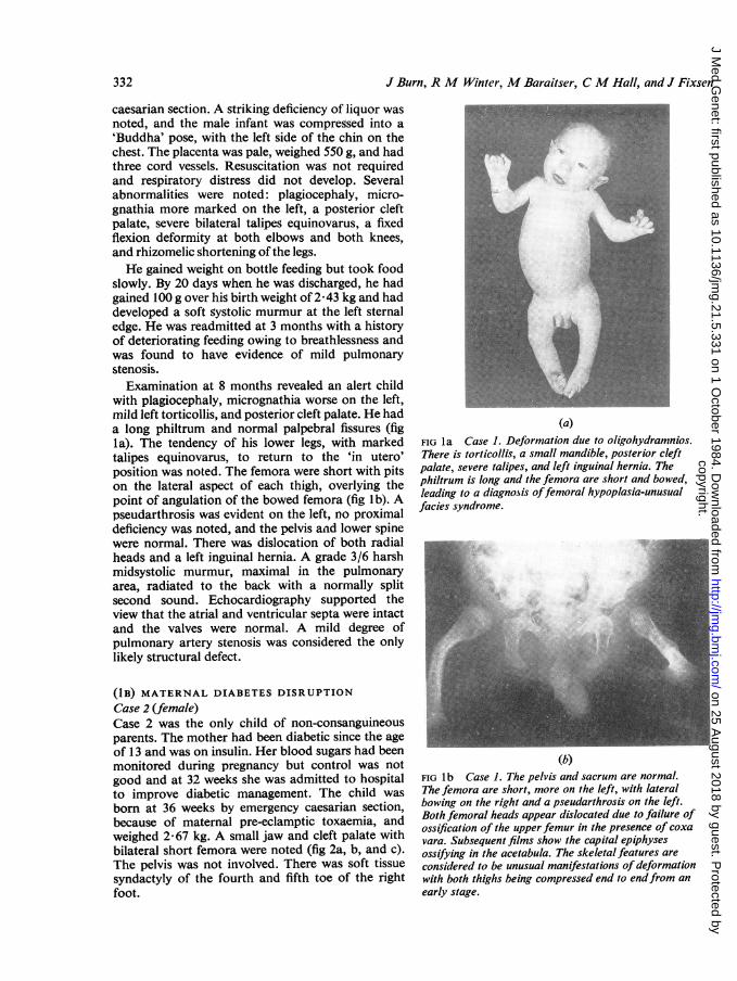
caesarian section. A striking deficiency of liquor wasnoted, and the male infant was compressed into a'Buddha' pose, with the left side of the chin on thechest. The placenta was pale, weighed 550 g, and hadthree cord vessels. Resuscitation was not requiredand respiratory distress did not develop. Severalabnormalities were noted: plagiocephaly, micro-gnathia more marked on the left, a posterior cleftpalate, severe bilateral talipes equinovarus, a fixedflexion deformity at both elbows and both knees,and rhizomelic shortening of the legs.He gained weight on bottle feeding but took food
slowly. By 20 days when he was discharged, he hadgained 100 g over his birth weight of 2 43 kg and haddeveloped a soft systolic murmur at the left sternaledge. He was readmitted at 3 months with a historyof deteriorating feeding owing to breathlessness andwas found to have evidence of mild pulmonarystenosis.
Examination at 8 months revealed an alert childwith plagiocephaly, micrognathia worse on the left,mild left torticollis, and posterior cleft palate. He hada long philtrum and normal palpebral fissures (figla). The tendency of his lower legs, with markedtalipes equinovarus, to return to the 'in utero'position was noted. The femora were short with pitson the lateral aspect of each thigh, overlying thepoint of angulation of the bowed femora (fig Ib). Apseudarthrosis was evident on the left, no proximaldeficiency was noted, and the pelvis and lower spinewere normal. There was dislocation of both radialheads and a left inguinal hernia. A grade 3/6 harshmidsystolic murmur, maximal in the pulmonaryarea, radiated to the back with a normally splitsecond sound. Echocardiography supported theview that the atrial and ventricular septa were intactand the valves were normal. A mild degree ofpulmonary artery stenosis was considered the onlylikely structural defect.
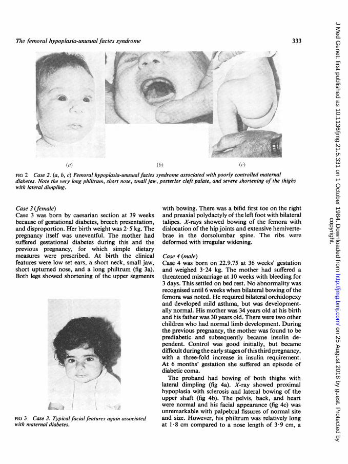
(IB) MATERNAL DIABETES DISRUPTIONCase 2 (female)Case 2 was the only child of non-consanguineousparents. The mother had been diabetic since the ageof 13 and was on insulin. Her blood sugars had beenmonitored during pregnancy but control was notgood and at 32 weeks she was admitted to hospitalto improve diabetic management. The child wasborn at 36 weeks by emergency caesarian section,because of maternal pre-eclamptic toxaemia, andweighed 2@67 kg. A small jaw and cleft palate withbilateral short femora were noted (fig 2a, b, and c).The pelvis was not involved. There was soft tissuesyndactyly of the fourth and fifth toe of the rightfoot.
(a)FIG la Case 1. Deformation due to oligohydramnios.There is torticollis, a small mandible, posterior cleftpalate, severe talipes, and left inguinal hernia. Thephiltrum is long and the femora are short and bowed,leading to a diagnosis offemoral hypoplasia-unusualfacies syndrome.
(b)FIG lb Case 1. The pelvis and sacrum are normal.The femora are short, more on the left, with lateralbowing on the right and a pseudarthrosis on the left.Both femoral heads appear dislocated due to failure ofossification of the upper femur in the presence of coxavara. Subsequent films show the capital epiphysesossifying in the acetabula. The skeletal features areconsidered to be unusual manifestations of deformationwith both thighs being compressed end to endfrom anearly stage.
332
copyright. on 25 A
ugust 2018 by guest. Protected by
http://jmg.bm
j.com/
J Med G
enet: first published as 10.1136/jmg.21.5.331 on 1 O
ctober 1984. Dow
nloaded from
The femoral hypoplasia-unusualfacies syndrome
h~~~~~~~~
*~~~~ 
FIG 2 Case 2. (a, b, c) Femoral hypoplasia-unusualfacies syndrome associated with poorly controlled maternaldiabetes. Note the very long philtrum, short nose, small.jaw, posterior cleft palate, and severe shortening of the thighswith lateral dimpling.
Case 3 (female)Case 3 was born by caesarian section at 39 weeksbecause of gestational diabetes, breech presentation,and disproportion. Her birth weight was 2 5 kg. Thepregnancy itself was uneventful. The mother hadsuffered gestational diabetes during this and theprevious pregnancy, for which simple dietarymeasures were prescribed. At birth the clinicalfeatures were low set ears, a short neck, small jaw,short upturned nose, and a long philtrum (fig 3a).Both legs showed shortening of the upper segments
./
FIG 3 Case 3. Typical facial features again associatedwith maternal diabetes.
with bowing. There was a bifid first toe on the rightand preaxial polydactyly of the left foot with bilateraltalipes. X-rays showed bowing of the femora withdislocation of the hip joints and extensive hemiverte-brae in the dorsolumbar spine. The ribs weredeformed with irregular widening.
Case 4 (male)Case 4 was born on 22.9.75 at 36 weeks' gestationand weighed 3X24 kg. The mother had suffered athreatened miscarriage at 10 weeks with bleeding for3 days. This settled on bed rest. No abnormality wasrecognised until 6 weeks when bilateral bowing of thefemora was noted. He required bilateral orchidopexyand developed mild asthma, but was development-ally normal. His mother was 34 years old at his birthand his father was 30 years old. There were two otherchildren who had normal limb development. Duringthe previous pregnancy, the mother was found to beprediabetic and subsequently became insulin de-pendent. Control was good initially, but becamedifficult during the early stages ofthis third pregnancy,with a three-fold increase in insulin requirement.At 6 months' gestation she suffered an episode ofdiabetic coma.The proband had bowing of both thighs with
lateral dimpling (fig 4a). X-ray showed proximalhypoplasia with sclerosis and lateral bowing of theupper shaft (fig 4b). The pelvis, back, and heartwere normal and his facial appearance (fig 4c) wasunremarkable with palpebral fissures of normal siteand size. However, his philtrum was relatively longat 1I8 cm compared to a nose length of 3 9 cm, a
333
1. g ,
copyright. on 25 A
ugust 2018 by guest. Protected by
http://jmg.bm
j.com/
J Med G
enet: first published as 10.1136/jmg.21.5.331 on 1 O
ctober 1984. Dow
nloaded from

J Burn, R M Winter, M Baraitser, C M Hall, andJ Fixsen
FIG 4 Case 4. (a, b, c) Third case of maternal diabetes. The long philtrum, short bowed femora, and lateraldimpling of the thighs are less striking but still easily recognised.
ratio of 0-46 (upper limit of normal range, based on22 age matched controls, being 0 36).
(Ic) UNCERTAIN AETIOLOGYCase 5 (female)Case S was born at 39 weeks' gestation and weighed2-2 kg. There was no relevant family history and alater born sib was normal. The mother was a 26 year
old teacher. The pregnancy, the mother's firsL, hadbeen uneventful. Fetal movement was reduced andat delivery there was little liquor, though no leakagehad been noted.
Fig 5a shows the facial features evident at birth:micrognathia and long philtrum. There was a wideposterior cleft palate. The legs were short withdimpling of both lateral thighs over angulated
(a) (c))
FIG 5 Case 5. (a, b, c) Femoral hypoplasia-unusualfacies syndrome of uncertain aetiology. In the neonatal periodthe micrognathia is striking, together with short thighs due to severe sclerosis and shortening and lateral bowingof both femora. The photograph at a later age shows the 'typical' face with short pinched nose with a broad tip, longphiltrum, and thin upper lip. There is a mild degree of upward slanting of the left palpebral fissure.
..z
334
copyright. on 25 A
ugust 2018 by guest. Protected by
http://jmg.bm
j.com/
J Med G
enet: first published as 10.1136/jmg.21.5.331 on 1 O
ctober 1984. Dow
nloaded from

The femoral hypoplasia-unusual facies syndrome
femora. X-ray examination (fig Sb) revealed hypo-plasia of the mid shaft of both femora. The pelvis,tibiae, and fibulae were not involved. Fig Sc shows herphysical appearance at 3 years: pinched nose, thinupper lip with rather long philtrum, and persistentsevere proximal limb shortening. Intellectualdevelopment had progressed normally and she wasmobile.
Case 6 (male)Case 6 was born at 33 weeks and weighed 1-5 kg.The mother was 32 years old and had a history of fitsbut was not on drug therapy. The father was 34 yearsold. His father and other family members were shortbut had normal body proportions. The motherreported heavy loss of fresh blood over 13 days at 8to 10 weeks' gestation. Extended breech presentationmade forceps extraction necessary. Bilateral femoralhypoplasia with hip dislocation, micrognathia, and aposterior cleft palate were noted, together with aright inguinal hernia. Fig 6a shows the facialappearance at 15 months of age. The micrognathiaremained evident, with a long philtrum. Fig 6b and cshow the clinical and radiological appearance of the
r.
A .,k
Ii
0010F
lower limbs. There was symmetrical hypoplasia of thefemora with lateral bowing. The tibiae, fibulae, andsacrum were normal.
(2A) FETAL CONSTRAINT SEQUENCECase 7 (male)Case 7 was born in 1971. His father was 27 and hismother 24 years of age. His mother had had amiscarriage at 6 weeks the previous year. Theysubsequently had two normal children. The earlystages of this pregnancy were uneventful and move-ment was felt at 18 weeks. Extended breech presenta-tion persisted until delivery at 41 weeks by caesariansection after a 24 hour labour. After birth the thighswere noted to be short and bowed laterally, withlimited abduction at the hips but normal joints.X-ray examination revealed lateral bowing withsclerosis of the shaft. Fig 7a, b, and c show theprogressive resolution of the femoral abnormality.At examination at the age of 11 years, slight valgusdeformity at the right knee and inturning of the feetduring walking were the only abnormalities. Intellec-tual development and facial morphology werenormal (fig 7d).
FIG 6 Case 6. (a, b, c) Again, short nose,long smooth philtrum, small jaw, and short
* ,~ laterally bowedfemora without a recognisedcause. The radiograph shows the typical pointof angulation between the proximal andmiddle thirds.
-
-
335
I
I}-ji.,W
.1I
#.--
-t
copyright. on 25 A
ugust 2018 by guest. Protected by
http://jmg.bm
j.com/
J Med G
enet: first published as 10.1136/jmg.21.5.331 on 1 O
ctober 1984. Dow
nloaded from

J Burn, R M Winter, M Baraitser, CM Hall, and J Fixsen
(a)
:s
|., |l
lIc)
(I)
FIG 7 Case 7. (a, b, c, d) Normalface in achild with a history offetal compression. Thesclerosis and mild lateral bowing of bothfemora is, again, regarded as a minor degree ofdeformation. The resolution of the bowingand normal later growth (b, c) are in keepingwith this explanation.
(2B) MATERNAL DIABETES DISRUPTION
Case 8 (female)Case 8 was born in March 1951, the third child of a 33year old mother and a 28 year old father. Themother had four healthy children by her previousmarriage. The father had a congenitally small leftarm. Otherwise, there was no family history ofmalformation. The mother fainted frequently in thefirst trimester of this pregnancy. A diagnosis ofepilepsy was considered, but in the postnatal periodpolyuria, polydipsia, and glycosuria were recog-nised and a diagnosis of diabetes mellitus was made.Control with oral hypoglycaemic agents and dietwas considered adequate. In the following decadecoronary artery disease developed and the motherdied in 1970 at the age of 52 years. The mother'sfather and sister also suffered maturity onset diabetes.Case 8 was found at birth to have short malformed
lower limbs. When seen at this hospital at the age of1 year she was noted to have a marked proximaldeficiency of both femora. The distal fifth of the left
femur and the distal three-fifths of the right werevisible on x-ray. A capital epiphysis on the right waslocated in the acetabulum. One metatarsal and toeon the right and two metatarsals and toes on the leftwere absent. Following prolonged traction and aleft knee arthrodesis she became mobile on artificiallimbs. Apart from her limb malformations she washealthy. She was found during her first pregnancy tohave a small distorted pelvis necessitating electivecaesarian section. This child and her two subsequentoffspring are normally developed. The youngest,aged 10 months, required surgery in infancy forpyloric stenosis.On review at the age of 32 years case 8 was found
to have normal facies apart from a rather shortphiltrum.
(2c) UNCERTAIN AETIOLOGYCase 9 (male)Case 9 was born in 1975. His mother, who laterdeveloped severe rheumatoid arthritis, had had one
j
336
copyright. on 25 A
ugust 2018 by guest. Protected by
http://jmg.bm
j.com/
J Med G
enet: first published as 10.1136/jmg.21.5.331 on 1 O
ctober 1984. Dow
nloaded from
The femoral hypoplasia-unusual facies syndrome
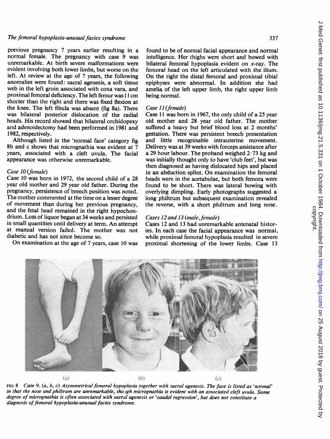
previous pregnancy 7 years earlier resulting in anormal female. The pregnancy with case 9 wasunremarkable. At birth severe malformations wereevident involving both lower limbs, but worse on theleft. At review at the age of 7 years, the followinganomalies were found: sacral agenesis, a soft tissueweb in the left groin associated with coxa vara, andproximal femoral deficiency. The left femur was 11 cmshorter than the right and there was fixed flexion atthe knee. The left fibula was absent (fig 8a). Therewas bilateral posterior dislocation of the radialheads. His record showed that bilateral orchidopexyand adenoidectomy had been performed in 1981 and1982, respectively.Although listed in the 'normal face' category fig
8b and c shows that micrognathia was evident at 7years, associated with a cleft uvula. The facialappearance was otherwise unremarkable.
Case 10 (female)Case 10 was born in 1972, the second child of a 28year old mother and 29 year old father. During thepregnancy, persistence of breech position was noted.The mother commented at the time on a lesser degreeof movement than during her previous pregnancy,and the fetal head remained in the right hypochon-drium. Loss of liquor began at 34 weeks and persistedin small quantities until delivery at term. An attemptat manual version failed. The mother was notdiabetic and has not since become so.On examination at the age of 7 years, case 10 was
337
found to be of normal facial appearance and normalintelligence. Her thighs were short and bowed withbilateral femoral hypoplasia evident on x-ray. Thefemoral head on the left articulated with the ilium.On the right the distal femoral and proximal tibialepiphyses were abnormal. In addition she hadamelia of the left upper limb, the right upper limbbeing normal.
Case 11 (female)Case 11 was born in 1967, the only child of a 25 yearold mother and 28 year old father. The mothersuffered a heavy but brief blood loss at 2 months'gestation. There was persistent breech presentationand little recognisable intrauterine movement.Delivery was at 39 weeks with forceps assistance aftera 29 hour labour. The proband weighed 2- 73 kg andwas initially thought only to have 'club feet', but wasthen diagnosed as having dislocated hips and placedin an abduction splint. On examination the femoralheads were in the acetabulae, but both femora werefound to be short. There was lateral bowing withoverlying dimpling. Early photographs suggested along philtrum but subsequent examination revealedthe reverse, with a short philtrum and long nose.
Cases 12 and 13 (male,female)Cases 12 and 13 had unremarkable antenatal histor-ies. In each case the facial appearance was normal,while proximal femoral hypoplasia resulted in severeproximal shortening of the lower limbs. Case 13
= _ *> waa X~~~~~o
-' Eli3t t Aff*l
(ci) (h) (C)FIG 8 Case 9. (a, b, c) Asymmetricalfemoral hypoplasia together with sacral agenesis. The face is listed as 'normal'in that the nose and philtrum are unremarkable, tho:igh micrognathia is evident with an associated cleft uvula. Somedegree of micrognathia is often associated with sacral agenesis or 'caudal regression', but does not constitute adiagnosis offemoral hypoplasia-unusualfacies syndrome.
copyright. on 25 A
ugust 2018 by guest. Protected by
http://jmg.bm
j.com/
J Med G
enet: first published as 10.1136/jmg.21.5.331 on 1 O
ctober 1984. Dow
nloaded from

J Burn, R M Winter, M Baraitser, CM Hall, anod J Fixsen
had the additional features of absence of the rightfibula and a left pseudarthrosis.
Discussion
Our findings suggest that the femoral hypoplasia-unusual facies syndrome is a recognisable entity. It isof interest that Lord and Beighton,4 who questionedthe existence of this syndrome, did not refer to thepatient reported by Bailey and Beighton in 1970,6who was described as having cleft palate andbilateral femoral dysgenesis. This young lady, fromthe published photograph, had very similar physicalfeatures to one of the cases reported by Daentl etal.1 This difference of terminology probably accountsfor the absence of this case, and the two reported byBurck et al,7 from the review of Pitt et al.2 Alsoomitted was the report by Graviss et al,8 under thetitle 'Proximal femoral focal deficiency associatedwith the Robin anomalad'. The term 'proximalfemoral focal deficiency' is well established inorthopaedics and it is reasonable to retain this incases of isolated femoral defect. Similarly, the termfemoral hypoplasia has now gained acceptance, eventhough 'dysgenesis' or 'dysplasia' might be con-sidered preferable terms.Femoral hypoplasia-unusual facies syndrome
must remain a descriptive term as it would appearthat cases with very similar clinical and radiologicalfeatures may have differing causes. We report onechild where severe intrauterine compression owingto oligohydramnios appeared to be the sole cause ofthe syndrome, though this is probably a rare explana-tion. Our finding of four cases associated withmaternal diabetes, on the other hand, suggests thatthis is a frequent cause of the syndrome. Thisfinding adds weight to the view that femoralhypoplasia-unusual facies syndrome is part of thespectrum of diabetic embryopathy or, to use themore recent terminology, maternal diabetes disrup-tion. This would make the femoral hypoplasia-unusual facies syndrome and sacral agenesis differingmanifestations of the caudal regression syndrome.9The features of case 9 confirmed this relationshipsince this child had sacral agenesis and vertebralanomalies together with severe femoral and lowerlimb involvement, a small mandible, and a cleftuvula.
Graviss et al8 suggested that the 'unusual face' wasmerely the typical face of Robin anomalad, nowRobin sequence. The basis of this sequence is asmall mandible. This causes the tongue to beposteriorly placed and often to interfere with closureof the palatal shelves. The mandible may fail togrow due to a genetic defect in the first branchialarch, or due to disruption by, for example, maternal
diabetes. Growth of the mandible may be preventedby extreme flexion of the neck, such as can occurwith oligohydramnios, so that some cases of Robinsequence are due to deformation. A short nose andlong philtrum may be associated with Robinsequence but are distinct from it. They implydisturbance of growth of the frontonasal andmaxillary processes, particularly the medial nasalprocess of the former. Case 5 illustrates how a smallmandible may be impressive in the neonate but, withthe passage of time, abnormal development of themidface to produce the long philtrum and pinchednose accounts for the 'unusual face'.The murmur of pulmonary stenosis and the
episode of right heart failure in case 1 may haveresulted from a degree of lung hypoplasia owing tothe prolonged loss of liquor. None of the fivechildren with the classical femoral hypoplasia-unusual facies syndrome had heart murmurs. Takingfrom published reports only those with a typical face,normal spine and sacrum, and bilateral femoralinvolvement there is also no case of heart malforma-tion, whereas cardiac defects are common in associa-tion with sacral agenesis and those 'mixed' cases withspinal and femoral involvement. In the diabetic casesthis may reflect a different timing of the insult.The striking analogy between these clinical obser-
vations and experiments on chickens ig worthy ofnote. Landauer10'11 in the 1940s showed that injectionof insulin into the yolk sac could produce a syndromeof short upper beak with proximal limb defects,closely analogous to the femoral hypoplasia-unusualfacies syndrome (fig 9). If the injection was per-formed earlier, rumplessness was produced. This wascharacterised by absence of the sacrum, similar tothat seen in the more classical caudal regressionsyndrome. Landauer postulated that the disturbanceof glucose metabolism affected metabolic pathwayscommon to proximal limb bud and facial develop-ment. The defect could, in addition, be more easilyprovoked in some strains than others. Chickenscould be bred which were particularly sensitive to theinsulin injection and in these supplementation of thediet with nicotinamide greatly reduced the frequencyof malformation. It is plausible to suggest that, as inthe chicken, disturbance of glucose pathways in earlydevelopment may produce the features of thissyndrome, but that some genetic predisposition isalso essential.
In a review of the subject, Landauer12 repeatedhis belief that insulin had direct teratogenic effectsin the chicken. In humans, however, the relationshipbetween poor control and frequency of malforma-tions suggests that instability of glucose homeostasisis more detrimental than the presence of exogenousinsulin. Case 8 is important in this regard. The
338
copyright. on 25 A
ugust 2018 by guest. Protected by
http://jmg.bm
j.com/
J Med G
enet: first published as 10.1136/jmg.21.5.331 on 1 O
ctober 1984. Dow
nloaded from

The femoral hypoplasia-unusual facies syndrome
(b)
'I..- -. (ease a- .. 5 S
,--e, Se tS IS-relies ices ci
a .::.:.-:- sees e eec I I
1 2
30h
Rumplessness
3 4 5Age of embryo (days)
Upper12B0ehShort Upper Beak
(Lcindauer 1947,1948)FIG 9 Experiments by Landauer10 11 which showed that the timing of an injection of insulin into the yolk sac determinedwhether rumplessness or short upper beak were produced. These patterns of malformation are very similar to thesacral agenesis andfemoral hypoplasia-unusualfacies syndromes seen in man.
mother had symptomatic diabetes mellitus duringpregnancy, but was not diagnosed or treated untilafter delivery of the proband. The rarity of limbmalformations in the offspring of diabetic motherssuggests that, as in the chicken, there is geneticvariation in susceptibility. The protective effect ofnicotinamide in chickens prone to malformationraises the intriguing question of whether similarsupplements might be beneficial for diabetic women.To establish a protective effect would require a studyof such proportions, however, that effort would bebetter devoted to improving the provision of goodantenatal care for women at risk.Though probably not completely analogous, the
experiments in the developing chicken lend supportto the belief that in pregnancies at risk from maternaldiabetes, the involvement of the proximal femora
and maxilla, rather than the sacrum and vertebralcolumn, is determined by the later timing of anabnormal fluctuation in glucose homeostasis. Thelow prevalence of these skeletal anomalies among theoffspring of diabetic mothers implies, in addition,genetic variation in susceptibility, as was seen in thechicken. The pattern of malformation suggests thatthe proximal lower limb buds and the first and secondbranchial arches share in common developmentalpathways liable to disturbance. In cases attributed todeformation or disruption, the risk of recurrencewill be substantial in cases of maternal uterineanomaly, such as bicornuate uterus, and withpersistently bad diabetic control in a mother.There were no familial instances in the 13 cases
reported, but on the basis of Lampert's report3 it islikely that, in some cases at least, the condition is
(a) '---
6 7
339
copyright. on 25 A
ugust 2018 by guest. Protected by
http://jmg.bm
j.com/
J Med G
enet: first published as 10.1136/jmg.21.5.331 on 1 O
ctober 1984. Dow
nloaded from

J Burn, R M Winter, M Baraitser, C M Hall, and J Fixsen
autosomal dominant. The possibility of fresh muta-tion accounting for some or all of the six patients ofunknown aetiology remains a possibility, but ingeneral the recurrence risk in the offspring of thisgroup is likely to be small. It is possible that the riskwill be greater to the offspring of those, such as cases2 and 3, who have the full syndrome with facialinvolvement.
In summary, it is postulated that bilateral femoralhypoplasia, with or without an unusual face, is aheterogeneous condition with three aetiologicalcategories identified to date. The descriptive term'femoral hypoplasia-unusual facies syndrome' isvalid and is of clinical value.
Reproduction of fig 9, with permission, from thereview by Hadorn E. Developmental genetics andlethal factors. London: Methuen, 1961: 241 (trans-lated by U Mittwoch).
We are grateful to Mrs Melanie Barham and MsCarol Reeves for preparation of the manuscript andillustrations respectively, and to the many paediatriccolleagues who have taken part in the care andevaluation of the families presented. JB is supportedby MRC grant number G8203878.
References
Daentl DL, Smith DW, Scott Cl, Hall BD, Gooding CA.Femoral hypoplasia-unusual facies syndrome. J Pediatr1975;86:107-l 1.
2 Pitt DB, Findlay H, Cole WG, Rogers JG. Femoralhypoplasia-unusual facies syndrome. Aust Paediatr J1982 ;18:63-6.
3 Lampert RP. Dominant inheritance of femoral hypo-plasia-unusual facies syndrome. Clin Genet 1980;17:255-8.
4 Lord J, Beighton P. The femoral hypoplasia-unusualfacies syndrome: a genetic entity? Clin Genet 1981;20:267-75.
5 Johnson JP, Carey JC, Manford Gooch W, Peterson J,Beattie JF. Femoral hypoplasia-unusual facies syn-drome in infants of diabetic mothers. JPediatr 1983;102:866-72.
6 Bailey JA, Beighton P. Bilateral femoral dysgenesis.Clin Pediatr 1970;9:668-74.Burck U, Riebel T, Held KR, Stoeckenius M. Bilateralfemoral dysgenesis with micrognathia, cleft palate,anomalies of the spine and pelvis, and foot deformities.Helv Paediatr Acta 1981 ;36:473-82.
8 Graviss ER, Monteleone PA, Wampler LR, SilbersteinMJ, Brodeur AE. Proximal femoral focal deficiencyassociated with the Robin anomalad. J Med Genet 1980;17:390-2.
9 Gleiser S, Weaver DD, Escobar V, Nichols G, EscobedoM. Femoral hypoplasia-unusual facies from anotherviewpoint. Eur J Pediatr 1978;128:1-5.
10 Landauer W. Insulin-induced abnormalities of beak,extremities and eyes in chickens. J Exp Zool 1947;105:147-72.Landauer W. The effect of nicotinamide and a keto-glutaric acid on the teratogenic action of insulin. J ExpZool 1948 ;109:283-90.
12 Landauer W. Is insulin a teratogen? Teratology 1972;5:129-36.
Correspondence and requests for reprints to DrJ Burn, Clinical Genetics Unit, Institute of ChildHealth, 30 Guilford Street, London WC1N IEH.
340
copyright. on 25 A
ugust 2018 by guest. Protected by
http://jmg.bm
j.com/
J Med G
enet: first published as 10.1136/jmg.21.5.331 on 1 O
ctober 1984. Dow
nloaded from