femoral condyle osteochondral fracture treated with bone...
TRANSCRIPT
r e v b r a s o r t o p . 2 0 1 8;5 3(5):636–642
SOCIEDADE BRASILEIRA DEORTOPEDIA E TRAUMATOLOGIA
www.rbo.org .br
Case Report
Femoral condyle osteochondral fracture treatedwith bone suture after acute patellar dislocation: acase report�
Camila Maftoum Cavalheiroa,∗, Riccardo Gomes Gobbia, Betina Bremer Hinckela,Marco Kawamura Demangea, José Ricardo Pécoraa, Gilberto Luis Camanhob
a Universidade de São Paulo, Faculdade de Medicina, Hospital das Clínicas, São Paulo, SP, Brazilb Universidade de São Paulo, Faculdade de Medicina, Departamento de Ortopedia e Traumatologia, São Paulo, SP, Brazil
a r t i c l e i n f o
Article history:
Received 25 February 2017
Accepted 17 April 2017
Available online 20 August 2018
Keywords:
Bone fractures
Patellar dislocation
Articular ligaments
Osteochondritis
a b s t r a c t
Osteochondral fracture after acute patellar dislocation in teenagers is relatively common (up
to 60% of cases of patellar dislocation), but poorly diagnosed. There are several treatments
proposed for this type of injury, but none well defined in the literature.
A male patient, 13 years old, with a diagnosis of osteochondral fracture of the lateral
femoral condyle after acute dislocation of the right patella. He underwent surgical treatment
of the chondral injury, which consisted of suturing of the chondral fragment to the cartilage
defect and, in a second approach, reconstruction of the medial patellotibial ligament and
medial patellofemoral ligament with autologous flexor graft. Currently, the patient has been
followed up for 16 months postoperatively for the suture of the chondral fragment and for
8 months for the ligament reconstruction. He has been evaluated through functional scores
and T2 weighted magnetic resonance imaging. Acute fixation through direct bone suturing
of a purely chondral fragment can be considered in special situations.
© 2018 Sociedade Brasileira de Ortopedia e Traumatologia. Published by Elsevier Editora
Ltda. This is an open access article under the CC BY-NC-ND license (http://
creativecommons.org/licenses/by-nc-nd/4.0/).
Lesão condral do fêmur tratada com sutura óssea após luxacão aguda depatela: um relato de caso
r e s u m o
Palavras-chave:
Fraturas ósseas
Luxacão patelar
Ligamentos articulares
Osteocondrite
A fratura osteocondral após luxacão aguda de patela em adolescentes é relativamente
comum (até 60% dos casos de luxacão patelar), porém pouco diagnosticada. Existem diversos
tratamentos propostos para esse tipo de lesão, mas nenhum está bem definido na liter-
atura. Paciente do sexo masculino, 13 anos, com diagnóstico de fratura osteocondral do
côndilo femoral lateral, após luxacão aguda da patela direita. Foi submetido a tratamento
� Study conducted at Universidade de São Paulo, Faculdade de Medicina, Hospital das Clínicas, Instituto de Ortopedia e Traumatologia,Grupo de Joelho, São Paulo, SP, Brazil.
∗ Corresponding author.E-mail: [email protected] (C.M. Cavalheiro).
https://doi.org/10.1016/j.rboe.2017.04.0082255-4971/© 2018 Sociedade Brasileira de Ortopedia e Traumatologia. Published by Elsevier Editora Ltda. This is an open access articleunder the CC BY-NC-ND license (http://creativecommons.org/licenses/by-nc-nd/4.0/).
r e v b r a s o r t o p . 2 0 1 8;5 3(5):636–642 637
cirúrgico da lesão condral, que consistiu em sutura do fragmento condral ao defeito da car-
tilagem e, em um segundo tempo, a reconstrucão do ligamento patelotibial medial (LPTM) e
reconstrucão do ligamento patelofemoral medial (LPFM) com enxerto autólogo de flexores.
Atualmente o paciente encontra-se com o seguimento de 16 meses de pós-operatório da
sutura do fragmento condral e oito meses da reconstrucão ligamentar, foi avaliado através
de escores funcionais e ressonância magnética com mapeamento de T2. Em casos espe-
ciais, pode-se considerar o uso de fixacão aguda por sutura óssea direta de um fragmento
puramente condral.
© 2018 Sociedade Brasileira de Ortopedia e Traumatologia. Publicado por Elsevier
Editora Ltda. Este e um artigo Open Access sob uma licenca CC BY-NC-ND (http://
I
Aiwpaltibo
oatcuahct
altToa
C
Atatwtkdtr
(
ntroduction
lateral patellofemoral dislocation is a relatively commonnjury in children and young adults; it is frequently associated
ith chondral or osteochondral injuries of the femur and/oratella.1,2 These injuries may occur in up to 60% of cases3 andre usually located in non-weight-bearing areas, such as theateral region of the trochlea or the lateral femoral condyle, orhe medial facet of the patella; the mechanism of trauma ismpaction.4,5 Less commonly, they may occur in the weight-earing area of the lateral femoral condyle, when a dislocationccurs with the knee in flexion.1,6
Several treatments have been proposed for injuries withsteochondral fragments, such as fixation with metallic orbsorbable materials, autologous osteochondral transplan-ation, and simple debridement.1,7,8 However, in some rareases, the injuries are solely cartilaginous or have minimalnderlying subchondral bone, preventing the use of bone fix-tion. In addition to the technical difficulty of the fixation, theealing potential is lower in cartilage than in bone. Few suc-essful cases of fragment reintegration have been reported forhis type of injury.2,6,9
The authors present a case of acute patellar dislocationssociated with a predominantly chondral fracture in theateral femoral condyle weight-bearing area, secured with aransosseous suture and presenting good functional results.o the best of the authors’ knowledge, this is the first reportf reinsertion of an extensive chondral fragment with this fix-tion technique.
ase report
13-year-old male patient, with no previous clinical his-ory and no comorbidities, suffered a sprain to his left kneefter a fall during a football match. He reported knee disloca-ion and severe pain. He was taken to the emergency room,here the initial assessment indicated the presence of impor-
ant pain and edema of the left knee, and a fixed position ofnee flexion. The anteroposterior and lateral view radiographsemonstrated an articular bone fragment, and that suggested
he diagnosis of acute patellofemoral dislocation. He was theneferred to the knee service of the Hospital das Clínicas.During the physical examination at the referral servicethree days after the trauma), it was observed that there
creativecommons.org/licenses/by-nc-nd/4.0/).
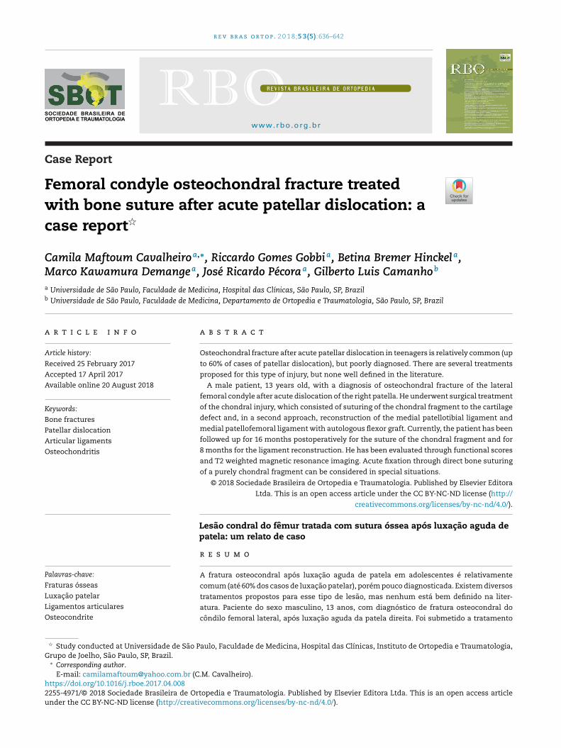
persisted pain on knee palpation, especially in the lateralregion; effusion and movement restriction were also observed(range of motion: 20–110◦), and the patient was unable toreach full extension. The patellar apprehension test waspositive. No other ligament instabilities were observed; themuscle tone was normal and the extensor mechanism wasintact. The radiographs showed an immature skeleton withunclosed physis and a bone fragment in the joint. A computedtomography of the knee was made for complementary evalu-ation, and associated injuries were excluded. The presence ofan osteochondral fragment from the weight-bearing area ofthe lateral femoral condyle (Fig. 1A) was confirmed, as well asan increased patellar tilt (29◦) and Dejour’s grade B trochleardysplasia (Fig. 1B).
The limb was immobilized with an inguino-malleolarsplint with maximal extension to await surgical intervention,which was indicated due to the presence of a free osteochon-dral fragment and articular blockage.
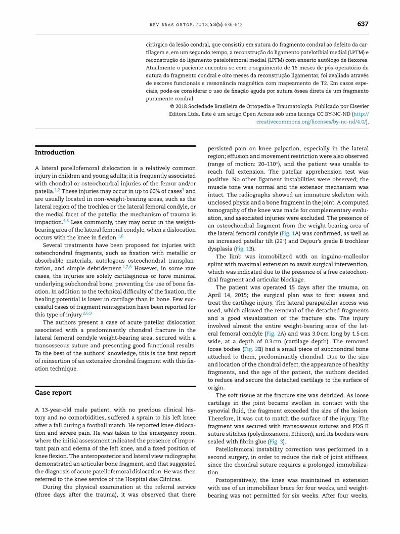
The patient was operated 15 days after the trauma, onApril 14, 2015; the surgical plan was to first assess andtreat the cartilage injury. The lateral parapatellar access wasused, which allowed the removal of the detached fragmentsand a good visualization of the fracture site. The injuryinvolved almost the entire weight-bearing area of the lat-eral femoral condyle (Fig. 2A) and was 3.0 cm long by 1.5 cmwide, at a depth of 0.3 cm (cartilage depth). The removedloose bodies (Fig. 2B) had a small piece of subchondral boneattached to them, predominantly chondral. Due to the sizeand location of the chondral defect, the appearance of healthyfragments, and the age of the patient, the authors decidedto reduce and secure the detached cartilage to the surface oforigin.
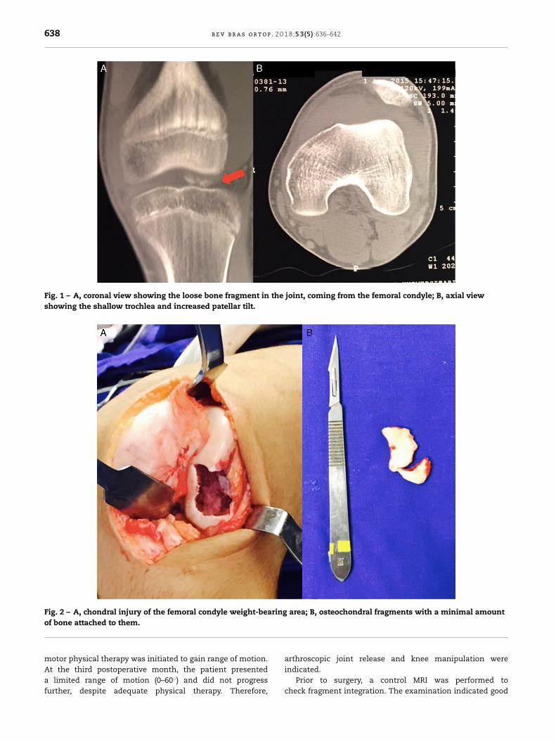
The soft tissue at the fracture site was debrided. As loosecartilage in the joint became swollen in contact with thesynovial fluid, the fragment exceeded the size of the lesion.Therefore, it was cut to match the surface of the injury. Thefragment was secured with transosseous sutures and PDS IIsuture stitches (polydioxanone, Ethicon), and its borders weresealed with fibrin glue (Fig. 3).
Patellofemoral instability correction was performed in asecond surgery, in order to reduce the risk of joint stiffness,since the chondral suture requires a prolonged immobiliza-tion.
Postoperatively, the knee was maintained in extensionwith use of an immobilizer brace for four weeks, and weight-bearing was not permitted for six weeks. After four weeks,
638 r e v b r a s o r t o p . 2 0 1 8;5 3(5):636–642
Fig. 1 – A, coronal view showing the loose bone fragment in the joint, coming from the femoral condyle; B, axial viewshowing the shallow trochlea and increased patellar tilt.
Fig. 2 – A, chondral injury of the femoral condyle weight-bearing area; B, osteochondral fragments with a minimal amount
of bone attached to them.motor physical therapy was initiated to gain range of motion.
At the third postoperative month, the patient presenteda limited range of motion (0–60◦) and did not progressfurther, despite adequate physical therapy. Therefore,arthroscopic joint release and knee manipulation were
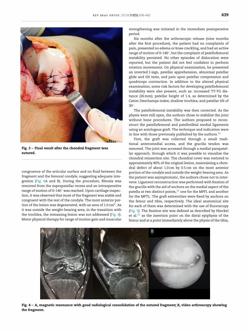
indicated.Prior to surgery, a control MRI was performed tocheck fragment integration. The examination indicated good
r e v b r a s o r t o p . 2 0 1 8
Fig. 3 – Final result after the chondral fragment wassutured.
cfgrrtctitM
Ft
ongruence of the articular surface and no fluid between theragment and the femoral condyle, suggesting adequate inte-ration (Fig. 4A and B). During the procedure, fibrosis wasemoved from the suprapatellar recess and an intraoperativeange of motion of 0–140◦ was reached. Upon cartilage inspec-ion, it was observed that most of the fragment was stable andongruent with the rest of the condyle. The most anterior por-ion of the lesion was degenerated, with an area of 1.0 cm2. As
t was outside the weight-bearing area, in the transition withhe trochlea, the remaining lesion was not addressed (Fig. 4).otor physical therapy for range of motion gain and muscularig. 4 – A, magnetic resonance with good radiological consolidathe fragment.
;5 3(5):636–642 639
strengthening was initiated in the immediate postoperativeperiod.
Six months after the arthroscopic release (nine monthsafter the first procedure), the patient had no complaints ofpain, presented no edema or knee crackling, and had an activerange of motion of 0–140◦, but the complaint of patellofemoralinstability persisted. No other episodes of dislocation werereported, but the patient did not feel confident to performrotation movements. On physical examination, he presentedan inverted J-sign, patellar apprehension, abnormal patellarglide and tilt tests, and pain upon patellar compression andquadriceps contraction. In addition to the altered physicalexamination, some risk factors for developing patellofemoralinstability were also present, such as: increased TT-TG dis-tance (26 mm); patellar height of 1.4, as determined by theCaton-Deschamps index; shallow trochlea; and patellar tilt of30◦.
The patellofemoral instability was then corrected. As thephysis were still open, the authors chose to stabilize the jointwithout bone procedures. The authors proposed to recon-struct the patellofemoral and patellotibial medial ligamentsusing an autologous graft. The technique and indication werein line with those previously published by the authors.10
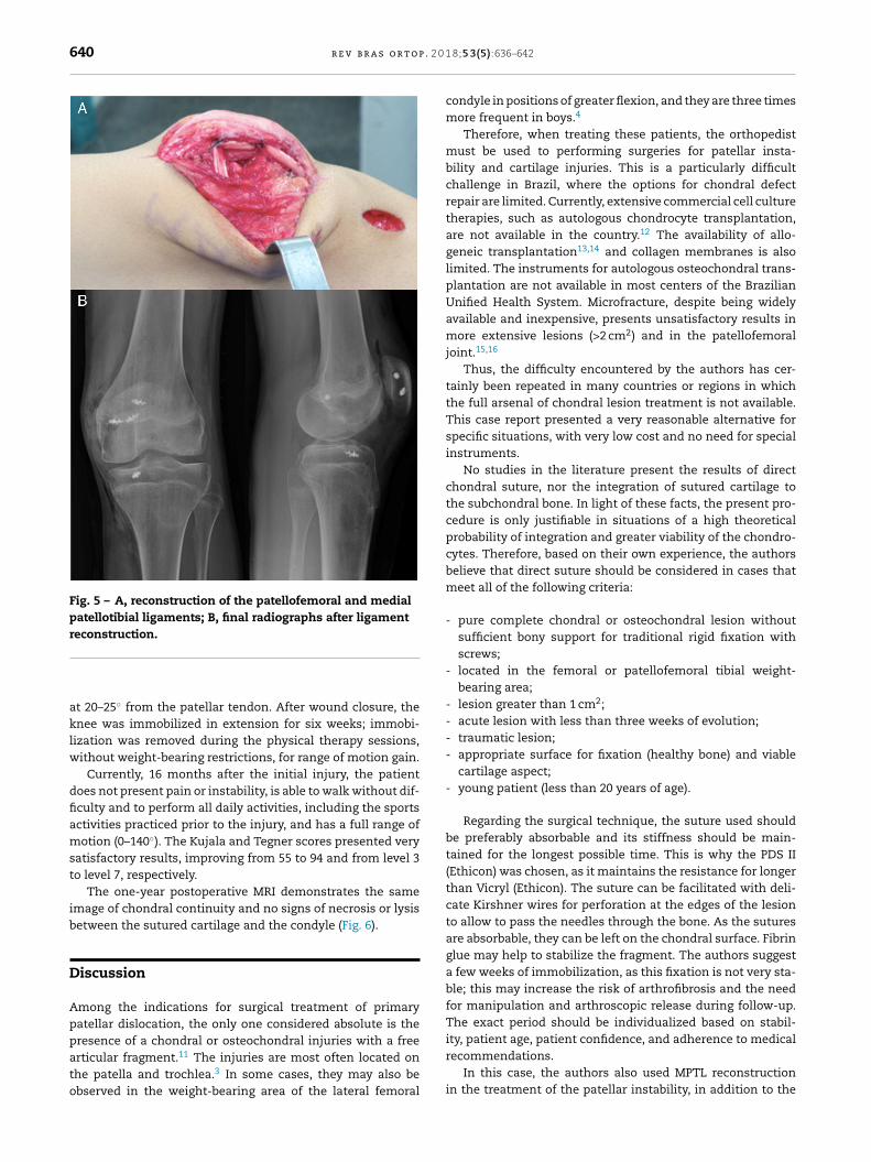
First, the graft was collected through a small tradi-tional anteromedial access, and the gracilis tendon wasremoved. The joint was accessed through a medial parapatel-lar approach, through which it was possible to visualize thechondral reinsertion site. The chondral cover was restored toapproximately 80% of the original lesion, maintaining a chon-dral defect of about 1.0 cm by 0.5 cm on the most anteriorportion of the condyle and outside the weight-bearing area. Asthe patient was asymptomatic, the authors chose not to inter-vene. Ligament reconstruction was performed with fixation ofthe gracilis with the aid of anchors on the medial aspect of thepatella at two distinct points,10 one for the MPFL and anotherfor the MPTL. The graft extremities were fixed by anchors onthe femur and tibia, respectively. The ideal anatomical sitefor each of them was determined with the use of fluoroscopy
(Fig. 5). The fixation site was defined as described by Hinckelet al.10 as the insertion point on the distal epiphysis of thefemur and at a point immediately above the physis of the tibia,ion of the sutured fragment; B, video arthroscopy showing
640 r e v b r a s o r t o p . 2 0
Fig. 5 – A, reconstruction of the patellofemoral and medialpatellotibial ligaments; B, final radiographs after ligamentreconstruction.
ity, patient age, patient confidence, and adherence to medicalrecommendations.
at 20–25◦ from the patellar tendon. After wound closure, theknee was immobilized in extension for six weeks; immobi-lization was removed during the physical therapy sessions,without weight-bearing restrictions, for range of motion gain.
Currently, 16 months after the initial injury, the patientdoes not present pain or instability, is able to walk without dif-ficulty and to perform all daily activities, including the sportsactivities practiced prior to the injury, and has a full range ofmotion (0–140◦). The Kujala and Tegner scores presented verysatisfactory results, improving from 55 to 94 and from level 3to level 7, respectively.
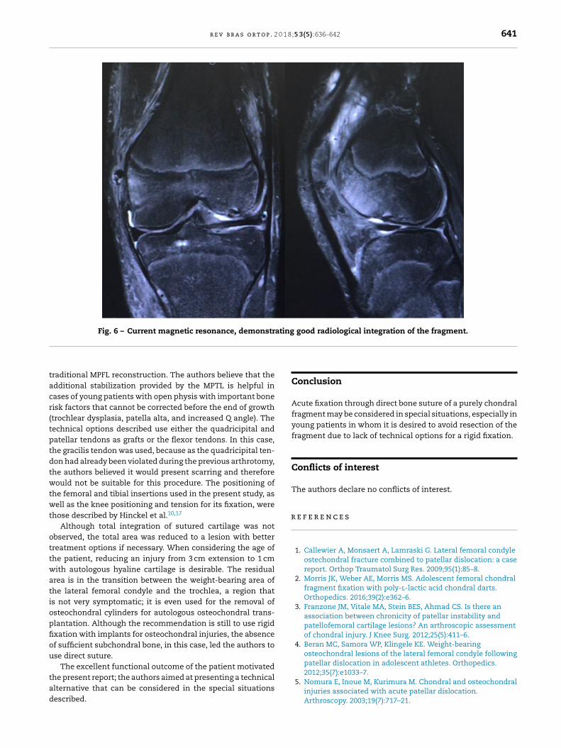
The one-year postoperative MRI demonstrates the sameimage of chondral continuity and no signs of necrosis or lysisbetween the sutured cartilage and the condyle (Fig. 6).
Discussion
Among the indications for surgical treatment of primarypatellar dislocation, the only one considered absolute is thepresence of a chondral or osteochondral injuries with a freearticular fragment.11 The injuries are most often located on
the patella and trochlea.3 In some cases, they may also beobserved in the weight-bearing area of the lateral femoral1 8;5 3(5):636–642
condyle in positions of greater flexion, and they are three timesmore frequent in boys.4
Therefore, when treating these patients, the orthopedistmust be used to performing surgeries for patellar insta-bility and cartilage injuries. This is a particularly difficultchallenge in Brazil, where the options for chondral defectrepair are limited. Currently, extensive commercial cell culturetherapies, such as autologous chondrocyte transplantation,are not available in the country.12 The availability of allo-geneic transplantation13,14 and collagen membranes is alsolimited. The instruments for autologous osteochondral trans-plantation are not available in most centers of the BrazilianUnified Health System. Microfracture, despite being widelyavailable and inexpensive, presents unsatisfactory results inmore extensive lesions (>2 cm2) and in the patellofemoraljoint.15,16
Thus, the difficulty encountered by the authors has cer-tainly been repeated in many countries or regions in whichthe full arsenal of chondral lesion treatment is not available.This case report presented a very reasonable alternative forspecific situations, with very low cost and no need for specialinstruments.
No studies in the literature present the results of directchondral suture, nor the integration of sutured cartilage tothe subchondral bone. In light of these facts, the present pro-cedure is only justifiable in situations of a high theoreticalprobability of integration and greater viability of the chondro-cytes. Therefore, based on their own experience, the authorsbelieve that direct suture should be considered in cases thatmeet all of the following criteria:
- pure complete chondral or osteochondral lesion withoutsufficient bony support for traditional rigid fixation withscrews;
- located in the femoral or patellofemoral tibial weight-bearing area;
- lesion greater than 1 cm2;- acute lesion with less than three weeks of evolution;- traumatic lesion;- appropriate surface for fixation (healthy bone) and viable
cartilage aspect;- young patient (less than 20 years of age).
Regarding the surgical technique, the suture used shouldbe preferably absorbable and its stiffness should be main-tained for the longest possible time. This is why the PDS II(Ethicon) was chosen, as it maintains the resistance for longerthan Vicryl (Ethicon). The suture can be facilitated with deli-cate Kirshner wires for perforation at the edges of the lesionto allow to pass the needles through the bone. As the suturesare absorbable, they can be left on the chondral surface. Fibringlue may help to stabilize the fragment. The authors suggesta few weeks of immobilization, as this fixation is not very sta-ble; this may increase the risk of arthrofibrosis and the needfor manipulation and arthroscopic release during follow-up.The exact period should be individualized based on stabil-
In this case, the authors also used MPTL reconstructionin the treatment of the patellar instability, in addition to the
r e v b r a s o r t o p . 2 0 1 8;5 3(5):636–642 641
Fig. 6 – Current magnetic resonance, demonstrating good radiological integration of the fragment.
tacr(tptdtwtwt
ottwatiopfiou
tad
r
raditional MPFL reconstruction. The authors believe that thedditional stabilization provided by the MPTL is helpful inases of young patients with open physis with important boneisk factors that cannot be corrected before the end of growthtrochlear dysplasia, patella alta, and increased Q angle). Theechnical options described use either the quadricipital andatellar tendons as grafts or the flexor tendons. In this case,he gracilis tendon was used, because as the quadricipital ten-on had already been violated during the previous arthrotomy,he authors believed it would present scarring and thereforeould not be suitable for this procedure. The positioning of
he femoral and tibial insertions used in the present study, asell as the knee positioning and tension for its fixation, were
hose described by Hinckel et al.10,17
Although total integration of sutured cartilage was notbserved, the total area was reduced to a lesion with betterreatment options if necessary. When considering the age ofhe patient, reducing an injury from 3 cm extension to 1 cmith autologous hyaline cartilage is desirable. The residual
rea is in the transition between the weight-bearing area ofhe lateral femoral condyle and the trochlea, a region thats not very symptomatic; it is even used for the removal ofsteochondral cylinders for autologous osteochondral trans-lantation. Although the recommendation is still to use rigidxation with implants for osteochondral injuries, the absencef sufficient subchondral bone, in this case, led the authors tose direct suture.
The excellent functional outcome of the patient motivated
he present report; the authors aimed at presenting a technicallternative that can be considered in the special situationsescribed.Conclusion
Acute fixation through direct bone suture of a purely chondralfragment may be considered in special situations, especially inyoung patients in whom it is desired to avoid resection of thefragment due to lack of technical options for a rigid fixation.
Conflicts of interest
The authors declare no conflicts of interest.
e f e r e n c e s
1. Callewier A, Monsaert A, Lamraski G. Lateral femoral condyleostechondral fracture combined to patellar dislocation: a casereport. Orthop Traumatol Surg Res. 2009;95(1):85–8.
2. Morris JK, Weber AE, Morris MS. Adolescent femoral chondralfragment fixation with poly-l-lactic acid chondral darts.Orthopedics. 2016;39(2):e362–6.
3. Franzone JM, Vitale MA, Stein BES, Ahmad CS. Is there anassociation between chronicity of patellar instability andpatellofemoral cartilage lesions? An arthroscopic assessmentof chondral injury. J Knee Surg. 2012;25(5):411–6.
4. Beran MC, Samora WP, Klingele KE. Weight-bearingosteochondral lesions of the lateral femoral condyle followingpatellar dislocation in adolescent athletes. Orthopedics.
2012;35(7):e1033–7.5. Nomura E, Inoue M, Kurimura M. Chondral and osteochondralinjuries associated with acute patellar dislocation.Arthroscopy. 2003;19(7):717–21.
p . 2 0
1
1
1
1
1
1
1
1Camanho GL, et al. Reconstrucão do ligamento patelofemoralmedial com tendão quadricipital combinada com patelotibialmedial com tendão patelar: experieˆncia inicial. Rev Bras
642 r e v b r a s o r t o
6. Chan CM, King J 3rd, Farmer KW. Fixation of chondral fractureof the weight-bearing area of the lateral femoral condyle inan adolescent. Knee Surg Sports Traumatol Arthrosc.2014;22(6):1284–7.
7. Argawala S, Mohrir GS, Mahajan BS. Osteochondral fracturelateral femoral condyle treated with ORIF using Z-Plasty: amodification of Coonse and Adams approach. Case RepOrthop. 2011;95:191–6.
8. Song KS, Min BW, Bae KC, Cho CH, Lee SW, et al. Chondralfracture of the lateral femoral condyle in children withdifferent treatment methods. J Pediatr Orthop B.2016;25(1):43–7.
9. Uchida R, Toritsuka Y, Yoneda K, Hamada M, Ohzono K,Horibe S, et al. Chondral fragment of the lateral femoraltrochlea of the knee in adolescents. Knee. 2012;19(5):719–23.
0. Hinckel BB, Gobbi RG, Demange MK, Bonadio MB, Pécora JR,Camanho GL, et al. Combined reconstruction of the medialpatellofemoral ligament with quadricipital tendon and themedial patellotibial ligament with patellar tendon. ArthroscTech. 2016;5(1):e79–84.
1. Arendt EA, Dejour D, Farr J. Patellofemoral instability. Sports
Med Arthrosc. 2012;20(3):127.2. Gobbi RG, Demange MK, Barreto RB, Pécora JR, Rezende MU,Barros Filho TEP, et al. Transplante autólogo de condrócitos:relato de três casos. Rev Bras Ortop. 2010;45(4):449–56.
1 8;5 3(5):636–642
3. Tirico LEP, Demange MK, Santos LAU, Rezende UM, Helito CP,Gobbi RC, et al. Development of a fresh osteochondralallograft program outside North America. Cartilage.2016;7(3):222–8.
4. Tirico LEP, Demange MK. O uso do transplante osteocondral afresco no tratamento das lesões osteocondrais do joelho. RevBras Ortop. 2012;47(6):694–700.
5. Gudas R, Gudaite A, Pocius A, Gudiene A, Cekanauskas E,Monastyreckiene E, et al. Ten-year follow-up of a prospective,randomized clinical study of mosaic osteochondralautologous transplantation versus microfracture for thetreatment of osteochondral defects in the knee joint ofathletes. Am J Sports Med. 2012;40(11):2499–508.
6. Kreuz PC, Steinwachs MR, Erggelet C, Krause SJ, Konrad G, UhlM, et al. Results after microfracture of full-thickness chondraldefects in different compartments in the knee. OsteoarthritisCartilage. 2006;14(11):1119–25.
7. Hinckel BB, Gobbi RG, Bonadio MB, Demange MK, Pécora JR,
Ortop. 2016;51(1):75–82.