femonum: a framework for whole body pregnant woman modeling from ante … · 2015-07-28 ·...
TRANSCRIPT
EUROGRAPHICS 2011/ K. Bühler, A. Vilanova Medical Prize
FEMONUM: A Framework for Whole Body PregnantWoman Modeling from Ante-Natal Imaging Data
Juan Pablo de la Plata Alcalde, Jérémie Anquez, Lazar Bibin, Tamy Boubekeur, Elsa Angelini and Isabelle Bloch
Telecom ParisTech - CNRS LTCI, Paris, France
AbstractAnatomical models of pregnant women can be used in several applications such as numerical dosimetry to assessthe potential effects of electromagnetic fields on biological tissues, or medical simulations for delivery planning.Recent advances in medical imaging have enabled the generation of realistic and detailed models of human be-ings. This paper describes FEMONUM, a complete methodological framework for the construction of pregnantwoman models based on medical images and their segmentation. FEMONUM combines several computer graph-ics methods, such as surface reconstruction and physics-based computer animation to model and deform pregnantwomen abdomens, to simulate different fetal positions and sizes and also different morphologies of the mother,represented with a synthetic woman body envelope. A set of 16 models, anatomically validated by clinical experts,is presented and is made available online to the scientific community. These models include detailed informationon the utero-fetal units and cover different gestational stages with various fetal positions.
1. Introduction
Any digital simulation requires at least two models to run:a physical model describing the simulated phenomenon anda geometrical model representing the object onto which thephenomenon happens. For instance, in the particular case ofdigital dosimetry in human tissues, the physical model is anelectromagnetic field propagation simulation and the geo-metrical model is a human body. Generating the latter canbe done from real world bodies by the means of 3D medicalimages. In order to explore the various parameters of a sim-ulation, physicists and other users need a segmented model,with different labels for different organs, and the ability tochange the pose of the input digital body, i.e. animating itintuitively. They also need smooth models due to the variousartifacts that arise during simulation on strong geometricaldifferential singularities and noisy areas, and may even re-quire an adaptively re-sampled model to keep computationtractable. Ultimately, generating new bodies out of existingmodels or hybrid bodies, constructed from both virtual andreal data, is what computer graphics can offer to developfurther the repository of models to simulate and study un-certainty and variability of such simulations. To some ex-tent, we demonstrate how computer graphics techniques –most particularly digital geometry processing and computeranimation – can bridge the gap between medical images andphysics applications. Moreover a number of technical ele-ments have been reflected to medical applications, based onboth our models and the resulting simulations (e.g. volumecomputations, interactive visualization).
Our work takes place in the context of medical imag-ing for digital modeling and focuses on the case of digi-tal dosimetry for pregnant woman models to help physicsdiscovering the real impact of radio waves onto humans.This paper summarizes our work to produce a set of preg-
nant woman models in standing position created usingFEMONUM, a new modeling framework based on varia-tional segmentation of medical images, 3D surface samplingand reconstruction as well as interactive physically-basedcomputer animation for fetus and pregnant woman mod-eling. One of the key aspects of FEMONUM is its abil-ity to convert data to different representations according tothe task: while segmentation is performed on (anisotropic)3D images, surface reconstruction from their sampling andmesh formats are generated automatically to enable affinetransformations and non-rigid deformations based on inter-active physics models or animation skeletons. The result-ing mesh model may then be directly used for FEM sim-ulation or converted to a discrete volumetric representationfor FDTD methods.
FEMONUM combines interactive and automated steps,depending on the modeling task. The most important fea-ture of this framework lies in the simulation of the intrinsicanatomical variability to generate a larger set of pregnantwoman models, performing realistic deformations on an ex-isting set. Our contribution – validated by obstetricians †
– participates in the build up of deeper clinical knowledgeof fetal morphology, anatomy and development of potentialpathologies. These models were used in digital dosimetrystudies performed by Orange Labs scientists ‡, measuringthe fetus exposure to different types of electromagnetic fieldsand helped to establish a basis for answering large scale pub-lic health questions §.
† Hospitals: St Vincent de Paul, Port-Royal and Beaujon, Paris‡ Orange Labs, http://www.orange.com/fr_FR/innovation§ http://www.who.int/peh-emf/research/rf_research_agenda_2006.pdf(WHO Report)
c© The Eurographics Association 2011.
J.P. de la Plata Alcalde, J. Anquez, L. Bibin, T. Boubekeur, E. Angelini and I. Bloch / FEMONUM
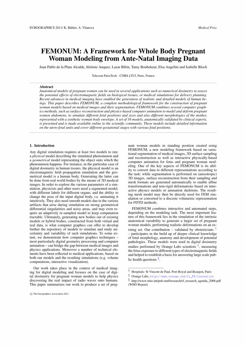
Figure 1: Overview of the FEMONUM methodology. WGstands for weeks of gestation.
2. A pregnant woman modeling framework
In this section we briefly describe the methodology used tocreate our set of pregnant woman models. This methodologycan be mainly decomposed into four steps (Figure 1). Firstly,from medical images, a segmentation of different visible or-gans in the utero-fetal unit (UFU) is performed using auto-mated or interactive tools. Secondly, in order to enable spa-tial transformations and non-rigid deformations, organ con-tours are extracted from the segmentations and representedas smooth meshed surfaces (by mesh reconstruction) avoid-ing undesirable singularities caused by "staircase" effects ofnaive direct meshing approaches. Thirdly, a fetal bone arma-ture is modeled to enable arbitrary positioning of the fetus.Finally, physics-based deformations are applied to automat-ically insert the UFU inside a generic non-pregnant womanbody in standing position, taking into account the variabil-ity of the muscle and the fat layers of the mother’s body(variable for different morphologies). We refer the readerto [BAd∗10] for a detailed description of this framework.
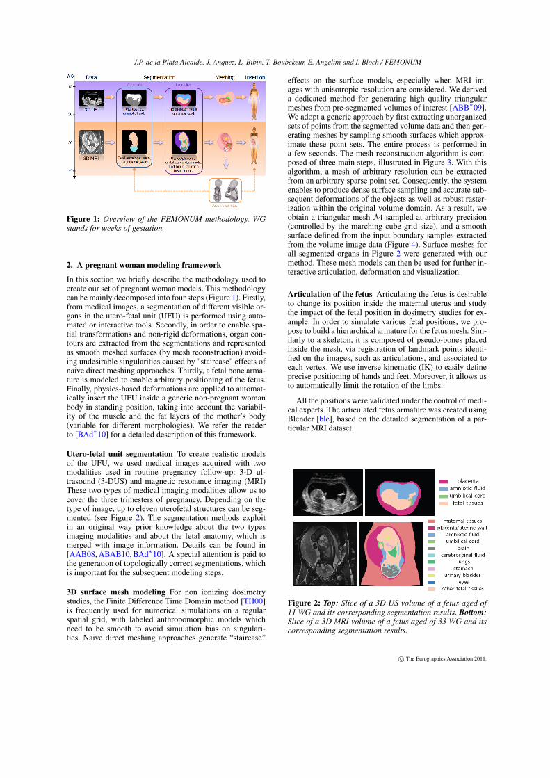
Utero-fetal unit segmentation To create realistic modelsof the UFU, we used medical images acquired with twomodalities used in routine pregnancy follow-up: 3-D ul-trasound (3-DUS) and magnetic resonance imaging (MRI)These two types of medical imaging modalities allow us tocover the three trimesters of pregnancy. Depending on thetype of image, up to eleven uterofetal structures can be seg-mented (see Figure 2). The segmentation methods exploitin an original way prior knowledge about the two typesimaging modalities and about the fetal anatomy, which ismerged with image information. Details can be found in[AAB08, ABAB10, BAd∗10]. A special attention is paid tothe generation of topologically correct segmentations, whichis important for the subsequent modeling steps.
3D surface mesh modeling For non ionizing dosimetrystudies, the Finite Difference Time Domain method [TH00]is frequently used for numerical simulations on a regularspatial grid, with labeled anthropomorphic models whichneed to be smooth to avoid simulation bias on singulari-ties. Naive direct meshing approaches generate “staircase”
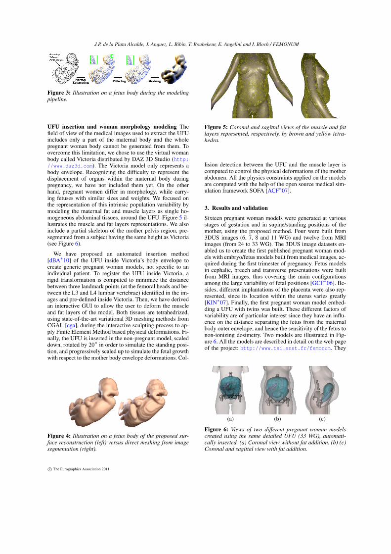
effects on the surface models, especially when MRI im-ages with anisotropic resolution are considered. We deriveda dedicated method for generating high quality triangularmeshes from pre-segmented volumes of interest [ABB∗09].We adopt a generic approach by first extracting unorganizedsets of points from the segmented volume data and then gen-erating meshes by sampling smooth surfaces which approx-imate these point sets. The entire process is performed ina few seconds. The mesh reconstruction algorithm is com-posed of three main steps, illustrated in Figure 3. With thisalgorithm, a mesh of arbitrary resolution can be extractedfrom an arbitrary sparse point set. Consequently, the systemenables to produce dense surface sampling and accurate sub-sequent deformations of the objects as well as robust raster-ization within the original volume domain. As a result, weobtain a triangular meshM sampled at arbitrary precision(controlled by the marching cube grid size), and a smoothsurface defined from the input boundary samples extractedfrom the volume image data (Figure 4). Surface meshes forall segmented organs in Figure 2 were generated with ourmethod. These mesh models can then be used for further in-teractive articulation, deformation and visualization.
Articulation of the fetus Articulating the fetus is desirableto change its position inside the maternal uterus and studythe impact of the fetal position in dosimetry studies for ex-ample. In order to simulate various fetal positions, we pro-pose to build a hierarchical armature for the fetus mesh. Sim-ilarly to a skeleton, it is composed of pseudo-bones placedinside the mesh, via registration of landmark points identi-fied on the images, such as articulations, and associated toeach vertex. We use inverse kinematic (IK) to easily defineprecise positioning of hands and feet. Moreover, it allows usto automatically limit the rotation of the limbs.
All the positions were validated under the control of medi-cal experts. The articulated fetus armature was created usingBlender [ble], based on the detailed segmentation of a par-ticular MRI dataset.
Figure 2: Top: Slice of a 3D US volume of a fetus aged of11 WG and its corresponding segmentation results. Bottom:Slice of a 3D MRI volume of a fetus aged of 33 WG and itscorresponding segmentation results.
c© The Eurographics Association 2011.
J.P. de la Plata Alcalde, J. Anquez, L. Bibin, T. Boubekeur, E. Angelini and I. Bloch / FEMONUM
Figure 3: Illustration on a fetus body during the modelingpipeline.
UFU insertion and woman morphology modeling Thefield of view of the medical images used to extract the UFUincludes only a part of the maternal body and the wholepregnant woman body cannot be generated from them. Toovercome this limitation, we chose to use the virtual womanbody called Victoria distributed by DAZ 3D Studio (http://www.daz3d.com). The Victoria model only represents abody envelope. Recognizing the difficulty to represent thedisplacement of organs within the maternal body duringpregnancy, we have not included them yet. On the otherhand, pregnant women differ in morphology, while carry-ing fetuses with similar sizes and weights. We focused onthe representation of this intrinsic population variability bymodeling the maternal fat and muscle layers as single ho-mogeneous abdominal tissues, around the UFU. Figure 5 il-lustrates the muscle and fat layers representations. We alsoinclude a partial skeleton of the mother pelvis region, pre-segmented from a subject having the same height as Victoria(see Figure 6).
We have proposed an automated insertion method[dBA∗10] of the UFU inside Victoria’s body envelope tocreate generic pregnant woman models, not specific to anindividual patient. To register the UFU inside Victoria, arigid transformation is computed to minimize the distancebetween three landmark points (at the femoral heads and be-tween the L3 and L4 lumbar vertebrae) identified in the im-ages and pre-defined inside Victoria. Then, we have derivedan interactive GUI to allow the user to deform the muscleand fat layers of the model. Both tissues are tetrahedrized,using state-of-the-art variational 3D meshing methods fromCGAL [cga], during the interactive sculpting process to ap-ply Finite Element Method based physical deformations. Fi-nally, the UFU is inserted in the non-pregnant model, scaleddown, rotated by 20◦ in order to simulate the standing posi-tion, and progressively scaled up to simulate the fetal growthwith respect to the mother body envelope deformations. Col-
Figure 4: Illustration on a fetus body of the proposed sur-face reconstruction (left) versus direct meshing from imagesegmentation (right).
Figure 5: Coronal and sagittal views of the muscle and fatlayers represented, respectively, by brown and yellow tetra-hedra.
lision detection between the UFU and the muscle layer iscomputed to control the physical deformations of the motherabdomen. All the physics constraints applied on the modelsare computed with the help of the open source medical sim-ulation framework SOFA [ACF∗07].
3. Results and validation
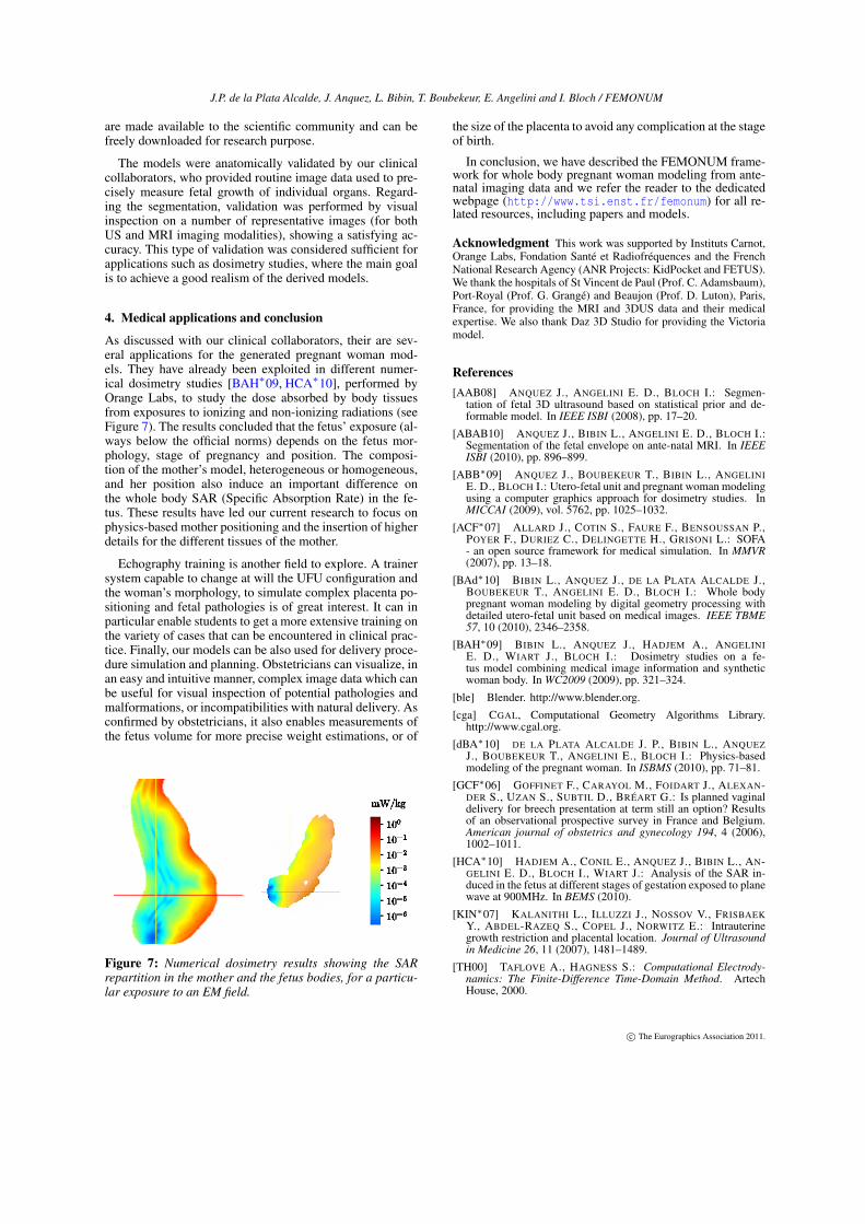
Sixteen pregnant woman models were generated at variousstages of gestation and in supine/standing positions of themother, using the proposed method. Four were built from3DUS images (6, 7, 8 and 11 WG) and twelve from MRIimages (from 24 to 33 WG). The 3DUS image datasets en-abled us to create the first published pregnant woman mod-els with embryo/fetus models built from medical images, ac-quired during the first trimester of pregnancy. Fetus modelsin cephalic, breech and transverse presentations were builtfrom MRI images, thus covering the main configurationsamong the large variability of fetal positions [GCF∗06]. Be-sides, different implantations of the placenta were also rep-resented, since its location within the uterus varies greatly[KIN∗07]. Finally, the first pregnant woman model embed-ding a UFU with twins was built. These different factors ofvariability are of particular interest since they have an influ-ence on the distance separating the fetus from the maternalbody outer envelope, and hence the sensitivity of the fetus tonon-ionizing dosimetry. Two models are illustrated in Fig-ure 6. All the models are described in detail on the web pageof the project: http://www.tsi.enst.fr/femonum. They
(a) (b) (c)
Figure 6: Views of two different pregnant woman modelscreated using the same detailed UFU (33 WG), automati-cally inserted. (a) Coronal view without fat addition. (b) (c)Coronal and sagittal view with fat addition.
c© The Eurographics Association 2011.
J.P. de la Plata Alcalde, J. Anquez, L. Bibin, T. Boubekeur, E. Angelini and I. Bloch / FEMONUM
are made available to the scientific community and can befreely downloaded for research purpose.
The models were anatomically validated by our clinicalcollaborators, who provided routine image data used to pre-cisely measure fetal growth of individual organs. Regard-ing the segmentation, validation was performed by visualinspection on a number of representative images (for bothUS and MRI imaging modalities), showing a satisfying ac-curacy. This type of validation was considered sufficient forapplications such as dosimetry studies, where the main goalis to achieve a good realism of the derived models.
4. Medical applications and conclusion
As discussed with our clinical collaborators, their are sev-eral applications for the generated pregnant woman mod-els. They have already been exploited in different numer-ical dosimetry studies [BAH∗09, HCA∗10], performed byOrange Labs, to study the dose absorbed by body tissuesfrom exposures to ionizing and non-ionizing radiations (seeFigure 7). The results concluded that the fetus’ exposure (al-ways below the official norms) depends on the fetus mor-phology, stage of pregnancy and position. The composi-tion of the mother’s model, heterogeneous or homogeneous,and her position also induce an important difference onthe whole body SAR (Specific Absorption Rate) in the fe-tus. These results have led our current research to focus onphysics-based mother positioning and the insertion of higherdetails for the different tissues of the mother.
Echography training is another field to explore. A trainersystem capable to change at will the UFU configuration andthe woman’s morphology, to simulate complex placenta po-sitioning and fetal pathologies is of great interest. It can inparticular enable students to get a more extensive training onthe variety of cases that can be encountered in clinical prac-tice. Finally, our models can be also used for delivery proce-dure simulation and planning. Obstetricians can visualize, inan easy and intuitive manner, complex image data which canbe useful for visual inspection of potential pathologies andmalformations, or incompatibilities with natural delivery. Asconfirmed by obstetricians, it also enables measurements ofthe fetus volume for more precise weight estimations, or of
Figure 7: Numerical dosimetry results showing the SARrepartition in the mother and the fetus bodies, for a particu-lar exposure to an EM field.
the size of the placenta to avoid any complication at the stageof birth.
In conclusion, we have described the FEMONUM frame-work for whole body pregnant woman modeling from ante-natal imaging data and we refer the reader to the dedicatedwebpage (http://www.tsi.enst.fr/femonum) for all re-lated resources, including papers and models.
Acknowledgment This work was supported by Instituts Carnot,Orange Labs, Fondation Santé et Radiofréquences and the FrenchNational Research Agency (ANR Projects: KidPocket and FETUS).We thank the hospitals of St Vincent de Paul (Prof. C. Adamsbaum),Port-Royal (Prof. G. Grangé) and Beaujon (Prof. D. Luton), Paris,France, for providing the MRI and 3DUS data and their medicalexpertise. We also thank Daz 3D Studio for providing the Victoriamodel.
References[AAB08] ANQUEZ J., ANGELINI E. D., BLOCH I.: Segmen-
tation of fetal 3D ultrasound based on statistical prior and de-formable model. In IEEE ISBI (2008), pp. 17–20.
[ABAB10] ANQUEZ J., BIBIN L., ANGELINI E. D., BLOCH I.:Segmentation of the fetal envelope on ante-natal MRI. In IEEEISBI (2010), pp. 896–899.
[ABB∗09] ANQUEZ J., BOUBEKEUR T., BIBIN L., ANGELINIE. D., BLOCH I.: Utero-fetal unit and pregnant woman modelingusing a computer graphics approach for dosimetry studies. InMICCAI (2009), vol. 5762, pp. 1025–1032.
[ACF∗07] ALLARD J., COTIN S., FAURE F., BENSOUSSAN P.,POYER F., DURIEZ C., DELINGETTE H., GRISONI L.: SOFA- an open source framework for medical simulation. In MMVR(2007), pp. 13–18.
[BAd∗10] BIBIN L., ANQUEZ J., DE LA PLATA ALCALDE J.,BOUBEKEUR T., ANGELINI E. D., BLOCH I.: Whole bodypregnant woman modeling by digital geometry processing withdetailed utero-fetal unit based on medical images. IEEE TBME57, 10 (2010), 2346–2358.
[BAH∗09] BIBIN L., ANQUEZ J., HADJEM A., ANGELINIE. D., WIART J., BLOCH I.: Dosimetry studies on a fe-tus model combining medical image information and syntheticwoman body. In WC2009 (2009), pp. 321–324.
[ble] Blender. http://www.blender.org.
[cga] CGAL, Computational Geometry Algorithms Library.http://www.cgal.org.
[dBA∗10] DE LA PLATA ALCALDE J. P., BIBIN L., ANQUEZJ., BOUBEKEUR T., ANGELINI E., BLOCH I.: Physics-basedmodeling of the pregnant woman. In ISBMS (2010), pp. 71–81.
[GCF∗06] GOFFINET F., CARAYOL M., FOIDART J., ALEXAN-DER S., UZAN S., SUBTIL D., BRÉART G.: Is planned vaginaldelivery for breech presentation at term still an option? Resultsof an observational prospective survey in France and Belgium.American journal of obstetrics and gynecology 194, 4 (2006),1002–1011.
[HCA∗10] HADJEM A., CONIL E., ANQUEZ J., BIBIN L., AN-GELINI E. D., BLOCH I., WIART J.: Analysis of the SAR in-duced in the fetus at different stages of gestation exposed to planewave at 900MHz. In BEMS (2010).
[KIN∗07] KALANITHI L., ILLUZZI J., NOSSOV V., FRISBAEKY., ABDEL-RAZEQ S., COPEL J., NORWITZ E.: Intrauterinegrowth restriction and placental location. Journal of Ultrasoundin Medicine 26, 11 (2007), 1481–1489.
[TH00] TAFLOVE A., HAGNESS S.: Computational Electrody-namics: The Finite-Difference Time-Domain Method. ArtechHouse, 2000.
c© The Eurographics Association 2011.