feature extraction of muscle fatigue on …eprints.utem.edu.my/15802/1/feature extraction of muscle...
TRANSCRIPT

0
FEATURE EXTRACTION OF MUSCLE FATIGUE ON FOREARM USING
SURFACE ELECTROMYOGRAPHY (sEMG) TECHNIQUE
KHOO HUI PING
A report submitted in partial fulfilment of requirements for the degree
of Bachelor of Electrical Engineering (Control, Instrumentation and Automation)
with Honors
Faculty of Electrical Engineering
Universiti Teknikal Malaysia Melaka
2014

1
“ I hereby declare that I have read through “Feature Extraction on Forearm using Surface
Electromyography (SEMG) Technique” and found that it has comply the partial fulfilment
for awarding the degree of Bachelor of Electrical Engineering (Control, Instrumentation
and Automation)”
Signature : .......................................................
Supervisor‟s Name : .......................................................
Date : ......................................................

2
DECLARATION
“I hereby declare that the work in this report is my own except for summaries and
quotations which have been duly acknowledged.”
Signature: ……………………………..
Author: ……………………………..
Date: ……………………………..

3
Gratitude to
My family
My FYP supervisor
My coursemates

ii
ACKNOWLEDGEMENT
In preparing this final year report, I was contacted with many people like Dr.
Chong Shin Horng who is my final year project supervisor. First of all, I would like to
express my gratitude Dr. Chong Shin Horng because she is willing to spend her precious
time to guide me throughout my final year and provide me with useful information.
Besides, I would like to express my sincere appreciation to my family for their providing
everything such as money and moral support to help me in accomplishing this final year
project.
I would like to thanks my senior, Mr. Abu Bakar bin Yahya that had provided me
lots of guidance and help in this final year project. Moreover, I would like to express my
sincere appreciation to Universiti Teknikal Malaysia Melaka (UTeM) because provided me
an opportunity to accomplish this final year project by my own at this beginning stage.
Besides, I would like to express my deepest appreciation towards Mr. Wan Bukhari and Dr.
Soo that had provided me some useful information during seminar FYP 1.
Last but not least, I wish to express again my appreciation to all the lecturers that
had taught me and guide me throughout my final year project. In addition, thanks to Mr.
Lee Yi Ming for his support, ideas and suggestions to help me solve my confusion during
completing this project and also my friends. Their view and ideas are useful for me and the
whole project really bought us to the true value of friendship and respect to each other.

iii
ABSTRACT
Nowadays, musculoskeletal disorder has becoming a common disease in society. Muscle
fatigue is one of the factor leads to musculoskeletal disorder. In this research, technique of
surface electromyography (sEMG) signal detection and processing will be implemented.
The main objective of this research is to extract features of muscle fatigue in order to
evaluate the muscle fatigue condition of males and females. sEMG signal were collected
from the forearm muscle - flexor carpi radialis of each volunteer. A group of 20 healthy
university students were recruited in order to determine muscle fatigue occur in real life. A
dynamic contraction and static contraction were implemented in order to understand the
relationship between motion and fatigue and relationship between force and fatigue.
Dynamic contraction experiment is done with subjects bent their wrist up to maximal joint
angle; whereas static contraction experiment is done with different percentage of maximal
voluntary contraction (MVC). For dynamic contraction, the feature of sEMG signal was
extracted using time domain (RMS) and time-frequency domain (Scalogram). For static
contraction, the feature of sEMG signal was extracted using time domain (RMS) and
frequency domain (MDF). While analysing the time domain, it is found that the amplitude
increased during fatigue in dynamic and static contraction experiment. For frequency
domain, MDF are found to be decreased during fatigue in static contraction experiment.
For time-frequency in terms of Scalogram, the energy distribution coefficients were found
to be shifted to lower frequency as shown in the result and discussion part. Validity test is
implemented in order to ensure the data collected is validated. Although the results were
promising, there will be some limitations that need to be overcome in the future such as
apply an online muscle fatigue progression test using Scalogram method for rehabilitation
purpose.

iv
ABSTRAK
Kini, keletihan otot telah menjadi satu penyakit yang biasa dalam masyarakat. Keletihan
otot adalah salah satu faktor yang membawa kepada masalah muskuloskeletal. Dalam
kajian ini, teknik permukaan Electromyography (EMG) pengesanan isyarat dan
pemprosesan akan dilaksanakan. Objektif utama kajian ini adalah untuk mendapatkan ciri-
ciri keletihan otot menggunakan analisis domain masa dan domain frekuensi. Isyarat EMG
dikumpulkan dari otot lengan bagi setiap pelajar. Sebanyak 20 pelajar universiti yang sihat
akan diambil untuk memahami hubungan antara pergerakan dan keletihan serta hubungan
antara kekuatan dan keletihan, pengecutan yang dinamik dan statik akan dijalankan. Isyarat
EMG akan dianalisiskan dengan domain masa (RMS), domain frekuensi (MDF) dan
domain masa-frekuensi (Scalogram). Semasa isyarat signal dianalisis dalam RMS,
ketinggian signal meningkat semasa keletihan otot. Manakala frekuensi untuk MDF dan
taburan tenaga untuk Scalogram pula menurun. Ujian kesahihan akan dijalankan untuk
memastikan data yang diambil adalah betul. Walaupun keputusan yang ditunjukkan adalah
sama dengan apa yang dijangkakan, ada juga sesetengah kelemahan yang kena dibaiki
pada masa hadapan dengan menjalankan kajian secara online untuk proses pemulihan.

v
LIST OF PUBLICATION
CONFERENCE PAPER (SUBMITTED)
1. “Feature Extraction of Muscle Fatigue on Forearm using Surface
Electromyography (sEMG) Technique”, IEEE-EMBS, 2014.

vi
TABLE OF CONTENTS
CHAPTER TITLE PAGE
ACKNOWLEDGEMENT ii
ABSTRACT iii
TABLE OF CONTENTS vi
LIST OF TABLES ix
LIST OF FIGURES x
LIST OF APPENDICES xii
1 INTRODUCTION
1.1 Project background 1
1.2 Motivation 2
1.3 Problem statement 2
1.4 Objectives 3
1.5 Scope 3
2 LITERATURE REVIEW 4
2.1 Muscle fatigue and its relationship with sEMG signal 4
2.1.1 Muscle fatigue stages and its experimental
implementation 7
2.2 Electromyography (EMG) 8
2.2.1 History of electromyography 8
2.2.2 Surface electromyography (sEMG) and its
application 9
2.3 Factors affecting EMG signal quality 11
2.3.1 EMG electrodes 11
2.3.2 Electrode types 11
2.3.4 Electrode placement and the innervation zone 12
2.3.5 Signal noise 12

vii
2.3.6 Other factors affecting signal quality 13
2.4 SEMG signal analysis and feature characterization 13
2.4.1 Time domain analysis 13
2.4.2 Frequency domain analysis 14
2.4.3 Time-frequency domain 15
3 METHODOLOGY
3.1 Subjects selection 16
3.2 Data pre-processing 17
3.3 Experimental procedure 18
3.3.1 Dynamic contraction
3.3.2 Static contraction
19
20
3.4 Experimental precaution 21
3.5 sEMG measurements 22
3.6 Data analysis and feature extraction 23
3.6.1 Time domain analysis
3.6.2 Frequency domain analysis
3.6.3 Time-frequency domain analysis
23
23
24
3.7 Validity of data 24
3.8 Reliability of data 25
4 RESULT AND DISCUSSION
4.1 Feature extraction in dynamic contraction 26
4.1.1 Time domain analysis: RMS 28
4.1.2 Time-frequency analysis: Scalogram 30
4.2 Static contraction 32
4.2.1 Maximal voluntary contraction analysis 33
4.2.2 Time domain analysis: RMS 35
4.2.3 Ratio between force to fatigue 37
4.2.4 Frequency domain analysis: Median frequency 38
4.3 Manifestation of muscle fatigue for dynamic and static
contraction 39
4.3.1 Amplitude (RMS) 39

viii
4.3.2 Median frequency (MDF) and Scalogram 40
5 CONCLUSION AND RECOMMENDATIONS 41
5.1 Conclusion 41
5.2 Future Works 42
REFERENCES
APPENDIX
43
46

ix
LIST OF TABLES
TABLE TITLE PAGE
2.1 Types of mother wavelets and its operation 15
3.1 Subject’s specification 16
3.2 Recommendation for electrode/skin impedance ranges 17
4.1 Average RMS of males and females for dynamic contraction 29
4.2 Average force losses for males and females 34
4.3 Average value of RMS before fatigue (BF) and during fatigue
(DF)
36
4.4 Average value of 30%, 50% and 70% MVC for males and
females
36
4.5 Ratio between force and fatigue 37
4.6 Median frequency (MDF) before fatigue (BF) and during fatigue
(DF)
39

x
LIST OF FIGURES
FIGURE TITLE PAGE
2.1 Leakage and storage of Calcium ions during muscle contraction
and relaxation
5
2.2 Experimental method in previous research 6
2.3 Muscle fatigue stages 7
2.4 Development of electromyography 8
2.5 Electrode placement 11
2.6 Preferred electrode location 12
3.1 Posture for dynamic contraction 18
3.2 Original condition with sitting position 19
3.3 Wrist up to maximal joint angle 19
3.4 Posture for static contraction 20
3.5 Location of muscle 21
3.6 Experimental setup 22
3.7 Flow chart for implementing data reliability process 25
4.1 Raw signal measurement for flexor carpi radialis 27
4.2 Raw EMG signal before fatigue and during fatigue 27
4.3 Rectification of raw signal 27
4.4 Filtering of signal using bandpass filter (a) before filter (b) after
filter for before fatigue signal
28
4.5 Rectified signal and moving RMS at 1000ms overlap time 29
4.6 Average RMS value for before and during fatigue 29
4.7 sEMG signal extracted from first contraction and the Scalogram
contour plot.
30
4.8 Comparison of Scalogram contour plot between (a) before fatigue
and (b) during fatigue
31
4.9 Figure 4.9: Raw signal and force display for (a) 30% MVC (b)
50% MVC (c) 70% MVC
32

xi
4.10 Performance of different percentage of MVC 33
4.11 MVC determination 33
4.12 Display of force losses based on Table 4.2 35
4.13 Rectified signal and moving rms at 1000ms overlap time 35
4.14 RMS feature extraction for different percentage of MVC from
two subjects (a) Male (b) Female
36
4.15 Ratio of force to fatigue between males and females 37
4.16 Frequency spectrum for one subject (before and during fatigue) 38
4.17 Comparison of RMS value for dynamic and static contraction 40

xii
LIST OF APPENDICES
APPENDIX TITLE PAGE
A Gantt chart 46
B Experimental setup for data acquisition 48
C Written consent form 55
D Schematic diagram for EMG Arduino 56
E Conference paper acceptance confirmation 57

1
CHAPTER 1
INTRODUCTION
This chapter will describes the problem of muscle fatigue problem and the
consequences of muscle fatigue experienced by human in their daily life. Therefore, sEMG
techniques are recommended for muscle fatigue detection in this research. Besides, the
objectives and scope will be covered in this chapter.
1.1 Project background
Nowadays, due to the advancement of industry world, the enhancement of human
performance is crucial for the improvement of quality of life. Repetitive works or
continuous similar types of motion happen in human’s daily life. However, people are just
evaluate their physical condition subjectively and ignoring their muscle status and this
issue may bring them to musculoskeletal disorder such as occupational overuse syndrome
(OOS) [1] and work-related musculoskeletal disorders (WMSDs) [1]. All these factors are
caused by the decline in motor unit firing rates and recruitment threshold of motor units
declined [25]. There are various methods to estimate body condition. However, muscle
fatigue will be the only consideration in this paper. Muscle fatigue is defined as failure to
maintain a desired force and it may occur due to isometric or non-isometric (dynamic)
muscle contraction. Recent physiological studies have demonstrated the crucial of muscle
fatigue detection in human’s daily lives in order to prevent any injury in muscle and
degradation in human performance efficiency. Electromyography (EMG) technique is
considered as a good solution to study about muscle activity either in motion, force and
fatigue. The most common technique used to evaluate muscle activity is surface
electromyography (sEMG). sEMG is a non-invasive, pain-free and easy to apply approach
to detect muscle activity. Result such as increasing in amplitude of EMG signals and
shifting in frequency spectrum from high frequency to lower frequency during muscle

2
fatigue have been observed by previous researchers. These changes can be measured using
time domain, frequency domain and time-frequency domain analysis by calculating its
mean frequency (MNF), median frequency (MDF), root mean square (RMS) and also
Scalogram. Muscular fatigue decreases the MDF value within the EMG power spectral
density, and increases the EMG signal amplitudes at the end of the experiment which
indicates increase in RMS. These factors happen due to the variations in the activation of
the muscle motor unit action potential (MUAP). Both MDF and MNF are considered as a
reliable estimator of the muscle fatigue. Scalogram is a visual method for displaying
wavelet transform. Energy distribution plays an important role while observing Scalogram.
1.2 Motivation
The number of patients suffers from muscle disorders are increasing. This brings
the important of muscle fatigue classification especially for those who are industry field.
The repetitive works by workers are able to bring an effect to their muscle tissues and
hence yield some disorders such as OOS and WMSDs. These disorders will cause a
uncomfortable feeling to human. Therefore, it is necessary for this research to implement
an analysis of muscle fatigue with the aids of hardware and software implementation. The
purpose behind this research is as a reminder how severe a muscle fatigue can affect our
lives.
1.3 Problem statement
Musculoskeletal disorder has become a common disease happen in human. The
necessity of muscle fatigue analysis should be apparent in order to prevent any disorders,
muscle injury and human performance degradation. By detecting and classifying muscle
fatigue, it adds important information to the fields of human-computer interactions (HCI),
sport injuries and performance, ergonomics, diagnosis and prosthetic purposes. However,
muscle fatigue is difficult to determine physically, it requires application tools. Therefore,
SEMG technique will be used to study the relationship between fatigue and SEMG signals.
Besides, non-fatigue condition and fatigue condition will be classified by extracting their

3
features using time domain and frequency domain analysis respectively in order to analyze
occurrence of muscle fatigue.
1.4 Objectives
The first objective of this research is to extract features of muscle fatigue using
time domain, frequency domain and time-frequency domain. All the features extracted are
analysed in Root Mean Square (RMS), Median Frequency (MDF) and lastly Scalogram.
The second objective is to analyse surface electromyography (sEMG) signals during
progression of muscle fatigue in static or dynamic contraction using statistical analysis.
Therefore, there are two types of experiments that will be conducted which are dynamic
contraction experiment and static contraction experiment in order to determine the
significant result from statistical analysis between males and females.
1.5 Scope
This research is primarily focus in wrist muscle analysis at flexor carpi radialis
using surface disposable electrodes. 20 subjects will be recruited. Dynamic contraction and
static contraction experimental setup will be conducted. For dynamic contraction, 20
subjects will be recruited while for static contraction, only 12 subjects are recruited.
Extracted feature from raw signal will be analyzed using time domain in terms of Root
Mean Square (RMS), frequency domain in terms of Median Frequency (MDF) and time-
frequency domain in terms of Scalogram. However, the correlation between hand size and
grip strength will not be covered in this research.

4
CHAPTER 2
LITERATURE REVIEW
This chapter first gives a general introduction about sEMG signal. After that, a
detailed literature review of various sEMG signal feature extraction analysis method and
procedure for estimating muscle fatigue is presented.
2.1 Muscle fatigue and its relationship with sEMG signal
Muscle fatigue has being studied by many researchers and discussed in their paper
in the past. The term ‘muscle fatigue’ was first introduced by Bills (1943) and it is divided
it into three different classes: subjective fatigue, which result from psychological factors
such as a lack of motivation; objective fatigue, which represents a decline in productivity;
and lastly, Physiological fatigue, which refers to muscle unable to maintain a desired force
[2]. Changes the nerve system and the muscle simultaneously is related to neuromuscular
mechanism of fatigue, which involved central fatigue (brain fatigue), fatigue in the
neuromuscular junction and fatigue occurring in the muscle (peripheral fatigue) [3].
Peripheral fatigue is the most common case for physical fatigue and this type of fatigue is
widely detected using EMG technique in most studies. It take place when the normal
functionality of the nerve fibers and the muscles that are contracting are impaired, that is
the muscle’s ability to utilize force is degrading due to the incapability of the body to reach
the increased energy demand in the contracting muscles [3]. The main reason that causes
muscle fatigue to occur is the release and storage of calcium ions within the muscle fibers.
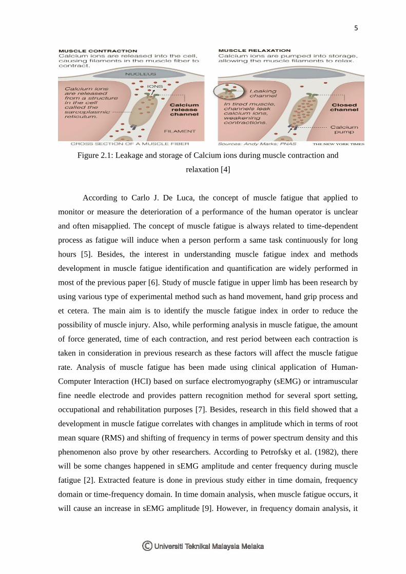
5
Figure 2.1: Leakage and storage of Calcium ions during muscle contraction and
relaxation [4]
According to Carlo J. De Luca, the concept of muscle fatigue that applied to
monitor or measure the deterioration of a performance of the human operator is unclear
and often misapplied. The concept of muscle fatigue is always related to time-dependent
process as fatigue will induce when a person perform a same task continuously for long
hours [5]. Besides, the interest in understanding muscle fatigue index and methods
development in muscle fatigue identification and quantification are widely performed in
most of the previous paper [6]. Study of muscle fatigue in upper limb has been research by
using various type of experimental method such as hand movement, hand grip process and
et cetera. The main aim is to identify the muscle fatigue index in order to reduce the
possibility of muscle injury. Also, while performing analysis in muscle fatigue, the amount
of force generated, time of each contraction, and rest period between each contraction is
taken in consideration in previous research as these factors will affect the muscle fatigue
rate. Analysis of muscle fatigue has been made using clinical application of Human-
Computer Interaction (HCI) based on surface electromyography (sEMG) or intramuscular
fine needle electrode and provides pattern recognition method for several sport setting,
occupational and rehabilitation purposes [7]. Besides, research in this field showed that a
development in muscle fatigue correlates with changes in amplitude which in terms of root
mean square (RMS) and shifting of frequency in terms of power spectrum density and this
phenomenon also prove by other researchers. According to Petrofsky et al. (1982), there
will be some changes happened in sEMG amplitude and center frequency during muscle
fatigue [2]. Extracted feature is done in previous study either in time domain, frequency
domain or time-frequency domain. In time domain analysis, when muscle fatigue occurs, it
will cause an increase in sEMG amplitude [9]. However, in frequency domain analysis, it

6
can be observed that power spectrum density is shifted to lower frequency [9]. All these
changes might be a result of concentration of blood lactate, muscle pH value, blood oxygen
saturation level, recruitment of motor unit action potential and motor unit firing rate [10].
These metrics are used to identify physiological phenomena during muscle contractions
that lead to muscle fatigue which is performed by those biomedical field researchers. After
review many of previous researcher’s experimental method, the method was concluded in
the flow chart shown below:
Figure 2.2: Experimental method in previous research
Signal
Acquisition
Signal
Conditioning
Feature
Extraction
Classification

7
2.1.1 Muscle fatigue stages and its experimental implementation
Figure 2.3: Muscle fatigue stages
Current research tends to focus on two classes of localized muscle fatigue: Non-
Fatigue and Fatigue. Fatigue is relates to the onset of fatigue during a muscle contraction;
while Non-Fatigue is define as the state of muscle during the contraction that occurs before
the onset of fatigue. However, there is also an additional class of fatigue, known as
transition to fatigue. This class is located in between of non-fatigue and fatigue. The
identification of this additional class helps in the autonomous detection and prediction of
muscle fatigue and differentiate between two classes of fatigue [11][12]. Although most
research on muscle fatigue only focuses in Non-Fatigue and Fatigue stages, the Transition-
to-Fatigue stage identified by Al-Mulla et al. is an important addition to this research field,
especially for the development of real-time systems that automate the process of detecting
and predicting fatigue. Previous research has conducted several researches in determining
fatigue condition in both isometric and dynamic environment. Isometric contraction is
often implemented by most of the researcher. For isometric contraction, the subjects will
require to maintain its posture and force throughout the experiment. One of the isometric
examples is conducted by Allmulla et al. [12]. In his research, he and his partner recorded
the sEMG signal accompanied with goniometer findings. Goniometer was placed on the
side of the arm to measure the elbow angle. The acquired sEMG signal is compared with
the goniometer finding to ensure the sEMG classification is correct. However, isometric
contraction is impractical in real life environment. To overcome this limitation, some
researchers conduct a dynamic contraction experiment. Movement and amount of force
exerted by each subject applied have become their main concern in dynamic contraction to
evaluate muscle fatigue index. Equipment such as strain gauge [13] and elbow angle [9]
are considered as they are reliable to measure muscle fatigue index and able to classify
sEMG signals correctly.
Non Fatigue Transition to
fatigue Fatigue

8
2.2 Electromyography (EMG)
Electromyography is the common tool used in detecting muscle status detection. An
overview about electromyography will be described in this section.
2.2.1 History of electromyography
Figure 2.4: Development of electromyography
Figure 2.4 shows the development of electromyography. Electromyography (EMG)
is widely discovered in the early 1950’s by many researchers. Electromyography had its
earliest roots where Greeks practice “shock” on electric eels (refer to first picture of figure
2.4) in order to make the eel to execute all the ailments out of its body. However, the origin
of shock that accompanied this earliest detection and application of EMG signal was not
highly appreciated. EMG techniques was first documented in early year of 1666 by an
Italian, named Francesco Redi realized that the spark is actually originated from muscle
tissue [14]. By the year 1773, Walsh showed that the muscle tissue of eel could generate a
spark of electricity [14]. The relationship between muscle contraction and electricity was
later proved by Luigi Galvani in the year 1792 [14]. Nevertheless, this relationship gets
disagreement by Volta. Volta stated that the phenomenon determined by Galvani may
result from the artifact of dissimilar metals touching the muscle tissue [14]. The history of
EMG is continued with the discovery of electricity and the development of the ability to
view through muscle activity with the aid of instruments in the year 1840s [14]. This
brought four new instruments such as cathode ray tube, vacuum tube amplifiers, metal
electrodes and the revolutionary needle electrode which used to detect EMG signal. In year
1849, the father of experimental electrophysiology, Du Bois-Reymond performed his
Electric eel Nobel Prize Carlo de Luca

9
experiment on subject’s forearm in electrical contact with electrodes during voluntary
contraction [14]. By implementing detection of muscle activity experiment, a conclusion
draws that signal amplitude will increase during wrist flexion [15]. By the early 1900s,
Pratt showed that the amplitude of energy associated with muscle contraction was related
to the recruitment of individual muscle fibers. In the 1920s, Gasser and Newcomer used
the cathode ray oscilloscope to display the signals from muscle and this brings them a
Nobel Prize in 1944 (refer to second picture in Figure 2.4) [15]. Researchers began to use
sEMG to study dynamic movement in the year 1940s, for example, Inman and Price. In the
early 1980s, Cram and Steger introduced a clinical method for scanning muscles using
handheld sEMG sensing device. Few years later, Cram and Engstrom collected signal from
104 normal subjects by scanning their muscle in different muscle area with different
posture either standing and sitting. All the efforts done by previous researchers are highly
appreciated and the efforts in discovering the application of EMG are still continued until
now. One of the famous sEMG researchers is Carlo de Luca. (Refer to third picture in
Figure 2.4)
2.2.2 Surface electromyography (sEMG) and its application
The term of electromyography has been defined by several researchers in
biomedical field. According to Carlo De Luca (2006), EMG signal is the electrical
manifestation of neuromuscular activation associated with a contracting muscle. Whereas
according to Christos (2013), electromyography refers to bio-signal that measure the
activity produced by skeletal muscles during contraction. The conclusion that can be made
from the two definitions above is that, electromyography are widely used to study muscle
activity. Electromyogram display an electrical signal generated by motor unit action
potential (MUAP) of muscles during either voluntary or involuntary contraction and it is a
result of summation of electrical of a large number of muscle fibers in the vicinity of
electrodes [16]. Besides, EMG provides the information about different features of muscle
activations associated with different types of contractions that are isometric and dynamic
contraction. It has been widely employed as an objective tool to study on the phenomenon
of muscle fatigue. EMG signals can be detected by using two types of techniques:
intramuscular fine wire electrodes and surface electrodes. Intramuscular fine wire requires