feature extraction and classification of mammographic masses
DESCRIPTION
ECE 8990: Automated Target Recognition Classification of Mammographic Masses. Feature Extraction and Classification of Mammographic Masses. Presented by, Jignesh Panchal Anuradha Agatheeswaran. - PowerPoint PPT PresentationTRANSCRIPT

Feature Extraction and Classification of Mammographic Masses
Presented by,
Jignesh PanchalAnuradha Agatheeswaran
ECE 8990: Automated Target Recognition Classification of Mammographic Masses
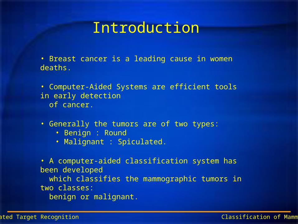
Introduction
• Breast cancer is a leading cause in women deaths.
• Computer-Aided Systems are efficient tools in early detection of cancer.
• Generally the tumors are of two types:• Benign : Round• Malignant : Spiculated.
• A computer-aided classification system has been developed which classifies the mammographic tumors in two classes: benign or malignant.
ECE 8990: Automated Target Recognition Classification of Mammographic Masses

System Overview
SegmentationFeature
ExtractionFeature
Optimization
ClassificationPerformanceEvaluation
ClassifiedData
ECE 8990: Automated Target Recognition Classification of Mammographic Masses
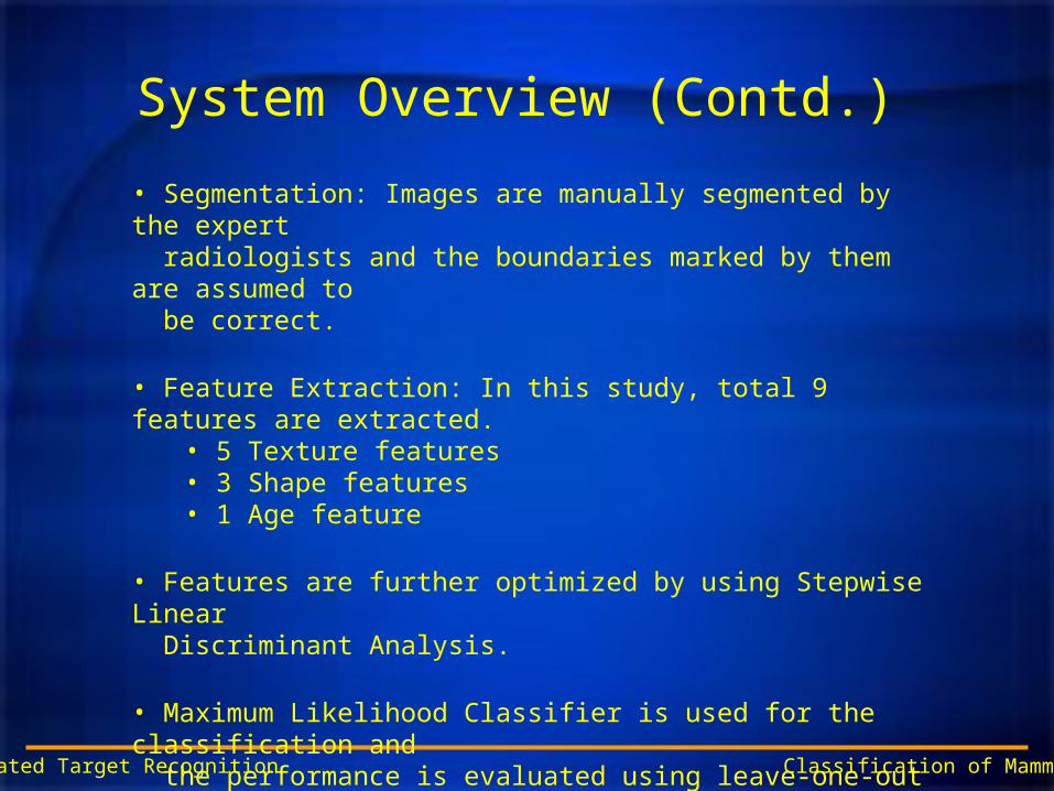
System Overview (Contd.)
ECE 8990: Automated Target Recognition Classification of Mammographic Masses
• Segmentation: Images are manually segmented by the expert radiologists and the boundaries marked by them are assumed to be correct.
• Feature Extraction: In this study, total 9 features are extracted.• 5 Texture features• 3 Shape features• 1 Age feature
• Features are further optimized by using Stepwise Linear Discriminant Analysis.
• Maximum Likelihood Classifier is used for the classification and the performance is evaluated using leave-one-out testing method.

Mammographic Dataset
ECE 8990: Automated Target Recognition Classification of Mammographic Masses
• Mammographic database for this system is obtained from the ‘Digital Database for Screening Mammography’, University of South Florida, Tampa.
• In this study, total 73 mammograms are used• 41 Benign• 32 Malignant
• The images are compressed to 8 bits/pixel using the software “heathusf v1.1.0”, provided by USF.
• Region of interest is cropped to a size of 1024 x 1024 pixels, rather than using the entire mammograms.

Mammographic Dataset (Contd.)
ECE 8990: Automated Target Recognition Classification of Mammographic Masses
(1024 x 1024)

Feature Extraction: Shape Features
ECE 8990: Automated Target Recognition Classification of Mammographic Masses
• Radial Distance Measure (RDM) is a very useful term in the shape analysis.
• RDM: It is basically the Euclidean distance calculated from the center of the tumor to the boundary pixels and normalized by dividing with the maximum length.
Mammogram Template
(1024 x 1024) (1024 x 1024)

Shape Features (Contd.)
ECE 8990: Automated Target Recognition Classification of Mammographic Masses
0 50 100 150 200 250 300 350 4000.8
0.85
0.9
0.95
1
Angle
Dis
tanc
e
Benign

Shape Features (Contd.)
ECE 8990: Automated Target Recognition Classification of Mammographic Masses
0 50 100 150 200 250 300 350 4000.4
0.6
0.8
1
Angle
Dis
tanc
e
Malignant
Shape Features (Contd.)
ECE 8990: Automated Target Recognition Classification of Mammographic Masses
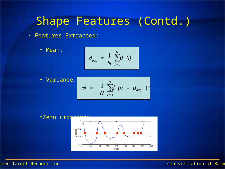
• Features Extracted:
• Mean:
• Variance:
•Zero crossings
davg = 1N
∑I = 1
N
d (i)
σ2 = 1N
∑I = 1
N
(d (i) - davg )2

Texture Analysis
• Texture features contains the information about the tonal variations in the spatial domain.
• Gray-tone spatial-dependence matrices
ECE 8990: Automated Target Recognition Classification of Mammographic Masses
6 7 8
5 * 1
4 3 2
135°90°
0°
45°
Direction considered

Texture Analysis (Cont.)• Calculation of all four distance 1 gray-tone spatial-dependence (GTSD) matrices
ECE 8990: Automated Target Recognition Classification of Mammographic Masses
0 0 1 1
0 0 1 1
0 2 2 2
2 2 3 3
4 X 4 image with 4 gray tone values
#(0,0) #(0,1) #(0,2) #(0,3)
#(1,0) #(1,1) #(1,2) #(1,3)
#(2,0) #(2,1) #(2,2) #(2,3)
#(3,0) #(31) #(3,2) #(3,3)
0 1 2 30
1
2
3
General form of GTSD matrix
4 2 1 0
2 4 0 0
1 0 6 1
0 0 1 2
6 0 2 0
0 4 2 0
2 2 2 2
0 0 2 0
4 1 0 0
1 2 2 0
0 2 4 1
0 0 1 0
2 1 3 0
1 2 1 0
3 1 0 2
0 0 2 0
0° 90° 45° 135°

Texture Analysis (Cont.)• Texture features extracted from different directions are
• For better accuracy, each texture feature in all direction are summed. Therefore there are 5 texture features instead of 20.
ECE 8990: Automated Target Recognition Classification of Mammographic Masses
i j
jipf ),(1
Energy
Uniformity of the region
Contrast
Amount of local variations
Correlation
Gray tone linear dependence
Inertia
Degree of fluctuations of image intensity
Homogeneity
i j
jipf 21 ),(
1
0 1 1
22 ),(
g g gN
n
N
i
N
j
jipnf
yx
i jyxjipij
f
),()(
3
i j
f jipji ),()( 24
i j
f jipji
),()(1
125

Feature optimization and Classification• To optimize the feature , stepwise LDA is used.
ECE 8990: Automated Target Recognition Classification of Mammographic Masses
Performance measure (PM) of N features
Sort according to PM values
Loop N times to get the optimum set of feature so that the performance measure improves.
Forward Selection
Optimum features Mfff~
,........~
,~
21
Features Nfff ,........, 21 Optimum features Mfff~
,........~
,~
21
Loop M times to get the “most” optimum set of features so as to improve the PM compared to the forward selection
“Most” optimum features Kfff~
,........~
,~
21
Backward Rejection

Feature optimization and Classification (Cont.)
• Maximum likelihood is used as a performance measure used to evaluate the features
• The classifier used is a maximum likelihood with LDA and method of testing was leave-one out
ECE 8990: Automated Target Recognition Classification of Mammographic Masses

Results and Discussions
ECE 8990: Automated Target Recognition Classification of Mammographic Masses
Feature AccuracyEnergy 0.5625Inertia 0.40625Entropy 0.65625Homogeinety 0.46875Correlation 0.46875RDM mean 0.53125RDM variance 0.46875Zero-crossings 0.375Age 0.65625
Benign MalignantBenign 34 7 0.8293Malignant 9 23 0.7188
0.7907 0.7667 0.7808
Benign MalignantBenign 38 3 0.9268Malignant 27 5 0.1563
0.5846 0.625 0.589
Table 1: Accuracies of individual features
Table 2 (a): Confusion Matrix for Texture Features
Table 2 (b): Confusion Matrix for Shape Features
Table 3: Confusion Matrix for the optimum set of features after performing stepwise LDA
Benign MalignantBenign 24 17 0.5854Malignant 19 13 0.4063
0.5581 0.4333 0.5068

Conclusion and Future Work
ECE 8990: Automated Target Recognition Classification of Mammographic Masses
•Accuracy of 78% is achieved with the combination of texture, shape and age feature
• Future work:•Better segmentation method •Implementations of rubber band straightening algorithm •Different algorithms for texture feature like gray-level run length method, gray level difference method can be implemented

References
ECE 8990: Automated Target Recognition Classification of Mammographic Masses
• “Normal mammogram classification based on regional analysis” -Yajie Sun; Babbs, C.F.; Delp, E.J.; Circuits and Systems, 2002. MWSCAS- 2002. The 2002 45th Midwest Symposium on, Volume: 2 , 4-7 Aug 2002 • http://marathon.csee.usf.edu/Mammography/Database.html• “Classification of Linear Structures in Mammographic Images - Reyer Zwiggelaar and Caroline R.M. Boggis, Division of Computer Science, University of Portsmouth, Greater Manchester Breast Screening Service, Withington Hospital, Manchester• “Gradient and texture analysis for the classification of Mammographic masses” Mudigonda, N.R.; Rangayyan, R.; Desautels, J.E.L.; Medical Imaging, IEEE Transactions on, Volume: 19, Issue: 10, Oct. 2000 Pages: 1032 – 1043• http://marathon.csee.usf.edu/Mammography/software/heat heathusf_v1.1.0.html• “Texture Features for image Classification” Haralick , R.M; Shanugam k; Dinstein, I; Systems,Man and Cybernetics,IEEE transactions on Vol.SMC- 3,No. 6 Nov. 1973 Pages 610 – 621• “Classifying Mammograhic Lesions Using Computerized Image Analysis” Kilday, J; Palmieri, F; Fox, M.D; Medical Imaging, IEEE Transactions on, Volume: 12, No.4, 1993, Pages: 664 – 669• “Classifying Mammographic Mass Shapes Using the wavelet transform Modulus-Maxima Method” Bruce, L.M; Adhami, R.R; Medical Imaging, IEEE Transactions on, Volume: 18,No.12,Dec 1999, Pages: 1170 – 1177• “Discrimination of subtly different vegetative species via hyperspectral data” Mathur, A.; Bruce, L.M.; Byrd, J; Geoscience and Remote Sensing Symposium, 2002. IGARSS '02. 2002 IEEE International Volume: 2 , 2002 Page(s): 805 –808• “A Theoretical Comparison of Texture Algorithms ” Conners, R.W Harlow, C.A; Pattern Analysis and Machine Intelligence, IEEE Transactions on, Vol: PAMI-2, No. 3, May 1980, Pages 204 - 222

ECE 8990: Automated Target Recognition Classification of Mammographic Masses

ECE 8990: Automated Target Recognition Classification of Mammographic Masses
Benign MalignantBenign 24 17 0.5854Malignant 19 13 0.4063
0.5581 0.4333 0.5068
Table 4: Confusion Matrix for all the features without age