fast parallel image registration on cpu and gpu ... -...
TRANSCRIPT
ORIGINAL RESEARCH ARTICLEpublished: 16 January 2014
doi: 10.3389/fninf.2013.00050
Fast parallel image registration on CPU and GPU fordiagnostic classification of Alzheimer’s diseaseDenis P. Shamonin1, Esther E. Bron2, Boudewijn P. F. Lelieveldt1,3, Marion Smits4, Stefan Klein2 and
Marius Staring1*, for the Alzheimer’s Disease Neuroimaging Initiative†
1 Division of Image Processing (LKEB), Department of Radiology, Leiden University Medical Center, Leiden, Netherlands2 Biomedical Imaging Group Rotterdam, Departments of Medical Informatics and Radiology, Erasmus MC, Rotterdam, Netherlands3 Intelligent Systems Group, Faculty of EEMCS, Delft University of Technology, Delft, Netherlands4 Department of Radiology, Erasmus MC, Rotterdam, Netherlands
Edited by:
Brian Avants, University ofPennsylvania, USA
Reviewed by:
Tjeerd O. Scheper, Oxford BrookesUniversity, UKBaohua Wu, University ofPennsylvania, USA
*Correspondence:
Marius Staring, Division of ImageProcessing (LKEB), Department ofRadiology, Leiden UniversityMedical Center, PO Box 9600,2300 RC Leiden, Netherlandse-mail: [email protected]†Data used in preparation of thisarticle were obtained from theAlzheimer’s Disease NeuroimagingInitiative (ADNI) database(adni.loni.ucla.edu). As such, theinvestigators within the ADNIcontributed to the design andimplementation of ADNI and/orprovided data but did not participatein analysis or writing of this report.A complete listing of ADNIinvestigators can be found at:http://adni.loni.ucla.edu/wp-content/uploads/how_to_apply/ADNI_Acknowledgement_List.pdf
Nonrigid image registration is an important, but time-consuming task in medical imageanalysis. In typical neuroimaging studies, multiple image registrations are performed,i.e., for atlas-based segmentation or template construction. Faster image registrationroutines would therefore be beneficial. In this paper we explore acceleration of the imageregistration package elastix by a combination of several techniques: (i) parallelizationon the CPU, to speed up the cost function derivative calculation; (ii) parallelization onthe GPU building on and extending the OpenCL framework from ITKv4, to speed upthe Gaussian pyramid computation and the image resampling step; (iii) exploitation ofcertain properties of the B-spline transformation model; (iv) further software optimizations.The accelerated registration tool is employed in a study on diagnostic classification ofAlzheimer’s disease and cognitively normal controls based on T1-weighted MRI. Weselected 299 participants from the publicly available Alzheimer’s Disease NeuroimagingInitiative database. Classification is performed with a support vector machine based ongray matter volumes as a marker for atrophy. We evaluated two types of strategies(voxel-wise and region-wise) that heavily rely on nonrigid image registration. Parallelizationand optimization resulted in an acceleration factor of 4–5x on an 8-core machine. UsingOpenCL a speedup factor of 2 was realized for computation of the Gaussian pyramids,and 15–60 for the resampling step, for larger images. The voxel-wise and the region-wiseclassification methods had an area under the receiver operator characteristic curve of88 and 90%, respectively, both for standard and accelerated registration. We concludethat the image registration package elastix was substantially accelerated, with nearlyidentical results to the non-optimized version. The new functionality will become availablein the next release of elastix as open source under the BSD license.
Keywords: image registration, parallelization, acceleration, OpenCL, elastix, Alzheimer’s disease
1. INTRODUCTIONImage registration is a frequently used technique in medicalimage processing. It refers to the process of automatically align-ing imaging data, where a moving (target) image IM is deformedto mimick a fixed (reference) image IF . In other words, registra-tion is the problem of finding a coordinate transformation T thatmakes IM(T) spatially aligned with IF . The quality of alignment isdefined by a cost function C. The optimal coordinate transforma-tion is estimated by minimizing the cost function with respect toT, usually by means of an iterative optimization method embed-ded in a hierarchical (multiresolution) scheme. Extensive reviewson the subject of image registration are given in Brown (1992);Maintz and Viergever (1998). Areas of application include thealignment of data sets from different modalities (Mattes et al.,2003) to fuse information, comparison of follow-up with base-line scans (Staring et al., 2007) to follow disease development,alignment of different MR sequences for extraction of quantita-tive MR parameters such as in diffusion tensor imaging or MRrelaxometry (Alexander et al., 2001; Bron et al., 2013), alignment
of pre- and post-contrast images (Rueckert et al., 1999) to aidbreast cancer detection and diagnosis, and updating treatmentplans for radiotherapy and surgery (Pennec et al., 2003).
Accordingly, most neuroimaging research also requires imageregistration. Registration is mainly needed to create a refer-ence frame, which enables comparison between subjects, betweenimage sequences and over time. This reference framework caneither be a common template space to which every subject’simage is registered (Mazziotta et al., 1995; Seghers et al., 2004;Ashburner, 2007), or a region-labeling system for exampleobtained with multi-atlas segmentation (Heckemann et al., 2006).Many different neuroimaging applications rely on such a ref-erence framework: statistical group comparisons (Friston et al.,1994), voxel-based morphometry (Ashburner and Friston, 2000),tissue segmentation (Fischl et al., 2002; Ashburner and Friston,2005), and diagnostic classification (Klöppel et al., 2008; Magninet al., 2009; Cuingnet et al., 2011). In these applications, reg-istration methods are used to align the data with the referenceframe.
Frontiers in Neuroinformatics www.frontiersin.org January 2014 | Volume 7 | Article 50 | 1
NEUROINFORMATICS
Shamonin et al. Fast parallel image registration
To create a reference frame that maps between different sub-jects, nonrigid image registration is applied, which can be verytime-consuming. Runtime depends on the specific cost function,transformation complexity, data size, and optimization strategy.The first three items have increased in complexity over the years:more complex cost functions were needed for multi-modal imageregistration (Maes et al., 1997), nonrigid transformations havemany parameters frequently generating a 106 dimensional spaceto be optimized, and data sizes have increased tremendously withthe advent of new scanners. This results in a typical runtimeof registration algorithms in the order of at best 15 min, up tohours (Klein et al., 2009a); future acquisition-side improvementsin image resolution may even increase that number. Moreover,for creating a reference frame, many registrations are required:every subject needs to be aligned with the template space, or,when using multi-atlas segmentation, every atlas image needs tobe aligned with every subject image.
One of the neuroimaging applications mentioned above isdiagnostic classification. As the incidence of Alzheimer’s Disease(AD) as well as the need for early and accurate diagnosis is dra-matically growing (Alzheimer’s Association, 2012), automatedclassification is an emerging research field. To advance the diag-nosis of AD in individual patients, machine-learning techniquescan be applied to imaging or other data. These techniques uselabeled data to train a classifier to categorize two groups (e.g.,patients and controls). Several studies demonstrated the suc-cessful classification of dementia based on atrophy using suchmachine-learning methods (e.g., Fan et al., 2008; Klöppel et al.,2008; Vemuri et al., 2008; Magnin et al., 2009; Cuingnet et al.,2011; Koikkalainen et al., 2012). The atrophy features used inthese studies are derived from structural MR using two mainapproaches: voxel-wise (e.g., Klöppel et al., 2008) and region-wise(e.g., Magnin et al., 2009) feature extraction. Voxel-wise meth-ods use a feature for each voxel in the brain, for example the graymatter (GM) density as an atrophy measure. In the region-wiseapproach, a region-labeling consisting of a set of brain regionsis used to calculate a feature, for example the GM volume ineach region of interest (ROI). Both approaches require many non-rigid image registrations: in the voxel-wise approach, to align allscans in a template space, and in the region-wise approach, toobtain a region-labeling for each individual scan using multi-atlassegmentation.
In this paper we explore the acceleration of image registrationin the context of neuroimaging applications, by a combination ofmethods. Critical registration components are parallelized, utiliz-ing the CPU as well as the GPU, certain properties of the B-splinetransformation model are exploited, and source code is opti-mized. These efforts are integrated in the popular open sourceregistration toolkit elastix (Klein et al., 2010), which is basedon the Insight ToolKit (ITK, (Ibánez et al., 2005)). elastixaims to deliver convenient access to a wide range of image reg-istration algorithms to end-users (researchers as well as medicalpractitioners). For the GPU implementation, the recently intro-duced OpenCL functionality in ITKv4 was improved, extendedand exploited. The developed functionality will become availablein the next release of elastix, as open source under the BSDlicense.
Others have also addressed registration performance by meansof parallel processing. An overview of both CPU and GPU workis given by Shams et al. (2010b). Many authors use derivative-freeoptimization techniques, and therefore focus on low dimensionaltransformations, on a cluster of computers (Warfield et al., 1998),using a GPU (Shams et al., 2010a) or an FPGA (Castro-Parejaet al., 2003). Rohlfing and Maurer (2003) proposed a schemefor nonrigid registration using finite differences for the deriva-tive computation, distributing the elements of the derivative overthe processing elements. Results were evaluated by visual inspec-tion. Saxena et al. (2010) implemented an analytical derivativebased nonrigid registration scheme on the GPU for mutual infor-mation, using CUDA. In this paper we present methods that(i) exploit both the CPU and hardware accelerators (GPU, andpotentially also the FPGA), (ii) do not require a cluster of com-puters but runs on a single computer, (iii) are based on the analyt-ical cost function derivative, enabling gradient based (stochastic)optimization, (iv) work for 2D and 3D image registration, imple-mented for various metrics and various transformation types,(v) will be made freely available, and (vi) are quantitatively val-idated to obtain similar results as the unoptimized registrationmethod.
The paper is outlined as follows. In Section 2 prelimi-nary information is given about image registration, elastix,OpenCL and ITK. The registration accelerations are describedin Section 3, together with the methodology for voxel-wise andregion-wise diagnostic classification of AD. Experiments andresults are given in Section 4, detailing the obtained speedupfactors (Section 4.2 and 4.3). In Section 4.4 an accuracy anal-ysis is made comparing original and optimized versions ofelastix. For this evaluation, we used structural MR dataof AD patients and healthy volunteers from the Alzheimer’sDisease Neuroimaging Initiative (ADNI) database. The paper isconcluded in Section 5.
2. PRELIMINARIES2.1. IMAGE REGISTRATIONImage registration is the process of aligning images, and can bedefined as an optimization problem:
µ̂ = arg minµ
C(IF, IM;µ), (1)
with IF(x) : x ∈ �F → R and IM(x) : x ∈ �M → R the d-dimensional fixed and moving image, respectively, on theirdomains �F and �M , and µ the vector of parameters of sizeN that model the transformation Tμ. The cost function C con-sists of a similarity measure S(IF, IM; µ) that defines the qualityof alignment, and optionally a regularizer. Examples of the firstare the mean square difference (MSD), normalized correlation(NC), and mutual information (MI) (Maes et al., 1997) mea-sure; examples of the last are the bending energy (Rueckertet al., 1999) and rigidity penalty term (Staring et al., 2007).Optimization is frequently performed using a form of gradientdescent:
µk + 1 = µk − ak∂C∂µ
, (2)
Frontiers in Neuroinformatics www.frontiersin.org January 2014 | Volume 7 | Article 50 | 2
Shamonin et al. Fast parallel image registration
with ak the step size at iteration k. The derivative of the costfunction can commonly be written as
∂C∂µ
= η∑
x∈�̃F
ξ (IF(x), IM(T(x)))∂T
∂µ
T
(x)∂IM
∂y(y)
∣∣∣∣y = T (x)
(3)
with ξ(·) a continuous function mapping to R, �̃F a discrete set ofcoordinates from �F , and η = 1/|�̃F| a normalization factor. Forthe MSD metric for example we have ξ(·) = IF(x) − IM(T(x)).This form holds for all the above mentioned similarity met-rics, while for regularizers a similar form can be derived. In thispaper we focus on stochastic optimization methods (Klein et al.,2007), where the derivative is computed with a small number |�̃F|of randomly drawn samples, newly selected in each iteration k.Specifically, we use the adaptive stochastic gradient descent opti-mizer (Klein et al., 2009b), which automatically computes the stepsize ak. The computation time of this step is addressed in otherwork (Qiao et al., 2014).
Image registration is usually embedded in a multi-resolutionframework, and after the optimization procedure (1) has fin-ished, a resampling of the moving image is desired to generatethe registration result IM(Tµ̂).
2.2. GPUs AND OPENCLMulti-core computers have enabled the acceleration of a widevariety of computationally intensive applications. Nowadays,another type of hardware promises even higher computationalperformance: the graphics processing unit (GPU), which hasa highly parallel hardware structure. This makes them moreeffective than general purpose CPUs for algorithms where pro-cessing of large blocks of data can be performed in parallel.The increasing computing power of GPUs gives them consid-erably higher peak computing power than CPUs. For example,NVidia’s GeForce GTX 780 GPU provides 3977 Gflop/s andAMDs HD7970 GPU 3788 Gflop/s, while Intels Xeon X5675 CPUreaches only 144 Gflop/s.
Writing parallel programs to take full advantage of this GPUpower is still a challenge. The OpenCL C programming language(www.khronos.org/opencl/) can be used to create programs thatcan be executed on one or more heterogeneous devices suchas CPUs, GPUs, FPGAs and potentially other devices developedin the future. CUDA (www.nvidia.com/object/cudahomenew.
html) on the other hand is NVidia’s C language targeted to NVidiaGPUs only. OpenCL is maintained by the non-profit technologyconsortium Khronos Group. An OpenCL program is similar to adynamic library, and an OpenCL kernel is similar to an exportedfunction from the dynamic library. In OpenCL programmers canuse OpenCL command queue execution and events to explicitlyspecify runtime dependencies between arbitrary queued com-mands, which is different from C(++) where sequential executionof commands is always implied. OpenCL is based on the C99 lan-guage specification with some restrictions and specific extensionsto the language for parallelism.
In this project we decided to adopt OpenCL for algorithmimplementation for two reasons: (i) OpenCL solutions are inde-pendent of the GPU hardware vendor, and can even be run on
other hardware accelerators, thereby broadening the applicabil-ity of this work; (ii) Our image registration package elastix islargely based on the Insight Toolkit (ITK), in which OpenCL alsowas adopted recently.
2.3. elastix AND ITKv4Parallelization is performed in the context of the image registra-tion software elastix (Klein et al., 2010), available at http://elastix.isi.uu.nl. The software is distributed as open source viaperiodic software releases under a BSD license. The software con-sists of a collection of algorithms that are commonly used to solve(medical) image registration problems. The modular design ofelastix allows the user to quickly configure, test, and com-pare different registration methods for a specific application. Acommand-line interface enables automated processing of largenumbers of data sets, by means of scripting.
elastix is based on the well-known open source InsightSegmentation and Registration Toolkit (ITK) (Ibánez et al., 2005)available at www.itk.org. This library contains a lot of image pro-cessing functionality, and delivers an extremely well tested codingframework. The ITK is implemented in C++, nightly tested, hasa rigorous collaboration process, and works on many platformsand compilers. The use of the ITK in elastix implies that thelow-level functionality (image classes, memory allocation, etc.) isthoroughly tested. Naturally, all image formats supported by theITK are supported by elastix as well. elastix can be com-piled on multiple operating systems (Windows, Linux, Mac OSX), using various compilers (MS Visual Studio, Clang, GCC), andsupports both 32 and 64 bit systems.
3. METHODSAs described in Section 2.1 the image registration algorithm con-sists of multiple parts: general tasks such as image reading andsetting up the registration pipeline, pyramid construction, theniteratively derivative computation and updating of the parametervector using (2), and finally resampling. To accelerate the reg-istration algorithm, we identified the pyramid construction, theoptimization routine and the resampling step as the most domi-nant parts in terms of performance. Acceleration possibilities forthe optimization routine are identified by recognizing paralleliza-tion options, by manual inspection of the source code, and by theuse of the Callgrind profiling tool (Weidendorfer et al., 2004), seeSection 3.1. This component of the registration algorithm is per-formed on the CPU. Both pyramid construction and resamplingare in this work off-loaded to the GPU, because these compo-nents exhibit clear opportunities for massive data parallelization,see Section 3.2. Finally, in Section 3.3, we present the methodsused for validation of the optimized registration procedure withan experiment on diagnostic classification of AD which heavilyrelies on image registration.
3.1. CPUConsidering Equation (3) we see that image registration con-stitutes a loop over the image samples as a key component ofthe algorithm. This part can be computed in parallel by dis-tributing the image samples in �̃F over different threads. This isimplemented by a fork-and-join model using the thread system
Frontiers in Neuroinformatics www.frontiersin.org January 2014 | Volume 7 | Article 50 | 3
Shamonin et al. Fast parallel image registration
of the ITK: in each iteration T threads are created (forking), Tderivatives gt
k = ∂Ct/∂µ over the sample subsets are computedin parallel (t denoting the thread id), and the results are joinedinto a single derivative. Functions that are used by the differ-ent threads were made thread-safe, and preparation functionalitywas refactored and called only once by the master thread. Wherepossible, we avoided false sharing of data (Bolosky and Scott,1993), which can substantially affect performance. This recipewas implemented in elastix for several similarity measures(MSD, NC, MI, kappa statistic), and the bending energy penaltyterm.
Parallel computation was also implemented at several otherplaces, namely for aggregation of the thread derivatives gt
k to asingle derivative gk, and for performing the update step of theoptimizer, see Equation (2). At these places some straightforwardvector arithmetic is performed on gk and µk, which are vectorsof possibly very long size (up to 106). Parallelization can be per-formed here by threads working on disjoint parts of the vectors.Implementations using the ITK thread model and OpenMP werecreated.
Again considering Equation (3) we can see that part of thecomputation is in calculating ∂T/∂µ
.= J. The Callgrind profilerconfirmed this as a performance bottleneck. For the general casethe matrix J has size d × N, N being the size of µ. In case of a B-spline transformation however, this matrix is mostly empty dueto the compact support of the B-spline basis function, resulting ina matrix of size d × dP, P = (O + 1)d � N, with O the B-splineorder (usually equal to 3). This much smaller matrix has the form:
J(x).= ∂T
∂µ(x) ≡
⎡⎣j1 · · · jP 0 · · · 0 0 · · · 0
0 · · · 0 j1 · · · jP 0 · · · 00 · · · 0 0 · · · 0 j1 · · · jP
⎤⎦ , (4)
where ji are products of the B-spline basis functions, followingfrom the definition (Rueckert et al., 1999). The derivative of theB-spline transformation is therefore a relatively small and sparsematrix, with repetitive elements, thus only P elements need to becomputed instead of d2P or even dN. Again examining (3) wecan see that the multiplication JT ∂IM
∂x can also be accelerated byomitting the empty parts.
Further optimizations to the source code resulted from acombination of Callgrind profiling and visual inspection of thesource code, and include: (i) Allocated large vectors or matri-ces only once and re-use them throughout the registration.Examples include the cost function derivative gk, the transfor-mation parameters µk and the transformation derivative J, andin the optimizer the new position µk + 1; (ii) Avoided repeatedinitializations of large arrays (fill with zeros), and additionallyoptimized this operation using std::fill (contributed backto ITKv4); (iii) Optimized some often used functions by avoid-ing ITK iterators, the use of loop unrolling, memcpy, etc; (iv)Compared to the previous implementation the amount of mem-ory accesses were reduced when interpolating the moving imagevalue and gradient; (v) Implemented gradient computation forthe linear interpolator, which can compute the moving image gra-dient ∂IM/∂x [see Equation (3)] much faster than the existing
implementation of the first order B-spline interpolator; (vi) Madeuse of a new ‘scan line’ iterator from ITKv4 with low overhead.
3.2. GPUFor implementing algorithms on the GPU we have chosen tobuild on ITKv4’s recent addition for GPU acceleration. This mod-ule wraps the OpenCL 1.2 API in an ITK-style API, while takingcare of OpenCL initialization, program compilation, and kernelexecution. It also provides convenience classes for interfacing withITK image classes and filtering pipelines.
In the OpenCL design of ITKv4 important parts of theOpenCL specification were missing, most notably the queueingmechanisms and event objects. We implemented a large part ofthe OpenCL class diagram, where classes are responsible for aspecific task conforming to the OpenCL standard. OpenCL eventobjects are used to synchronize execution of multiple kernels, incase a program consists of multiple kernels. We take advantageof the scheduling and synchronization mechanisms of OpenCLfor the implementation of the GPU version of the resampler,see Section 3.2.2, where individual kernels have to be executedin order. In addition, we have added debugging and profilingfunctionality, which are useful features during development andfor understanding performance bottlenecks of GPU architectures.A number of modifications have been made to improve design,implementation, and platform support (Intel, AMD, NVidia),thereby enhancing the existing ITKv4 GPU design.
We identified two independent registration components thatallow for parallelism: the Gaussian pyramids and the resamplingstep. The Gaussian filtering relies on a line-by-line causal andanti-causal filtering, where all image scan lines can be indepen-dently processed; The resampling step requires for every voxelthe same independent operation (transformation followed byinterpolation).
3.2.1. PyramidsIt is common to start the registration process (1) using imagesthat have lower complexity, to increase the chance of success-ful registration. To this end images are smoothed and option-ally downsampled, the latter either using linear interpolation(resampling) or by subsampling without interpolation (shrink-ing). The Gaussian pyramid is by far the most common one forimage registration, and the computation of this pyramid we tar-get to accelerate. The Gaussian filter computes infinite impulseresponse convolution with an approximation of the Gaussian ker-nel G(x; σ) = 1
σ√
2πexp
(−x2/2σ2)
(Deriche, 1990). This filter
smoothes the image in a single direction only, and is thereforesubsequently called for each direction to perform full smoothing.
The filter performs execution row-by-row for the direction xor column-by-column for the direction y, and similarly for direc-tion z. All rows or columns can be processed independently, butcolumns can only be processed when all rows have finished. Thisexecution model is therefore suitable for the GPU, by assigningeach row or column to a different thread, which can then be exe-cuted in parallel. The column kernel is scheduled to start after therow kernel, using the OpenCL queues.
To achieve better performance each thread uses the local GPUmemory, which is fastest, but this introduces a limitation on the
Frontiers in Neuroinformatics www.frontiersin.org January 2014 | Volume 7 | Article 50 | 4
Shamonin et al. Fast parallel image registration
input image size. Current GPUs usually only have 16kB of localmemory, and the algorithm allocates three floating point buffersthe size of the row/column (input, output plus temporary buffer).This results in a maximum image size of 1365 pixels, and thereforeour GPU implementation works only for images of maximum size[1365,1365] or [1365,1365,1365]. This limitation can be avoidedby using other platforms with a larger local memory (e.g., IntelCPUs allow 32kB), or by changing the algorithm altogether (e.g.,by direct convolution with a truncated Gaussian kernel).
3.2.2. ResamplingResampling is the process of computing the value IM(T(x)) forevery voxel x inside some domain. Usually, the fixed imagedomain �F is chosen, meaning that the computational complex-ity is linearly dependent on the number of voxels in the fixedimage. The procedure is simple: 1) loop over all voxels x ∈ �F ,2) compute its mapped position y = T(x), 3) obtain the mov-ing image intensity IM(y) by interpolation, since y is generally anon-voxel position, and 4) copy this value to the output image.
Notice from above that the procedure is dependent on achoice of the interpolator and the transform. Several methods forinterpolation exist, varying in quality and speed. Available imple-mentations inelastix are nearest neighbor, linear and B-splineinterpolation. There are also many flavors of transformations.The ones available in elastix in order of increasing flexibil-ity, are the translation, the rigid, the similarity, the affine, thenonrigid B-spline and the nonrigid thin-plate-spline-like trans-formations, as well as arbitrary combinations of them by functioncomposition, i.e., T(x) = Tn(. . . T2(T1(x))). The latter is fre-quently used in image registration, for example when a rigidor affine registration is performed prior to a nonrigid B-splineregistration.
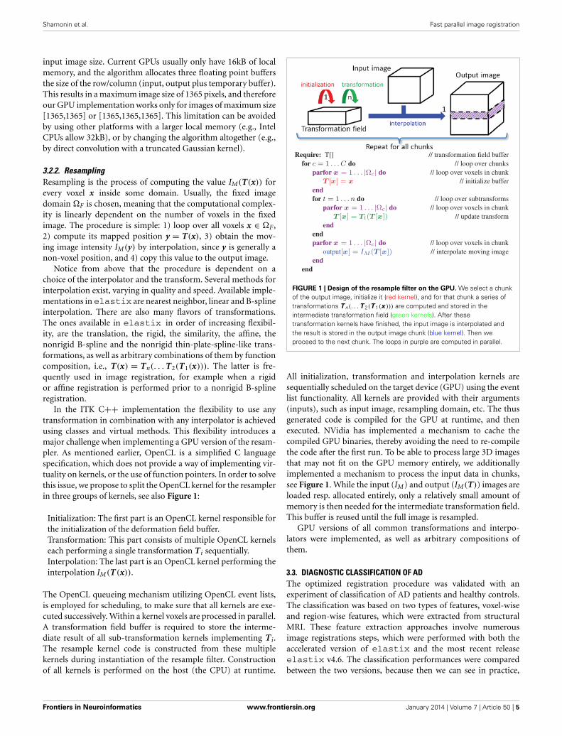
In the ITK C++ implementation the flexibility to use anytransformation in combination with any interpolator is achievedusing classes and virtual methods. This flexibility introduces amajor challenge when implementing a GPU version of the resam-pler. As mentioned earlier, OpenCL is a simplified C languagespecification, which does not provide a way of implementing vir-tuality on kernels, or the use of function pointers. In order to solvethis issue, we propose to split the OpenCL kernel for the resamplerin three groups of kernels, see also Figure 1:
Initialization: The first part is an OpenCL kernel responsible forthe initialization of the deformation field buffer.Transformation: This part consists of multiple OpenCL kernelseach performing a single transformation Ti sequentially.Interpolation: The last part is an OpenCL kernel performing theinterpolation IM(T(x)).
The OpenCL queueing mechanism utilizing OpenCL event lists,is employed for scheduling, to make sure that all kernels are exe-cuted successively. Within a kernel voxels are processed in parallel.A transformation field buffer is required to store the interme-diate result of all sub-transformation kernels implementing Ti.The resample kernel code is constructed from these multiplekernels during instantiation of the resample filter. Constructionof all kernels is performed on the host (the CPU) at runtime.
FIGURE 1 | Design of the resample filter on the GPU. We select a chunkof the output image, initialize it (red kernel), and for that chunk a series oftransformations T n(. . . T 2(T 1(x ))) are computed and stored in theintermediate transformation field (green kernels). After thesetransformation kernels have finished, the input image is interpolated andthe result is stored in the output image chunk (blue kernel). Then weproceed to the next chunk. The loops in purple are computed in parallel.
All initialization, transformation and interpolation kernels aresequentially scheduled on the target device (GPU) using the eventlist functionality. All kernels are provided with their arguments(inputs), such as input image, resampling domain, etc. The thusgenerated code is compiled for the GPU at runtime, and thenexecuted. NVidia has implemented a mechanism to cache thecompiled GPU binaries, thereby avoiding the need to re-compilethe code after the first run. To be able to process large 3D imagesthat may not fit on the GPU memory entirely, we additionallyimplemented a mechanism to process the input data in chunks,see Figure 1. While the input (IM) and output (IM(T)) images areloaded resp. allocated entirely, only a relatively small amount ofmemory is then needed for the intermediate transformation field.This buffer is reused until the full image is resampled.
GPU versions of all common transformations and interpo-lators were implemented, as well as arbitrary compositions ofthem.
3.3. DIAGNOSTIC CLASSIFICATION OF ADThe optimized registration procedure was validated with anexperiment of classification of AD patients and healthy controls.The classification was based on two types of features, voxel-wiseand region-wise features, which were extracted from structuralMRI. These feature extraction approaches involve numerousimage registrations steps, which were performed with both theaccelerated version of elastix and the most recent releaseelastix v4.6. The classification performances were comparedbetween the two versions, because then we can see in practice,
Frontiers in Neuroinformatics www.frontiersin.org January 2014 | Volume 7 | Article 50 | 5
Shamonin et al. Fast parallel image registration
in an application that makes heavy use of rigid and nonrigidregistration, if and how much the results are affected by theacceleration. In this section the methods for the classificationexperiment are explained.
3.3.1. DataData from the ADNI 1 database was used. The ADNI cohortused for our experiments is adopted from the study of Cuingnetet al. (2011), from which we selected the AD patient groupand the normal elderly control group. The inclusion criteriafor participants were defined in the ADNI GO protocol (www.adni-info.org/Scientists/AboutADNI.aspx\#). The patient groupconsisted of 137 patients (67 males, age = 76.0 ± 7.3 years,Mini Mental State Examination (MMSE) score = 23.2 ± 2.0),and the control group of 162 participants (76 males, age =76.3 ± 5.4 years, MMSE = 29.2 ± 1.0). The participants wererandomly split into two groups of the same size, a training setand a test set, while preserving the age and sex distribution(Cuingnet et al., 2011). Structural MRI (T1w) data were acquiredat 1.5T according to the ADNI acquisition protocol (Jack et al.,2008).
3.3.2. Image processingTissue segmentations were obtained for GM, white matter (WM),and cerebrospinal fluid (CSF) using SPM8 (Statistical ParametricMapping, London, UK). For estimation of intracranial volume,a brain mask was required for each subject. This brain maskwas constructed using a multi-atlas segmentation approach using30 atlases (see Section 3.3.3). We performed brain extraction(Smith, 2002) on the T1w images associated with the 30 atlases(Hammers et al., 2003; Gousias et al., 2008), checked the brainextractions visually, and adjusted extraction parameters if needed.The extracted brains were transformed to each subject’s imageand the labels were fused, resulting in a brain mask for eachsubject.
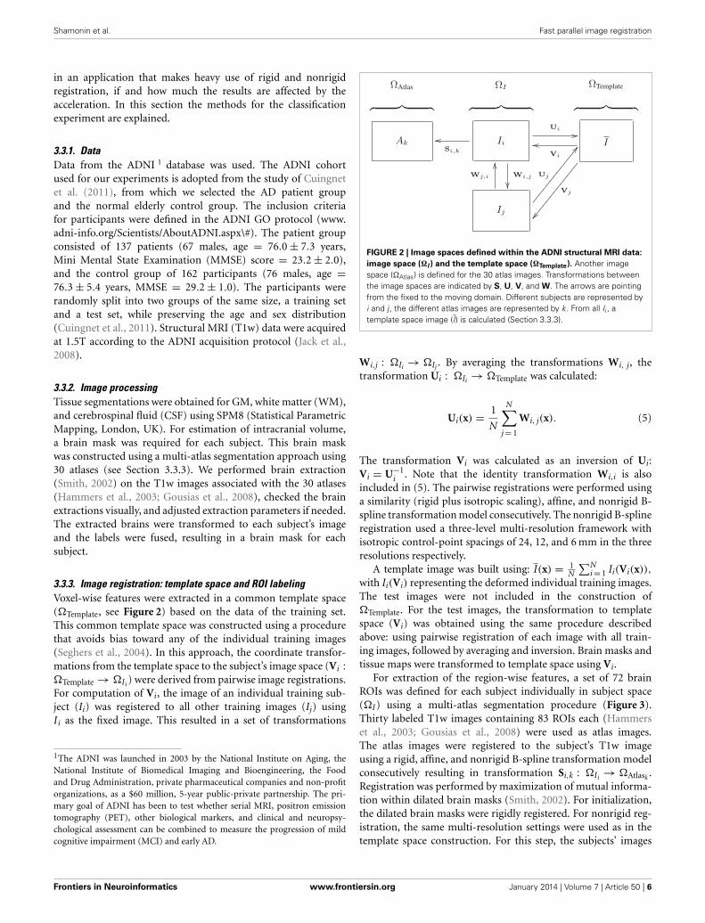
3.3.3. Image registration: template space and ROI labelingVoxel-wise features were extracted in a common template space(�Template, see Figure 2) based on the data of the training set.This common template space was constructed using a procedurethat avoids bias toward any of the individual training images(Seghers et al., 2004). In this approach, the coordinate transfor-mations from the template space to the subject’s image space (Vi :�Template → �Ii ) were derived from pairwise image registrations.For computation of Vi, the image of an individual training sub-ject (Ii) was registered to all other training images (Ij) usingIi as the fixed image. This resulted in a set of transformations
1The ADNI was launched in 2003 by the National Institute on Aging, theNational Institute of Biomedical Imaging and Bioengineering, the Foodand Drug Administration, private pharmaceutical companies and non-profitorganizations, as a $60 million, 5-year public-private partnership. The pri-mary goal of ADNI has been to test whether serial MRI, positron emissiontomography (PET), other biological markers, and clinical and neuropsy-chological assessment can be combined to measure the progression of mildcognitive impairment (MCI) and early AD.
FIGURE 2 | Image spaces defined within the ADNI structural MRI data:
image space (�I ) and the template space (�Template). Another imagespace (�Atlas) is defined for the 30 atlas images. Transformations betweenthe image spaces are indicated by S, U, V, and W. The arrows are pointingfrom the fixed to the moving domain. Different subjects are represented byi and j, the different atlas images are represented by k. From all Ii , atemplate space image (I) is calculated (Section 3.3.3).
Wi,j : �Ii → �Ij . By averaging the transformations Wi, j, thetransformation Ui : �Ii → �Template was calculated:
Ui(x) = 1
N
N∑j = 1
Wi, j(x). (5)
The transformation Vi was calculated as an inversion of Ui:Vi = U−1
i . Note that the identity transformation Wi,i is alsoincluded in (5). The pairwise registrations were performed usinga similarity (rigid plus isotropic scaling), affine, and nonrigid B-spline transformation model consecutively. The nonrigid B-splineregistration used a three-level multi-resolution framework withisotropic control-point spacings of 24, 12, and 6 mm in the threeresolutions respectively.
A template image was built using: I(x) = 1N
∑Ni = 1 Ii(Vi(x)),
with Ii(Vi) representing the deformed individual training images.The test images were not included in the construction of�Template. For the test images, the transformation to templatespace (Vi) was obtained using the same procedure describedabove: using pairwise registration of each image with all train-ing images, followed by averaging and inversion. Brain masks andtissue maps were transformed to template space using Vi.
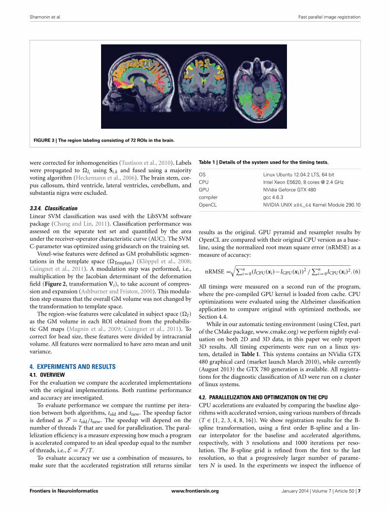
For extraction of the region-wise features, a set of 72 brainROIs was defined for each subject individually in subject space(�I) using a multi-atlas segmentation procedure (Figure 3).Thirty labeled T1w images containing 83 ROIs each (Hammerset al., 2003; Gousias et al., 2008) were used as atlas images.The atlas images were registered to the subject’s T1w imageusing a rigid, affine, and nonrigid B-spline transformation modelconsecutively resulting in transformation Si,k : �Ii → �Atlask .Registration was performed by maximization of mutual informa-tion within dilated brain masks (Smith, 2002). For initialization,the dilated brain masks were rigidly registered. For nonrigid reg-istration, the same multi-resolution settings were used as in thetemplate space construction. For this step, the subjects’ images
Frontiers in Neuroinformatics www.frontiersin.org January 2014 | Volume 7 | Article 50 | 6
Shamonin et al. Fast parallel image registration
FIGURE 3 | The region labeling consisting of 72 ROIs in the brain.
were corrected for inhomogeneities (Tustison et al., 2010). Labelswere propagated to �Ii using Si,k and fused using a majorityvoting algorithm (Heckemann et al., 2006). The brain stem, cor-pus callosum, third ventricle, lateral ventricles, cerebellum, andsubstantia nigra were excluded.
3.3.4. ClassificationLinear SVM classification was used with the LibSVM softwarepackage (Chang and Lin, 2011). Classification performance wasassessed on the separate test set and quantified by the areaunder the receiver-operator characteristic curve (AUC). The SVMC-parameter was optimized using gridsearch on the training set.
Voxel-wise features were defined as GM probabilistic segmen-tations in the template space (�Template) (Klöppel et al., 2008;Cuingnet et al., 2011). A modulation step was performed, i.e.,multiplication by the Jacobian determinant of the deformationfield (Figure 2, transformation Vi), to take account of compres-sion and expansion (Ashburner and Friston, 2000). This modula-tion step ensures that the overall GM volume was not changed bythe transformation to template space.
The region-wise features were calculated in subject space (�I)as the GM volume in each ROI obtained from the probabilis-tic GM maps (Magnin et al., 2009; Cuingnet et al., 2011). Tocorrect for head size, these features were divided by intracranialvolume. All features were normalized to have zero mean and unitvariance.
4. EXPERIMENTS AND RESULTS4.1. OVERVIEWFor the evaluation we compare the accelerated implementationswith the original implementations. Both runtime performanceand accuracy are investigated.
To evaluate performance we compare the runtime per itera-tion between both algorithms, told and tnew. The speedup factoris defined as F = told/tnew. The speedup will depend on thenumber of threads T that are used for parallelization. The paral-lelization efficiency is a measure expressing how much a programis accelerated compared to an ideal speedup equal to the numberof threads, i.e., E = F/T.
To evaluate accuracy we use a combination of measures, tomake sure that the accelerated registration still returns similar
Table 1 | Details of the system used for the timing tests.
OS Linux Ubuntu 12.04.2 LTS, 64 bit
CPU Intel Xeon E5620, 8 cores @ 2.4 GHz
GPU NVidia Geforce GTX 480
compiler gcc 4.6.3
OpenCL NVIDIA UNIX x86_64 Kernel Module 290.10
results as the original. GPU pyramid and resampler results byOpenCL are compared with their original CPU version as a base-line, using the normalized root mean square error (nRMSE) as ameasure of accuracy:
nRMSE =√∑n
i = 0(ICPU(xi)−IGPU(xi))2 /
∑ni = 0ICPU(xi)
2.(6)
All timings were measured on a second run of the program,where the pre-compiled GPU kernel is loaded from cache. CPUoptimizations were evaluated using the Alzheimer classificationapplication to compare original with optimized methods, seeSection 4.4.
While in our automatic testing environment (using CTest, partof the CMake package, www.cmake.org) we perform nightly eval-uation on both 2D and 3D data, in this paper we only report3D results. All timing experiments were run on a linux sys-tem, detailed in Table 1. This systems contains an NVidia GTX480 graphical card (market launch March 2010), while currently(August 2013) the GTX 780 generation is available. All registra-tions for the diagnostic classification of AD were run on a clusterof linux systems.
4.2. PARALLELIZATION AND OPTIMIZATION ON THE CPUCPU accelerations are evaluated by comparing the baseline algo-rithms with accelerated version, using various numbers of threads(T ∈ {1, 2, 3, 4, 8, 16}). We show registration results for the B-spline transformation, using a first order B-spline and a lin-ear interpolator for the baseline and accelerated algorithms,respectively, with 3 resolutions and 1000 iterations per reso-lution. The B-spline grid is refined from the first to the lastresolution, so that a progressively larger number of parame-ters N is used. In the experiments we inspect the influence of
Frontiers in Neuroinformatics www.frontiersin.org January 2014 | Volume 7 | Article 50 | 7
Shamonin et al. Fast parallel image registration
the number of samples |�̃F| (2000 vs. 20,000), the B-splinegrid spacing in the last resolution (10 mm vs. 5 mm, result-ing in N = 2 · 103, 9 · 103, 5 · 104 vs. N = 9 · 103, 5 · 105, 3 · 105
parameters at each resolution, respectively), and the cost function(MSD vs. NC vs. MI).
Figure 4 displays the performance results for MI, 2000 sam-ples, N = 5·104, showing the reduction in runtime per iteration,the speedup factor and the parallelization efficiency. It can beseen that using more threads steadily increases the performance,until T matches the number of CPU cores. Further increas-ing parallelization decreases performance. The efficiency plot
A
B
C
FIGURE 4 | Registration performance as a function of the number of
threads. Ri denotes the resolution number, b refers to the baselineun-accelerated algorithm, and the numbers 1–16 refer to the number ofthreads used when running the parallel accelerated algorithm. The blue lineshows ideal linear speedup. Results are shown for MI, N = 5·104,|�̃F | = 2000. (A) Shows the runtime per iteration, (B) the speedup factorF , and (C) the efficiency E.
shows that although the performance increases with increasingT, the benefits are gradually diminished. An efficiency of 60–70%(Figure 4C) was obtained for 8 threads, which is influenced bythe overhead of thread creation and destruction and by the factthat derivative joining (aggregating gt
k to gk) is not free of cost.Comparing the columns “b” and “1” we can see that the generaloptimizations described in Section 3.1 already reduce runtimefrom 27 ms to 18 ms per iteration (R2), showing the overallbenefits of these modifications. Separate tests used during devel-opment showed for example that computing ∂IM/∂x using thelinear interpolator instead of a first order B-spline was about10–15x faster stand-alone, and using the new scan line iteratorfrom ITKv4 when computing T(x) for the B-spline transform wasabout 15% faster. Overall, the image registration was acceleratedby a factor of 4–5x, when using 8 threads on our 8-core machine.
Figure 5 shows the experimental results when varying thenumber of samples |�̃F|, parameters length N and cost functiontype. The speedup remains much closer to the theoretical limitwhen using 20,000 samples instead of 2000 (Figure 5A), althoughof course the former is 10 times as slow. This may be attributedto the fact that for many samples the overhead of thread cre-ation and destruction is relatively small wrt computation time.In our current design we employ ITK’s threading mechanism,which may be suboptimal for short tasks. Figure 5B shows thatspeedup decreases when the number of parameters is large (R2).In this case vector arithmetic [joining the derivatives gt
k and per-forming the optimization step (2)] is starting to take a largerportion of an iteration. According to the Callgrind profiler about15% of the time was spend for derivative joining and an addi-tional ∼7% for threading related initialization, and ∼3% for theoptimization step. In a separate test program we tested the per-formance of these operations comparing three versions: singlethreaded, multi-threaded using ITK and multi-threaded usingOpenMP. We found that multi-threading was unsuccessful forthe optimization step, only deteriorating performance, and suc-cessful for derivative joining, mostly so when using OpenMP. Wetherefore opted to only use multi-threading with OpenMP for thederivative joining. Finally, Figure 5C shows that all metrics almostequally well benefit from parallelization. Overall, the accelerationsreduced the registration runtimes from 52, 57, and 80s to 10, 12,and 17 s for MSD, NC and MI, respectively (|�̃F| = 2000, N =5 · 104), excluding optimization step size computation (∼22s) ofthe ASGD optimizer.
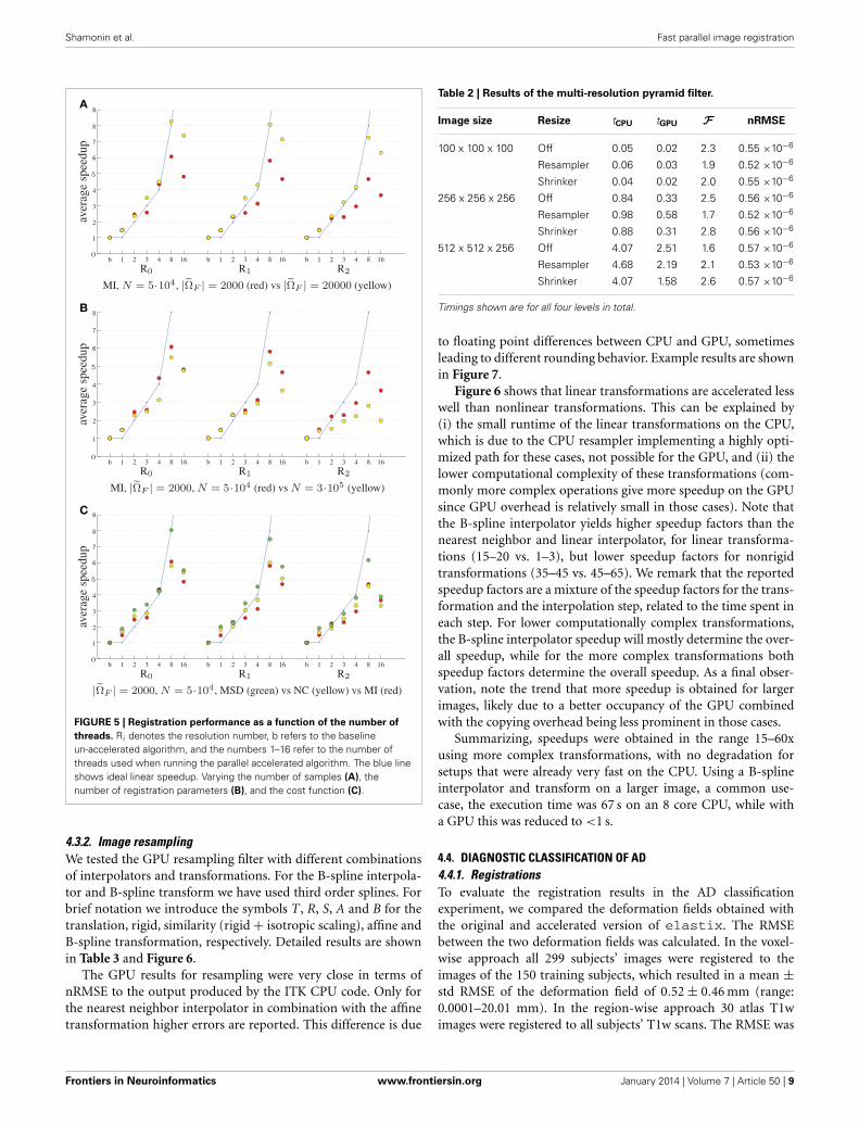
4.3. PARALLELIZATION ON THE GPU4.3.1. Gaussian image pyramidsFor testing the Gaussian pyramid accelerations we chose defaultscaling and smoothing schedules using 4 resolutions: images weredownsized by a factor of 8, 4, 2, and 1 and smoothed with aGaussian kernel with σ = 4, 2, 1 and 0 for the four resolutions,respectively. The results are shown in Table 2.
The imprecision as measured by the nRMSE was quite small(<10−6), meaning that the the CPU and GPU returns almostexactly identical smoothed images. Small speedup factors ofabout two were measured, which may be an indication that thespecific Gaussian smoothing algorithm is not very well suited foracceleration on the GPU.
Frontiers in Neuroinformatics www.frontiersin.org January 2014 | Volume 7 | Article 50 | 8
Shamonin et al. Fast parallel image registration
A
B
C
FIGURE 5 | Registration performance as a function of the number of
threads. Ri denotes the resolution number, b refers to the baselineun-accelerated algorithm, and the numbers 1–16 refer to the number ofthreads used when running the parallel accelerated algorithm. The blue lineshows ideal linear speedup. Varying the number of samples (A), thenumber of registration parameters (B), and the cost function (C).
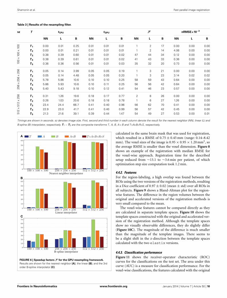
4.3.2. Image resamplingWe tested the GPU resampling filter with different combinationsof interpolators and transformations. For the B-spline interpola-tor and B-spline transform we have used third order splines. Forbrief notation we introduce the symbols T, R, S, A and B for thetranslation, rigid, similarity (rigid + isotropic scaling), affine andB-spline transformation, respectively. Detailed results are shownin Table 3 and Figure 6.
The GPU results for resampling were very close in terms ofnRMSE to the output produced by the ITK CPU code. Only forthe nearest neighbor interpolator in combination with the affinetransformation higher errors are reported. This difference is due
Table 2 | Results of the multi-resolution pyramid filter.
Image size Resize tCPU tGPU F nRMSE
100 x 100 x 100 Off 0.05 0.02 2.3 0.55 ×10⁻⁶
Resampler 0.06 0.03 1.9 0.52 ×10⁻⁶
Shrinker 0.04 0.02 2.0 0.55 ×10⁻⁶
256 x 256 x 256 Off 0.84 0.33 2.5 0.56 ×10⁻⁶
Resampler 0.98 0.58 1.7 0.52 ×10⁻⁶
Shrinker 0.88 0.31 2.8 0.56 ×10⁻⁶
512 x 512 x 256 Off 4.07 2.51 1.6 0.57 ×10⁻⁶
Resampler 4.68 2.19 2.1 0.53 ×10⁻⁶
Shrinker 4.07 1.58 2.6 0.57 ×10⁻⁶
Timings shown are for all four levels in total.
to floating point differences between CPU and GPU, sometimesleading to different rounding behavior. Example results are shownin Figure 7.
Figure 6 shows that linear transformations are accelerated lesswell than nonlinear transformations. This can be explained by(i) the small runtime of the linear transformations on the CPU,which is due to the CPU resampler implementing a highly opti-mized path for these cases, not possible for the GPU, and (ii) thelower computational complexity of these transformations (com-monly more complex operations give more speedup on the GPUsince GPU overhead is relatively small in those cases). Note thatthe B-spline interpolator yields higher speedup factors than thenearest neighbor and linear interpolator, for linear transforma-tions (15–20 vs. 1–3), but lower speedup factors for nonrigidtransformations (35–45 vs. 45–65). We remark that the reportedspeedup factors are a mixture of the speedup factors for the trans-formation and the interpolation step, related to the time spent ineach step. For lower computationally complex transformations,the B-spline interpolator speedup will mostly determine the over-all speedup, while for the more complex transformations bothspeedup factors determine the overall speedup. As a final obser-vation, note the trend that more speedup is obtained for largerimages, likely due to a better occupancy of the GPU combinedwith the copying overhead being less prominent in those cases.
Summarizing, speedups were obtained in the range 15–60xusing more complex transformations, with no degradation forsetups that were already very fast on the CPU. Using a B-splineinterpolator and transform on a larger image, a common use-case, the execution time was 67 s on an 8 core CPU, while witha GPU this was reduced to <1 s.
4.4. DIAGNOSTIC CLASSIFICATION OF AD4.4.1. RegistrationsTo evaluate the registration results in the AD classificationexperiment, we compared the deformation fields obtained withthe original and accelerated version of elastix. The RMSEbetween the two deformation fields was calculated. In the voxel-wise approach all 299 subjects’ images were registered to theimages of the 150 training subjects, which resulted in a mean ±std RMSE of the deformation field of 0.52 ± 0.46 mm (range:0.0001–20.01 mm). In the region-wise approach 30 atlas T1wimages were registered to all subjects’ T1w scans. The RMSE was
Frontiers in Neuroinformatics www.frontiersin.org January 2014 | Volume 7 | Article 50 | 9
Shamonin et al. Fast parallel image registration
Table 3 | Results of the resampling filter.
sz T tCPU tGPU F nRMSE×10−3
NN L B NN L B NN L B NN L B
100
x10
0x
100 T 1 0.00 0.01 0.25 0.01 0.01 0.01 1 2 17 0.00 0.00 0.00
T 2 0.00 0.01 0.21 0.01 0.01 0.01 1 2 14 4.06 0.00 0.00
T 3 0.38 0.39 0.60 0.01 0.01 0.02 47 44 34 0.12 0.00 0.00
T 4 0.38 0.39 0.61 0.01 0.01 0.02 41 43 33 0.36 0.00 0.00
T 5 0.36 0.36 0.56 0.01 0.01 0.03 35 32 20 0.73 0.00 0.00
256
x25
6x
256 T 1 0.05 0.14 3.99 0.05 0.05 0.19 1 3 21 0.00 0.00 0.00
T 2 0.05 0.14 4.48 0.05 0.05 0.20 1 3 23 3.14 0.02 0.02
T 3 5.78 5.86 10.6 0.10 0.10 0.25 58 59 43 0.64 0.00 0.00
T 4 5.86 5.93 10.6 0.10 0.11 0.25 56 56 42 0.64 0.00 0.00
T 5 5.40 5.43 9.18 0.10 0.12 0.41 54 46 23 0.57 0.00 0.00
512
x51
2x
256 T 1 0.31 1.26 19.6 0.18 0.17 0.77 2 8 26 0.00 0.00 0.00
T 2 0.26 1.03 20.6 0.18 0.18 0.78 1 6 27 1.26 0.00 0.00
T 3 23.4 24.4 66.7 0.41 0.40 0.96 56 62 70 0.41 0.00 0.00
T 4 22.9 23.0 41.7 0.41 0.40 0.99 56 57 42 0.45 0.00 0.00
T 5 21.3 21.6 39.1 0.39 0.44 1.47 54 49 27 0.53 0.00 0.01
Timings are shown in seconds. sz denotes image size. First, second and third number in each column denote the result for the nearest neighbor (NN), linear (L) and
B-spline (B) interpolator, respectively. T1 − T5 are the composite transforms T , A, B, A ◦ B and T◦A◦B◦R◦S, respectively.
A
B
C
FIGURE 6 | Speedup factors F for the GPU resampling framework.
Results are shown for the nearest neighbor (A), the linear (B), and the 3rdorder B-spline interpolator (C).
calculated in the same brain mask that was used for registration,which resulted in a RMSE of 0.75 ± 0.45 mm (range: 0.14–8.42mm). The voxel sizes of the image is 0.95 × 0.95 × 1.20 mm3, sothe average RMSE is smaller than the voxel dimension. Figure 8shows an example of the registration with median RMSE forthe voxel-wise approach. Registration time for the describedsetup reduced from ∼13.1 to ∼3.6 min per patient, of whichoptimization step size computation took 1.2 min.
4.4.2. FeaturesFor the region-labeling, a high overlap was found between theROIs using the two versions of the registration methods, resultingin a Dice coefficient of 0.97 ± 0.02 (mean ± std) over all ROIs inall subjects. Figure 9 shows a Bland-Altman plot for the region-wise features. The difference in the region volumes between theoriginal and accelerated versions of the registration methods isvery small compared to the mean.
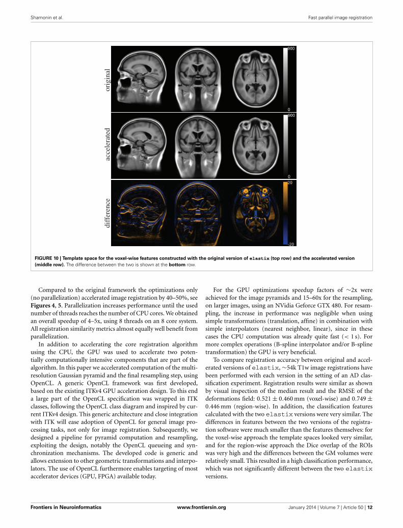
The voxel-wise features cannot be compared directly as theyare calculated in separate template spaces. Figure 10 shows thetemplate spaces constructed with the original and accelerated ver-sion of the registration method. Although the template spacesshow no visually observable differences, they do slightly differ(Figure 10C). The magnitude of the difference is much smallerthan the magnitude of the template images. There seems tobe a slight shift in the z-direction between the template spacescalculated with the two elastix versions.
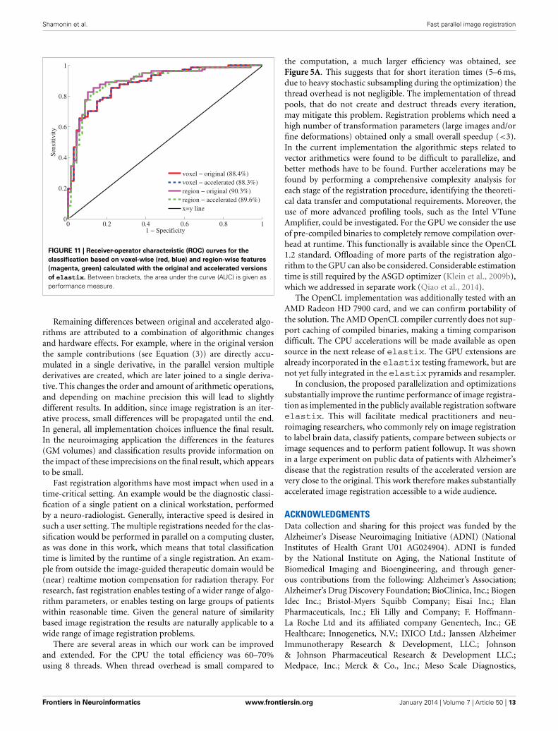
4.4.3. Classification performanceFigure 11 shows the receiver-operator characteristic (ROC)curves for the classifications on the test set. The area under thiscurve (AUC) is a measure for classification performance. For thevoxel-wise classifications, the features calculated with the original
Frontiers in Neuroinformatics www.frontiersin.org January 2014 | Volume 7 | Article 50 | 10
Shamonin et al. Fast parallel image registration
A B C
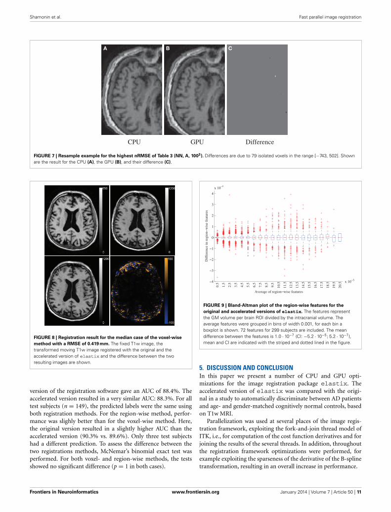
FIGURE 7 | Resample example for the highest nRMSE of Table 3 (NN, A, 1003). Differences are due to 79 isolated voxels in the range [−743, 502]. Shownare the result for the CPU (A), the GPU (B), and their difference (C).
FIGURE 8 | Registration result for the median case of the voxel-wise
method with a RMSE of 0.419 mm. The fixed T1w image, thetransformed moving T1w image registered with the original and theaccelerated version of elastix and the difference between the tworesulting images are shown.
version of the registration software gave an AUC of 88.4%. Theaccelerated version resulted in a very similar AUC: 88.3%. For alltest subjects (n = 149), the predicted labels were the same usingboth registration methods. For the region-wise method, perfor-mance was slighly better than for the voxel-wise method. Here,the original version resulted in a slightly higher AUC than theaccelerated version (90.3% vs. 89.6%). Only three test subjectshad a different prediction. To assess the difference between thetwo registrations methods, McNemar’s binomial exact test wasperformed. For both voxel- and region-wise methods, the testsshowed no significant difference (p = 1 in both cases).
FIGURE 9 | Bland-Altman plot of the region-wise features for the
original and accelerated versions of elastix. The features representthe GM volume per brain ROI divided by the intracranial volume. Theaverage features were grouped in bins of width 0.001, for each bin aboxplot is shown. 72 features for 299 subjects are included. The meandifference between the features is 1.0 · 10−7 (CI: −5.2 · 10−5; 5.2 · 10−7),mean and CI are indicated with the striped and dotted lined in the figure.
5. DISCUSSION AND CONCLUSIONIn this paper we present a number of CPU and GPU opti-mizations for the image registration package elastix. Theaccelerated version of elastix was compared with the origi-nal in a study to automatically discriminate between AD patientsand age- and gender-matched cognitively normal controls, basedon T1w MRI.
Parallelization was used at several places of the image regis-tration framework, exploiting the fork-and-join thread model ofITK, i.e., for computation of the cost function derivatives and forjoining the results of the several threads. In addition, throughoutthe registration framework optimizations were performed, forexample exploiting the sparseness of the derivative of the B-splinetransformation, resulting in an overall increase in performance.
Frontiers in Neuroinformatics www.frontiersin.org January 2014 | Volume 7 | Article 50 | 11
Shamonin et al. Fast parallel image registration
FIGURE 10 | Template space for the voxel-wise features constructed with the original version of elastix (top row) and the accelerated version
(middle row). The difference between the two is shown at the bottom row.
Compared to the original framework the optimizations only(no parallelization) accelerated image registration by 40–50%, seeFigures 4, 5. Parallelization increases performance until the usednumber of threads reaches the number of CPU cores. We obtainedan overall speedup of 4–5x, using 8 threads on an 8 core system.All registration similarity metrics almost equally well benefit fromparallelization.
In addition to accelerating the core registration algorithmusing the CPU, the GPU was used to accelerate two poten-tially computationally intensive components that are part of thealgorithm. In this paper we accelerated computation of the multi-resolution Gaussian pyramid and the final resampling step, usingOpenCL. A generic OpenCL framework was first developed,based on the existing ITKv4 GPU acceleration design. To this enda large part of the OpenCL specification was wrapped in ITKclasses, following the OpenCL class diagram and inspired by cur-rent ITKv4 design. This generic architecture and close integrationwith ITK will ease adoption of OpenCL for general image pro-cessing tasks, not only for image registration. Subsequently, wedesigned a pipeline for pyramid computation and resampling,exploiting the design, notably the OpenCL queueing and syn-chronization mechanisms. The developed code is generic andallows extension to other geometric transformations and interpo-lators. The use of OpenCL furthermore enables targeting of mostaccelerator devices (GPU, FPGA) available today.
For the GPU optimizations speedup factors of ∼2x wereachieved for the image pyramids and 15–60x for the resampling,on larger images, using an NVidia Geforce GTX 480. For resam-pling, the increase in performance was negligible when usingsimple transformations (translation, affine) in combination withsimple interpolators (nearest neighbor, linear), since in thesecases the CPU computation was already quite fast (< 1 s). Formore complex operations (B-spline interpolator and/or B-splinetransformation) the GPU is very beneficial.
To compare registration accuracy between original and accel-erated versions of elastix, ∼54k T1w image registrations havebeen performed with each version in the setting of an AD clas-sification experiment. Registration results were similar as shownby visual inspection of the median result and the RMSE of thedeformations field: 0.521 ± 0.460 mm (voxel-wise) and 0.749 ±0.446 mm (region-wise). In addition, the classification featurescalculated with the two elastix versions were very similar. Thedifferences in features between the two versions of the registra-tion software were much smaller than the features themselves: forthe voxel-wise approach the template spaces looked very similar,and for the region-wise approach the Dice overlap of the ROIswas very high and the differences between the GM volumes wererelatively small. This resulted in a high classification performance,which was not significantly different between the two elastixversions.
Frontiers in Neuroinformatics www.frontiersin.org January 2014 | Volume 7 | Article 50 | 12
Shamonin et al. Fast parallel image registration
FIGURE 11 | Receiver-operator characteristic (ROC) curves for the
classification based on voxel-wise (red, blue) and region-wise features
(magenta, green) calculated with the original and accelerated versions
of elastix. Between brackets, the area under the curve (AUC) is given asperformance measure.
Remaining differences between original and accelerated algo-rithms are attributed to a combination of algorithmic changesand hardware effects. For example, where in the original versionthe sample contributions (see Equation (3)) are directly accu-mulated in a single derivative, in the parallel version multiplederivatives are created, which are later joined to a single deriva-tive. This changes the order and amount of arithmetic operations,and depending on machine precision this will lead to slightlydifferent results. In addition, since image registration is an iter-ative process, small differences will be propagated until the end.In general, all implementation choices influence the final result.In the neuroimaging application the differences in the features(GM volumes) and classification results provide information onthe impact of these imprecisions on the final result, which appearsto be small.
Fast registration algorithms have most impact when used in atime-critical setting. An example would be the diagnostic classi-fication of a single patient on a clinical workstation, performedby a neuro-radiologist. Generally, interactive speed is desired insuch a user setting. The multiple registrations needed for the clas-sification would be performed in parallel on a computing cluster,as was done in this work, which means that total classificationtime is limited by the runtime of a single registration. An exam-ple from outside the image-guided therapeutic domain would be(near) realtime motion compensation for radiation therapy. Forresearch, fast registration enables testing of a wider range of algo-rithm parameters, or enables testing on large groups of patientswithin reasonable time. Given the general nature of similaritybased image registration the results are naturally applicable to awide range of image registration problems.
There are several areas in which our work can be improvedand extended. For the CPU the total efficiency was 60–70%using 8 threads. When thread overhead is small compared to
the computation, a much larger efficiency was obtained, seeFigure 5A. This suggests that for short iteration times (5–6 ms,due to heavy stochastic subsampling during the optimization) thethread overhead is not negligible. The implementation of threadpools, that do not create and destruct threads every iteration,may mitigate this problem. Registration problems which need ahigh number of transformation parameters (large images and/orfine deformations) obtained only a small overall speedup (<3).In the current implementation the algorithmic steps related tovector arithmetics were found to be difficult to parallelize, andbetter methods have to be found. Further accelerations may befound by performing a comprehensive complexity analysis foreach stage of the registration procedure, identifying the theoreti-cal data transfer and computational requirements. Moreover, theuse of more advanced profiling tools, such as the Intel VTuneAmplifier, could be investigated. For the GPU we consider the useof pre-compiled binaries to completely remove compilation over-head at runtime. This functionally is available since the OpenCL1.2 standard. Offloading of more parts of the registration algo-rithm to the GPU can also be considered. Considerable estimationtime is still required by the ASGD optimizer (Klein et al., 2009b),which we addressed in separate work (Qiao et al., 2014).
The OpenCL implementation was additionally tested with anAMD Radeon HD 7900 card, and we can confirm portability ofthe solution. The AMD OpenCL compiler currently does not sup-port caching of compiled binaries, making a timing comparisondifficult. The CPU accelerations will be made available as opensource in the next release of elastix. The GPU extensions arealready incorporated in the elastix testing framework, but arenot yet fully integrated in the elastix pyramids and resampler.
In conclusion, the proposed parallelization and optimizationssubstantially improve the runtime performance of image registra-tion as implemented in the publicly available registration softwareelastix. This will facilitate medical practitioners and neu-roimaging researchers, who commonly rely on image registrationto label brain data, classify patients, compare between subjects orimage sequences and to perform patient followup. It was shownin a large experiment on public data of patients with Alzheimer’sdisease that the registration results of the accelerated version arevery close to the original. This work therefore makes substantiallyaccelerated image registration accessible to a wide audience.
ACKNOWLEDGMENTSData collection and sharing for this project was funded by theAlzheimer’s Disease Neuroimaging Initiative (ADNI) (NationalInstitutes of Health Grant U01 AG024904). ADNI is fundedby the National Institute on Aging, the National Institute ofBiomedical Imaging and Bioengineering, and through gener-ous contributions from the following: Alzheimer’s Association;Alzheimer’s Drug Discovery Foundation; BioClinica, Inc.; BiogenIdec Inc.; Bristol-Myers Squibb Company; Eisai Inc.; ElanPharmaceuticals, Inc.; Eli Lilly and Company; F. Hoffmann-La Roche Ltd and its affiliated company Genentech, Inc.; GEHealthcare; Innogenetics, N.V.; IXICO Ltd.; Janssen AlzheimerImmunotherapy Research & Development, LLC.; Johnson& Johnson Pharmaceutical Research & Development LLC.;Medpace, Inc.; Merck & Co., Inc.; Meso Scale Diagnostics,
Frontiers in Neuroinformatics www.frontiersin.org January 2014 | Volume 7 | Article 50 | 13
Shamonin et al. Fast parallel image registration
LLC.; NeuroRx Research; Novartis Pharmaceuticals Corporation;Pfizer Inc.; Piramal Imaging; Servier; Synarc Inc.; and TakedaPharmaceutical Company. The Canadian Institutes of HealthResearch is providing funds to support ADNI clinical sitesin Canada. Private sector contributions are Rev November 7,2012 facilitated by the Foundation for the National Institutes ofHealth (www.fnih.org). The grantee organization is the NorthernCalifornia Institute for Research and Education, and the studyis coordinated by the Alzheimer’s Disease Cooperative Study atthe University of California, San Diego. ADNI data are dissemi-nated by the Laboratory for Neuro Imaging at the University ofCalifornia, Los Angeles. This research was also supported by NIHgrants P30 AG010129 and K01 AG030514.
FUNDINGThis research was funded by the Netherlands Organization forScientific Research (NWO), grants NWO VENI 639.021.919 and639.021.124 and NWO NRG-2010.02, and by an Erasmus MCgrant on “Advanced MR neuroimaging in presenile dementia.”
SUPPLEMENTARY MATERIALThe Supplementary Material for this article can be foundonline at: http://www.frontiersin.org/journal/10.3389/fninf.2013.00050/abstract
REFERENCESAlexander, D., Pierpaoli, C., Basser, P., and Gee, J. (2001). Spatial transformation
of diffusion tensor magnetic resonance images. IEEE Trans. Med. Imag. 20,1131–1139. doi: 10.1109/42.963816
Alzheimer’s Association. (2012). 2012 Alzheimer’s disease facts and figures.Alzheimers Dement. 8, 113–168. doi: 10.1016/j.jalz.2012.02.001
Ashburner, J. (2007). A fast diffeomorphic image registration algorithm.Neuroimage 38, 95–113. doi: 10.1016/j.neuroimage.2007.07.007
Ashburner, J., and Friston, K. (2000). Voxel-based morphometry–the methods.Neuroimage 11, 805–821. doi: 10.1006/nimg.2000.0582
Ashburner, J., and Friston, K. (2005). Unified segmentation. Neuroimage 26,839–851. doi: 10.1016/j.neuroimage.2005.02.018
Bolosky, W., and Scott, M. (1993). “False sharing and its effect on shared memoryperformance,” in SEDMS IV (San Diego, CA), 57–71.
Bron, E., van Tiel, J., Smit, H., Poot, D., Niessen, W., Krestin, G., et al. (2013).Image registration improves human knee cartilage T1 mapping with delayedgadolinium-enhanced MRI of cartilage (dGEMRIC). Eur. Radiol. 23, 246–252.doi: 10.1007/s00330-012-2590-3
Brown, L. (1992). A survey of image registration techniques. ACM Comput. Surv.24, 325–376. doi: 10.1145/146370.146374
Castro-Pareja, C. R., Jagadeesh, J. M., and Shekhar, R. (2003). FAIR: a hardwarearchitecture for real-time 3-D image registration. IEEE Trans. Info. Tech. Biomed.7, 426–434. doi: 10.1109/TITB.2003.821370
Chang, C., and Lin, C. (2011). LIBSVM: a library for support vector machines.ACM TIST 2, 27:1–27:27. doi: 10.1145/1961189.1961199
Cuingnet, R., Gerardin, E., Tessieras, J., Auzias, G., Lehéricy, S., Habert, M., et al.(2011). Automatic classification of patients with Alzheimer’s disease from struc-tural MRI: a comparison of ten methods using the ADNI database. Neuroimage56, 766–781. doi: 10.1016/j.neuroimage.2010.06.013
Deriche, R. (1990). Fast algorithms for low-level vision. IEEE Trans. Pattern. Anal.Mach. Intell. 12, 78–87. doi: 10.1109/34.41386
Fan, Y., Resnick, S., Wu, X., and Davatzikos, C. (2008). Structural andfunctional biomarkers of prodromal Alzheimer’s disease: a high-dimensional pattern classification study. Neuroimage 41, 277–285. doi:10.1016/j.neuroimage.2008.02.043
Fischl, B., Salat, D., Busa, E., Albert, M., Dieterich, M., Haselgrove, C., et al. (2002).Whole brain segmentation: automated labeling of neuroanatomical structuresin the human brain. Neuron 33, 341–355. doi: 10.1016/S0896-6273(02)00569-X
Friston, K., Holmes, A., Worsley, K., Poline, J.-P., Frith, C., and Frackowiak, R.(1994). Statistical parametric maps in functional imaging: a general linearapproach. Hum. Brain Mapp. 2, 189–210. doi: 10.1002/hbm.460020402
Gousias, I., Rueckert, D., Heckemann, R., Dyet, L., Boardman, J., Edwards, A., et al.(2008). Automatic segmentation of brain MRIs of 2-year-olds into 83 regions ofinterest. Neuroimage 40, 672–684. doi: 10.1016/j.neuroimage.2007.11.034
Hammers, A., Allom, R., Koepp, M. J., Free, S. L., Myers, R., Lemieux, L., et al.(2003). Three-dimensional maximum probability atlas of the human brain,with particular reference to the temporal lobe. Hum. Brain Mapp. 19, 224–247.doi: 10.1002/hbm.10123
Heckemann, R., Hajnal, J., Aljabar, P., Rueckert, D., and Hammers, A.(2006). Automatic anatomical brain MRI segmentation combininglabel propagation and decision fusion. Neuroimage 33, 115–126. doi:10.1016/j.neuroimage.2006.05.061
Ibánez, L., Schroeder, W., Ng, L., and Cates, J. (2005). The ITK Software Guide.New York, NY: Kitware Inc.
Jack, C., Bernstein, M., Fox, N., Thompson, P., Alexander, G., Harvey, D., et al.(2008). The Alzheimer’s disease neuroimaging initiative (ADNI): MRI methods.J. Magn. Reson. Imag. 27, 685–691. doi: 10.1002/jmri.21049
Klein, A., Andersson, J., Ardekani, B., Ashburner, J., Avants, B., Chiang,M., et al. (2009a). Evaluation of 14 nonlinear deformation algorithmsapplied to human brain MRI registration. Neuroimage 46, 786–802. doi:10.1016/j.neuroimage.2008.12.037
Klein, S., Pluim, J., Staring, M., and Viergever, M. (2009b). Adaptive stochasticgradient descent optimisation for image registration. Int. J. Comput. Vis. 81,227–239. doi: 10.1007/s11263-008-0168-y
Klein, S., Staring, M., Murphy, K., Viergever, M., and Pluim, J. (2010). elastix: atoolbox for intensity-based medical image registration. IEEE Trans. Med. Imag.29, 196–205. doi: 10.1109/TMI.2009.2035616
Klein, S., Staring, M., and Pluim, J. (2007). Evaluation of optimization methods fornonrigid medical image registration using mutual information and B-splines.IEEE Trans. Image. Proc. 16, 2879–2890. doi: 10.1109/TIP.2007.909412
Klöppel, S., Stonnington, C., Chu, C., Draganski, B., Scahill, R., Rohrer, J., et al.(2008). Automatic classification of MR scans in Alzheimer’s disease. Brain 131,681–689. doi: 10.1093/brain/awm319
Koikkalainen, J., Pölönen, H., Mattila, J., van Gils, M., Soininen, H., Lötjönen,J., et al. (2012). Improved classification of Alzheimer’s disease data viaremoval of nuisance variability. PLoS ONE 7:e31112. doi: 10.1371/journal.pone.0031112
Maes, F., Collignon, A., Vandermeulen, D., Marchal, G., and Suetens, P. (1997).Multimodality image registration by maximization of mutual information.IEEE Trans. Med. Imag. 16, 187–198. doi: 10.1109/42.563664
Magnin, B., Mesrob, L., Kinkingnéhun, S., Pélégrini-Issac, M., Colliot, O., Sarazin,M., et al. (2009). Support vector machine-based classification of Alzheimer’sdisease from whole-brain anatomical MRI. Neuroradiology 51, 73–83. doi:10.1007/s00234-008-0463-x
Maintz, J., and Viergever, M. (1998). A survey of medical image registration. Med.Image. Anal. 2, 1–36. doi: 10.1016/S1361-8415(01)80026-8
Mattes, D., Haynor, D., Vesselle, H., Lewellen, T., and Eubank, W. (2003). PET-CTimage registration in the chest using free-form deformations. IEEE Trans. Med.Imag. 22, 120–128. doi: 10.1109/TMI.2003.809072
Mazziotta, J., Toga, A., Evans, A., Fox, P., and Lancaster, J. (1995). A probabilisticatlas of the human brain: theory and rationale for its development the inter-national consortium for brain mapping (ICBM). Neuroimage 2, 89–101. doi:10.1006/nimg.1995.1012
Pennec, X., Cachier, P., and Ayache, N. (2003). Tracking brain deformations intime sequences of 3D US images. Pattern Recognit. Lett. 24, 801–813. doi:10.1016/S0167-8655(02)00183-6
Qiao, Y., Lelieveldt, B., and Staring, M. (2014). “Fast automatic estimation of theoptimization step size for nonrigid image registration,” in SPIE Medical Imaging:Image Processing, Proceedings of SPIE eds N. Karssemeijer and E. Samei (SanDiego, CA).
Rohlfing, T., and Maurer Jr., C. (2003). Nonrigid image registration in shared-memory multiprocessor environments with application to brains, breasts, andbees. IEEE Trans. Inf. Technol. Biomed. 7, 16–25. doi: 10.1109/TITB.2003.808506
Rueckert, D., Sonoda, L., Hayes, C., Hill, D., Leach, M., and Hawkes, D. (1999).Nonrigid registration using free-form deformations: application to breast MRimages. IEEE Trans. Med. Imag. 18, 712–721. doi: 10.1109/42.796284
Frontiers in Neuroinformatics www.frontiersin.org January 2014 | Volume 7 | Article 50 | 14
Shamonin et al. Fast parallel image registration
Saxena, V., Rohrer, J., and Gong, L. (2010). “A parallel GPU algorithm for mutualinformation based 3D nonrigid image registration,” in Euro-Par 2010 - ParallelProcessing, Vol. 6272 of LNCS (Ischia), 223–234.
Seghers, D., D’Agostino, E., Maes, F., Vandermeulen, D., and Suetens, P. (2004).“Construction of a brain template from MR images using state-of-the-artregistration and segmentation techniques,” in Proceedings of InternationalConference Medical Image Computing and Computer-Assisted Intervention(Berlin/Heidelberg: Springer-Verlag), 696–703.
Shams, R., Sadeghi, P., Kennedy, R., and Hartley, R. (2010a). Parallel computationof mutual information on the GPU with application to real-time registra-tion of 3D medical images. Comput. Methods Prog. Biomed. 99, 133–146. doi:10.1016/j.cmpb.2009.11.004
Shams, R., Sadeghi, P., Kennedy, R., and Hartley, R. (2010b). A survey of medi-cal image registration on multicore and the GPU. Signal Process. Mag. IEEE 27,50–60. doi: 10.1109/MSP.2009.935387
Smith, S. (2002). Fast robust automated brain extraction. Hum. Brain Map. 17,143–155. doi: 10.1002/hbm.10062
Staring, M., Klein, S., and Pluim, J. (2007). A rigidity penalty term for nonrigidregistration. Med. Phys. 34, 4098–4108. doi: 10.1118/1.2776236
Tustison, N., Avants, B., Cook, P., Zheng, Y., Egan, A., Yushkevich, P. et al. (2010).N4ITK: improved N3 bias correction. IEEE Trans. Med. Imag. 29, 1310–1320.doi: 10.1109/TMI.2010.2046908
Vemuri, P., Gunter, J., Senjem, M., Whitwell, J., Kantarci, K., Knopman, D.,et al. (2008). Alzheimer’s disease diagnosis in individual subjects usingstructural MR images: validation studies. Neuroimage 39, 1186–1197. doi:10.1016/j.neuroimage.2007.09.073
Warfield, S., Jolesz, F., and Kikinis, R. (1998). A high performance computingapproach to the registration of medical imaging data. Parallel Comput. 24,1345–1368. doi: 10.1016/S0167-8191(98)00061-1
Weidendorfer, J., Kowarschik, M., and Trinitis, C. (2004). “A tool suite forsimulation based analysis of memory access behavior,” in Proceedings of 4thInternational Conference on Computatation Science (Krakow), 440–447.
Conflict of Interest Statement: The authors declare that the research was con-ducted in the absence of any commercial or financial relationships that could beconstrued as a potential conflict of interest.
Received: 28 August 2013; paper pending published: 05 December 2013; accepted: 21December 2013; published online: 16 January 2014.Citation: Shamonin DP, Bron EE, Lelieveldt BPF, Smits M, Klein S and Staring M(2014) Fast parallel image registration on CPU and GPU for diagnostic classificationof Alzheimer’s disease. Front. Neuroinform. 7:50. doi: 10.3389/fninf.2013.00050This article was submitted to the journal Frontiers in Neuroinformatics.Copyright © 2014 Shamonin, Bron, Lelieveldt, Smits, Klein and Staring. This is anopen-access article distributed under the terms of the Creative Commons AttributionLicense (CC BY). The use, distribution or reproduction in other forums is permit-ted, provided the original author(s) or licensor are credited and that the originalpublication in this journal is cited, in accordance with accepted academic prac-tice. No use, distribution or reproduction is permitted which does not comply withthese terms.
Frontiers in Neuroinformatics www.frontiersin.org January 2014 | Volume 7 | Article 50 | 15