fasciae and topography
TRANSCRIPT

Fasciae and topography
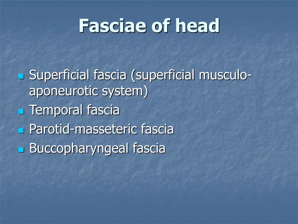
Superficial fascia (superficial musculo-aponeurotic system)
Temporal fascia
Parotid-masseteric fascia
Buccopharyngeal fascia
Fasciae of head

Superficial musculo-aponeurotic system

Temporal fascia
• Lamina superficialis
• Lamina profunda
Between these two layers there is a temporal space, filled with cellulose and fatty tissue (2).

Parotid-masseteric fascia
Unique fascia that forms a capsule for parotid salivary gland and covers m. masseter

Buccopharyngeal fascia
Buccopharyngeal fascia covers the posterior section of m.buccinator and superior constrictor of the pharynx
Place, where buccopharyngeal fascia inserts into pterygoid process of sphenoid bone, is called pterygomandibular raphe

Anatomic borders of the neck
Upper border:
Protuberantiaoccipitalis externa
Linea nuchaesuperior
Top of the mastoid process of the temporal bone
Ramus and base of the mandible

Anatomic borders of the neck
Inferior border:- Line passing along clavicles and jugular notch of the sternum
- Line connecting acromial ends of clavicles and spinous process of the VII cervical vertebrae

Regions of the neck
A - Regio sternocleidomastoidea
B - Regio cervicalis posterior
C - Regio cervicalis lateralis
D - Regio cervicalis anterior
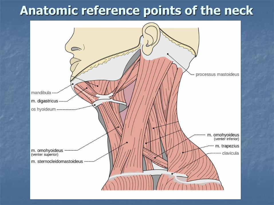
Anatomic reference points of the neck

Triangles of neck
Omotrapezoid triangle
Retromandibular fossa

Lingual triangle (Pirogov`s triangle)
Borders:
the posterior border of the mylohyoid
intermediate tendon of the digastricus
the hypoglossalnerve

Fasciae of neck
1 - m. trapezius; 2 - deep muscles of the neck; 3 - oesophagus; 4 - mm. scaleni; 5 - a. carotis communis, v. jugularis interna et n. vagus; 6 - m. omohyoideus; 7 - m. sternocleidomastoideus; 8 - platysma; 9 - trachea; 10 - spatium previscerale; 11 - gl. thyroidea

Fasciae of neck
I - superficial
F.propria
- II - superficial layer
- III - deep layer
IV - endocervical
V - prevertebral

Fascia superficialis
Part of the common superficial fascia of the body
Contains platysma
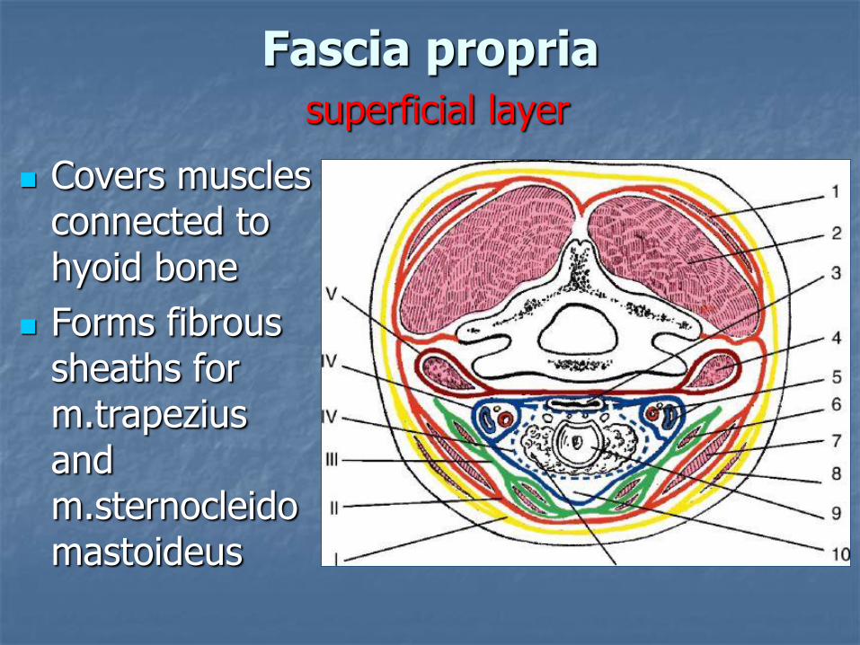
Fascia propriasuperficial layer
Covers muscles connected to hyoid bone
Forms fibrous sheaths for m.trapeziusand m.sternocleidomastoideus

Fascia propriadeep layer
Is present only in middle part of neck
Forms fibrous sheaths for infrahyoid muscles
Joins with the superficila layer along the m. omohyoideus and linea alba of neck.

Endocervical fascia Covers intercervical
space
It has 2 layers Parietal (cover all
organs together). Forms fibrous canal for nerves and vessels of neck.
Visceral (covers each organ separately) –dotted line.
Pretracheal(previsceral) space -in front of layers.

Prevertebral fascia
Covers deep muscles of neck anteriorly
Retrovisceral space – between prevertebraland intracervical fascias

Spaces of neck
Interaponeuroticalsuprasternal space (between the layers of f.propria – II and III)
Cellulose tissue with lymphatic nodules and arcus venosusjuguli
Recesssusretoristernocleidomastoideus(Gruber`s pockets)

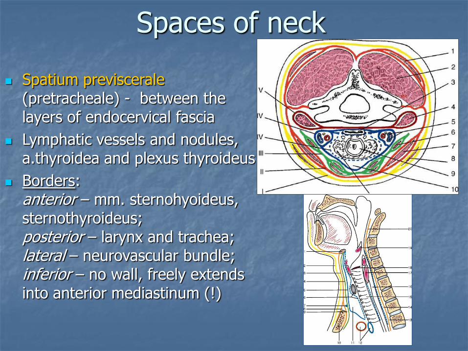
Spatium previscerale(pretracheale) - between the layers of endocervical fascia
Lymphatic vessels and nodules, a.thyroidea and plexus thyroideus
Borders: anterior – mm. sternohyoideus, sternothyroideus; posterior – larynx and trachea; lateral – neurovascular bundle; inferior – no wall, freely extends into anterior mediastinum (!)
Spaces of neck

Spatium retroviscerale (retropharingeale) – between endocervical (IV) and prevertebral (V) fascias
It continuous with the posterior mediastinum (!)
Spaces of neck

Mediastinum

Spaces of neck
Spatium interscalenum – between the anterior, middle scalene muscles and the first rib. It transmits the subclavian artery and the brachial plexus.
Spatium antescalenum – in front of the anterior scalene muscle. It transmits the subclavian vein.

Spaces of neck
Spatium interaponeuroticum laterale – between lamina superficialis fasciae colli propriae (II) and fascia prevertebralis (V)
Connected with axillary fossa

Axillary fossa
1 – axillary fossa2 – border of m. latissimus dorsi3 – border of m. pectoralis major4 – m. serratus anterior

Axillary cavityis bordered by:
anteriorly –mm.pectorales major et minor
posteriorly –m.latissimus dorsi, m.teres major and m.subscapularis
medially– m.serratusanterior
laterally – humerus and mm. of anterior side of the arm

Axillary cavity
Apertura superior
Apertura inferior

Foramen trilaterum:
above –m.subscapularis
below – m.teresmajor
laterally – long head of m.tricepsbrachii
Foramen quadrilaterum:
above –m.subscapularis
below – m.teresmajor
medially – long head of m.triceps brachii
laterally – humerus

Canalis nervi radialis(canalis humeromusularis)
anteriorly –humerus
posteriorly– m.tricepsbrachii
Inlet:
between the upper and middle thirds of the arm on medial side
humerus and the medial and lateral heads of the triceps muscle
Outlet:
between the middle and lower thirds of the arm on lateral side
It is bounded by the brachialis and brachioradialismuscles

Fasciae of the arm
• The axillary fascia • The deltoid fascia• The brachial fascia (medial and lateral intermuscular septum)

The lateral and medial bicipital grooves(sulcus bicipitalis lateralis et medialis)

Fasciae of the arm

Cubital fossa

The medial ulnar groovelies between flexor carpi ulnaris and the flexor digitorumsuperficialis (laterally). It transmits the ulnar nerve, artery and veins.
Grooves between forearm muscles

The median groove lies between the flexor carpi radialis (laterally) and the flexor digitorumsuperficialis (medially). It transmits the median nerve.
Grooves between forearm muscles

The lateral radial groove lies between brachioradialis (laterally) and the flexor carpi radialis(medially). It transmits the radial nerve, artery and veins.
Grooves between forearm muscles

Fasciae of the arm (dorsal surface)

Fasciae of the arm (palmar surface)


tunnel
n. medianus
tendons
Carpal tunnel syndrome

Fasciae of thorax
The superficial fascia
The pectoral fascia
The thoracic fascia
The endothoracic fascia

Topography of thorax
A - Tr. clavipectorale
B - Tr. pectorale
C - Tr. subpectorale
A
B
C

Lines of thorax
Anterior median
sternalis
parasternalis
medioclavicularias
anterior, median, posterior axial
scapularis
paravertebralis
Posterior median

Lines of thorax

Diaphragm


Hiatal hernia

Triangles of back
Triangle for lungs auscultation
Inferior – superior border of m.latissimus dorsi
Medial – inferior border of m.trapezius
Lateral – posterior border of m.infraspinatus
Triangle for lungs auscultation

Triangles of backTrigonum lumbale(Petit trigonum)
Inferior – crista iliaca
Medial – anterior border of m. latissimus dorsi
Lateral – posterior border of m.obliquusexternusabdominis

Regions of the abdomen
epigastrium
mesogastrium
hypogastrium
Linea bicostarum(X costae)
Linea bispinarum(spina iliaca
anterior superior)

Epigastrium
right hypochondric
left hypochondric
epigastric
Vertical lines:- midclavicular line (mammary line)- correspond to the lateral borders
of m.rectus abdominis

Mesogastrium
umbilical
right lateral
left lateral

Hypogastrium
pubic
right inguinal
left inguinal
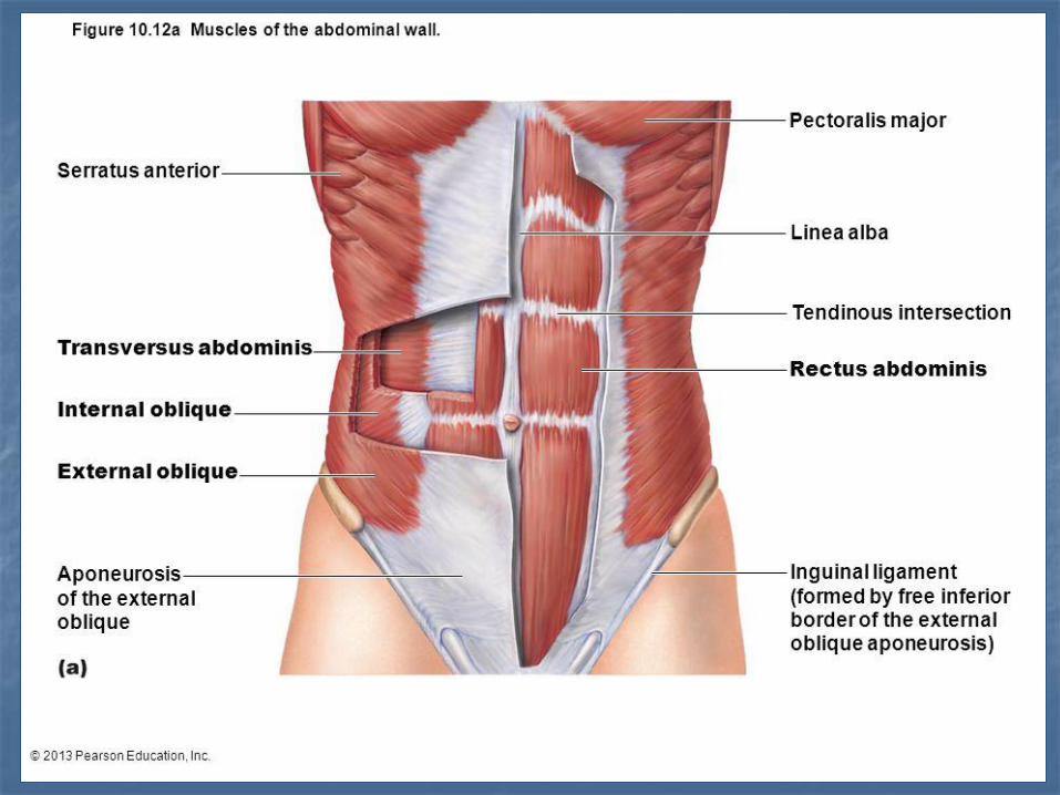

Fascia superficialis
Fascia propria(covers muscular part of m. obliquusexternusabdominis and fuses with aponeurosis of this muscle)
Fascia transversalis
Fasciae of abdomen

Rectal sheath
It is formed by aponeuroses of wide muscles of abdomen and transverse fascia
Its structure is different in upper and lower parts of the abdomen

1 - m. rectus abdominis; 2 - mm. intercostales; 3 – posterior layer of aponeurosis of m. obliquus internus abdominis; 4 – anterior layer of aponeurosis of m. obliquus internusabdominis; 5 - m. transversus abdominis; 6 - m. obliquus internus abdominis; 7 - m. obliquus externus abdominis; 8 – anterior plate of m.rectus abdominis sheath
Split of the aponeurosis of m.obliquus internusabdominis to 2 layers (anterior and posterior) with further formation of the sheath of m.rectus abdominis

Anterior wall• Aponeurosis of m.obliquus
abdominis externus• ½ of aponeurosis of
m.obliquus abdominis internus
Posterior wall• ½ of aponeurosis of m.obliquus
abdominis internus• Aponeurosis of m.transversus
abdominis• Fascia transversalis

Anterior wall• Aponeuroses of all 3 muscles
Posterior wall• Fascia transversalis

Linea arcuata(Douglas line)
Linea semilunaris(Spiegel line)
- border between muscle fibers and
aponeurosis of m.transversus
abdominis

Linea alba abdominis tendinous raphe extending from xiphoid process to the
symphysis pubis and pubic crest
formed by interlacing of wide muscles of the abdomen
it is used for laparotomy in surgery
umbilical ring is usually bypassed at the left side during the surgery

Umbilical hernia

The inguinal ligament (Poupart ligament) is formed by the margin of the aponeurosis of m.obliquus externus abdominis between the superior iliac spine and the pubic tubercule.

Inguinal canal
It is located above medial half of inguinal ligament
It has 4 walls and 2 openings (apertures)
Contains round ligament of uterus (ligamentum teres) or spermatic code (funiculusspermaticus)


Walls of inguinal canal
Anterior – aponeurosis of m.obliquus abdominisexternus (9)
Inferior – lig.inguinale (4)
Superior – borders of m.obliquus abdominisinternus (10) and m.transversus abdominis (2)
Posterior – fascia transversalis (1)

Deep ring Depression of transverse fascia
(corresponds to fossa inguinalis lateralis)


Superficial ring (4 walls)
Superior – crus mediale of lig.inguinale
Inferior – crus lateral of lig.inguinale
Lateral – fibrae intercrurales
Medial – lig.reflexum

Inguinal hernia


(Gimbernat`s)
(Poupart`s)

Lacuna vasorum
located medially
for passage of femoral artery (4) and vein (5)
1 – lacuna musculorum; 2 – arcus ilipectineus; 3 – lig. inguinale; 4 – a. femoralis; 5 – v. femoralis; 6 – lacuna vasorum; 7 – anulus femoralis; 8 – deep inguinal lymphatic nodule; 9 – lig. Lacunare; 10 – funiculus spermaticus; 11 – m. pectineus; 12 – n., a. et v. obturatoriae; 13 – n. femoralis; 14 – m. iliopsoas

Lacuna musculorum
located laterally
m.iliopsoas and n.femoralispass through it
1 – lacuna musculorum; 2 – arcus ilipectineus; 3 – lig. inguinale; 4 – a. femoralis; 5 – v. femoralis; 6 – lacuna vasorum; 7 – anulus femoralis; 8 – deep inguinal lymphatic nodule; 9 – lig. Lacunare; 10 – funiculus spermaticus; 11 – m. pectineus; 12 – n., a. et v. obturatoriae; 13 – n. femoralis; 14 – m. iliopsoas

Femoral canal
Appears only in case of femoral herniation
It is located below the inguinal ligament
It has 3 walls and 2 openings

Walls of femoral canal
in front – fusion of lig.inguinale with cornu superius of hiatus saphenus
posteriorly – fascia pectinea
laterally – vena femoralis

Superficial ring - hiatus saphenus

Deep (femoral) ringis bordered by:
anteriorly – lig.inguinale
posteriorly – pectineal ligament
laterally – vena femoralis
medially – lig.lacunare
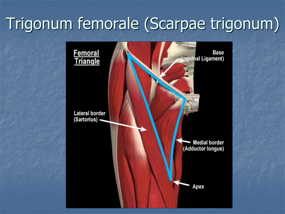
Trigonum femorale (Scarpae trigonum)

Canalis adductorius
located at the thigh
has 3 walls and 3 openings
vessels and nerves pass through it from anterior side of the thigh to popliteal fossa

Walls of canalis adductorius
lateral – m.vastusmedialis (1)
medial –m.adductormagnus (2)
anterior – septum between these muscles (3)
1
2
3
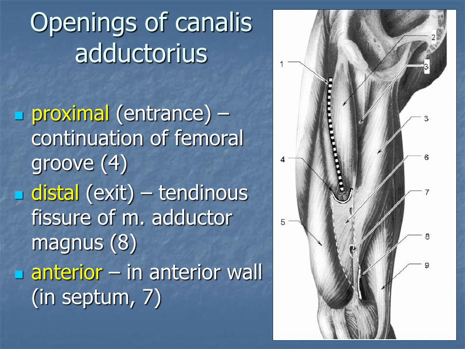
Openings of canalisadductorius
proximal (entrance) –continuation of femoral groove (4)
distal (exit) – tendinousfissure of m. adductor magnus (8)
anterior – in anterior wall (in septum, 7)


Canalis cruropopliteus
Between deep (m.tibialisposterior) and superficial (m.soleus) muscles
Has two walls and three openings
Transmits tibial vessels and nerves

Walls of canalis cruropopliteus
anterior – m.tibialisposterior
posterior – m.soleus

Openings of canaliscruropopliteus
Entrance – below the arcustendineus of m.soleus (1)
Exit – medially from lig.calcaneus (5) (medially from Achill tendon - 4)
Anterior – in membranainterossea cruris (not shown)

Canalis musculoperoneus superior
located between:
Upper part of fibula
m.fibularis (peroneus) longus
Transmits n. fibularis(peroneus) superficialis

Canalis musculoperoneus inferior
located (Б) between:
inferior part of fibula
m. flexor hallucis longus
m. tibialis posterior
Transmits a. et v. fibulares

Cross section through the shin in the middle third : 1 – fascia of the shin; 2 – posterior intermuscular partition of the shin; 3 - fibula; 4 – m. peroneus longus; 5 – anterior intermuscular partition of the shin; 6 – membrana interossea; 7 – m. extensor digitorum longus; 8 – m. tibialis anterior; 9 - tibia; 10 – m. tibialis posterior; 11 – m. flexor digitorum longus; 12 – m. flexor hallucis longus; 13 – m. soleus; 14 – m. gastrocnemius
Canaliscruropopliteus
Canalismusculoperoneus
inferior
Canalismusculoperoneus
superior


Articulatio tarsi transversa(Chopart`s joint) combines two joints:- Calcaneocuboid joint- Talonavicular joint
Ligamentum bifurcatum:- lig. calcaneonaviculare- lig. calcaneocuboideum
- “key” of Chopart`s joint
Articulatio tarsimetatarsales(Lisfrank`s joint)
Ligg. cuneometatarsaliainterossea:
The “key” of Lisfrank`sjoint – cuneometatarsalinterosseus ligament between medial cuneiform bone and second metatarsal bone