factors affecting the variable outcomes of juvenile nasopharyngeal
TRANSCRIPT

Department of Otorhinolaryngology – Head and Neck Surgery, Helsinki University Central Hospital,
Department of Pathology, Haartman Institute, University of HelsinkiHelsinki, Finland
FACTORS AFFECTING THE VARIABLE OUTCOMES OF JUVENILE
NASOPHARYNGEAL ANGIOFIBROMA
Suvi Renkonen
ACADEMIC DISSERTATION
To be publicly discussed, with the permission of the Faculty of Medicine, University of Helsinki, in the Small Lecture Hall of Haartman Institute, Haartmaninkatu 3,
on Saturday, September 29th, 2012, at 12 noon.

ISBN 978-952-10-8228-3 (paperback)ISBN 978-952-10-8229-0 (PDF)
Unigrafi aHelsinki 2012
3
ABSTRACT
Background Juvenile Nasopharyngeal Angiofi broma (JNA) is a rare, benign tumour affecting adolescent males. Due to its capability to erode bone, JNA can be accompanied by life-threatening complications when growing into the cranium. The etiology of JNA is unknown, as are the factors determing the variable growth patterns of individual tumours. The treatment of choice for JNA is surgery, but high recurrence rates or persistent growth are charasteristic.
Aims The aim of this study was to elucidate factors determing the variable outcomes of JNA. For this purpose the various surgical techniques used to resect JNA during the past 40 years at the Helsinki University Central Hospital (HUCH) were investigated, immunohistochemistry was performed and the gene copy number and mRNA expression data of two phenotypically different JNA tumours were combined to seek for processes putatively determining their growth pattern.
Methods Retrospective clinicopathological data of all JNA patients diagnosed and treated, during the years 1970–2011, were reviewed. By immunohistochemistry, we investigated the cellular distribution and expression levels of C-KIT, C-MYC, BMI-1, GLUT-1, tenascin-C, syndecan-1, syndecan-2 and SYK in JNA samples, in order to fi nd their possible correlations with clinicopathological factors. Comparative genomic hybridisation and gene expression analyses were performed for two phenotypically different tumour samples to investigate the possible processes leading to more aggressive growth. A gene ontology enrichment analysis for the in silico translated proteins of genes with altered gene expression status was performed to detect the categories with enrichments.
Results The primary recurrence rate was 37% and correlated with the surgical approach used, transpalatal approach linked with higher risk of recurrence, vascularity of the tumour and young age of the patient. Contrary to previous reports, C-KIT was expressed in both stromal and endothelial cells of JNA samples and was more prominent in slit-like vessels. A correlation between its endothelial expression and cellular density of the tumour was found. Frequent stromal tenascin-C expression had a strong correlation with vessel density and higher tumour stage. When present, endothelial GLUT-1 expression correlated with higher tumour stage. SYK expression was found to correlate with lower tumour stage.
Between the low and high stage JNA tumours studied, 1245 genes showed at least a two-fold- change in their expression. The corresponding proteins of these transcripts were enriched in different biological processes, e.g., hypoxia in the low stage tumour and signal transduction activity in the high stage tumour.

4
Conclusions The type of surgical approach seemed crucial for the outcome. Other clinical factors affecting the recurrence rate were young age of the patient and vascular density of the tumour. In selective cases of JNA, we could thus carefully recommend the consideration of antiangiogenic treatment. Based on our fi nding of protein expressions, we suggest that at least C-KIT, tenascin-C, GLUT-1 and SYK might be substantial in the growth of JNA by having an effect on tumour cellularity, vessel density and the stage of the tumour. Based on our result of GLUT-1 positivity correlating with higher tumour stage, we suggest that JNAs are not likely to be vascular malformations. Although we were able to identify gene expression changes that relate to particular biological processes, the assessing of clinically relevant molecular profi les of JNA still requires further characterization. In the future, combinating molecular profi ling data from several studies will be useful to better understand the molecular background of this rare tumour.
5
CONTENTS
Abstract ..............................................................................................................3List of original publications ..............................................................................7Abbreviations .....................................................................................................81 Introduction ...............................................................................................102 Review of the literature ...............................................................................11 2.1 Juvenile nasopharyngeal angiofi broma................................................11 2.2 Proteins known to act as tumour markers for cell proliferation and
angiogenesis ........................................................................................ 213 Aims of the study ........................................................................................264 Materials and Methods ..............................................................................27 4.1 Patient material ....................................................................................27 4.2 Histology of the tumours and immunohistochemistry
(Study II, III and IV) ........................................................................... 28 4.3 Scoring of immunostainings (Study II, III and IV) .............................29 4.4 Sample preparation for gene expression and comparative genomic
hybridization microarrays (Study IV) .................................................29 4.5 Gene expression data analysis (Study IV) .......................................... 30 4.6 Comparative genomic hybridization analysis (Study IV) .................. 30 4.7 Gene enrichment set analysis (Study IV) ............................................ 30 4.8 Protein-protein interaction networks (Study IV) ................................ 31 4.9 Statistical analysis (Study II, III and IV) ............................................. 31 4.10Ethical considerations (Study I, II, III and IV) .................................. 315 Results ........................................................................................................32 5.1 Patients (Study I, II, III and IV) ..........................................................32 5.2 Histology of the JNA samples (Study II) ............................................33 5.3 Protein expression (Study II and III) ..................................................34 5.4 Correlations with clinocopathological factors (Study II and III) ........35 5.5 Survival analysis (Study I and III) .......................................................35 5.6 Gene copy number aberrations (Study IV)..........................................36 5.7 Gene expression levels (Study IV) .......................................................36 5.8 Gene ontology (Study IV) ....................................................................376 Discussion .................................................................................................. 38Acknowledgements ..........................................................................................507 References ..................................................................................................52

6
7
LIST OF ORIGINAL PUBLICATIONS
This thesis is based on the following original publications:
I Renkonen S, Hagström J, Vuola J, Niemelä M, Porras M, Kivivuori S-M, Leivo I, Mäkitie AA. The changing surgical management of juvenile nasopharyngeal angiofi broma. Eur Arch Otorhinolaryngol. 2011 268:599-607
II Renkonen S, Häyry V, Heikkilä P, Leivo I, Haglund C, Mäkitie AA, Hagström J. Stem cell-related proteins C-KIT, C-MYC and BMI-1 in juvenile nasopharyngeal angiofi broma--do they have a role? Virchows Arch. 2011 458:189-95
III Renkonen S, Heikkilä P, Haglund C, Mäkitie AA, Hagström J. Tenascin-C, GLUT-1 and syndecan-2 expression in JNA – correlations to vessel density and tumor stage. Head & Neck (Epub 7/2012)
IV Renkonen S, Kankainen M, Hagström J,Haglund C, Monni O, Mäkitie AA. Systems level analysis of clinically different phenotypes of juvenile nasopharyngeal angiofi bromas. Laryngoscope (accepted)
The publications are referred to in the text by their Roman numerals.
8
ABBREVIATIONS
APC Adenomatous polyposis coliAR Androgen receptoraRNA Amplifi ed ribonucleic acidBMI-1 B-lymphoma Moloney murine leukemia virus insertionCGH Comparative genomic hybridisationCK1 Casein kinase 1C-MYC Myelocytomatosis viral oncogene homologCNV Copy number variationCT Computed tomographyCUSA Cavitron ultrasonic surgical aspiratorDNA Deoxyribonucleic acidDvl Segment polarity protein dishevelled homologECM Extracellular matrixFz Frizzled receptorGLUT-1 Glucose transporter 1GO Gene ontologyGSK3b Glycogen synthase kinase 3b GW Great wingHIF Hypoxia induced factorHS High stage tumourICA Internal catorid arteryIGF Insulin-like growth factorITF Infratemporal fossaIH Infantile hemangioma JNA Juvenile nasopharyngeal angiofi bromaLrp 5/6 Low density lipoprotein receptor-related proteins-5/6LS Low stage tumourMRI Magnetic resonance imagingmRNA Messanger ribonucleic acidMSCT Multislice computed tomographyNP NasopharynxOH HydroxideP Phosphoric groupPcG Plycomb group proteinPDGF Platelet derived growth factorPDGFR Platelet derived growth factor receptor
9
PHDs Prolyl hydroxylasesPINA Protein interaction network analysisPMF Pterygomaxillar fossaPPF Pterygopalatal fossaPVA Polyvinyl alcoholRAC2 Ras-related C3 botulinum toxin substrate 2RNA Ribonucleic acidSPF Sphenopalatine foramenTFG Transforming growth factorTNC Tenascin-CTNF Tumour necrosis factorUb UbiquitinVEGF Vascular endothelial growth factorVHL Von Hippel Lindau protein
10
1 Introduction
1 INTRODUCTION
Juvenile nasopharyngeal angiofi broma (JNA) is a rare tumour with unknown etiology. It is considered to be the most common benign neoplasm of the nasopharynx, accounting for 0.5 % of all head and neck neoplasms (1, 2). JNA originates from the superior margin of sphenopalatine foramen, also serving as the route for the sphenopalatine artery, a branch of internal maxillar artery. Histologically JNA is composed of various amounts of fi brous stroma, rich in collagen and fi broblasts, and numerous blood vessels with different calibers and shapes. Although histologically classifi ed as benign, JNA’s growth pattern can be locally destructive and due to its capability to erode bone, it can have life-threathning complications, when extending intracranially. Also fatal epistaxis and intraoperative massive haemorrhage are possible severe complications of JNA.
The extremely rich vascularisation, complex anatomical structures of the cranium and the possible aggressive growth pattern of JNA make its treatment a challenge. Surgical removal is accepted to be the treatment of choice, and traditionally the used approaches have been transpalatal, transmaxillar, Le Fort I osteotomy, and infratemporal fossa craniotomy. In recent decades, preoperative selective embolization of the feeding arteries has substantially decreased intraoperative bleeding and enabled the resection of even the largest tumours. Yet, recurrence rates as high as 50% have been associated with JNA, usually thought to be caused by persistent growth of the residual tumour. Trying to overcome this conundrum, new treatment strategies have been developed. Among these, endoscopic resection has gained popularity, due to its minimal invasiveness, low morbidity rate and most importantly, typically low rates of recurrence. The consensus is, however, that also this approach has its limitations and is not recommended for tumours with extensive growth to the cranium or to the cavernous sinuses.
The pathogenesis and stem cell population of JNA remain unknown. It has even been suggested that JNA is not a true neoplasm. A common feature of JNA is strong vasculature. Current studies on the proliferation of stromal and vascular components of this tumour have supported the hypothesis of angiogenic factors playing a centred role in its pathogenesis. This is of great importance, because during the last decade, increased understanding of angiogenesis has allowed the development of novel therapeutic substances, also considered for use in some selective cases of JNA. Uncovering new aspects of the molecular mechanisms of the pathogenesis of JNA will hopefully continue to provide future targets for individual treatment strategies for JNA patients.
11
2 REVIEW OF THE LITERATURE
2.1 JUVENILE NASOPHARYNGEAL ANGIOFIBROMA
Juvenile nasopharyngeal angiofi broma (JNA) is thought to represent only 0.05–0.5% of all head and neck tumours. Its incidence is approximately 1:150 000 per year, although incidences in Egypt and India have been reported to be slightly higher that in USA and Europe (1, 3). Also males with fair-skin and red hair are more often affected (4). It is generally thought that the anatomical origin of JNA is the superior part of sphenopalatine foramen, in the posterolateral wall of the nasal cavity. However, by analyzing CT and MRI data, Lloyd et al. concluded, that the site of origin is in pterygopalatal fossa (5). In most cases, JNA receives its blood supply from the internal maxillary artery, a branch of external carotid artery, and it is suggested that this tumour originates from vascular plexus that remains after involution of fi rst branchial artery (6). In selective cases, feeders from internal carotid artery may also be present (7). The fact that almost all JNA cases occur in young males, is postulated to be explained by the impact of androgens and the precence of hormonal receptors expressed in this tumour (8–10).
HISTOLOGY AND PATHOGENESIS
JNA is known to spread in the submucosal plane and macroscopically this tumour is a soft, multilobular, rounded, circumscribed, non-encapsulated, mucosa covered mass (11, 12). Depending on the vascular component, the colour of the tumour varies from pink to pale-whitish (13), and the base of the tumour tends to be sessile or pedunculated (4). The mean size of JNA is ca. 4 cm, but some tumours might grow much larger (4, 14). Histologically, JNA consists of an abnormal vascular network, fi brous connective tissue stroma and stromal cells (4, 14). It has been shown that the larger the tumour, the greater the proportion of fi brous tissue and the lesser the number of vessels (15, 16). Mast cells may be seen, but otherwise no infl ammatory elements are present, as far as there is no surface ulceration (4, 14). Mitotic activity and nuclear atypia are not typical features of JNA. The vasculature is mostly made up by either thin-walled, slit-like (“staghorn”) or dilated vessels, calibres ranging from capillary to large, patulous vessels (4, 14). Muscular lining in the vessels walls may be totally absent, focal and pad-like, or circumferential (4, 14). The stroma consists of stromal cells of different shapes (plump spindle, angular, stellate-shaped) and varying amounts of fi ne and coarse collagen fi bers (4). Because
12
2 Review of the literature
of the single layered intratumoural vessel walls with few or no smooth muscle cells and no ability to contract, together with the rigidity of the fi brous stroma, JNAs are highly hemorrhagic (17–19). Sarcomatous transformation, typically following radiation therapy, is utmost uncommon (4, 14).
Several aspects in the pathogenesis of JNA remain unsolved. Because JNA affects mostly adolescent males and regression of these tumours has been reported to happen after the full development of secondary sex characteristics, there are several studies aiming to elucidate the role of androgen and estrogen receptors in JNA’s growth and regression (9, 20–22). However, the results still seem inconsistent. Because of its rich vascularity, JNA has been proposed to be a hemangioma (2, 23), a vascular malformation (17), or an incomplete regression of the fi rst brachial artery (2, 24). Due to the histologically benign nature of JNA, only a few studies exist aiming to solve the role of tumour suppressors and oncogenes in JNA. The expression of tumour suppressor adenomatous polyposis coli (APC) gene in JNA has been investigated in numerous studies, led by the discovery of increased frequency of JNA among patients with familial adenomatous polyposis (FAP) (25–27). The gene product of APC is known to regulate β-catenin, a downstream activator of the Wnt signalling pathway, as APC is needed for the degradation of β-catenin (25). Although evidence of APC gene mutations have not been found (27), activating β-catenin gene alterations are frequently detected in JNA (25). More interestingly, β-catenin is known to act as a co-activator of androgen receptors, increasing androgen sensitivity – which might explain the exclusive occurrence of JNA in adolescent males (28). The roles of tumour suppressor p53 and oncogenes C-MYC and C-FOS in JNA remain to be confi rmed (29, 30), while no mutations have been reported link with oncogenes Ki-ras, Ha-ras and Her-2/neu (30, 31).
ROUTES OF INVASION FROM THE SITE OF ORIGIN
Starting to grow from its site of origin, the superior margin of sphenopalatine foramen, JNA typically fi rst reaches the nasal septum and the posterior parts of the nose, causing mass effect and obstruction of the airways (13). As a general view of JNA growth, it is believed that from here the growth proceeds and destroys the anterior face of the sphenoidal sinus invading the sinus. Medially the tumour grows towards nasal fossa and extends to the posterior parts of the middle turbinate. Laterally it extends towards pterygomaxullary fossa, destroying the posterior wall of maxillary sinus and causing the charasteristc Holman-Miller sign of anterior bowing of the posterior maxillar wall (4). Finally, the tumour invades the infratemporal fossa and the middle cranial fossa (13, 32, 33). On the whole, approximately 30% of all JNAs extend to the orbit and 10–20% to the cranium (34, 35).
13
SYMPTOMS AND DIAGNOSIS
The classical clinical presentation of JNA is a triad of nasal obstruction, epistaxis and tumour mass in the nasopharynx (1, 12, 36–40). When invading the orbit, it can also cause proptosis and diplopia, when causing pressure to the optic nerve changes in visus and when reaching intracranial region, even cranial nerve palsy (7, 13, 41, 42). The diagnosis of JNA is based on clinical history and examination together with radiological fi ndings. Biopsy is contraindicated because of the risk of severe bleeding. Because nasal obstruction and epistaxis are common symptoms, one should consider also other possible diagnosis options, such as infl ammatory polyps, angiomatous polyps, nasopharyngeal cysts, nasopharyngeal malignancies, e.g., fi brosarcomas, upper maxillary malignancies, adenoid hypertrophy and cervical vertebra chordomas (13). The defi nite diagnosis is histologically verifi ed after surgery.
As biopsies should be avoided, imaging studies are of great importance in the management of JNAs. These are needed preoperatively for establishing the diagnosis and the extent of the tumour and postoperatively for showing the extent of any persistent recurrent disease (5). Head and neck computed tomographic (CT) scans have been used since 1974 to identify typical patterns of bone erosion by JNA (43). For over a decade, however, also magnetic resonance imaging (MRI) has been used, offering improved soft tissue resolution and thus facilitating the assessment of spatial relationships between tumour and intracranial structures (34, 38, 44–46). Traditionally, CT is regarded as the most important imaging technique for JNA because the complexity of bony structures at skull base invaded by the tumour demands highly accurate bone imaging. In their review article of the evolution of the management of JNA, Nicolai et al. suggest the use of either multislice computed tomography (MSCT) or magnetic resonance imaging (MRI) and recommend the administration of contrast agent before use, to confi rm the clinical suspicion pattern of vascularisation and to estimate the extension of the lesion (2). The authors also state that: “Without doubt, MR better depicts cancellous bone invasion” (2), leading to the conclusion that as CT better depicts the damage of cortex, both techniques have their strengths, when concerning the invasion of skull base.
14
2 Review of the literature
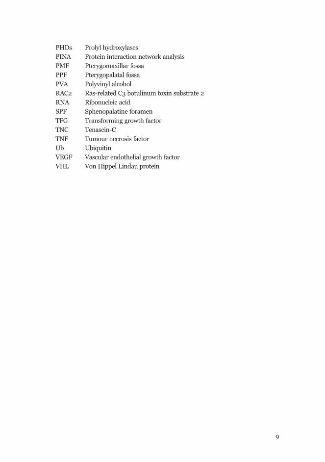
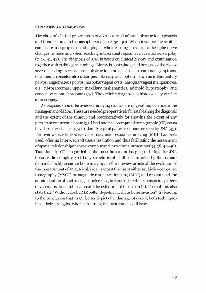
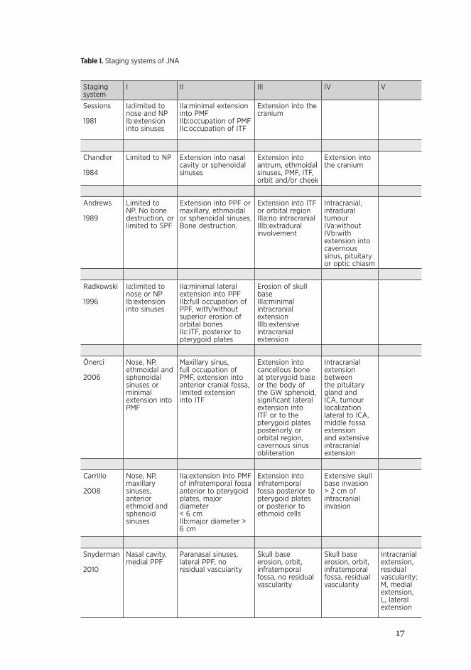
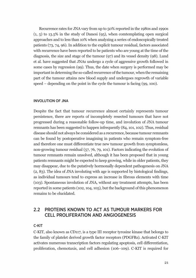
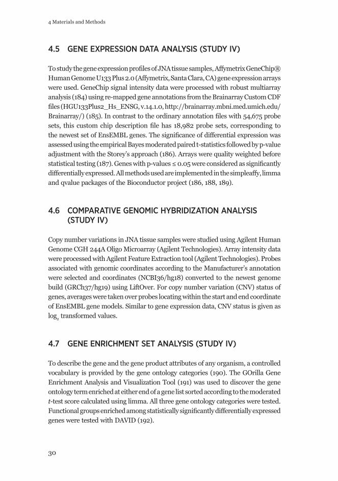
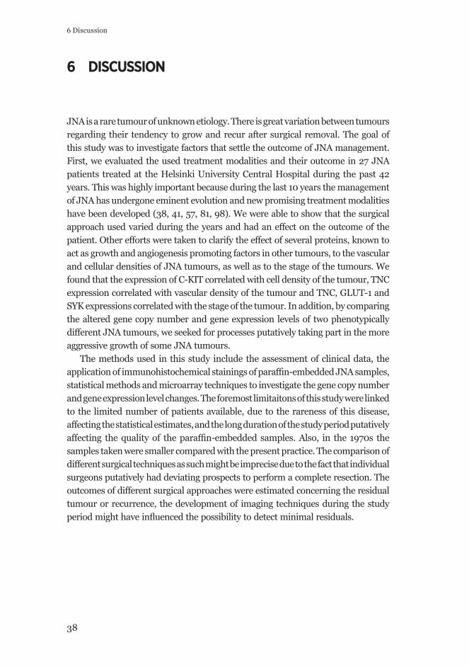
Figure I. Axial CT image showing tumour invading the pterygopalatinal fossa and remodelling its bony walls (arrow). Coronal CT image showing tumour extension to the Vidian canal (left arrow) and widening it compared to the normal left side (right arrow). The foramen rotundum is intact (little arrow).
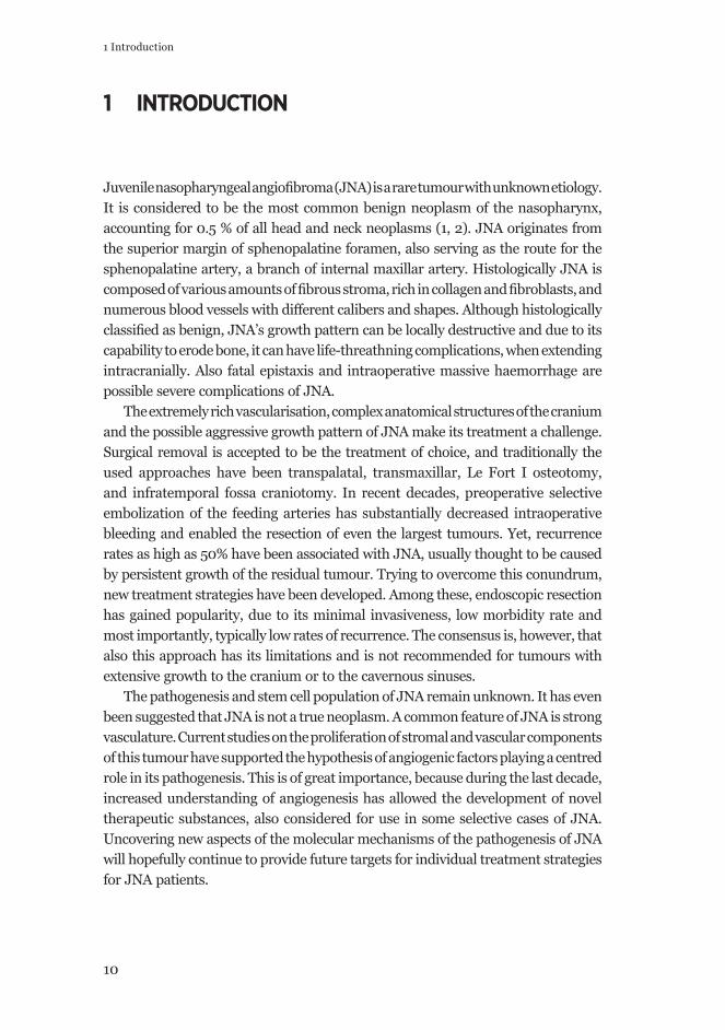
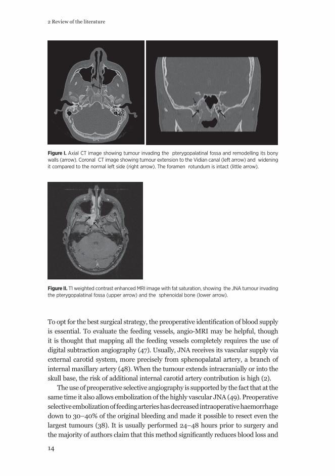
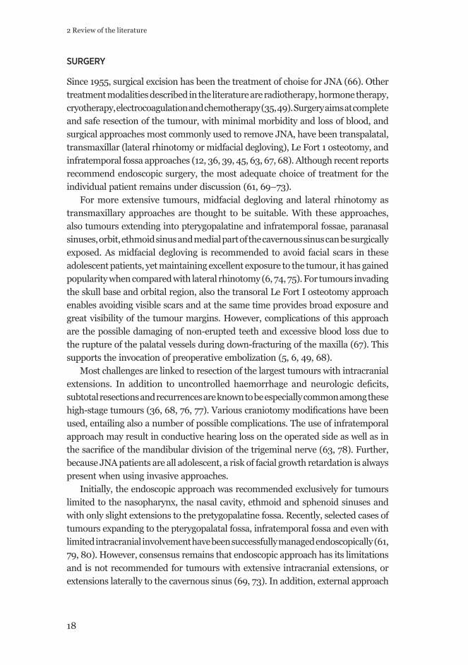
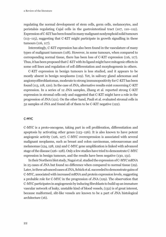
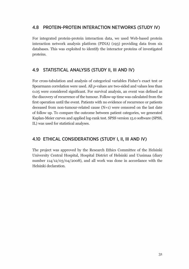
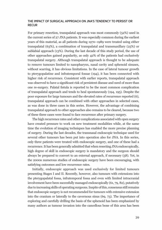
Figure II. T1 weighted contrast enhanced MRI image with fat saturation, showing the JNA tumour invading the pterygopalatinal fossa (upper arrow) and the sphenoidal bone (lower arrow).
To opt for the best surgical strategy, the preoperative identifi cation of blood supply is essential. To evaluate the feeding vessels, angio-MRI may be helpful, though it is thought that mapping all the feeding vessels completely requires the use of digital subtraction angiography (47). Usually, JNA receives its vascular supply via external carotid system, more precisely from sphenopalatal artery, a branch of internal maxillary artery (48). When the tumour extends intracranially or into the skull base, the risk of additional internal carotid artery contribution is high (2).
The use of preoperative selective angiography is supported by the fact that at the same time it also allows embolization of the highly vascular JNA (49). Preoperative selective embolization of feeding arteries has decreased intraoperative haemorrhage down to 30–40% of the original bleeding and made it possible to resect even the largest tumours (38). It is usually performed 24–48 hours prior to surgery and the majority of authors claim that this method signifi cantly reduces blood loss and
15
results in better surgical visualizion (6, 13, 38, 50). Regardless, Lloyd et al. state that embolization might lead to poor tumour removal, should there be deep invasion of the sphenoid (5). Arguments also exist about increased recurrence following embolization (39) and additionally, that embolization of the vessels from internal carotid artery might lead to occlusion of the ophthalmic or cerebral arteries with severe consequences (41, 51). However, in their review in 2012, Nicolai et al. state that during the last decade refi nements of techniques together with the availability of new materials have minimized the risk of leaving residual material, and the new small particles and microcatheters have enabled to avoid the risk of neurologic sequelae following the unintentional embolization of small vessels supplied from internal catorid artery (2).
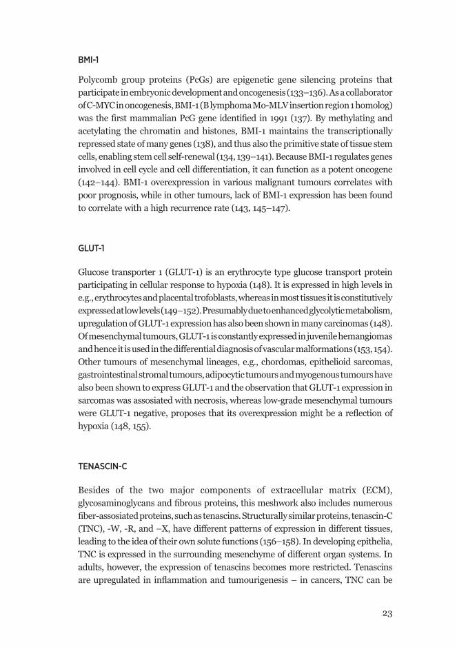
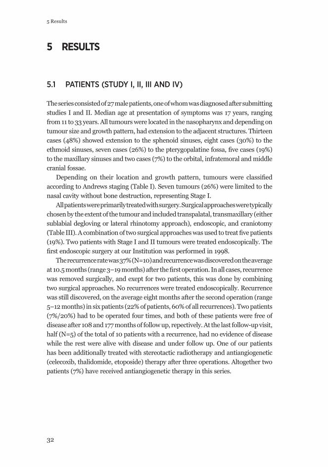
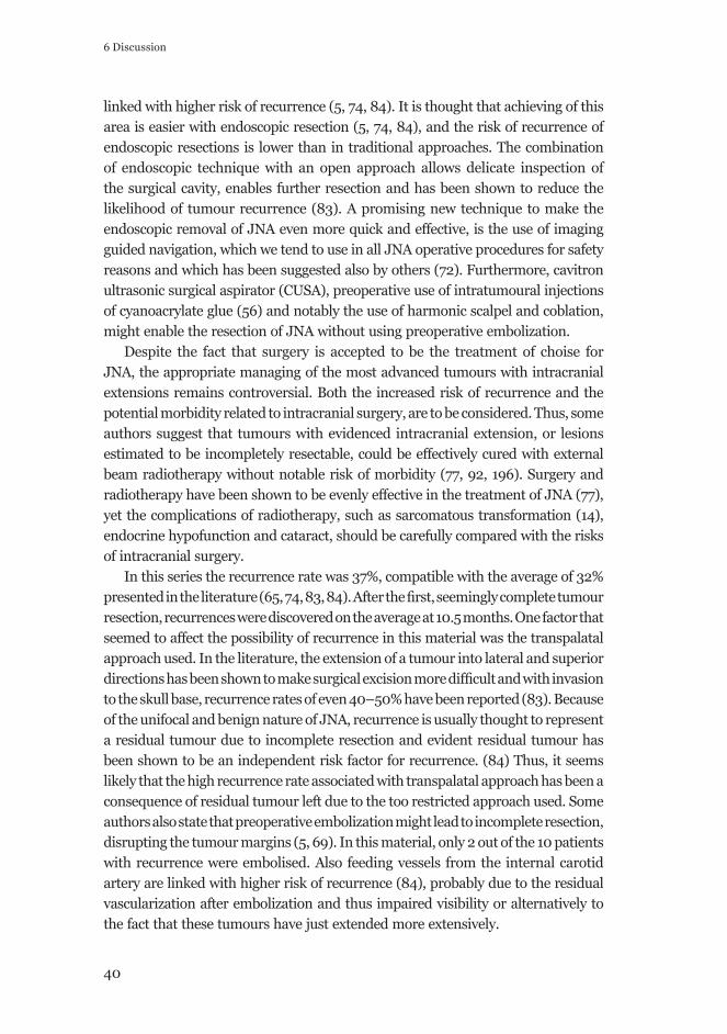
A B
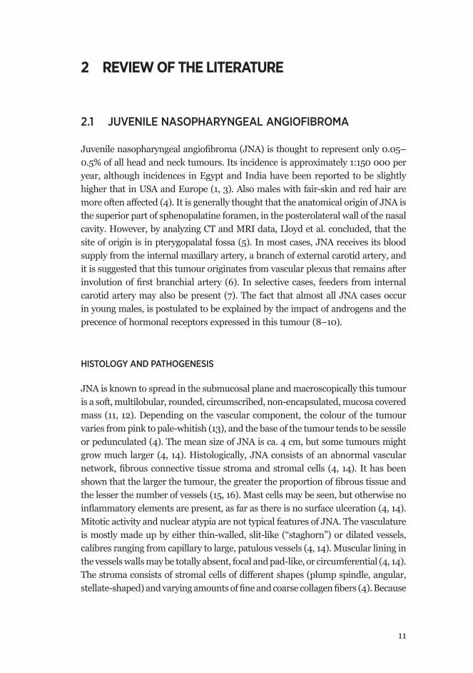
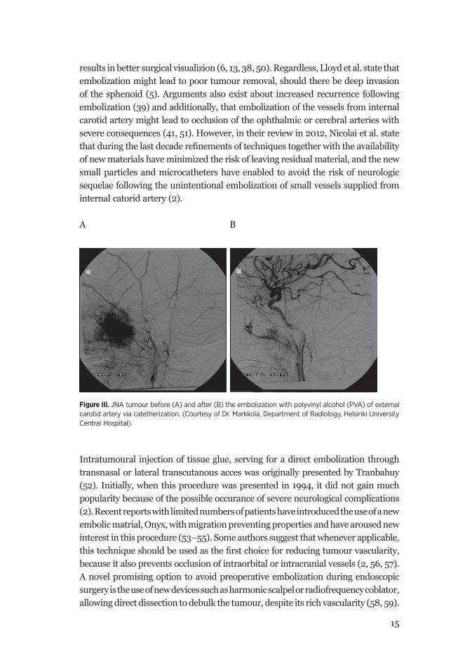
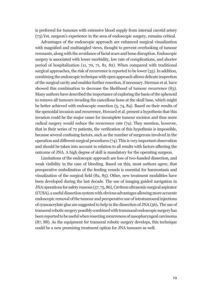
Figure III. JNA tumour before (A) and after (B) the embolization with polyvinyl alcohol (PVA) of external carotid artery via catetherization. (Courtesy of Dr. Markkola, Department of Radiology, Helsinki University Central Hospital).
Intratumoural injection of tissue glue, serving for a direct embolization through transnasal or lateral transcutanous acces was originally presented by Tranbahuy (52). Initially, when this procedure was presented in 1994, it did not gain much popularity because of the possible occurance of severe neurological complications (2). Recent reports with limited numbers of patients have introduced the use of a new embolic matrial, Onyx, with migration preventing properties and have aroused new interest in this procedure (53–55). Some authors suggest that whenever applicable, this technique should be used as the fi rst choice for reducing tumour vascularity, because it also prevents occlusion of intraorbital or intracranial vessels (2, 56, 57). A novel promising option to avoid preoperative embolization during endoscopic surgery is the use of new devices such as harmonic scalpel or radiofrequency coblator, allowing direct dissection to debulk the tumour, despite its rich vascularity (58, 59).
16
2 Review of the literature
Whether this technique will become established in the management of JNA remains to be seen, but so far two reports on the use of a coblator in endoscopic surgery of JNA have been promising (59, 60).
STAGING OF THE TUMOUR
Precise staging of the tumour is important for the selection of surgical approach, for comparing the surgical results and for predicting tumour recurrence (61). In general, low stages of JNA are limited in extension, whereas advanced stages are associated with the involvement of skull base and with intracranial extensions (51). Based on advances in diagnostic and treatment techniques, numerous staging systems have been adopted over the years (Table I). The fi rst staging system was introduced in 1981 by Sessions et al. (62), and after that, those poroposed by Andrews et al. (63) and Radkowski et al. (64) have been broadly adopted (2). In the 2000th century, the evolution of endoscopic surgery has led to introduction of new staging systems, some including also other parameters than the tumour extent and the sites of tumour involvement. In 2006 Önerci et al. felt that advances in imaging, embolization and surgical methods, especially endoscopic resection, have changed the sites, associated with a high risks of persistent disease and therefore suggested a new classifi cation for JNA (61). In their staging system they, for example, consider that with endoscopic surgery, ethmoid or sphenoid invasion has no effect on persistent disease and can be completely removed – thus tumours invading these areas should be classifi ed as Stage I (61). In 2008 Carrillo et al. proposed another staging system, which includes the tumour’s size as a new parameter which was shown to be an independent prognostic factor in multivariate-analysis aiming to predict the risk of recurrence (65). Based on a retrospective review of the outcomes of open and endoscopic surgery on JNA patients, in 2010, Snyderman et al. presented their staging system, where both endoscopic and open approaches were applicable and thus better predicted the immediate morbidity and tumour recurrence (51). This staging system, called the University of Pittsburgh Medical Center (UPCM) staging system for Angiofi broma, incorporates the route of invasion and residual vascularity following embolization, two factors that were not addressed by prior staging systems. According to this system, Stages I and II are considered as minimal stage, Stage III represents tumours that are locally advanced, e.g., with skull base erosion, requiring greater acces, but because of lack of residual vascularization after embolization, are not plagued with excessive bleeding. Stages IV and V represent tumours with residual vascularity after embolization and Stage V true intracranial tumours. Stage V is divided to medial (defi ned by extension from medial to the paraclival and cavernous segments of internal carotid artery), and lateral (defi ned by extension to the middle cranial fossa lateral to the same segments of internal carotid artery) divisions (51).
17
Table I. Staging systems of JNA
Staging system
I II III IV V
Sessions
1981
Ia:limited to nose and NPIb:extension into sinuses
IIa:minimal extension into PMFIIb:occupation of PMF IIc:occupation of ITF
Extension into the cranium
Chandler
1984
Limited to NP Extension into nasal cavity or sphenoidal sinuses
Extension into antrum, ethmoidal sinuses, PMF, ITF, orbit and/or cheek
Extension into the cranium
Andrews
1989
Limited to NP. No bone destruction, or limited to SPF
Extension into PPF or maxillary, ethmoidal or sphenoidal sinuses. Bone destruction.
Extension into ITF or orbital regionIIIa:no intracranialIIIb:extradural involvement
Intracranial, intradural tumourIVa:withoutIVb:with extension into cavernous sinus, pituitary or optic chiasm
Radkowski
1996
Ia:limited to nose or NPIb:extension into sinuses
IIa:minimal lateral extension into PPFIIb:full occupation of PPF, with/without superior erosion of orbital bonesIIc:ITF, posterior to pterygoid plates
Erosion of skull baseIIIa:minimal intracranial extensionIIIb:extensive intracranial extension
Önerci
2006
Nose, NP, ethmoidal and sphenoidal sinuses or minimal extension into PMF
Maxillary sinus, full occupation of PMF, extension into anterior cranial fossa, limited extension into ITF
Extension into cancellous bone at pterygoid base or the body of the GW sphenoid, signifi cant lateral extension into ITF or to the pterygoid plates posteriorly or orbital region, cavernous sinus obliteration
Intracranial extension between the pituitary gland and ICA, tumour localization lateral to ICA, middle fossa extension and extensive intracranial extension
Carrillo
2008
Nose, NP, maxillary sinuses, anterior ethmoid and sphenoid sinuses
IIa:extension into PMF of infratemporal fossa anterior to pterygoid plates, major diameter < 6 cmIIb:major diameter > 6 cm
Extension into infratemporal fossa posterior to pterygoid plates or posterior to ethmoid cells
Extensive skull base invasion > 2 cm of intracranial invasion
Snyderman
2010
Nasal cavity, medial PPF
Paranasal sinuses, lateral PPF, no residual vascularity
Skull base erosion, orbit, infratemporal fossa, no residual vascularity
Skull base erosion, orbit, infratemporal fossa, residual vascularity
Intracranial extension, residual vascularity; M, medial extension, L, lateral extension
18
2 Review of the literature
SURGERY
Since 1955, surgical excision has been the treatment of choise for JNA (66). Other treatment modalities described in the literature are radiotherapy, hormone therapy, cryotherapy, electrocoagulation and chemotherapy (35, 49). Surgery aims at complete and safe resection of the tumour, with minimal morbidity and loss of blood, and surgical approaches most commonly used to remove JNA, have been transpalatal, transmaxillar (lateral rhinotomy or midfacial degloving), Le Fort 1 osteotomy, and infratemporal fossa approaches (12, 36, 39, 45, 63, 67, 68). Although recent reports recommend endoscopic surgery, the most adequate choice of treatment for the individual patient remains under discussion (61, 69–73).
For more extensive tumours, midfacial degloving and lateral rhinotomy as transmaxillary approaches are thought to be suitable. With these approaches, also tumours extending into pterygopalatine and infratemporal fossae, paranasal sinuses, orbit, ethmoid sinus and medial part of the cavernous sinus can be surgically exposed. As midfacial degloving is recommended to avoid facial scars in these adolescent patients, yet maintaining excellent exposure to the tumour, it has gained popularity when compared with lateral rhinotomy (6, 74, 75). For tumours invading the skull base and orbital region, also the transoral Le Fort I osteotomy approach enables avoiding visible scars and at the same time provides broad exposure and great visibility of the tumour margins. However, complications of this approach are the possible damaging of non-erupted teeth and excessive blood loss due to the rupture of the palatal vessels during down-fracturing of the maxilla (67). This supports the invocation of preoperative embolization (5, 6, 49, 68).
Most challenges are linked to resection of the largest tumours with intracranial extensions. In addition to uncontrolled haemorrhage and neurologic defi cits, subtotal resections and recurrences are known to be especially common among these high-stage tumours (36, 68, 76, 77). Various craniotomy modifi cations have been used, entailing also a number of possible complications. The use of infratemporal approach may result in conductive hearing loss on the operated side as well as in the sacrifi ce of the mandibular division of the trigeminal nerve (63, 78). Further, because JNA patients are all adolescent, a risk of facial growth retardation is always present when using invasive approaches.
Initially, the endoscopic approach was recommended exclusively for tumours limited to the nasopharynx, the nasal cavity, ethmoid and sphenoid sinuses and with only slight extensions to the pretygopalatine fossa. Recently, selected cases of tumours expanding to the pterygopalatal fossa, infratemporal fossa and even with limited intracranial involvement have been successfully managed endoscopically (61, 79, 80). However, consensus remains that endoscopic approach has its limitations and is not recommended for tumours with extensive intracranial extensions, or extensions laterally to the cavernous sinus (69, 73). In addition, external approach
19
is preferred for tumours with extensive blood supply from internal carotid artery (73).Yet, surgeon’s experience in the area of endoscopic surgery, remains critical.
Advantages of the endoscopic approach are enhanced surgical visualization with magnifi ed and multiangled views, thought to prevent overlooking of tumour remnants, along with the avoidance of facial scars and bone disruption. Endoscopic surgery is associated with lower morbidity, low rate of complications, and shorter period of hospitalization (11, 70, 71, 81, 82). When compared with traditional surgical approaches, the risk of recurrence is reported to be lower (35). In addition, combining the endoscopic technique with open approach allows delicate inspection of the surgical cavity and enables further resection, if necessary. Herman et al. have showed this combination to decrease the likelihood of tumour recurrence (83). Many authors have described the importance of exploring the basis of the sphenoid to remove all tumours invading the cancellous bone at the skull base, which might be better achieved with endoscopic resection (5, 74, 84). Based on their results of the spenoidal invasion and recurrence, Howard et al. present a hypothesis that this invasion could be the major cause for incomplete tumour excision and thus more radical surgery would reduce the recurrence rate (74). They mention, however, that in their series of 72 patients, the verifi cation of this hypothesis is impossible, because several confusing factors, such as the number of surgerons involved in the operation and different surgical procedures (74). This is very important observation and should be taken into account in relation to all results with factors affecting the outcome of JNA. A high degree of skill is mandatory for the operating surgeon.
Limitations of the endoscopic approach are loss of two-handed dissection, and weak visibility in the case of bleeding. Based on this, most authors agree, that preoperative embolization of the feeding vessels is essential for haemostasis and visualization of the surgical fi eld (82, 85). Other, new treatment modalities have been developed during the last decade. The use of imaging guided navigation in JNA operations for safety reasons (57, 75, 86), Cavitron ultrasonic surgical aspirator (CUSA), a useful dissection system with obvious advantages allowing more accurate endoscopic removal of the tumour and peroperative use of intratumoural injections of cyanoacrylate glue are suggested to help in the dissection of JNA (56). The use of transoral robotic surgery possibly combined with transnasal endoscopic surgery has been reported to be useful when resecting recurrences of nasopharyngeal carcinoma (87, 88). As the equipment for transoral robotic surgery develops, this technique could be a new promising treatment option for JNA tumours as well.
20
2 Review of the literature
RADIOTHERAPY
Although surgery is considered as the treatment of choise for JNA, the potential morbidity and mortality associated with operating intracranial tumours has motivated others to investigate the role of radiotherapy for these patients (9). At present, external radiation therapy, either conventional, intensity-modulated or Gamma knife is generally used for patients with unresectable tumour or for those whose clinical condition prevents surgery (57, 89–91). It has been shown that radiotherapy and surgery are evenly effective in the treatment of JNA (77). However, the well known long-term complications of radiotherapy, such as secondary malignancies, endocrine hypofunction, cataract and glaucoma, must be taken into account and compared with the risks of intracranial surgery (92).
FOLLOW UP
Because of the submucosal growth of JNA, MRI and MSCT are superior to endoscopic examination to identify any residual or recurrence (2). Due to the infl ammatory changes usually seen 3–4 months after surgery, differentation between JNA and active scar tissue might be highly challenging (2, 93). This is why a number of authors have recommended postoperative MRI to be performed after the removal of nasal packaging until 72 hours for early identifi cation of any questionable residual disease (2, 93). Other authors state that follow-up imaging should not be performed earlier than at 6 weeks, as an early CT or MRI scan can give a false impression of a residual tumour due to various postoperative artefacts (84). In their review published in 2012, Nicolai et al. recommend that regardless of the follow-up technique selected, it should be performed every 6–8 months for at least 3 years after surgery (2).
RECURRENCES
Recurrences are common in the management of JNA, the majority of which occurs during the fi rst year after surgery (38). JNA is a unifocal benign disease and thus recurrence is often thought to represent a residual tumour due to incomplete resection. In 2007, Tyagi et al. showed that an evident residual tumour is an independent risk factor for recurrence (84). It has been proposed and later accepted by others that high rates of recurrence might be consequent on failure to recognize the skull base erosion (38, 57, 83). Yet, in their study of chromosomal alterations in JNA, Heinrich et al. reported few or no alterations in recurrent tumours, compared with numerous DNA gains in primary tumours (94). They conclude that this supports a concept of newly formed tumours, in that the same patterns of chromosomal aberrations would be expected in recurrence from residual tumour cells (94).
21
Recurrence rates for JNA vary from up to 50% reported in the 1980s and 1990s (1, 5) to 13.5% in the study of Danesi (95), when comtemplating open surgical approaches and to less than 10% when analyzing a series of endoscopically treated patients (73, 74, 96). In addition to the explicit tumour residual, factors associated with recurrence have been reported to be patients who are young at the time of the diagnosis, the size and stage of the tumour (97) and its vessel density (98). Lund et al. have suggested that JNAs undergo a cycle of aggressive growth followed in some cases by regression (99). Thus, the date when surgery is performed may be important in determing the so-called recurrence of the tumour, when the remaining part of the tumour attains new blood supply and undergoes regrowth of variable speed – depending on the point in the cycle the tumour is facing (99, 100).
INVOLUTION OF JNA
Despite the fact that tumour recurrence almost certainly represents tumour persistence, there are reports of incompletely resected tumours that have not progressed during a reasonable follow-up time, and involution of JNA tumour remnants has been suggested to happen infrequently (84, 101, 102). Thus, residual disease should not always be considered as a recurrence, because tumour remnants can be found by postoperative imagining in patients who remain symptom-free and therefore one must differentiate true new tumour growth from symptomless, non-growing tumour residual (57, 76, 79, 101). Factors indicating the evolution of tumour remnants remain unsolved, although it has been proposed that in young patients remnants might be expected to keep growing, while in older patients, they may disappear, due to the putatively hormonally dependent pathogenesis on JNA (2, 83). The idea of JNA involuting with age is supported by histological fi ndings, as individual tumours tend to express an increase in fi brous elements with time (103). Spontaneous involution of JNA, without any treatment attempts, has been reported in some patients (102, 104, 105), but the background of this phenomenon remains to be elucidated.
2.2 PROTEINS KNOWN TO ACT AS TUMOUR MARKERS FOR CELL PROLIFERATION AND ANGIOGENESIS
C-KIT
C-KIT, also known as CD117, is a type III receptor tyrosine kinase that belongs to the family of platelet derived growth factor receptors (PDGFRs). Activated C-KIT activates numerous transcription factors regulating apoptosis, cell differentiation, proliferation, chemotaxis, and cell adhesion (106–109). C-KIT is required for
22
2 Review of the literature
regulating the normal development of stem cells, germ cells, melanocytes, and peristalsis regulating Cajal cells in the gastrointestinal tract (107, 110–112). Expression of C-KIT has been found in many malignant nonlymphoid solid tumours (113–115), suggesting that C-KIT might participate in growth signalling in these tumours (116, 117).
Interestingly, C-KIT expression has also been found in the vasculature of many types of malignant tumours (118). However, in some tumours, when compared to corresponding normal tissue, there has been loss of C-KIT expression (116, 117). Thus, it has been proposed that C-KIT with its ligand might have mitogenic effects in some cell lines and regulation of cell differentation and morphogenesis in others.
C-KIT expression in benign tumours is less studied, and it appears to be mostly absent in benign neoplasms (119). Yet, in salivary gland adenomas and angiomyofi broblastomas, moderate to strong immunopositivity for C-KIT has been found (113, 118, 120). In the case of JNA, alternative results exist concerning C-KIT expression. In a series of 12 JNA samples, Zhang et al. reported strong C-KIT expression in stromal cells only and suggested that C-KIT might have a role in the progression of JNA (121). On the other hand, Pauli et al. evaluated stromal cells in 52 samples of JNA and found all of them to be C-KIT negative (122).
C-MYC
C-MYC is a proto-oncogene, taking part in cell proliferation, differentiation and apoptosis by activating other genes (123–126). It is also known to have potent angiogenic activity (126, 127). C-MYC overexpression is associated with several malignant neoplasms, such as breast and colon carcinomas, osteosarcomas and melanomas (124, 128, 129) and C-MYC gene amplifi cation is linked with advanced stage of the disease (126–128). Only a few studies have tried to demonstrate C-MYC expression in benign tumours, and the results have been negative (130, 131).
In their Northern blot study, Nagai et al. studied the expression of C-MYC mRNA in 25 cases of JNA but found no difference when compared to normal tissue (29). Later, in three advanced cases of JNA, Schick et al. succeeded to demonstrate gains of C-MYC, associated with increased mRNA and protein expression levels, suggesting a probable role for C-MYC in the progression of JNA (129). The observation that C-MYC participates in angiogenesis by inducing fi broblasts to build up an immature vascular network of leaky, unstable kind of blood vessels, (132) is of great interest, because malformed, slit-like vessels are known to be a part of JNA histological architecture (16).
23
BMI-1
Polycomb group proteins (PcGs) are epigenetic gene silencing proteins that participate in embryonic development and oncogenesis (133–136). As a collaborator of C-MYC in oncogenesis, BMI-1 (B lymphoma Mo-MLV insertion region 1 homolog) was the fi rst mammalian PcG gene identifi ed in 1991 (137). By methylating and acetylating the chromatin and histones, BMI-1 maintains the transcriptionally repressed state of many genes (138), and thus also the primitive state of tissue stem cells, enabling stem cell self-renewal (134, 139–141). Because BMI-1 regulates genes involved in cell cycle and cell differentiation, it can function as a potent oncogene (142–144). BMI-1 overexpression in various malignant tumours correlates with poor prognosis, while in other tumours, lack of BMI-1 expression has been found to correlate with a high recurrence rate (143, 145–147).
GLUT-1
Glucose transporter 1 (GLUT-1) is an erythrocyte type glucose transport protein participating in cellular response to hypoxia (148). It is expressed in high levels in e.g., erythrocytes and placental trofoblasts, whereas in most tissues it is constitutively expressed at low levels (149–152). Presumably due to enhanced glycolytic metabolism, upregulation of GLUT-1 expression has also been shown in many carcinomas (148). Of mesenchymal tumours, GLUT-1 is constantly expressed in juvenile hemangiomas and hence it is used in the differential diagnosis of vascular malformations (153, 154). Other tumours of mesenchymal lineages, e.g., chordomas, epithelioid sarcomas, gastrointestinal stromal tumours, adipocytic tumours and myogenous tumours have also been shown to express GLUT-1 and the observation that GLUT-1 expression in sarcomas was assosiated with necrosis, whereas low-grade mesenchymal tumours were GLUT-1 negative, proposes that its overexpression might be a refl ection of hypoxia (148, 155).
TENASCIN-C
Besides of the two major components of extracellular matrix (ECM), glycosaminoglycans and fi brous proteins, this meshwork also includes numerous fi ber-assosiated proteins, such as tenascins. Structurally similar proteins, tenascin-C (TNC), -W, -R, and –X, have different patterns of expression in different tissues, leading to the idea of their own solute functions (156–158). In developing epithelia, TNC is expressed in the surrounding mesenchyme of different organ systems. In adults, however, the expression of tenascins becomes more restricted. Tenascins are upregulated in infl ammation and tumourigenesis – in cancers, TNC can be

24
2 Review of the literature
secreted either by tumour cells themselves or more commonly, by fi broblasts in the surrounding stroma. Its contribution to tumour development is thought to be a direct stimulation of tumour cells to proliferate or promotion of angiogenesis via its impact on endothelium (159–163).
SYNDECANS
Syndecans are membrane proteoglycans consisting of a core protein and heparan sulphate chains and constitute the major class of heparan sulphate proteoglycans in vasculature. Their expression varies both temporally and spatially, but it is acknowledged, that almost all nucleated cells express at least one member of the syndecan family (164–166).
Syndecan-1 is the most studied of the four mammalian syndecans. In adult tissues, it is mostly expressed by epithelial cells, but during development also in both epithelial and mesenchymal cells (167, 168). Syndecan-2 is expressed in mesenchymal, neuronal and muscle cells and is the major syndecan in microvascular endothelial cells (169, 170). Syndecans interact with various ligands through their heparan sulphate chains and are known to be involved in cell-cell and cell-matrix interactions, cell proliferation, migration and differentiation. Although traditionally classifi ed as adhesion molecules, syndecans have been shown to have an essential role in vascular development and repair by binding and tranducing the signals of growth factors, e.g., of vascular endothelial growth factor (VEGF). Syndecan-2 plays an essential role in angiogenesis by stimulating endothelial cells to proliferate, migrate and generate new tubular structures. Its downregulation is known to reduce the spreading and adhesion of endothelial cells, enchance their migration, and impair the formation of normal, capillary-like structures (169, 170).
SYK
Over 500 kinases are encoded by the human genome and the interest in investigating these proteins is partly explained by the development of kinase inhibitors for oncological targets. Spleen tyrosine kinase SYK is a non-receptor tyrosine kinase originally cloned from porcine spleen (171, 172). Activated SYK conducts numerous cellular responses, such as cell proliferation, differentiation, survival and phagosytosis (171, 172). Although, initially SYK signalling pathway was thought to be restricted only to hematopoietic cell responses, in particular to immunoreceptor signalling events, recent works have demonstrated that SYK exhibits a widespread expression pattern in various nonhematopoietic cells. It appears to have a functionally centered
25
role on, e.g., nasal fi broblasts, vascular endothelial cells, epithelial cells, breast tissue, neuronal cells and hepatocytes (173).
SYK has been linked to the development of haematological and non-haematological malignancies and is required for the oncogenic acitivity of several viruses, such as Epstein-Barr virus (174–177). In these virus-induced tumours SYK has a putative tumour-promoting role, whereas in nonhaematopoietic tumours it has been proposed to suppress tumour growth (172). This was fi rst suggested by Cooper et al. in 2000, as in their material, SYK was expressed in normal breast tissue, benign breast lesions and low-tumourigenic breast cancer cell lines, but in invasive breast cancer SYK mRNA and protein were low or undetectable (171). Transfection of SYK in these tumours suppressed tumour growth and metastasis formation (171). Later this study has been substantiated and complemented by various clinical investigations (178–181). However, in e.g., chronic leukemia, gastric cancer, and oral squamous cell cancer high SYK expression correlates with tumour progression and has prognostic value, thus SYK acting as an oncogene (182).
26
3 Aims of the study
3 AIMS OF THE STUDY
The main goal of this study was to elucidate factors determining the outcome of juvenile nasopharyngeal angiofi broma, JNA.
For this purpose we investigated the various surgical techniques used to resect JNA •
during the past 40 years at our Institution, in order to settle their effect on the outcome and possible tumour recurrence. In addition, other potential clinical factors influencing the tumours’ tendency to recur in this series were recorded (Study I);
studied the expression of several proteins known to • participate in tumour- and angiogenesis, to find out if they were predictive also in JNA (Study II and III);
combined the copy number and gene expression data of two • phenotypically different JNA tumours to seek for processes putatively determining their growth pattern (Study IV).
27
4 MATERIALS AND METHODS
4.1 PATIENT MATERIAL
4.11 RETROSPECTIVE MATERIAL
The clinicopathological data of all patients diagnosed for a histologically verifi ed JNA during the 42-year period between January 1, 1970 and December 31, 2011 at the Helsinki University Central Hospital (HUCH), Helsinki, Finland were retrospectively reviewed. The health-care district included in this study represents almost one third of the whole country, including approximately 1.5 million inhabitants. To identify the patients, hospital surgical and discharge registries as well as the database of the Department of Pathology, HUCH, were used. A total of 27 male patients were included, the mean age being 17 years. Details on the patients’ age, sex, presenting signs and symptoms, the duration of symptoms, date of diagnosis, tumour site, imaging, whether preoperative embolization was performed, histopathology of the neoplasm, stage of the tumour, feeding arteries, treatment modality and the surgical approach performed, rate of recurrence, date of last follow up, and status at last follow up were collected from hospital records. All patients had a minimum follow up of seven months.
4.12 PROSPECTIVE MATERIAL (STUDY IV)
Fresh tissue samples were prospectively collected from two patients who underwent surgery at our Institution between 2008 and 2011. The fi rst patient (HS as High Stage) was 15 years old at the time of diagnosis and had a tumour extending to pterygopalatinal fossa, sphenoid, ethmoid, and/or maxillary sinuses, orbit and middle cranial fossa, representing Stage IIIa according to Andrew’s staging system. After the fi rst operation this patient got a recurrent disease and hade to face two re-operations. Additionally, radiotherapy and antiangiogenic therapy were administered, due to persistent intracranial growth. The other patient (LS as Low Stage) had a small tumour (3 cm in diameter), limited only to the nasopharynx. After endoscopic removal, this patient had no evidence of disease. These two phenotypically different JNA tumours were sampled and snap-frozen in liquid nitrogen within 20 minutes of devascularisation and stored at -80 C to be used for microarray analysis.
28
4 Materials and Methods
4.2 HISTOLOGY OF THE TUMOURS AND IMMUNOHISTOCHEMISTRY (STUDY II, III AND IV)
Formalin fi xed and paraffi n-embebbed samples of JNA tumours from the fi les of the Department of Pathology, University of Helsinki were used. The diagnoses were histologically confi rmed by experienced pathologists (PH and JH). Samples were scored for cell number per mm2 and from these the mean cell density was calculated. For vessel density analysis, vessels marked by CD31 were counted per mm2 and the mean counts were calculated. All CD31 positive vessels were included in this counting, and at this point, no separation between smaller and larger vessels was made.
For immunohistochemical staining, formalin fi xed and paraffi n-embebbed samples were cut into 4 μm micrometer-thick sections deparaffi nized in xylene and rehydrated through a graded alcohol series. For antigen retrieval, slides were treated in a PT-module (LabVision UK Ltd, UK) with Tris-HCl buffer (pH 8.5), Tris-EDTA for 20 minutes at +98°C, or with Trypsin (1g/200 ml PBS) for 30 minutes at +37°C. Immunohistochemical stainings were performed in Autostainer 480 (LabVision) using Dako REAL EnVision Detection System, Peroxidase/DAB+, Rabbit/Mouse (Dako, Glostrup, Denmark). The slides were treated with 0.3% Dako REAL Peroxidase-Blocking Solution (Dako) to block endogenous peroxidase activity followed by primary antibody incubation with specifi c antibodies for each antigen (Table II) for one hour, followed by a 30-min incubation with Dako REAL EnVision/HRP detection system, Rabbit/Mouse (ENV) reagent (Dako). The slides were fi nally visualized by Dako REAL DAB+ Chromogen (Dako) for 10 min. PBS-0.04%-Tween20 washing was accomplished between each step. Slides were counterstained with Meyer’s hematoxylin and mounted in mounting medium (Aquamount, BDH, Poole, UK). Mouse monoclonal antibody for CD31 (clone JC70A, Dako) mouse monoclonal antibody for SYK (clone 4D10.1, Abcam, Cambridge, UK), mouse monoclonal antibody for GLUT-1 (clone SPM498, Thermo scientifi c, Cheshire, UK), monoclonal antibody for tenascin –C (clone DB7 (IgG2a), Biohit, Helsinki, Finland), mouse anti-human syndecan-1 (clone β-B4, Serotec, Hämeenlinna, Finland), mouse monoclonal C-MYC (clone 9E10, Santa Cruz, CA), mouse monoclonal BMI-1 (ab 14389, Abcam), or polyclonal C-KIT (A4502, Dako) and rabbit anti-syndecan-2 (clone ZMD.308, Invitrogen, Nuppulinna, Finland) were used for one hour, followed by a 30-min incubation with Dako REAL EnVision/HRP detection system, Rabbit/Mouse (ENV) reagent.
29
Table II. Antibodies used in immunohistochemistry (Study II, III and IV)
Antibody Dilution Pre-treatment StudyCD31 1:20 Tris-HCl I, II, IV
C-KIT 1:400 Tris-HCl II
C-MYC 1:400 Tris-EDTA II
BMI-1 1:300 Tris-EDTA III
GLUT-1 1:1000 Trypsin III
TNC 1:50 Tris-EDTA III
Syndecan 1 1:2000 Tris-EDTA III
Syndecan 2 1:250 Tris-EDTA III
SYK 1:2000 Tris-EDTA IV
4.3 SCORING OF IMMUNOSTAININGS (STUDY II, III AND IV)
All stained samples were evaluated (N= 26 for study II and N=27 for studies III and IV) except for C-MYC, BMI-1, TNC (N=25) and syndecan 1 (N=21) because of the insuffi ciency of samples.
Independently and without knowledge of the clinical data, two assessors (JH and PH or JH and SR) scored the immunostainings by evaluating the percentage of positive tumour cells. The average staining was estimated by evaluating the whole tissue slide. No positivity was scored as 0, up to 30% positive cells as 1 (very low), 30–50% as 2 (low), 50–80 % as 3 (high) and over 80% as 4 (very high). This scoring system has been modifi ed from Häyry et al. (183). In the case of GLUT-1 and syndecan-2, however, the expression was scored positive or negative. Stroma, large and slit-like vessels were scored separately for these samples.
4.4 SAMPLE PREPARATION FOR GENE EXPRESSION AND COMPARATIVE GENOMIC HYBRIDIZATION MICROARRAYS (STUDY IV)
To extract total RNA from two JNA tissue samples, Qiagen RNeasy mini kit (Qiagen) was used according to the Manufacturer’s instructions. The integrity of RNA was measured using Agilent Bioanalyzer (Agilent Technologies, Palo Alto, CA). Two-hundred and fi fty nanograms of total RNA was used for labelling and 12.5 μg of fragmented aRNA was hybridized on microarrays according to the manufacturer’s instructions (Affymetrix, Santa Clara, CA). For DNA copy number analysis, DNA was extracted using DNeasy Blood & Tissue Kit (Qiagen). Two micrograms of genomic DNA was used for array hybridizations according to the manufacturer’s instructions (Agilent Technologies).
30
4 Materials and Methods
4.5 GENE EXPRESSION DATA ANALYSIS (STUDY IV)
To study the gene expression profi les of JNA tissue samples, Affymetrix GeneChip® Human Genome U133 Plus 2.0 (Affymetrix, Santa Clara, CA) gene expression arrays were used. GeneChip signal intensity data were processed with robust multiarray analysis (184) using re-mapped gene annotations from the Brainarray Custom CDF fi les (HGU133Plus2_Hs_ENSG, v.14.1.0, http://brainarray.mbni.med.umich.edu/Brainarray/) (185). In contrast to the ordinary annotation fi les with 54,675 probe sets, this custom chip description fi le has 18,982 probe sets, corresponding to the newest set of EnsEMBL genes. The signifi cance of differential expression was assessed using the empirical Bayes moderated paired t-statistics followed by p-value adjustment with the Storey’s approach (186). Arrays were quality weighted before statistical testing (187). Genes with p-values ≤ 0.05 were considered as signifi cantly differentially expressed. All methods used are implemented in the simpleaffy, limma and qvalue packages of the Bioconductor project (186, 188, 189).
4.6 COMPARATIVE GENOMIC HYBRIDIZATION ANALYSIS (STUDY IV)
Copy number variations in JNA tissue samples were studied using Agilent Human Genome CGH 244A Oligo Microarray (Agilent Technologies). Array intensity data were processed with Agilent Feature Extraction tool (Agilent Technologies). Probes associated with genomic coordinates according to the Manufacturer’s annotation were selected and coordinates (NCBI36/hg18) converted to the newest genome build (GRCh37/hg19) using LiftOver. For copy number variation (CNV) status of genes, averages were taken over probes locating within the start and end coordinate of EnsEMBL gene models. Similar to gene expression data, CNV status is given as log2 transformed values.
4.7 GENE ENRICHMENT SET ANALYSIS (STUDY IV)
To describe the gene and the gene product attributes of any organism, a controlled vocabulary is provided by the gene ontology categories (190). The GOrilla Gene Enrichment Analysis and Visualization Tool (191) was used to discover the gene ontology term enriched at either end of a gene list sorted according to the moderated t-test score calculated using limma. All three gene ontology categories were tested. Functional groups enriched among statistically signifi cantly differentially expressed genes were tested with DAVID (192).
31
4.8 PROTEIN-PROTEIN INTERACTION NETWORKS (STUDY IV)
For integrated protein-protein interaction data, we used Web-based protein interaction network analysis platform (PINA) (193) providing data from six databases. This was exploited to identify the interactor proteins of investigated proteins.
4.9 STATISTICAL ANALYSIS (STUDY II, III AND IV)
For cross-tabulation and analysis of categorical variables Fisher’s exact test or Spearmann correlation were used. All p-values are two-sided and values less than 0.05 were considered signifi cant. For survival analysis, an event was defi ned as the discovery of recurrence of the tumour. Follow-up time was calculated from the fi rst operation until the event. Patients with no evidence of recurrence or patients deceased from non-tumour-related cause (N=1) were censored on the last date of follow up. To compare the outcome between patient categories, we generated Kaplan-Meier curves and applied log-rank test. SPSS version 15.0 software (SPSS, IL) was used for statistical analyses.
4.10 ETHICAL CONSIDERATIONS (STUDY I, II, III AND IV)
The project was approved by the Research Ethics Committee of the Helsinki University Central Hospital, Hospital District of Helsinki and Uusimaa (diary number 114/12/03/04/2008), and all work was done in accordance with the Helsinki declaration.
32
5 Results
5 RESULTS
5.1 PATIENTS (STUDY I, II, III AND IV)
The series consisted of 27 male patients, one of whom was diagnosed after submitting studies I and II. Median age at presentation of symptoms was 17 years, ranging from 11 to 33 years. All tumours were located in the nasopharynx and depending on tumour size and growth pattern, had extension to the adjacent structures. Thirteen cases (48%) showed extension to the sphenoid sinuses, eight cases (30%) to the ethmoid sinuses, seven cases (26%) to the pterygopalatine fossa, fi ve cases (19%) to the maxillary sinuses and two cases (7%) to the orbital, infratemoral and middle cranial fossae.
Depending on their location and growth pattern, tumours were classifi ed according to Andrews staging (Table I). Seven tumours (26%) were limited to the nasal cavity without bone destruction, representing Stage I.
All patients were primarily treated with surgery. Surgical approaches were typically chosen by the extent of the tumour and included transpalatal, transmaxillary (either sublabial degloving or lateral rhinotomy approach), endoscopic, and craniotomy (Table III). A combination of two surgical approaches was used to treat fi ve patients (19%). Two patients with Stage I and II tumours were treated endoscopically. The fi rst endoscopic surgery at our Institution was performed in 1998.
The recurrence rate was 37% (N=10) and recurrence was discovered on the average at 10.5 months (range 3–19 months) after the fi rst operation. In all cases, recurrence was removed surgically, and exept for two patients, this was done by combining two surgical approaches. No recurrences were treated endoscopically. Recurrence was still discovered, on the average eight months after the second operation (range 5–12 months) in six patients (22% of patients, 60% of all recurrences). Two patients (7%/20%) had to be operated four times, and both of these patients were free of disease after 108 and 177 months of follow up, repectively. At the last follow-up visit, half (N=5) of the total of 10 patients with a recurrence, had no evidence of disease while the rest were alive with disease and under follow up. One of our patients has been additionally treated with stereotactic radiotherapy and antiangiogenetic (celecoxib, thalidomide, etoposide) therapy after three operations. Altogether two patients (7%) have received antiangiogenetic therapy in this series.
33
Table III. Management of JNAs in this series
SURGICAL APPROACH
Transpalatal 14 (52%)
Transmaxillar 2 (7%)
Endoscopic 3 (11%)
Craniotomy 2 (7%)
Combinded 6 (22%)
SURGERY FOR RESIDUAL DISEASE
Once 10 (37%)
More than once 6 (22%)
STATUS AT LAST FOLLOW UP
No evidence of disease 21 (78%)
Alive with minimum residual disease under surveillance 6 (22%)
One patient (4%) received postoperative radiotherapy and two patients (7%) were additionally treated with antiangiogenic agents.
Except for one 24 years old patient, patients with a recurrence were between 10 and 17 years old at the time of diagnosis. All patients who had more than two reoperations were under the age of 15 at the time of diagnosis. Among the nine patients older than 17 years at the time of diagnosis, only one had a recurrence. A correlation was found between the number of needed reoperations and the surgical approach used, as the tumours removed transpalatally had higher risk of multiple recurrences when compared to other approaches (Fisher’s exact test p=0.004)
The median follow-up time was 106 months (range 7–360 months). All patients remained either with no evidence of disease (N=20, 74%) or were alive with minimal signs of persistent disease (N=6, 22%) at the last follow-up visit, except for one patient, who perished for another reason during the follow up. There was no evidence of malignant transformation in any patient.
5.2 HISTOLOGY OF THE JNA SAMPLES (STUDY II)
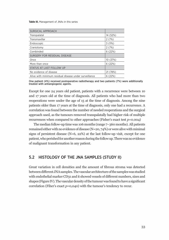
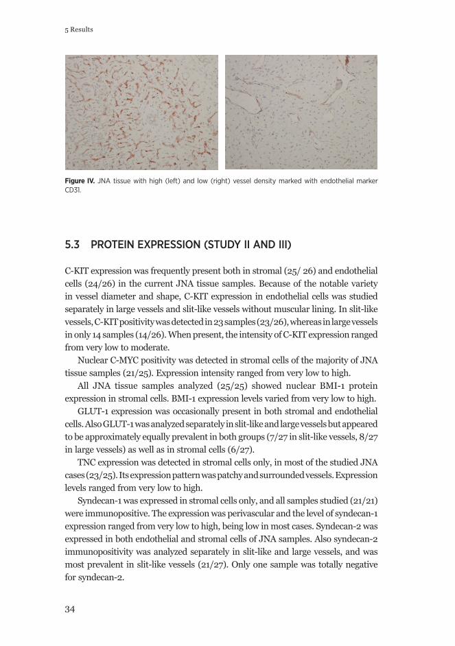
Great variation in cell densities and the amount of fi brous stroma was detected between different JNA samples. The vascular architecture of the samples was studied with endothelial marker CD31 and it showed vessels of different numbers, sizes and shapes (Figure IV). The vascular density of the tumour was found to have a signifi cant correlation (Fiher’s exact p=0,040) with the tumour’s tendency to recur.
34
5 Results
Figure IV. JNA tissue with high (left) and low (right) vessel density marked with endothelial marker CD31.
5.3 PROTEIN EXPRESSION (STUDY II AND III)
C-KIT expression was frequently present both in stromal (25/ 26) and endothelial cells (24/26) in the current JNA tissue samples. Because of the notable variety in vessel diameter and shape, C-KIT expression in endothelial cells was studied separately in large vessels and slit-like vessels without muscular lining. In slit-like vessels, C-KIT positivity was detected in 23 samples (23/26), whereas in large vessels in only 14 samples (14/26). When present, the intensity of C-KIT expression ranged from very low to moderate.
Nuclear C-MYC positivity was detected in stromal cells of the majority of JNA tissue samples (21/25). Expression intensity ranged from very low to high.
All JNA tissue samples analyzed (25/25) showed nuclear BMI-1 protein expression in stromal cells. BMI-1 expression levels varied from very low to high.
GLUT-1 expression was occasionally present in both stromal and endothelial cells. Also GLUT-1 was analyzed separately in slit-like and large vessels but appeared to be approximately equally prevalent in both groups (7/27 in slit-like vessels, 8/27 in large vessels) as well as in stromal cells (6/27).
TNC expression was detected in stromal cells only, in most of the studied JNA cases (23/25). Its expression pattern was patchy and surrounded vessels. Expression levels ranged from very low to high.
Syndecan-1 was expressed in stromal cells only, and all samples studied (21/21) were immunopositive. The expression was perivascular and the level of syndecan-1 expression ranged from very low to high, being low in most cases. Syndecan-2 was expressed in both endothelial and stromal cells of JNA samples. Also syndecan-2 immunopositivity was analyzed separately in slit-like and large vessels, and was most prevalent in slit-like vessels (21/27). Only one sample was totally negative for syndecan-2.
35
Cytoplasmic SYK protein expression was detected in stromal cells of all JNA tissue samples analysed (27/27). The frequency of SYK expression levels varied from very low to high.
5.4 CORRELATIONS WITH CLINOCOPATHOLOGICAL FACTORS (STUDY II AND III)
The protein expression levels of C-KIT, C-MYC and BMI- 1, GLUT-1, TNC, syndecan-1, syndecan-2 and SYK were compared with each other, cell and vessel density, tumour stage, and patient age. A signifi cant correlation between endothelial expression of C-KIT and cell density was found (Spearman correlation p=0.009). Endothelial C-KIT protein expression was also found to correlate with stromal C-MYC protein expression (Fisher’s exact p=0.006), while no correlation between the C-MYC protein expression and cellular or vascular density was found. No correlation between the immunoexpression of BMI-1 and other parameters studied was found.
TNC seemed to correlate with many other factors, including vessel density (Fisher’s exact p=0.01, Spearman correlation p=0.004), tumour stage using Andrew’s staging system (Spearman correlation p=0.001), endothelial C-KIT expression (Spearman correlation p=0.027) and stromal GLUT-1 (Fisher’s exact p=0.002, Spearman correlation p=0.001). Also, the expression of endothelial GLUT-1, when present, had a signifi cant correlation with tumour stage (Fisher’s exact p=0.011). Syndecan-1 and syndecan-2 had no correlations with any of the above-mentioned factors. SYK had a signifi cant correlation with lower tumour stage (Fisher’s exact p=0.048).
5.5 SURVIVAL ANALYSIS (STUDY I AND III)
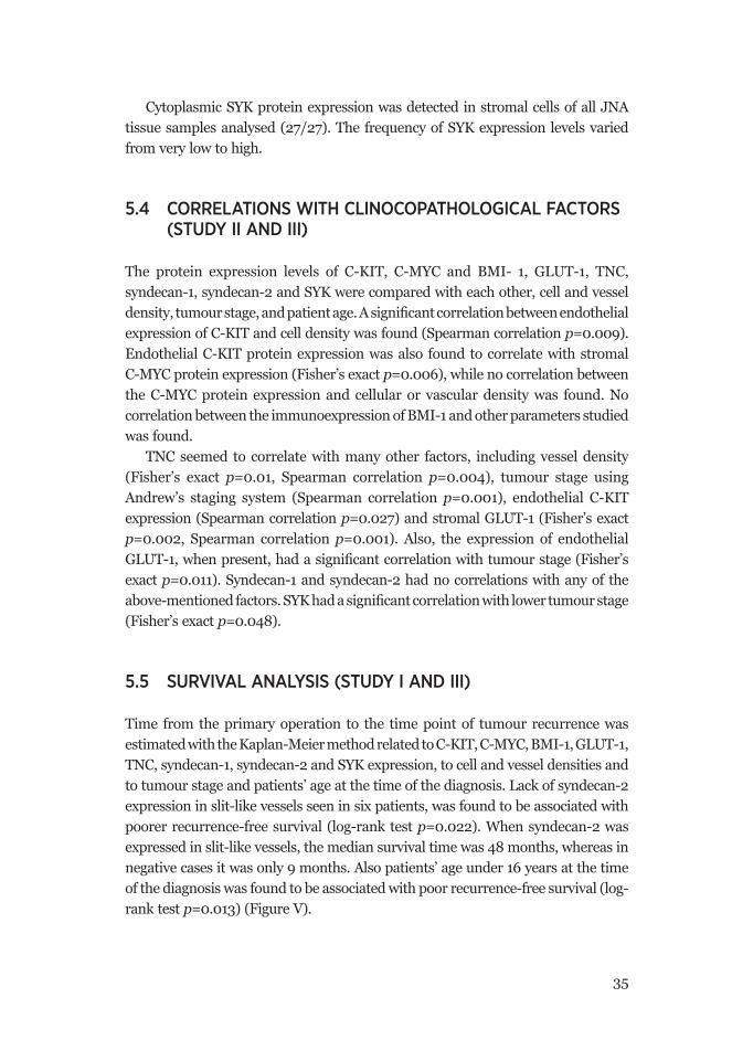
Time from the primary operation to the time point of tumour recurrence was estimated with the Kaplan-Meier method related to C-KIT, C-MYC, BMI-1, GLUT-1, TNC, syndecan-1, syndecan-2 and SYK expression, to cell and vessel densities and to tumour stage and patients’ age at the time of the diagnosis. Lack of syndecan-2 expression in slit-like vessels seen in six patients, was found to be associated with poorer recurrence-free survival (log-rank test p=0.022). When syndecan-2 was expressed in slit-like vessels, the median survival time was 48 months, whereas in negative cases it was only 9 months. Also patients’ age under 16 years at the time of the diagnosis was found to be associated with poor recurrence-free survival (log-rank test p=0.013) (Figure V).
36
5 Results
Figure V. Young age aff ecting the recurrence-free survival of JNA patients.
5.6 GENE COPY NUMBER ABERRATIONS (STUDY IV)
The gene copy numbers from the low stage (LS) and high stage (HS) specimens were compared to gender-matched DNA obtained from white blood cells of healthy individuals (Promega, Wisconsin, WI). Then the copy number gains and losses were compared between the two different samples. The log2 cut-off values for gains and losses were 1 and -1, respectively, indicating a two-fold difference in copy number. Between the LS and HS samples and their corresponding controls, 46 genes showed at least a two-fold copy number difference. Compared with each other, the copy number change between these two samples was at least two-fold in 11 genes.
5.7 GENE EXPRESSION LEVELS (STUDY IV)
Between LS and HS, at least two-fold change in the gene expression levels was discovered in 1383 transcripts, of which in 1245 cases the q-value was <0.05. The level was higher in LS in 773 transcripts and in HS in 610 transcripts. Expression level changes related to copy number changes were seen in three genes.
37
5.8 GENE ONTOLOGY (STUDY IV)
Assessment with GOrilla revealed some strongly enriched categories that where different in LS and HS tumours. Genes overexpressed in the LS tumour were commonly associated with biological functions linked to the tumour organization, like development of the vasculature and epithelium, cell adhesion and collagen catabolic functions, and also hypoxia. As in the HS tumour with recurrences, the upregulated genes were enriched into categories like signal transduction activity, including positive regulation of phosphorylation, tumour necrosis factor (TNF) production and Wnt-activated receptor activity.
One of the genes with an over two-fold change in expression levels between LS and HS was tyrosine kinase SYK, known to participate in tumourigenesis of various other tumours. This protein was decided to be studied further, both computationally as well with immunohistochemical analyses. To identify proteins interacting with SYK, we used protein interaction database tool, PINA (193), and found 95 proteins interacting with SYK. In 12 of these, the gene expression change between LS and HS was over two-fold. These genes, together with SYK, were more frequently present in the GO-categories enriched in HS, when compared to those enriched in LS.
38
6 Discussion
6 DISCUSSION
JNA is a rare tumour of unknown etiology. There is great variation between tumours regarding their tendency to grow and recur after surgical removal. The goal of this study was to investigate factors that settle the outcome of JNA management. First, we evaluated the used treatment modalities and their outcome in 27 JNA patients treated at the Helsinki University Central Hospital during the past 42 years. This was highly important because during the last 10 years the management of JNA has undergone eminent evolution and new promising treatment modalities have been developed (38, 41, 57, 81, 98). We were able to show that the surgical approach used varied during the years and had an effect on the outcome of the patient. Other efforts were taken to clarify the effect of several proteins, known to act as growth and angiogenesis promoting factors in other tumours, to the vascular and cellular densities of JNA tumours, as well as to the stage of the tumours. We found that the expression of C-KIT correlated with cell density of the tumour, TNC expression correlated with vascular density of the tumour and TNC, GLUT-1 and SYK expressions correlated with the stage of the tumour. In addition, by comparing the altered gene copy number and gene expression levels of two phenotypically different JNA tumours, we seeked for processes putatively taking part in the more aggressive growth of some JNA tumours.
The methods used in this study include the assessment of clinical data, the application of immunohistochemical stainings of paraffi n-embedded JNA samples, statistical methods and microarray techniques to investigate the gene copy number and gene expression level changes. The foremost limitaitons of this study were linked to the limited number of patients available, due to the rareness of this disease, affecting the statistical estimates, and the long duration of the study period putatively affecting the quality of the paraffi n-embedded samples. Also, in the 1970s the samples taken were smaller compared with the present practice. The comparison of different surgical techniques as such might be imprecise due to the fact that individual surgeons putatively had deviating prospects to perform a complete resection. The outcomes of different surgical approaches were estimated concerning the residual tumour or recurrence, the development of imaging techniques during the study period might have infl uenced the possibility to detect minimal residuals.
39
THE IMPACT OF SURGICAL APPROACH ON JNA’S TENDENCY TO PERSIST OR RECUR
For primary resection, transpalatal approach was most commonly (52%) used in the current series of 27 JNA patients. It was especially common during the earliest years of this material, as all patients during 1970–1989 were treated using either transpalatal (63%), a combination of transpalatal and transmaxillary (25%) or sublabial approach (13%). During the last decade of this study period, the use of other approaches gained popularity, as only 42% of the patients had exclusively transpalatal surgery. Although transpalatal approach is thought to be adequate to remove tumours limited to nasopharynx, nasal cavity and sphenoid sinuses, without scarring, it has obvious limitations. In the case of lateral tumour growth to pterygopalatine and infratemporal fossae (194), it has been connected with higher risk of recurrence. Consistent with earlier reports, transpalatal approach was observed to have a signifi cant risk of persistent disease and need of more than one re-surgery. Palatal fi stula is reported to be the most common complication of transpalatal approach and tends to heal spontaneously (194, 195). Despite the poor exposure for large tumours and the elevated rates of recurrence reported (6), transpalatal approach can be combined with other approaches in selected cases, as was done in three cases in this series. However, the advantage of combining transpalatal approach to other approaches also remains to be cleared, because two of these three cases were found to face recurrence after primary surgery.
The high recurrence rates and other complications associated with open surgery have created pressure to work on new treatment modalities while, at the same time the evolution of imaging techniques has enabled the more precise planning of surgery. During the last decades, the transnasal endoscopic technique used for several other tumours has been put into operation also for JNA. In this series, only three patients were treated with endoscopic surgery, and one of these had a recurrence. It has been generally admitted that when resecting JNA endoscopically, high degree of skill in endoscopic surgery is mandatory and the surgeon should always be prepared to convert to an external approach, if necessary (58). Yet, in the 2000s numerous studies of endoscopic surgery have been encouraging, with satisfying outcomes and low recurrence rates.
Initially, endoscopic approach was used exclusively for limited tumours presenting Stages I and II. Recently, however, also tumours with extensions into the pterygopalatal fossa, infratemporal fossa and even with limited intracranial involvement have been succesfully managed endoscopically (61, 79, 82), putatively due to increasing skills of operating surgeons. Inspite of this, consensus still remains that endoscopic surgery is not recommended for tumours with extensive extension into the cranium or laterally to the cavernous sinus (69, 73). The importance of exploring and carefully drilling the basis of the sphenoid has been emphasized by many authors as tumour invasion into the cancellous bone of this area has been
40
6 Discussion
linked with higher risk of recurrence (5, 74, 84). It is thought that achieving of this area is easier with endoscopic resection (5, 74, 84), and the risk of recurrence of endoscopic resections is lower than in traditional approaches. The combination of endoscopic technique with an open approach allows delicate inspection of the surgical cavity, enables further resection and has been shown to reduce the likelihood of tumour recurrence (83). A promising new technique to make the endoscopic removal of JNA even more quick and effective, is the use of imaging guided navigation, which we tend to use in all JNA operative procedures for safety reasons and which has been suggested also by others (72). Furthermore, cavitron ultrasonic surgical aspirator (CUSA), preoperative use of intratumoural injections of cyanoacrylate glue (56) and notably the use of harmonic scalpel and coblation, might enable the resection of JNA without using preoperative embolization.
Despite the fact that surgery is accepted to be the treatment of choise for JNA, the appropriate managing of the most advanced tumours with intracranial extensions remains controversial. Both the increased risk of recurrence and the potential morbidity related to intracranial surgery, are to be considered. Thus, some authors suggest that tumours with evidenced intracranial extension, or lesions estimated to be incompletely resectable, could be effectively cured with external beam radiotherapy without notable risk of morbidity (77, 92, 196). Surgery and radiotherapy have been shown to be evenly effective in the treatment of JNA (77), yet the complications of radiotherapy, such as sarcomatous transformation (14), endocrine hypofunction and cataract, should be carefully compared with the risks of intracranial surgery.
In this series the recurrence rate was 37%, compatible with the average of 32% presented in the literature (65, 74, 83, 84). After the fi rst, seemingly complete tumour resection, recurrences were discovered on the average at 10.5 months. One factor that seemed to affect the possibility of recurrence in this material was the transpalatal approach used. In the literature, the extension of a tumour into lateral and superior directions has been shown to make surgical excision more diffi cult and with invasion to the skull base, recurrence rates of even 40–50% have been reported (83). Because of the unifocal and benign nature of JNA, recurrence is usually thought to represent a residual tumour due to incomplete resection and evident residual tumour has been shown to be an independent risk factor for recurrence. (84) Thus, it seems likely that the high recurrence rate associated with transpalatal approach has been a consequence of residual tumour left due to the too restricted approach used. Some authors also state that preoperative embolization might lead to incomplete resection, disrupting the tumour margins (5, 69). In this material, only 2 out of the 10 patients with recurrence were embolised. Also feeding vessels from the internal carotid artery are linked with higher risk of recurrence (84), probably due to the residual vascularization after embolization and thus impaired visibility or alternatively to the fact that these tumours have just extended more extensively.
41
YOUNG AGE LINKED TO POOR RECURRENCE-FREE SURVIVAL
Along with evident residual tumour, wide extension of the tumour likely to lead to residual tumour, embolization of the tumour according to some authors and feeding vessels from internal carotid artery, an other factor linked with recurrence in the literature is the patient’s young age at the time of the diagnosis (84). Also in this material, young age was linked with higher risk of recurrence, as only one out of nine patients over 17 years old had a recurrence (Figure V). The correlation between recurrence rate and young age of the patient has been presented in numerous studies (50, 83, 84), and JNA growth has been considered to relate with the hormonal environment during male puberty (9, 20, 85, 102). Although several studies have strove to demonstrate the precise role of androgen, estrogen and progesterone and their nuclear receptors in this tumour, the results still remain controversial (8–10). The use of nonsteroidal androgen receptor blocker fl utamide in the treatment of JNA has been reported in several studies with varying results. In their work with fi ve patients, Gates et al. showed a 44% decrease in tumour size after administering fl utamide (21), whereas Labra et al. found no effect of fl utamide in their 7 JNA patients (22). In the most recent study, Thackar et al. were able to show with their 20 patients, that fl utamide-induced regression was demonstrated only in post-pubertal patients (197). Earlier it has already been stated that in older patients, JNA tumours and tumour residuals are more likely to involute during follow up than in younger ones (2, 83). That is why in some patient populations, the “watchful waiting” might be a possibility, especially with tumour remnants in surgically challenging places. Yet, besides the patient’s age other factors predicting the residual tumour’s tendency to involute in progress, remain unknown.
VASCULARITY OF JNA AFFECTING THE TUMOURS’ TENDENCY TO GROW LARGE
JNAs are known for their hypervascularity and studies investigating the proliferation of stromal and vascular components of JNA have raised interest regarding the hypothesis of the angiogenic growth factors taking a crucial part in the pathogenesis of this esoteric tumour (10, 18, 29, 121, 198, 199). In their immunohistochemical study, Brieger et al. showed that VEGF is secreted by cells of JNA (18) and in this material tumour vascularity was found to have a signifi cant correlation with tumour recurrence. These fi ndings are of great importance, since within the last decade increased understanding of the process of angiogenesis has enabled the development of new therapeutic substances. Studies presenting the important role of VEGF in the regulation of angiogenesis have allowed the blockade of VEGF signalling and made it a major target for the therapy of several tumours (200). In the present series, two cases with persistent tumour growth in the sphenoid bone, were successfully
42
6 Discussion
experimentally treated with antiangiogenic drugs (selecoxib, thalidomide, etoposide) (98). In cancer therapy, the use of VEGF inhibitors is common (201), but so far none of our patients has been treated with these agents. Yet, in the future, this might be possible in selected JNA patients, knowing that JNAs are positive for VEGF.
TUMOUR SUPPRESSORS, ONCOGENES AND PROTO-ONCONGENES IN JNA
While most efforts aiming to elucidate the pathogenesis of JNA have been made to investigate growth factors, e.g., VEGF, transforming growth factor (TGF), insulinlike growth factor (IGF) and PDGF and platelet derived growth factor (PDGF) – all proteins known to be related to stromal or vascular growth and to correlate with high vessel density (10, 19, 29, 202, 203), fewer studies have been performed on actual tumour suppressors, oncogenes or proto-oncogenes. This might be due to the fact that inspite of its potential to aggressive behavior, JNA is classifi ed as a benign tumour. However, the expression of tumour suppressor adenomatous polyposis coli (APC) gene in JNA has been investigated in numerous studies, led by the discovery of increased frequency of JNA among patients with familial adenomatous polyposis (FAP) (25–27). The gene product of APC is known to regulate β-catenin, a downstream activator of the Wnt signalling pathway, as APC is needed for the degradation of β-catenin (25). Interestingly, β-catenin is known to act as a co-activator of androgen receptors (ARs), increasing androgen sensitivity –possibly explaining the exclusive occurrence of JNA in adolescent males (28). None of the JNA patients in the current patient series had relatives with FAP or JNA (204).
Protein expressions of oncogene C-KIT and proto-oncogene C-MYC have been studied in JNA tissue and based on these fi ndings the possible involvement of these proteins in the growth and promotion on JNA has been discussed (29, 121, 122, 129). Interestingly, in 2007 Sihto et al. investigated the expression of C-KIT in endothelial cells in several malignant tumours and concluded that it might be associated in tumour angiogenesis and furthermore in the development of morphologically characteristic features of coiled microvessels in certain juvenile brain tumours, as also in other tissues (118). They proposed hypoxia to be the triggering factor for C-KIT overexpression in tumour cells promoting angiogenesis and survival of tumour microvessel endothelial cells. C-MYC has also been thought to have a potential role in angiogenesis, as it induces fi broblasts to build up an immature vascular network of leaky, unstable blood vessels (132). Both observations are of great interest, because slit- and immature-like blood vessels are part of JNA’s histological architecture (15).
In this study, frequent immunoexpression of C-MYC in stromal cells and C-KIT in both stromal and endothelial cells of JNA samples was shown. Interestingly, the
43
C-KIT expression was more abundant in the endothelium of slit-like vessels than in larger vessels. Endothelial C-KIT expression showed a signifi cant correlation to the cell density of the tumour and to the stromal expression of C-MYC. Based on these results, we ended up with a proposal that both the stromal and vascular component of JNA possibly take part in the neoplastic growth of the tumour. Additionally, the correlation between endothelial C-KIT and stromal C-MYC expression led to the hypothesis of stromal C-MYC putatively infl uencing the growth of immature blood vessels by regulating the expression and effect of C-KIT on endothelial cells. The role of these two proteins in JNA should, however, be elucidated. As a third known oncogene, the immunoexpression of BMI-1 in JNA samples was studied. This is an epigenetic silencing protein known to be dysregulated in many cancers and to correlate with poor prognosis (145, 147, 183). As far as we know, BMI-1 has not been studied in JNA tissue before. In this study, stromal cells frequently expressed BMI-1, suggesting that it might possibly be involved in the pathogenesis of JNA. However, no correlations between BMI-1 and the other proteins studied, or with clinical parameters, were found.
To elucidate the possible formation of impaired capillaries, the immunoexpression of syndecan-2, a marker known to function as a co-receptor for growth factors and participate in numerous signalling cascades (169) was studied. In 2008, Noguer et al. demonstrated that syndecan-2 downregulation reduced the adhesion of endothelial cells leading to impaired formation of capillary vessels and enhanced migration of endothelial cells (170). In this fi nite material on JNA tumours, in the six cases when slit-like vessels lacked the syndecan-2 expression, the overall recurrence-free survival of these patients was worse, referring to the tendency of the tumour to recur and need of reoperations. This can be speculated to result from the absence of syndecan-2 leading to greater migration potential of endothelial cells, making radical surgical management more diffi cult, as the migrative endothelial cells have covered a wider boarder of the surgical fi eld.
TUMOUR- AND ANGIOGENESIS MARKERS IN JNA – ARE THEY PREDICTIVE?
Next, we aimed to fi nd proteins whose expression could predict the tendency of extensive tumour growth. Led by the fact that there are slit-like vessels without normal lumen in JNA tissue, and that phenotypes of this tumour vary so much, the aim was to test our hypothesis that part of JNAs are vascular malformations. This seemed warranted because the treatment of JNA still remains a challenge and more importantly, there are new treatment options available for lesions with vascular malformation characteristics. Infantile hemangiomas (IHs) are vascular tumours composed of endothelial cells without the ability to be organized in lumenized
44
6 Discussion
structures. Tissue hypoxia is thought to precede the proliferative phase of IHs and this hypoxic insult contributes to the elevated levels of GLUT-1 in the lesion (148, 153, 154). GLUT-1 is used in histopathological routine to distinguish hemangiomas from vascular malformations that do not express GLUT-1. The differentiation of these two entities is essential because only hemangiomas are self-limiting. Our aim was to test, if the high-stage JNA tumours were GLUT-1 negative and thus resembled vascular malformations. GLUT-1 negativity in our samples, however, did not show correlation with higher tumour stage and thus putatively more aggressive growth, often seen in vascular malformations. Quite on the contrary, endothelial GLUT-1 positivity, when present, was correlated with higher tumour stage. Based on this fi nding, it seems unlikely that JNAs or a part of them were vascular malformations.
The fi nding of GLUT-1 expression correlating with higher tumour stage is somewhat compatible with literature, because GLUT-1 is known to be overexpressed in numerous neoplasms, especially in high grade tumours, as a marker of increased need of glucose metabolism in hypoxic lesions (77). The idea of possible hypoxia preceding the growth and angiogenesis of JNA has been presented (19). Hypoxia inducible factor (HIF)-1α expression data have been studied in two sets of JNA tumours and it can be concluded that HIF and its induced factors could be important in the promotion of JNA and maintenance on tumour cells (19, 198). In 2007 Schuon et al. investigated the HIF-1α expression in JNA and found it to be highest in well-vascularized tumours. They suggest that this is due to the fact that infunctional vessels cause the tumour to become hypoxic (19). HIF is known to be the main hypoxia signalling and VEGF activating transcription factor (205). Other downstream activation targets of HIFs are, e.g., GLUT-1, C-KIT and TNC (150, 151, 206, 207).
TNC expression in adult tissues is known to be tightly regulated and restricted mostly to conditions requiring active remodelling, such as wound healing and infl ammation following tissue injury (208). However, it is known to be stromally re-expressed in many tumours, and it acts as a predictive factor for local recurrence and mestatasis, e.g., in breast cancer, brain tumours and head and neck cancers (208). In addition, TNC has been classifi ed as a marker of vascular proliferation in lesions growing within brain tissue, and it has been proposed to stimulate angiogenesis by establishing tumour cell-derived vessels (209–211). Angiogenesis in tumours may be started by hypoxia, stromal and tumour cells or ECM compounds, resulting in leaky vessels, with only partial pericyte coverage (208, 212). TNC is thought to regulate angiogenesis by activating other factors, such as VEGF (213). However, taking part in remodelling of the ECM, TNC could have a direct effect in controlling of vascular instability of the tumour vessels (208, 212). Although most antiangiogenic treatments applied are targeted against VEGFs, in some tumours, these agents have been shown to actually promote metastasis, due to the resistance and escape
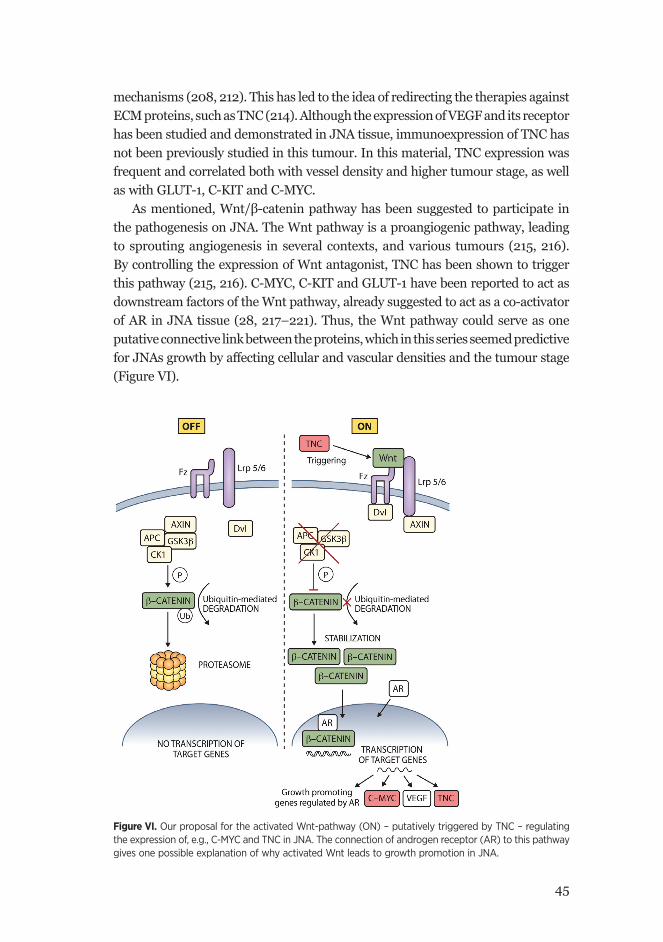
45
mechanisms (208, 212). This has led to the idea of redirecting the therapies against ECM proteins, such as TNC (214). Although the expression of VEGF and its receptor has been studied and demonstrated in JNA tissue, immunoexpression of TNC has not been previously studied in this tumour. In this material, TNC expression was frequent and correlated both with vessel density and higher tumour stage, as well as with GLUT-1, C-KIT and C-MYC.
As mentioned, Wnt/β-catenin pathway has been suggested to participate in the pathogenesis on JNA. The Wnt pathway is a proangiogenic pathway, leading to sprouting angiogenesis in several contexts, and various tumours (215, 216). By controlling the expression of Wnt antagonist, TNC has been shown to trigger this pathway (215, 216). C-MYC, C-KIT and GLUT-1 have been reported to act as downstream factors of the Wnt pathway, already suggested to act as a co-activator of AR in JNA tissue (28, 217–221). Thus, the Wnt pathway could serve as one putative connective link between the proteins, which in this series seemed predictive for JNAs growth by affecting cellular and vascular densities and the tumour stage (Figure VI).
Figure VI. Our proposal for the activated Wnt-pathway (ON) – putatively triggered by TNC – regulating the expression of, e.g., C-MYC and TNC in JNA. The connection of androgen receptor (AR) to this pathway gives one possible explanation of why activated Wnt leads to growth promotion in JNA.
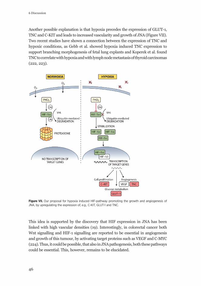
46
6 Discussion
Another possible explanation is that hypoxia precedes the expression of GLUT-1, TNC and C-KIT and leads to increased vascularity and growth of JNA (Figure VII). Two recent studies have shown a connection between the expression of TNC and hypoxic conditions, as Gebb et al. showed hypoxia induced TNC expression to support branching morphogenesis of fetal lung explants and Koperek et al. found TNC to correlate with hypoxia and with lymph node metastasis of thyroid carcinomas (222, 223).
Figure VII. Our proposal for hypoxia induced HIF-pathway promoting the growth and angiogenesis of JNA, by upregulating the expression of, e.g., C-KIT, GLUT-1 and TNC.
This idea is supported by the discovery that HIF expression in JNA has been linked with high vascular densities (19). Interestingly, in colorectal cancer both Wnt signalling and HIF-1 signalling are reported to be essential in angiogenesis and growth of this tumour, by activating target proteins such as VEGF and C-MYC (224). Thus, it could be possible, that also in JNA pathogenesis, both these pathways could be essential. This, however, remains to be elucidated.
47
THE PUTATIVE ROLE OF ALTERED GENE COPY NUMBERS AND GENE EXPRES-SION LEVELS IN THE GROWTH OF JNA
To control the expression of genes essential to survival and tumour progression, a neoplastic cell population has the ability to change the copy numbers of these genes. These alterations usually involve a large group of closely located genes and only a minority of these truly participate in malignant growth (225–229). Integrating genome-wide copy number data with gene expression data is one way to identify the true oncogenic driver genes (230–232). A few genome-wide studies of copy number and gene expression changes in JNA have been reported. In one of them chromosomal alterations were studied separately for stromal and endothelial cells and most of the imbalances were detected in both components (28, 30, 94, 129, 233–236). Although numerous chromosomal alterations have been demonstrated by these studies, explicit target genes for gains and deletions in JNA still remain unknown.
To achieve a wider vision of processes determining the different growth patterns and outcomes of JNA patients, we decided to explore the molecular genetic background of JNA by comparing gene copy number and gene expression level changes between two clinically different phenotypes of JNA of which we received fresh material. The two JNAs were both manifested in 15-year old male patients. One was an endoscopically removed, clearly limited tumour of 3 cm in diameter while the other was an extensive tumour infi ltrating intracranial space and warranting several surgical interventions, radiation treatment and antiangiogenic treatment. When analyzing the data, a few gene copy number changes that link to changes in gene expression were found. This might be due to the heterogeneity of the JNA tumour samples, consisting of, e.g., stromal, endothelial and epithelial cells. Another explanation for the low number of gene copy number changes is that in these subjects, the central level of regulation is not due to copy number abnormalities.
1245 genes had a signifi cant, over two-fold change in their expression, between low and high stage JNA tumours. In these phenotypically different tumours, the in silico translated proteins of the differently expressed transcripts showed enrichement for different biological processes and functions. Genes over-expressed in the low stage tumour were commonly associated with biological functions linked to the tumour organization, like the development of the vasculature and epithelium, cell adhesion and collagen catabolic functions, and interestingly also hypoxia. On the contrary, in high-stage tumour with recurrences, the upregulated genes were associated with, e.g., signal transduction activity, including positive regulation of phosphorylation, tumour necrosis factor (TNF) production and Wnt-activated receptor activity. These results were in concordance with earlier immunohistochemical reports of, e.g., vascular growth factors, matrix metalloproteinases and HIFs being expressed in the JNA tissue (18, 19, 198, 202, 237). Based on the present results on gene
48
6 Discussion
expression and prior knowledge of the proteins signifi cance for growth, we ended up investigating the immunoexpression of protein kinase SYK. SYK is a protein previously known to participate in tumourigenesis of different tumours and depending on the tumour, it can act as a tumour suppressor or as an oncogene (172). We found SYK expression to correlate with lower tumour stage also in JNA.
The possible connections of TNF and Wnt to the more aggressive growth pattern of JNA are of interest and should be confi rmed in the future. If the fi nding of TNF will be corroborated, it can offer a new treatment option for JNA, as TNF-targeted molecular therapies are already in wide use in immunological and cancer diseases (238, 239). The discovery of Wnt being putatively linked to the more aggressive growth of JNA is compatible with earlier reports of APC and β-catenin gene mutations and β-catenin protein expression in JNA, as well as with the suggestion that increased β-catenin expression might lead to increased AR sensitivity and thus explain the predominance of JNA in young boys (28). Also this observation supports our preliminary idea of Wnt possibly being the linking factor of the altered expression patterns of TNC, C-MYC, C-KIT and GLUT-1 in JNA, putatively promoting angiogenesis and growth in JNA, as presented earlier in this discussion.
Since the incidence of JNA tumours is very low, all the molecular data published on this disease will be valuable in understanding the molecular genetic background of this esoteric tumour. In addition, the application of discovery-driven methodologies on diseases with unknown ethiologies can provide new hypotheses worth further analyses. This is of great importance because that learning novel aspects from molecular mechanisms of complex diseases, like JNA, can provide future targets for individual treatment strategies.
CONCLUSIONS
This study was done to elucidate the factors and processes determing the variable outcomes of JNA. For this purpose we
- investigated the signifi cance of the treatment modalities used and of other clinical factors for tumour residuals and recurrent disease. The risk of recurrence in this series was linked with transpalatal approach, young age of the patient and vascular density of the tumour. These results emphasize the importance of choosing the right surgical approach for each case, other approaches probably being more preferable than transpalatal, at least for more extensive tumours. For selected cases, the consideration of antiangiogenic therapy could also be justifi ed by these results.
49
- studied the expressions of C-KIT, BMI-1, C-MYC, TNC, GLUT-1, syndecan-1 and 2 and SYK in JNA samples and compared these with cell and vessel densities and tumour stage in order to investigate their putative signifi cance for JNA’s growth and vascularity. Endothelial C-KIT expression was found to correlate with cell density, proposing this protein to have a putative infl uence in JNA growth. Expression of stromal TNC was found to have a correlation with vessel density and higher tumour stage, as well as with numerous other factors, and it appears to have a role in tumourigenesis of JNA, possibly by promoting angiogenesis. The expression of SYK, a protein known to act as tumour suppressor in, e.g., breast cancer, was found to correlate with lower tumour stage in JNA. Based on our fi nding of GLUT-1 expression correlating with higher tumour stage, it seems unlikely that JNAs or even a part of them were vascular malformations. One explanation for our observation that the lack of syndecan-2 expression was linked with poorer recurrence-free survival could be that the loss of syndecan-2 expression leads to greater migration potential of tumour cells thus making the complete removal of JNA more diffi cult.
- sought for processes determing the different outcomes of JNA by comparing the gene copy number and gene expression levels of two phenotypically different JNA tumours. By performing a gene ontology enrichement analysis for the in silico translated proteins with altered expression status, we were able to show that different processes were essential in low and high stage tumours. Certain interesting categories, among the ones with most enriched proteins, were, e.g., hypoxia in the low stage tumour and signal transduction activity in the high stage tumour. However, the assessing of clinically relevant molecular profi les of JNA still requires further characterization and because of the low frequency of JNA tumours, in the future, combining molecular profi ling data from several studies will be usefull to better undestrand the molecular background of this rare tumour.
50
Acknowledgements
ACKNOWLEDGEMENTS
This study was carried out at the Department of Otorhinolaryngology, Head and Neck Surgery, Helsinki University Central Hospital and University of Helsinki during years 2009–2012. I thank Professor Anne Pitkäranta, M.D., the Head of Department of Otorhinolaryngology for providing me the excellent facilities to perform this work.
I wish to express my warmest thanks to my supervisors Professor Antti Mäkitie, M.D. and Docent Jaana Hagström, DSS. To Antti I owe my infi nite gratitude – for introducing the fascinating world of science to me, for his everlasting interest not only in my work but also in my medical studies and life in general, and once and for all for being such an optimistic, encouraging, determined, and in every way wonderful supervisor. Antti fi rst suggested the topic of this study to me and his broad experience in the fi eld has been essential for the successful completion of this work. To Jaana I am most grateful for her neverfailing enthusiasm, interest to generate and solve scientifi c problems and for her encouraging support towards me and my work during these years. Jaana has been my teacher in histology and has answered hundreds of my immunopathological questions – I thank for her exceptional patience.
I owe my respectful thanks to the offi cial reviewers of this thesis; Professors Ilmari Pyykkö, M.D. and Veli-Matti Kosma, M.D. for their constructive critique and for giving me valuable suggestions for improving my thesis. I express my sincere gratitude to my collaborators Caj Haglund, M.D., Jyrki Vuola, M.D., Mika Niemelä, M.D., Sanna-Maria Kivivuori, M.D., Matti Porras, M.D., Päivi Heikkilä, M.D., Valtteri Häyry, M.D., Matti Kankainen MSc. and Outi Monni PhD. It has been an incomparable opportunity to work with such professional and skilled people. Caj Haglund I want to thank for the valuable contribution to this work but also for encouraging me with my clinical career. Matti Kankainen deserves special thanks for patiently trying to teach me something about biochemistry and statistics, and Valtteri Häyry for introducing the fascinating world of SPSS to me.
I wish to thank Antti Markkola, M.D. for helping me to choose the best CT and MRI images and introducing the basics of radiology to me.
Ville Parviainen, Niina Tohmola, Sakari Joenväärä and Pike Mattila are thanked for offering me the opportunity to be part of their gang and for encouraging me during the diffi cult times. Ville and Sakke have also friendly helped me with some technical diffi culties, of which I am truly grateful.
Päivi Peltokangas and Tuire Koskinen are warmly thanked for their valuable contribution and Elina Aspiala for all her help and for the cheerfulness and supportive attitude towards me.

51
I want to thank Leena Saraste for revising the language of this thesis and also for the numeral times she helped me out with practicalities.
I wish to thank my colleagues in Lappeenranta, Rovaniemi and Leppävaara for reminding me that life and medicine also exists outside the world of science.
The support of my friends has been extremely essential. I thank them all for their care and sympathy on the tough days but also for sharing the happiness and optimism on the good days. I thank my relatives for their support and for their trust that this thesis project will not last forever.
I wish to thank my friend Miia for sharing both fun and misery of doing science and living with me through these years. I am grateful for her fl exibility, understanding, good sense of humor and fi nally for holding on with me.
My family has been closely involved in my thesis work. It is almost impossible to express my gratitude to my father for his advice in the ways of science and medicine. I thank my parents for always believing in me and Meri, Maija and Olli for always being there for me.
This thesis work was fi nancially supported by The Orion-Farmos Research Foundation, Duodecim and Emil Aaltonen foundation.
Helsinki, 1.9.2012, Suvi Renkonen
52
7 References
7 REFERENCES
1. Gullane PJ, Davidson J, O’Dwyer T, Forte V. Juvenile angiofi broma: a review of the literature and a case series report. Laryngoscope 1992;102:928-33.
2. Nicolai P, Schreiber A, Bolzoni Villaret A. Juvenile angiofi broma: evolution of management. Int J Pediatr 2012;2012:Article ID 412545, 11 pages.
3. El-Banhawy OA, Ragab A, El-Sharnoby MM. Surgical resection of type III juvenile angiofi broma without preoperative embolization. Int J Pediatr Otorhinolaryngol 2006;70:1715-23.
4. Thompson LDR. Head and neck pathology. 1. ed. Cleveland; 2006.5. Lloyd G, Howard D, Phelps P, Cheesman A. Juvenile angiofi broma: the
lessons of 20 years of modern imaging. J Laryngol Otol 1999;113:127-34.6. Marshall AH, Bradley PJ. Management Dilemmas in the Treatment and
Follow-Up of Advanced Juvenile Nasopharyngeal Angiofi broma. ORL 2006;68:211-216.
7. Tang IP, Shashinder S, Gopala Krishnan G, Narayanan P. Juvenile nasopharyngeal angiofi broma in a tertiary centre: ten-year experience. Singapore Med J 2009;50:261-4.
8. Gatalica Z. Immunohistochemical analysis of steroid hormone receptors in nasopharyngeal angiofi bromas. Cancer Lett 1998;127:89-93.
9. Lee DA, Rao BR, Meyer JS, Prioleau PG, Bauer WC. Hormonal receptor determination in juvenile nasopharyngeal angiofibromas. Cancer 1980;46:547-51.
10. Saylam G, Yucel OT, Sungur A, Onerci M. Proliferation, angiogenesis and hormonal markers in juvenile nasopharyngeal angiofi broma. Int J Pediatr Otorhinolaryngol 2006;70:227-34.
11. Banhiran W, Casiano RR. Endoscopic sinus surgery for benign and malignant nasal and sinus neoplasm. Curr Opin Otolaryngol Head Neck Surg 2005;13:50-4.
12. Cansiz H, Guvenc MG, Sekercioglu N. Surgical approaches to juvenile nasopharyngeal angiofi broma. J Craniomaxillofac Surg 2006;34:3-8.
13. Gaillard AL, Anastacio VM, Piatto VB, Maniglia JV, Molina FD. A seven-year experience with patients with juvenile nasopharyngeal angiofi broma. Braz J Otorhinolaryngol 2010;76:245-50.
14. Thompson LDR, Fanburg-Smith JC. Pathology and Genetics of Head and Neck Tumors. Third edition ed. Lyon: IARC Press; 2005.
15. Sennes LU, Butugan O, Sanchez TG, Bernardi Fdel C, Saldiva PH. Tissue maturation during the growth of juvenile nasopharyngeal angiofi broma. Ann Otol Rhinol Laryngol 2004;113:34-8.
53
16. Sennes LU, Fortes FS, Butugan O, Saldiva PH, Bernardi FC. Tissue maturation correlating to clinical manifestations in juvenile angiofi broma. Ann Otol Rhinol Laryngol 2005;114:705-8.
17. Beham A, Beham-Schmid C, Regauer S, Aubock L, Stammberger H. Nasopharyngeal angiofi broma: true neoplasm or vascular malformation? Adv Anat Pathol 2000;7:36-46.
18. Brieger J, Wierzbicka M, Sokolov M, Roth Y, Szyfter W, Mann WJ. Vessel density, proliferation, and immunolocalization of vascular endothelial growth factor in juvenile nasopharyngeal angiofi bromas. Arch Otolaryngol Head Neck Surg 2004;130:727-31.
19. Schuon R, Brieger J, Heinrich UR, Roth Y, Szyfter W, Mann WJ. Immunohistochemical analysis of growth mechanisms in juvenile nasopharyngeal angiofi broma. Eur Arch Otorhinolaryngol 2007;264:389-94.
20. Farag MM, Ghanimah SE, Ragaie A, Saleem TH. Hormonal receptors in juvenile nasopharyngeal angiofi broma. Laryngoscope 1987;97:208-11.
21. Gates GA, Rice DH, Koopmann CF, Jr., Schuller DE. Flutamide-induced regression of angiofi broma. Laryngoscope 1992;102:641-4.
22. Labra A, Chavolla-Magana R, Lopez-Ugalde A, Alanis-Calderon J, Huerta-Delgado A. Flutamide as a preoperative treatment in juvenile angiofi broma (JA) with intracranial invasion: report of 7 cases. Otolaryngol Head Neck Surg 2004;130:466-9.
23. Sternberg SS. Pathology of juvenile nasopharyngeal angiofi broma; a lesion of adolescent males. Cancer 1954;7:15-28.
24. Schick B, Plinkert PK, Prescher A. [Aetiology of Angiofibromas: Refl ection on their Specifi c Vascular Component]. Laryngorhinootologie 2002;81:280-4.
25. Abraham SC, Montgomery EA, Giardiello FM, Wu TT. Frequent beta-catenin mutations in juvenile nasopharyngeal angiofi bromas. Am J Pathol 2001;158:1073-8.
26. Ferouz AS, Mohr RM, Paul P. Juvenile nasopharyngeal angiofi broma and familial adenomatous polyposis: an association? Otolaryngol Head Neck Surg 1995;113:435-9.
27. Valanzano R, Curia MC, Aceto G, Veschi S, De Lellis L, Catalano T, et al. Genetic evidence that juvenile nasopharyngeal angiofi broma is an integral FAP tumour. Gut 2005;54:1046-7.
28. Schick B, Rippel C, Brunner C, Jung V, Plinkert PK, Urbschat S. Numerical sex chromosome aberrations in juvenile angiofi bromas: genetic evidence for an androgen-dependent tumor? Oncol Rep 2003;10:1251-5.
54
7 References
29. Nagai MA, Butugan O, Logullo A, Brentani MM. Expression of growth factors, proto-oncogenes, and p53 in nasopharyngeal angiofi bromas. Laryngoscope 1996;106:190-5.
30. Schick B, Veldung B, Wemmert S, Jung V, Montenarh M, Meese E, et al. p53 and Her-2/neu in juvenile angiofi bromas. Oncol Rep 2005;13:453-7.
31. Coutinho CM, Bassini AS, Gutierrez LG, Butugan O, Kowalski LP, Brentani MM, et al. Genetic alterations in Ki-ras and Ha-ras genes in juvenile nasopharyngeal angiofi bromas and head and neck cancer. Sao Paulo Med J 1999;117:113-20.
32. Sennes LU, Butugan O, Sanchez TG, Bento RF, Tsuji DH. Juvenile nasopharyngeal angiofi broma: the routes of invasion. Rhinology 2003;41:235-40.
33. Zito J, Fitzpatrick P, Amedee R. Juvenile nasopharyngeal angiofi broma. J La State Med Soc 2001;153:395-8.
34. Bales C, Kotapka M, Loevner LA, Al-Rawi M, Weinstein G, Hurst R, et al. Craniofacial resection of advanced juvenile nasopharyngeal angiofi broma. Arch Otolaryngol Head Neck Surg 2002;128:1071-8.
35. Pryor SG, Moore EJ, Kasperbauer JL. Endoscopic versus traditional approaches for excision of juvenile nasopharyngeal angiofibroma. Laryngoscope 2005;115:1201-7.
36. Antonelli AR, Cappiello J, Di Lorenzo D, Donajo CA, Nicolai P, Orlandini A. Diagnosis, staging, and treatment of juvenile nasopharyngeal angiofi broma (JNA). Laryngoscope 1987;97:1319-25.
37. Doyle PJ, Riding K, Kahn K. Management of nasopharyngeal angiofi broma. J Otolaryngol 1977;6:224-32.
38. Glad H, Vainer B, Buchwald C, Petersen BL, Theilgaard SA, Bonvin P, et al. Juvenile nasopharyngeal angiofi bromas in Denmark 1981-2003: diagnosis, incidence, and treatment. Acta Otolaryngol 2007;127:292-9.
39. Mann WJ, Jecker P, Amedee RG. Juvenile angiofi bromas: changing surgical concept over the last 20 years. Laryngoscope 2004;114:291-3.
40. Mistry RC, Qureshi SS, Gupta S. Juvenile nasopharyngeal angiofi broma: a single institution study. Indian J Cancer 2005;42:35-9.
41. Hackman T, Snyderman CH, Carrau R, Vescan A, Kassam A. Juvenile nasopharyngeal angiofi broma: The expanded endonasal approach. Am J Rhinol Allergy 2009;23:95-9.
42. Hillel AT, Metzinger RC, Nemechek AJ, Nuss DW. Loss of refl ex tearing: an expected consequence of juvenile nasopharyngeal angiofi broma. Otolaryngol Head Neck Surg 2005;133:605-10.
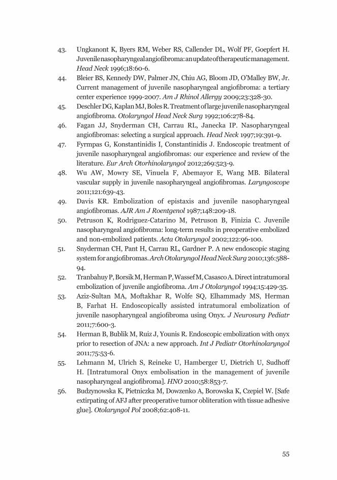
55
43. Ungkanont K, Byers RM, Weber RS, Callender DL, Wolf PF, Goepfert H. Juvenile nasopharyngeal angiofi broma: an update of therapeutic management. Head Neck 1996;18:60-6.
44. Bleier BS, Kennedy DW, Palmer JN, Chiu AG, Bloom JD, O’Malley BW, Jr. Current management of juvenile nasopharyngeal angiofi broma: a tertiary center experience 1999-2007. Am J Rhinol Allergy 2009;23:328-30.
45. Deschler DG, Kaplan MJ, Boles R. Treatment of large juvenile nasopharyngeal angiofi broma. Otolaryngol Head Neck Surg 1992;106:278-84.
46. Fagan JJ, Snyderman CH, Carrau RL, Janecka IP. Nasopharyngeal angiofi bromas: selecting a surgical approach. Head Neck 1997;19:391-9.
47. Fyrmpas G, Konstantinidis I, Constantinidis J. Endoscopic treatment of juvenile nasopharyngeal angiofi bromas: our experience and review of the literature. Eur Arch Otorhinolaryngol 2012;269:523-9.
48. Wu AW, Mowry SE, Vinuela F, Abemayor E, Wang MB. Bilateral vascular supply in juvenile nasopharyngeal angiofi bromas. Laryngoscope 2011;121:639-43.
49. Davis KR. Embolization of epistaxis and juvenile nasopharyngeal angiofi bromas. AJR Am J Roentgenol 1987;148:209-18.
50. Petruson K, Rodriguez-Catarino M, Petruson B, Finizia C. Juvenile nasopharyngeal angiofi broma: long-term results in preoperative embolized and non-embolized patients. Acta Otolaryngol 2002;122:96-100.
51. Snyderman CH, Pant H, Carrau RL, Gardner P. A new endoscopic staging system for angiofi bromas. Arch Otolaryngol Head Neck Surg 2010;136:588-94.
52. Tranbahuy P, Borsik M, Herman P, Wassef M, Casasco A. Direct intratumoral embolization of juvenile angiofi broma. Am J Otolaryngol 1994;15:429-35.
53. Aziz-Sultan MA, Moftakhar R, Wolfe SQ, Elhammady MS, Herman B, Farhat H. Endoscopically assisted intratumoral embolization of juvenile nasopharyngeal angiofi broma using Onyx. J Neurosurg Pediatr 2011;7:600-3.
54. Herman B, Bublik M, Ruiz J, Younis R. Endoscopic embolization with onyx prior to resection of JNA: a new approach. Int J Pediatr Otorhinolaryngol 2011;75:53-6.
55. Lehmann M, Ulrich S, Reineke U, Hamberger U, Dietrich U, Sudhoff H. [Intratumoral Onyx embolisation in the management of juvenile nasopharyngeal angiofi broma]. HNO 2010;58:853-7.
56. Budzynowska K, Pietniczka M, Dowzenko A, Borowska K, Czepiel W. [Safe extirpating of AFJ after preoperative tumor obliteration with tissue adhesive glue]. Otolaryngol Pol 2008;62:408-11.
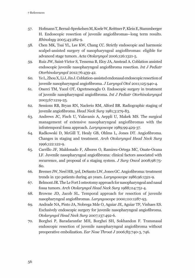
56
7 References
57. Hofmann T, Bernal-Sprekelsen M, Koele W, Reittner P, Klein E, Stammberger H. Endoscopic resection of juvenile angiofi bromas--long term results. Rhinology 2005;43:282-9.
58. Chen MK, Tsai YL, Lee KW, Chang CC. Strictly endoscopic and harmonic scalpel-assisted surgery of nasopharyngeal angiofi bromas: eligible for advanced stage tumors. Acta Otolaryngol 2006;126:1321-5.
59. Ruiz JW, Saint-Victor S, Tessema B, Eloy JA, Anstead A. Coblation assisted endoscopic juvenile nasopharyngeal angiofi broma resection. Int J Pediatr Otorhinolaryngol 2012;76:439-42.
60. Ye L, Zhou X, Li J, Jin J. Coblation-assisted endonasal endoscopic resection of juvenile nasopharyngeal angiofi broma. J Laryngol Otol 2011;125:940-4.
61. Onerci TM, Yucel OT, Ogretmenoglu O. Endoscopic surgery in treatment of juvenile nasopharyngeal angiofi broma. Int J Pediatr Otorhinolaryngol 2003;67:1219-25.
62. Sessions RB, Bryan RN, Naclerio RM, Alford BR. Radiographic staging of juvenile angiofi broma. Head Neck Surg 1981;3:279-83.
63. Andrews JC, Fisch U, Valavanis A, Aeppli U, Makek MS. The surgical management of extensive nasopharyngeal angiofibromas with the infratemporal fossa approach. Laryngoscope 1989;99:429-37.
64. Radkowski D, McGill T, Healy GB, Ohlms L, Jones DT. Angiofi broma. Changes in staging and treatment. Arch Otolaryngol Head Neck Surg 1996;122:122-9.
65. Carrillo JF, Maldonado F, Albores O, Ramirez-Ortega MC, Onate-Ocana LF. Juvenile nasopharyngeal angiofi broma: clinical factors associated with recurrence, and proposal of a staging system. J Surg Oncol 2008;98:75-80.
66. Bremer JW, Neel HB, 3rd, DeSanto LW, Jones GC. Angiofi broma: treatment trends in 150 patients during 40 years. Laryngoscope 1986;96:1321-9.
67. Belmont JR. The Le Fort I osteotomy approach for nasopharyngeal and nasal fossa tumors. Arch Otolaryngol Head Neck Surg 1988;114:751-4.
68. Browne JD, Jacob SL. Temporal approach for resection of juvenile nasopharyngeal angiofi bromas. Laryngoscope 2000;110:1287-93.
69. Andrade NA, Pinto JA, Nobrega Mde O, Aguiar JE, Aguiar TF, Vinhaes ES. Exclusively endoscopic surgery for juvenile nasopharyngeal angiofi broma. Otolaryngol Head Neck Surg 2007;137:492-6.
70. Borghei P, Baradaranfar MH, Borghei SH, Sokhandon F. Transnasal endoscopic resection of juvenile nasopharyngeal angiofi broma without preoperative embolization. Ear Nose Throat J 2006;85:740-3, 746.
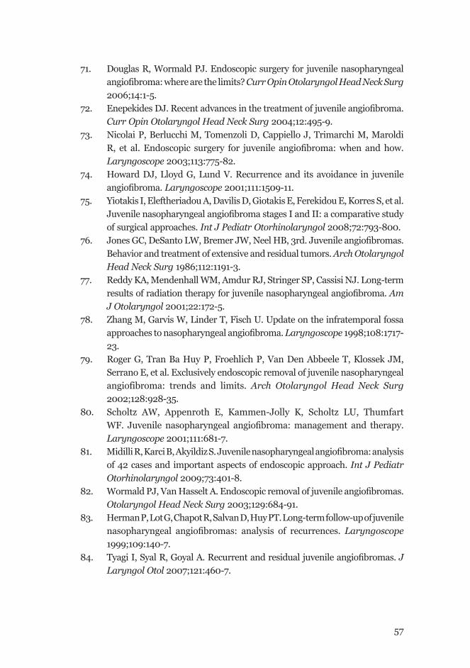
57
71. Douglas R, Wormald PJ. Endoscopic surgery for juvenile nasopharyngeal angiofi broma: where are the limits? Curr Opin Otolaryngol Head Neck Surg 2006;14:1-5.
72. Enepekides DJ. Recent advances in the treatment of juvenile angiofi broma. Curr Opin Otolaryngol Head Neck Surg 2004;12:495-9.
73. Nicolai P, Berlucchi M, Tomenzoli D, Cappiello J, Trimarchi M, Maroldi R, et al. Endoscopic surgery for juvenile angiofi broma: when and how. Laryngoscope 2003;113:775-82.
74. Howard DJ, Lloyd G, Lund V. Recurrence and its avoidance in juvenile angiofi broma. Laryngoscope 2001;111:1509-11.
75. Yiotakis I, Eleftheriadou A, Davilis D, Giotakis E, Ferekidou E, Korres S, et al. Juvenile nasopharyngeal angiofi broma stages I and II: a comparative study of surgical approaches. Int J Pediatr Otorhinolaryngol 2008;72:793-800.
76. Jones GC, DeSanto LW, Bremer JW, Neel HB, 3rd. Juvenile angiofi bromas. Behavior and treatment of extensive and residual tumors. Arch Otolaryngol Head Neck Surg 1986;112:1191-3.
77. Reddy KA, Mendenhall WM, Amdur RJ, Stringer SP, Cassisi NJ. Long-term results of radiation therapy for juvenile nasopharyngeal angiofi broma. Am J Otolaryngol 2001;22:172-5.
78. Zhang M, Garvis W, Linder T, Fisch U. Update on the infratemporal fossa approaches to nasopharyngeal angiofi broma. Laryngoscope 1998;108:1717-23.
79. Roger G, Tran Ba Huy P, Froehlich P, Van Den Abbeele T, Klossek JM, Serrano E, et al. Exclusively endoscopic removal of juvenile nasopharyngeal angiofibroma: trends and limits. Arch Otolaryngol Head Neck Surg 2002;128:928-35.
80. Scholtz AW, Appenroth E, Kammen-Jolly K, Scholtz LU, Thumfart WF. Juvenile nasopharyngeal angiofi broma: management and therapy. Laryngoscope 2001;111:681-7.
81. Midilli R, Karci B, Akyildiz S. Juvenile nasopharyngeal angiofi broma: analysis of 42 cases and important aspects of endoscopic approach. Int J Pediatr Otorhinolaryngol 2009;73:401-8.
82. Wormald PJ, Van Hasselt A. Endoscopic removal of juvenile angiofi bromas. Otolaryngol Head Neck Surg 2003;129:684-91.
83. Herman P, Lot G, Chapot R, Salvan D, Huy PT. Long-term follow-up of juvenile nasopharyngeal angiofi bromas: analysis of recurrences. Laryngoscope 1999;109:140-7.
84. Tyagi I, Syal R, Goyal A. Recurrent and residual juvenile angiofi bromas. J Laryngol Otol 2007;121:460-7.
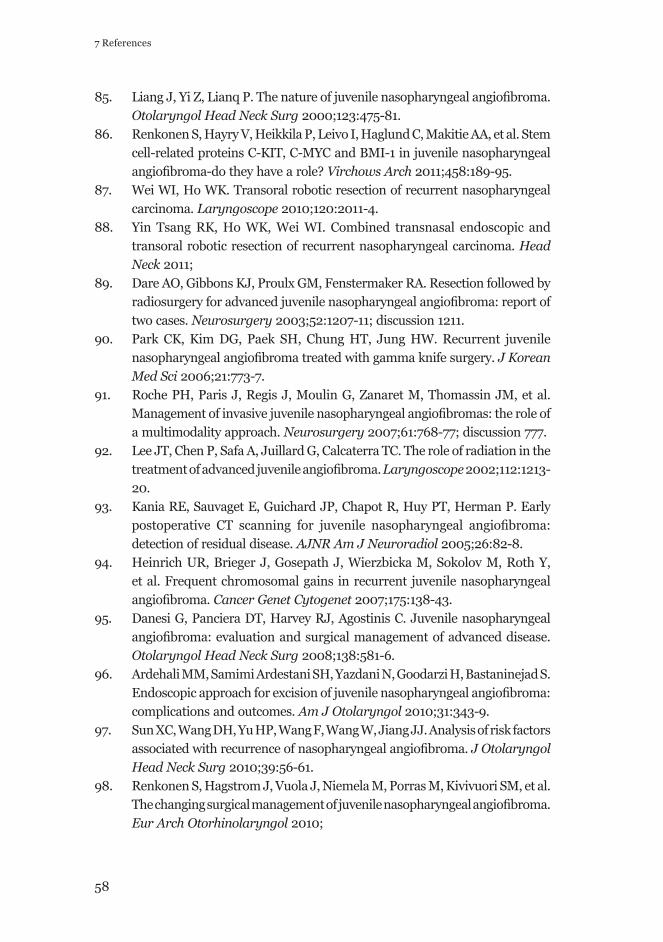
58
7 References
85. Liang J, Yi Z, Lianq P. The nature of juvenile nasopharyngeal angiofi broma. Otolaryngol Head Neck Surg 2000;123:475-81.
86. Renkonen S, Hayry V, Heikkila P, Leivo I, Haglund C, Makitie AA, et al. Stem cell-related proteins C-KIT, C-MYC and BMI-1 in juvenile nasopharyngeal angiofi broma-do they have a role? Virchows Arch 2011;458:189-95.
87. Wei WI, Ho WK. Transoral robotic resection of recurrent nasopharyngeal carcinoma. Laryngoscope 2010;120:2011-4.
88. Yin Tsang RK, Ho WK, Wei WI. Combined transnasal endoscopic and transoral robotic resection of recurrent nasopharyngeal carcinoma. Head Neck 2011;
89. Dare AO, Gibbons KJ, Proulx GM, Fenstermaker RA. Resection followed by radiosurgery for advanced juvenile nasopharyngeal angiofi broma: report of two cases. Neurosurgery 2003;52:1207-11; discussion 1211.
90. Park CK, Kim DG, Paek SH, Chung HT, Jung HW. Recurrent juvenile nasopharyngeal angiofi broma treated with gamma knife surgery. J Korean Med Sci 2006;21:773-7.
91. Roche PH, Paris J, Regis J, Moulin G, Zanaret M, Thomassin JM, et al. Management of invasive juvenile nasopharyngeal angiofi bromas: the role of a multimodality approach. Neurosurgery 2007;61:768-77; discussion 777.
92. Lee JT, Chen P, Safa A, Juillard G, Calcaterra TC. The role of radiation in the treatment of advanced juvenile angiofi broma. Laryngoscope 2002;112:1213-20.
93. Kania RE, Sauvaget E, Guichard JP, Chapot R, Huy PT, Herman P. Early postoperative CT scanning for juvenile nasopharyngeal angiofi broma: detection of residual disease. AJNR Am J Neuroradiol 2005;26:82-8.
94. Heinrich UR, Brieger J, Gosepath J, Wierzbicka M, Sokolov M, Roth Y, et al. Frequent chromosomal gains in recurrent juvenile nasopharyngeal angiofi broma. Cancer Genet Cytogenet 2007;175:138-43.
95. Danesi G, Panciera DT, Harvey RJ, Agostinis C. Juvenile nasopharyngeal angiofi broma: evaluation and surgical management of advanced disease. Otolaryngol Head Neck Surg 2008;138:581-6.
96. Ardehali MM, Samimi Ardestani SH, Yazdani N, Goodarzi H, Bastaninejad S. Endoscopic approach for excision of juvenile nasopharyngeal angiofi broma: complications and outcomes. Am J Otolaryngol 2010;31:343-9.
97. Sun XC, Wang DH, Yu HP, Wang F, Wang W, Jiang JJ. Analysis of risk factors associated with recurrence of nasopharyngeal angiofi broma. J Otolaryngol Head Neck Surg 2010;39:56-61.
98. Renkonen S, Hagstrom J, Vuola J, Niemela M, Porras M, Kivivuori SM, et al. The changing surgical management of juvenile nasopharyngeal angiofi broma. Eur Arch Otorhinolaryngol 2010;
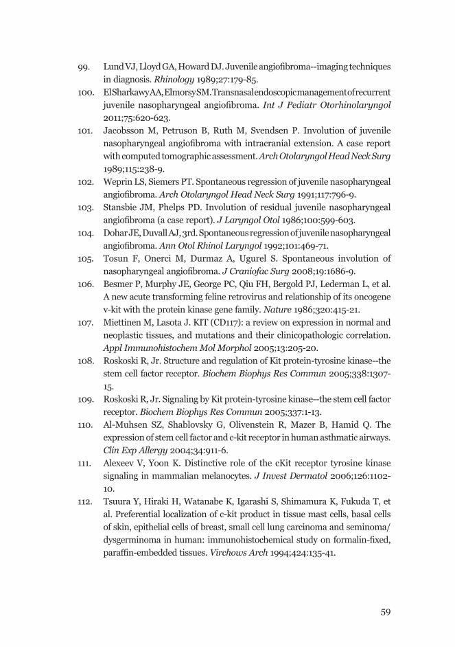
59
99. Lund VJ, Lloyd GA, Howard DJ. Juvenile angiofi broma--imaging techniques in diagnosis. Rhinology 1989;27:179-85.
100. El Sharkawy AA, Elmorsy SM. Transnasal endoscopic management of recurrent juvenile nasopharyngeal angiofi broma. Int J Pediatr Otorhinolaryngol 2011;75:620-623.
101. Jacobsson M, Petruson B, Ruth M, Svendsen P. Involution of juvenile nasopharyngeal angiofi broma with intracranial extension. A case report with computed tomographic assessment. Arch Otolaryngol Head Neck Surg 1989;115:238-9.
102. Weprin LS, Siemers PT. Spontaneous regression of juvenile nasopharyngeal angiofi broma. Arch Otolaryngol Head Neck Surg 1991;117:796-9.
103. Stansbie JM, Phelps PD. Involution of residual juvenile nasopharyngeal angiofi broma (a case report). J Laryngol Otol 1986;100:599-603.
104. Dohar JE, Duvall AJ, 3rd. Spontaneous regression of juvenile nasopharyngeal angiofi broma. Ann Otol Rhinol Laryngol 1992;101:469-71.
105. Tosun F, Onerci M, Durmaz A, Ugurel S. Spontaneous involution of nasopharyngeal angiofi broma. J Craniofac Surg 2008;19:1686-9.
106. Besmer P, Murphy JE, George PC, Qiu FH, Bergold PJ, Lederman L, et al. A new acute transforming feline retrovirus and relationship of its oncogene v-kit with the protein kinase gene family. Nature 1986;320:415-21.
107. Miettinen M, Lasota J. KIT (CD117): a review on expression in normal and neoplastic tissues, and mutations and their clinicopathologic correlation. Appl Immunohistochem Mol Morphol 2005;13:205-20.
108. Roskoski R, Jr. Structure and regulation of Kit protein-tyrosine kinase--the stem cell factor receptor. Biochem Biophys Res Commun 2005;338:1307-15.
109. Roskoski R, Jr. Signaling by Kit protein-tyrosine kinase--the stem cell factor receptor. Biochem Biophys Res Commun 2005;337:1-13.
110. Al-Muhsen SZ, Shablovsky G, Olivenstein R, Mazer B, Hamid Q. The expression of stem cell factor and c-kit receptor in human asthmatic airways. Clin Exp Allergy 2004;34:911-6.
111. Alexeev V, Yoon K. Distinctive role of the cKit receptor tyrosine kinase signaling in mammalian melanocytes. J Invest Dermatol 2006;126:1102-10.
112. Tsuura Y, Hiraki H, Watanabe K, Igarashi S, Shimamura K, Fukuda T, et al. Preferential localization of c-kit product in tissue mast cells, basal cells of skin, epithelial cells of breast, small cell lung carcinoma and seminoma/dysgerminoma in human: immunohistochemical study on formalin-fi xed, paraffi n-embedded tissues. Virchows Arch 1994;424:135-41.
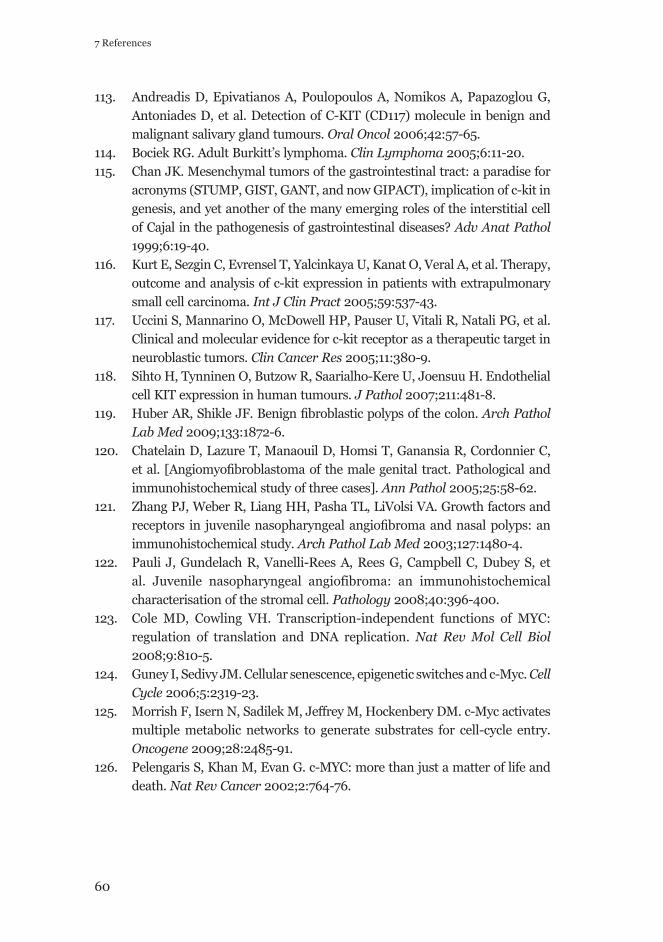
60
7 References
113. Andreadis D, Epivatianos A, Poulopoulos A, Nomikos A, Papazoglou G, Antoniades D, et al. Detection of C-KIT (CD117) molecule in benign and malignant salivary gland tumours. Oral Oncol 2006;42:57-65.
114. Bociek RG. Adult Burkitt’s lymphoma. Clin Lymphoma 2005;6:11-20.115. Chan JK. Mesenchymal tumors of the gastrointestinal tract: a paradise for
acronyms (STUMP, GIST, GANT, and now GIPACT), implication of c-kit in genesis, and yet another of the many emerging roles of the interstitial cell of Cajal in the pathogenesis of gastrointestinal diseases? Adv Anat Pathol 1999;6:19-40.
116. Kurt E, Sezgin C, Evrensel T, Yalcinkaya U, Kanat O, Veral A, et al. Therapy, outcome and analysis of c-kit expression in patients with extrapulmonary small cell carcinoma. Int J Clin Pract 2005;59:537-43.
117. Uccini S, Mannarino O, McDowell HP, Pauser U, Vitali R, Natali PG, et al. Clinical and molecular evidence for c-kit receptor as a therapeutic target in neuroblastic tumors. Clin Cancer Res 2005;11:380-9.
118. Sihto H, Tynninen O, Butzow R, Saarialho-Kere U, Joensuu H. Endothelial cell KIT expression in human tumours. J Pathol 2007;211:481-8.
119. Huber AR, Shikle JF. Benign fi broblastic polyps of the colon. Arch Pathol Lab Med 2009;133:1872-6.
120. Chatelain D, Lazure T, Manaouil D, Homsi T, Ganansia R, Cordonnier C, et al. [Angiomyofi broblastoma of the male genital tract. Pathological and immunohistochemical study of three cases]. Ann Pathol 2005;25:58-62.
121. Zhang PJ, Weber R, Liang HH, Pasha TL, LiVolsi VA. Growth factors and receptors in juvenile nasopharyngeal angiofi broma and nasal polyps: an immunohistochemical study. Arch Pathol Lab Med 2003;127:1480-4.
122. Pauli J, Gundelach R, Vanelli-Rees A, Rees G, Campbell C, Dubey S, et al. Juvenile nasopharyngeal angiofibroma: an immunohistochemical characterisation of the stromal cell. Pathology 2008;40:396-400.
123. Cole MD, Cowling VH. Transcription-independent functions of MYC: regulation of translation and DNA replication. Nat Rev Mol Cell Biol 2008;9:810-5.
124. Guney I, Sedivy JM. Cellular senescence, epigenetic switches and c-Myc. Cell Cycle 2006;5:2319-23.
125. Morrish F, Isern N, Sadilek M, Jeffrey M, Hockenbery DM. c-Myc activates multiple metabolic networks to generate substrates for cell-cycle entry. Oncogene 2009;28:2485-91.
126. Pelengaris S, Khan M, Evan G. c-MYC: more than just a matter of life and death. Nat Rev Cancer 2002;2:764-76.

61
127. Vora HH, Shah NG, Trivedi TI, Goswami JV, Shukla SN, Shah PM. Expression of C-Myc mRNA in squamous cell carcinoma of the tongue. J Surg Oncol 2007;95:70-8.
128. Gil J, Kerai P, Lleonart M, Bernard D, Cigudosa JC, Peters G, et al. Immortalization of primary human prostate epithelial cells by c-Myc. Cancer Res 2005;65:2179-85.
129. Schick B, Wemmert S, Jung V, Steudel WI, Montenarh M, Urbschat S. Genetic heterogeneity of the MYC oncogene in advanced juvenile angiofi bromas. Cancer Genet Cytogenet 2006;164:25-31.
130. Corzo C, Corominas JM, Tusquets I, Salido M, Bellet M, Fabregat X, et al. The MYC oncogene in breast cancer progression: from benign epithelium to invasive carcinoma. Cancer Genet Cytogenet 2006;165:151-6.
131. Manjarrez ME, Ocadiz R, Valle L, Pacheco C, Marroquin A, De la Torre C, et al. Detection of human papillomavirus and relevant tumor suppressors and oncoproteins in laryngeal tumors. Clin Cancer Res 2006;12:6946-51.
132. Evan GI, Vousden KH. Proliferation, cell cycle and apoptosis in cancer. Nature 2001;411:342-8.
133. Haupt Y, Bath ML, Harris AW, Adams JM. bmi-1 transgene induces lymphomas and collaborates with myc in tumorigenesis. Oncogene 1993;8:3161-4.
134. Lao TT, Chan BC, Leung WC, Ho LF. Postpartum anemia unrelated to maternal prepregnancy BMI. Obes Res 2004;12:1903-4; author reply 1904-5.
135. Levy LS, Lobelle-Rich PA, Overbaugh J. fl vi-2, a target of retroviral insertional mutagenesis in feline thymic lymphosarcomas, encodes bmi-1. Oncogene 1993;8:1833-8.
136. Martin EC, Adler PN. The Polycomb group gene Posterior Sex Combs encodes a chromosomal protein. Development 1993;117:641-55.
137. van Lohuizen M, Frasch M, Wientjens E, Berns A. Sequence similarity between the mammalian bmi-1 proto-oncogene and the Drosophila regulatory genes Psc and Su(z)2. Nature 1991;353:353-5.
138. Spivakov M, Fisher AG. Epigenetic signatures of stem-cell identity. Nat Rev Genet 2007;8:263-71.
139. Iwama A, Oguro H, Negishi M, Kato Y, Morita Y, Tsukui H, et al. Enhanced self-renewal of hematopoietic stem cells mediated by the polycomb gene product Bmi-1. Immunity 2004;21:843-51.
140. Molofsky AV, Pardal R, Iwashita T, Park IK, Clarke MF, Morrison SJ. Bmi-1 dependence distinguishes neural stem cell self-renewal from progenitor proliferation. Nature 2003;425:962-7.
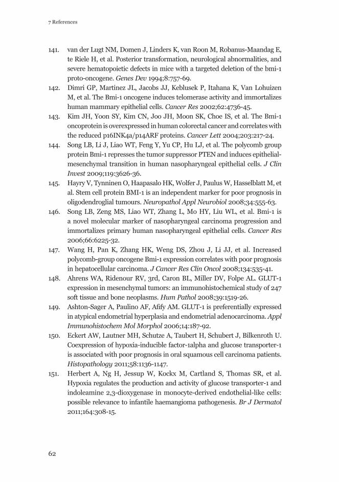
62
7 References
141. van der Lugt NM, Domen J, Linders K, van Roon M, Robanus-Maandag E, te Riele H, et al. Posterior transformation, neurological abnormalities, and severe hematopoietic defects in mice with a targeted deletion of the bmi-1 proto-oncogene. Genes Dev 1994;8:757-69.
142. Dimri GP, Martinez JL, Jacobs JJ, Keblusek P, Itahana K, Van Lohuizen M, et al. The Bmi-1 oncogene induces telomerase activity and immortalizes human mammary epithelial cells. Cancer Res 2002;62:4736-45.
143. Kim JH, Yoon SY, Kim CN, Joo JH, Moon SK, Choe IS, et al. The Bmi-1 oncoprotein is overexpressed in human colorectal cancer and correlates with the reduced p16INK4a/p14ARF proteins. Cancer Lett 2004;203:217-24.
144. Song LB, Li J, Liao WT, Feng Y, Yu CP, Hu LJ, et al. The polycomb group protein Bmi-1 represses the tumor suppressor PTEN and induces epithelial-mesenchymal transition in human nasopharyngeal epithelial cells. J Clin Invest 2009;119:3626-36.
145. Hayry V, Tynninen O, Haapasalo HK, Wolfer J, Paulus W, Hasselblatt M, et al. Stem cell protein BMI-1 is an independent marker for poor prognosis in oligodendroglial tumours. Neuropathol Appl Neurobiol 2008;34:555-63.
146. Song LB, Zeng MS, Liao WT, Zhang L, Mo HY, Liu WL, et al. Bmi-1 is a novel molecular marker of nasopharyngeal carcinoma progression and immortalizes primary human nasopharyngeal epithelial cells. Cancer Res 2006;66:6225-32.
147. Wang H, Pan K, Zhang HK, Weng DS, Zhou J, Li JJ, et al. Increased polycomb-group oncogene Bmi-1 expression correlates with poor prognosis in hepatocellular carcinoma. J Cancer Res Clin Oncol 2008;134:535-41.
148. Ahrens WA, Ridenour RV, 3rd, Caron BL, Miller DV, Folpe AL. GLUT-1 expression in mesenchymal tumors: an immunohistochemical study of 247 soft tissue and bone neoplasms. Hum Pathol 2008;39:1519-26.
149. Ashton-Sager A, Paulino AF, Afi fy AM. GLUT-1 is preferentially expressed in atypical endometrial hyperplasia and endometrial adenocarcinoma. Appl Immunohistochem Mol Morphol 2006;14:187-92.
150. Eckert AW, Lautner MH, Schutze A, Taubert H, Schubert J, Bilkenroth U. Coexpression of hypoxia-inducible factor-1alpha and glucose transporter-1 is associated with poor prognosis in oral squamous cell carcinoma patients. Histopathology 2011;58:1136-1147.
151. Herbert A, Ng H, Jessup W, Kockx M, Cartland S, Thomas SR, et al. Hypoxia regulates the production and activity of glucose transporter-1 and indoleamine 2,3-dioxygenase in monocyte-derived endothelial-like cells: possible relevance to infantile haemangioma pathogenesis. Br J Dermatol 2011;164:308-15.
63
152. Mendez LE, Manci N, Cantuaria G, Gomez-Marin O, Penalver M, Braunschweiger P, et al. Expression of glucose transporter-1 in cervical cancer and its precursors. Gynecol Oncol 2002;86:138-43.
153. Dyduch G, Okon K, Mierzynski W. Benign vascular proliferations--an immunohistochemical and comparative study. Pol J Pathol 2004;55:59-64.
154. Leon-Villapalos J, Wolfe K, Kangesu L. GLUT-1: an extra diagnostic tool to differentiate between haemangiomas and vascular malformations. Br J Plast Surg 2005;58:348-52.
155. Rathmell WK, Acs G, Simon MC, Vaughn DJ. HIF transcription factor expression and induction of hypoxic response genes in a retroperitoneal angiosarcoma. Anticancer Res 2004;24:167-9.
156. Chiquet-Ehrismann R, Hagios C, Matsumoto K. The tenascin gene family. Perspect Dev Neurobiol 1994;2:3-7.
157. Jones FS, Jones PL. The tenascin family of ECM glycoproteins: structure, function, and regulation during embryonic development and tissue remodeling. Dev Dyn 2000;218:235-59.
158. Tucker RP, Chiquet-Ehrismann R. The regulation of tenascin expression by tissue microenvironments. Biochim Biophys Acta 2009;1793:888-92.
159. Atula T, Hedstrom J, Finne P, Leivo I, Markkanen-Leppanen M, Haglund C. Tenascin-C expression and its prognostic signifi cance in oral and pharyngeal squamous cell carcinoma. Anticancer Res 2003;23:3051-6.
160. Jahkola T, Toivonen T, Virtanen I, von Smitten K, Nordling S, von Boguslawski K, et al. Tenascin-C expression in invasion border of early breast cancer: a predictor of local and distant recurrence. Br J Cancer 1998;78:1507-13.
161. Koljonen V, Bohling T, Tukiainen E, Haglund C, Jahkola T. Tenascin-C expression in Merkel cell carcinoma lymph node metastasis. APMIS 2006;114:39-42.
162. Lundin M, Nordling S, Lundin J, Haglund C. Tenascin-C expression and its prognostic signifi cance in colorectal cancer. Oncology 2007;72:403-9.
163. Wiksten JP, Lundin J, Nordling S, Lundin M, Kokkola A, von Boguslawski K, et al. Tenascin-C expression correlates with prognosis in gastric cancer. Oncology 2003;64:245-50.
164. Alexopoulou AN, Multhaupt HA, Couchman JR. Syndecans in wound healing, infl ammation and vascular biology. Int J Biochem Cell Biol 2007;39:505-28.
165. Fears CY, Woods A. The role of syndecans in disease and wound healing. Matrix Biol 2006;25:443-56.
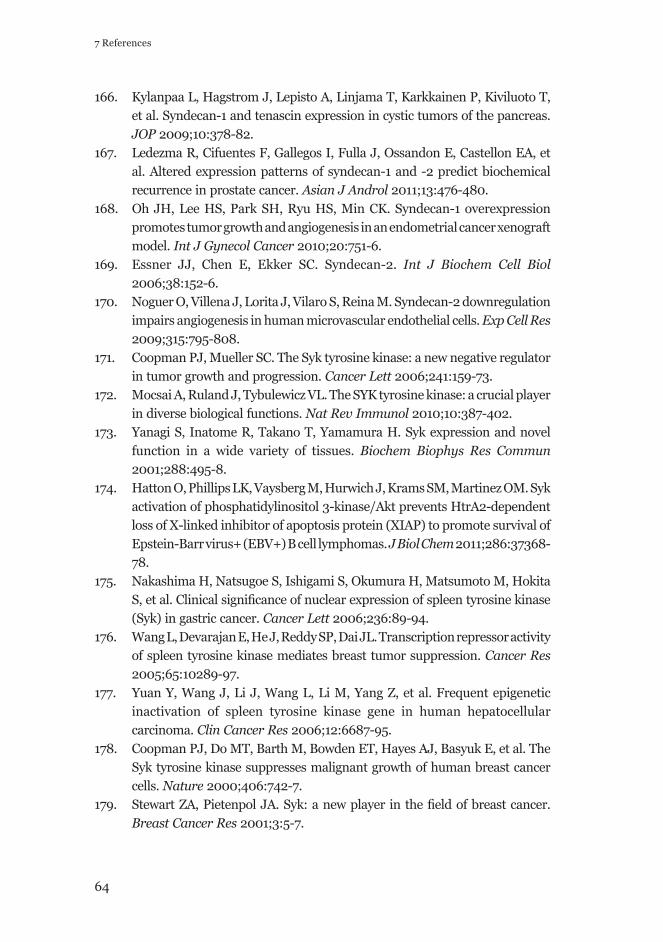
64
7 References
166. Kylanpaa L, Hagstrom J, Lepisto A, Linjama T, Karkkainen P, Kiviluoto T, et al. Syndecan-1 and tenascin expression in cystic tumors of the pancreas. JOP 2009;10:378-82.
167. Ledezma R, Cifuentes F, Gallegos I, Fulla J, Ossandon E, Castellon EA, et al. Altered expression patterns of syndecan-1 and -2 predict biochemical recurrence in prostate cancer. Asian J Androl 2011;13:476-480.
168. Oh JH, Lee HS, Park SH, Ryu HS, Min CK. Syndecan-1 overexpression promotes tumor growth and angiogenesis in an endometrial cancer xenograft model. Int J Gynecol Cancer 2010;20:751-6.
169. Essner JJ, Chen E, Ekker SC. Syndecan-2. Int J Biochem Cell Biol 2006;38:152-6.
170. Noguer O, Villena J, Lorita J, Vilaro S, Reina M. Syndecan-2 downregulation impairs angiogenesis in human microvascular endothelial cells. Exp Cell Res 2009;315:795-808.
171. Coopman PJ, Mueller SC. The Syk tyrosine kinase: a new negative regulator in tumor growth and progression. Cancer Lett 2006;241:159-73.
172. Mocsai A, Ruland J, Tybulewicz VL. The SYK tyrosine kinase: a crucial player in diverse biological functions. Nat Rev Immunol 2010;10:387-402.
173. Yanagi S, Inatome R, Takano T, Yamamura H. Syk expression and novel function in a wide variety of tissues. Biochem Biophys Res Commun 2001;288:495-8.
174. Hatton O, Phillips LK, Vaysberg M, Hurwich J, Krams SM, Martinez OM. Syk activation of phosphatidylinositol 3-kinase/Akt prevents HtrA2-dependent loss of X-linked inhibitor of apoptosis protein (XIAP) to promote survival of Epstein-Barr virus+ (EBV+) B cell lymphomas. J Biol Chem 2011;286:37368-78.
175. Nakashima H, Natsugoe S, Ishigami S, Okumura H, Matsumoto M, Hokita S, et al. Clinical signifi cance of nuclear expression of spleen tyrosine kinase (Syk) in gastric cancer. Cancer Lett 2006;236:89-94.
176. Wang L, Devarajan E, He J, Reddy SP, Dai JL. Transcription repressor activity of spleen tyrosine kinase mediates breast tumor suppression. Cancer Res 2005;65:10289-97.
177. Yuan Y, Wang J, Li J, Wang L, Li M, Yang Z, et al. Frequent epigenetic inactivation of spleen tyrosine kinase gene in human hepatocellular carcinoma. Clin Cancer Res 2006;12:6687-95.
178. Coopman PJ, Do MT, Barth M, Bowden ET, Hayes AJ, Basyuk E, et al. The Syk tyrosine kinase suppresses malignant growth of human breast cancer cells. Nature 2000;406:742-7.
179. Stewart ZA, Pietenpol JA. Syk: a new player in the fi eld of breast cancer. Breast Cancer Res 2001;3:5-7.
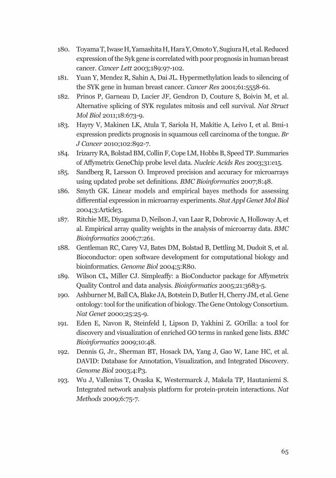
65
180. Toyama T, Iwase H, Yamashita H, Hara Y, Omoto Y, Sugiura H, et al. Reduced expression of the Syk gene is correlated with poor prognosis in human breast cancer. Cancer Lett 2003;189:97-102.
181. Yuan Y, Mendez R, Sahin A, Dai JL. Hypermethylation leads to silencing of the SYK gene in human breast cancer. Cancer Res 2001;61:5558-61.
182. Prinos P, Garneau D, Lucier JF, Gendron D, Couture S, Boivin M, et al. Alternative splicing of SYK regulates mitosis and cell survival. Nat Struct Mol Biol 2011;18:673-9.
183. Hayry V, Makinen LK, Atula T, Sariola H, Makitie A, Leivo I, et al. Bmi-1 expression predicts prognosis in squamous cell carcinoma of the tongue. Br J Cancer 2010;102:892-7.
184. Irizarry RA, Bolstad BM, Collin F, Cope LM, Hobbs B, Speed TP. Summaries of Affymetrix GeneChip probe level data. Nucleic Acids Res 2003;31:e15.
185. Sandberg R, Larsson O. Improved precision and accuracy for microarrays using updated probe set defi nitions. BMC Bioinformatics 2007;8:48.
186. Smyth GK. Linear models and empirical bayes methods for assessing differential expression in microarray experiments. Stat Appl Genet Mol Biol 2004;3:Article3.
187. Ritchie ME, Diyagama D, Neilson J, van Laar R, Dobrovic A, Holloway A, et al. Empirical array quality weights in the analysis of microarray data. BMC Bioinformatics 2006;7:261.
188. Gentleman RC, Carey VJ, Bates DM, Bolstad B, Dettling M, Dudoit S, et al. Bioconductor: open software development for computational biology and bioinformatics. Genome Biol 2004;5:R80.
189. Wilson CL, Miller CJ. Simpleaffy: a BioConductor package for Affymetrix Quality Control and data analysis. Bioinformatics 2005;21:3683-5.
190. Ashburner M, Ball CA, Blake JA, Botstein D, Butler H, Cherry JM, et al. Gene ontology: tool for the unifi cation of biology. The Gene Ontology Consortium. Nat Genet 2000;25:25-9.
191. Eden E, Navon R, Steinfeld I, Lipson D, Yakhini Z. GOrilla: a tool for discovery and visualization of enriched GO terms in ranked gene lists. BMC Bioinformatics 2009;10:48.
192. Dennis G, Jr., Sherman BT, Hosack DA, Yang J, Gao W, Lane HC, et al. DAVID: Database for Annotation, Visualization, and Integrated Discovery. Genome Biol 2003;4:P3.
193. Wu J, Vallenius T, Ovaska K, Westermarck J, Makela TP, Hautaniemi S. Integrated network analysis platform for protein-protein interactions. Nat Methods 2009;6:75-7.
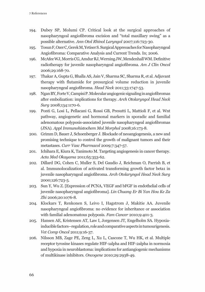
66
7 References
194. Dubey SP, Molumi CP. Critical look at the surgical approaches of nasopharyngeal angiofi broma excision and “total maxillary swing” as a possible alternative. Ann Otol Rhinol Laryngol 2007;116:723-30.
195. Tosun F, Ozer C, Gerek M, Yetiser S. Surgical Appreoaches for Nasopharyngeal Angiofi broma: Comparative Analysis and Current Trends. In; 2006.
196. McAfee WJ, Morris CG, Amdur RJ, Werning JW, Mendenhall WM. Defi nitive radiotherapy for juvenile nasopharyngeal angiofi broma. Am J Clin Oncol 2006;29:168-70.
197. Thakar A, Gupta G, Bhalla AS, Jain V, Sharma SC, Sharma R, et al. Adjuvant therapy with flutamide for presurgical volume reduction in juvenile nasopharyngeal angiofi broma. Head Neck 2011;33:1747-53.
198. Ngan BY, Forte V, Campisi P. Molecular angiogenic signaling in angiofi bromas after embolization: implications for therapy. Arch Otolaryngol Head Neck Surg 2008;134:1170-6.
199. Ponti G, Losi L, Pellacani G, Rossi GB, Presutti L, Mattioli F, et al. Wnt pathway, angiogenetic and hormonal markers in sporadic and familial adenomatous polyposis-associated juvenile nasopharyngeal angiofi bromas (JNA). Appl Immunohistochem Mol Morphol 2008;16:173-8.
200. Grimm D, Bauer J, Schoenberger J. Blockade of neoangiogenesis, a new and promising technique to control the growth of malignant tumors and their metastases. Curr Vasc Pharmacol 2009;7:347-57.
201. Ichihara E, Kiura K, Tanimoto M. Targeting angiogenesis in cancer therapy. Acta Med Okayama 2011;65:353-62.
202. Dillard DG, Cohen C, Muller S, Del Gaudio J, Reichman O, Parrish B, et al. Immunolocalization of activated transforming growth factor beta1 in juvenile nasopharyngeal angiofi broma. Arch Otolaryngol Head Neck Surg 2000;126:723-5.
203. Sun Y, Wu Z. [Expression of PCNA, VEGF and bFGF in endothelial cells of juvenile nasopharyngeal angiofi broma]. Lin Chuang Er Bi Yan Hou Ke Za Zhi 2006;20:1076-8.
204. Klockars T, Renkonen S, Leivo I, Hagstrom J, Makitie AA. Juvenile nasopharyngeal angiofi broma: no evidence for inheritance or association with familial adenomatous polyposis. Fam Cancer 2010;9:401-3.
205. Hansen AE, Kristensen AT, Law I, Jorgensen JT, Engelholm SA. Hypoxia-inducible factors--regulation, role and comparative aspects in tumourigenesis. Vet Comp Oncol 2011;9:16-37.
206. Nilsson MB, Zage PE, Zeng L, Xu L, Cascone T, Wu HK, et al. Multiple receptor tyrosine kinases regulate HIF-1alpha and HIF-2alpha in normoxia and hypoxia in neuroblastoma: implications for antiangiogenic mechanisms of multikinase inhibitors. Oncogene 2010;29:2938-49.
67
207. Tojyo I, Yamaguchi A, Nitta T, Yoshida H, Fujita S, Yoshida T. Effect of hypoxia and interleukin-1beta on expression of tenascin-C in temporomandibular joint. Oral Dis 2008;14:45-50.
208. Midwood KS, Hussenet T, Langlois B, Orend G. Advances in tenascin-C biology. Cell Mol Life Sci 2011;68:3175-99.
209. Behrem S, Zarkovic K, Eskinja N, Jonjic N. Distribution pattern of tenascin-C in glioblastoma: correlation with angiogenesis and tumor cell proliferation. Pathol Oncol Res 2005;11:229-35.
210. Orend G, Chiquet-Ehrismann R. Tenascin-C induced signaling in cancer. Cancer Lett 2006;244:143-63.
211. Pezzolo A, Parodi F, Marimpietri D, Raffaghello L, Cocco C, Pistorio A, et al. Oct-4+/Tenascin C+ neuroblastoma cells serve as progenitors of tumor-derived endothelial cells. Cell Res 2011;21:1470-86.
212. Baeriswyl V, Christofori G. The angiogenic switch in carcinogenesis. Semin Cancer Biol 2009;19:329-37.
213. Tanaka K, Hiraiwa N, Hashimoto H, Yamazaki Y, Kusakabe M. Tenascin-C regulates angiogenesis in tumor through the regulation of vascular endothelial growth factor expression. Int J Cancer 2004;108:31-40.
214. Carmeliet P, Jain RK. Principles and mechanisms of vessel normalization for cancer and other angiogenic diseases. Nat Rev Drug Discov 2011;10:417-27.
215. Dejana E. The role of wnt signaling in physiological and pathological angiogenesis. Circ Res 2010;107:943-52.
216. Stefater JA, 3rd, Lewkowich I, Rao S, Mariggi G, Carpenter AC, Burr AR, et al. Regulation of angiogenesis by a non-canonical Wnt-Flt1 pathway in myeloid cells. Nature 2011;474:511-5.
217. Daneman R, Agalliu D, Zhou L, Kuhnert F, Kuo CJ, Barres BA. Wnt/beta-catenin signaling is required for CNS, but not non-CNS, angiogenesis. Proc Natl Acad Sci U S A 2009;106:641-6.
218. Diaz-Molina JP, Llorente JL, Vivanco B, Martinez-Camblor P, Fresno MF, Perez-Escuredo J, et al. Wnt-pathway activation in intestinal-type sinonasal adenocarcinoma. Rhinology 2011;49:593-9.
219. Formosa R, Gruppetta M, Falzon S, Santillo G, Degaetano J, Xuereb-Anastasi A, et al. Expression and Clinical Signifi cance of Wnt Players and Survivin in Pituitary Tumours. Endocr Pathol 2012;23:123-131.
220. Tickenbrock L, Hehn S, Sargin B, Evers G, Ng PR, Choudhary C, et al. Activation of Wnt signaling in cKit-ITD mediated transformation and imatinib sensitivity in acute myeloid leukemia. Int J Hematol 2008;88:174-80.
68
7 References
221. Trowbridge JJ, Guezguez B, Moon RT, Bhatia M. Wnt3a activates dormant c-Kit(-) bone marrow-derived cells with short-term multilineage hematopoietic reconstitution capacity. Stem Cells 2010;28:1379-89.
222. Gebb SA, Fox K, Vaughn J, McKean D, Jones PL. Fetal oxygen tension promotes tenascin-C-dependent lung branching morphogenesis. Dev Dyn 2005;234:1-10.
223. Koperek O, Prinz A, Scheuba C, Niederle B, Kaserer K. Tenascin C in medullary thyroid microcarcinoma and C-cell hyperplasia. Virchows Arch 2009;455:43-8.
224. Mizukami Y, Chung DC. Hypoxia, Angiogenesis, and Colorectal Cancer. Current colorectal cancer report 2007;3:71-75.
225. Hyman E, Kauraniemi P, Hautaniemi S, Wolf M, Mousses S, Rozenblum E, et al. Impact of DNA amplifi cation on gene expression patterns in breast cancer. Cancer Res 2002;62:6240-5.
226. Jarvinen AK, Autio R, Haapa-Paananen S, Wolf M, Saarela M, Grenman R, et al. Identifi cation of target genes in laryngeal squamous cell carcinoma by high-resolution copy number and gene expression microarray analyses. Oncogene 2006;25:6997-7008.
227. Jarvinen AK, Autio R, Kilpinen S, Saarela M, Leivo I, Grenman R, et al. High-resolution copy number and gene expression microarray analyses of head and neck squamous cell carcinoma cell lines of tongue and larynx. Genes Chromosomes Cancer 2008;47:500-9.
228. Pollack JR, Perou CM, Alizadeh AA, Eisen MB, Pergamenschikov A, Williams CF, et al. Genome-wide analysis of DNA copy-number changes using cDNA microarrays. Nat Genet 1999;23:41-6.
229. Wolf M, Mousses S, Hautaniemi S, Karhu R, Huusko P, Allinen M, et al. High-resolution analysis of gene copy number alterations in human prostate cancer using CGH on cDNA microarrays: impact of copy number on gene expression. Neoplasia 2004;6:240-7.
230. Leary RJ, Lin JC, Cummins J, Boca S, Wood LD, Parsons DW, et al. Integrated analysis of homozygous deletions, focal amplifi cations, and sequence alterations in breast and colorectal cancers. Proc Natl Acad Sci U S A 2008;105:16224-9.
231. Myllykangas S, Junnila S, Kokkola A, Autio R, Scheinin I, Kiviluoto T, et al. Integrated gene copy number and expression microarray analysis of gastric cancer highlights potential target genes. Int J Cancer 2008;123:817-25.
232. Torkamani A, Verkhivker G, Schork NJ. Cancer driver mutations in protein kinase genes. Cancer Lett 2009;281:117-27.
69
233. Brunner C, Urbschat S, Jung V, Praetorius M, Schick B, Plinkert PK. [Chromosomal alterations in juvenile angiofibromas]. HNO 2003;51:981-5.
234. Nascimento EPMG, da Silveira SM, de Souza Trindade Filho JC, Rogatto SR, Viana de Camargo JL. Chromosomal imbalances in successive moments of human bladder urothelial carcinoma. Urol Oncol 2011;
235. Schick B, Wemmert S, Bechtel U, Nicolai P, Hofmann T, Golabek W, et al. Comprehensive genomic analysis identifi es MDM2 and AURKA as novel amplifi ed genes in juvenile angiofi bromas. Head Neck 2007;29:479-87.
236. Schick B, Wemmert S, Willnecker V, Dlugaiczyk J, Nicolai P, Siwiec H, et al. Genome-wide copy number profi ling using a 100K SNP array reveals novel disease-related genes BORIS and TSHZ1 in juvenile angiofi broma. Int J Oncol 2011;39:1143-51.
237. Duerr S, Wendler O, Aigner T, Karosi S, Schick B. Metalloproteinases in juvenile angiofi broma--a collagen rich tumor. Hum Pathol 2008;39:259-68.
238. Thalayasingam N, Isaacs JD. Anti-TNF therapy. Best Pract Res Clin Rheumatol 2011;25:549-67.
239. Wang S. The promise of cancer therapeutics targeting the TNF-related apoptosis-inducing ligand and TRAIL receptor pathway. Oncogene 2008;27:6207-15.
