facial emotion processing in major depression: a systematic
TRANSCRIPT
RESEARCH Open Access
Facial emotion processing in major depression: asystematic review of neuroimaging findingsAnja Stuhrmann1, Thomas Suslow1,2 and Udo Dannlowski1*
Abstract
Background: Cognitive models of depression suggest that major depression is characterized by biased facialemotion processing, making facial stimuli particularly valuable for neuroimaging research on the neurobiologicalcorrelates of depression. The present review provides an overview of functional neuroimaging studies on abnormalfacial emotion processing in major depression. Our main objective was to describe neurobiological differencesbetween depressed patients with major depressive disorder (MDD) and healthy controls (HCs) regarding brainresponsiveness to facial expressions and, furthermore, to delineate altered neural activation patterns associatedwith mood-congruent processing bias and to integrate these data with recent functional connectivity results. Wefurther discuss methodological aspects potentially explaining the heterogeneity of results.
Methods: A Medline search was performed up to August 2011 in order to identify studies on emotional faceprocessing in acutely depressed patients compared with HCs. A total of 25 studies using functional magneticresonance imaging were reviewed.
Results: The analysis of neural activation data showed abnormalities in MDD patients in a common faceprocessing network, pointing to mood-congruent processing bias (hyperactivation to negative and hypoactivationto positive stimuli) particularly in the amygdala, insula, parahippocampal gyrus, fusiform face area, and putamen.Furthermore, abnormal activation patterns were repeatedly found in parts of the cingulate gyrus and theorbitofrontal cortex, which are extended by investigations implementing functional connectivity analysis. However,despite several converging findings, some inconsistencies are observed, particularly in prefrontal areas, probablycaused by heterogeneities in paradigms and patient samples.
Conclusions: Further studies in remitted patients and high-risk samples are required to discern whether thedescribed abnormalities represent state or trait characteristics of depression.
Keywords: Facial emotion processing, fMRI, neuroimaging, depression, emotion, amygdala, anterior cingulate, orbi-tofrontal cortex, functional connectivity
BackgroundMajor depression ranks among the most debilitating dis-eases worldwide and is estimated to produce the secondlargest disease burden by the year 2020 [1]. Despite anincreasing amount of empirical studies investigatingabnormalities in affective processing in unipolar depres-sion, understanding the neurobiological underpinningsis still a major research goal and is essential for noveltreatment developments. In a large body of behavioral
studies, depression has been characterized by mood con-gruent emotion processing biases in different aspects ofcognition [2-5]. Apparently, these cognitive biases havebeen reported to be particularly prominent for emo-tional faces. Depressed patients seem to be less sensitivein the identification of emotional faces and, in addition,a negative response bias was found: they tend to inter-pret neutral faces as sad and happy faces as neutral (forreview see [6,7]).While negative faces seem to be processed more
rapidly and deeply, processing of positive facial expres-sions appears to be impaired [8-10]. Furthermore, beha-vioral biases towards sad faces seem to persist even after
* Correspondence: [email protected] of Münster, Department of Psychiatry, Albert-Schweitzer-Campus1, Building, A9, 48149 Münster, GermanyFull list of author information is available at the end of the article
Stuhrmann et al. Biology of Mood & Anxiety Disorders 2011, 1:10http://www.biolmoodanxietydisord.com/content/1/1/10 Biology of
Mood & Anxiety Disorders
© 2011 Stuhrmann et al; licensee BioMed Central Ltd. This is an Open Access article distributed under the terms of the CreativeCommons Attribution License (http://creativecommons.org/licenses/by/2.0), which permits unrestricted use, distribution, andreproduction in any medium, provided the original work is properly cited.
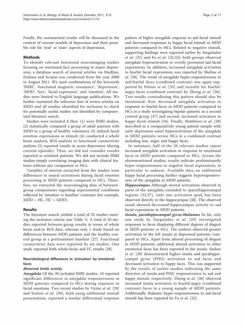
recovery from depression [11], increasing the risk forfuture depressive episodes [12]. Interestingly, rapid,automatic stages of emotion processing are also affectedin depression, as suggested by studies employing sublim-inal presentation conditions [13,14]. Figure 1 presentsthe main emotion processing stages as supposed byPhillips et al. [15], extended about separate pathways forstimulus presentation with or without consciousawareness.Faces are a very important component of daily human
visual communication. Since the processing of facialexpressions is a fundamental step in social functioning,guiding adequate social interaction [16], biased proces-sing of emotional faces in depression could be a strongdeterminant of the frequently observed interpersonalproblems, including social withdrawal, feelings of inter-personal rejection and restriction of non-verbal expres-siveness [17].Brain imaging techniques, such as functional magnetic
resonance imaging (fMRI), have already made substan-tial contributions to the understanding of how faces andfacial expressions are processed in humans [18-21].
According to neurobiological models of emotional faceprocessing, successful encoding of emotional expressionsdepends on multiple interactions between complimen-tary systems: a neural core system for the visual analysisof faces consists of the bilateral inferior occipital gyrus,the lateral fusiform gyrus and the superior temporal sul-cus. Changeable and invariant aspects of the face repre-sentation have distinct representations in this system. Asecond, extended system supports the processing offacial information such as meaning and significance. Itis composed of additional brain areas generally involvedin representing and producing emotions. Major compo-nents include the amygdala, insula, orbitofrontal areasand somatosensory cortex [22]. Notably, most if not allof these areas have already been implicated in the etiol-ogy of major depression (see [23-25] for reviews). Thus,presenting facial emotional stimuli is a valid and reliableapproach in order to activate brain areas crucial foremotion processing in general and crucial for the patho-physiology of depression specifically [18]. Unsurpris-ingly, emotional faces have been frequently employed inneuroimaging studies in depressed patients, contributingto the refinement of neurobiological models of depres-sion [24-26]. Put simply, these models postulateincreased activity in brain regions essential for emo-tional identification and production (that is, amygdala,orbitofrontal cortex (OFC), striatum) and decreasedneural activity within regions important for emotionregulation such as the dorsolateral prefrontal cortex(DLPFC) and anterior cingulate cortex (ACC).However, currently available data on emotional face
processing in depression are far from being consistent.The heterogeneity of study samples (for example, stateof illness, medication status and so on), imaging para-digms (for example, implicit or explicit processing para-digms, stimulus material, baseline condition), andanalysis strategies (for example, activation or connectiv-ity analyses) is reflected in apparently heterogeneousand partly conflicting findings at first sight. Given theimportance of emotional face processing in majordepression, the goal of the present review is to provide acomprehensive overview of neuroimaging studies inves-tigating facial emotion processing in acutely depressedpatients compared with healthy controls. Particulareffort was made to delineate altered neural activationpatterns associated with mood-congruent processingbias and to integrate these findings with functional con-nectivity results.First, we describe in detail the results of all available
fMRI studies comparing facial emotion-related brainactivation in patients with major depressive disorder(MDD) and healthy control (HC) subjects. In additionto whole brain and region of interest (ROI) data, recentfunctional connectivity data will also be considered.
Figure 1 Emotional perception and processing stages. Afterstimulus presentation (subliminal or supraliminal) the centralemotion perception and processing stages are: (1) the identificationand appraisal of stimulus significance, taking place with or withoutconscious awareness; (2) the generation of an affective state,expression of emotion and behavioral response; and (3) up or downregulation (circles with positive/negative signs) of the affective stateand identification process. Modified from Phillips et al. [15].
Stuhrmann et al. Biology of Mood & Anxiety Disorders 2011, 1:10http://www.biolmoodanxietydisord.com/content/1/1/10
Page 2 of 17
Finally, the summarized results will be discussed in thecontext of current models of depression and their possi-ble role for ‘trait’ or ‘state’ aspects of depression.
MethodsTo identify relevant functional neuroimaging studiesfocusing on emotional face processing in major depres-sion, a database search of journal articles via Medline,Embase and Scopus was conducted from the year 2000to August 2011. We used combinations of the keywords‘fMRI’, ‘functional magnetic resonance’, ‘depression’,‘MDD’, ‘face’, ‘facial expression’, and ‘emotion’. All stu-dies were limited to English language publications. Wefurther examined the reference lists of review articles onMDD and all studies identified for inclusion to checkfor potentially useful studies not identified by computer-ized literature search.Studies were included if they: (1) were fMRI studies,
(2) statistically compared a group of adult patients withMDD to a group of healthy volunteers (3) utilized facialemotion expressions as stimuli (4) conducted a wholebrain analysis, ROI analysis or functional connectivityanalysis (5) reported results in acute depression (duringcurrent episode). Thus, we did not consider resultsreported in remitted patients. We did not include fMRIstudies simply correlating imaging data with clinical fea-tures without any comparison to HCs.Variables of interest extracted from the studies were
differences in neural activations during facial emotionprocessing in MDD patients compared to HCs. There-fore, we extracted the neuroimaging data of between-group comparisons regarding experimental conditionsreflected by ‘emotion vs baseline’ contrasts (for example,MDD > HC, HC > MDD).
ResultsThe literature search yielded a total of 25 studies meet-ing the inclusion criteria (see Table 1). A total of 20 stu-dies reported between-group results in terms of wholebrain and/or ROI data, whereas only 1 study found nodifferences between MDD patients and the healthy con-trol group at a pretreatment baseline [27]. Functionalconnectivity data were reported by six studies. Onestudy reported both whole-brain and FC results [28].
Neurobiological differences in ‘activation’ by emotionalfacesAbnormal limbic activityAmygdala Of the 20 included fMRI studies, 10 reportedsignificant differences in amygdala responsiveness inMDD patients compared to HCs during exposure tofacial emotions. Two recent studies by Victor et al. [29]and Suslow et al. [30], both using subliminal stimulipresentation, reported a similar differential response
pattern of higher amygdala response to sad facial stimuliand decreased responses to happy facial stimuli in MDDpatients compared to HCs. Related to negative stimuli,supporting findings were reported earlier by Surguladzeet al. [31] and Fu et al. [32,33]; both groups observedamygdala hyperactivation to overtly presented sad facialexpressions. In addition, increased amygdala activationto fearful facial expressions was reported by Sheline etal. [34]. The result of amygdala hyper-responsiveness tosad/fearful faces (combined contrast) was again sup-ported by Peluso et al. [35] and recently for fearful/angry faces (combined contrast) by Zhong et al. [36].Two results contradicting this pattern should also bementioned: first, decreased amygdala activation inresponse to fearful faces in MDD patients compared toHCs in a study investigating bipolar patients as a secondcontrol group [37] and second, increased activation tohappy facial stimuli [34]. Finally, Matthews et al. [28]described in a comparatively young patient sample withearly depression onset hyperactivation of the amygdalain MDD patients versus HCs in a combined contrastincluding fear, angry and happy faces.In summary, half of the 20 relevant studies report
increased amygdala activation in response to emotionalfaces in MDD patients compared to HCs. Across theaforementioned studies, results indicate predominantlyhyper-responsiveness to negative facial expressions, inparticular to sadness. Available data on subliminalhappy facial processing further suggests hyporesponsive-ness of the amygdala in MDD patients.Hippocampus Although several activations observed inparts of the amygdala extended to (para)hippocampalregions [33,37], only one activation peak has beenobserved directly in the hippocampus [38]. The observedresult showed decreased hippocampus activity to sadfacial expressions in MDD patients.Insula, parahippocampal gyrus/thalamus So far, onlyone study by Surguladze et al. [39] investigatedresponses to faces displaying different degrees of disgustin MDD patients vs HCs. The authors observed greateractivation in the left insula in depressed patients com-pared to HCs. Apart from altered processing of disgustin MDD patients, additional altered activation to otheremotional faces has been reported in the insula: Suslowet al. [30] demonstrated higher insula and parahippo-campal gyrus (PHG) activation to sad faces anddecreased activation to happy faces. This was supportedby the results of earlier studies indicating the samedirection of insula and PHG responsiveness to sad andhappy stimuli, respectively. Zhong et al. [36] observedincreased insula activation to fearful/angry (combinedcontrast) faces in a young sample of MDD patients.Additionally, thalamic hyper-responsiveness to sad facialstimuli has been reported by Fu et al. [32].
Stuhrmann et al. Biology of Mood & Anxiety Disorders 2011, 1:10http://www.biolmoodanxietydisord.com/content/1/1/10
Page 3 of 17

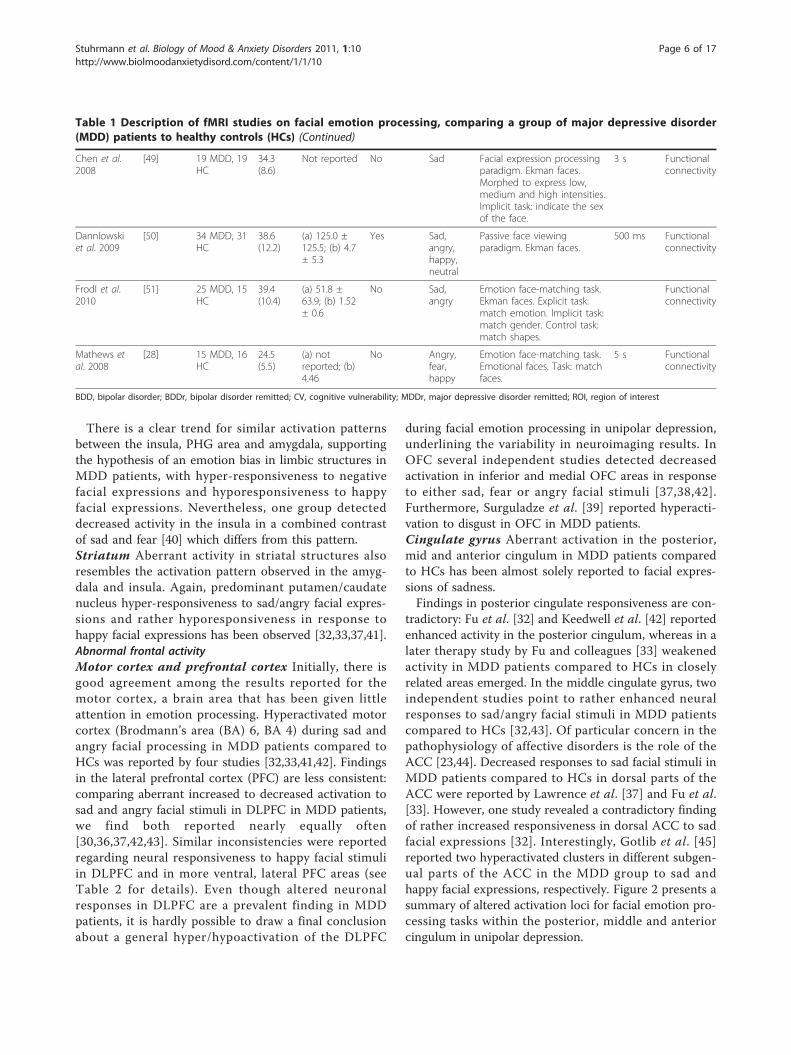
Table 1 Description of fMRI studies on facial emotion processing, comparing a group of major depressive disorder(MDD) patients to healthy controls (HCs)
Author/year Reference Participants Patientmeanage(SD)
Patient (a)meanduration ofillness inmonths; (b)meanepisodes
Medication Emotions Paradigm and stimulustype
Stimulusduration
Analysisapproach
Whole brain and/or ROI data:
Almeida et al.2010
[62] 15 MDD, 15HC, (15BDD), (15BDDr)
32.74(9.87)
(a) 13.67 ±9.87;(b) notreported
Yes Fear, sad,happy
Facial expression processingparadigm. Ekman faces.Morphed 50% and 100%intensity. Explicit task: labelemotion.
2 s ROI
Frodl et al.2009
[43] 12 MDD, 12HC
43.3(11.2)
Not reported Yes Sad,angry
Emotion face-matching task.Ekman faces. Explicit task:match emotion. Implicit task:match gender. Control task:match shapes.
5.3 s Wholebrain, ROIs
Frodl et al.2011
[27] 24 MDD, 15HC
38.9(10.4)
(a) 56.0 ±63.4; (b) 1.6 ±0.7
No Sad,angry
Emotion face-matching task.Faces from Gur andcolleagues. Explicit task:match the emotion. Implicittask: match the gender.Control task: match shapes.
5.3 s Wholebrain
Fu et al. 2004;Fu et al. 2007
[32,63] 19 MDD, 19HC
43.2(8.8)
Not reported No Sad,happy
Facial expression processingparadigm. Ekman faces.Morphed to express low,medium and high intensities.Implicit task: indicate the sexof the face.
3 s Wholebrain
Fu et al. 2008 [33] 16 MDD, 16HC
40.0(9.4)
(a) notreported; (b)0.63
No Sad Facial expression processingparadigm. Ekman faces.Morphed to express low,medium and high intensities.Implicit task: indicate the sexof the face.
3 s Wholebrain
Gotlib et al.2005
[45] 18 MDD, 18HC
35.2 Not reported Yes Sad,happy,neutral
Facial expression processingparadigm. Ekman faces.Implicit task: indicate the sexof the face.
3 s Wholebrain
Keedwell etal. 2005
[42] 12 MDD, 12HC
43 (9.8) Not reported Yes Sad,happy,neutral
Mood provocation paradigm.Individual autobiographicalmemory prompts playedprior to the presentation ofmood congruent facialexpressions. Ekman faces.Task: oral subjective rating ofmood.
2 s Wholebrain
Lawrence etal. 2004
[37] 9 MDD, 11HC, (12BDD)
41a (11) (a) 96 ± 60;(b) notreported
Yes Sad, fear,happy,neutral
Facial expression processingparadigm. Ekman faces.Morphed 50% and 100%intensity. Implicit task:indicate the sex of the face.
2 s Wholebrain, ROIs
Lee et al.2008
[38] 21 MDD, 15HC
46.8(9.1)
(a) 14.8 ± 3.3;(b) 1.9 ± 0.8
Yes Sad,angry,neutral
Face viewing paradigm. Dataset of Korean faces. Task:evaluative ratings (arousal,valence).
1.5 s ROIs
Matthews etal. 2008
[28] 15 MDD, 16HC
24.5(5.5)
(a) notreported; (b)4.46
No Angry,fear,happy
Emotion face-matching task.Emotional faces. Task: matchfaces.
5 s ROI
Stuhrmann et al. Biology of Mood & Anxiety Disorders 2011, 1:10http://www.biolmoodanxietydisord.com/content/1/1/10
Page 4 of 17
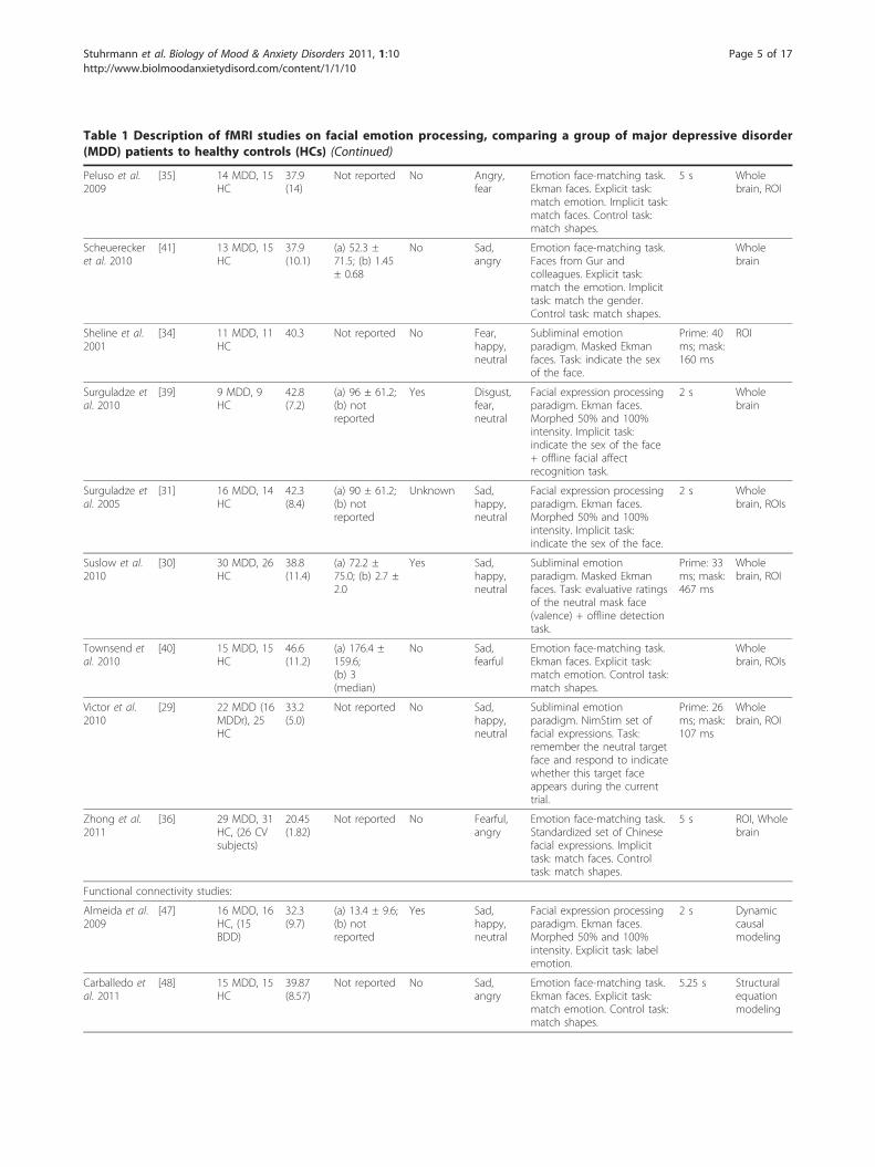
Table 1 Description of fMRI studies on facial emotion processing, comparing a group of major depressive disorder(MDD) patients to healthy controls (HCs) (Continued)
Peluso et al.2009
[35] 14 MDD, 15HC
37.9(14)
Not reported No Angry,fear
Emotion face-matching task.Ekman faces. Explicit task:match emotion. Implicit task:match faces. Control task:match shapes.
5 s Wholebrain, ROI
Scheuereckeret al. 2010
[41] 13 MDD, 15HC
37.9(10.1)
(a) 52.3 ±71.5; (b) 1.45± 0.68
No Sad,angry
Emotion face-matching task.Faces from Gur andcolleagues. Explicit task:match the emotion. Implicittask: match the gender.Control task: match shapes.
Wholebrain
Sheline et al.2001
[34] 11 MDD, 11HC
40.3 Not reported No Fear,happy,neutral
Subliminal emotionparadigm. Masked Ekmanfaces. Task: indicate the sexof the face.
Prime: 40ms; mask:160 ms
ROI
Surguladze etal. 2010
[39] 9 MDD, 9HC
42.8(7.2)
(a) 96 ± 61.2;(b) notreported
Yes Disgust,fear,neutral
Facial expression processingparadigm. Ekman faces.Morphed 50% and 100%intensity. Implicit task:indicate the sex of the face+ offline facial affectrecognition task.
2 s Wholebrain
Surguladze etal. 2005
[31] 16 MDD, 14HC
42.3(8.4)
(a) 90 ± 61.2;(b) notreported
Unknown Sad,happy,neutral
Facial expression processingparadigm. Ekman faces.Morphed 50% and 100%intensity. Implicit task:indicate the sex of the face.
2 s Wholebrain, ROIs
Suslow et al.2010
[30] 30 MDD, 26HC
38.8(11.4)
(a) 72.2 ±75.0; (b) 2.7 ±2.0
Yes Sad,happy,neutral
Subliminal emotionparadigm. Masked Ekmanfaces. Task: evaluative ratingsof the neutral mask face(valence) + offline detectiontask.
Prime: 33ms; mask:467 ms
Wholebrain, ROI
Townsend etal. 2010
[40] 15 MDD, 15HC
46.6(11.2)
(a) 176.4 ±159.6;(b) 3(median)
No Sad,fearful
Emotion face-matching task.Ekman faces. Explicit task:match emotion. Control task:match shapes.
Wholebrain, ROIs
Victor et al.2010
[29] 22 MDD (16MDDr), 25HC
33.2(5.0)
Not reported No Sad,happy,neutral
Subliminal emotionparadigm. NimStim set offacial expressions. Task:remember the neutral targetface and respond to indicatewhether this target faceappears during the currenttrial.
Prime: 26ms; mask:107 ms
Wholebrain, ROI
Zhong et al.2011
[36] 29 MDD, 31HC, (26 CVsubjects)
20.45(1.82)
Not reported No Fearful,angry
Emotion face-matching task.Standardized set of Chinesefacial expressions. Implicittask: match faces. Controltask: match shapes.
5 s ROI, Wholebrain
Functional connectivity studies:
Almeida et al.2009
[47] 16 MDD, 16HC, (15BDD)
32.3(9.7)
(a) 13.4 ± 9.6;(b) notreported
Yes Sad,happy,neutral
Facial expression processingparadigm. Ekman faces.Morphed 50% and 100%intensity. Explicit task: labelemotion.
2 s Dynamiccausalmodeling
Carballedo etal. 2011
[48] 15 MDD, 15HC
39.87(8.57)
Not reported No Sad,angry
Emotion face-matching task.Ekman faces. Explicit task:match emotion. Control task:match shapes.
5.25 s Structuralequationmodeling
Stuhrmann et al. Biology of Mood & Anxiety Disorders 2011, 1:10http://www.biolmoodanxietydisord.com/content/1/1/10
Page 5 of 17
There is a clear trend for similar activation patternsbetween the insula, PHG area and amygdala, supportingthe hypothesis of an emotion bias in limbic structures inMDD patients, with hyper-responsiveness to negativefacial expressions and hyporesponsiveness to happyfacial expressions. Nevertheless, one group detecteddecreased activity in the insula in a combined contrastof sad and fear [40] which differs from this pattern.Striatum Aberrant activity in striatal structures alsoresembles the activation pattern observed in the amyg-dala and insula. Again, predominant putamen/caudatenucleus hyper-responsiveness to sad/angry facial expres-sions and rather hyporesponsiveness in response tohappy facial expressions has been observed [32,33,37,41].Abnormal frontal activityMotor cortex and prefrontal cortex Initially, there isgood agreement among the results reported for themotor cortex, a brain area that has been given littleattention in emotion processing. Hyperactivated motorcortex (Brodmann’s area (BA) 6, BA 4) during sad andangry facial processing in MDD patients compared toHCs was reported by four studies [32,33,41,42]. Findingsin the lateral prefrontal cortex (PFC) are less consistent:comparing aberrant increased to decreased activation tosad and angry facial stimuli in DLPFC in MDD patients,we find both reported nearly equally often[30,36,37,42,43]. Similar inconsistencies were reportedregarding neural responsiveness to happy facial stimuliin DLPFC and in more ventral, lateral PFC areas (seeTable 2 for details). Even though altered neuronalresponses in DLPFC are a prevalent finding in MDDpatients, it is hardly possible to draw a final conclusionabout a general hyper/hypoactivation of the DLPFC
during facial emotion processing in unipolar depression,underlining the variability in neuroimaging results. InOFC several independent studies detected decreasedactivation in inferior and medial OFC areas in responseto either sad, fear or angry facial stimuli [37,38,42].Furthermore, Surguladze et al. [39] reported hyperacti-vation to disgust in OFC in MDD patients.Cingulate gyrus Aberrant activation in the posterior,mid and anterior cingulum in MDD patients comparedto HCs has been almost solely reported to facial expres-sions of sadness.Findings in posterior cingulate responsiveness are con-
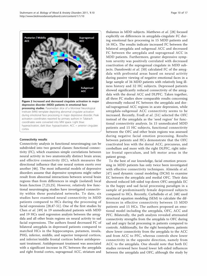
tradictory: Fu et al. [32] and Keedwell et al. [42] reportedenhanced activity in the posterior cingulum, whereas in alater therapy study by Fu and colleagues [33] weakenedactivity in MDD patients compared to HCs in closelyrelated areas emerged. In the middle cingulate gyrus, twoindependent studies point to rather enhanced neuralresponses to sad/angry facial stimuli in MDD patientscompared to HCs [32,43]. Of particular concern in thepathophysiology of affective disorders is the role of theACC [23,44]. Decreased responses to sad facial stimuli inMDD patients compared to HCs in dorsal parts of theACC were reported by Lawrence et al. [37] and Fu et al.[33]. However, one study revealed a contradictory findingof rather increased responsiveness in dorsal ACC to sadfacial expressions [32]. Interestingly, Gotlib et al. [45]reported two hyperactivated clusters in different subgen-ual parts of the ACC in the MDD group to sad andhappy facial expressions, respectively. Figure 2 presents asummary of altered activation loci for facial emotion pro-cessing tasks within the posterior, middle and anteriorcingulum in unipolar depression.
Table 1 Description of fMRI studies on facial emotion processing, comparing a group of major depressive disorder(MDD) patients to healthy controls (HCs) (Continued)
Chen et al.2008
[49] 19 MDD, 19HC
34.3(8.6)
Not reported No Sad Facial expression processingparadigm. Ekman faces.Morphed to express low,medium and high intensities.Implicit task: indicate the sexof the face.
3 s Functionalconnectivity
Dannlowskiet al. 2009
[50] 34 MDD, 31HC
38.6(12.2)
(a) 125.0 ±125.5; (b) 4.7± 5.3
Yes Sad,angry,happy,neutral
Passive face viewingparadigm. Ekman faces.
500 ms Functionalconnectivity
Frodl et al.2010
[51] 25 MDD, 15HC
39.4(10.4)
(a) 51.8 ±63.9; (b) 1.52± 0.6
No Sad,angry
Emotion face-matching task.Ekman faces. Explicit task:match emotion. Implicit task:match gender. Control task:match shapes.
Functionalconnectivity
Mathews etal. 2008
[28] 15 MDD, 16HC
24.5(5.5)
(a) notreported; (b)4.46
No Angry,fear,happy
Emotion face-matching task.Emotional faces. Task: matchfaces.
5 s Functionalconnectivity
BDD, bipolar disorder; BDDr, bipolar disorder remitted; CV, cognitive vulnerability; MDDr, major depressive disorder remitted; ROI, region of interest
Stuhrmann et al. Biology of Mood & Anxiety Disorders 2011, 1:10http://www.biolmoodanxietydisord.com/content/1/1/10
Page 6 of 17
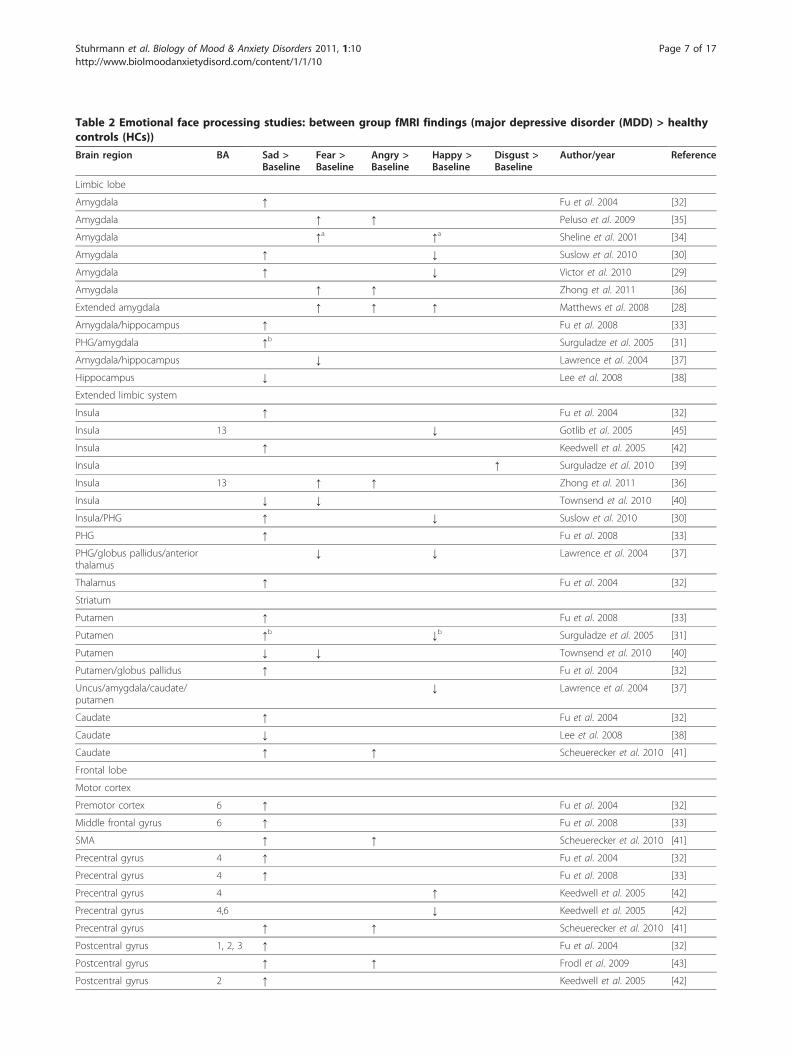
Table 2 Emotional face processing studies: between group fMRI findings (major depressive disorder (MDD) > healthycontrols (HCs))
Brain region BA Sad >Baseline
Fear >Baseline
Angry >Baseline
Happy >Baseline
Disgust >Baseline
Author/year Reference
Limbic lobe
Amygdala ↑ Fu et al. 2004 [32]
Amygdala ↑ ↑ Peluso et al. 2009 [35]
Amygdala ↑a ↑a Sheline et al. 2001 [34]
Amygdala ↑ ↓ Suslow et al. 2010 [30]
Amygdala ↑ ↓ Victor et al. 2010 [29]
Amygdala ↑ ↑ Zhong et al. 2011 [36]
Extended amygdala ↑ ↑ ↑ Matthews et al. 2008 [28]
Amygdala/hippocampus ↑ Fu et al. 2008 [33]
PHG/amygdala ↑b Surguladze et al. 2005 [31]
Amygdala/hippocampus ↓ Lawrence et al. 2004 [37]
Hippocampus ↓ Lee et al. 2008 [38]
Extended limbic system
Insula ↑ Fu et al. 2004 [32]
Insula 13 ↓ Gotlib et al. 2005 [45]
Insula ↑ Keedwell et al. 2005 [42]
Insula ↑ Surguladze et al. 2010 [39]
Insula 13 ↑ ↑ Zhong et al. 2011 [36]
Insula ↓ ↓ Townsend et al. 2010 [40]
Insula/PHG ↑ ↓ Suslow et al. 2010 [30]
PHG ↑ Fu et al. 2008 [33]
PHG/globus pallidus/anteriorthalamus
↓ ↓ Lawrence et al. 2004 [37]
Thalamus ↑ Fu et al. 2004 [32]
Striatum
Putamen ↑ Fu et al. 2008 [33]
Putamen ↑b ↓b Surguladze et al. 2005 [31]
Putamen ↓ ↓ Townsend et al. 2010 [40]
Putamen/globus pallidus ↑ Fu et al. 2004 [32]
Uncus/amygdala/caudate/putamen
↓ Lawrence et al. 2004 [37]
Caudate ↑ Fu et al. 2004 [32]
Caudate ↓ Lee et al. 2008 [38]
Caudate ↑ ↑ Scheuerecker et al. 2010 [41]
Frontal lobe
Motor cortex
Premotor cortex 6 ↑ Fu et al. 2004 [32]
Middle frontal gyrus 6 ↑ Fu et al. 2008 [33]
SMA ↑ ↑ Scheuerecker et al. 2010 [41]
Precentral gyrus 4 ↑ Fu et al. 2004 [32]
Precentral gyrus 4 ↑ Fu et al. 2008 [33]
Precentral gyrus 4 ↑ Keedwell et al. 2005 [42]
Precentral gyrus 4,6 ↓ Keedwell et al. 2005 [42]
Precentral gyrus ↑ ↑ Scheuerecker et al. 2010 [41]
Postcentral gyrus 1, 2, 3 ↑ Fu et al. 2004 [32]
Postcentral gyrus ↑ ↑ Frodl et al. 2009 [43]
Postcentral gyrus 2 ↑ Keedwell et al. 2005 [42]
Stuhrmann et al. Biology of Mood & Anxiety Disorders 2011, 1:10http://www.biolmoodanxietydisord.com/content/1/1/10
Page 7 of 17
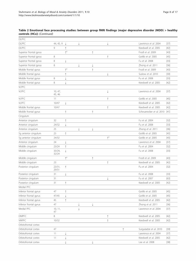
Table 2 Emotional face processing studies: between group fMRI findings (major depressive disorder (MDD) > healthycontrols (HCs)) (Continued)
DLPFC
DLPFC 44, 45, 9 ↓ ↓ ↓ Lawrence et al. 2004 [37]
DLPFC 9 ↑ Keedwell et al. 2005 [42]
Superior frontal gyrus ↑a ↑ Frodl et al. 2009 [43]
Superior frontal gyrus 8 ↑ Gotlib et al. 2005 [45]
Superior frontal gyrus 8 ↓ Fu et al. 2008 [33]
Superior frontal gyrus 8 ↓ ↓ Zhong et al. 2011 [36]
Middle frontal gyrus ↑a ↑ Frodl et al. 2009 [43]
Middle frontal gyrus ↑ ↓ Suslow et al. 2010 [30]
Middle frontal gyrus 8 ↓ Fu et al. 2008 [33]
Middle frontal gyrus 8 ↓ Keedwell et al. 2005 [42]
VLPFC
VLPFC 10, 47,45, 46
↓ Lawrence et al. 2004 [37]
VLPFC 11 ↑ Gotlib et al. 2005 [45]
VLPFC 10/47 ↓ Keedwell et al. 2005 [42]
Middle frontal gyrus 10/47 ↑ Keedwell et al. 2005 [42]
Middle frontal gyrus ↑ ↑ Scheuerecker et al. 2010 [41]
Cingulum
Anterior cingulum 32 ↑ Fu et al. 2004 [32]
Anterior cingulum 24/32 ↓ Fu et al. 2008 [33]
Anterior cingulum 25 ↓ ↓ Zhong et al. 2011 [36]
Sg anterior cingulum 25 ↑ Gotlib et al. 2005 [45]
Sg anterior cingulum 24/32 ↑a Gotlib et al. 2005 [45]
Anterior cingulum 24 ↓ Lawrence et al. 2004 [37]
Middle cingulum 23/24 ↑ Fu et al. 2004 [32]
Middle cingulum 33/24,32/24
↓ Fu et al. 2008 [33]
Middle cingulum ↑a ↑ Frodl et al. 2009 [43]
Middle cingulum 23 ↑ Keedwell et al. 2005 [42]
Posterior cingulum 23/31,29/31
↑ Fu et al. 2004 [32]
Posterior cingulum 31 ↓ Fu et al. 2008 [33]
Posterior cingulum 31 ↓ Fu et al. 2007 [63]
Posterior cingulum 31 ↑ Keedwell et al. 2005 [42]
Medial PFC
Inferior frontal gyrus 47 ↑ Gotlib et al. 2005 [45]
Inferior frontal gyrus 47/45 ↓ Gotlib et al. 2005 [45]
Inferior frontal gyrus 45 ↑ Keedwell et al. 2005 [42]
Inferior frontal gyrus 47 ↓ ↓ Zhong et al. 2011 [36]
Medial PFC 10, 11,47
↓ Lawrence et al. 2004 [37]
DMPFC 8 ↑ Keedwell et al. 2005 [42]
VMPFC 10/32 ↑ Keedwell et al. 2005 [42]
Orbitofrontal cortex
Orbitofrontal cortex 47 ↑ Surguladze et al. 2010 [39]
Orbitofrontal cortex 11 ↓ Lawrence et al. 2004 [37]
Orbitofrontal cortex 11 ↓ Keedwell et al. 2005 [42]
Orbitofrontal cortex ↓ ↓ Lee et al. 2008 [38]
Stuhrmann et al. Biology of Mood & Anxiety Disorders 2011, 1:10http://www.biolmoodanxietydisord.com/content/1/1/10
Page 8 of 17
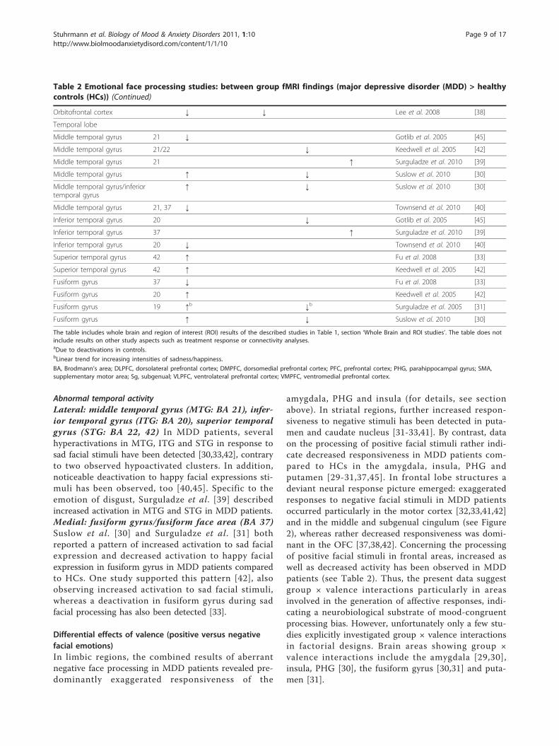
Abnormal temporal activityLateral: middle temporal gyrus (MTG: BA 21), infer-ior temporal gyrus (ITG: BA 20), superior temporalgyrus (STG: BA 22, 42) In MDD patients, severalhyperactivations in MTG, ITG and STG in response tosad facial stimuli have been detected [30,33,42], contraryto two observed hypoactivated clusters. In addition,noticeable deactivation to happy facial expressions sti-muli has been observed, too [40,45]. Specific to theemotion of disgust, Surguladze et al. [39] describedincreased activation in MTG and STG in MDD patients.Medial: fusiform gyrus/fusiform face area (BA 37)Suslow et al. [30] and Surguladze et al. [31] bothreported a pattern of increased activation to sad facialexpression and decreased activation to happy facialexpression in fusiform gyrus in MDD patients comparedto HCs. One study supported this pattern [42], alsoobserving increased activation to sad facial stimuli,whereas a deactivation in fusiform gyrus during sadfacial processing has also been detected [33].
Differential effects of valence (positive versus negativefacial emotions)In limbic regions, the combined results of aberrantnegative face processing in MDD patients revealed pre-dominantly exaggerated responsiveness of the
amygdala, PHG and insula (for details, see sectionabove). In striatal regions, further increased respon-siveness to negative stimuli has been detected in puta-men and caudate nucleus [31-33,41]. By contrast, dataon the processing of positive facial stimuli rather indi-cate decreased responsiveness in MDD patients com-pared to HCs in the amygdala, insula, PHG andputamen [29-31,37,45]. In frontal lobe structures adeviant neural response picture emerged: exaggeratedresponses to negative facial stimuli in MDD patientsoccurred particularly in the motor cortex [32,33,41,42]and in the middle and subgenual cingulum (see Figure2), whereas rather decreased responsiveness was domi-nant in the OFC [37,38,42]. Concerning the processingof positive facial stimuli in frontal areas, increased aswell as decreased activity has been observed in MDDpatients (see Table 2). Thus, the present data suggestgroup × valence interactions particularly in areasinvolved in the generation of affective responses, indi-cating a neurobiological substrate of mood-congruentprocessing bias. However, unfortunately only a few stu-dies explicitly investigated group × valence interactionsin factorial designs. Brain areas showing group ×valence interactions include the amygdala [29,30],insula, PHG [30], the fusiform gyrus [30,31] and puta-men [31].
Table 2 Emotional face processing studies: between group fMRI findings (major depressive disorder (MDD) > healthycontrols (HCs)) (Continued)
Orbitofrontal cortex ↓ ↓ Lee et al. 2008 [38]
Temporal lobe
Middle temporal gyrus 21 ↓ Gotlib et al. 2005 [45]
Middle temporal gyrus 21/22 ↓ Keedwell et al. 2005 [42]
Middle temporal gyrus 21 ↑ Surguladze et al. 2010 [39]
Middle temporal gyrus ↑ ↓ Suslow et al. 2010 [30]
Middle temporal gyrus/inferiortemporal gyrus
↑ ↓ Suslow et al. 2010 [30]
Middle temporal gyrus 21, 37 ↓ Townsend et al. 2010 [40]
Inferior temporal gyrus 20 ↓ Gotlib et al. 2005 [45]
Inferior temporal gyrus 37 ↑ Surguladze et al. 2010 [39]
Inferior temporal gyrus 20 ↓ Townsend et al. 2010 [40]
Superior temporal gyrus 42 ↑ Fu et al. 2008 [33]
Superior temporal gyrus 42 ↑ Keedwell et al. 2005 [42]
Fusiform gyrus 37 ↓ Fu et al. 2008 [33]
Fusiform gyrus 20 ↑ Keedwell et al. 2005 [42]
Fusiform gyrus 19 ↑b ↓b Surguladze et al. 2005 [31]
Fusiform gyrus ↑ ↓ Suslow et al. 2010 [30]
The table includes whole brain and region of interest (ROI) results of the described studies in Table 1, section ‘Whole Brain and ROI studies’. The table does notinclude results on other study aspects such as treatment response or connectivity analyses.aDue to deactivations in controls.bLinear trend for increasing intensities of sadness/happiness.
BA, Brodmann’s area; DLPFC, dorsolateral prefrontal cortex; DMPFC, dorsomedial prefrontal cortex; PFC, prefrontal cortex; PHG, parahippocampal gyrus; SMA,supplementary motor area; Sg, subgenual; VLPFC, ventrolateral prefrontal cortex; VMPFC, ventromedial prefrontal cortex.
Stuhrmann et al. Biology of Mood & Anxiety Disorders 2011, 1:10http://www.biolmoodanxietydisord.com/content/1/1/10
Page 9 of 17
Connectivity resultsConnectivity analysis in functional neuroimaging can besubdivided into two general classes: functional connec-tivity (FC), which examines simple correlations betweenneural activity in two anatomically distinct brain areas;and effective connectivity (EC), which measures thedirectional influence that one neural system exerts overanother [46]. The most influential models of depressivedisorders assume that depressive symptoms might ratherresult from abnormal interactions between several brainregions than from differences in single (isolated) localbrain function [7,23,25]. However, relatively few func-tional neuroimaging studies have investigated connectiv-ity within these postulated networks. To date, sixstudies have examined neural connectivity in MDDpatients compared to HCs during the processing offacial expressions [28,47-51]. One of the first studies byChen et al. [49] in 19 unmedicated patients with MDDand 19 HCs used regression analysis between the amyg-dala and all other brain regions on neural activity to sadfacial expressions. The authors found decreased FC ofbilateral amygdala in depressed patients compared tomatched HCs in the hippocampus, putamen, insula,PHG, inferior, middle, and superior temporal cortices,and inferior/middle frontal cortices before antidepres-sant treatment. Antidepressant treatment was associatedwith a significant increase in FC between the amygdalaand right frontal cortex, supragenual ACC, striatum and
thalamus in MDD subjects. Matthews et al. [28] focusedexplicitly on differences in amygdala-cingulate FC dur-ing emotional face processing in 15 MDD patients and16 HCs. The results indicate increased FC between thebilateral amygdala and subgenual ACC and decreasedFC between the amygdala and supragenual ACC inMDD patients. Furthermore, greater depressive symp-tom severity was positively correlated with decreasedcoactivation of the supragenual cingulate in MDD sub-jects. Dannlowski et al. [50] calculated FC of the amyg-dala with prefrontal areas based on neural activityduring passive viewing of negative emotional faces in alarge sample of 34 MDD patients with relatively long ill-ness history and 32 HC subjects. Depressed patientsshowed significantly reduced connectivity of the amyg-dala with the dorsal ACC and DLPFC. Taken together,all three FC studies show comparable results concerningabnormally reduced FC between the amygdala and dor-sal/supragenual ACC regions in acute depression, whileamygdala-subgenual ACC connectivity seems to beincreased. Recently, Frodl et al. [51] selected the OFCinstead of the amygdala as the ‘seed region’ for func-tional connectivity analysis. In 15 unmedicated MDDpatients and 15 HC subjects, functional connectivitybetween the OFC and other brain regions was assessedduring negative facial emotion processing. Resultsbetween patients and HCs demonstrate that the OFCcoactivated less with the dorsal ACC, precuneus, andcerebellum and more with the right DLPFC, right infer-ior frontal operculum, and left motor areas in thepatient group.To the best of our knowledge, facial emotion proces-
sing in MDD patients has only twice been investigatedwith effective connectivity techniques. Almeida et al.[47] used dynamic causal modeling (DCM) to examineEC between the amygdala and medial OFC. Their datashowed reduced left-sided top-down OFC-amygdala ECin the happy and sad facial processing paradigm in asample of predominantly female depressed subjectscompared to HCs. Recently, Carballedo et al. [48] usedstructural equation modeling (SEM) to calculate the dif-ferences in effective connectivity between 15 MDDpatients and 15 HCs. The authors proposed an emo-tional model including the amygdala, OFC, ACC andPFC. Bilaterally, the path analysis revealed attenuatedconnectivity strengths from the amygdala to OFC duringsad and angry facial processing in patients compared tocontrols. Additionally, for the right hemisphere, patientsshow lower connectivity from the amygdala to the ACCand from ACC to PFC, whereas controls show lowerconnectivity in the opposite direction, namely fromACC to the amygdala. One should note that both ECstudies reviewed here found lower left-sided influencesbetween the amygdala and OFC, although the study by
Figure 2 Increased and decreased cingulate activation in majordepressive disorder (MDD) patients in emotional faceprocessing studies. Paramedian slice of a Montreal NeurologicalInstitute (MNI) template depicting abnormal cingulate activationduring emotional face processing in major depressive disorder. Peakactivation coordinates reported by primary authors in Talairachcoordinates were converted into MNI space. Light blue:hyperactivation, dark blue: hypoactivation. ACC = anterior cingulatecortex.
Stuhrmann et al. Biology of Mood & Anxiety Disorders 2011, 1:10http://www.biolmoodanxietydisord.com/content/1/1/10
Page 10 of 17

Almeida et al. [47] showed top-down alterations and theone by Carballedo et al. [48] bottom-up alterations.In summary, functional connectivity between the
amygdala and other brain areas shows (a) decreasedamygdala coupling with other limbic regions (hippocam-pus, putamen, insula, PHG), temporal regions, and inparticular with the supragenual/dorsal ACC and DLPFC,and (b) increased coupling with subgenual ACC. Of par-ticular concern seems to be the role of the ACC, resem-bling results identified with conventional fMRI analysis.The longitudinal data by Chen et al. [49] provide firstevidence that decreased FC coupling between the amyg-dala and supragenual ACC increases after pharmacologi-cal intervention.
DiscussionThe present review aimed to summarize availableempirical data regarding the neural correlates of abnor-mal emotional face processing in acute unipolar depres-sion (during the current episode). Presenting differentialfacial expressions activates a common face-processingnetwork in HCs and MDD patients, including primaryvisual pathways as well as further supporting brain areascrucial for emotion processing in general. The amygdalabelongs to the latter group, the extended limbic systemand specific frontal areas, namely the ACC, OFC andventromedial prefrontal cortex (VMPFC). These regionsare of particular interest for understanding the patho-physiology of unipolar depression. Our analysis indicatesevidence of abnormal neural face processing in MDDpatients, especially in the amygdala, the insula, PHG,ACC and OFC. Although neural alterations werereported in several other brain regions, the Discussionsection focuses on these areas because they are (a) cru-cial for evaluating the neural mood-congruent face pro-cessing hypothesis, and (b) are core domains in analtered functional connectivity network in MDD patientsduring emotional face processing.
Neural mood-congruent face processingNeural responses in MDD patients associated withmood-congruent processing patterns are most evident inthe amygdala [29,30], insula and PHG [30], the fusiformgyrus [30,31] and putamen [31].The amygdala plays a pivotal role in emotion proces-
sing and in the perception and processing of emotionalsalience in facial expressions (for reviews see [52-54]).Furthermore, the amygdala is a key region within theneurobiological framework of depressive disorder. Sev-eral authors have suggested that, for MDD, mood-con-gruent bias in behavioral measures is strongly linked toamygdala hyper-responsiveness to negative stimuli[2,55,56]. Findings of increased amygdala responsivenessto negative emotional faces are well in line with several
imaging studies employing other stimuli, including nega-tive words [57,58], individualized self-referential sen-tences [59], or in expectation of negative pictures [60].Furthermore, these findings are supported by studies indepressed adolescents [61].However, not all fMRI studies have found evidence for
altered amygdala activation in MDD. In detail, 10 of the20 included studies reported differences in amygdalaactivation between MDD patients and HCs using faceemotion processing tasks [28-37], while the other stu-dies found no significant group effects[27,38-43,45,62,63]. Nevertheless, focusing on theobserved differences in amygdala responsiveness, studiesprovide compelling support for amygdala mood-congru-ent processing in MDD patients. First, abnormal amyg-dala responsiveness has been shown to negative andpositive facial expressions, corroborating amygdala func-tion in processing salient stimuli, independent of stimulivalence [64]. Second, as hypothesized in mood congru-ent processing theories of depression, the majority ofresults show exaggerated amygdala response to sad sti-muli, and in addition decreased amygdala response tohappy facial stimuli, although replications with happyfacial expressions are still rare. These results indicatethat, in convergence with behavioral measures, neuro-biological assessment can be a sensitive measure formood-congruent biases in unipolar depression. Of noteare two recent studies using subliminal presentationconditions pointing to mood congruency effects to nega-tive and positive stimuli already at early, automatic pro-cessing stages [29,30].In conclusion, the findings of our analysis support the
assumption that amygdala hyperactivity is associatedwith negatively biased facial emotion processing impli-cated in the pathophysiology of major depression,although this became evident in only one-half of thereviewed studies. Studies investigating the question ofwhether abnormalities in amygdala responses to emo-tional faces demonstrated in acute depression representa state marker of acute depressive episodes or vulner-ability factors for depression are rare. In remittedpatients, Neumeister et al. [65] demonstrated enhancedregional cerebral blood flow responses to sad facialexpressions in the amygdala relative to HCs, but othershave failed to replicate these findings in remittedpatients [66,67]. In people at risk for depression, van derVeen et al. [68] and Monk et al. [69] reported greaterabnormal amygdala activation to negative facial expres-sion, but again inconsistent findings exist [70]. Interest-ingly, Zhong et al. [36] reported higher amygdalaactivation evident in both MDD subjects and a sampleof healthy people with high cognitive vulnerability todepression compared to HCs. Increased left amygdalaresponsiveness was positively associated with CSQ
Stuhrmann et al. Biology of Mood & Anxiety Disorders 2011, 1:10http://www.biolmoodanxietydisord.com/content/1/1/10
Page 11 of 17
scores (measures causal attributions, consequences andself-worth characteristics). In addition, Cremers et al.[71] reported that right amygdala-dorsomedial PFC con-nectivity for negative faces vs neutral faces was posi-tively associated with neuroticism scores, a personalitytrait related to the development of affective disorders.Finally, a recent study by Dannlowski et al. [72] investi-gated long-term effects of childhood maltreatment withfMRI in psychologically healthy participants. Theobserved association between childhood maltreatmentand amygdala responsiveness during emotional face pro-cessing resembles findings in depressed patients, sug-gesting that these functional changes might constitute apredisposition for developing affective disorders.Hyperactivated amygdala to negative emotional faces
in remitted patients and people at high risk for depres-sion is indicative of trait vulnerability. This interpreta-tion receives support from imaging genetics and twinstudies, suggesting that amygdala responsiveness toemotional faces as well as amygdala prefrontal connec-tivity are under strong genetic influence [73-78].Some methodological aspects explaining the heteroge-
neity of studies should be discussed here. With regardto presentation modus, all three studies using subliminalpresentation of facial expressions reported differences inamygdala activation [29,30,34]. Victor et al. [29] evenobserved differences in amygdala activation specific tomasked presentation of sad and happy faces, absent tounmasked stimuli, supporting the assumption that sub-liminal stimuli presentation maybe an advantage inidentifying emotional-processing biases in MDD withfocus on amygdala activation. It may be subliminal sti-mulus presentation prevents confounding with othercognitive processes prevalent in depression such asrumination on negative thoughts/preservation of atten-tion to negative faces [34]. Comparing paradigms pre-senting facial stimuli supraliminally, only about half ofthe investigations implementing either face-matchingparadigms or the ‘face recognition task’ observed amyg-dala differences. Scheuerecker et al. [41] suggested thatparticipants probably used more visual and cognitivestrategies to solve the face-matching task, causing ACCand PFC activation maybe inhibiting amygdala activa-tion. Concerning task type (that is, explicit or implicit),an implicit task, requiring participants to focus on gen-der aspects of the face, seems to be sufficient to elicitamygdala activation [28,31-33,35,36]. As amygdala andfrontal responsiveness depends on task complexity, facetype and attention focus, future research should takeinto account such variations in designing facial proces-sing paradigms.Furthermore, medication status has an important
impact on neural activation patterns: seven of the tenstudies reporting altered amygdala activation were
performed on unmedicated patients. This result is notsurprising regarding the converging evidence, that amyg-dala is a key region for antidepressant effects, reducingabnormal amygdala responsiveness to negativelyvalenced faces in MDD patients (for a recent meta-ana-lysis see [79]). Other possible influencing factors may bemethodological aspects such as experimental design (forexample, event-related vs block design) or the selectionof different baseline conditions (for example, neutralfaces or a no-face condition) as well as clinical and non-diagnostic variables such as age, comorbidity, treatmenthistory and number of prior episodes (for details seeTable 1). Furthermore, difficulties in detecting alteredamygdala responsiveness in MDD patients may becaused by a ‘ceiling’ effect. As noted by Townsend et al.[40], several PET studies have shown increased restingblood flow in the amygdala in MDD patients [80-84],making it difficult to detect group differences in activa-tion tasks if amygdala baseline activation was alreadyincreased.Aside from the amygdala, several other subcortical
brain structures show activation patterns supportingmood-congruent processing in depressed patients. Insulahyperactivation to sad facial stimuli is a prominentresult, and furthermore two independent studiesobserved hypoactivation to happy facial stimuli (seeTable 2). Apart from having a pivotal role in the proces-sing of disgust [39] the insula has strong functional con-nections to the amygdala [85]. Insula projections toinferior parietal cortex and the amygdala are involved inidentifying/representing motivational salient informa-tion, social cues and the expression of conditionedresponses: particularly on implicit processing pathways[86,87]. Furthermore, activity in the putamen and cau-date nucleus also resembles mood-congruent activationpatterns in MDD patients, although contributions to theprocessing of facial expressions are still under debate[87]. In visual face areas, fusiform gyrus responsivenessalso indicates mood-congruent processing in terms ofincreased activation to sad facial expressions anddecreased activation to happy faces. In addition toencoding face traits and facial identity [20], recent stu-dies revealed that fusiform regions are also sensitive tofacial emotional expression (for a review see [88]). Theauthors suggest that the modulation by emotional effectscan be explained by direct connections between visualcortex and the amygdala, facilitating direct feedback sig-nals from the amygdala [89] to visual processing areas.In summary, neuronal correlates of mood-congruent
facial affect processing in MDD patients are most pro-minent in limbic and subcortical regions, compromisingthe amygdala, insula and putamen/caudate nucleus. In alarger context these regions are hypothesized to be partof an extended emotional face processing system [20]
Stuhrmann et al. Biology of Mood & Anxiety Disorders 2011, 1:10http://www.biolmoodanxietydisord.com/content/1/1/10
Page 12 of 17
and furthermore constitute a ventral stream in emotion-cognition processing, appraising emotional behavior andproducing affective states, altered in unipolar depression[25]. As described, these alterations may even influencevisual processing areas such as the fusiform face gyrus.Studies in remitted patients and in people at risk fordepression provide the first indications that enhancedlimbic neural responses to negative emotional materialmay contribute to vulnerability to MDD [65,68,69].
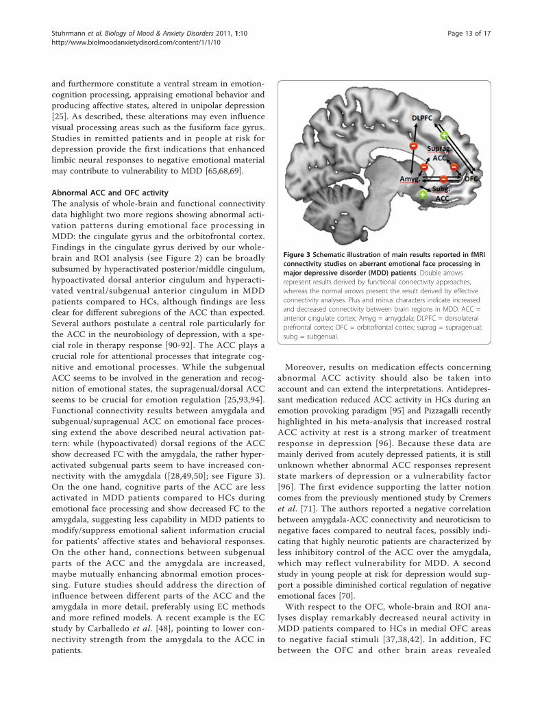
Abnormal ACC and OFC activityThe analysis of whole-brain and functional connectivitydata highlight two more regions showing abnormal acti-vation patterns during emotional face processing inMDD: the cingulate gyrus and the orbitofrontal cortex.Findings in the cingulate gyrus derived by our whole-brain and ROI analysis (see Figure 2) can be broadlysubsumed by hyperactivated posterior/middle cingulum,hypoactivated dorsal anterior cingulum and hyperacti-vated ventral/subgenual anterior cingulum in MDDpatients compared to HCs, although findings are lessclear for different subregions of the ACC than expected.Several authors postulate a central role particularly forthe ACC in the neurobiology of depression, with a spe-cial role in therapy response [90-92]. The ACC plays acrucial role for attentional processes that integrate cog-nitive and emotional processes. While the subgenualACC seems to be involved in the generation and recog-nition of emotional states, the supragenual/dorsal ACCseems to be crucial for emotion regulation [25,93,94].Functional connectivity results between amygdala andsubgenual/supragenual ACC on emotional face proces-sing extend the above described neural activation pat-tern: while (hypoactivated) dorsal regions of the ACCshow decreased FC with the amygdala, the rather hyper-activated subgenual parts seem to have increased con-nectivity with the amygdala ([28,49,50]; see Figure 3).On the one hand, cognitive parts of the ACC are lessactivated in MDD patients compared to HCs duringemotional face processing and show decreased FC to theamygdala, suggesting less capability in MDD patients tomodify/suppress emotional salient information crucialfor patients’ affective states and behavioral responses.On the other hand, connections between subgenualparts of the ACC and the amygdala are increased,maybe mutually enhancing abnormal emotion proces-sing. Future studies should address the direction ofinfluence between different parts of the ACC and theamygdala in more detail, preferably using EC methodsand more refined models. A recent example is the ECstudy by Carballedo et al. [48], pointing to lower con-nectivity strength from the amygdala to the ACC inpatients.
Moreover, results on medication effects concerningabnormal ACC activity should also be taken intoaccount and can extend the interpretations. Antidepres-sant medication reduced ACC activity in HCs during anemotion provoking paradigm [95] and Pizzagalli recentlyhighlighted in his meta-analysis that increased rostralACC activity at rest is a strong marker of treatmentresponse in depression [96]. Because these data aremainly derived from acutely depressed patients, it is stillunknown whether abnormal ACC responses representstate markers of depression or a vulnerability factor[96]. The first evidence supporting the latter notioncomes from the previously mentioned study by Cremerset al. [71]. The authors reported a negative correlationbetween amygdala-ACC connectivity and neuroticism tonegative faces compared to neutral faces, possibly indi-cating that highly neurotic patients are characterized byless inhibitory control of the ACC over the amygdala,which may reflect vulnerability for MDD. A secondstudy in young people at risk for depression would sup-port a possible diminished cortical regulation of negativeemotional faces [70].With respect to the OFC, whole-brain and ROI ana-
lyses display remarkably decreased neural activity inMDD patients compared to HCs in medial OFC areasto negative facial stimuli [37,38,42]. In addition, FCbetween the OFC and other brain areas revealed
Figure 3 Schematic illustration of main results reported in fMRIconnectivity studies on aberrant emotional face processing inmajor depressive disorder (MDD) patients. Double arrowsrepresent results derived by functional connectivity approaches,whereas the normal arrows present the result derived by effectiveconnectivity analyses. Plus and minus characters indicate increasedand decreased connectivity between brain regions in MDD. ACC =anterior cingulate cortex; Amyg = amygdala; DLPFC = dorsolateralprefrontal cortex; OFC = orbitofrontal cortex; suprag = supragenual;subg = subgenual.
Stuhrmann et al. Biology of Mood & Anxiety Disorders 2011, 1:10http://www.biolmoodanxietydisord.com/content/1/1/10
Page 13 of 17

decreased FC to the amygdala and supragenual ACC aswell as increased FC to the DLPFC. The two availableEC studies [47,48] in MDD patients further specified thedirectionality of these brain abnormalities. Both studiesindicate reduced left-sided connectivity between theOFC and the amygdala in patients, but show, at firstglance, contradictory results with regard to the directionof influence on another (top down vs bottom up). Inboth paradigms participants were instructed to explicitlylabel emotions, but different paradigms were used (face-matching task vs morphed facial expression processingparadigm); therefore this may be, next to medicationeffects, one reason explaining the results. Future studiesare needed to further investigate on this issue.The OFC is a central part of the frontosubcortical cir-
cuits, connecting the frontal and limbic systems witheach other, and is crucial for mood regulatory processes[97,98]. Relative uncoupling of connections betweenheightened activity in the limbic system and the OFCduring negative facial processing in MDD may accountfor depressive symptoms such as negative emotionalexperiences and impaired regulation of emotional andsocial behavior [41]. Increased FC between OFC and lat-eral PFC systems could be the neural substrate of amore voluntary compensatory mechanism in MDD [99]for the described altered automatic emotional faceprocessing.
Unresolved questionsTo date, it is not clear whether the neurobiologicalabnormalities described above represent state or traitmarkers of depression. As highlighted above, a few stu-dies have demonstrated a normalization of abnormalneurobiological response patterns after antidepressantmedication (for example, [29,32]). Moreover, these stu-dies are in line with several pharmaco-fMRI studies inhealthy subjects, showing that limbic responsiveness tonegative facial stimuli can be attenuated even by short-term antidepressant administration [100-102]. However,although it seems that antidepressants modify pathologi-cal emotional face processing in depression, it stillremains to be clarified whether these functionalabnormalities in emotional face processing represent afeature of acute depressed state and would thereforealso resolve without medication after remission orwhether they represent a risk factor preceding the onsetof depression. The first studies in remitted patients andin high-risk subjects [36,65,68,69,71,103], as well as datafrom imaging genetics and twin studies [73-78] suggestthat amygdala responsiveness to emotional faces as wellas amygdala-prefrontal and amygdala-ACC connectivitymay represent vulnerability factors for MDD.A second unresolved question concerns possible later-
ality effects of valence-specific emotion processing in
the depressed brain. Although this aspect may be raisedby the data, it was not the focus of our analysis and stillneeds further clarification. As noted above, other unre-solved issues concern the heterogeneity of presentationparadigms. For example, studies investigating automaticfacial emotion processing are likely to target other brainareas compared to explicit emotion processing para-digms. Obviously, this is particularly important forinvestigating prefrontal areas and might explain theapparently contradictive results in brain areas involvedin emotion regulation, for example the DLPFC. Next tothe methodological aspect, variability between patientsamples due to different symptom characteristics maybe a further critical, influential factor. Age, comorbidity,treatment history, number of prior episodes or age onillness onset may confound the reported results [7].Unfortunately, information about clinical variables wasprovided by less than half of the reviewed studies, leav-ing these variables relatively uncontrolled for in thisreview and therefore limiting the described results andtheir interpretation. As described in the Discussion sec-tion, differences in medication status and low samplesizes could further contribute to inconsistencies amongstudy results.The research field would benefit from larger studies
with well characterized patient samples (that is, detaileddescription of clinical variables), particularly multicenterstudies. Furthermore, investigators should carry onemploying standardized paradigms in order to replicateresults and to resolve conflicting findings. For example,the comparison of subliminal and supraliminal stimuluspresentation in one patient sample and the influence ofattentional mechanisms on a neural level are still rarelyinvestigated. Future studies should explicitly focus ongroup × valence interactions in factorial designs toexplore differential effects of valence and should useconnectivity analysis strategies (FC and/or EC) todescribe the interplay of core regions such as the amyg-dala, ACC and OFC more precisely. Longitudinal stu-dies, including relatives or other high-risk subjects arevery essential and may ultimately answer the question ifthe described anomalies represent ‘trait’ or ‘state’ markerof depression.Finally, one should notice that facial processing is only
one aspect of altered cognitive/emotional processingamong several others in MDD described by behavioral(for review see [104]) and neuroimaging (for review see[7]) studies. Thus, one must caution against overinter-pretation of the presented results on altered neural facialprocessing in MDD.
ConclusionsBased on cognitive models of depression and behavioralstudies pointing to an emotion processing bias in acute
Stuhrmann et al. Biology of Mood & Anxiety Disorders 2011, 1:10http://www.biolmoodanxietydisord.com/content/1/1/10
Page 14 of 17
depression, several neuroimaging studies have investi-gated the neuronal underpinnings of these emotionalprocessing abnormalities. It has been shown that the useof emotional face processing tasks is a reliable and validapproach to pinpoint most if not all relevant areas. Theanalysis of neural activation data shows that MDDpatients are characterized by abnormalities within thecommon face processing network, indicating a mood-congruent processing bias particularly in the amygdala,insula and PHG, fusiform face area and putamenresponsiveness. Furthermore, abnormalities in the cingu-late gyrus and OFC are obvious, which are refined byinvestigations implementing functional connectivity ana-lysis. A pathologically altered emotion processing andemotion regulation network emerged, including theamygdala, the ACC, OFC and DLPFC as core compo-nents. Further neuroimaging studies will be needed toextend these findings, especially by replicating data withsame activation paradigms and larger sample sizes inorder to enable researchers to make more valid assump-tions on neural emotional processing mechanisms, con-tributing to a better understanding of depressivedisorders.
AcknowledgementsThe study was supported by grants from Innovative Medizinische Forschung(IMF) of the Medical Faculty of Münster (DA120309 to UD)
Author details1University of Münster, Department of Psychiatry, Albert-Schweitzer-Campus1, Building, A9, 48149 Münster, Germany. 2University of Leipzig, Departmentof Psychosomatic Medicine and Psychotherapy, Semmelweisstraße 10, 04103Leipzig, Germany.
Authors’ contributionsAS performed the literature research and wrote major parts of the article. TScontributed to the Introduction and Discussion sections. UD selected topics,article structure, and inclusion criteria, supervised the literature research, andwrote major parts of the discussion section. All authors read and approvedthe final manuscript.
Competing interestsThe authors declare that they have no competing interests.
Received: 28 July 2011 Accepted: 7 November 2011Published: 7 November 2011
References1. World Health Organization: Mental Health: New Understanding, New Hope
Geneva, Switzerland: WHO; 2001.2. Leppänen JM: Emotional information processing in mood disorders: a
review of behavioral and neuroimaging findings. Curr Opin Psychiatry2006, 19:34-39.
3. Mathews A, MacLeod C: Cognitive vulnerability to emotional disorders.Annu Rev Clin Psychol 2005, 1:167-195.
4. Ridout N, Astell AJ, Reid IC, Glen T, O’Carroll RE: Memory bias foremotional facial expressions in major depression. Cognition Emotion 2003,17:101-122.
5. Williams JMG, Watts FN, MacLeod C, Mathews A: Cognitive psychology andemotional disorders. 2 edition. Chichester, UK: John Wiley & Sons; 1997.
6. Bourke C, Douglas K, Porter R: Processing of facial emotion expression inmajor depression: a review. Aust N Z J Psychiatry 2010, 44:681-96.
7. Elliott R, Zahn R, Deakin JFW, Anderson IM: Affective cognition and itsdisruption in mood disorders. Neuropsychopharmacol 2010, 36:153-182.
8. Gotlib IH, Krasnoperova E, Yue DN, Joormann J: Attentional biases fornegative interpersonal stimuli in clinical depression. J Abnorm Psychol2004, 113:127-135.
9. Leyman L, De Raedt R, Schacht R, Koster EHW: Attentional biases for angryfaces in unipolar depression. Psychol Med 2007, 37:393-402.
10. Suslow T, Dannlowski U, Lalee-Mentzel J, Donges U-S, Arolt V: Spatialprocessing of facial emotion in patients with unipolar depression: alongitudinal study. J Affect Disord 2004, 83:59-63.
11. Joormann J, Gotlib IH: Selective attention to emotional faces followingrecovery from depression. J Abnorm Psychol 2007, 116:80-85.
12. Bouhuys AL, Geerts E, Gordijn M: Depressed patients’ perceptions of facialemotions in depressed and remitted states are associated with relapse:a longitudinal study. J Nerv Ment Dis 1999, 187:595-692.
13. Dannlowski U, Kersting A, Donges U-S, Lalee-Mentzel J, Arolt V, Suslow T:Masked facial affect priming is associated with therapy response inclinical depression. Eur Arch Psychiatry Clin Neurosci 2006, 256:215-221.
14. Dannlowski U, Kersting A, Lalee-Mentzel J, Donges U-S, Arolt V, Suslow T:Subliminal affective priming in clinical depression and comorbid anxiety:a longitudinal investigation. Psychiatry Res 2006, 143:63-75.
15. Phillips M: Neurobiology of emotion perception I: the neural basis ofnormal emotion perception. Biol Psychiatry 2003, 54:504-514.
16. Blair RJR: Facial expressions, their communicatory functions and neuro-cognitive substrates. Philos Trans R Soc Lond B Biol Sci 2003, 358:561-572.
17. Suslow T, Dannlowski U: Detection of facial emotion in depression. InMood State and Health. Edited by: Clark AV. Hauppauge, NY: NovaBiomedical Books; 2005:1-32.
18. Fusar-Poli P, Placentino A, Carletti F, Landi P, Allen P, Surguladze S,Benedetti F, Abbamonte M, Gasparotti R, Barale F, Perez J, McGuire P,Politi P: Functional atlas of emotional faces processing: a voxel-basedmeta-analysis of 105 functional magnetic resonance imaging studies. JPsychiatr Neurosci 2009, 34:418-432.
19. Haxby J, Gobbini MI: The perception of emotion and social cues in faces.Neuropsychologia 2007, 45:1.
20. Haxby J, Hoffman E, Gobbini MI: The distributed human neural system forface perception. Trends Cogn Sci 2000, 4:223-233.
21. Posamentier MT, Abdi H: Processing faces and facial expressions.Neuropsychol Rev 2003, 13:113-143.
22. Haxby J, Hoffman E, Gobbini MI: Human neural systems for facerecognition and social communication. Biol Psychiatry 2002, 51:59-67.
23. Mayberg HS: Limbic-cortical dysregulation: a proposed model ofdepression. J Neuropsychiatry Clin Neurosci 1997, 9:471-481.
24. Phillips ML, Ladouceur CD, Drevets WC: A neural model of voluntary andautomatic emotion regulation: implications for understanding thepathophysiology and neurodevelopment of bipolar disorder. MolPsychiatry 2008, 13:829, 833-857..
25. Phillips ML, Drevets WC, Rauch SL, Lane R: Neurobiology of emotionperception II: Implications for major psychiatric disorders. Biol Psychiatry2003, 54:515-528.
26. Mayberg HS: Defining the neural circuitry of depression: toward a newnosology with therapeutic implications. Biol Psychiatry 2007, 61:729-730.
27. Frodl T, Scheuerecker J, Schoepf V, Linn J, Koutsouleris N, Bokde A,Hampel H, Möller H-J, Brückmann H, Wiesmann M, Meisenzahl E: Differenteffects of mirtazapine and venlafaxine on brain activation: an openrandomized controlled fMRI study. J Clin Psychiatry 2011, 72:448-457.
28. Matthews SC, Strigo IA, Simmons AN, Yang TT, Paulus MP: Decreasedfunctional coupling of the amygdala and supragenual cingulate isrelated to increased depression in unmedicated individuals with currentmajor depressive disorder. J Affect Disord 2008, 111:13-20.
29. Victor TA, Furey ML, Fromm S, Ohman A, Drevets WC: Relationshipbetween amygdala responses to masked faces and mood state andtreatment in major depressive disorder. Arch Gen Psychiatry 2010,67:1128-1138.
30. Suslow T, Konrad C, Kugel H, Rumstaedt D, Zwitserlood P, Schöning S,Ohrmann P, Bauer J, Pyka M, Kersting A, Arolt V, Heindel W, Dannlowski U:Automatic mood-congruent amygdala responses to masked facialexpressions in major depression. Biol Psychiatry 2010, 67:155-160.
31. Surguladze S, Brammer M, Keedwell P, Giampietro V, Young AW, Travis MJ,Williams SCR, Phillips ML: A differential pattern of neural response toward
Stuhrmann et al. Biology of Mood & Anxiety Disorders 2011, 1:10http://www.biolmoodanxietydisord.com/content/1/1/10
Page 15 of 17

sad versus happy facial expressions in major depressive disorder. BiolPsychiatry 2005, 57:201-209.
32. Fu CHY, Williams SCR, Cleare AJ, Brammer M, Walsh ND, Kim J, Andrew CM,Pich EM, Williams PM, Reed LJ, Mitterschiffthaler MT, Suckling J, Bullmore ET:Attenuation of the neural response to sad faces in major depression byantidepressant treatment: a prospective, event-related functionalmagnetic resonance imaging study. Arch Gen Psychiatry 2004, 61:877-889.
33. Fu CHY, Williams SCR, Cleare AJ, Scott J, Mitterschifthaler MT, Walsh ND,Donaldson C, Suckling J, Andrew CM, Steiner H, Murray RM: Neuralresponses to sad facial expressions in major depression followingcognitive behavioral therapy. Biol Psychiatry 2008, 64:505-512.
34. Sheline YI, Barch DM, Donnelly JM, Ollinger JM, Snyder AZ, Mintun MA:Increased amygdala response to masked emotional faces in depressedsubjects resolves with antidepressant treatment: an fMRI study. BiolPsychiatry 2001, 50:651-658.
35. Peluso MAM, Glahn DC, Matsuo K, Monkul ES, Najt P, Zamarripa F, Li J,Lancaster JL, Fox PT, Gao J-H, Soares JC: Amygdala hyperactivation inuntreated depressed individuals. Psychiatry Res 2009, 173:158-161.
36. Zhong M, Wang X, Xiao J, Yi J, Zhu X, Liao J, Wang W, Yao S: Amygdalahyperactivation and prefrontal hypoactivation in subjects with cognitivevulnerability to depression. Biol Psychiatry 2011, 88:233-242.
37. Lawrence NS, Williams AM, Surguladze S, Giampietr V, Brammer M,Andrew CM, Frangou S, Ecker C, Phillips ML: Subcortical and ventralprefrontal cortical neural responses to facial expressions distinguishpatients with bipolar disorder and major depression. Biol Psychiatry 2004,55:578-87.
38. Lee B-T, Seok J-H, Lee B-C, Cho SW, Yoon B-J, Lee K-U, Chae J-H, Choi I-G,Ham B-J: Neural correlates of affective processing in response to sadand angry facial stimuli in patients with major depressive disorder. ProgNeuropsychopharmacol Biol Psychiatry 2008, 32:778-785.
39. Surguladze S, El-Hage W, Dalgleish T, Radua J, Gohier B, Phillips ML:Depression is associated with increased sensitivity to signals of disgust:a functional magnetic resonance imaging study. J Psychiatr Res 2010,44:894-902.
40. Townsend JD, Eberhart NK, Bookheimer SY, Eisenberger NI, Foland-Ross LC,Cook IA, Sugar CA, Altshuler LL: fMRI activation in the amygdala and theorbitofrontal cortex in unmedicated subjects with major depressivedisorder. Psychiatry Res 2010, 183:209-217.
41. Scheuerecker J, Meisenzahl EM, Koutsouleris N, Roesner M, Schöpf V, Linn J,Wiesmann M, Brückmann H, Möller HJ, Frodl T: Orbitofrontal volumereductions during emotion recognition in patients with majordepression. J Psychiatr Neurosci 2010, 35:311-320.
42. Keedwell P, Andrew CM, Williams SCR, Brammer M, Phillips ML: A doubledissociation of ventromedial prefrontal cortical responses to sad andhappy stimuli in depressed and healthy individuals. Biol Psychiatry 2005,58:495-503.
43. Frodl T, Scheuerecker J, Albrecht J, Kleemann AM, Müller-Schunk S,Koutsouleris N, Möller HJ, Brückmann H, Wiesmann M, Meisenzahl EM:Neuronal correlates of emotional processing in patients with majordepression. World J Biol Psychiatry 2009, 10:202-208.
44. Mayberg HS, Lozano AM, Voon V, McNeely HE, Seminowicz DA, Hamani C,Schwalb JM, Kennedy S: Deep brain stimulation for treatment-resistantdepression. Neuron 2005, 45:651-660.
45. Gotlib IH, Sivers H, Gabrieli JDE, Whitfield-Gabrieli S, Goldin P, Minor KL,Canli T: Subgenual anterior cingulate activation to valenced emotionalstimuli in major depression. Neuroreport 2005, 16:1731-1734.
46. Friston KJ: Functional and effective connectivity in neuroimaging: asynthesis. Hum Brain Mapp 1994, 2:56-78.
47. Almeida JRC, Versace A, Mechelli A, Hassel S, Quevedo K, Kupfer DJ,Phillips ML: Abnormal amygdala-prefrontal effective connectivity tohappy faces differentiates bipolar from major depression. Biol Psychiatry2009, 66:451-459.
48. Carballedo A, Scheuerecker J, Meisenzahl E, Schoepf V, Bokde A, Möller H-J,Doyle M, Wiesmann M, Frodl T: Functional connectivity of emotionalprocessing in depression. J Affect Disord 2011, 134:272-279.
49. Chen C-H, Suckling J, Ooi C, Fu CHY, Williams SCR, Walsh ND,Mitterschiffthaler MT, Pich EM, Bullmore E: Functional coupling of theamygdala in depressed patients treated with antidepressant medication.Neuropsychopharmacol 2008, 33:1909-1918.
50. Dannlowski U, Ohrmann P, Konrad C, Domschke K, Bauer J, Kugel H,Hohoff C, Schöning S, Kersting A, Baune BT, Mortensen LS, Arolt V,
Zwitserlood P, Deckert J, Heindel W, Suslow T: Reduced amygdala-prefrontal coupling in major depression: association with MAOAgenotype and illness severity. Int J Neuropsychopharmacol 2009, 12:11-22.
51. Frodl T, Bokde ALW, Scheuerecker J, Lisiecka D, Schoepf V, Hampel H,Möller H-J, Brückmann H, Wiesmann M, Meisenzahl EM: Functionalconnectivity bias of the orbitofrontal cortex in drug-free patients withmajor depression. Biol Psychiatry 2010, 67:161-167.
52. Adolphs R, Spezio M: Role of the amygdala in processing visual socialstimuli. Brain 2006, 156:363-78.
53. Davis M, Whalen PJ: The amygdala: vigilance and emotion. Mol Psychiatry2001, 6:13-34.
54. Phan KL, Wager T, Taylor SF, Liberzon I: Functional neuroanatomy ofemotion: a meta-analysis of emotion activation studies in PET and fMRI.NeuroImage 2002, 16:331-348.
55. Dannlowski U, Ohrmann P, Bauer J, Kugel H, Arolt V, Heindel W, Kersting A,Baune BT, Suslow T: Amygdala reactivity to masked negative faces isassociated with automatic judgmental bias in major depression: a 3 TfMRI study. J Psychiatr Neurosci 2007, 32:423-429.
56. Dannlowski U, Ohrmann P, Bauer J, Kugel H, Arolt V, Heindel W, Suslow T:Amygdala reactivity predicts automatic negative evaluations for facialemotions. Psychiatry Res 2007, 154:13-20.
57. Siegle GJ, Steinhauer SR, Thase ME, Stenger VA, Carter CS: Can’t shake thatfeeling: event-related fMRI assessment of sustained amygdala activity inresponse to emotional information in depressed individuals. BiolPsychiatry 2002, 51:693-707.
58. Siegle GJ, Thompson WK, Carter CS, Steinhauer SR, Thase ME: Increasedamygdala and decreased dorsolateral prefrontal BOLD responses inunipolar depression: related and independent features. Biol Psychiatry2007, 61:198-209.
59. Kessler H, Taubner S, Buchheim A, Münte TF, Stasch M, Kächele H, Roth G,Heinecke A, Erhard P, Cierpka M, Wiswede D: Individualized and clinicallyderived stimuli activate limbic structures in depression: an fMRI study.PLoS ONE 2011, 6:e15712.
60. Abler B, Erk S, Herwig U, Walter H: Anticipation of aversive stimuliactivates extended amygdala in unipolar depression. J Psychiat Res 2007,41:511-522.
61. Yang TT, Simmons AN, Matthews SC, Tapert SF, Frank GK, May JE, Bischoff-Grethe A, Lansing AE, Brown G, Strigo IA, Wu J, Paulus MP: Adolescentswith major depression demonstrate increased amygdala activation. J AmAcad Child Psy 2010, 49:42-51.
62. Almeida JRC, Versace A, Hassel S, Kupfer DJ, Phillips ML: Elevated amygdalaactivity to sad facial expressions: a state marker of bipolar but notunipolar depression. Biol Psychiatry 2010, 67:414-421.
63. Fu CHY, Williams SCR, Brammer M, Suckling J, Cleare AJ, Walsh ND,Mitterschiffthaler MT, Andrew CM, Pich EM, Bullmore ET: Neural responsesto happy facial expressions in major depression followingantidepressant treatment. Am J Psychiatry 2007, 164:599-607.
64. Fitzgerald DA, Angstadt M, Jelsone LM, Nathan PJ, Phan KL: Beyond threat:amygdala reactivity across multiple expressions of facial affect.NeuroImage 2006, 30:1441-1448.
65. Neumeister A, Drevets WC, Belfer I, Luckenbaugh DA, Henry S, Bonne O,Herscovitch P, Goldman D, Charney DS: Effects of a alpha 2C-adrenoreceptor gene polymorphism on neural responses to facialexpressions in depression. Neuropsychopharmacol 2006, 31:1750-1756.
66. Norbury R, Selvaraj S, Taylor MJ, Harmer C, Cowen PJ: Increased neuralresponse to fear in patients recovered from depression: a 3T functionalmagnetic resonance imaging study. Psychol Med 2010, 40:425-432.
67. Thomas EJ, Elliott R, McKie S, Arnone D, Downey D, Juhasz G, Deakin JFW,Anderson IM: Interaction between a history of depression andrumination on neural response to emotional faces. Psychol Med 2011,41:1-11.
68. van der Veen FM, Evers EA, Deutz NE, Schmitt JA: Effects of acutetryptophan depletion on mood and facial emotion perception relatedbrain activation and performance in healthy women with and without afamily history of depression. Neuropsychopharmacol 2007, 32:216-224.
69. Monk CS, Klein RG, Telzer EH, Schroth EA, Mannuzza S, Moulton JL,Guardino M, Masten CL, McClure EB, Fromm SJ, Blair RJ, Pine DS, Ernst M:Amygdala and nucleus accumbens activation to emotional facialexpressions in children and adolescents at risk for major depression. AmJ Psychiatry 2008, 165:90-98.
Stuhrmann et al. Biology of Mood & Anxiety Disorders 2011, 1:10http://www.biolmoodanxietydisord.com/content/1/1/10
Page 16 of 17
70. Mannie ZN, Taylor MJ, Harmer CJ, Cowen PJ, Norbury R: Frontolimbicresponses to emotional faces in young people at familial risk ofdepression. J Affect Disord 2011, 130:127-132.
71. Cremers HR, Demenescu LR, Aleman A, Renken R, van Tol MJ, van derWee NJ, Veltman DJ, Roelofs K: Neuroticism modulates amygdala-prefrontal connectivity in response to negative emotional facialexpressions. NeuroImage 2010, 49:963-970.
72. Dannlowski U, Stuhrmann A, Beutelmann V, Zwanzger P, Lenzen T,Grotegerd D, Domschke K, Hohoff C, Ohrmann P, Bauer J, Lindner C,Posterts C, Konrad C, Arolt V, Heindel W, Kugel H: Limbic scars: Long-termconsequences of childhood maltreatment revealed by functional andstructural MRI. Biol Psychiatry .
73. Dannlowski U, Ohrmann P, Bauer J, Kugel H, Baune BT, Hohoff C,Kersting A, Arolt V, Heindel W, Deckert J, Suslow T: Serotonergic genesmodulate amygdala activity in major depression. Genes Brain Behav 2007,6:672-676.
74. Dannlowski U, Konrad C, Kugel H, Zwitserlood P, Domschke K, Schöning S,Ohrmann P, Bauer J, Pyka M, Hohoff C, Zhang W, Baune BT, Heindel W,Arolt V, Suslow T: Emotion specific modulation of automatic amygdalaresponses by 5-HTTLPR genotype. NeuroImage 2009, 53:893-898.
75. Dannlowski U, Ohrmann P, Bauer J, Deckert J, Hohoff C, Kugel H, Arolt V,Heindel W, Kersting A, Baune BT, Suslow T: 5-HTTLPR biases amygdalaactivity in response to masked facial expressions in major depression.Neuropsychopharmacol 2008, 33:418-424.
76. Hariri AR, Weinberger DR: Imaging genomics. Br Med Bull 2003, 65:259-270.77. Munafò MR, Brown SM, Hariri AR: Serotonin transporter (5-HTTLPR)
genotype and amygdala activation: a meta-analysis. Biol Psychiatry 2008,63:852-857.
78. Wolfensberger SPA, Veltman DJ, Hoogendijk WJG, Boomsma DI, GeusEJC de: Amygdala responses to emotional faces in twins discordant orconcordant for the risk for anxiety and depression. NeuroImage 2008,41:544-552.
79. Delaveau P, Jabourian M, Lemogne C, Jabourian M, Lemogne C,Guionnet S, Bergouignan L, Fossati P: Brain effects of antidepressants inmajor depression: a meta-analysis of emotional processing studies. JAffect Disord 2010, 130:66-74.
80. Davidson R, Irwin W: The functional neuroanatomy of emotion andaffective style. Trends Cogn Sci 1999, 3:11-21.
81. Drevets WC: Neuroimaging studies of mood disorders. Biol Psychiatry2000, 48:813-829.
82. Drevets WC: Neuroimaging and neuropathological studies of depression:implications for the cognitive-emotional features of mood disorders. CurrOpin Neurobiol 2001, 11:240-249.
83. Drevets WC, Raichle ME: Neuroanatomical circuits in depression:implications for treatment mechanisms. Psychopharmacol Bull 1992,28:261-274.
84. Grady CL, Keightley ML: Studies of altered social cognition inneuropsychiatric disorders using functional neuroimaging. Can J Psychiat2002, 47:327-336.
85. Stein JL, Wiedholz LM, Bassett DS, Weinberger DR, Zink CF, Matty VS,Meyer-Lindenberg A: A validated network of effective amygdalaconnectivity. Neuroimage 2007, 36:736-745.
86. Calder AJ, Lawrence AD, Young AW: Neuropsychology of fear andloathing. Nat Rev Neurosci 2001, 2:352-363.
87. Critchley H, Daly E, Phillips ML, Brammer M, Bullmore E, Williams S, VanAmelsvoort T, Robertson D, David A, Murphy D: Explicit and implicit neuralmechanisms for processing of social information from facial expressions:a functional magnetic resonance imaging study. Hum Brain Mapp 2000,9:93-105.
88. Vuilleumier P, Pourtois G: Distributed and interactive brain mechanismsduring emotion face perception: evidence from functionalneuroimaging. Neuropsychologia 2007, 45:174-194.
89. Amaral D: Topographic organization of projections from the amygdala tothe visual cortex in the macaque monkey. Neuroscience 2003,118:1099-1120.
90. Davidson R, Irwin W, Anderle MJ, Kalin NH: The neural substrates ofaffective processing in depressed patients treated with venlafaxine. Am JPsychiatry 2003, 160:64-75.
91. Keedwell P, Drapier D, Surguladze S, Giampietro V, Brammer M, Phillips ML:Subgenual cingulate and visual cortex responses to sad faces predict
clinical outcome during antidepressant treatment for depression. J AffectDisord 2010, 120:120-125.
92. Mayberg HS: Modulating dysfunctional limbic-cortical circuits indepression: towards development of brain-based algorithms fordiagnosis and optimised treatment. Br Med Bull 2003, 65:193-207.
93. Bush G, Luu P, Posner M: Cognitive and emotional influences in anteriorcingulate cortex. Trends Cogn Sci 2000, 4:215-222.
94. Devinsky O, Morrell MJ, Vogt BA: Contributions of anterior cingulatecortex to behaviour. Brain 1995, 118:279-306.
95. Almeida JRC, Phillips ML, Cerqueira CT, Zilberman M, Lobo D, Henna E,Tavares H, Amaro E, Gorenstein C, Gentil V, Busatto GF: Neural activitychanges to emotional stimuli in healthy individuals under chronic use ofclomipramine. J Psychopharmacol 2010, 24:1165-1174.
96. Pizzagalli DA: Frontocingulate dysfunction in depression: towardbiomarkers of treatment response. Neuropsychopharmacol 2010,36:183-206.
97. Seminowicz DA, Mayberg HS, McIntosh AR, Goldapple K, Kennedy S,Segal Z, Rafi-Tari S: Limbic-frontal circuitry in major depression: a pathmodeling metanalysis. NeuroImage 2004, 22:409-418.
98. Tekin S, Cummings JL: Frontal-subcortical neuronal circuits and clinicalneuropsychiatry: an update. J Psychosom Res 2002, 53:647-654.
99. Price JL, Drevets WC: Neurocircuitry of mood disorders.Neuropsychopharmacol 2010, 35:192-216.
100. Harmer CJ, Mackay CE, Reid CB, Cowen PJ, Goodwin GM: Antidepressantdrug treatment modifies the neural processing of nonconscious threatcues. Biol Psychiatry 2006, 59:816-820.
101. Harmer CJ, Shelley NC, Cowen PJ, Goodwin GM: Increased positive versusnegative affective perception and memory in healthy volunteersfollowing selective serotonin and norepinephrine reuptake inhibition.Am J Psychiatry 2004, 161:1256-1263.
102. Norbury R, Taylor MJ, Selvaraj S, Taylor MJ, Harmer C, Cowen PJ: Short-termantidepressant treatment modulates amygdala response to happy faces.Psychopharmacol 2009, 206:197-204.
103. Joormann J, Cooney RE, Henry ML, Gotlib IH: Neural correlates ofautomatic mood regulation in girls at high risk for depression. J AbnormPsychol 2011.
104. Gotlib IH, Joormann J: Cognition and depression: current status andfuture directions. Annu Rev Clin Psychol 2010, 6:285-312.
doi:10.1186/2045-5380-1-10Cite this article as: Stuhrmann et al.: Facial emotion processing in majordepression: a systematic review of neuroimaging findings. Biology ofMood & Anxiety Disorders 2011 1:10.
Submit your next manuscript to BioMed Centraland take full advantage of:
• Convenient online submission
• Thorough peer review
• No space constraints or color figure charges
• Immediate publication on acceptance
• Inclusion in PubMed, CAS, Scopus and Google Scholar
• Research which is freely available for redistribution
Submit your manuscript at www.biomedcentral.com/submit
Stuhrmann et al. Biology of Mood & Anxiety Disorders 2011, 1:10http://www.biolmoodanxietydisord.com/content/1/1/10
Page 17 of 17