face-selective cells in the temporal cortex of … review face-selective cells in the temporal...
TRANSCRIPT
Critical Review
Face-Selective Cells in the Temporal Cortex of Monkeys
Robert Desimone Laboratory of Neuropsychology National Institute of Mental Health
Abstract
The notion of a neuron that responds selectively to the image of a particular complex object has been controversial ever since Gross and his colleagues reported neurons in the temporal cortex of monkeys that were selective for the sight of a monkey’s hand (Gross, Rocha-Miranda, & Bender, 1972). Since that time, evidence has mounted for neurons in the temporal lobe that respond selectively to faces. The present
paper presents a critical analysis of the evidence for face new rons and discusses the implications of these neurons for mod- els of object recognition. The paper also presents some possible reasons for the evolution of face neurons and sug- gests some analogies with the development of language in humans.
INTRODUCTION
In the early 1970s when nearly all neurophysiological work in vision was concentrated on the properties of simple and complex cells in striate cortex, Gross and his colleagues jumped several anatomical steps ahead to the inferior temporal (IT) cortex, or cytoarchitectonic area TE (see Fig. 1). There they found a number of unusual neuronal properties, including extremely large, bilateral, receptive fields (Gross et al., 1972). Further, although they could activate most cells with oriented bars of light, they described an accidentally discovered neuron that responded almost exclusively to the outline of a hand (a monkey’s hand was best). The authors also mentioned a few cells that responded best to faces, but these cells attracted little attention. The monkey-hand cell, by con- trast, attracted a great deal of attention in both textbooks and in some theoretical speculations on visual recogni- tion. This cell seemed to fulfill Konorski’s (1967) predic- tion of a “gnostic unit.” Gnostic units were supposed to be at the pinnacle of a processing pyramid that began with line and edge detectors in striate cortex and contin- ued with detectors of ever increasing complexity until reaching a neuron that represented one specific object. In the extreme, it was thought that even one’s grand- mother might be represented by an individual neuron in the cortex, and the notion became half-seriously known as the “grandmother cell” theory of recognition.
The grandmother cell theory lost favor over the years,
0 1991 Mmssachusetts Institute of Technology
in part because of advances in theoretical approaches to object recognition and in part because of empirical find- ings in the temporal cortex. Most modern theoretical approaches that incorporate neurons at all rely on dis- tributed neuronal representations of general object fea- tures, such as neural ensembles or neural networks, rather than individual object-detector cells. Correspond- ingly, subsequent studies by Gross and others found that most IT neurons respond selectively to general object features, such as color, shape, and texture, rather than to one specific object (Schwartz, Desimone, Albright, & Gross, 1983; Desimone, Albright, Gross, & Bruce, 1984; Saito, Tanaka, Fukumoto, & Fukada, 1987). Shape-selec- tive IT cells appear to be tuned to the overall shape of a stimulus rather than to the orientation of local edges, and they often maintain their selectivity for shape over changes in stimulus size and position (Schwartz et al., 1983).
Ironically, during the time that grandmother cell the- ories went into decline, the evidence for a subpopulation of IT cells selective for a particular class of object, namely faces, mounted. Bruce, Desimone, and Gross (1981) pub- lished the first full description of a face-selective cell in temporal cortex, and this report has been followed by more than a dozen detailed studies of these cells, span- ning at least six laboratories. Face cells have been found in anesthetized monkeys, in awake but naive monkeys, and in monkeys performing a behavioral task. In addition to the face cells in the temporal cortex, there is also a
Journal of Cognitive Neuroscience Volume 3, Number I

Figure 1. Some o f the known visual areas in the occipital, tempora and parietal lobes of the macaque, shown on a two-dimensional un- folded cortical map (adapted from Boussaoud et al., 1990). Face- selective neurons have been reported in both the inferior temporal cortex, or cytwarchitectonic area TE, and the superior temporal poly- sensor) area, or STP. Heavy lines indicate the boundaries of sulci. Thin line around the perimeter o f the map indicates where the map was “cut” from the rest of the cortex. Dashed lines on the perimeter indicate boundaries between isocortex and allocortex. Striate cortex is not shown. The cortex included in the map is indicated by the shaded region o n the lateral view of the macaque brain at bottom right. Sulci (shaded) and sulcal labels are shown on the small map at upper left Bottom left: The superior temporal sulcus has heen en- larged to show the subdivisions proposed by Seltzer and Pandva (1978). Within the sulcus, face-selective neurons are reported to be most prevalent in areas TPO and E m . Sulcal abbreviations: amt, an- terior middle temporal sulcus; ca, calcarine fissure; ci, cingukate sul- cus; co, collateral sulcus; io, inferior occipital sulcus; ip, intraparietal sulcus; la, lateral sulcus; lu, h a t e sulcus; ot, occipitotemporal sulcus; pmt, posterior middle temporal sulcus; po, parietooccipiral sulcus; pom, medial parietooccipital sulcus; rh, rhinal sulcus; sp, subparietal sulcus; st, superior temporal sulcus.
report of face cells in the amygdala (Rolls, 1984), and there is even a report of face cells in the cortex of the sheep (Kendrick & Baldwin, 1987). Further, distributed among the face cells in the monkey temporal cortex there are reported to be cells selective for parts of faces, such as the eyes or hair (Perrett, Rolls, & Caan, 1982). Naturally, the first question raised by all of these reports is whether the alleged face cells are truly selective for faces. In this paper, I will consider only the face cells of the monkey temporal cortex.
ARE THE ALLEGED FACE CELLS TRULY SELECTIVE FOR FACES?
The famous hand cell of Gross et al. (1972) and, to some extent, the first face cells of subsequent studies, engen- dered skepticism among neurophysiologists. Simply be- cause a cell responds well (even fantastically well) to a face does not mean that it is specialized to code only the properties of faces, i.e., that it is a “face cell.” Every neurophysiologist who has worked in striate cortex has had the experience of accidentally walking in front of the testing screen, provoking a huge response from a cell that had previously been hard to activate with con- ventional stimuli. With careful investigation, it turns out that the cell that initially seemed to respond only to “noses,” for example, is actually an end-stopped complex cell selective for pink edges of a particular orientation and location in the visual field. To prove that a cell is a face cell is to prove that it is not actually selective for a simpler or more general object feature than a face per se. Given that most (if not all) complex object feature classes are unknown, this proof is not currently possible, in a strict sense.
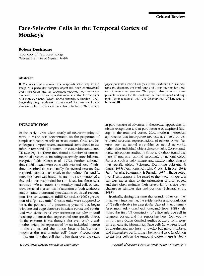
Although they do not provide absolute proof, several studies have tried and failed to identify alternative fea- tures that could explain the properties of face cells. For example, in several studies, most face cells gave virtually no response to any other stimulus tested, including tex- tures, brushes, gratings, bars and edges of various colors, and models of complex objects that might be expected to elicit emotional responses, such as snakes, spiders, and food (Bruce et al., 1981; Perrett et al., 1982; Desi- mone et al., 1984; Baylis, Rolls, & Leonard, 1985; Rolls and Baylis, 1986; Saito, Yukie, Tanaka, Hikosaka, Fukada, & Iwai, 1986). By contrast, each responded to a variety of faces, including real ones, plastic models, and photo- graphs of human and monkey faces (see Figs. 2 and 3) . Many face cells continue to respond well to faces over more than a 12-fold range in the size of the face (Rolls & Baylis, 1986) and many respond over a wide range of orientations in the horizontal plane (Perrett et al., 1982; Desimone et al., 1984; Perrett, Mistlin, Chitty, Smith, Pot- ter, Broennimann, & Harries, 1988; Hasselmo, Rolls, Bay- lis, & Nalwa, 198913). Further, in anesthetized monkeys, face cells have been shown to respond to faces posi- tioned anywhere within a large, bilateral receptive field (Desimone et al., 1984). None responds well to pictures of faces that have the components rearranged, even though the outer shape remains intact and all of the components are still present (Perrett et al., 1982; Desi- mone et al., 1984; Perrett et al., 1988). Likewise, for most cells, no single component of the face is sufficient to elicit a response comparable to the entire face, nor does the elimination of only one specific component cause an elimination of the neuronal response (Perrett et al., 1982; Desimone et al., 1984). Rolls, Baylis, and Leonard (1985) have shown that face cells will continue to respond to
2 Journal of Cognitive Neuroscience Volume 3, Number 1
I A I
1 ' O0 30" 60" 80' 100" 180" NO EYES BRUSH -
5"
Figure 2. Responses o f a neuron within the superior temporal sulcus in IT cortex that responded selectively to Paces. Stimuli were colored slides, projected with a slight oscillatory motion onto a tangent screen for 2.5 sec each trial, with the center o f each stimulus on the fovea. The stimulus on period is indicated by the bar under each histogram, and each histogram is based on a minimum of 10 trials per stimulus. The drawings under each histogram were traced from the stimuli. (A) The neuron responded well to two different monkey fxes and to a human face, but not when the internal components were rearranged. Eliminating the eyes, or the nose, or the color, reduced but did not eliminate the response. The neuron was also tested with a large number of additional nonface stimuli (not shown), and it responded very poorly or not at all to any of them. (B) This neuron was tuned to the frontal view o f the face. Adapted from Desimone et al. (1984).
pictures of faces that have been either low- or high-pass filtered such that no spatial frequencies are in common (responses were dependent on the faces being recog- nizable as faces). Removing the color of the face (Desi- mone et al., 1984; Rolls & Baylis, 1986), altering the color (Perrett et al., 1982), or reducing the contrast to a very low level (Rolls & Baylis, 1986) reduces but does not eliminate the response. Line drawings of faces produce weak responses (Bruce et al., 1981; Perrett et a]., 1982). Taken together, no hypothesis, other than face selectivity, has yet been advanced that could explain such complex neuronal properties.
On the other hand, it is very likely that some of the cells that have been classified as "face cells" actually serve as general-purpose feature analyzers or code the overall significance of stimuli. In some studies, a few cells clas- sified as face selective responded only twice as well to faces as to nonface stimuli (e.g., Perrett et al., 1982; Baylis et al., 1985). Yet, the greater the response to nonface stimuli, the more likely it is that a cell is actually tuned to some more general object feature, such as shape or texture. Because cells that have only a relative preference for faces appear to be sensitive to the configuration of
facial features and are located near cells that are more highly face-selective, Rolls and his colleagues argued that they are also likely to contribute to a face-recognition network (Baylis et al., 1985). While this may be so, their contribution may well not be exclusive.
In addition to cells that respond best or only to intact faces, there have been reports of cells that respond se- lectively to face components, such as the mouth, hair, or (especially) eyes (Perrett et al., 1982; Perrett, Mistlin, & Chitty, 1987). If there is truly a neural network dedicated to the analysis of faces, it is not surprising that it would contain cells selective for meaningful components, such as eyes. In fact, many face cells appear to be sensitive to the direction of gaze of the eyes in the head (of the stimulus, not of the observer) (Perrett, Smith, Potter, Mistlin, Head, Milner, & Jeeves, 1985). Perrett and his colleagues (Perrett et al., 1987) argued that the properties of face cells may be built up from inputs derived from face-component cells. Yet, the simpler the face compo- nents, the more difficult it will be to show that a cell is actually selective for the component, rather than some more general shape feature. For example, without very elaborate tests, it would be hard to reject the possibility
Desimone 3

1. 30" 60" 80" loo" 180" NOEYES BRUSH -
5"
2 3 4 5 6 7 8 - 2.5'
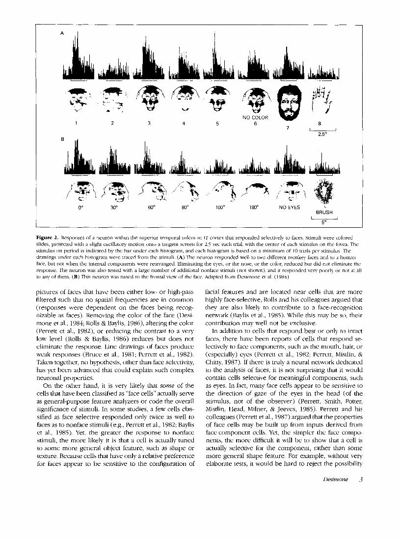
Figure 3. Responses o f a different IT neuron, found in the same cortical region as the cell shown in Figure 2, but which responded best to profile views of faces. Other conditions as in Figure 2 . (A) Responses to one monkey face in different degrees of rotation. (B) Responses to profile of face and to profile with components removed or altered. Removing or altering any of the components of the profile eliminated the response. Adapted from Desimone et al. (1984).
that an "eye" cell is actually a cell that is selective for any dark spot on a light background or that a "hair" cell is actually selective for a fine oriented texture. Indeed, it would seem economical for the visual system to use general-purpose shape and texture-selective cells to an- alyze relatively simple features such as eyes and hair. In any case, detailed tests on the cells selective for face components remain to be conducted.
PROPERTIES OF FACE CELLS
As expected, individual face cells vary in their response to different faces. After all, why would the visual system need a large population of cells just to signal the pres- ence of any face? On the other hand, there is no evidence for a face cell that responds exclusively to the face of one individual. In this respect, the cells are not, strictly speaking, "grandmother" cells. Rather, because individual face-selective cells vary in their response to different facial features, facial orientations, expressions, orienta- tions, and individuals, they likely comprise a distributed network for the coding of faces, just as other cells in IT cortex seem to comprise a distributed network for the coding of general object features.
Yamdne, Kaji, and Kawano (1988) studied a group of IT face cells with a large number of pictures of human
faces and face montages that varied parametrically along several facial feature dimensions, such as the distance between the eyes and width of eyebrows. For some of the cells, they were able to fit the distribution of re- sponses with a regression equation based on a small number of component features. Although cells varied in their selectivity, features that frequently influenced re- sponses were the intereye distance, the distance from the eyes to mouth, and the style of hair on the forehead. They found that, in general, increasing the amount of hair on the forehead improved the response, and in- creasing the distance from the eyes to the mouth de- creased the response.
In addition to the arrangement of facial features, some face cells give graded responses to faces depending on either the identity of the face or on the facial expression (Perrett, Smith, Potter, Mistlin, Head, Milner, & Jeeves, 1984; Baylis et al., 1985; Hasselmo, Rolls, & Baylis, 1989a). Hasselmo et al. (1989a) measured the responses of a group of face cells to nine pictures of three different monkey faces, each with three different facial expressions (calm, slight threat, or full threat). The cells were broadly tuned to the faces; no cell responded solely to one in- dividual or to one expression. However, when the re- sponses of each cell to the nine faces were analyzed with a two-way analysis of variance, 20% of the cells showed significant selectivity for expression independent of iden-
4 Journal of Cognitive Neuroscience Volume 3, Number 1

tity, and 33% showed significant selectivity for identity independent of expression. Only 7% of the cells showed a significant interaction effect. Thus, expression and iden- tity appear to be coded by separate populations of cells. Interestingly, the cells sensitive to expression tended to be located within the superior temporal sulcus, whereas the cells sensitive to identity tended to be located on the inferior temporal gyrus (see below).
Some face cells appear to be sensitive to the orienta- tion of the face, in either the two-dimensional or three- dimensional plane, with the least sensitivity to orientation in the two-dimensional plane (Perrett et al., 1982; Desi- mone et al., 1984; Perrett et al., 1988; Hasselmo et al., 1989b). Nearly all cells that respond to rightside up faces continue to respond to faces rotated on their side or upside down (Hasselmo et al., 1989b), although re- sponses are commonly reduced in magnitude and show an increase in latency of 10-60 msec (Perrett et al., 1988). Correspondingly, monkeys appear to discriminate upside down faces as well as rightside up ones (Bruce, 1982; Overman & Doty, 1982; Perrett et al., 1988), but when the monkeys are trained to respond differentially to face and nonface stimuli, they show an increase in reaction time for responding to upside down faces (Perrett et al., 1988).
Compared to the effects of two-dimensional rotation, the majority of face cells show a much greater sensitivity to rotations of a head in the three dimensional plane, i.e., rotated from frontal to profile or tipped upward or downward (Desimone et al., 1984; Perrett et al., 1988; Hasselmo et al., 1989b). Some cells respond exclusively to frontal views of faces (Fig. 2 ) , some to profiles (Fig. 3), and some appear to respond regardless of rotation. Cells sensitive to rotated views, such as the profile, main- tain this selectivity even when the head is rotated on its side or upside down, suggesting that some cells code profiles in an object-centered rather than viewer-cen- tered frame (Hasselmo et a]., 1989b). Many cells that are selective for full-face or profile views are also sensitive to the direction of gaze of the eyes in the stimulus, some preferring eye contact and others preferring averted gaze (Perrett et al., 1985). Finally, it should be noted that in addition to the face cells that respond to static rotated views of faces, some cells respond specifically to the motion of a face rotating into profile or full-face views (Hasselmo et al., 1989b).
THE ANATOMY OF FACE RECOGNITION
The face cells reported in the temporal cortex are con- tained within both the inferior temporal gyrus and the banks and floor of the superior temporal sulcus. The inferior temporal gyrus and the lower bank of the sulcus are located within cytoarchitectonic area TE and have been traditionally included within “inferior temporal cortex,” or IT cortex, which is an exclusively visual area
(Gross et al., 1972; Desimone & Gross, 1979). The upper bank of the sulcus contains a high proportion of poly- sensory neurons and has been termed the superior tem- poral polysensory area, or STP (Desimone & Gross, 1979; Bruce et al., 1981; Baylis, Rolls, & Leonard, 1987). The boundary between the two areas lies somewhere in the floor of the sulcus (Desimone & Gross, 1979).
In addition to the overall division of the superior temporal sulcus into STP and IT components, Seltzer and Pandya (1978) proposed six additional subdivisions based on cytoarchitecture and anatomical connections. The subdivisions are shown in Figure 1. According to this scheme, STP in the upper bank of the sulcus contains areas T h , TPO, and PGa, whereas IT cortex in the lower bank of the sulcus contains areas TEm and TEa. It is not clear if IPa, in the floor of the sulcus, should be consid- ered part of STP or IT cortex. Although these six subdi- visions are still tentative, there is some recent support for them from a physiological mapping study of Baylis et al. (1987) (see also Boussaoud, Ungerleider, & Desi- mone, 1990).
If one considers the locations of all the face cells that have been reported in the literature, they span all regions of the inferior temporal gyrus and superior temporal sulcus, but they appear to be most prevalent in the sulcus. Consistent with this, in a physiological survey of the temporal cortex, Baylis et al. (1987) found that cells preferring faces to a variety of nonface stimuli were distributed throughout all divisions of the sulcus and on the inferior temporal gyrus, but were especially heavy concentrated in area TPO in the upper bank of the su- perior temporal sulcus (within area STP) and in area TEm in the lower bank and lip of the sulcus (within IT cortex). Cells preferring faces totaled over 20% of the recorded cells in TPO and TEm; however, this 20% al- most certainly included some shape-selective cells that were not specialized exclusively for faces. Other studies have found concentrated pockets or columns of cells with extremely high selectivity for faces, but it is not clear what proportion they comprise of the total cells in temporal cortex.
Interestingly, the two regions across which face cells are distributed, STP and IT cortex, have very different physiological properties and anatomical connections. IT neurons are commonly selective for object features such as shape and color and have large receptive fields that are most sensitive at the fovea. IT cortex receives inputs from both area TEO at the occipitotemporal junction and area V4 in prestriate cortex, which contain cells sensitive to orientation, size, and color (see Desimone & Unger- leider, 1989). By contrast, except for the face cells, STP neurons are rarely sensitive to shape or color, and their receptive fields are at least as sensitive to the far periph- ery of the visual field as to the fovea (Bruce et al., 1981). Further, many cells in STP are selective for stimulus motion (Bruce et al., 1981; Baylis et al., 1987), including the motion of moving people and monkeys (Bruce et al.,
Desimone 5

1981; Perrett, Smith, Mistlin, Chitty, Head, Potter, Broen- nimann, Milner, & Jeeves, 1984). Consistent with these neuronal properties, STP receives inputs from the pos- terior parietal cortex (Jones & Powell, 1970; Anderson, Asanuma, Essick, & Siegel, 1990; Morel & Bullier, 1990; Baizer, Ungerleider, & Desimone, 1990), which is asso- ciated with spatial analyses, and from areas MST and FST in the posterior portion of the sulcus, which are areas concerned with motion analysis (Boussaoud et al., 1990). The region that spans the boundary between STP and IT in the floor of the sulcus receives converging inputs from the posterior parietal cortex, the inferior temporal gyrus, areas MST and FST, and the remainder of STP and thus may be a site of convergence of object, spatial, and mo- tion information (Baizer et al., 1990; Morel & Bullier, 1990; Boussaoud et al., 1990).
Why would face cells be distributed across cortical regions with such diverse properties? A likely reason is that face cells contribute to several functionally distinct neural circuits. As noted above, there is eivdence that different face cells contribute to the analysis of emotional expression, to the recognition of specific individuals, and to the construction of prototypical representations from different face orientations. Each of these operations may be associated with different subdivisions of the temporal cortex. In this regard, it is interesting to recall the ob- servation by Hasselmo et al. (1989a) that cells selective for facial expression tend to be located within the su- perior temporal sulcus, whereas cells selective for iden- tity tend to be located on the inferior temporal gyrus.
WHY FACE CELLS?
Why should faces be treated differently from other classes of objects by the visual system? One possible reason is that faces are extremely important to primates, not only for the recognition of specific individuals in the troop but also for social communication by facial ex- pression. Because of this importance, there may have been selective pressure that led to the evolution of neural mechanisms for the analysis of faces and facial expres- sion. In fact, Allman and McGuinness (1988) argued that the use of facial expression for social communication in higher primates has evolved to largely replace the use of olfaction for the same purpose. As Allman and Mc- Guinness point out, nonprimates as well as primitive primates, such as galagos, make more use of scent glands and scent marking for social purposes than do higher primates, such as macaques and humans. Yet, using facial expression, higher primates can communicate social in- formation much more rapidly and with finer differentia- tion. This ability of higher primates evolved in parallel with the development of a more differentiated facial musculature and the combination of a furry rhinarium and mobile upper lip, which can participate in facial expressions more easily than can the fixed, moist rhi-
narium of primitive primates and other mammals. Allman and McGuinness further note that these behavioral and facial changes in higher primates are paralleled by a relative decrease in the size of the olfactory bulb and an increase in visual inputs to the amygdala, compared to those of primitive primates.
In this context, we have previously noted at least two other examples of specialized neural mechanisms that have evolved to facilitate social communication in other species (Desimone et al., 1984). One is the specialized structures that mediate the perception and generation of song in birds (Leppelsack & Vogt, 1966; McCasland & Konishi, l9Sl), and another is the cortex specialized for language in humans. In each case, specialized perceptual mechanisms have evolved that are separate from those of audition in general. In man, the supramodal language cortex of the temporal lobe is located adjacent to the auditory association cortex, in the left hemisphere (Ras- mussen & Milner, 1975). In monkeys, the cortex that lies adjacent to the auditory association cortex is the superior temporal sulcus, which contains both face-selective and polysensory cells. This region could be a fertile zone for the development of supramodal mechanisms for com- munication. Indeed, Perrett et al. (1988) reported sug- gestive evidence that face-selective cells are more prevalent in the left temporal cortex of monkeys than in the right. Although this does not fit with the fact that, in humans, the right hemisphere is superior to the left in visual pattern recognition, it makes sense if the face cells are more involved in communication than in pattern recognition per se.
Another possibility to consider is that faces are not actually treated differently from other objects by the vis- ual system. This possibility would be consistent with recent clinical studies, which have shown that many pa- tients diagnosed as prosopagnosic have difficulty dis- criminating other classes of objects in addition to faces (Damasio, Damasio, & Van Hoesen, 1982; also see Farah, 1990, for a recent review). As noted above, face cells are probably not “grandmother” cells, but rather give graded responses to many faces, depending on their character- istics. Thus, the existence of face cells is not inconsistent with the notion that specific objects are represented by distributed networks, rather than by individual cells. Faces may be different from other objects only in that they are so important to monkeys, and therefore are represented by a large proportion of the cells in tem- poral cortex. It would not be surprising if future studies found other populations of temporal lobe neurons rel- atively specialized for other object classes, probably due to the monkey’s experience with those objects (Rolls, Baylis, Hasselmo, & Nalwa, 1989). Gross’s ori- ginal “hand cell” could even be such an example (Gross et al., 1972). Ironically then, face cells could turn out to be a model system for studying the neural mech- anisms of complex object recognition, rather than an exception.
6 Journal of Cognitive Neuroscience Volume 3, Number 1

Acknowledgments 1 thank Leslie Ungerleider and Mortimer Mishkin for valuable comments on the manuscript.
Reprint requests should be sent to Robert Desimone, Labora- tory of Neuropsychology, NIMH, Building 9, Bethesda, MD 20892.
REFERENCES Allman, J., & McGuinness, E. (1988). Visual cortex in primates.
In Comparativeprimate biology (Vol. 4). New York: Alan R. Liss, 279-326.
(1990). Cortico-cortical connections of anatomically and physiologically defined subdivisions within the inferior pari- etal lobule. Journal of Comparative Neurology, 296, 65- 113.
Baizer, J. S., Ungerleider, L. G., & Desimone, R. (1990). Orga- nization of visual inputs to inferior temporal and posterior parietal cortex in macaques. Jozirnal of Neuroscience, in press.
Baylis, G. C., Rolls, E. T., & Leonard, C. M. (1985). Selectivity between faces in the responses of a population of neurons in the cortex in the superior temporal sulcus of the mon- key. Brain Research, 342, 91-102.
Baylis, G. C., Rolls, E. T., & Leonard, C. M. (1987). Functional subdivisions of the temporal lobe neocortex. Journal of Neuroscience, 7, 330-342.
Boussaoud, D. B., Ungerleider, L. G., & Desimone, R. (1990). Pathways for motion analysis: cortical connections of the medial superior temporal and fundus of the superior tem- poral visual areas in the macaque.. Journal of Comparative Neurology, 296, 462-495.
Bruce, C. J. (1982). Face recognition by monkeys: Absence of an inversion effect. Neuropsychologia, 20, 515-521.
Bruce, C. J., Desimone, R., & Gross, C. G. (1981). Visual prop- erties of neurons in a polysensory area in superior tem- poral sulcus of the macaque. Journal of Neurophysiology,
Andersen, R. A,, Asanuma, C., Essick, G., & Siegel, R. M.
46, 369-384. Damasio, A. R., Damasio, H., & Van Hoesen, G. W. (1982).
Prosopagnosia: Anatomical basis and behavioral mecha- nisms. Neurology, 32, 331-341.
Desimone, R., Albright, T. D., Gross, C. G., & Bruce, C. (1984). Stimulus-selective properties of inferior temporal neurons in the macaque. Journal of Neuroscience, 4, 2051-2062.
Desimone, R., & Gross, C. G. (1979). Visual areas in the tem- poral cortex of the macaque. Brain Research, 178, 363- 380.
Desimone, R., & Ungerleider, L. G. (1989). Neural mecha- nisms of visual processing in monkeys. In F. Boller & J. Grafman (Eds.), Handbook of neuropsychology (Vol. 2). New York: Elsevier, 267-299.
Fdrah, M. J. (1990). Visual agnosia. Cambridge: MIT Press. Gross, C. G., Rocha-Miranda, C. E., & Bender, D. B. (1972).
Visual properties of neurons in inferotemporal cortex of the macaque. Journal of Neurophysiology, 35,96-111.
Hasselmo, M. E., Rolls, E. T., & Baylis, G. C. (1989a). The role of expression and identity in the face-selective responses of neurons in the temporal visual cortex of the monkey. Be- havioural Brain Research, 32, 203-2 18.
(1989b). Object-centered encoding by face-selective neu- rons in the cortex in the superior temporal sulcus of the monkey. Experimental Brain Research, 75, 41 7-429.
Hasselmo, M. E., Rolls, E. T., Baylis, G. C., & Nalwa, V.
Jones, E. G., & Powell, T. P. S. (1970). An anatomical study of converging sensory pathways within the cerebral cortex of the monkey. Brain Research, 93, 793-820.
cortex of conscious sheep can respond preferentially to the sight of faces. Science, 236, 448-450.
Konorski, J. (1967). Integrative activily of the brain: An inter- d&ciplinay approach. Chicago: University of Chicago Press.
Leppelsack, H. J., & Vogt, M. (1966). Responses of auditory neurons in the forebrain of the songbird to stimulation with species-specific sounds. Journal of Comparative Physi-
McCasland, J., & Konishi, M. (1981). Interactions between au- ditory and motor activities in an avian song control nu- cleus. Proceedings of the National Academy of Sciences of the United States of America, 178, 781 5-78 19.
Morel, A,, & Bullier, J. (1990). Anatomical segregation of two cortical visual pathways in the macaque monkey. Visual Neuroscience, 4, 555-578.
Overman, W. M., Jr., & Doty, R. W. (1982). Hemispheric spe- cialization displayed by man but not macques for analysis of faces. Neuropsychologia, 20, 113-128,
Perrett, D. I., Mistlin, A. J., & Chitty, A. J. (1987). Visual neu- rones responsive to faces. Trenak in Neurosciences, 10,
Perrett, D. I., Mistlin, A. J., Chitty, A. J., Smith, P. A. J., Potter, D. D., Broennimann, R., & Harries, M. (1988). Special- ized face processing and hemispheric asymmetry in man and monkey: Evidence from single unit and reaction time studies. Behavioral Brain Research, 29,
Kendrick, K. M., & Baldwin, B. A. (1987). Cells in temporal
ology, 107, 263-274.
358-364.
245-258. Perrett, D. I., Rolls, E. T., & Caan, W. (1982). Visual neurones
responsive to faces in the monkey temporal cortex. Experi- mental Brain Research, 47, 329-342.
Perrett, D. I., Smith, A. J., Potter, D. D., Mistlin, A. J , , Head, A. S., Milner, A. D., & Jeeves, M. A. (1984). Neurones re- sponsive to faces in the temporal cortex: Studies of func- tional organization, sensitivity to identity and relation to perception. Human Neurobiology, 3, 197-208.
Perrett, D. I., Smith, P. A. J., Mistlin, A. J., Chitty, A. J., Head, A. S., Potter, D. D., Broennimann, R., Milner, A. D., & Jeeves, M. A. (1984). Visual analysis of body movements by neu- rones in the temporal cortex of the macaque monkey: a preliminary report. Behavioural Brain Research, 16, 153- 170.
Perrett, D. I., Smith, P. A. J,, Potter, D. D., Mistlin, A. J., Head, A. S., Milner, A. D., & Jeeves, M. A. (1985). Visual cells in the temporal cortex sensitive to face view and gaze direc- tion. Proceedings of the Royal Sociely of London, 223, 293- 317.
Rasmussen, T., & Milner, B. (1975). Clinical and surgical stud- ies of the cerebral speech areas in man. In K. J. Zulch, 0. Creutzfeldt, & G. C. Galbraith (Eds.), Cerebral localization. Berlin: Springer-Verlag, pp. 238-257.
Rolls, E. T. (1984). Neurons in the cortex of the temporal lobe and in the amygdala of the monkey with responses selective for faces. Human Neurobiology, 3, 209-222.
Rolls, E. T., & Baylis, G. C . (1986). Size and contrast have only small effects on the responses to faces of neurons in the cortex of the superior temporal sulcus of the monkey. Ex- perimental Brain Research, 65, 38-48.
Rolls, E. T. Baylis, G. C., Hasselmo, M. E., & Nalwa, V. (1989). The effect of learning on the face selective responses of neurons in the cortex in the superior temporal sulcus of the monkey. Experimental Brain Research, 76, 153- 164.
Rolls, E. T., Baylis, G. C., & Leonard, C. M. (1985). Role of low and high spatial frequencies in the face-selective responses
Desimone 7

of neurons in the cortex in the superior temporal sulcus in the monkey. %ion Research, 25, 1021-1035.
Saito, H., Yukie, M., Tanaka, K., Hikosaka, K., Fukada, Y., & Iwai, E. (1986). Integration of direction signals of image motion in the superior temporal sulcus of the macaque monkey. Journal of Neuroscience, 6, 145-157.
Saito, H., Tanaka, K., Fukumoto, M., & Fukada, Y. (1987). The inferior temporal cortex of the macaque monkey. 11. The level of complexity in the integration of pattern informa- tion. Society for Neuroscience Abstracts, 13, 628.
Schwartz, E. L., Desimone, R., Albright, T. D., & Gross, C. G.
8 Journal of Cognitive Neuroscience
(1983). Shape recognition and inferior temporal neurons. Proceedings of the National Academy of Sciences of the United States of America, 80, 5776-5778.
Seltzer, B., & Pandya, D. N. (1978). Afferent cortical connec- tions and architectonics of the superior temporal sulcus and surrounding cortex in the rhesus monkey. Brain Re- search, 149, 1-24.
Yamane, S., Kaji, S., & Kawano, K. (1988). What facial features activate face neurons in the inferotemporal cortex of the monkey? Experimental Brain Research, 73, 209-214.
Volume 3, Number 1

This article has been cited by:
1. Bruno Rossion, Daniel Collins, Valérie Goffaux, Tim Curran. 2007. Long-term Expertise with Artificial Objects Increases VisualCompetition with Early Face Categorization Processes. Journal of Cognitive Neuroscience 19:3, 543-555. [Abstract] [PDF] [PDFPlus]
2. Edmund T. Rolls, Simon M. Stringer. 2007. Invariant Global Motion Recognition in the Dorsal Visual System: A UnifyingTheory. Neural Computation 19:1, 139-169. [Abstract] [PDF] [PDF Plus]
3. Gillian Rhodes, Graham Byatt, Patricia T. Michie, Aina Puce. 2004. Is the Fusiform Face Area Specialized for Faces, Individuation,or Expert Individuation?. Journal of Cognitive Neuroscience 16:2, 189-203. [Abstract] [PDF] [PDF Plus]
4. Simon M. Stringer , Edmund T. Rolls . 2002. Invariant Object Recognition in the Visual System with Novel Views of 3D Objects.Neural Computation 14:11, 2585-2596. [Abstract] [PDF] [PDF Plus]
5. Noam Sagiv, Shlomo Bentin. 2001. Structural Encoding of Human and Schematic Faces: Holistic and Part-Based Processes.Journal of Cognitive Neuroscience 13:7, 937-951. [Abstract] [PDF] [PDF Plus]
6. Bruno Laeng, Verne S. Caviness. 2001. Prosopagnosia as a Deficit in Encoding Curved Surface. Journal of Cognitive Neuroscience13:5, 556-576. [Abstract] [PDF] [PDF Plus]
7. Edmund T. Rolls , T. Milward . 2000. A Model of Invariant Object Recognition in the Visual System: Learning Rules, ActivationFunctions, Lateral Inhibition, and Information-Based Performance Measures. Neural Computation 12:11, 2547-2572. [Abstract][PDF] [PDF Plus]
8. Sidney R. Lehky . 2000. Fine Discrimination of Faces can be Performed Rapidly. Journal of Cognitive Neuroscience 12:5, 848-855.[Abstract] [PDF] [PDF Plus]
9. Morris Moscovitch, Gordon Winocur, Marlene Behrmann. 1997. What Is Special about Face Recognition? Nineteen Experimentson a Person with Visual Object Agnosia and Dyslexia but Normal Face Recognition. Journal of Cognitive Neuroscience 9:5, 555-604.[Abstract] [PDF] [PDF Plus]
10. Guy Wallis, Roland Baddeley. 1997. Optimal, Unsupervised Learning in Invariant Object Recognition. Neural Computation 9:4,883-894. [Abstract] [PDF] [PDF Plus]
11. Stephen Grossberg, John W.L. Merrill. 1996. The Hippocampus and Cerebellum in Adaptively Timed Learning, Recognition,and Movement. Journal of Cognitive Neuroscience 8:3, 257-277. [Abstract] [PDF] [PDF Plus]