fabrication of photonic transfer dna-quantum dot … · 2018-12-11 · biology, and materials:...
TRANSCRIPT

Fabrication of Photonic Transfer DNA-Quantum Dot Nanostructures
M. J. Heller, B. Sullivan, D. Dehlinger,
University of California San Diego Departments of Bioengineering/Electrical & Computer Engineering
PFBE Bldg. Rm 429 9500 Gilman Dr.
La Jolla, CA 92093-0412 [email protected]
ABSTRACT The fabrication of viable linear DNA photonic/electronic transfer nanostructures is an important prerequisite for the subsequent self-assembly into higher-order 2D/3D devices, structures and materials (high density memory, photonic antennas, nanoscale “fiber optics”, nanocomputers, nano-sensors etc.). Unfortunately, it is very difficult to maintain the self-assembling and self-recognition properties of derivatized molecules such as biotin functionalized DNA sequences, when attempting to further derivatize the structure with multiply functionalized nanoparticles (streptavidin-quantum dots, gold nanoparticles, fluorescent polymer nanoparticles). Generally, overwhelming intra- and intermolecular crosslinking reactions prevent the formation of viable linear structures. Our goal is the development of nanofabrication techniques which will allow quantum dot functionalized linear DNA chains to be constructed without the need for complex chemical blocking group procedures. Keywords: DNA, quantum dots, nanofabrication, self-assembly, nanophotonics, nanoelectronics, nanopores
INTRODUCTION Presently, it is very difficult to maintain the self-assembling properties of derivatized molecules such as a biotin functionalized DNA sequences, when attempting to further derivatize it with multiply functionalized nanoparticles (streptavidin-quantum dots, gold nanoparticles, fluorescent polymer nanoparticles). Our goal is the development of nanofabrication techniques which will allow the formation of quantum dot functionalized linear DNA chains of more than one micron in length which retain their photonic transfer, self-assembly and recognition (DNA hybridization) properties. To achieve this, we have first constructed linear biotinylated DNA backbone structures which can be used for further modification. DNA ligation procedures were used to produce these long DNA structures. To achieve large-scale precision functionalization of DNA, we start with multiple DNA
subunits that self-assemble when mixed. Once mixed, the fragments are enzymatically ligated to prevent dissociation back into the individual subunits. Once the larger DNA backbones have been manufactured, the goal is to further functionalize the backbones with nanoparticles (quantum-dots) at regular intervals (every 34 bases). The regularity is achieved by the placement of chemical binding groups (biotins) which have been located on each subunit of DNA before they assembly into the larger linear structures. The further linkages make use of biotin/streptavidin ligand binding reaction. Direct mixing of biotinylated DNA backbones with streptavidin-quantum dots leads to considerable inter- and intramolecular strand crosslinking of the biotinylated DNA with quantum dots (see Figure 1A and 1B).
Under some very stringent conditions, the stoichiometric mixing of biotinylated DNA with streptavidin quantum dots sometime produces the desired structures as shown in Figure 2B.
5 um
B C A
5um
Figure 2, Various DNA/quantum dot nanoparticle mixtures; (A) Q-dots, with no DNA; (B) 2-3 DNA/Q-dots showing a linear structure; and (C) more than 4 DNA/Q-dots, showing some linear aggregated structures.
Figure 1A and 1B - Inter- and intramolecular crosslinked (aggregated) DNA/quantum dot structures
NSTI-Nanotech 2005, www.nsti.org, ISBN 0-9767985-0-6 Vol. 1, 2005 769

Unfortunately, the linear structures (Figure 2B) are transient when they appear, and further biotin/streptavidin reactions soon aggregate them into non-viable materials. To prevent this, we are attempting to control and direct the assembly of the quantum dots with the biotinylated DNA by first isolating the two materials and separating them by a semipermeable nanopore membrane device. By applying a DC electric field, we intend to transport the biotinylated DNA (electrophoretically) across the membrane and slowly expose each biotinylated segment to the streptavidin quantum dots. At the same time, DNA strands are kept spatially isolated from one another to prevent crosslinking. Figure 3 below shows a schematic diagram of the nanopore membrane device and the process for functionalizing biotinylated DNA strands with streptavidin quantum dots.
Figure 3 – Nanopore membrane fabrication device with chambers for biotinylated DNA fragments and streptavidin quantum dots. Upon application of DC electric field biotinylated DNA fragments are transported in a controlled manner into a second chamber containing the streptavidin quantum dots, where biotin/streptavidin binding reactions can occur with limited intra- and intermolecular crosslinking. The final goal is to produce linear DNA quantum dot structures of greater than one micron in length which exhibit long distance photonic energy transfer properties and still have ability to be further self-assembled via specific DNA hybridization interactions into higher order 2D/3D structures.
Materials and Methods (DNA Ligation Procedure) Unmodified and biotinylated DNA fragments, as shown in Figure 4, were designed and synthesized by automated oligonucleotide synthesis and then purified by conventional HPLC and gel electrophoretic techniques. DNA fragments (Figure 4) were mixed in equal concentrations, and then ligated using an enzyme called DNA ligase. (This is a standard molecular biological enzymatic process used for making large DNA constructs. It uses a mixture of bacterial DNA ligase, buffers, and ATP to covalently link DNA fragments together. Ligase repairs certain kinds of chemical breaks in the DNA structure). The result is that two
fragments of DNA become one larger fragment. Figure 5 and 6 shows the results of running the DNA ligase reactions on agarose gels, and the resulting high molecular weight DNA fragments which were produced. This technique is used to go from our modified monomer DNA fragments (34 bp) to the much larger biotin modified DNA backbone structures we wish to use for further functionalization with quantum dots.
Figure 4 – DNA fragments which were designed and synthesized for ligating into high molecular weight linear DNA constructs.
Directed Assembly We are developing an electrophoretic device with multiple chambers separated by micron thick membranes with nanometer sized pores to control the reaction of biotinylated DNA fragments with streptavidin quantum dots. Nanopore membranes are generated by exposing a suitable membrane to radiation that causes tracks in the material that are then etched. This causes holes that go straight through the material, as opposed to other materials, such as gels that have a mesh of fibers. The nanopore hole density of these membranes is such that they (the nanopores) will be farther apart on average then the length of the DNA to be modified. The DNA is placed on the negative side of the device, and quantum dots on the positive side. Since the DNA is negatively charged, it will be pulled towards the positive electrode. The quantum dot’s charge can be changed by
Electrode Buffer Chamber
Streptavidin Qdot Negative Biotinylated
DNA Chain
10 nm pore Membrane
V-
V+
Applied Electric Field
Biotinylated “Main” Strand : 34mer 5’ GTG AAC GTG GAT GAA G (dT Biotin) AGG TGG TGA GGC CCT GA 3’ “Main” Terminator Strand : 51mer 5’ GTG AAC GTG GAT GAA GT AGG TGG TGA GGC CCT GA GTG AAC GTG GAT GAA GT 3’ “Link” Strand : 34mer 5’ ACT TCA TCC ACG TTC AC TCA GGG CCT CAC CAC CT 3’ Biotinylated “Link” Strand : 34mer 5’ ACT TCA TCC ACG TTC AC (dT biotin) CA GGG CCT CAC CAC CT 3’ “Link” Terminator Strand : 51mer 5’ TCA GGG CCT CAC CAC CT ACT TCA TCC ACG TTC AC TCA GGG CCT CAC
B
B
5’ 3’
3’ 5’
B B B B
10kpb Ladder 1x Lig Ligation controls No Lig 5x Lig 25 bp
10k bp
500 bp
100 bp
300 bp
1000
Figure 5 and 6 – Agarose gel electrophoretic separation of high molecular weight DNA constructs ligated from much smaller modified fragments.
V-
V+
Applied Electric Field
NSTI-Nanotech 2005, www.nsti.org, ISBN 0-9767985-0-6 Vol. 1, 2005770
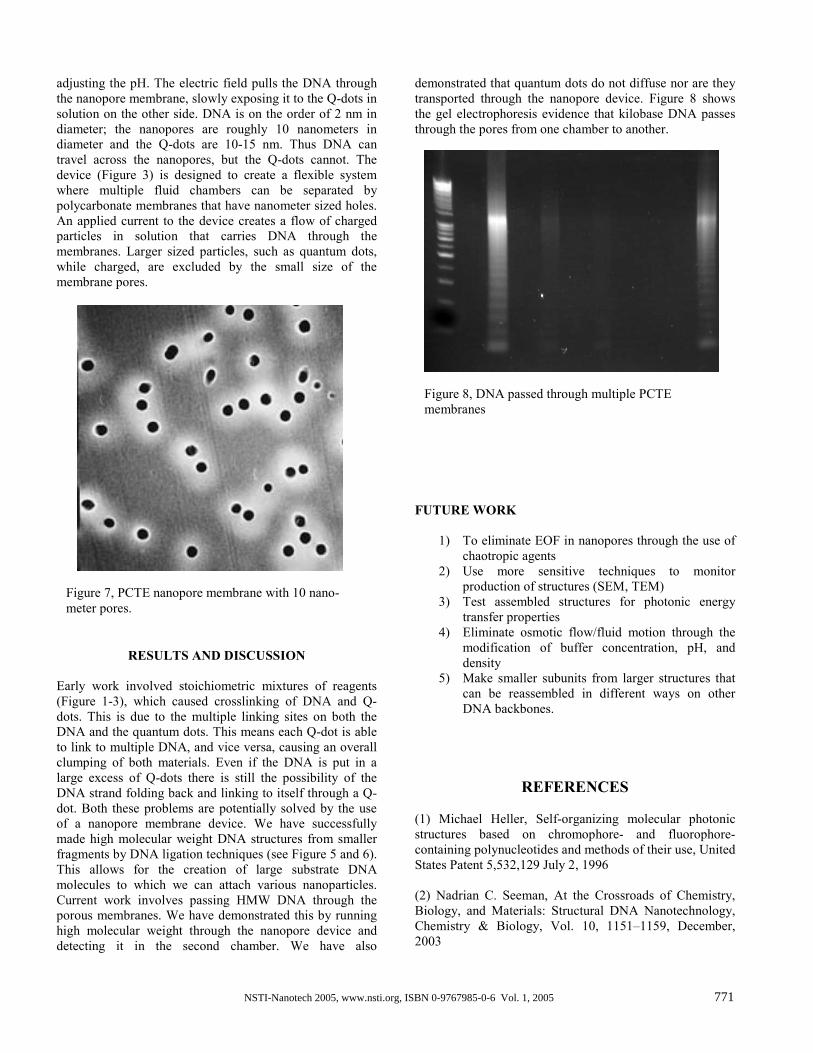
adjusting the pH. The electric field pulls the DNA through the nanopore membrane, slowly exposing it to the Q-dots in solution on the other side. DNA is on the order of 2 nm in diameter; the nanopores are roughly 10 nanometers in diameter and the Q-dots are 10-15 nm. Thus DNA can travel across the nanopores, but the Q-dots cannot. The device (Figure 3) is designed to create a flexible system where multiple fluid chambers can be separated by polycarbonate membranes that have nanometer sized holes. An applied current to the device creates a flow of charged particles in solution that carries DNA through the membranes. Larger sized particles, such as quantum dots, while charged, are excluded by the small size of the membrane pores.
RESULTS AND DISCUSSION Early work involved stoichiometric mixtures of reagents (Figure 1-3), which caused crosslinking of DNA and Q-dots. This is due to the multiple linking sites on both the DNA and the quantum dots. This means each Q-dot is able to link to multiple DNA, and vice versa, causing an overall clumping of both materials. Even if the DNA is put in a large excess of Q-dots there is still the possibility of the DNA strand folding back and linking to itself through a Q-dot. Both these problems are potentially solved by the use of a nanopore membrane device. We have successfully made high molecular weight DNA structures from smaller fragments by DNA ligation techniques (see Figure 5 and 6). This allows for the creation of large substrate DNA molecules to which we can attach various nanoparticles. Current work involves passing HMW DNA through the porous membranes. We have demonstrated this by running high molecular weight through the nanopore device and detecting it in the second chamber. We have also
demonstrated that quantum dots do not diffuse nor are they transported through the nanopore device. Figure 8 shows the gel electrophoresis evidence that kilobase DNA passes through the pores from one chamber to another.
FUTURE WORK
1) To eliminate EOF in nanopores through the use of chaotropic agents
2) Use more sensitive techniques to monitor production of structures (SEM, TEM)
3) Test assembled structures for photonic energy transfer properties
4) Eliminate osmotic flow/fluid motion through the modification of buffer concentration, pH, and density
5) Make smaller subunits from larger structures that can be reassembled in different ways on other DNA backbones.
REFERENCES (1) Michael Heller, Self-organizing molecular photonic structures based on chromophore- and fluorophore-containing polynucleotides and methods of their use, United States Patent 5,532,129 July 2, 1996 (2) Nadrian C. Seeman, At the Crossroads of Chemistry, Biology, and Materials: Structural DNA Nanotechnology, Chemistry & Biology, Vol. 10, 1151–1159, December, 2003
Figure 7, PCTE nanopore membrane with 10 nano-meter pores.
Figure 8, DNA passed through multiple PCTE membranes
NSTI-Nanotech 2005, www.nsti.org, ISBN 0-9767985-0-6 Vol. 1, 2005 771

(3) Hao Yan, Sung Ha Park, Gleb Finkelstein, John H. Reif, Thomas H. LaBean, DNA-Templated Self-Assembly of Protein Arrays and Highly Conductive Nanowires, 26 SEPTEMBER 2003VOL 301 SCIENCE (4) Hao Yan, Thomas H. LaBean, Liping Feng, and John H. Reif, Directed nucleation assembly of DNA tile complexes for barcode-patterned lattices, PNAS July 8, 2003 vol. 100 no. 14 8103–8108 (5) Sarah J. Kodumal, Kedar G. Patel, Ralph Reid, Hugo G. Menzella, Mark Welch, and Daniel V. Santi, Total synthesis of long DNA sequences: Synthesis of a contiguous 32-kb polyketide synthase gene cluster PNAS November 2, 2004 vol. 101 no. 44 15575
NSTI-Nanotech 2005, www.nsti.org, ISBN 0-9767985-0-6 Vol. 1, 2005772