f2 intercross cross-references definition
TRANSCRIPT

F
F2 Intercross
Definition
The second-generation descendents of a cross of
two contrasting populations (e.g., inbred strains).The offspring of the cross (i.e., F1 hybrids) are
sib-mated to produce F2 hybrids, in which homol-
ogous recombination has “shuffled” the genomesof the progenitors in a unique manner. F2 hybrids
from inbred strain progenitors are useful for
quantitative trait locus (QTL) mapping.
Cross-References
▶Quantitative Trait Locus Mapping
Fabry’s Disease
▶Metabolic and Nutritional Neuropathies
Faces Pain Scale
Definition
Visual pain scale of seven faces now tested in
older adults as well as children.
Cross-References
▶Cancer Pain, Assessment in the CognitivelyImpaired
Facet Denervation
▶ Facet Joint Procedures for Chronic Back Pain
Facet Joint
Definition
Facet is a flat, platelike surface that acts as part of
a joint, as seen in the vertebrae of the spine and in
the subtalar joint of the ankle. Each vertebra hastwo superior and two inferior facets. Facet joints
are small stabilizing synovial joints located
between and behind adjacent vertebrae.
Cross-References
▶Chronic Back Pain and Spinal Instability
▶Chronic Low Back Pain: Definitions andDiagnosis
▶ Facet Joint Pain
▶ Pain Treatment: Spinal Nerve Blocks
G.F. Gebhart, R.F. Schmidt (eds.), Encyclopedia of Pain, DOI 10.1007/978-3-642-28753-4,# Springer-Verlag Berlin Heidelberg 2013

Facet Joint Injection
▶ Facet Joint Procedures for Chronic Back Pain
Facet Joint Pain
Jenna E. Zauk1, Alfred T. Ogden2 andChristopher J. Winfree2
1St. George’s University, School of Medicine,
Grenada, West Indies2Department of Neurological Surgery,
Neurological Institute Columbia University,
New York, NY, USA
Synonyms
Facet syndrome; Sciatica; Zygapophysial joint
pain
Definition
Although the intervertebral joint has long been
known to be a common generator of low backpain and leg pain, the facet joint has been
proposed as another potential focus of degenera-
tive pathology that can produce similar symp-toms. Structurally analogous to joints in the
extremities, the facet joint has the capacity todegenerate over time and cause pain through the
same mechanisms that are at play in osteoarthritis
of the hip or the knee. This pain can be “felt” bythe patient in the area of the facet, producing low
back pain, or it can potentially be referred to other
parts of the body through the activation ofadjacent nociceptive fibers. Radiofrequency,
cryoneuroablation, and chemical neurolysis are
all possible treatment modalities for lumbar facetsyndrome (Wheeler and May 2010). Although
a “facet syndrome” including a component of
sciatica was initially proposed, stimulation ofthe nerve supply to facet joints in live subjects
has shown reproducible patterns of pain that are
limited to the low back, flank, abdomen, and
buttock. Many studies have claimed a benefitfrom treatment of low back pain through inter-
ventions aimed at disrupting the primary affer-ents supplying the facet joint, but the handful of
randomized clinical trials of these interventions
has generally failed to demonstrate any benefitbeyond placebo (Carette et al. 1991; Leclaire
et al. 2001; Lilius et al. 1989, Slipman 2003).
These studies have been criticized, however,and their generally negative results may stem
from poor patient selection and improper selec-
tion of therapeutic target. In 2009, a panel ofexperts in interventional therapies and surgery
recommended against the use of facet joint
steroid injections as a treatment for lower backpain. Their decision was based upon insufficient
evidence from previously conducted studies
(Chou et al. 2009b). Although rates of use forfacet joint injections increased by 231 % from
1994 to 2001, these therapies have shown to only
improve short-term pain management (Friedlyet al. 2007; Chou et al. 2009a).
Characteristics
Anatomical ConsiderationsGhormley is credited with coining the phrase the
“facet syndrome” in 1933 (Ghormley 1933).
In his seminal article, he noted that facet jointswere the only “true” joints in the spinal column,
meaning that they contain a complete joint cap-
sule with a clear synovial membrane and hyalinecartilage at the articular surface. Such true joints
are termed zygapophysial joints and exist in all
the typical weight-bearing places affected byosteoarthritis such as the hip, the knee, and the
ankle. He conjectured and offered histopatholog-
ical evidence that these joints degenerate overtime like their counterparts in the extremities.
He theorized that this process could produce
a syndrome of low back pain, scoliosis, andsciatica, perhaps induced by rotatory strain of
the lumbosacral region.
Detailed anatomical studies of the nerve sup-ply of facet joints have provided the blueprint for
testing and treating the “facet syndrome.”
Each joint is innervated by spinal nerves from
F 1246 Facet Joint Injection

the adjacent and superior vertebral level on theipsilateral side (Bogduk and Long 1979;
Maldjian et al. 1998; Mooney and Robertson1976). Arising from the dorsal root ganglion,
the medial branch of the posterior ramus passes
through a notch at the base of the transverseprocess. Twigs are given off to the facet joint at
the same level before the nerve descends inferi-
orly, giving off muscular and cutaneousbranches, as well as a branch to the superior
aspect of the facet joint one vertebral level
below. Bogduk and Long published the mostdetailed anatomical study of these relationships,
and proposed calling the branches comprising
this dual nerve supply the proximal and distalzygapophysial nerves (Bogduk and Long 1979).
These same authors recognized variability at
L5–S1 mandated by the absence of transverseprocesses at this level, and noted the medial
branch of the posterior ramus of L5 passes
through a groove between the sacral ala and theroot of the superior articular process of the
sacrum. The key points from this anatomical
study were that denervation of a facet jointwould require a lesion of the medial branch of
the posterior ramus at the same vertebral level
and the level above, and that a denervation pro-cedure directed at the facet joint proper might
result in an incomplete lesion.
Provocative StudiesThe first report of nervous stimulation of facet
joints that induced back and leg pain was byHirsch et al. in 1963 (Hirsch et al. 1963). The
first systematic analysis of referred lumbar
facet joint pain was written by Mooney andRobertson in 1975 (Mooney and Robertson
1976). These authors studied five normal sub-
jects and 15 patients with low back pain, andreported a nonspecific pattern of pain referral
to the flank, buttock, and the leg upon injec-
tion of 5 % hypertonic saline into the facetjoint. An important discrepancy was noted
between normal and low back pain patients
in that the latter complained of more frequentand more widespread patterns of pain referral.
The only reports of induced pain in a sciatic
distribution came from the low back pain
cohort. These authors also related their resultswith fluoroscopically guided anesthetic injec-
tions, and offered a detailed description of theprocedure, which served as a model for future
studies. Due to difficulty in locating the pre-
cise vertebral level of the pain generator,injections at three lumbar levels were advo-
cated and injections were directed into the
facet joint proper. The facet joint, as the targetof treatment, has been used by many clinicians
and in many studies ever since, but this
approach has come under criticism by some,who maintain that a more efficacious target of
anesthetization or neurotomy is the medial
branch of the posterior ramus (Bogduk andLong 1979).
The technique of Mooney and Robertson
was applied (McCall et al. 1979) to six healthyindividuals who received fluoroscopically
guided injections of 6 % hypertonic saline into
lumbar facet joints of L1–2 and L4–5. Theynoted several important findings. L1–2 injec-
tions reproduced pain to the adjacent lower
back, flank, and groin. L4–5 injections pro-duced pain in the adjacent lower back, buttock,
groin, and lateral thigh. There was significant
overlap in the distribution of the patterns ofreferred pain, even though the facet joints
selected were three levels apart, and there was
no patient that complained of pain below themid-thigh.
Provocative studies have shown fairly clearly
that stimulation of the sensory nerves aroundfacet joints can induce pain, and that this stimu-
lation can reproducibly generate pain that is
referred to other parts of the body. It is stillunclear to what extent this induced pain has
a relationship to the clinical entity of low back
pain in the general population. The paucity ofprovocative evidence for the induction of
referred leg upon stimulation of facet joints in
control populations casts some doubt on thesciatic component of the alleged “facet syn-
drome.” The lack of dermatomal specificity for
referred pain from facet joint stimulation mayreflect the dual level innervation, and contribute
to the difficulty in localization of the potential
pain-generating level.
Facet Joint Pain 1247 F
F
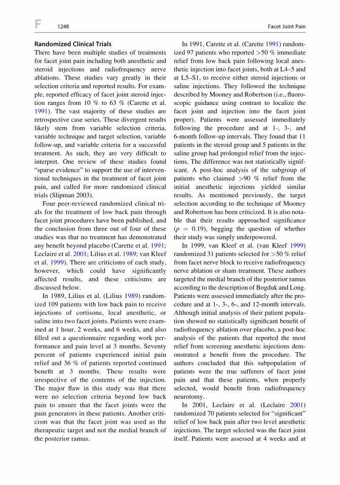
Randomized Clinical TrialsThere have been multiple studies of treatments
for facet joint pain including both anesthetic and
steroid injections and radiofrequency nerveablations. These studies vary greatly in their
selection criteria and reported results. For exam-
ple, reported efficacy of facet joint steroid injec-tion ranges from 10 % to 63 % (Carette et al.
1991). The vast majority of these studies are
retrospective case series. These divergent resultslikely stem from variable selection criteria,
variable technique and target selection, variable
follow-up, and variable criteria for a successfultreatment. As such, they are very difficult to
interpret. One review of these studies found
“sparse evidence” to support the use of interven-tional techniques in the treatment of facet joint
pain, and called for more randomized clinical
trials (Slipman 2003).Four peer-reviewed randomized clinical tri-
als for the treatment of low back pain through
facet joint procedures have been published, andthe conclusion from three out of four of these
studies was that no treatment has demonstrated
any benefit beyond placebo (Carette et al. 1991;Leclaire et al. 2001; Lilius et al. 1989; van Kleef
et al. 1999). There are criticisms of each study,
however, which could have significantlyaffected results, and these criticisms are
discussed below.In 1989, Lilius et al. (Lilius 1989) random-
ized 109 patients with low back pain to receive
injections of cortisone, local anesthetic, orsaline into two facet joints. Patients were exam-
ined at 1 hour, 2 weeks, and 6 weeks, and also
filled out a questionnaire regarding work per-formance and pain level at 3 months. Seventy
percent of patients experienced initial pain
relief and 36 % of patients reported continuedbenefit at 3 months. These results were
irrespective of the contents of the injection.
The major flaw in this study was that therewere no selection criteria beyond low back
pain to ensure that the facet joints were the
pain generators in these patients. Another criti-cism was that the facet joint was used as the
therapeutic target and not the medial branch of
the posterior ramus.
In 1991, Carette et al. (Carette 1991) random-ized 97 patients who reported >50 % immediate
relief from low back pain following local anes-thetic injection into facet joints, both at L4–5 and
at L5–S1, to receive either steroid injections or
saline injections. They followed the techniquedescribed by Mooney and Robertson (i.e., fluoro-
scopic guidance using contrast to localize the
facet joint and injection into the facet jointproper). Patients were assessed immediately
following the procedure and at 1-, 3-, and
6-month follow-up intervals. They found that 11patients in the steroid group and 5 patients in the
saline group had prolonged relief from the injec-
tions. The difference was not statistically signif-icant. A post-hoc analysis of the subgroup of
patients who claimed >90 % relief from the
initial anesthetic injections yielded similarresults. As mentioned previously, the target
selection according to the technique of Mooney
and Robertson has been criticized. It is also nota-ble that their results approached significance
(p ¼ 0.19), begging the question of whether
their study was simply underpowered.In 1999, van Kleef et al. (van Kleef 1999)
randomized 31 patients selected for>50 % relief
from facet nerve block to receive radiofrequencynerve ablation or sham treatment. These authors
targeted the medial branch of the posterior ramus
according to the description of Bogduk and Long.Patients were assessed immediately after the pro-
cedure and at 1-, 3-, 6-, and 12-month intervals.
Although initial analysis of their patient popula-tion showed no statistically significant benefit of
radiofrequency ablation over placebo, a post-hoc
analysis of the patients that reported the mostrelief from screening anesthetic injections dem-
onstrated a benefit from the procedure. The
authors concluded that this subpopulation ofpatients were the true sufferers of facet joint
pain and that these patients, when properly
selected, would benefit from radiofrequencyneurotomy.
In 2001, Leclaire et al. (Leclaire 2001)
randomized 70 patients selected for “significant”relief of low back pain after two level anesthetic
injections. The target selected was the facet joint
itself. Patients were assessed at 4 weeks and at
F 1248 Facet Joint Pain

12 weeks using two measures of functionalabilityand one pain scale. Although one of the func-
tional assessments showed a small but statisti-cally significant improvement in the treatment
group at 4 weeks, there were no statistically
significant differences in the other functionalassessment or the pain assessment at 4 weeks, or
in any of the outcome measures at 12 weeks. The
authors concluded that beyond a mild transientreduction in functional disability, radiofrequency
facet joint neurotomy had no proven benefit in the
treatment of low back pain. No post-hoc analysisof the patients who were most relieved by the
selecting anesthetic injections was done. This
study was criticized for its vague selectioncriteria and for its use of the facet joint as
a target (Dreyfuss et al. 2002).
In 2005, Fuchs et al. (Fuchs 2005) randomized60 patients suffering from chronic lumbar pain in
a blind-observer clinical trial to evaluate the ther-
apeutic results of intra-articular sodiumhyaluronate (SH) injections versus glucocorti-
coid (TA) injections. The patients were divided
into two groups to be treated with either 10-mgSH or 10-mg TA. The injections were delivered
per facet joint bilaterally (from L3-S1) on a
weekly basis. Level of pain was determined bythe Visual Analog Scale (VAS), and life quality
was determined by the Roland Morris question-
naire, Oswestry Disability questionnaire, LowBack Outcome Score and the Short Form 36
(SF-36) questionnaire. Throughout the 180-day
trial period, both groups of patients demonstratedincreased positive results in pain relief and qual-
ity of life. As for long-term results (>6 months),
the sodium hyaluronate injections were poten-tially more beneficial for patients when compared
to the glucocorticoid injection therapy (Fuchs
et al. 2005).While the existence of a “facet syndrome”
that includes sciatica seems unlikely,
a syndrome of low back pain caused by degen-erative changes in the facet joints seems plau-
sible. Provocative studies of sensory nerves to
facet joints, as well as the close anatomicalassociation between the nervous supply to the
facet joint and the dorsal root ganglion, pro-
vide evidence of a pattern of referred pain to
areas as distant as the buttocks and inguinalregion. The prevalence of this entity within
the vast population of patients with low backpain remains unknown.
Randomized clinical trials have failed to
demonstrate convincing data to justify facetjoint steroid injections or radiofrequency
neurotomy within the populations of patients
studied, but these results could easily be theresult of improper patient selection. In the
absence of a reliable radiographic diagnostic
tool, more stringent screening criteria arerequired before these procedures should be
dismissed. The cut-off of >50 % pain relief
after a single session of anesthetic injectionsused by the studies reviewed may be too liberal
and/or too unreliable. One interesting study
probed this issue. Starting with 176 patientswith low back pain, 47 were selected that
reported a “definite” or “complete” response
after facet block with a short-actinganesthetic. When this cohort was brought back
for a confirmatory block 2 weeks later, only
15 % reported >50 % response (Schwarzer1994). Facet joint degeneration may thus be
a relatively rare cause of low back pain. Perhaps,
anesthetic injections are simply not a reliablescreening tool. Also, the difficulty to locate the
exact site of the pain is a common problem in
injection therapy (Fuchs et al. 2005). Anotherexplanation for the negative results from clinical
trials may lie in target selection. It has yet to be
determined whether the facet capsule or themedial branch of the posterior ramus is pre-
ferred. Only randomized trials of steroid injec-
tions or ablation procedures that use morestringent selection criteria and compare results
using different therapeutic targets will answer
these questions.
References
Bogduk, N., & Long, D. M. (1979). The anatomy of theso-called “Articular Nerves” and their relationship tofacet denervation in the treatment of low-back pain.Journal of Neurosurgery, 51, 172–177.
Carette, S., Marcoux, S., Truchon, R., et al. (1991).A controlled trial of corticosteroid injections into
Facet Joint Pain 1249 F
F
facet joints for chronic low back pain. The NewEngland Journal of Medicine, 325, 1002–1007.
Chou, R., Atlas, S., Stanos, S., & Rosenquist, R. (2009a).Nonsurgical interventional therapies for low backpain, a review of the evidence for an American painsociety clinical practice guideline. Spine, 34(10),1078–1093.
Chou, R., Loeser, J., Owens, D., Rosenquist, R., Atlas,S., Baisden, J., Carragee, E., Grabois, M., Murphy,D., Resnick, D., Stanos, S., Shaffer, W., & Wall, E.(2009b). Interventional therapies, surgery, andinterdisciplinary rehabilitation for low back pain,an evidence-based clinical practice guideline fromthe American pain society. Spine, 34(10), 1066–1077.
Dreyfuss, P., Baker, R., & Leclaire, R. (2002).Radiofrequency facet joint denervation in the treat-ment of low back pain: A placebo-controlled clinicaltrial to assess efficacy. Spine, 27, 556–557.
Friedly, J., Chan, L., & Deyo, R. (2007). Increase inlumbosacral injections in the medicare population.Spine, 32, 1754–1760.
Fuchs, S., Erbe, T., Fischer, H. L., & Tibesku, C. O.(2005). Intraarticular hyaluronic acid versus glucocor-ticoid injections for nonradicular pain in the lumbarspine. Journal of Vascular and InterventionalRadiology, 16(11), 1493–1498.
Ghormley, R. K. (1933). Low back pain: With specialreference to the articular facets. Journal of theAmerican Medical Association, 101, 1773–1777.
Hirsch, D., Ingelmark, B., & Miller, M. (1963). The ana-tomical basis for low back pain. Acta OrthopaedicaScandinavica, 33, 1.
Leclaire, R., Fortin, L., Lambert, R., Bergeron, Y. M., &Rossignol, M. (2001). Radiofrequency facet jointdenervation in the treatment of low back pain:A placebo-controlled clinical trial to assess efficacy.Spine, 26, 1411–1417.
Lilius, G., Laasonen, E. M., Myllynen, P., Harilainen, A.,& Gronlund, G. (1989). Lumbar facet joint syndrome.A randomised clinical trial. Journal of Bone and JointSurgery, 71, 681–684.
Maldjian, C., Mesgarzadeh, M., & Tehranzadeh, J. (1998).Diagnostic and therapeutic features of facet and sacro-iliac joint injection. Anatomy, pathophysiology, andtechnique. Radiologic Clinics of North America, 36,497–508.
McCall, I. W., Park, W. M., & O’Brien, J. P. (1979).Induced pain referral from posterior lumbar elementsin normal subjects. Spine, 4, 441–446.
Mooney, V., & Robertson, J. (1976). The facet syndrome.Clinic Orthopaedics and Related Research, 1976, 115,149–156.
van Kleef, M., Barendse, G. A., Kessels, A., Voets, H. M.,Weber, W. E., & de Lange, S. (1999). Randomizedtrial of radiofrequency lumbar facet denervation forchronic low back pain. Spine, 24, 1937–1942.
Wheeler, A. H. May 2010 – http://emedicine.medscape.com/article/1144130-overview
Facet Joint Procedures for ChronicBack Pain
David M. Sibell
Comprehensive Pain Center/Spine Center,
Oregon Health and Science University, Portland,OR, USA
Synonyms
Facet denervation; Facet joint injection; Facetrhizolysis; Medial branch block; Median branch
block; Radiofrequency ablation; Zygapophyseal
joint injection; Zygapophysial joint injection
Characteristics
Zygapophyseal (facet) joints are synovial diar-
throses and are present from C1 to S1, inclusive.
These joints allow for articular motion in theposterior spinal column and are innervated by
medial branch of the primary posterior ramus ofthe segmental spinal nerves. Each articular pro-
cess receives innervation from a spinal nerve, so
each joint, comprised of two articular processes,is innervated by two medial branches (Fig. 1).
The medial branch is primarily sensory to the
joint and surrounding structures and is innervatedrichly with nociceptive fibers. Numerous pain-
mediating neurotransmitters (e.g., bradykinin,
substance P, and neuropeptide Y) are alsofound in these neurons (Morinaga et al. 1996).
These nerves are also motor to the multifidus
muscles, and multifidus EMG studies have beenused to validate the results of radiofrequency
medial branch denervation (Dreyfuss et al. 2000).
Initially, the approach to treating facetarthropathy-related pain was limited to surgical
excision and/or stabilization. It is difficult to
assess the results of the surgical approaches tofacet arthropathy, as patients do not uniformly
have diagnostic procedures first, and the surgical
treatment is almost always a part of anothersurgical procedure (e.g., fusion, laminectomy).
F 1250 Facet Joint Procedures for Chronic Back Pain

Joint injections with local anesthetic andsteroid are still popular in many practices, but
these injections have not been demonstrated to
be reliably diagnostic (due to potential epiduralspread of injectate) or of any prolonged thera-
peutic value (Dreyfuss and Dreyer 2003).
Numerous prospective, double-blinded, random-ized controlled trials have shown these injections
to be no better than placebo in the treatment ofchronic back and neck pain (Barnsley et al. 1994;
Carette et al. 1991). There have been no signifi-
cant studies since these indicate that there isa role for intraarticular steroid injections.
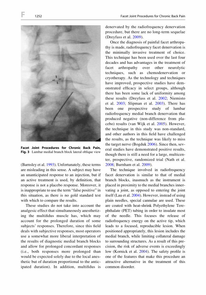
Fluoroscopically guided diagnostic medial
branch blocks anesthetize the facet joint selec-tively and are used to provide prognostic infor-
mation for radiofrequency medial branch
denervation. They are not intended for prolongedanalgesia. Generally, a two-block paradigm is
used: one injection of short-acting local
anesthetic and one of long-acting anesthetic(Lord et al. 1995). When performed correctly,
these blocks have high specificity for anesthetiz-
ing the facet joint (Dreyfuss et al. 1997)(Figs. 2 and 3). Some authors have attempted to
use ultrasound as guidance for this procedure,
and in ideal settings, it is possible that theremay be a role. However, in clinical practice,
body habitus and the inability to detect intravas-
cular injections or perform injections at morethan one site at a time will remain significant
barriers (Rauch et al. 2009).
There has been debate regarding the exactinterpretation of these blocks, fueled by the prob-
lems inherent in attempting to make an objective
diagnosis in a subjective disorder (i.e., pain).Much of this debate has focused on the test
characteristics of the procedure and uses termssuch as “placebo response” and “false positive”
Facet Joint Procedures for Chronic Back Pain,Fig. 2 Lumbar medial branch block AP view with local-ized spread of contrast medium
Facet Joint Procedures for Chronic Back Pain,Fig. 1 Illustration of right posterior view of lumbosacralspine showing key right posterior neural structures. L2through S1 spinous processes labeled. Right:MB1 medialbranch of L1 dorsal primary ramus, NR2 L2 nerve root,DPR2 L2 dorsal primary ramus, LB2 lateral branch of L2dorsal primary ramus, TP3 L3 transverse process, NR3 L3nerve root,MBmedial branch of L3 dorsal primary ramusthat extends around the base of the right superior articularprocess (S) of L4 and innervates portions of the right L3–4and L4–5 facet joint capsules, NR4 L4 nerve root, IC iliaccrest, DPRL5 L5 dorsal primary ramus, DPRS1 S1 dorsalprimary ramus, I inferior articular process L3, S superiorarticular process of L4. Left: FJ L2–3 facet(zygapophysial) joint, which is innervated by branchesof L1 and L2 medial branch nerves, IAB inferior articularbranches from medial branch of L4 dorsal primary ramus,SAB superior articular branches from medial branch of L4dorsal primary ramus (FromCzervionke and Fenton 2003)
Facet Joint Procedures for Chronic Back Pain 1251 F
F
(Barnsley et al. 1993). Unfortunately, these terms
are misleading in this sense. A subject may have
an unanticipated response to an injection, but ifan active treatment is used, by definition, that
response is not a placebo response. Moreover, it
is inappropriate to use the term “false positive” inthis situation, as there is no gold standard test
with which to compare the results.
These studies do not take into account theanalgesic effect that simultaneously anesthetiz-
ing the multifidus muscle has, which may
account for the prolonged duration of somesubjects’ responses. Therefore, since this field
deals with subjective responses, most operators
use a somewhat more liberal interpretation ofthe results of diagnostic medial branch blocks
and allow for prolonged concordant responses
(i.e., both responses more prolonged thanwould be expected solely due to the local anes-
thetic but of duration proportional to the antic-ipated duration). In addition, multifidus is
denervated by the radiofrequency denervationprocedure, but there are no long-term sequelae
(Dreyfuss et al. 2009).Once the diagnosis of painful facet arthropa-
thy is made, radiofrequency facet denervation is
the minimally invasive treatment of choice.This technique has been used over the last four
decades and has advantages in the treatment of
facet arthropathy over other neurolytictechniques, such as chemodenervation or
cryotherapy. As the technology and techniques
have improved, prospective studies have dem-onstrated efficacy in select groups, although
there has been some lack of uniformity among
these results (Dreyfuss et al. 2002; Niemistoet al. 2003; Slipman et al. 2003). There has
been one prospective study of lumbar
radiofrequency medial branch denervation thatproduced negative (non-difference from pla-
cebo) results (van Wijk et al. 2005). However,
the technique in this study was non-standard,and other authors in this field have challenged
the results, as the technique was likely to miss
the target nerve (Bogduk 2006). Since then, sev-eral studies have demonstrated positive results,
though there is still a need for a large, multicen-
ter, prospective, randomized trial (Nath et al.2008; Burnham et al. 2009).
The technique involved in radiofrequency
facet denervation is similar to that of medialbranch blocks, inasmuch as the instrument is
placed in proximity to the medial branches inner-
vating a joint, as opposed to entering the jointitself (Lau et al. 2004). However, instead of using
plain needles, special cannulae are used. These
are coated with heat-shrink Polyethylene Tere-phthalate (PET) tubing in order to insulate most
of the needle. This focuses the release of
radiofrequency energy on the active tip, whichleads to a focused, reproducible lesion. When
positioned appropriately, this lesion includes the
medial branch, while limiting collateral damageto surrounding structures. As a result of this pre-
cision, the risk of adverse events is exceedingly
low (Kornick et al. 2004). The safety profile isone of the features that make this procedure an
attractive alternative in the treatment of this
common disorder.
Facet Joint Procedures for Chronic Back Pain,Fig. 3 Lumbar medial branch block lateral oblique view
F 1252 Facet Joint Procedures for Chronic Back Pain
The desired outcome in this procedure is thefocal denervation of the joints in which the
patient’s back pain was relieved upon perfor-mance of diagnostic medial branch blocks. This
does not treat the underlying arthropathy but
reduces the painful limitation to mobility that itcauses. The nature of radiofrequency denervation
does allow for regrowth of the medial branch
nerve. Therefore, the procedure may requirerepetitive treatments over time. Longer-term
cohorts and studies of repeated intervention
indicate that the clinical results of the procedurelast approximately 12 months, which is consis-
tent with a period of denervation, followed
by regrowth of the medial branch nerve(Tome-Bermejo et al. 2011; Rambaransingh
et al. 2010). Since the denervation procedure
does not address comorbidities, such asmyofascial pain, post-denervation physical ther-
apy may be used to extend the benefits of the
procedure, to include relief of myofascial painand associated loss of range of motion.
Due to the increasing use of this procedure in
the United States (Friedly et al. 2007), insurershave sought to limit its availability by placing
stringent requirements on patients with
facetogenic pain prior to approvingradiofrequency medial branch denervation
(Noridian Local Coverage Determination; United
Health Care Medical Policy). While the datasupporting this procedure are imperfect, there is
only one significant negative study, which used
a nonstandard technique. The weight of evidencefor the positive symptomatic and functional
effects of radiofrequency medial branch denerva-
tion is far greater than for most analgesic inter-ventional procedures. It remains to be seen
whether this useful, safe procedure will continue
to be available to those who most benefit fromits use.
References
Barnsley, L., Lord, S., Wallis, B., et al. (1993). False-positive rates of cervical zygapophysial joint blocks.The Clinical Journal of Pain, 9, 124–130.
Barnsley, L., Lord, S. M., Wallis, B. J., et al. (1994). Lackof effect of intraarticular corticosteroids for chronic
pain in the cervical zygapophyseal joints. The NewEngland Journal of Medicine, 330, 1047–1050.
Bogduk, N. (2006). Lumbar radiofrequency neurotomy.The Clinical Journal of Pain, 22(4), 409.
Burnham, R. S. MSc, MD, FRCPC, Holitski, S. BScPT, &Dinu, I. PhD. (2009). A prospective outcome study onthe effects of facet joint radiofrequency denervation onpain, analgesic intake, disability, satisfaction, cost, andemployment. Archives of Physical Medicine andRehabilitation, 90, 201–205.
Carette, S., Marcoux, S., Truchon, R., et al. (1991).A controlled trial of corticosteroid injectionsinto facet joints for chronic low back pain.The New England Journal of Medicine, 325,1002–1007.
Czervionke, L. F., & Fenton, D. S. (2003). Facet jointinjection and medial branch block. In L. F. Czervionke& D. S. Fenton (Eds.), Image-guided spine interven-tion. Philadelphia: WB Saunders.
Dreyfuss, P., & Dreyer, S. J. (2003). Lumbarzygapophysial (facet) joint injections. The SpineJournal, 3, 50S–59S.
Dreyfuss, P., Schwarzer, A. C., Lau, P., et al. (1997).Specificity of lumbar medial branch and L5 dorsalramus blocks. A computed tomography study. Spine,22, 895–902.
Dreyfuss, P., Halbrook, B., Pauza, K., et al. (2000).Efficacy and validity of radiofrequency neurotomyfor chronic lumbar zygapophysial joint pain. Spine,25, 1270–1277.
Dreyfuss, P., Baker, R., Leclaire, R., et al. (2002).Radiofrequency facet joint denervation in the treat-ment of low back pain: A placebo-controlled clin-ical trial to assess efficacy. Spine, 27, 556–557.
Dreyfuss, P. MD, Stout, A. DO, Aprill, C. MD, Pollei, S.MD, Johnson, B. MD, & Bogduk, N. MD, PhD.(2009). The significance of multifidus atrophy afterradiofrequency neurotomy for low back pain suc-cessful radiofrequency neurotomy for low backpain. Physical Medicine and Rehabilitation, 1,719–722.
Friedly, J. MD, Chan, L. MD, MPH, & Deyo, R. MD,MPH. (2007). Increases in lumbosacral injections inthe medicare population 1994 to 2001. Spine, 32(16),1754–1760.
Kornick, C., Kramarich, S. S., Lamer, T. J., et al. (2004).Complications of lumbar facet radiofrequency dener-vation. Spine, 29, 1352–1354.
Lau, P., Mercer, S., Govind, J., et al. (2004). The surgicalanatomy of lumbar medial branch neurotomy (facetdenervation). Pain Medicine, 5, 289–298.
Local Coverage Determination (LCD) for paravertebralfacet nerve blockade (L30813). www.cms.gov.Accessed 1 Feb 2013.
Lord, S. M., Barnsley, L., & Bogduk, N. (1995). Theutility of comparative local anesthetic blocks versusplacebo-controlled blocks for the diagnosis of cervicalzygapophysial joint pain. The Clinical Journal of Pain,11, 208–213.
Facet Joint Procedures for Chronic Back Pain 1253 F
F

Morinaga, T., Takahashi, K., Yamagata, M., et al.(1996). Sensory innervation to the anteriorportion of lumbar intervertebral disc. Spine, 21,1848–1851.
Nath, S. MD, FRCA, Nath, C. A. SRN, & Pettersson, K.MD, PhD. (2008). Percutaneous lumbar zygapophysial(facet) joint neurotomy using radiofrequency current,in the management of chronic low backpain a randomized double-blind trial. Spine, 33(12),1291–1297.
Niemisto, L., Kalso, E., Malmivaara, A., et al. (2003).Radiofrequency denervation for neck and back pain:A systematic review within the framework of theCochrane Collaboration Back Review Group. Spine,28, 1877–1888.
Rambaransingh, B. BSc, MD, Stanford, G. DC{, &Burnham, R. MSc, MD, FRCPC. (2010). The effectof repeated zygapophysial joint radiofrequencyneurotomy on pain, disability, and improvement dura-tion. Pain Medicine, 11, 1343–1347.
Rauch, S. MD, Kasuya, Y. MD, Turan, A. MD, Neamtu,A. MD, Vinayakan, A. MD, Sessler, D. I. MD, et al.(2009). Ultrasound-guided lumbar medial branchblock in obese patients a fluoroscopically confirmedclinical feasibility study. Regional Anesthesia andPain Medicine, 34(4), 340–342.
Slipman, C. W., Bhat, A. L., Gilchrist, R. V., et al. (2003).A critical review of the evidence for the use ofzygapophysial injections and radiofrequency denerva-tion in the treatment of low back pain. The SpineJournal, 3, 310–316.
Tome-Bermejo, F. MD, Barriga-Martin, A. PhD, & Mar-tin, J. L. R. PhD. (2011). Identifying patients withchronic low back pain likely to benefit from lumbarfacet radiofrequency denervation. Journal of SpinalDisorders & Techniques, 24, 69–75.
United Healthcare Medical Policy. Ablative treatment forspinal pain policy number: 2013T0107M. https://www.unitedhealthcareonline.com. Accessed 8 May2013.
van Kleef, M., Barendse, G. A. M., Kessels, A., et al.(1999). Randomized trial of radiofrequency lumbarfacet denervation for chronic low back pain. Spine,24, 1937–1942.
Van Wijk, R. M. A. W. MD, PhD, Geurts, J. W. M. MD,PhD, Wynne, H. J. PhD, Hammink, E. MD, Buskens,E. MD, PhD, Lousberg, R. PhD, et al. (2005).Radiofrequency denervation of lumbar facet joints inthe treatment of chronic low back pain a randomized,double-blind, sham lesion-controlled trial. TheClinical Journal of Pain, 21(4), 335–344.
Facet Rhizolysis
▶ Facet Joint Procedures for Chronic Back Pain
Facet Syndrome
▶ Facet Joint Pain
Facial Ganglion Neuralgia
▶Geniculate Neuralgia
Facial Pain
Definition
Facial pain is identified by its location, usually
excluding tic douloureux.
Cross-References
▶ Pain Treatment, Motor Cortex Stimulation
Facial Pain Associated withDisorders of the Cranium
▶Headache from Cranial Bone
Facilitative Tucking
Definition
A caregiver uses his/her hands to swaddle aninfant by placing a hand on the infant’s head
and feet while providing flexion and
containment.
Cross-References
▶Acute Pain Management in Infants
F 1254 Facet Rhizolysis

Factor Loading
Definition
Factor analysis is a statistical procedure that
groups together variables that share common var-iance. Variables that “load” on the same factor
are presumed to reflect a similar underlying
process.
Cross-References
▶ Psychology of Pain, Self-Efficacy
Factors Associated with LowBack Pain
▶Low Back Pain, Epidemiology
Failed Back
Definition
Clinical syndrome characterized by back or lower
extremity pain or both following surgery fordecompression of neural elements in the lower
back.
Cross-References
▶ Pain Treatment: Spinal Cord Stimulation
Failed Back Surgery Syndrome
Definition
Failed back surgery syndrome is axial or radicu-
lar pain persisting after surgical approaches to
relieve the pain. It is also known as failed backsyndrome.
Cross-References
▶Central Nervous System Stimulation for Pain
▶Dorsal Root Ganglionectomy and Dorsal
Rhizotomy
False Affirmative Rate
Definition
False affirmative rate is the probability of
response “A” when event B has occurred.
Familial Adenomatous Polyposis
Synonyms
FAP
Definition
An inherited disease which is characterized by
the formation of numerous polyps on the inside
walls of the colon and rectum. The FAP disease isassociated with a 100 % risk for developing colo-
rectal cancer.
Cross-References
▶NSAIDs and Cancer
Familial Amyloid Polyneuropathy(FAP)
▶Hereditary Neuropathies
Familial Amyloid Polyneuropathy (FAP) 1255 F
F
Familial Factors
▶ Impact of Familial Factors on Children’sChronic Pain
Familial Hemiplegic Migraine
Definition
Familial hemiplegic migraine is an inherited
form of migraine with aura in which patients
experience weakness and other neurological dis-turbances as their aura.
Cross-References
▶Migraine, Pathophysiology
Familial Polyposis Coli
Definition
People with this syndrome have massive numbersof colonic polyps and almost invariably develop
cancer of the colon.
Cross-References
▶NSAIDs and Their Indications
Family Environment
▶ Impact of Familial Factors on Children’sChronic Pain
Family-Centered Care
Lisa M. PetersDepartment of Anesthesiology and Pain
Medicine, Seattle Children’s Hospital, Seattle,
WA, USA
Synonyms
Family-centered theory; Patient-centered care
Definition
What Is Family-Centered Care?The practice of pediatrics focuses on the health
of a child. The experience of pediatric care, how-ever, is not isolated to the child; the experience
impacts the whole family. In fact, a bidirectional
relationship exists whereby efforts to provideholistic care to a child necessarily involve
accounting for relevant contextual factors.
Context includes elements internal to the child,such as developmental level, as well as external
to the child, such as familial influences. The
method and degree to which this relationship isembraced in the care setting is the focus of an
approach called family-centered care.While the central tenants of family-centered
care may be practiced to varying degrees around
the world, most of the published literature on thistopic has originated from the United States
and United Kingdom in the last two decades.
Definitions articulate an approach to decisionmaking and information sharing, captured most
powerfully in one word, partnership. Together,
healthcare practitioners, patient, and familynavigate each step along the care pathway with
intention and respect.
A framework for family-centered care inpediatrics, originally described by Shelton in
1987, includes the following nine elements:
• Recognizing the family as a constant in thechild’s life
F 1256 Familial Factors

• Facilitating parent-professional collaboration
at all levels of health care• Honoring the racial, ethnic, cultural, and
socioeconomic diversity of families
• Recognizing family strengths and individualityand respecting different methods of coping
• Sharing complete and unbiased informationwith families on a continuous basis
• Encouraging and facilitating family-to-family
support and networking• Responding to child and family developmen-
tal needs as part of healthcare practices
• Adopting policies and practices that providefamilies with emotional and financial
support
• Designing health care that is flexible, cultur-ally competent, and responsive to family
needs
According to the Institute for Family-Centered Care, established in 1992 in the USA,
the summary themes include: respect and dignity;
information sharing; participation; and collabo-ration (Committee on Hospital Care. American
Academy of Pediatrics & Institute for Patient-
and Family-Centered Care, 2003).
Characteristics
Pediatric Pain and Family-Centered CarePain in children is complex. Yet, the fundamen-tal human right to pain care drives the field to
further scientific study and the individual prac-
titioner to prevent and alleviate children’s pain.From parents’ perspective, data show that the
top two (among 36) issues of priority for their
children admitted to an acute care setting were“Find out what is wrong with my child” and
“taking care of my child’s pain if it is relevant.”
And yet, pain presented the greatest discrepancybetween priority and level of satisfaction
(Ammentorp 2005). Through the alignment of
science and humanity, the greatest possibleimpact on the short- and long-term conse-
quences of poorly treated or untreated pain in
children can be realized. Therein lays the
synergy of a family-centered care approach toimprove a child’s experience with pain;
a partnership of evidence-based health care andthe expertise of self-report or parents’ knowl-
edge of their child.
Clinical: A Call to Action
A commitment to partner along the clinical care
continuum particularly illustrates the powerfulopportunity, including assessment, planning,
intervention, and evaluation. First, the corner-
stone of effective pain care is assessment. Evolu-tion of study has led to an iterative set of
developmental-age specific tools to assess the
intensity of children’s pain. However, healthcarepractitioners are not treating an intensity number,
they are treating a person. Thus, it becomes
essential to engage a parent to share and observebehavioral cues noticed in their child, or honor an
adolescent’s self-report of the quality, location,
duration, and aggravating and relieving factors(Reid et al. 1995). From the outset, it must be
noted that communication is foundational and
continuous (Garland and Kenny 2006). Thus,eliminating language disparity via routine and
readily accessible interpreter services is critical
(Guerrero et al. 2010). This exchange informsplanning for pain care and draws upon the critical
component of respect. The well-recognized, pos-
itive effects of an interdisciplinary approach topain care match the collaborative model of fam-
ily-centered care (Huth et al. 2003; Shields et al.
2006; Simons et al. 2001). This is an opportunityto welcome and optimize a family’s strengths and
an individual child’s coping style. The impor-
tance of optimizing patient and parent participa-tion and coping has been clearly demonstrated,
such as in a cohort of adolescents with sickle cell
disease (Mitchell 2007). A partnership to shareinformation allows for unbiased presentation of
evidence-based interventions and nonjudgmental
discussion. It becomes essential to recognize thatall parties may hold different elements of the care
at a premium, including evaluation of the risk and
benefit of the pain experience itself, as well as theinterventions and goals.
Family-Centered Care 1257 F
F

Education: Empowering AdvocatesEfforts to enhance awareness and knowledge of
children’s pain through education can give new
voice to all. Education offers a link to close thegap between the prevalence of undertreated or
untreated pain in children and the lack oftimely, effective interventions to prevent or
alleviate that pain. In its 2011 publication titled
“Relieving Pain in America,” the Institute ofMedicine cites education as a foundational
endeavor to transform care (IOM 2011). The
aim of educational activities may focus onexpectations and current evidence of pain care
to inform a shared mental model, yet impor-
tantly also focus on the benefit of each mem-ber’s individual expertise. In other words, to
recognize that a healthcare provider has the
responsibility to contribute expertise in currentevidence-based treatment options, while
a parent has the responsibility to contribute
expertise in their child’s context and currentexperience. Such messaging again emphasizes
communication as key; there is support for any-
one to speak up and advocate on behalf ofa child, including truly creating space for and
hearing the voice of a parent.
Research: Data to Improve Outcomes
In recent years, further collaboration between
the scientific community and the healthcareconsumer offers another example of, and
opportunity for, a powerful partnership
(MacKean et al. 2005). For example, the Patient-Centered Outcomes Research Institute (PCORI) is
an independent, nonprofit organization in the USA
created to contribute specifically to shared,informed decision making by patients, families,
and healthcare providers. Patients play a central
role to define research priorities, contribute to thedirection of clinical queries, and gain access to
results. Another example is the Patient Reported
Outcomes Measurement Information System(PROMIS), which is funded by the US National
Institutes of Health (NIH). These measures
become the tools of scientific study to inform theeffectiveness of interventions and have already
invested in pain as a focal area. Specifically inpediatrics, this entity has made available validated
tools to assess pain interference and intensity, aswell as focusing on areas of physical function,
emotional distress, fatigue, and peer relationships.
Thus, research offers an arena not only for patientand parent participation, but also for collaboration
to define priorities for study and new forums in
which to share results.
What Do the Data Show with Regard toFamily-Centered Care?A systematic review (Shields et al. 2008)
underscored the paucity of solid quantitative
research on the effectiveness of family-centeredcare. Qualitative studies, and subsequent quanti-
tative efforts, have shown the impact of family-
centered care to (Bamm and Rosenbaum 2008;Kuo 2012):
• Improve patient outcomes
• Improve patient and family satisfaction• Improve healthcare professionals’ satisfaction
• Decrease cost
• Improve utilization of resources in health careIn line with these results, leading professional
and safety organizations have endorsed family-
centered care. In the USA, for example, theInstitute of Medicine (IOM) identified family-
centered care as one of the six attributes of
high-quality health care (IOM 1999, 2001), andthe Joint Commission incorporated a bill of rights
that included patient comfort and that active
involvement of patients and families is a strongstrategy to ensure patient safety.
Summary
The synergy between family-centered care andthe practice of pediatric pain medicine is unde-
niable. The data available assert its value to the
extent that it is sanctioned as a primary safetyand quality strategy. Yet, the challenge remains
in the translation of a philosophy in approach to
bedside practice. For some healthcare settingsthis would present a complete interdisciplinary
F 1258 Family-Centered Care

paradigm shift, while at the same time offera tremendous opportunity for culture change
in treating pain in children. It is possible.First, the system must define the professional
standards, set expectations for behavior, and
create the space for a rich partnership. Then,the system must maintain accountability to
that model, including individual practitioner
responsibility. In time, a culture will emergein which the partnership of family-centered
care will transform the experience of a child
in pain.
References
Ammentorp, J., Mainz, J., & Sabroe, S. (2005). Parents’priorities and satisfaction with acute pediatric care.Archives of Pediatric and Adolescent Medicine,159(2), 127–131.
Bamm, E. L., & Rosenbaum, P. (2008). Family-centeredtheory: Origins, development, barriers, and supports toimplementation in rehabilitation medicine. Archivesof Physical Medicine and Rehabilitation, 89(8),1618–1624.
Committee on Hospital Care. American Academy ofPediatrics & Institute for Patient- and Family-Centered Care. (2003). Family-centered care andthe pediatrician’s role. Pediatrics, 112(3 Pt 1),691–697.
Garland, L., & Kenny, G. (2006). Family nursing and themanagement of pain in children. Paediatric Nursing,18(6), 18–21.
Guerrero, A. D., et al. (2010). Racial and ethnic disparitiesin pediatric experiences of family centered care. Med-ical Care, 48(4), 388–393.
Huth, M. M., Broome, M. E., Mussatto, K. A., & Morgan,S. W. (2003). A study of the effectiveness of a painmanagement education booklet for parents of childrenhaving cardiac surgery. Pain Management Nursing,4(1), 31–39.
Institute for Family-Centered Care. Available at: www.familycenteredcare.org. Accessed 11 Apr 2012.
Institute of Medicine. (1999). To err is human: Buildinga safer health system. Washington, DC: NationalAcademies Press.
Institute of Medicine. (2001). Crossing the quality chasm:A new health system for the 21st Century. Washington,DC: National AcademiesPress.
Institute of Medicine. (2011). Relieving pain in America:A blueprint for transforming prevention, care, educa-tion, and research. Washington, DC: NationalAcademiesPress.
Kuo, D. (2012). Family-centered care: Current applica-tions and future directions in pediatric health care.Maternal and child health journal, (1092–7875),16 (2), 297.
MacKean, G. L., Thurston, W. E., & Scott, C. M. (2005).Bridging the divide between families and health pro-fessionals’ perspectives on family-centered care.Health Expectations, 8, 74–85.
Mitchell, M. J., Lemanek, K., Palermo, T. M., Crosby,L. E., Nichols, A., & Powers, S. W. (2007). Parentperspectives on pain management, coping, and familyfunctioning in pediatric sickle cell disease. ClinicalPediatrics, 46(4), 311–319.
Patient Reported Outcomes Measurement InformationSystem. Available at: www.nihpromis.org. Accessed11 Apr 2012.
Patient-Centered Outcomes Research Institute. Availableat: www.pcori.org. Accessed 11 Apr 2012.
Reid, G. J., Hebb, J. P., McGrath, P. J., Finley, G. A., &Forward, S. P. (1995). Cues parents use to assesspostoperative pain in their children. The Clinical Jour-nal of Pain, 11(3), 229–235.
Shields, L., Pratt, J., & Hunter, J. (2006). Family-centredcare: A review of qualitative studies. Journal ofClinical Nursing, 15(10), 1317–1323.
Shields, L., Pratt, J., Davis, L. M., & Hunter, J. (2008).Family-centred care for children in hospital. CochraneDatabase of Systematic Reviews, (1), CD004811.
Simons, J.,Franck,L.,&Roberson,E. (2001).Parent involve-ment in children’s pain care: Views of parents and nurses.Journal of Advanced Nursing, 36(4), 591–599.
Family-Centered Care, Approachand Basis
Definition
The American Academy of Pediatrics Committee
on Hospital Care and the Institute forFamily-Centered Care issued a joint policy state-
ment in which they defined terms as follows: “Fam-
ily-centered care is an approach to health care thatshapes health care policies, programs, facility
design, and day-to-day interactions among patients,
families, physicians, and other health care profes-sionals.” Further, “Family-centered care in pediat-
rics is based on the understanding that the family is
the child’s primary source of strength and supportand that the child’s and family’s perspectives and
information are important in clinical decision
making.”
Family-Centered Care, Approach and Basis 1259 F
F

References
American Academy of Pediatrics. (2003). Pediatrics, 112,691–696 (http://www.umassmed.edu/uploadedFiles/shriver/education/LEND/Courses/Pediatrics_FCC_and_Peds_Role_2003.pdf)
Family-Centered Care, Objective
Definition
Caredirectedat improving thehealthandwell-being
ofthefamilyanditsmembersbyassessingthefamily
health needs and identifying potential obstacles.
Cross-References
▶Chronic Pain in Children, Physical Medicine
and Rehabilitation
Family-centered Theory
▶ Family-Centered Care
FAP
▶ Familial Adenomatous Polyposis
Fascia Iliaca Compartment Block
Definition
Injection via needle or catheter of local anesthetic
deep to the fascia lata and iliaca medial to theanterior superior iliac spine and inferior to the
inguinal ligament.
Cross-References
▶Acute Pain in Children, Postoperative
Fasciculus Cuneatus
Definition
The lateral bundle of nerves in the dorsal column
referred to as the cuneate fasciculus, which ter-minates in the cuneate nucleus just off the dorsal
midline in the caudal medulla.
Cross-References
▶ Postsynaptic Dorsal Column Projection,
Anatomical Organization
Fasciculus Gracilis
Definition
The medial bundle nerves in the dorsal column
referred to as the fasciculus gracilis, which ter-minates in the gracile nucleus in the dorsal mid-
line of the caudal medulla.
Cross-References
▶ Postsynaptic Dorsal Column Projection,
Anatomical Organization
Fast-Track Surgery
▶ Postoperative Pain, Importance of
Mobilization
F 1260 Family-Centered Care, Objective
Fatigue
Definition
Fatigue is a decrement of response seen with
repeated stimulation and is a prominent attributeof nociceptors and other primary afferents.
Cross-References
▶ Pain in Humans, Electrical Stimulation (Skin,Muscle, and Viscera)
▶ Polymodal Nociceptors, Heat Transduction
FCA
▶ Freund’s Complete Adjuvant
FCA-Induced Arthritis
Definition
This is the same as CFA (Complete Freund’sAdjuvant)-induced Arthritis. An experimental
model of inducing rheumatoid arthritis byinjecting a suspension of killed mycobacteria
(Freund’s complete adjuvant, FCA) into various
tissues in rats. The classical model involves injec-tion of high doses into the tail base which pro-
duces multiple joint arthritis (polyarthritis)
accompanied by wide-spread lesions of skin andother organs. A modified method uses low-dose
local injections around a joint to produce unilat-
eral single joint arthritis.
Cross-References
▶Opioids and Inflammatory Pain
FCE
▶ Functional Capacity Evaluation
Fear and Pain
Kim Helsen1,3, Maaike Leeuw2 andJohan W. S. Vlaeyen4,5
1Research Group on Health Psychology,
KU Leuven Katholieke Universiteit Leuven,Leuven, Belgium2Department of Medical, Clinical and
Experimental Psychology, MaastrichtUniversity, Maastricht, The Netherlands3Department of Experimental–Clinical and
Health Psychology, Ghent University, Ghent,Belgium4Research Group Health Psychology, Catholic
University of Leuven, Leuven, Belgium5Department of Clinical Psychological Science,
Maastricht University, Maastricht,
The Netherlands
Synonyms
Fear of movement/(re)injury; Kinesiophobia;Pain-related anxiety; Pain-related fear
Definition
▶ Fear of pain is a general term used to describeseveral forms of fear with respect to pain.
Depending on the anticipated source of threat,
the content of fear of pain varies considerably.For example, fear of pain can be directed toward
the occurrence or continuation of pain, toward
physical activity, or toward the induction of (re)injury or physical harm. A more specific fear of
pain concerns ▶ fear of movement/(re)injury,
which is the specific fear that physicalactivity will cause (re)injury. Synonymously,
▶ kinesiophobia is defined as “an excessive,
Fear and Pain 1261 F
F

irrational, and debilitating fear of physical move-ment and activity resulting from a feeling of
vulnerability to painful injury or re-injury”(Kori et al. 1990; Lundberg et al. 2006).
Characteristics
In recent years, chronic pain has no longer beenconceptualized as purely a medical problem but
rather as a complex biopsychosocial phenome-
non in which the relationship among impair-ments, pain, and disability is weak. In chronic
pain patients, anxiety disorders frequently co-
occur, indicating that patients with persistentmusculoskeletal pain fear a variety of situations
that are not essentially related to pain
(Asmundson and Katz 2009; Asmundson et al.2004; Gatchel et al. 2007). Besides the finding
that chronic pain patients seem to suffer more
frequently from anxiety symptoms, fear and anx-iety are often an integral part of the chronic pain
problem. The experience of pain can be charac-
terized by psychophysiological (e.g., musclereactivity), cognitive (e.g., worry), and behav-
ioral (e.g., escape and avoidance) responses,
showing similarities with responses regardingfear and anxiety (Leeuw et al. 2007; Vlaeyen
and Linton 2000).
There are multiple pathways by which pain-related fear mediates disability, namely, through
escape and avoidance behaviors, through inter-
ference with cognitive functioning, throughreduced opportunities to correct the erroneous
underlying cognitions guiding the avoidance
behaviors, and through detrimental effects oflong-lasting avoidance on various physiological
systems (Crombez et al. 1999; Gheldof et al.
2006; Turk and Wilson 2010). Empirical findingssupport the notion that fear of pain is a significant
contributor to the chronification and maintenance
of chronic pain syndromes (Hirsh et al. 2008; Turkand Wilson 2010; Vlaeyen and Linton 2000).
Fear and AnxietyIn the literature describing fear of pain, the con-
cepts of fear and anxiety are often used inter-
changeably. Despite the fact that these concepts
are substantially related, some differences can bedistinguished (Asmundson et al. 2004; Leeuw
et al. 2007; Sylvers et al. 2011).Fear is the emotional expression of the fight-
flight response, which is the immediate readiness
of the body to respond to an event that is per-ceived as dangerous or threatening. Fear is there-
fore a present-oriented state that is designed to
protect the individual from the perceived imme-diate threat. ▶Anxiety, however, is a cognitive-
affective state that is rather future oriented. It
tends to occur in the anticipation of a dangerousor threatening event and is therefore more indef-
inite and uncertain in nature. Instead of initiating
the fight-flight response as in case of fear, thestate of anxiety seems to facilitate and stimulate
the fight-flight response only in case the threat-
ening event occurs. Both in fear and anxiety,cognitive, physiological, and behavioral dimen-
sions of responses can be distinguished. Physio-
logically, fear and anxiety responses arecharacterized by the activation of the sympathetic
nervous system, designed to increase the likeli-
hood of survival by promoting escape from orprotection against the perceived threat. In anxi-
ety, these physiological responses are less present
than in fear. The cognitive element, relativelymore present in anxiety, is more narrowed in
anxiety and directed in such a way that a source
of threat, when present, will be detected. Fear, onthe other hand, comprises thoughts of danger,
threat, or death, through which the attention
toward the threat is advanced, while irrelevantdistracters are ignored and the initiation of action
is stimulated. On a behavioral level, anxiety
guides motivation to engage in preventative andavoidance behaviors, while fear motivates to
engage in defensive behaviors. Despite the defi-
nition of pain-related fear, both fear and anxietyare distinct processes that contribute significantly
to chronic pain (Asmundson et al. 2004).
Hierarchy of FearBesides the important distinction between fear
and anxiety in chronic pain, understandingabout the hierarchical nature of fear and anxiety
is also an important consideration. The hierarchi-
cal structure of anxiety is displayed in Fig. 1.
F 1262 Fear and Pain
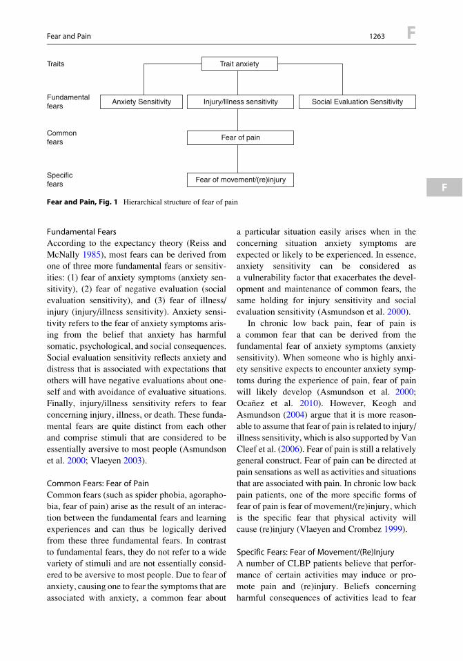
Fundamental Fears
According to the expectancy theory (Reiss and
McNally 1985), most fears can be derived fromone of three more fundamental fears or sensitiv-
ities: (1) fear of anxiety symptoms (anxiety sen-
sitivity), (2) fear of negative evaluation (socialevaluation sensitivity), and (3) fear of illness/
injury (injury/illness sensitivity). Anxiety sensi-
tivity refers to the fear of anxiety symptoms aris-ing from the belief that anxiety has harmful
somatic, psychological, and social consequences.
Social evaluation sensitivity reflects anxiety anddistress that is associated with expectations that
others will have negative evaluations about one-
self and with avoidance of evaluative situations.Finally, injury/illness sensitivity refers to fear
concerning injury, illness, or death. These funda-
mental fears are quite distinct from each otherand comprise stimuli that are considered to be
essentially aversive to most people (Asmundson
et al. 2000; Vlaeyen 2003).
Common Fears: Fear of Pain
Common fears (such as spider phobia, agorapho-bia, fear of pain) arise as the result of an interac-
tion between the fundamental fears and learning
experiences and can thus be logically derivedfrom these three fundamental fears. In contrast
to fundamental fears, they do not refer to a wide
variety of stimuli and are not essentially consid-ered to be aversive to most people. Due to fear of
anxiety, causing one to fear the symptoms that are
associated with anxiety, a common fear about
a particular situation easily arises when in the
concerning situation anxiety symptoms are
expected or likely to be experienced. In essence,anxiety sensitivity can be considered as
a vulnerability factor that exacerbates the devel-
opment and maintenance of common fears, thesame holding for injury sensitivity and social
evaluation sensitivity (Asmundson et al. 2000).
In chronic low back pain, fear of pain isa common fear that can be derived from the
fundamental fear of anxiety symptoms (anxiety
sensitivity). When someone who is highly anxi-ety sensitive expects to encounter anxiety symp-
toms during the experience of pain, fear of pain
will likely develop (Asmundson et al. 2000;Ocanez et al. 2010). However, Keogh and
Asmundson (2004) argue that it is more reason-
able to assume that fear of pain is related to injury/illness sensitivity, which is also supported by Van
Cleef et al. (2006). Fear of pain is still a relatively
general construct. Fear of pain can be directed atpain sensations as well as activities and situations
that are associated with pain. In chronic low back
pain patients, one of the more specific forms offear of pain is fear of movement/(re)injury, which
is the specific fear that physical activity will
cause (re)injury (Vlaeyen and Crombez 1999).
Specific Fears: Fear of Movement/(Re)Injury
A number of CLBP patients believe that perfor-mance of certain activities may induce or pro-
mote pain and (re)injury. Beliefs concerningharmful consequences of activities lead to fear
Trait anxiety
Anxiety Sensitivity
Traits
Fundamentalfears
Commonfears
Specificfears
Social Evaluation SensitivityInjury/lllness sensitivity
Fear of pain
Fear of movement/(re)injury
Fear and Pain, Fig. 1 Hierarchical structure of fear of pain
Fear and Pain 1263 F
F

of movement/(re)injury and consequently to theavoidance of these activities, although medical
indications for this behavioral pattern of avoid-
ance are lacking. Despite the fact that in acutepain the avoidance of daily activities may be
adaptive in facilitating healing and recovery,
avoidance behavior is no longer necessary forrecovery in chronic pain (Leeuw et al. 2007;
Vlaeyen and Linton 2000).
Cognitive Behavioral ModelsA cognitive behavioral model of chronic low
back pain has been proposed, which emphasizesthe crucial importance of the role of fear of
movement/(re)injury and avoidance behavior in
chronic low back pain patients (Vlaeyen 2003;Vlaeyen and Linton 2000). According to the
model, two opposing behavioral responses may
occur in response to acute pain: “confrontation”and “avoidance.” A gradual confrontation and
resumption of daily activities despite pain is con-
sidered as an adaptive response that eventuallyleads to the reduction of fear, the encouragement
of physical recovery, and functional rehabilita-
tion. In contrast, a catastrophic interpretation ofpain is considered to be a maladaptive response,
which initiates a vicious circle in which fear
of movement/(re)injury and the subsequentavoidance of activities augment functional
disability and the pain experience by means
of hypervigilance, depression, and disuse.Substantial support for this cognitive behavioral
model and the role of the specific fear ofmovement/(re)injury has been found (Leeuw
et al. 2007; Vlaeyen and Linton 2000).
In addition to this cognitive behavioral model,Asmundson et al. (2004) propose to update
the model by integrating the concept of anxiety
in addition to fear, referring to this as thefear-anxiety-avoidance model (Fig. 2).
This model states that catastrophizing about
pain produces fear of pain, designed to protect theindividual from the perceived immediate threat.
This fear of pain in turn might promote pain-
related anxiety. Pain-producing stimuli resultthrough pain-related fear in escape and protecting
behaviors aimed at reducing pain intensity. These
behaviors in turn strengthen erroneous beliefsabout pain, increase catastrophizing, and further
enhance pain-related fear. The addition of an
anxiety-related pathway to the pathway of pain-related fear provides a more accurate explanation
for the fact that chronic pain interferes with one’s
daily life. In the anticipation, rather than in thepresence of pain and/or injury, anxiety is evoked,
leading to an increased attention (hypervigilance)
for evidence of potential pain or injury. Thishypervigilance and psychical responses may
interact with memories and may promote mis-
interpretations of harmless stimuli as impendingdanger of pain or injury. Behaviorally, anxiety
results in avoidance and preventative behaviors,
increasing disability and disuse (Asmundsonet al. 2004).
DisuseDisability
Depression
Catastrophizing
Injury/Strain
Recovery
Exposure
Low fear
Pain experience
Avoidance
Escape
Threat perception
Hypervigilance
Painanxiety
Fearof pain
Defensive
motivation
Preventative
motivation
Arou
sal
Arou
sal
Fear and Pain,Fig. 2 Fear-anxiety-avoidance model of chronicpain (Adapted from thecognitive behavioral modelof Vlaeyen and Linton(2000) and the fear-anxiety-avoidance modelof Asmundson et al. (2004))
F 1264 Fear and Pain

Recently, the sequential relationships betweenchanges in pain catastrophizing, subsequent fear
and avoidance, and functional outcomes such asdisability or return to work were examined using
a prospective design (Wideman et al. 2009).
Although overall changes in pain catastrophizingand fear of movement were predictive for return
to work, no evidence was found that early
changes in catastrophizing were associated withlate changes in fear of movement. These findings
suggest that both components might indepen-
dently influence disability. At present, new cog-nitive behavioral models regarding pain-related
fear are evolving, focussing on behavior in con-
text. Fear of pain might result in diverse behav-iors depending on the current goal context, and
likewise, seemingly similar behaviors may be
driven by dissimilar motivational strategies(Van Damme et al. 2008; Vlaeyen et al. 2009).
Other Objects of Fear in PainMorley and Eccleston (2004) propose the exis-
tence of a range of “feared objects” in chronic
pain, because of the overwhelming threat value ofpain and three associated capacities to (1) inter-
rupt, (2) interfere, and (3) impact on one’s iden-
tity. Many potential fears arise because of theability of pain to threaten the whole range of
a person’s existence. Interruption is established
because the immediate pain experience interruptsbehavior and influences the person’s cognitive
functioning (e.g., thoughts about possible harm).
Interference is visible in the diminished accom-plishment of daily functional activities. Finally,
when repeated interference occurs to a degree
that it concerns major goals, a threat to the iden-tity is instigated. As chronic pain interferes with
current tasks, plans, and goals, the person’s per-
spective of oneself is changed, both with respectto the future and the past. Fear and anxiety are
likely to occur when the goals and the identity of
a person are threatened (Morley 2008).
Assessment of Fear of PainSeveral measurements of fear of pain are available(for an overview see McNeil and Vowles 2004).
Anxiety sensitivity can be measured by the 16-itemAnxiety Sensitivity Index (ASI) (Peterson and
Reiss 1987), measuring the degree to which peopleare concerned about the possible negative conse-
quences of anxiety symptoms. Injury/illness sensi-
tivity can be measured with the correspondingsubscale of the sensitivity index (Taylor 1993).
Fear of pain can be measured by, for example, the
Pain Anxiety Symptoms Scale (PASS)(McCracken and Dhingra 2002; McCracken et al.
1993), designed to assess pain-specific fearful
appraisals, cognitive symptoms of anxiety, physio-logical symptoms of anxiety, and escape and avoid-
ance behavior. Another way to assess pain-related
fear is by means of the Fear of Pain Questionnaire(FPQ), a self-report measure utilized in clinical as
well as in nonclinical populations (McNeil and
Rainwater 1998). Fear of movement/(re)injury canbest be measured with the 17-item Tampa Scale for
Kinesiophobia (TSK; Kori et al. 1990).
Treatment ImplicationsDue to the inextricable binding between fear and
(chronic) pain, treatment of chronic pain shouldaim to focus on these perpetuating factors. In graded
exposure in vivo, a hierarchy of fearful activities is
established, which leads to disconfirmation of painbeliefs and reduction of fear, thereby promoting
recovery of activities and functional abilities
(Vlaeyen et al. 2004; Woods and Asmundson2008). Fear- and anxiety-focused treatments seem
toprovidepromisingresults inchronic lowbackpain
patients (Bailey et al. 2010; Lohnberg 2007).
References
Asmundson,G.J.G.,&Katz, J. (2009).Understanding theco-occurrence of anxiety disorders and chronic pain: State-of-the-art.Depression and Anxiety, 26(10), 888–901.
Asmundson, G. J., Wright, K. D., & Hadjistavropoulos,H. D. (2000). Anxiety sensitivity and disabling chronichealth conditions: State of the arts and future direc-tions. Scandinavian Journal of Behaviour Therapy,29, 100–117.
Asmundson, G. J., Norton, P. J., & Vlaeyen, J. W. S.(2004). Fear-avoidance models of chronic pain: Anoverview. In G. J. G. Asmundson, J. W. S. Vlaeyen,
Fear and Pain 1265 F
F

&G. Crombez (Eds.),Understanding and treating fearof pain. Oxford: Oxford University Press.
Bailey, K. M., Carleton, R. N., Vlaeyen, J. W. S., &Asmundson, G. J. G. (2010). Treatments addressingpain-related fear and anxiety in patients with chronicmusculoskeletal pain: A preliminary review.CognitiveBehaviour Therapy, 39(1), 46–63.
Crombez, G., Vlaeyen, J. W. S., Heuts, P. H. T. G., et al.(1999). Pain-related fear is more disabling than painitself: Evidence on the role of pain-related fear inchronic back pain disability. Pain, 80, 329–339.
Gatchel, R. G., Peng, Y. B., Peters, M. L., Fuchs, P. N., &Turk, D. C. (2007). The biopsychosocial approach tochronic pain: Scientific advances and future directions.Psychological Bulletin, 133(4), 581–624.
Gheldof, E. L. M., Vinck, J., Van den Bussche, E.,Vlaeyen, J. W. S., Hidding, A., & Crombez, G.(2006). Pain and pain-related fear are associated withfunctional and social disability in an occupational set-ting: Evidence of mediation by pain-related fear. Euro-pean Journal of Pain, 10(6), 513–525.
Hirsh, A. T., George, S. Z., Bialosky, J. E., & Robinson,M. E. (2008). Fear of pain, pain catastrophizing,and acute pain perception: Relative prediction andtiming of assessment. The Journal of Pain, 9(9),806–812.
Keogh, E., & Asmundson, G. J. G. (2004). Negative affec-tivity, catastrophizing, and anxiety sensitivity. InG. J. G. Asmundson, J. W. S. Vlaeyen, & G. Crombez(Eds.), Understanding and treating fear of pain.Oxford: Oxford University Press.
Kori, S. H., Miller, R. P., & Todd, D. D. (1990).Kinesiophobia: A new view of chronic pain behavior.Pain Manage, 35–43.
Leeuw, M., Goossens, M. E. J. B., Linton, S. J., Crombez,G., Boersma, K., & Vlaeyen, J. W. S. (2007). The fear-avoidance model of musculoskeletal pain: Currentstate of scientific evidence. Journal of BehavioralMedicine, 30(1), 77–94.
Lohnberg, J. A. (2007). A review of outcome studies oncognitive behavioral therapy for reducing fear-avoidance beliefs among individuals with chronicpain. Journal of Clinical Psychology in Medical Set-tings, 14, 113–122.
Lundberg, M., Larsson, M., Ostlund, H., & Styf, J. (2006).Kinesiophobia among patients with musculoskeletalpain in primary healthcare. Journal of RehabilitationMedicine, 38, 37–43.
McCracken, L. M., & Dhingra, L. (2002). A short versionof the pain anxiety symptoms scale (PASS-20): Pre-liminary development and validity. Pain Research &Management, 7(1), 45–50.
McCracken, L. M., Zayfert, C., & Gross, R. T. (1993). Thepain anxiety symptoms scale (PASS):A multidimensional measure of pain-specific anxietysymptoms. Behavior Therapist, 16, 183–184.
McNeil, D. W., & Rainwater, A. J. (1998). Developmentof the fear of pain questionnaire-III. Journal of Behav-ioral Medicine, 21(4), 389–410.
McNeil, D. W., & Vowles, K. E. (2004). Assessment offear and anxiety associated with pain: Conceptua-lisation, methods, and measures. In G. J. G.Asmundson, J. W. S. Vlaeyen, & G. Crombez (Eds.),Understanding and treating fear of pain. Oxford:Oxford University Press.
Morley, S. (2008). Psychology of pain. British Journal ofAnaesthesia, 101(1), 25–31.
Morley, S., & Eccleston, C. (2004). The object of fearin pain. In G. J. G. Asmundson, J. W. S. Vlaeyen, &G. Crombez (Eds.),Understanding and treating fear ofpain. Oxford: Oxford University Press.
Ocanez, K. L. S., McHugh, R. K., & Otto, M. W. (2010).A meta-analytic review of the association betweenanxiety sensitivity and pain. Depression and Anxiety,27, 760–767.
Peterson, R. A., & Reiss, S. (1987). Anxiety sensitivityindex manual. Palos Heights: International DiagnosticSystems.
Reiss, S., & McNally, R. J. (1985). The expectancymodel of fear. In S. Reis & R. R. Bootzin (Eds.),Theoretical issues in behavior therapy. New York:Academic.
Sylvers, P., Lilienfeld, S. O., & LaPrairie, J. L. (2011).Differences between trait fear and trait anxiety: Impli-cations for psychopathology. Clinical PsychologyReview, 31(1), 122–137.
Taylor, S. (1993). The structure of fundamental fears.Journal of Behavior Therapy and Experimental Psy-chiatry, 24, 289–299.
Turk, D. C., & Wilson, H. D. (2010). Fear of pain asa prognostic factor in chronic pain: Conceptualmodels, assessment, and treatment implications. Cur-rent Pain and Headache Reports, 14(2), 88–95.
Van Cleef, L. M. G., Peters, M. L., Roelofs, J., &Asmundson, G. J. G. (2006). Do fundamentalfears differentially contribute to pain-related fearand pain catastrophizing? An evaluation of thesensitivity index. European Journal of Pain, 10(6),527–536.
Van Damme, S., Crombez, G., & Eccleston, C. (2008).Coping with pain: A motivational perspective. Pain,139, 1–4.
Vlaeyen, J. W. S. (2003). Fear in musculoskeletal pain. InO. Dostrovsky, D. B. Carr, & M. Koltzenburg (Eds.),Proceedings of the 10th World Congress on pain,progress in pain research and management (Vol. 24,pp. 631–650). Seattle: IASP Press.
Vlaeyen, J. W. S., & Crombez, G. (1999). Fear of move-ment/(re)injury, avoidance and pain disability inchronic low back pain patients. Manual Therapy,4(4), 187–195.
Vlaeyen, J. W. S., & Linton, S. J. (2000). Fear-avoidanceand its consequences in chronic musculoskeletal pain:A state of the art. Pain, 85, 317–332.
Vlaeyen, J. W. S., de Jong, J. R., Leeuw, M., & Crombez,G. (2004). Fear reduction in chronic pain: Gradedexposure in vivo with behavioral experiments. InG. J. G. Asmundson, P. J. Norton, G. R. Norton,
F 1266 Fear and Pain

J. W. S. Vlaeyen, & G. Crombez (Eds.), Understand-ing and treating fear of pain (pp. 313–343). Oxford:Oxford University Press.
Vlaeyen, J. W. S., Hanssen, M., Goubert, L., Vervoort, T.,Peters, M. L., Van Breukelen, G., et al. (2009). Threatof pain influences social context effects on verbal painreport and facial expression. Behaviour Research andTherapy, 47(9), 774–782.
Wideman, T. H., Adams, H., & Sullivan, M. J. L. (2009).A prospective sequential analysis of the fear-avoidance model of pain. Pain, 145, 45–51.
Woods, M. P., & Asmundson, G. J. G. (2008). Evaluatingthe efficacy of graded in vivo exposure for the treat-ment of fear in patients with chronic back pain:A randomized controlled clinical trial. Pain, 136,271–280.
Fear Avoidance
Definition
Fear avoidance is the avoidance of activities inorder to prevent injury, reinjury, or exacerbation
of any injury or pain. It is a learned behavior to
reduce negative pain-related emotions. In thisway, pain-related fear can lead to disability,
catastrophizing beliefs, hypervigilance to bodily
signals, and avoidance behavior.
Cross-References
▶Disability, Fear of Movement
▶ Pain in the Workplace, Risk Factors forChronicity, and Psychosocial Factors
Fear Avoidance Beliefs
Definition
Fear avoidance beliefs are cognitions often foundin individuals with pain, which support avoid-
ance behavior and make participation in physical
activity programs more difficult. Patients believethat physical activity initiated their pain and that
physical activity is bound to aggravate the pain in
the long run.
Cross-References
▶Disability
▶ Fear Avoidance▶ Pain
▶ Psychological Treatment of Pain in Older
Populations
Fear of Movement/(Re)Injury
Definition
Fear related to movement and physical activity,
inextricably associated with the fear that physical
activity will cause pain and (re)injury. Fear ofmovement is most prominent in patients with
chronic pain syndromes.
Cross-References
▶Disability, Fear of Movement
▶ Fear and Pain
Fear of Pain
Definition
Pain-related fear is a general term to describe
several forms of fear with respect to pain.
Depending on the anticipated source of threat,the content of fear of pain varies considerably.
Fear of pain can be directed toward the occur-rence or continuation of pain, toward physical
activity, or toward the induction of (re)injury or
physical harm.
Cross-References
▶Disability, Fear of Movement
▶ Fear and Pain▶Muscle Pain, Fear-Avoidance Model
Fear of Pain 1267 F
F
Fear Reduction Through ExposureIn Vivo
Jeroen R. De Jong1 and Johan W. S. Vlaeyen2,3
1Department of Rehabilitation, University
Hospital Maastricht, Maastricht,The Netherlands2Research Group Health Psychology, Catholic
University of Leuven, Leuven, Belgium3Department of Clinical Psychological Science,
Maastricht University, Maastricht,
The Netherlands
Synonyms
Exposure in vivo; Exposure treatment; Extinc-
tion; Graded exposure; Graded exposure in vivowith behavioral experiments
Definition
Exposure in vivo, originally based on ▶ extinc-tion of ▶ Pavlovian conditioning (Bouton 1988),
is currently viewed as a cognitive process during
which fear is activated and catastrophic expecta-tions are being challenged and disconfirmed,
resulting in reduction of the threat value of theoriginally fearful stimuli. During graded expo-
sure, special attention goes to the establishment
of an individual hierarchy of the ▶ pain-relatedfear stimuli. Exposure in vivo includes activities
that are selected based on the fear hierarchy and
the idiosyncratic aspects of the fear stimuli.
Characteristics
Inorder toproducedisconfirmationsbetweenexpec-
tations of pain and harm, the actual pain, and theother consequences of the activity or movement,
Philips (1987) was one of the first to argue for
repeated, graded, and controlled exposures to suchsituations. Experimental support for this idea is pro-
vided by the match-mismatch model of pain
(RachmanandArntz 1991),which states that people
initially tend tooverpredict howmuchpain theywillexperience, but after some exposures these predic-
tions tend to be corrected to match with the actualexperience.Crombez et al. (2002) andGoubert et al.
(2002) found a similar pattern in a sample of chronic
low back pain patients, who were requested toperform certain physical activities. As predicted,
the chronic low back pain patients initially
overpredicted pain, but after repetition of the activ-ity, the overprediction was readily corrected.
Overpredictions of pain and the negative
consequences of pain are more pronounced in indi-viduals reporting increasedfearofpain.Anumberof
studies examined the effectiveness of a graded
▶ exposure in vivo treatment in reducing pain-related fear, ▶pain catastrophizing, and pain dis-
ability in chronic pain patients who were referred
for outpatient behavioral rehabilitation (Vlaeyenet al. 2002a, b, c; Vlaeyen et al. 2001; de Jong
et al. 2005a, b, 2008, 2012; Leeuw et al. 2008;
Linton et al. 2008; Woods and Asmundson 2008).Theses showed improvements in pain-related fear,
pain catastrophizing, and pain disability whenever
the graded exposure was initiated. Measured withambulatory activity monitors, the improvements
also generalized to increases in physical activity
in the home situation (Vlaeyen et al. 2002a).Besides behavioral and cognitive changes, one
study in patients with ▶Complex Regional Pain
Syndrome (CRPS) even found observed positivechanges in edema, skin color, excessive sweating,
and motor function disturbances (de Jong et al.
2005a). It seems that graded exposure to painfulactivities, movements, and/or situations that were
previously avoided may indeed be a successful
treatment approach for pain patients reportingsubstantial pain-related fear.
Graded Exposure In VivoWe suggest that the intervention generally be
designed in three steps: cognitive-behavioral
assessment, education, and exposure in vivowith ▶ behavioral experiments.
Cognitive-Behavioral AssessmentSpecific Questionnaires A basic question that
may be asked is as follows: “What is the nature of
the perceived threat?” The answer is not as simple
F 1268 Fear Reduction Through Exposure In Vivo
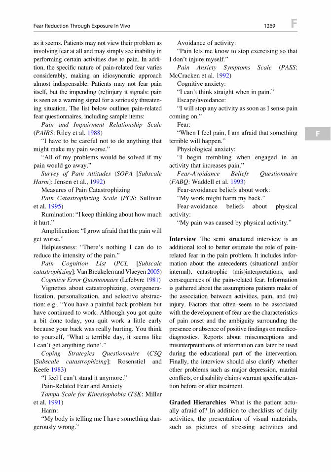
as it seems. Patients may not view their problem asinvolving fear at all and may simply see inability in
performing certain activities due to pain. In addi-tion, the specific nature of pain-related fear varies
considerably, making an idiosyncratic approach
almost indispensable. Patients may not fear painitself, but the impending (re)injury it signals: pain
is seen as a warning signal for a seriously threaten-
ing situation. The list below outlines pain-relatedfear questionnaires, including sample items:
Pain and Impairment Relationship Scale(PAIRS: Riley et al. 1988)
“I have to be careful not to do anything that
might make my pain worse.”
“All of my problems would be solved if mypain would go away.”
Survey of Pain Attitudes (SOPA [SubscaleHarm]: Jensen et al., 1992)
Measures of Pain Catastrophizing
Pain Catastrophizing Scale (PCS: Sullivan
et al. 1995)Rumination: “I keep thinking about howmuch
it hurt.”
Amplification: “I grow afraid that the pain willget worse.”
Helplessness: “There’s nothing I can do to
reduce the intensity of the pain.”Pain Cognition List (PCL [Subscale
catastrophizing]: VanBreukelen andVlaeyen 2005)Cognitive Error Questionnaire (Lefebvre 1981)Vignettes about catastrophizing, overgenera-
lization, personalization, and selective abstrac-
tion: e.g., “You have a painful back problem buthave continued to work. Although you got quite
a bit done today, you quit work a little early
because your back was really hurting. You thinkto yourself, ‘What a terrible day, it seems like
I can’t get anything done’.”
Coping Strategies Questionnaire (CSQ[Subscale catastrophizing]: Rosenstiel and
Keefe 1983)
“I feel I can’t stand it anymore.”Pain-Related Fear and Anxiety
Tampa Scale for Kinesiophobia (TSK: Miller
et al. 1991)Harm:
“My body is telling me I have something dan-
gerously wrong.”
Avoidance of activity:“Pain lets me know to stop exercising so that
I don’t injure myself.”Pain Anxiety Symptoms Scale (PASS:
McCracken et al. 1992)
Cognitive anxiety:“I can’t think straight when in pain.”
Escape/avoidance:
“I will stop any activity as soon as I sense paincoming on.”
Fear:
“When I feel pain, I am afraid that somethingterrible will happen.”
Physiological anxiety:
“I begin trembling when engaged in anactivity that increases pain.”
Fear-Avoidance Beliefs Questionnaire(FABQ: Waddell et al. 1993)
Fear-avoidance beliefs about work:
“My work might harm my back.”
Fear-avoidance beliefs about physicalactivity:
“My pain was caused by physical activity.”
Interview The semi structured interview is an
additional tool to better estimate the role of pain-
related fear in the pain problem. It includes infor-mation about the antecedents (situational and/or
internal), catastrophic (mis)interpretations, and
consequences of the pain-related fear. Informationis gathered about the assumptions patients make of
the association between activities, pain, and (re)
injury. Factors that often seem to be associatedwith the development of fear are the characteristics
of pain onset and the ambiguity surrounding the
presence or absence of positive findings onmedico-diagnostics. Reports about misconceptions and
misinterpretations of information can later be used
during the educational part of the intervention.Finally, the interview should also clarify whether
other problems such as major depression, marital
conflicts, or disability claims warrant specific atten-tion before or after treatment.
Graded Hierarchies What is the patient actu-ally afraid of? In addition to checklists of daily
activities, the presentation of visual materials,
such as pictures of stressing activities and
Fear Reduction Through Exposure In Vivo 1269 F
F

movements reflecting the full range of situationsavoided by the patient, can be quite helpful in the
development of graded hierarchies. They startwith activities or situations that provoke only
mild discomfort and end with those that are
beyond the patient’s present abilities. The Photo-graph Series of Daily Activities uses photographs
representing various physical daily activities to
be placed along a fear thermometer (Dubberset al. 2003; Jelinek et al. 2003; Kugler et al.
1999; Leeuw et al. 2007).
Behavioral Tests Sometimes, patients find it
hard to really estimate the harmfulness of an
activity when it has been avoided extensively.For cases in which even pictorial methods
(PHODA) do not work, behavioral tests can be
introduced. These consist of performing anactivity that has been avoided previously, while
performance indices (e.g., time, distance, number
of repetitions) are measured. Target behaviorscan be derived from PHODA items, and in most
cases the behavioral tests can be considered as
a variant of the exercise tolerance test describedby Fordyce (Fordyce 1976).
EducationOne of themajor goals of the educational section is
to increase the willingness of patients to finally
engage in activities they avoided for a long time.The aim is to correct the misinterpretations and
misconceptions that occurred early on during the
development of the pain-related fear. The educa-tional section is more than just reassuring that there
are no specific physical abnormalities. Unambigu-
ously educating the patient in a way that the patientviews his or her pain as a common condition that
can be self-managed, rather than a serious disease
or condition that needs careful protection, isa useful first step. It can be explained to patients
that they may have probably overestimated the
value of diagnostic tests and that in symptom-freepeople similar abnormalities can also be found.
Graded Exposure In Vivo with BehavioralExperiments
Graded Exposure In Vivo As firsthand
evidence of actually experiencing oneselves
behaving differently is far more convincing thanrational argument, the most essential step
consists of graded exposure to the situations thefearful patient has identified as “dangerous” or
“threatening.” The patient is encouraged to
engage in these fearful activities as much aspossible, until disconfirmation has occurred and
anxiety levels have decreased. If the rating has
decreased significantly, the therapist mayconsider moving on to the next item of the hier-
archy. Each activity or movement is first modeled
by the therapist. His presence, initially acting asa safety signal to promote more exposures, is
gradually withdrawn to facilitate independence
and to create contexts that mimic those of thehome situation (Vlaeyen et al. 2012).
Behavioral Experiments The graded exposureto fear-eliciting activities can be carried out in the
form of a behavioral experiment in which
a collaborative empiricism is the bottom line.The essence of a behavioral experiment is that
the patient performs an activity to challenge the
validity of his catastrophic assumptions andmisinterpretations. These assumptions take the
form of “If . . . then . . .” statements and are
empirically tested in such a behavioralexperiment.
Generalization and Maintenance of ChangeExposure to physical activities is not likely to
result in a fundamental change in the belief of
the pain patient that certain movements areharmful or painful (Goubert et al. 2002). More
likely, the patient will learn that the movements
involved in the exposure treatment are lessharmful or painful than anticipated. In other
words, during successful exposure, exceptions
to the rule are learned, rather than there beinga fundamental change of that rule. Generaliza-
tion and maintenance can be enhanced by the
following measures. First, exposure to the fullspectrum of contexts and natural settings in
which fear has been experienced is required.
Second, during the exposure, it is best that stim-uli be varied as much as possible. Third,
expanded-spaced, rather than a massed expo-
sure, is preferred (Vlaeyen et al. 2012).
F 1270 Fear Reduction Through Exposure In Vivo
References
Bouton, M. E. (1988). Context and ambiguity in theextinction of emotional learning: Implications forexposure therapy. Behaviour Research and Therapy,26, 137–149.
Crombez, G., Eccleston, C., Vlaeyen, J. W.,Vansteenwegen, D., Lysens, R., & Eelen, P. (2002).Exposure to physical movements in low back painpatients: Restricted effects of generalization. HealthPsychology, 21, 573–578.
de Jong, J. R., Vlaeyen, J.W. S., Onghena, P., Cuypers, C.,den Hollander, M., & Ruygrok, J. (2005a). Reductionof pain-related fear in complex regional pain syndrometype I: The application of graded exposure in vivo.Pain, 116, 264–275.
de Jong, J. R., Vlaeyen, J. W., Onghena, P., Goossens,M. E., Geilen, M., & Mulder, M. (2005b). Fear ofmovement/(Re) injury in chronic low back pain: Edu-cation or exposure In Vivo as mediator to fear reduc-tion? The Clinical Journal of Pain, 21, 9–17.
de Jong, J. R., Vangronsveld, K., Peters, M. L., Goossens,M. E., Onghena, P., Bulte, I., et al. (2008). Reductionof pain-related fear and disability in post-traumaticneck pain: A replicated single-case experimentalstudy of exposure in vivo. The Journal of Pain,9(12), 1123–1134.
de Jong, J. R., Vlaeyen, J. W. S., van Eijsden, M., Loo, C.,& Onghena, P. (2012). Reduction of pain-related fearand increased function and participation in work-related upper extremity pain (WRUEP): Effects ofexposure in vivo. Pain, 10, 2109–2118.
Dubbers,A.T.,Vikstrom,M.H.,&DeJong, J.R. (2003).ThePhotographseriesofDailyActivities (PHODA):Cervicalspine and shoulder [CD-ROM version 1.2]. The Nether-lands:HogeschoolZuyd,UniversityMaastricht and Insti-tute for Rehabilitation Research (iRv).
Fordyce, W. E. (1976). Behavioral methods for chronicpain and illness. St. Louis: Mosby.
Goubert, L., Francken, G., Crombez, G., Vansteenwegen,D., & Lysens, R. (2002). Exposure to physical move-ment in chronic back pain patients: No evidence forgeneralization across different movements. BehaviourResearch and Therapy, 40, 415–429.
Jelinek, S., Germes, D., Leyckes, N., & De Jong, J. R.(2003). The Photograph Series of Daily Activities(PHODA): Low extremities [CD-ROM Version 1.2].The Netherlands: Hogeschool Zuyd, UniversityMaastricht and Institute for Rehabilitation Research(iRv).
Jensen, M. P., & Karoly, P. (1992). Pain-specific beliefs,perceived symptom severity, and adjustmentto chronic pain. The Clinical Journal of Pain, 8,123–130.
Kugler, K., Wijn, J., Geilen, M., De Jong, J. R., &Vlaeyen, J. W. S. (1999). The Photograph series ofDaily Activities (PHODA) [CD-ROM version 1.0].The Netherlands: Institute for Rehabilitation Researchand School for Physiotherapy Heerlen.
Leeuw, M., Goossens, M. E. J. B., van Breukelen, G.,Boersma, K., & Vlaeyen, J. W. S. (2007). Measuringperceived harmfulness of physical activities in patientswith chronic low back pain: The photograph series ofdaily activities [Short electronic version]. The Journalof Pain, 8, 840–849.
Leeuw, M., Goossens, M. E. J. B., van Breukelen, G. J. P.,de Jong, J. R., Heuts, P. H. T. G., Smeets, R. J. E. M.,et al. (2008). Exposure in vivo versus operantgraded activity in chronic low back pain patients:Results of a randomized controlled trial. Pain, 138,192–207.
Lefebvre, M. F. (1981). Cognitive distortion and cognitiveerrors in depressed psychiatric and low back painpatients. Journal of Consulting and ClinicalPsychology, 49, 517–525.
Linton, S. J., Boersma, K., Jansson, M., Overmeer, T.,Lindblom, K., & Vlaeyen, J. W. (2008). A randomizedcontrolled trial of exposure in vivo for patients withspinal pain reporting fear of work-related activities.European Journal of Pain, 12, 722–730.
McCracken, L. M., Zayfert, C., & Gross, R. T. (1992). ThePain Anxiety Symptoms Scale: Development valida-tion of a scale to measure fear of pain. Pain, 50, 67–73.
Miller, R. P., Kori, S. H., & Todd, D. D. (1991). TheTampa Scale for Kinisophobia. Unpublished Report,Tampa, FL.
Philips, H. C. (1987). Avoidance behaviour and its role insustaining chronic pain. Behaviour Research andTherapy, 25, 273–279.
Rachman, S., & Arntz, A. R. (1991). The overpredictionand underprediction of pain. Clinical PsychologyReview, 11, 339–355.
Riley, J. F., Ahern, D. K., & Follick, M. J. (1988). Chronicpain and functional impairment: Assessing beliefsabout their relationship. Archives of Physical Medicineand Rehabilitation, 69, 579–582.
Rosenstiel, A. K., & Keefe, F. J. (1983). The use of copingstrategies in chronic low back pain. Pain, 17, 33–44.
Sullivan, M. J., Bishop, S. R., & Pivik, J. (1995). The paincatastrophizing scale: Development and validation.Psychological Assessment, 7, 524–532.
Van Breukelen, G. J., & Vlaeyen, J. W. (2005). Normingclinical questionnaires with multiple regression:The pain cognition list. Psychological Assessment,17, 336–444.
Vlaeyen, J. W. S., De Jong, J. R., Geilen, M., Heuts, P. H.T. G., & van Breukelen, G. (2001). Graded exposure invivo in the treatment of pain-related fear: A replicatedsingle-case experimental design in four patients withchronic low back pain. Behaviour Research and Ther-apy, 39, 151–166.
Vlaeyen, J. W., de Jong, J. R., Geilen, M., Heuts, P. H., &van Breukelen, G. (2002a). The treatment of fear ofmovement/(Re) injury in chronic low back pain: Fur-ther evidence on the effectiveness of exposure in vivo.The Clinical Journal of Pain, 18, 251–261.
Vlaeyen, J. W., de Jong, J. R., Onghena, P., Kerckhoffs-Hanssen, M., & Kole-Snijders, A. M. (2002b).
Fear Reduction Through Exposure In Vivo 1271 F
F

Can pain-related fear be reduced? The application ofcognitive-behavioral exposure in vivo. Pain Research& Management, 7, 144–153.
Vlaeyen, J.W., de Jong, J. R., Sieben, J.M., &Crombez, G.(2002c). Graded exposure in vivo for pain-related fear.In D. C. Turk & R. J. Gatchel (Eds.), Psychologicalapproaches to pain management a practitioner’s hand-book (pp. 210–233). New York: Guilford.
Vlaeyen, J. W., Morley, S. J., Linton, S. J., Boersma, K., &de Jong, J. R. (2012). Pain-related fear: Exposure-basedtreatment of chronic pain. Seattle: IASP Press.
Waddell, G., Newton, M., Henderson, I., Somerville, D.,&Main, C. J. (1993). A Fear-Avoidance Beliefs Ques-tionnaire (FABQ) and the role of fear-avoidancebeliefs in chronic low back pain and disability. Pain,52, 157–168.
Woods, M. P., & Asmundson, G. J. G. (2008). Evaluatingthe efficacy of graded in vivo exposure for the treatmentof fear in patients with chronic back pain: A randomizedcontrolled clinical trial. Pain, 136, 271–280.
Fear-Anxiety-Avoidance Model
Definition
The Fear-Anxiety-Avoidance Model states that
catastrophic misinterpretations in response toacute pain can lead to fear of pain and subse-
quently to pain-related anxiety. Fear urges the
escape from the pain stimulus, while anxietyin anticipation of pain or threat urges avoidance
of those situations. As a result of this, a
self-perpetuating cycle develops in whichsubsequent avoidance of activities augments
functional disability and the pain experience
by means of hypervigilance, depression, anddecreased physical fitness, thereby further
advancing chronicity.
Cross-References
▶ Fear and Pain
Fear-Avoidance
▶Disability, Fear of Movement
Fear-Avoidance Model
Definition
The basic tenet of the fear-avoidance model is
that catastrophic misinterpretations in response toacute pain lead to pain-related fear, which subse-
quently urges the escape from the painful stimuli,
and to selectively attend to bodily sensations. Asa result, a self-perpetuating cycle develops in
which subsequent avoidance of activities aug-
ments functional disability, depression, anddecreased physical fitness.
Cross-References
▶ Fear Reduction through Exposure in Vivo▶Muscle Pain, Fear-Avoidance Model
Feasible
Definition
The simple, economic, and easy application of
a measure
Cross-References
▶ Pain Assessment in Neonates
Feedback Control of Pain
Definition
Ascending nociceptive signals activate supra-
spinal mechanisms (e.g., an inhibitory brainstem-spinal pathway) that suppress spinal/trigeminal
nociceptive transmission.
F 1272 Fear-Anxiety-Avoidance Model

Cross-References
▶Descending Modulation and Persistent Pain
Fee-for-Service
Definition
Patients or insurance pay for medical care as it is
needed and according to the service provided.
Cross-References
▶Disability Management in Managed Care
System
Female Reproductive Organ PainModel
▶Visceral Pain Models, Female Reproductive
Organ Pain
Female Sex Steroid Hormonesand Pain
Jennifer Brawn1 and Katy Vincent2
1P.A.I.N. Group, Center for Pain and the Brain,
Boston Children’s Hospital, Harvard MedicalSchool, Waltham, MA, USA2Nuffield Department of Obstetrics and
Gynaecology, The Women’s Centre, JohnRadcliffe Hospital, University of Oxford,
Oxford, UK
Introduction
It widely understood that females are dispropor-
tionately affected by pain syndromes whencompared to their male counterparts. This clinical
skew is notable in conditions such as migraine
headache, temporomandibular joint disorder(TMJD), irritable bowel syndrome (IBS), and
fibromyalgia (Unruh 1996). In addition to thehigher prevalence of females with pain condi-
tions, there have been reports of cyclical patterns
of pain onset and severity (Martin 2009). Thisdisparity in pain experience has resulted in
a significant body of research comparing
gender-specific physiological differences. Mostexperimental pain studies suggest that females
are more sensitive to pain than males, although
there are inconsistencies in the literature (Martin2009; Fillingim et al. 2009). Consequently,
researchers have focused on female biology, spe-
cifically the menstrual cycle, to untangle thisimportant clinical and biological conundrum.
The two main female sex steroid hormones, pro-
gesterone and estradiol, are thought to influencepain processing through alteration of a variety of
neural mechanisms.
Characteristics
Pain Perception in Healthy Females over theMenstrual CycleAnimal models have allowed for improvedmechanistic understanding of the central effects
of progesterone and estradiol; however, differ-
ences in the physiology of the ovarian cyclebetween species causes difficulty in extrapolating
some of these findings to humans. In rodents,
these hormones have been implicated ina variety of pain-related pathways including the
gamma-aminobutyric acid (GABA), glutamate,
opioid, and dopamine systems. Thus, estradiolappears to mediate inflammatory responses,
increase opioidergic tone, and promote reuptake
of glutamate, while progesterone is thought toalter GABAergic inhibitory activity by increas-
ing binding affinity through its metabolite
allopregnanolone. Certainly, in rodents there isa notable cyclical trend toward greater pain
sensitivity in the proestrus phase, a time that is
characterized by dramatic fluctuations of proges-terone and estradiol. However, researchers
have also observed increased sensitivity at other
Female Sex Steroid Hormones and Pain 1273 F
F

points including estrus, metestrus, and diestrus(Reviewed in Martin 2009). In humans, results
are more difficult to interpret. A number of fac-tors contribute to this problem, not least the dif-
ferent definitions and methods of verification of
the menstrual cycle phases (Sherman andLeResche 2006).
Evaluating Menstrual Cycle Changes in PainPerceptionIn a meta-analysis performed by Riley et al.
1999, 16 studies were categorized by stimuliand assessed (Fillingim et al. 2009). For the
cold pressor test, pressure, heat, and ischemic
pain, sensitivity was lowest in the follicularphase, characterized by low hormone levels
with estradiol increasing in the latter half. Elec-
trical pain sensitivity, however, was lowest in theluteal and highest in the periovulatory phases.
Only one of the included studies obtained hor-
monal measures. In a subsequent methodologicalreview, researchers were skeptical of this analysis.
They found inconsistent cyclical patterns in
thermal, mechanical, and ischemic pain percep-tion. The only notable pattern involved electrical
pain with studies reporting decreased sensitivity in
the early luteal phase (Sherman and LeResche2006).
In a recent narrative review of cyclical pain
response in healthy females, Martin 2009 ana-lyzed the results of 19 human studies. To make
findings comparable, the author redefined the
reported cycle phases based on the actual testdates. Observed trends were summarized using
three categories. The author noted that six studies
reported no cyclical changes in pain sensitivity.However, five studies observed that pain sensi-
tivity increased in phases characterized by fluc-
tuating estradiol and increasing progesterone(late follicular and early luteal). Seven studies
found that pain sensitivity increased in phases
characterized by low or decreasing estradiol andprogesterone (late luteal or early follicular). It
should be noted that only four of the studies
analyzed had blood progesterone and estradiollevels to verify menstrual cycle phase. An addi-
tional three studies confirmed ovulation through
biological measures.
Hormonal Measures and Pain PerceptionHormonal assays are key to untangling the
complex relationship between progesterone, estra-
diol, and pain. While countless studies have eval-uated changes in pain perception over the
menstrual cycle, only a small number have
obtained either plasma or serum hormone levels.These measures allow for analysis of pain changes
independent of experimental sessions or some-
what arbitrary divisions of the menstrual cycle(Sherman and LeResche 2006). Fillingim et al.
1997 conducted the first study that used blood-
derived estrogen and progesterone levels to eval-uate changes in pain sensitivity, specifically pain
onset and threshold, over the menstrual cycle.
A significant correlation was observed betweenelevated plasma estrogen levels and low thermal
pain onset and threshold measures. In a study that
examined diffuse noxious inhibitory control(DNIC), within the ovulatory phase, pain-induced
analgesia was positively correlated with progester-
one levels (Tousignant-LaFlamme and Marchand2009). Ring et al. 2009 observed that within the
luteal phase, progesterone, estradiol, and pain
ratings from needle and catheter insertion werepositively correlated. While Stening et al. 2007
used the cold pressor test to measure transition
from mild to moderate pain (activation time) andnoted that shorter activation times were signifi-
cantly correlated with high progesterone levels;as were maximal pain ratings. Estradiol, however,
was not significantly correlated with any of the
results. Stening and colleagues (2007) alsoanalyzed the combined impact of estradiol and
progesterone on pain perception. When present
together at high levels, estradiol and progesteroneappeared to decrease pain sensitivity, with
estradiol mediating progesterone’s apparent
pronociceptive influence. Alternatively, severalstudies have not observed any correlations
between hormone levels and pain measures
(Craft 2007; Klatzkin et al. 2010; Kowalczyket al. 2006).
Menstrual Cycle DisordersDue to their close correlation with hormonal
changes, several pain studies have evaluated
females with cyclical medical conditions.
F 1274 Female Sex Steroid Hormones and Pain

However, a major difficulty with this strategy isdisentangling hormonal influences on pain per-
ception per se from influences on the pathologycausing the symptoms. Dysmenorrhea is
a menstrual cycle condition characterized by
extreme abdominal and back pain during menses.It has been speculated that low or decreasing
progesterone levels may trigger dysmenorrheic
symptoms (Martin 2009). In a clinical study,Granot et al. 2001 compared heat and laser pain
thresholds in dysmenorrheic women and healthy
females over four experimental sessions.Dysmenorrheic women were more sensitive to
pain from both stimuli. For the laser stimuli, both
groups appeared to have the lowest pain sensitivityin the luteal phase (high plasma progesterone and
estradiol) and the highest during the follicular
phase (increasing plasma estradiol). Cycle phase,however, did not affect dysmenorrheic women’s
response to the heat stimuli. Similarly, Vincent
and colleagues (2011) demonstrated increasedsensitivity to noxious heat in dysmenorrheic
women compared to pain-free controls, but found
no influence of cycle phase in either group. In thisstudy, the timing of the three experimental
sessions was chosen to maximize endogenous
variation in levels of estradiol and progesterone.Premenstrual dysphoric disorder (PMDD) is
a luteal phase condition characterized by extreme
physical and emotional changes. While theunderlying pathology of the condition remains
unclear, it has been postulated that women with
PMDD may have abnormal levels of sex steroidhormones and/or altered neurotransmitter regula-
tion by these hormones. In support of this, two
studies have shown luteal phase estradiol andprogesterone levels to be higher in women with
PMDD than controls (Straneva et al. 2002;
Epperson et al. 2002). Interestingly, althoughthere was no difference in cortical GABA levels
between the groups, GABA levels decreased
across the cycle in healthy women but increasedin those with PMDD, with the steroid hormones
appearing to exert opposing effects in the two
groups (e.g., a positive correlation betweenGABA and both estradiol and progesterone in
the women with PMDD and a negative correla-
tion in the control women) (Epperson et al. 2002).
PMDD sufferers demonstrated higher pain andunpleasantness ratings in response to noxious
stimuli in the luteal compared to the follicularphase, and significantly lower pain tolerance
and threshold measures than healthy controls in
all menstrual cycle phases (Straneva et al. 2002).
Chronic Pain Conditions and HormonalTreatmentGiven the prevalence of pain conditions in the
female population, studies have focused on both
evaluating differences in experimental painresponse and deconstructing patterns of increased
pain. Chronic pain conditions such as fibromyal-
gia, temporomandibular joint disorder (TMJD),and migraine appear to worsen at times of low or
rapidly decreasing estradiol (LeResche et al.
2003), while symptoms from IBS, interstitialcystitis/painful bladder syndrome, and migraine
as well as a variety of gynecological pain
conditions can be improved by pharmacologicalmanipulation of hormonal status (Mathias et al.
1994; Lentz et al. 2002; Silberstein and Goldberg
2007). Once again, effects of hormones on theunderlying pathologies cannot be excluded;
however, Sherman et al. 2005 did find that
females with TMJD were more sensitiveto experimental pain during the mid-luteal and
menstrual phases.
Neuroimaging StudiesNeuroimaging technology has added an interest-
ing dimension to the field (reviewed by Vincentand Tracey 2010). Studies of pain perception in
healthy subjects suggest alterations in the cere-
bral response to noxious stimuli in differinghormonal states, although the findings from
individual studies are not entirely consistent
(Choi et al. 2006; de Leeuw et al. 2006). Unfor-tunately, to date, no studies have used
neuroimaging techniques to investigate the
relationship between steroid hormones and thecerebral response to pain in patients with
a chronic pain condition. However, studies in
healthy subjects further strengthen the evidencefor interactions between sex hormones and
endogenous pain modulatory systems; most strik-
ingly for estradiol and the mu-opioid system
Female Sex Steroid Hormones and Pain 1275 F
F

(Smith et al. 2006), but also with GABA asdiscussed earlier and potentially other systems
including the descending pain inhibitory system.
Summary
ConclusionDespite the sheer number of studies, much workis needed to develop a cohesive understanding of
individual and combined effects of estradiol and
progesterone on pain perception. As noted bymany reviews, reproducible, standardized meth-
odologies are greatly needed (Martin 2009;
Sherman and LeResche 2006; Riley et al. 1999).The field of pain and hormones is complex, thus it
is important to minimize as much research varia-
tion as possible.
References
Choi, J. C., Park, S. K., Kim, Y. H., Shin, Y. W.,Kwon, J. S., Kim, J. S., et al. (2006). Different brainactivation patterns to pain and pain-related unpleasant-ness during the menstrual cycle. Anesthesiology,105(1), 120–127.
Craft, R. M. (2007). Modulation of pain by estrogens.Pain, 132(Suppl. 1), S3–S12.
de Leeuw, R., Albuquerque, R. J., Andersen, A. H., &Carlson, C. R. (2006). Influence of estrogen on brainactivation during stimulation with painful heat.Journal of Oral and Maxillofacial Surgery: OfficialJournal of the American Association of Oral andMaxillofacial Surgeons, 64(2), 158–166.
Epperson, C. N., Haga, K., et al. (2002). Cortical gamma-aminobutyric acid levels across the menstrual cycle inhealthy women and those with premenstrual dysphoricdisorder: a proton magnetic resonance spectroscopystudy. Archives of General Psychiatry, 59(9), 851–858.
Fillingim, R. B., Maixner, W., Girdler, S. S., Light, K. C.,Harris, M. B., Sheps, D. S., et al. (1997). Ischemic butnot thermal pain sensitivity varies across the menstrualcycle. Psychosomatic Medicine, 59(5), 512–520.
Fillingim, R. B., King, C. D., Ribeiro-Dasilva, M. C.,Rahim-Williams, B., & III Riley, J. L. (2009). Sex,gender, and pain: A review of recent clinical and exper-imental findings. The Journal of Pain, 10(5), 447–485.
Granot,M.,Yarnitsky,D., Itskovitz-Eldor, J.,Granovsky,Y.,Peer, E., & Zimmer, E. Z. (2001). Pain perception inwomen with dysmenorrhea. Obstetrics andGynecology, 98(3), 407–411.
Klatzkin, R. R., Mechlin, B., & Girdler, S. S. (2010).Menstrual cycle phase does not influence gender
differences in experimental pain sensitivity. EuropeanJournal of Pain, 14(1), 77–82.
Kowalczyk, W. J., Evans, S. M., Bisaga, A. M.,Sullivan, M. A., & Comer, S. D. (2006). Sex differ-ences and hormonal influences on response to coldpressor pain in humans. The Journal of Pain: OfficialJournal of the American Pain Society, 7(3), 151–160.
Lentz, G. M., Bavendam, T., Stenchever, M. A.,Miller, J. L., & Smalldridge, J. (2002). Hormonalmanipulation in women with chronic, cyclic irritablebladder symptoms and pelvic pain. American Journalof Obstetrics and Gynecology, 186(6), 1268–1271;discussion 1271–1263.
LeResche, L., Mancl, L., Sherman, J. J., Gandara, B., &Dworkin, S. F. (2003). Changes in temporomandibularpain and other symptoms across the menstrual cycle.Pain, 106(3), 253–261.
Martin, V. T. (2009). Ovarian hormones andpain response: A review of clinical and basic sciencestudies. Gender Medicine, 6(Suppl. 2), 168–192.
Mathias, J. R., Clench, M. H., Roberts, P. H., &Reeves-Darby, V. G. (1994). Effect of leuprolideacetate in patients with functional bowel disease.Long-term follow-up after double-blind, placebo-controlled study. Digestive Diseases and Sciences,39(6), 1163–1170.
Riley, J. L., 3rd, Robinson,M. E.,Wise, E. A., & Price, D. D.(1999). Ameta-analytic review of pain perception acrossthe menstrual cycle. Pain, 81(3), 225–235.
Ring, C., Veldhuijzen van Zanten, J. J., & Kavussanu, M.(2009). Effects of sex, phase of the menstrual cycleand gonadal hormones on pain in healthy humans.Biological Psychology, 81(3), 189–191.
Sherman, J. J., & LeResche, L. (2006). Does experimentalpain response vary across the menstrual cycle?A methodological review. American Journal ofPhysiology. Regulatory, Integrative and ComparativePhysiology, 291(2), R245–R256. doi:10.1152/ajpregu.00920.2005.
Sherman, J. J., LeResche, L., Mancl, L. A., Huggins, K.,Sage, J. C., & Dworkin, S. F. (2005). Cyclic effects onexperimental pain response in women with temporo-mandibular disorders. Journal of Orofacial Pain,19(2), 133–143.
Silberstein, S. D., & Goldberg, J. (2007). Menstruallyrelated migraine: Breaking the cycle in your clinicalpractice. The Journal of Reproductive Medicine,52(10), 888–895.
Smith, Y. R., Stohler, C. S., Nichols, T. E., Bueller, J. A.,Koeppe, R. A., & Zubieta, J. K. (2006). Pronociceptiveand antinociceptive effects of estradiol through endog-enous opioid neurotransmission in women.The Journal of Neuroscience: The Official Journal ofthe Society for Neuroscience, 26(21), 5777–5785.
Stening, K., Eriksson, O., Wahren, L., Berg, G.,Hammar, M., & Blomqvist, A. (2007). Pain sensationsto the cold pressor test in normally menstruatingwomen: Comparison with men and relation to men-strual phase and serum sex steroid levels. American
F 1276 Female Sex Steroid Hormones and Pain
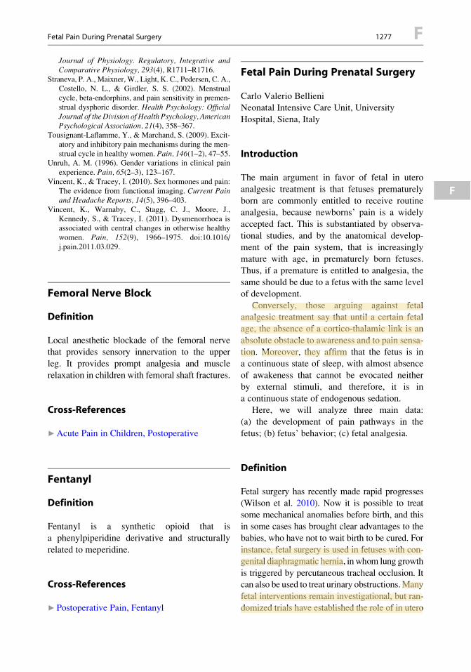
Journal of Physiology. Regulatory, Integrative andComparative Physiology, 293(4), R1711–R1716.
Straneva, P. A., Maixner, W., Light, K. C., Pedersen, C. A.,Costello, N. L., & Girdler, S. S. (2002). Menstrualcycle, beta-endorphins, and pain sensitivity in premen-strual dysphoric disorder. Health Psychology: OfficialJournal of theDivision of Health Psychology, AmericanPsychological Association, 21(4), 358–367.
Tousignant-Laflamme, Y., & Marchand, S. (2009). Excit-atory and inhibitory pain mechanisms during the men-strual cycle in healthy women. Pain, 146(1–2), 47–55.
Unruh, A. M. (1996). Gender variations in clinical painexperience. Pain, 65(2–3), 123–167.
Vincent, K., & Tracey, I. (2010). Sex hormones and pain:The evidence from functional imaging. Current Painand Headache Reports, 14(5), 396–403.
Vincent, K., Warnaby, C., Stagg, C. J., Moore, J.,Kennedy, S., & Tracey, I. (2011). Dysmenorrhoea isassociated with central changes in otherwise healthywomen. Pain, 152(9), 1966–1975. doi:10.1016/j.pain.2011.03.029.
Femoral Nerve Block
Definition
Local anesthetic blockade of the femoral nervethat provides sensory innervation to the upper
leg. It provides prompt analgesia and muscle
relaxation in children with femoral shaft fractures.
Cross-References
▶Acute Pain in Children, Postoperative
Fentanyl
Definition
Fentanyl is a synthetic opioid that is
a phenylpiperidine derivative and structurally
related to meperidine.
Cross-References
▶ Postoperative Pain, Fentanyl
Fetal Pain During Prenatal Surgery
Carlo Valerio BellieniNeonatal Intensive Care Unit, University
Hospital, Siena, Italy
Introduction
The main argument in favor of fetal in utero
analgesic treatment is that fetuses prematurely
born are commonly entitled to receive routineanalgesia, because newborns’ pain is a widely
accepted fact. This is substantiated by observa-
tional studies, and by the anatomical develop-ment of the pain system, that is increasingly
mature with age, in prematurely born fetuses.
Thus, if a premature is entitled to analgesia, thesame should be due to a fetus with the same level
of development.
Conversely, those arguing against fetalanalgesic treatment say that until a certain fetal
age, the absence of a cortico-thalamic link is an
absolute obstacle to awareness and to pain sensa-tion. Moreover, they affirm that the fetus is in
a continuous state of sleep, with almost absence
of awakeness that cannot be evocated neitherby external stimuli, and therefore, it is in
a continuous state of endogenous sedation.Here, we will analyze three main data:
(a) the development of pain pathways in the
fetus; (b) fetus’ behavior; (c) fetal analgesia.
Definition
Fetal surgery has recently made rapid progresses
(Wilson et al. 2010). Now it is possible to treatsome mechanical anomalies before birth, and this
in some cases has brought clear advantages to the
babies, who have not to wait birth to be cured. Forinstance, fetal surgery is used in fetuses with con-
genital diaphragmatic hernia, inwhom lung growth
is triggered by percutaneous tracheal occlusion. Itcan also be used to treat urinary obstructions.Many
fetal interventions remain investigational, but ran-
domized trials have established the role of in utero
Fetal Pain During Prenatal Surgery 1277 F
F
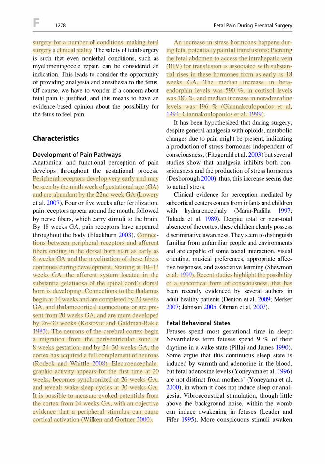
surgery for a number of conditions, making fetalsurgery a clinical reality. The safety of fetal surgery
is such that even nonlethal conditions, such asmyelomeningocele repair, can be considered an
indication. This leads to consider the opportunity
of providing analgesia and anesthesia to the fetus.Of course, we have to wonder if a concern about
fetal pain is justified, and this means to have an
evidence-based opinion about the possibility forthe fetus to feel pain.
Characteristics
Development of Pain PathwaysAnatomical and functional perception of pain
develops throughout the gestational process.
Peripheral receptors develop very early and maybe seen by the ninth week of gestational age (GA)
and are abundant by the 22nd week GA (Lowery
et al. 2007). Four or five weeks after fertilization,pain receptors appear around the mouth, followed
by nerve fibers, which carry stimuli to the brain.
By 18 weeks GA, pain receptors have appearedthroughout the body (Blackburn 2003). Connec-
tions between peripheral receptors and afferent
fibers ending in the dorsal horn start as early as8 weeks GA and the myelination of these fibers
continues during development. Starting at 10–13
weeks GA, the afferent system located in thesubstantia gelatinosa of the spinal cord’s dorsal
horn is developing. Connections to the thalamus
begin at 14 weeks and are completed by 20 weeksGA, and thalamocortical connections or are pre-
sent from 20 weeks GA, and are more developed
by 26–30 weeks (Kostovic and Goldman-Rakic1983). The neurons of the cerebral cortex begin
a migration from the periventricular zone at
8 weeks gestation, and by 24–30 weeks GA, thecortex has acquired a full complement of neurons
(Rodeck and Whittle 2008). Electroencephalo-
graphic activity appears for the first time at 20weeks, becomes synchronized at 26 weeks GA,
and reveals wake-sleep cycles at 30 weeks GA.
It is possible to measure evoked potentials fromthe cortex from 24 weeks GA, with an objective
evidence that a peripheral stimulus can cause
cortical activation (Wilken and Gortner 2000).
An increase in stress hormones happens dur-ing fetal potentially painful transfusions: Piercing
the fetal abdomen to access the intrahepatic vein(IHV) for transfusion is associated with substan-
tial rises in these hormones from as early as 18
weeks GA. The median increase in beta-endorphin levels was 590 %, in cortisol levels
was 183 %, and median increase in noradrenaline
levels was 196 % (Giannakoulopoulos et al.1994, Giannakoulopoulos et al. 1999).
It has been hypothesized that during surgery,
despite general analgesia with opioids, metabolicchanges due to pain might be present, indicating
a production of stress hormones independent of
consciousness, (Fitzgerald et al. 2003) but severalstudies show that analgesia inhibits both con-
sciousness and the production of stress hormones
(Desborough 2000), thus, this increase seems dueto actual stress.
Clinical evidence for perception mediated by
subcortical centers comes from infants and childrenwith hydranencephaly (Marın-Padilla 1997;
Takada et al. 1989). Despite total or near-total
absence of the cortex, these children clearly possessdiscriminative awareness. They seem to distinguish
familiar from unfamiliar people and environments
and are capable of some social interaction, visualorienting, musical preferences, appropriate affec-
tive responses, and associative learning (Shewmon
et al. 1999). Recent studies highlight the possibilityof a subcortical form of consciousness, that has
been recently evidenced by several authors in
adult healthy patients (Denton et al. 2009; Merker2007; Johnson 2005; Ohman et al. 2007).
Fetal Behavioral StatesFetuses spend most gestational time in sleep:
Nevertheless term fetuses spend 9 % of their
daytime in a wake state (Pillai and James 1990).Some argue that this continuous sleep state is
induced by warmth and adenosine in the blood,
but fetal adenosine levels (Yoneyama et al. 1996)are not distinct from mothers’ (Yoneyama et al.
2000), in whom it does not induce sleep or anal-
gesia. Vibroacoustical stimulation, though littleabove the background noise, within the womb
can induce awakening in fetuses (Leader and
Fifer 1995). More conspicuous stimuli awaken
F 1278 Fetal Pain During Prenatal Surgery
the fetus at birth. Fetal sleep can increase painthreshold, but cannot annihilate the possibility of
experiencing pain.
Fetal AnalgesiaIn the case of mayor surgery, some researchers rec-ommend to administer 20microg/kg of intramuscu-
lar fentanyl to the fetus prior to the procedure, to
increase the effect of volatile anesthetics that arriveto it through mother’s circulation (Schwarz and
Galinkin 2003). Recent studies show that direct
administration of 10 microg/kg fentanyl blunts thefetal stress response to intrauterine needling (Fisk
et al. 2001): These authors showed that the magni-
tude of the beta-endorphin response to pain withthese doses was halved, and the cerebral Doppler
response was ablated. Comparison with control
fetuses transfused without fentanyl indicated thatthe beta endorphin and cerebral Doppler response
to intrahepatic vein transfusion with fentanyl
approached that of nonstressful placental cord trans-fusions. Fentanyl significantly attenuated the endor-
phin and cerebrovascular response, but not the
cortisol response (Fisk et al. 2001). This differentialeffect is not surprising. Using the same dose, intra-
venous fentanyl in preterm babies ablated most of
the stress responses to surgery, but the reduction inthe cortisol response after fentanyl failed to achieve
statisticalsignificance(Anandetal.2001). It isworth
highlighting that the amount of opioids that canarrive to the mother from the fetus is clearly low
and relatively safe.
During open fetal surgery under maternalgeneral anesthesia, inhalational agents given to
the mother are still considered to provide adequate
fetal anesthesia and produce uterine relaxationessential for successful surgery, so that additional
analgesia for the fetus would be unnecessary. Nev-
ertheless, several evidences against this approachshould be considered: for instance after cesarean
sections made with maternal general anesthesia,
fetuses are born awake or are easily awakened atbirth, providing evidence that maternal anesthetic
drugs are not sufficiently anesthetic to the fetus.
Endoscopic procedures performed directly onthe fetus, such as tracheal occlusion or
meningomyelocele repair, are usually performed
under maternal local or regional anesthesia.
As these procedures do not requirematernal generalanesthesia, additional fetal anesthesia is desired and
is usually done by direct administration of opioidsand muscle relaxants to the fetus. Two possible
routes of administration for these drugs are injection
into the umbilical cord and intramuscular injectioninto the fetus. Sometimes direct fetal administration
of fentanyl and pancuronium is sometimes limited
to caseswhere the fetusmoves during the procedurein other cases, it is given routinely (Sutton 2008).
Some researchers utilized intra-amniotic opioids
for fetal analgesia on lamb fetuses: They showedthat greater plasma concentrations were obtained in
the fetal lamb as comparedwith the ewe, suggesting
that this route might be utilizable for humans.The presence of an active cortical subplate
(Anand et al. 2001) and of spino-thalamic con-
nections seem sufficient to transmit the painfulstimulus during the second half of pregnancy.
Thus, the use of fetal analgesia is at least a due
precautionary procedure.
Summary
In the second trimester of gestation, the incom-
plete development of the cortex does not yetconsent awareness, but this does not seem to be
an absolute obstacle to pain from the 20th week
of GA. Evidence is present in adult patients forskills that do not need an involvement of the
cortex. Several stimuli are processed without the
need of the cortex (Mulckhuyse and Theeuwes2010); nevertheless, these stimuli give useful
visual information to the individual (Sewards
and Sewards 2000; Pasley et al. 2004), or provokecomplex experiences such as fear (Ohman et al.
2007). A typical example of this is the perception
of fearful stimuli or of unusual objects(for instance, during a mechanical skill like driv-
ing a car) that do not arrive to awareness, but
that nevertheless can provoke complex reactionsor behaviors: These stimuli do not arrive to
awareness, but can affect consciousness. We
can hypothesize a similar scenario for subcorticalfetal processing of pain in this trimester:
After the 20th week of GA, we should not
exclude that pain can be felt by the fetus
Fetal Pain During Prenatal Surgery 1279 F
F

(Mahieu-Caputo et al. 2000), with possible long-term consequences (Lowery et al. 2007).
In the second half of pregnancy, fetuses havea higher pain threshold due to their sleep state,
but this does not absolutely rule out pain. The
presence of thalamocortical pathways and of thecortical subplate point out the possibilities of pain
at this stage of development.
References
Anand, K. J., & Sippel, W. G. (2001). Aynsley green a:Randomized trial of fentanyl anesthesia in pretermbabies undergoing surgery: Effects on the stressresponse. Lancet, 1, 62–66.
Blackburn, S. T. (2003). Maternal, fetal, and neonatalphysiology (2nd ed.). Philadelphia: WB Saunders.
Denton, D. A., McKinley, M. J., Farrell, M., & Egan, G. F.(2009). The role of primordial emotions in the evolu-tionary origin of consciousness. Consciousness andCognition, 18(2), 500–514.
Desborough, J. P. (2000). The stress response to trauma andsurgery. British Journal of Anaesthesia, 85, 109–117.
Fisk, N. M., Gitau, R., Teixeira, J. M., Giannakou-lopoulos, X., Cameron, A. D., & Glover, V. A.(2001). Effect of direct fetal opioid analgesia on fetalhormonal and hemodynamic stress response to intra-uterine needling. Anesthesiology, 95(4), 828–835.
Fitzgerald, R. D., Hieber, C., Schweitzer, E., Luo, A.,Oczenski, W., & Lackner, F. X. (2003). Intraoperativecatecholamine release in brain-dead organ donors isnot suppressed by administration of fentanyl.European Journal of Anaesthesiology, 20, 952–956.
Giannakoulopoulos, X., Sepulveda, W., Kourtis, P.,Glover, V., & Fisk, N. M. (1994). Fetal plasma cortisoland betaendorphin response to intrauterine needling.Lancet, 344, 77–81.
Giannakoulopoulos, X., Teixeira, J., Fisk, N., & Glover,V. (1999). Human fetal and maternal noradrenalineresponses to invasive procedures. Pediatric Research,45, 494–499.
Johnson, M. H. (2005). Subcortical face processing.Nature Reviews Neuroscience, 6(10), 766–774.
Kostovic, I., & Goldman-Rakic, P. S. (1983). Transientcholinesterase staining in the mediodorsal nucleus ofthe thalamus and its connections in the developinghuman and monkey brain. The Journal of ComparativeNeurology, 219, 431–447.
Leader, L. R., & Fifer,W. P. (1995). The potential value ofhabituation in the prenate. In J. P. Lecanuet (Ed.),Fetal development: A psychobiological perspective(pp. 83–404). Hillstdale, NJ: Lawrence Erlbaum.
Lowery, C. L., Hardman, M. P., Manning, N., Hall, R. W.,Anand, K. J., & Clancy, B. (2007). Neurodevelopmentalchanges of fetal pain. Semin Perinatol, 31(5), 275–82.
Mahieu-Caputo, D., Dommergues, M., Muller, F., &Dumez, Y. (2000). Fetal pain. Presse Medicale,29(12), 663–669.
Marın-Padilla, M. (1997). Developmental neuropathologyand impact of perinatal brain damage. II: White matterlesions of the neocortex. Journal of Neuropathologyand Experimental Neurology, 56(3), 219–235.
Merker, B. (2007). Consciousness without a cerebral cor-tex: A challenge for neuroscience and medicine. TheBehavioral and Brain Sciences, 30, 63–81.
Mulckhuyse, M., & Theeuwes, J. (2010). Unconsciousattentional orienting to exogenous cues: A review ofthe literature. Acta Psychologica, 134(3), 299–309.
Ohman, A., Carlsson, K., Lundqvist, D., & Ingvar, M.(2007). On the unconscious subcortical origin ofhuman fear. Physiology & Behavior, 92(1–2), 180–185.
Pasley, B. N., Mayes, L. C., & Schultz, R. T. (2004).Subcortical discrimination of unperceived objects dur-ing binocular rivalry. Neuron, 42(1), 163–172.
Pillai, M., & James, D. (1990). Behavioural states innormal mature human fetuses. Archives of Disease inChildhood, 65(1), 39–43. Spec No.
Rodeck, C. H., & Whittle, M. J. (2008). Fetal medicine:Basic science and clinical practice. London: ElsevierHealth Sciences.
Schwarz, U., & Galinkin, J. L. (2003). Anesthesia for fetalsurgery. Seminars in Pediatric Surgery, 12(3), 196–201.
Sewards, T. V., & Sewards, M. A. (2000). Visual aware-ness due to neuronal activities in subcortical struc-tures: A proposal. Consciousness and Cognition,9(1), 86–116.
Shewmon, D. A., Holmes, G. L., & Byrne, P. A. (1999).Consciousness in congenitally decorticate children:Developmental vegetative state as self-fulfillingprophecy.Developmental Medicine and Child Neurol-ogy, 41(6), 364–374.
Sutton, L. N. (2008). Fetal surgery for neural tube defects.Best Practice & Research Clinical Obstetrics &Gynaecology, 22(1), 175–188.
Takada, K., Shiota, M., Ando, M., Kimura, M., &Inoue, K. (1989). Porencephaly and hydranencephaly:A neuropathological study of four autopsy cases.Brain & Development, 11(1), 51–56.
Wilken, B., & Gortner, L. (2000). Early auditory evokedpotentials in very small premature infants. Zeitschriftf€ur Geburtshilfe und Neonatologie, 204(1), 14–19.
Wilson, R. D., Lemerand, K., Johnson, M. P., Flake,A. W., Bebbington, M., Hedrick, H. L., & Adzick,N. S. (2010). Reproductive outcomes in subsequentpregnancies after a pregnancy complicated by openmaternal-fetal surgery (1996–2007). American Jour-nal of Obstetrics and Gynecology, 203(3), 209e1–209–e6.
Yoneyama, Y., Sawa, R., Suzuki, S., Shin, S., Power,G. G., & Araki, T. (1996). The relationship betweenuterine artery Doppler velocimetry and umbilicalvenous adenosine levels in pregnancies complicatedby preeclampsia. American Journal of Obstetrics andGynecology, 174(1Pt 1), 267–271.
F 1280 Fetal Pain During Prenatal Surgery
Yoneyama,Y.,Suzuki,S.,Sawa,R.,Takeuchi,T.,Kobayashi,H., Takei, R., Kiyokawa, Y., Otsubo, Y., Hayashi, Z., &Araki, T. (2000). Changes in plasma adenosine concen-trations during normal pregnancy. Gynecologic andObstetric Investigation, 50(3), 145–148.
Fibroblast
Definition
Fibroblast is a connective tissue cell.
Cross-References
▶Wallerian Degeneration
Fibrocartilage
Definition
Fibrocartilage is cartilage that is largely composedof fibers like those in ordinary connective tissue.
Cross-References
▶ Sacroiliac Joint Pain
Fibromyalgia
David Vivian1 and Nikolai Bogduk21Metro Spinal Clinic, South Caulfield, VIC,
Australia2University of Newcastle, Newcastle Bone &Joint Institute, Royal Newcastle Centre,
Newcastle, NSW, Australia
Synonyms
Chronic widespread pain; Fibrositis; Muscular
rheumatism; Psychogenic rheumatism
Definition
Fibromyalgia is a condition characterized by
chronic widespread pain, fatigue, unrefreshedsleep, and a tendency to experience generalized
somatic symptoms.
Characteristics
Fibromyalgia is present in about 5 % of the
population. It is a syndrome of symptoms
that include chronic widespread pain, fatigue,disordered sleep, and increased stress in associa-
tion with abnormal neurosensory processing
and nonspecific endocrine and autonomicdysfunction.
The original American College of Rheumatol-
ogy (ACR) classification criteria for fibromyalgiaincluded tenderness on pressure palpation at 11 or
more of 18 specific tender points (Wolfe et al.
1990). Subsequently, the recognition that exami-nation for tender points lacks validity and reli-
ability has lead to the deletion of tender point
examination from the diagnostic formulation forfibromyalgia.
Fibromyalgia is now a symptom-based diag-
nosis with the primary feature being wide-spread pain present for at least 3 months in
association with other symptoms including
fatigue, non-restorative sleep (NRS), and“somatic” symptoms such as anxiety, irritable
bowel and bladder syndrome, cognitive ineffi-
ciency, tinnitus, and nausea. Justification forthe shift from the physical examination crite-
rion has been elicited by large studies that have
developed diagnostic classification criteriascales. The ACR has now combined the wide-
spread pain index (WPI) and the symptom
severity (SS) scale to produce “simple, practi-cal criteria for the clinical diagnosis of fibro-
myalgia” (Wolfe et al. 2010).The Symptom Intensity Scale (SIS) is
another scoring system for fibromyalgia
(Wilke 2009). It combines the number of areasof pain present in the previous 7 days with
a subjective measurement of fatigue as indi-
cated on a 10-cm line to produce an overall
Fibromyalgia 1281 F
F
SIS score, the level of which categorizespatients with possible fibromyalgia as having
features “probably consistent with fibromyal-gia” or “diagnostic.”
PathophysiologyThe pathophysiology of fibromyalgia is unknown
and remains in dispute. Patients with fibromyal-
gia exhibit increased pain sensitivity to pressure,heat, cold, and electrical stimulation, but these
features are not unique to fibromyalgia (Gracely
et al. 2003).Other markers of the disorder have been
explored, but the results have not been consistent
or reproducible. Nor are the abnormalities, whenevident, expressed by all patients with fibromy-
algia. Nor are they unique to these patients.
NRS, which is defined variably but includesthe feeling of being unrefreshed upon awakening
in spite of normal sleep duration, is common in
the general population but more prevalent in FMand chronic fatigue syndrome (CFS) (Staud
2010). Fragmented sleep or difficulty getting to
sleep is also common in FM patients. Althoughnot unique to NRS, ▶ alpha(a)-delta(d) sleep is
more common in subjects with NRS than in
healthy controls. The same anomaly occurs inother conditions and in 15 % of healthy individ-
uals (Carette 1995). Histological abnormalities
have been reported in the muscles of patientswith fibromyalgia, but no differences have been
found in adequately controlled studies (Carette
1995). Nor have disturbances in muscle metabo-lism been verified (Carette 1995). Patients with
fibromyalgia exhibit deficiencies in ▶ serotonin,
increased levels of ▶ substance P in their cere-brospinal fluid, and abnormalities of the ▶ hypo-
thalamic pituitary axis (Carette 1995), but these
differences have not been shown to be unique tofibromyalgia.
One model that has been proposed is that
fibromyalgia is due to an impairment of thediffuse noxious inhibitory control system
(DNIC) (Gracely et al. 2003). The implication
is that patients perceive spontaneous painbecause of a lack of tonic inhibition of the cen-
tral nociceptive pathways. The circumstantial
evidence is that of conditioning, that painful
stimuli produce analgesia in normal subjectsbut fail to do so in patients with fibromyalgia
(Gracely et al. 2003).Although this may be so, commentators have
questioned whether the syndrome is due to
altered central nociception or to hypervigilance,and if there is altered nociception, does it arise
because of somatic factors or psychogenic influ-
ences (Cohen and Quintner 1998).
NosologyThe relegation of tenderness as a diagnostic cri-terion for diagnosis of fibromyalgia together with
the development of symptom-based standards
has provided clinicians with a more robust andreliable method for managing and studying
patients with these symptoms. As a minimum,
the diagnosis of fibromyalgia offers patientsa diagnostic label that is more palatable than
a diagnosis of widespread pain (Carette 1995).
TreatmentA variety of treatments have been applied to
patients with fibromyalgia. Many treatments arebased on hearsay or anecdote, and some of the
conclusions from studies are based on previous
diagnostic criteria for fibromyalgia. The EULARreport in 2008 found 146 studies eligible for
review and made 9 recommendations for the man-
agement of fibromyalgia (Carville 2008). Exercisein general including heated pool exercise had
some limited evidence and was included on the
basis of expert opinion; tramadol had benefits over13 weeks in a randomized controlled trial, but
simple analgesics were supported and opioids
refuted by expert opinion; antidepressants tendedto reduce pain and improve function over a 12-
week period but not over longer periods.
A multidisciplinary approach to managementwas considered relevant, but this was again based
on expert opinion rather than any evidence.
Ameta-analysis of pregabalin found five high-quality trials (Straube 2010). Pregabalin seems to
have similar efficacy to other medication treat-
ments including duloxetine, combined paraceta-mol and tramadol, and amitriptyline 25 mg/day.
Pregabalin enhances slow-wave sleep, and
sodium oxybate (gamma-hydroxybutyrate)
F 1282 Fibromyalgia
reduces alpha EEG sleep, and both may havea role in symptomatic control of FM for those
reasons (Staud 2010).
Cross-References
▶Chronic Low Back Pain: Definitions and
Diagnosis▶ Fibromyalgia, Mechanisms, and Treatment
▶Human Thalamic Response to Experimental
Pain (Neuroimaging)▶Muscle Pain, Fibromyalgia Syndrome
(Primary, Secondary)
▶Myalgia▶Nocifensive Behaviors, Muscle and Joint
▶Opioids and Muscle Pain
▶ Physical Exercise▶ Psychological Aspects of Pain in Women
References
Carette, S. (1995). Fibromyalgia 20 years later. What havewe accomplished? The Journal of Rheumatology, 22,590–594.
Carville, S. F., Arendt-Nielsen, S., Bliddal, H., et al.(2008). EULAR evidence-based recommendationsfor the management of fibromyalgia syndrome. Annalsof the Rheumatic Diseases, 67, 536–541.
Cohen, M. L., & Quintner, J. L. (1998). Fibromyalgia syn-drome and disability: A failed construct fails those inpain.TheMedical Journal of Australia, 168(8), 402–404.
Gracely, R. H., Grant, M. A. B., & Giesecke, T. (2003).Evoked pain measures in fibromyalgia. Best Practice& Research Clinical Rheumatology, 17, 593–609.
Staud, S. (2010).Nonrestorative sleep in fibromyalgia: Fromassessment to therapy – focus on diagnosis. Medscape.http://cme.medscape.com.viewarticle/727292
Straube, S., Derry, S., Moore, R. A., et al. (2010).Pregabalin in fibromyalgia: Meta-analysis of efficacyand safety from company clinical trial reports. Rheu-matology, 49, 706–715.
Wilke,W. S. (2009). New developments in the diagnosis offibromyalgia syndrome: Say goodbye to tender points?Cleveland Clinic Journal of Medicine, 76, 345–352.
Wolfe, F., Smythe, H. A., Yunus, M. B., et al. (1990). TheAmerican college of rheumatology 1990 criteria forthe classification of fibromyalgia. Arthritis & Rheuma-tism, 33, 160–172.
Wolfe, F., Clauw, D. J., Fitzcharles, M., et al. (2010). TheAmerican college of rheumatology preliminary diag-nostic criteria for fibromyalgia and measurement ofsymptom severity. Arthritis Care & Research, 62,600–610.
Fibromyalgia Syndrome
▶Muscle Pain, Fibromyalgia Syndrome(Primary, Secondary)
Fibromyalgia, Mechanisms, andTreatment
Alice A. Larson and Katalin J. Kovacs
Department of Veterinary and BiomedicalSciences, University of Minnesota, St. Paul,
MN, USA
Introduction
Historically, fibromyalgia syndrome (FMS) was
referred to as fibrositis, a term coined by Sir
Edward Gowers at the turn of the century todescribe the inflammation responsible for the
stiffness and pain experienced by a group of
non-arthritic patients. Fibromyalgia syndromewas the name adopted in 1990 by the American
College of Rheumatology (Wolfe et al. 1990),
and the consensus document on fibromyalgia atthe MYOPAIN conference held in Copenhagen,
Denmark, in 1992 provided the original descrip-tion of the syndrome, the diagnostic criteria, and
its prevalence. More recently, the American
College of Rheumatology has provisionallyapproved a revised set of criteria for the diagnosis
of fibromyalgia that does not require a tender
point assessment (Wolfe et al. 2010).
Definition
Many chronic pain conditions display symptoms
that overlap with those of FMS. Those conditionsmust be diagnosed separately and eliminated as
the first step in the diagnostic protocol. The diag-
nostic criteria include widespread pain in allfour quadrants of the body for a minimum of
3 months, originally based on tenderness to dig-
ital palpation at 11 of the 18 anatomically defined
Fibromyalgia, Mechanisms, and Treatment 1283 F
F
tender points that are sensitive to pressure(mechanical pain). Wolfe et al. (2010) now rec-
ommend the diagnosis be based on the combina-tion of a widespread pain index (WPI) and
a symptom severity scale (SS) as follows: (WPI
" 7 and SS " 5) or (WPI 3–6 and SS "9).Reductions in electrical and thermal pain thresh-
olds have also been reported in patients with
FMS, suggesting multimodal changes in painsensitivity.
Characteristics
PrevalenceIn North America, approximately 2 % of the
population, or greater than 3.7 million people,
are estimated to suffer from FMS. The majorityof these patients, approximately three out of four,
are female. While it is the second most common
syndrome presented in rheumatology clinics, thetreatment is typically unsatisfactory, resulting in
disability and handicap. Studies in several coun-
tries and ethnicities suggest a poor prognosis overseveral years of treatment.
ComorbidityChronic widespread pain is the primary com-
plaint bringing most patients with FMS into the
clinic. In addition to pain, non-restorative sleep,fatigue, anxiety and depression, interstitial cysti-
tis, and irritable bowel syndrome are symptoms
frequently diagnosed in patients with FMS.A higher incidence of cold intolerance, restless
leg syndrome, cognitive dysfunction, rhinitis, and
multiple chemical sensitivities is also more com-mon in FMS patients than in healthy normal
controls, adding to the perplexing mosaic of the
disease. The etiology of FMS is unknown, and therelationships between pain and the other symp-
toms of FMS are unclear. Acute pain usually
results in increased activation of the pituitary-adrenal and sympathomedullary pathways as
well as growth hormone production. Patients
with FMS, in contrast, present with hypofunctionof the HPA, thyroidal, and gonadal axis; dimin-
ished growth hormone production (reviewed by
Bennett 1998); and abnormally low sympathetic
output (Clauw and Chrousos 1997). While expo-sure to stressful situations, including noise, lights,
and weather, exacerbates symptoms of FMS,patients have an impaired ability to activate the
hypothalamic-pituitary portion of the HPA axis
as well as the sympathoadrenal system, leading toreduced adrenocorticotropic hormone (ACTH)
and epinephrine responses. Yet many symptoms
of FMS are sympathetically maintained, such asthe neuropathic-type pain (reviewed by
Martinez-Lavin 2007), abnormal heart rate
variability, tilttable responses, and inappropriateresponses to daily stressful situations.
Unique Location of Tender PointsWhile tender points are no longer necessary
diagnostically, they offer insight into the etiol-
ogy of pain. Tender points are generally distrib-uted in areas whose primary afferent nerves
project to spinal lumbar levels 3–5 and cervical
levels 2–7 spinal cord segments. It may be ofimportance that these areas surround the tho-
racic 1 through lumbar 3 spinal cord segments
that are involved with regulation of the sympa-thetic nervous system, whose function is signif-
icantly altered in patients with FMS. It is
noteworthy that the pain of FMS is often inareas that receive a relatively low density of
afferent innervation (trunk and proximal limbs)
compared to areas normally considered “sensi-tive” by virtue of their dense innervation, e.g.,
the fingers, mouth, feet, and genitals. The sensi-
tivity of these tender points does not, then, orig-inate from a simple hyperactivity of all
mechanically sensitive tissues but rather from
an unknown, symmetrically distributed changein afferent input. Based on this and other psy-
chophysical evidence of windup, the etiology of
this syndrome is widely believed to includethe CNS.
Excitatory Amino AcidsExcitatory amino acids, such as glutamate and
aspartate, transmit pain signals, including those
in patients with FMS.While the concentrations ofmost amino acids in the cerebrospinal fluid do not
differ between subjects with FMS compared to
healthy normal controls, a high degree of
F 1284 Fibromyalgia, Mechanisms, and Treatment
correspondence was found between specificamino acids and the degree of pain reported at
the 18 tender points (tender point index, TPI).Excitatory amino acid activity is known to trigger
synthesis of nitric oxide (NO), a gaseous-
signaling compound that is critical for the devel-opment and expression of chronic pain.
Enhanced synthesis of NO results from a variety
of depolarizing events, including excitatoryamino acid and substance P activity, both of
which lead to influx of calcium and activation of
the enzyme NO synthase. NMDA activity is alsoassociated with the production of nerve growth
factor (NGF), a compound that regulates the syn-
thesis of peptides in primary afferent C-fiberssuch as substance P.
Substance PThe first documentation of a biochemical charac-
teristic consistent with chronic pain in patients
with FMS was the increased concentration ofsubstance P in their cerebrospinal fluid (Vaeroy
et al. 1988; Russell et al. 1994;Welin et al. 1995),
similar to that in many other pain states. Sub-stance P is a neuroactive peptide released from
small-diameter, unmyelinated primary afferent
fibers, called nociceptors. Substance P isupregulated in chronic, inflammatory pain condi-
tions by increased concentrations of NGF.
Because substance P does not mediate acutepain but supports hyperalgesia, enhanced sub-
stance P content in the cerebrospinal fluid of
patients with FMS is consistent with a chronichyperalgesic state.
Growth FactorsSynthesis of nerve growth factor (NGF),
a neurotrophin, in inflamed tissue is responsible
for the upregulation of SP synthesis duringchronic pain and the development of mechanical
hyperalgesia and allodynia. Intravenous adminis-
tration of NGF in humans causes muscle pain ina dose-dependent manner, primarily in bulbar
and truncal musculature, and affects women
more than men. While there is no gross inflam-mation at tender points to account for a peripheral
source of NGF, delivery of NGF to the spinal area
of mice or rats is sufficient to cause hyperalgesia.
The concentration of NGF in the cerebrospinalfluid is normally low or unmeasurable in healthy
individuals, but NGF is elevated in patients withprimary but not secondary FMS (Giovengo et al.
1999). In addition, the concentration of brain-
derived neurotrophic factor (BDNF), a mediatorof pain in the peripheral nervous system, is ele-
vated in the serum of patients with FMS (Laske
et al. 2007). It is, therefore, possible that elevatedconcentrations of NGF and or BDNF are respon-
sible for the hyperalgesia and allodynia associ-
ated with FMS, while secondary FMS resultsfrom conditions producing large areas of
enhanced pain sensitivity.
FMS and Thalamic ActivityPositron emission tomography (PET) and func-
tional magnetic resonance imaging (fMRI) stud-ies in humans indicate that the thalamus as well as
the anterior insula and S2 area are important in
the perception of pain. Activation of the thalamusby pain normally initiates descending inhibitory
activity that controls pain. Stimulation of
the somatosensory thalamus has even been suc-cessfully used to treat extreme conditions of
chronic pain in humans. Descending activity
originating from the thalamus is sufficientlyimportant that thalamotomy in rats results in
both thermal as well as mechanical hyperalgesia.
Although pain normally stimulates the thalamusin healthy individuals, a different pattern of acti-
vation occurs in patients with chronic pain.
Regional blood flow in response to painful stim-ulation also differs in patients with FMS com-
pared to that in normal control individuals
(Risberg et al. 1995; Mountz et al. 1998; Petzkeet al. 2000). These studies suggest that either the
ascending spinothalamic nociceptive pathways
are not fully functional or the thalamus respondsabnormally to input from spinothalamic neurons.
Because other parts of the CNS respond appro-
priately in patients with FMS, ascending activityappears sufficient. Rather, thalamic neurons
likely fail to initiate sufficient descending inhib-
itory activity controlling nociceptive processingat the spinal cord level, a concept supported by
altered nociceptive responses in patients with
FMS (Staud et al. 2003).
Fibromyalgia, Mechanisms, and Treatment 1285 F
F

TreatmentA variety of muscle relaxants, sedatives, analge-
sics, and drugs that promote sleep or combat
fatigue are frequently used to treat symptoms ofFMS. Unfortunately, even nonnarcotic analgesic
drugs provide only temporary relief in most
patients. When all else fails, narcotic analgesicsare transiently effective in controlling flares, but
not only does tolerance develop to their analgesic
effect but long-term use can unmask an opioid-induced hyperalgesia. The efficacy and side
effects of each compound vary greatly from one
patient to another, but in general, their sideeffects are minimized and efficacy maximized
by combination treatments using two drugs with
distinct mechanisms of action. For example,gabapentin (Neurontin) is a compound used for
neurogenic pain either alone or in combination
with opioids. Gabapentin inhibits calcium chan-nel activity, thereby minimizing the release of
neurotransmitters.
Low doses of tricyclic antidepressants, such asamitriptyline, provide immediate but temporary
analgesia, similar to their efficacy in other
chronic pain conditions (O’Malley et al. 2000).The efficacy of this, and related drugs, is likely
due to their action on nociceptive pathways and is
not related to their antidepressant activity, whichrequires higher doses and longer periods of treat-
ment to develop. Because tolerance to their anal-gesic activity develops, low doses (10 mg or less)
are recommended in the beginning, gradually
increasing by 10 mg as needed until the optimaldose of 70–80 mg is reached.
Only pregabalin (Lyrica), duloxetine
(Cymbalta), and milnacipran (Savella) are FDAapproved for the treatment of FMS symptoms.
Duloxetine and milnacipran are antidepressants
that inhibit serotonin and norepinephrine reup-take. Over the short term, this increases the syn-
aptic concentration of these transmitters but, by
doing so, influences their receptor sensitivityover the long term. In contrast, pregabalin binds
to a voltage-dependent calcium channel in the
central nervous system, reducing calcium influxinto the nerve terminals and decreasing release of
neurotransmitters such as glutamate, noradrena-
line, and substance P. Pregabalin also increases
the concentration of GABA, an inhibitory neuro-transmitter, by increasing glutamic acid decar-
boxylase activity, the enzyme required for thesynthesis of GABA.
Drug treatment to temporarily alleviate pain,
including amitriptyline, NSAIDS, and evennarcotic analgesics, should be geared to facili-
tate a new level of physical activity (reviewed
by Clauw and Crofford 2003) as the most effec-tive treatment for FMS is exercise (Sim and
Adams 2002). Patients need to understand that
exercise is the key to recovery, even if thismeans merely doubling their distance walked
within the house from one day to the next. The
exercise program should be initiated gradually,or it will temporarily exacerbate their symp-
toms. Heat therapy, in the form of a long hot
bath immediately prior to bedtime, has beenanecdotally reported to be effective for epi-
sodic bouts of intense pain. In contrast, cold
exacerbates their condition and should beavoided.
Cross-References
▶Chronic Low Back Pain: Definitions andDiagnosis
▶Disability in Fibromyalgia Patients
▶ Fibromyalgia, Mechanisms, and Treatment▶Human Thalamic Response to Experimental
Pain (Neuroimaging)
▶Muscle Pain, Fibromyalgia Syndrome(Primary, Secondary)
▶Myalgia
▶Nocifensive Behaviors, Muscle and Joint▶Opioids and Muscle Pain
▶ Physical Exercise
▶ Psychological Aspects of Pain in Women
References
Bennett, R. M. (1998). Disordered growth hormone secre-tion in fibromyalgia: A review of recent findings anda hypothesized etiology. Zeitschrift f€urRheumatologie, 57(Suppl 2), 72–762.
Clauw, D. J., & Chrousos, G. P. (1997). Chronic pain andfatigue syndromes: Overlapping clinical and
F 1286 Fibromyalgia, Mechanisms, and Treatment
neuroendocrine features and potential pathogenicmechanisms. Neuroimmunomodulation, 4, 143–1533.
Clauw, D. J., & Crofford, L. J. (2003). Chronic widespreadpain and fibromyalgia: What we know, and what weneed to know. Best Practice & Research ClinicalRheumatology, 17, 685–7014.
Giovengo, S. L., Russell, I. J., & Larson, A. A. (1999).Increased concentrations of nerve growth factor incerebrospinal fluid of patients with fibromyalgia.Journal of Rheumatology, 26, 1564–15695.
Laske, C., Stransky, E., Eschweiler, G. W., et al. (2007).Increased BDNF serum concentration in fibromyalgiawith or without depression or antidepressants. Journalof Psychiatric Research, 41, 600–605.
Martinez-Lavin, M. (2007). Stress, the stress responsesystem, and fibromyalgia. Arthritis Research &Therapy, 9, 216.
Mountz, J. M., Bradley, L. A., & Alarcon, G. S. (1998).Abnormal functional activity of the central nervoussystem in fibromyalgia syndrome. The AmericanJournal of the Medical Sciences, 315, 385–3968.
O’Malley, P. G., Balden, E., Tomkins, G., et al.(2000). Treatment of fibromyalgia with antidepres-sants. Journal of General Internal Medicine, 15,659–666.
Petzke, F., Clauw, D. J., Wolf, J. M., & Gracely, R. H.(2000). Pressure pain in fibromyalgia and healthy con-trols: Functional MRI subjective pain experienceversus objective stimulus intensity. Arthritis andRheumatism, 43, 200010.
Risberg, J. G., Rosenhall, U., Orndahl, G., et al. (1995).Cerebral dysfunction in fibromyalgia: Evidence fromregional cerebral blood flow measurements,otoneurological tests and cerebrospinal fluid analysis.Acta Psychiatrica Scandinavica, 91, 86–9411.
Russell, I. J., Orr, M. D., Littman, B., et al. (1994).Elevated cerebrospinal fluid levels of substance P inpatients with the fibromyalgia syndrome. Arthritis andRheumatism, 37, 1593–160112.
Sim, J., & Adams, N. (2002). Systematic review of ran-domized controlled trials of nonpharmacological inter-ventions for fibromyalgia. The Clinical Journal ofPain, 18, 324–33613.
Staud, R., Robinson, M. E., Vierck, C. J., et al. (2003).Diffuse noxious inhibitory controls (DNIC) attenuatetemporal summation of second pain in normal malesbut not in normal females or fibromyalgia patients.Pain, 101, 167–17414.
Vaeroy, H., Helle, R., Forre, O., et al. (1988). ElevatedCSF levels of substance P and high incidence ofRaynaud’s phenomenon in patients withfibromyalgia: New features for diagnosis. Pain,32, 21–2615.
Welin, M., Bragee, B., Nyberg, F., et al. (1995). Elevatedsubstance P levels are contrasted by a decrease in met-enkephalin-arg-phe levels in CSF from fibromyalgiapatients. Journal of Musculoskeletal Pain, 3, 4.
Wolfe, F., Clauw, D. J., Fitzcharles, M.-A., et al. (2010).The American college of rheumatology preliminary
diagnostic criteria for fibromyalgia and measurementof symptom severity. Arthritis Care & Research, 62,600–610.
Wolfe, F., Smythe, H. A., Yunus, M. B., et al. (1990). TheAmerican college of rheumatology 1990 criteria forthe classification of fibromyalgia. Report of the multi-center criteria committee. Arthritis and Rheumatism,33, 160–172.
Fibrositis
▶ Fibromyalgia▶Myalgia
▶ Psychiatric Aspects of the Epidemiology ofPain
Fibrositis Syndrome
▶Muscle Pain, Fibromyalgia Syndrome
(Primary, Secondary)▶Myalgia
Field Block
Definition
Local anesthetic injected through intact skin
adjacent to the surgical site to createa subcutaneous wall encompassing the injury.
Cross-References
▶Acute Pain in Children, Postoperative
Fifth Lobe
▶ Insular Cortex, Neurophysiology, and Func-
tional Imaging of Nociceptive Processing
Fifth Lobe 1287 F
F

Firing of Suburothelial AfferentNerves
Definition
Firing of suburothelial afferent nerves and thethreshold for bladder activation may be modified
by both inhibitory (e.g., NO) and stimulatory (e.g.,
ATP, tachykinins, prostanoids) mediators. Thesemechanisms can be involved in the generation of
detrusor overactivity causing urgency, frequency,
and incontinence, but also bladder pain.
Cross-References
▶Opioids and Bladder Pain/Function
First and Second Pain
Definition
When a needle is pierced into the skin of an extrem-
ity, the subject feels typically an immediate sting-ing pain followed after a short delay by a second
wave of predominantly burning pain. These two
pain phenomena are called “first” and “second”pain. They are attributed to the excitation of A-
delta and C-fibers. Since the former have conduc-
tion velocities up to 30 m/s, the nerve impulsesreach the central nervous system so quickly that
no delay is experienced. C-fibers having conduc-
tion velocities around 1 m/s convey their impulsesconsiderably slower. If the needle prick is applied
to the foot of a grown-up person, the delay could be
more than a second. If the sting is applied to theface, the two pains are not separated by a delay due
to the short conductance distance.
Cross-References
▶Encoding of Noxious Information in the SpinalCord
▶Opioids, Effects of Systemic Morphine on
Evoked Pain
First and Second Pain Assessment(First Pain, Pricking Pain, Pin-PrickPain, Second Pain, Burning Pain)
Donald D. PriceOral Surgery and Neuroscience, McKnight Brain
Institute University of Florida, Gainesville,
FL, USA
Synonyms
Burning pain; First pain assessment; Pin-prickpain; Pricking pain; Second pain assessment
Definition
Prior to the development of methods for physio-logical and anatomical characterization of axons
within peripheral nerves, Henry Head concluded
that the skin was served by epicritic and proto-pathic afferent systems in which each gave rise to
its own particular qualities of sensations (Head
1920). Epicritic pain, for example, was said to beaccurately localized, to not outlast the stimulus,
and to provide precise qualitative information
about the nature of the stimulus. Thus, epicriticpain could be elicited by mild pricking of the skin
with a needle, a form of pain known to almost
everyone as pricking pain. In contrast, proto-pathic pain was described as less well localized,
slow in onset, often outlasting the stimulus, and
summating with repeated stimulus application.Protopathic pain was considered more difficult
to endure and contained special feelings of
unpleasantness or “feeling tone.” The concept of“protopathic” has been applied to burning pain,
aching pain, throbbing pain, and dull pain. Head
based these ideas largely on observations made ofhis own experiences of pain, after nerve division
and during nerve regeneration.
With the advent of modern electrophysiologi-cal and neuroanatomical techniques, it became
clear that pain depended on two types of periph-
eral nerve axons. The first is that of thinly mye-linated A-delta axons, whose conduction
F 1288 Firing of Suburothelial Afferent Nerves

velocities range between 3 and 30 m/s, and thesecond is that of unmyelinated C axons, whose
conduction velocities range between 0.5 and2.0 m/s. Based on their differences in conduction
velocity and reminiscent of the functional dichot-
omy proposed by Head, Zotterman proposed thatA-delta and C afferent axons could account for
first and second pain, which often occur in
response to a brief intense stimulus to the handor foot (Zotterman 1933). Landau and Bishop
explicitly related first pain to epicritic pain and
second pain to protopathic pain (Landau andBishop 1953). Interestingly, they based their con-
clusions as a result of an approach similar to that
of Head. They carefully observed and recordedtheir own pain experiences in response to “exper-
imental” pain stimuli, before and after selective
conduction block of A-delta or C axons of periph-eral nerves. They used local injections of dilute
solutions of procaine to selectively block C axons
within small nerve branches in order to study painfrom impulses in A-delta axons. They selectively
blocked all myelinated axons in peripheral nerves
of their lower arms by means of a 250 mmHgpressure cuff in order to assess the types of pains
evoked by impulses in C axons. They applied
various types of painful stimuli to skin, fascia,and periosteal surfaces, including bee stings, tur-
pentine injections, intramuscular KCL injections,
and application of deep and sharp pressure withmechanical probes. When they blocked C axons
with procaine, well-localized brief-duration
stinging sharp pains, such as those elicited bypinpricks (i.e., pricking pain or first pain), were
preserved, but prolonged, deep, and diffuse burn-
ing pains evoked by inflammatory stimuli, suchas the second pain from a bee sting, could no
longer be elicited. When they blocked all mye-
linated (A) axons by means of the blood pressurecuff, the latter types of pain could be elicited and
were more intense than before blockade of mye-
linated axons.These general observations have since been
corroborated in conventional studies, wherein
investigators studied responses of volunteer par-ticipants (Price 1972, 1999; Collins et al. 1960).
An important aspect of the study by Collins et al.
was that compound action potentials were
monitored (Collins et al. 1960). They placedstimulating and recording electrodes under the
sural nerve of cancer patients undergoinganterolateral chordotomy for relief of pain.
They found that stimulation of A-delta axons
produced sharp pricking pain sensations thatwere accurately localized. When A-delta axons
were blocked by cold, stimulation of C axons at
a rate of 3/s resulted in summating unbearablediffuse burning pain that was not as well
localized.
The association of first and second pain withimpulses in A-delta and C axons and the relation-
ship of these two types of pain to epicritic and
protopathic pain have pivotal roles in the historyof pain research. However, like all functional
dichotomies, it is important not to overgeneralize
their explanatory role, for example, to prema-turely label different central pain-related path-
ways as “epicritic” or “protopathic.” Even Head
warned against this type of error, when he con-cluded that epicritic and protopathic systems
recombined once they entered the dorsal horn
(Head 1920). Thus, although zones of relativelypure epicritic or protopathic sensibility could be
found after dorsal root or peripheral nerve
lesions, he noted that such zones were neverobserved after lesions of the spinal cord or
brain. More than 40 years before electrophysio-
logical studies were carried out on dorsal hornnociceptive neurons, Head anticipated the synap-
tic convergence of two functional types of
primary nociceptive afferents (known since asA-delta and C) onto neurons of the dorsal horn.
One must also take note of the fact that A-delta
and C nociceptive afferents are rarely activated inisolation. Most acute pains are likely to reflect
a combination of input from A-delta and
C nociceptive afferents and, in most cases, fromnon-nociceptive afferents as well. Indeed, the
composition of input from different types of noci-
ceptive and non-nociceptive afferents undoubt-edly contributes a lot to the diverse qualities of
both painful and non-painful somatic sensation
(Price 1999). However, many long-durationpains, especially those that are diffuse, spatially
spreading, and especially unpleasant in their
“feeling tone” may depend to a greater extent
First and Second Pain Assessment 1289 F
F
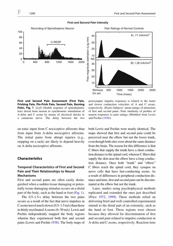
on tonic input from C nociceptive afferents than
from input from A-delta nociceptive afferents.The initial pains from abrupt injuries (e.g.,
stepping on a tack) are likely to depend heavily
on A-delta nociceptive afferents.
Characteristics
Temporal Characteristics of First and SecondPain and Their Relationships to NeuralMechanismsFirst and second pains are often easily distin-
guished when a sudden tissue damaging or poten-tially tissue-damaging stimulus occurs on a distal
part of the body, such as the hand or foot (Fig. 1).
The 0.5–1.5-s delay between the two painsoccurs as a result of the fact that nerve impulses in
Caxons travelmuch slower (0.5–1.5m/s) than those
in thinlymyelinatedA axons (6–30m/s). Lewis andPochin independently mapped the body regions
wherein they experienced both first and second
pains (Lewis and Pochin 1938). The body maps of
both Lewis and Pochin were nearly identical. The
maps showed that first and second pain could beperceived near the elbow but not the lower trunk,
even though both siteswere about the same distance
from the brain. The reason for this difference is thatC fibers that supply the trunk have a short conduc-
tion distance to the spinal cord,whereasCfibers that
supply the skin near the elbow have a long conduc-tion distance. Once both “trunk” and “elbow”
C fibers reach the spinal cord, they synapse on
nerve cells that have fast-conducting axons. Asa result of differences in peripheral conduction dis-
tance and time, first and second pain can be discrim-
inated at the elbow but not the trunk.Later, studies using psychophysical methods
replicated and extended the ones just described
(Price 1972, 1999). These methods relied ondelivering brief and well-controlled experimental
stimuli to the distal part of an extremity, such as
the hand or foot. These regions were chosenbecause they allowed for discrimination of first
and second pain related to impulse conduction inA-delta and C axons, respectively. Reaction time
Pain Ratings of Normal Controls
A) .71 mA/mm2
B) 1.28 mA/mm2
First and Second Pain Intensity
Recording of Spinothalamic Neuron
C-RESP
00
20
20
10NS
40
40
30
20
10
40
30
60
80
100
400 800 1200 1600 2000
2000 25001500
Time (msec)
StimulusOn set
1000500Fr
ee m
agni
tude
est
imat
ion
(Log
mm
)
First and Second Pain Assessment (First Pain,Pricking Pain, Pin-Prick Pain, Second Pain, BurningPain), Fig. 1 (Left) Double response of spinothalamictract dorsal horn neuron to synchronous stimulation ofA-delta and C axons by means of electrical shocks toa cutaneous nerve. The delay between the two
postsynaptic impulse responses is related to the fasterand slower conduction velocities of A and C axons,respectively. (Right) Subjects’ mean ratings of intensitiesof first and second pains. Note similarity of profiles ofneuron responses to pain ratings (Modified from Lewisand Pochin (1938))
F 1290 First and Second Pain Assessment

measurements were used to confirm that subjectscould indeed distinguish the two pains. Both
trained and untrained subjects reported qualities
of first pain as “pricking,” “stinging,” or “sharp”(i.e., pin-prick pain), without provocation or sug-
gestion that such qualities existed. Subjects were
trained to judge the perceived magnitudes of firstand second pain by squeezing a handgrip dyna-
mometer in some experiments (Price et al. 1977)
or using a mechanical visual analogue scale inothers (Price et al. 1994). Mean psychophysical
ratings of intensities of first pain during trains of
computer-driven heat pulses (2.5 s duration, peaktemperature 52#C) decreased progressively
(Price 1999; Price et al. 1977, 1994) and stayedthe same in the case of 5–9 mA electric shocks
(Price 1972; Price et al. 1994). Unlike first pain,second pain progressively increased in mean
intensity and duration throughout a series of
shocks or heat pulses, when the interstimulusinterval was less than 3 s but not when it was
5 s (Price 1972, 1999; Price et al. 1977, 1994)
(Fig. 2). Moreover, second pain became stronger,more diffuse, and more unpleasant with repeated
heat pulses or repeated electrical shocks.
When the same types of heat pulses or electricalshocks described above are applied to the skin of
monkeys, spinothalamic tract neurons within the
spinal cord dorsal horn respond with a doubleresponse (i.e., two sets of impulse discharges)
(Price 1999), as shown in Fig. 1. The earlier of thetwo is related to synaptic input fromA-nociceptors,
First and Second PainAssessment (First Pain,Pricking Pain, Pin-PrickPain, Second Pain,Burning Pain),Fig. 2 Subjects’ meanratings of second pain inresponse to series of 9-mAelectric shocks. Notetemporal summation ofsecond pain at interstimulusintervals of 3 s but not 5 sand the fact that first painremains at the sameintensity throughout seriesof shocks (From Price(1999))
First and Second Pain Assessment 1291 F
F

and the delayed response is related to synaptic inputfrom C nociceptors (Price 1999; Price et al. 1977).
Similar to first pain, the first response decreases orremains the same during heat pulses or shocks,
respectively, whereas the second delayed response
to heat pulses or shocks increases progressivelyboth in magnitude and duration. Similar to second
pain, temporal summation of the delayed neural
response was observed when the interstimulusinterval was 3 s or less but not 5 s (Price 1999).
For neurons, this temporal summation has been
termed “windup” (Price 1999).Moreover, this sum-mation must occur within the spinal cord dorsal
horn because similar experiments conducted on
peripheral A and C nociceptors show that theirresponses do not increase with stimulus repetition
(Price et al. 1977, 1994). Thus, temporal summa-
tion of second pain depends on mechanisms of thecentral nervous system (i.e., dorsal horn neurons),
not changes in peripheral receptors.
These psychophysical-neural parallels havebeen confirmed not only in the case of single
neurons of the spinal cord dorsal horn but also in
the case of neural imaging at the level of thesomatosensory region of the cerebral cortex
(Tommerdahl et al. 1996). Using a brain imaging
method of intrinsic optical density measurements(OIS), Tommerdahl and colleagues (Tommerdahl
et al. 1996) imaged neural activity within the pri-
mary somatosensory cortex of anesthetized squirrelmonkeys as their hands were repetitively tapped
with a heated thermode. These taps reliably evoke
first and second pain in human subjects. Theirmethod of neural imaging has a high degree of
both spatial and temporal resolution, measuring
local cortical neural activity within 50–100 mmand sampling neural activity that has occurred
within a third of a second. Heat taps produced
localized activity in two regions of the primarysomatosensory cortex, termed 3a and 1. When
a train of heat taps was presented at a rate of once
per 3 s, each tap evoked delayed neural activitywithin these regions. The neural response to each
tap grew progressively more intense and larger in
area with each successive tap. This temporal sum-mation of this neural response paralleled human
psychophysical experiences of second pain in sev-
eral distinctways. Both types of responses summate
at the same rate of stimulus repetition and havea similar growth in intensity during a series of
heat taps. The perceived skin area in which secondpain is perceived and the area of cortical neural
activity both increase with repeated heat taps.
Windup and temporal summation of secondpain are related to synaptic interactions between
C-nociceptive afferents and dorsal horn neurons.
These interactions involve long-duration excit-atory processes related to the release of neuro-
transmitters such as glutamate/aspartate and
neuromodulators such as substance P. Theseagents respectively activate NMDA (N-methyl-
D-aspartate) receptors and neurokinin 1 recep-
tors, leading to prolonged depolarizations(Thompson and Woolf 1990, for review). Thus,
NMDA receptor antagonists block both temporal
summation of second pain and the “windup”responses of dorsal horn neurons to repeated
C fiber input (Price 1999). Windup is related to
hyperalgesic states that can be produced experi-mentally as well as those occurring in pathophys-
iological pain, such as postherpetic neuralgia
(Arendt-Nielsen 1997; Dubner 1991). Indeed,slow temporal summation has long been consid-
ered a central neural mechanism that has a role in
pathophysiological pain (Noordenbos 1959).
References
Arendt-Nielsen, L. (1997). Induction and assessment ofexperimental pain from human skin, muscle, and vis-cera. In T. S. Jensen, J. A. Turner, & Z. Wiesenfeld-Hallin (Eds.), Proceedings of the 8th World Congresson pain (pp 393–403). Seattle: IASP Press.
Collins, W. F., Nulsen, F. E., & Randt, C. T. (1960).Relation of peripheral nerve fiber size and sensationin man. Archive of Neurology (Chicago), 3, 381–385.
Dubner, R. (1991). Neuronal plasticity and pain followingperipheral tissue inflammation or nerve injury. In M.Bond, E. Charlton, & C. J. Woolf (Eds.), Proceedings ofVth World Congress on pain. Pain research and clinicalmanagement (Vol.5,pp.263–276).Amsterdam:Elsevier.
Head, H. (1920). Studies in neurology. London: OxfordUniversity Press.
Landau, W., & Bishop, G. H. (1953). Pain from dermal,periosteal, and fascial endings and from inflammation.Archives of Neurology and Psychiatry, 69, 490–504.
Lewis, T., & Pochin, E. E. (1938). The double response ofthe human skin to a single stimulus. Clinical Science,3, 67–76.
F 1292 First and Second Pain Assessment
Noordenbos, W. (1959). Pain. Amsterdam: Elsevier.Price, D. D. (1972). Characteristics of second pain and
flexion reflexes indicative of prolonged centralsummation. Experimental Neurology, 37, 371–387.
Price, D. D. (1999). Psychological mechanisms of painand analgesia (p. 250). Seattle: IASP Press.
Price, D. D., Hu, J. W., Dubner, R., & Gracely, R. (1977).Peripheral suppression of first pain and central sum-mation of second pain evoked by noxious heat pulses.Pain, 3, 57–68.
Price, D. D., Frenk, H., Mao, J., & Mayer, D. J. (1994).The NMDA receptor antagonist dextromethorphanselectively reduces temporal summation of secondpain. Pain, 59, 165–174.
Thompson, S. W. N., & Woolf, C. J. (1990). Primaryafferent-evoked prolonged potentials in the spinalcord and their central summation: Role of the NMDAreceptor. In: M. R. Bond, J. Carlton, & C. J. Woolf(Eds.), Proceedings of the VIth World Congress onpain (pp. 291–298). Amsterdam: Elsevier.
Tommerdahl, M., Delemos, K. A., Vierck, C. J.,Favorov, O. V., & Whitsel, B. L. (1996). Anteriorparietal cortical response to tactile and skin heatingstimulation. Journal of Neurophysiology, 75(6),2662–2670.
Zotterman, Y. (1933). Studies in the peripheral nervousmechanisms of pain. Acta Medica Scandinavica, 80,185–242.
First Pain
Definition
First pain is a rapid onset, sharp, pricking nox-ious sensation that is associated with the activa-
tion of Ad nociceptors. When a noxious stimulus
is sufficient to activate both Ad andC nociceptors, first pain is perceived before sec-
ond pain due to differences in nerve conduction
velocity.
Cross-References
▶Encoding of Noxious Information in the Spinal
Cord▶ First and Second Pain Assessment (First Pain,
Pricking Pain, Pin-Prick Pain, Second Pain,
Burning Pain)▶Opioids, Effects of Systemic Morphine on
Evoked Pain
First Pain Assessment
▶ First and Second Pain Assessment (First Pain,Pricking Pain, Pin-Prick Pain, Second Pain,
Burning Pain)
First-Generation Antidepressants
▶Antidepressant Analgesics in PainManagement
First-Order Sensory Neurons
▶Opioid Modulation of Nociceptive Afferents
In Vivo
First-Order Subtype: MedialThalamotomy
▶Thalamotomy for Human Pain Relief
First-Pass Metabolism
Definition
The first-pass metabolism describes the transfor-
mation of the active drug after absorptionprior to reaching the systemic circulation,
i.e., pre-systemic elimination of the drug. It
mainly occurs after oral or deep rectal adminis-tration and can be avoided by intravenous, intra-
muscular, sublingual, buccal, or topical
administration.
Cross-References
▶NSAIDs, Pharmacokinetics
First-Pass Metabolism 1293 F
F

Fitness Training
▶ Physical Exercise
Fits of Pain
▶ Pain Paroxysms
Flare
▶Nociceptor, Axonal Branching
Flare, Flare Response
Definition
See also “neurogenic inflammation” and “axon
reflex.” Since the flare response is dependent on
the extension of the terminal arborization of theC-fibers mediating this axon reflex response,
i.e., their receptive fields, flare sizes vary in
different species and depend also on the typeof nerve fibers involved. Rodents do not show
a visible flare response since the receptive fields
of their C-afferents are small. They produce,however, a neurogenic plasma extravasation
upon the stimulation of C-fibers. In humans,
the flare responses following histamine stimu-lation are particularly large due to the large
receptive fields of the histamine-sensitive
C-fibers.
Cross-References
▶Nociceptor, Axonal Branching
▶ Polymodal Nociceptors, Heat Transduction▶Quantitative Thermal Sensory Testing of
Inflamed Skin
Flexion Exercise
▶Exercise
Flexion Withdrawal Reflex
Definition
This is a protective reflex usually elicited in the
lower limb and was originally described byCharles Sherrington as “the withdrawal of a limb
from an offending stimulus.” As originally char-
acterized, it involved the activation of flexor mus-cles via a group of afferent nerve fibers called
“flexor reflex afferents” with corresponding inhi-
bition of extensor muscles. However, more recentresearch has revealed that the flexion withdrawal
reflex has a more complex modular organization,
its activation being contingent upon the area ofskin being stimulated. Nevertheless, in the adult,
the nociceptive (i.e., produced by noxious stimuli)
flexion withdrawal reflex has proved invaluable asa model of nociceptive processing, and shows
a clear correlation with pain perception in terms
of threshold, peak intensity, and sensitivity toanalgesics. However, in the newborn infant, the
flexion withdrawal reflex can also be evoked withlow-intensity mechanical stimuli to the foot, such
as calibrated monofilaments (also called von Frey
hairs), and has a much lower threshold than thenociceptive flexion withdrawal reflex in the adult.
Cross-References
▶ Infant Pain Mechanisms
Flinching
Definition
At its most vigorous, flinching of the paw and/or
hindquarters is paw shaking, and when less
F 1294 Fitness Training
vigorous, rapid paw lifting. It is usually seen asdrawing the paw under the body and rapidly
vibrating it, and this causes a shudder or ripplingmotion across the back which is easy to observe
even when the paw is not visible. Each episode is
recorded as a single flinch.
Cross-References
▶ Formalin Test
Flip-Flop Isoform of AMPA Receptors
Definition
AMPA receptors have four subunits named
GluR1-4 (or GluRA-D), respectively. Each
subunit of the AMPA receptor exists in twoisoforms, so-called flip and flop due to alternative
splicing of a 115-base pair region encoding 38
amino acid residues immediately preceding thepredicted fourth membrane domain. The flip and
flop isoforms of AMPA receptors may be
differentially involved in pain transmission andresponse to injury.
Cross-References
▶Descending Circuitry, Molecular Mechanismsof Activity-Dependent Plasticity
Flunarizine
Definition
Calcium channel, dopamine, and histamine antag-onist used in the preventive treatment of migraine.
Cross-References
▶Headache Due to Arteritis
Fluorocitrate
Definition
Fluorocitrate blocks the activation of glial cells,
without directly affecting neurons, and functionsto inhibit the activity of aconitase, an enzyme
in the Krebs cycle of glia, but not neurons.
Peri-spinal administration of fluorocitrate blocksexaggerated pain.
Cross-References
▶Cord Glial Activation
Flurbiprofen
Definition
Flurbiprofen is a nonsteroid anti-inflammatory
drug (NSAID), which inhibits the formation ofprostaglandins by cyclooxygenase. It is selective
for COX-1, thus inhibits COX-1 at lower concen-
trations than COX-2.
Cross-References
▶Cyclooxygenases in Biology and Disease
fMRI
▶ Functional Magnetic Resonance Imaging
fMRI Imaging and PET in ParietalCortex
▶ PET and fMRI Imaging in Parietal Cortex
(SI, SII, Inferior Parietal Cortex BA40)
fMRI Imaging and PET in Parietal Cortex 1295 F
F
FMS
▶Muscle Pain, Fibromyalgia Syndrome(Primary, Secondary)
Focal Pain
Definition
Focal pain is that which is experienced at one site,superficial to the underlying nociceptive lesion.
Cross-References
▶Cancer Pain, Goals of a ComprehensiveAssessment
Focused Analgesia
Definition
Focused analgesia is based on increased anddirected attention to that part of the body for
which suggestions of analgesia have been given.
For example, suggestions for numbness in thehand may produce the experience of numbness
as well as reduced pain sensation.
Cross-References
▶Hypnotic Analgesia
Follicular Phase
Definition
The follicular phase is the time during which
a single dominant ovarian follicle develops. The
follicle should be mature at mid-cycle for
ovulation. The average length of this phase isabout 10–14 days. Variability in the length of
this phase is responsible for variations in totalcycle length.
Cross-References
▶ Premenstrual Syndrome
Forced-Choice Procedure
Definition
Forced-choice procedure is a statistical deci-
sion theory method in which a sensory deci-sion is made after two or more stimulus
presentations.
Forearm Ischemia Procedure
▶Tourniquet Test
Forearm Occlusion Pain
▶Tourniquet Test
Forebrain
Definition
Forebrain is the part of the brain including
the cerebral cortex, limbic system, and
hypothalamus.
Cross-References
▶ Forebrain Modulation of the Periaqueductal
Gray and Its Role in Pain
F 1296 FMS
Forebrain Modulation of thePeriaqueductal Gray and Its Rolein Pain
Dayna R. Loyd1 and Anne Z. Murphy2
1US Army Institute of Surgical Research,Fort Sam Houston, TX, USA2Neuroscience Institute, Georgia State
University, Atlanta, GA, USA
Introduction
Forebrain Inputs to PAGIn the 1980s and 1990s, tract-tracing studies froma number of laboratories (e.g., Beitz 1982;
Shipley et al. 1991) revealed an important feature
of the PAG; afferent inputs to the PAG arise froma staggering number of cortical and subcortical
forebrain sites. These inputs to the PAG are much
heavier than previously suspected and terminatewith a remarkable degree of topographic
specificity. Major identified forebrain afferents
to the PAG include the medial prefrontal cortex(MPF), the medial preoptic area (MPO), the
central and medial nuclei of the amygdala
(CeA, MeA), the ventromedial hypothalamus(VMH), and the paraventricular nucleus of the
hypothalamus (PVN).
Definition
The midbrain ▶ periaqueductal gray (PAG) and
its descending projections to the▶ rostral ventro-medial medulla (RVM) comprise a critical
descending neuroanatomical pathway modulat-
ing pain and analgesia. The PAG projects heavilyand directly to the RVM, which in turn projects to
the ▶ dorsal horn of the spinal cord. Both pain
and analgesia are modulated by emotional,motivational, and cognitive factors indicating
higher-order cortical and subcortical ▶ forebrain
modulation of the PAG-RVM-spinal cordpathway. Anatomical studies have demonstrated
that the PAG predominantly receives afferents
from the forebrain, including cortical and
subcortical sites involved in nociception. Theseforebrain projections terminate with a high
degree of topographical specificity within thePAG, forming longitudinal input columns
extending throughout the rostrocaudal extent of
the PAG and activating output neurons projectingto the RVM. Major identified forebrain afferents
to the PAG include the medial prefrontal cortex
(MPF), the medial preoptic area (MPO), thecentral and medial nuclei of the ▶ amygdala
(CeA, MeA), the ventromedial ▶ hypothalamus
(VMH), and the paraventricular nucleus of thehypothalamus (PVN).
Characteristics
Medial Prefrontal Cortex (MPF)The PAG receives dense inputs from large
groups of neurons in the orbital, medial pre-
frontal, and lateral (insular and perirhinal) cor-tices (Beitz 1982; Shipley et al. 1991; Floyd
et al. 2000). In total, inputs to the PAG arise
from at least 8 distinct cytoarchitectonic fieldsin the medial prefrontal and lateral cortices
(Shipley et al. 1991). ▶Retrograde tracing
demonstrates that projections to the PAG fromthe cerebral cortex arise from all fields of the
medial prefrontal cortex infralimbic, prelimbic,
anterior cingulate, and precentral medialis.Equally extensive projections arise from the
lateral suprarhinal (insular cortex) and
perirhinal cortical areas. Anterograde tracingreveals that the pattern of terminal labeling
from each medial or lateral cortical field is
highly organized and selectively targets dis-crete, columnar subregions of the PAG along
its entire rostrocaudal axis. Inputs from differ-
ent cortical fields terminate as different, largelycomplementary, longitudinal columns. Imag-
ing studies in humans have shown that anterior
cingulate and insular orbital cortices are con-sistently activated by the sensory-perceptual
and affective (i.e., emotional or unpleasant)
aspects of pain (Price 2000; Rolls et al. 2003).Analgesia, including opiate receptor-
dependent analgesia, can be elicited from the
anterior cingulate and insular cortices (see
Forebrain Modulation of the Periaqueductal Gray and Its Role in Pain 1297 F
F
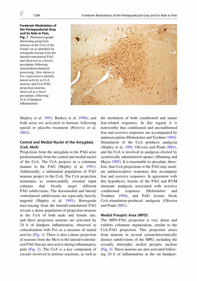
Shipley et al. 1991; Burkey et al. 1996), and
both areas are activated in humans followingopioid or placebo treatment (Petrovic et al.
2002).
Central and Medial Nuclei of the Amygdala(CeA, MeA)Projections from the amygdala to the PAG arisepredominantly from the central and medial nuclei
of the CeA. The CeA projects in a columnar
manner to the PAG (Shipley et al. 1991).Additionally, a substantial population of PAG
neurons project to the CeA. The CeA projection
terminates as rostrocaudally oriented inputcolumns that focally target different
PAG subdivisions. The dorsomedial and lateral/
ventrolateral subdivisions are especially heavilytargeted (Shipley et al. 1991). Retrograde
tract-tracing from the lateral/ventrolateral PAG
reveals a dense population of projection neuronsin the CeA of both male and female rats,
and these projection neurons are activated by
24 h of hindpaw inflammation, observed ascolocalization with Fos as a measure of neural
activity (Fig. 1). There is also a dense projection
of neurons from the MeA to the lateral/ventrolat-eral PAG that are also active during inflammatory
pain (Fig. 2). The CeA is a key component ofcircuits involved in defense reactions, as well as
the mediation of both conditioned and innate
fear-related responses. In this regard, it isnoteworthy that conditioned and unconditioned
fear and aversive responses are accompanied by
antinociception (Helmstetter and Tershner 1994).Stimulation of the CeA produces analgesia
(Shipley et al. 1991; Oliveira and Prado 2001),
and the CeA is involved in analgesia elicited bysystemically administered opiates (Manning and
Mayer 1995). It is reasonable to speculate, there-
fore, that CeA projections to the PAG may medi-ate antinociceptive responses that accompany
fear and aversive responses. In agreement with
this hypothesis, lesions of the PAG and RVMattenuate analgesia associated with aversive
conditioned responses (Helmstetter and
Tershner 1994), and PAG lesions blockCeA-stimulation-produced analgesia (Oliveira
and Prado 2001).
Medial Preoptic Area (MPO)The MPO-PAG projection is very dense and
exhibits columnar organization, similar to theCeA-PAG projection. This projection arises
from neurons in several cytoarchitectonically
distinct subdivisions of the MPO, including thesexually dimorphic medial preoptic nucleus
(Fig. 3). These neurons are also activated follow-ing 24 h of inflammation in the rat hindpaw.
Forebrain Modulation ofthe Periaqueductal Grayand Its Role in Pain,Fig. 1 Photomicrographillustrating projectionneurons in the CeA of thefemale rat as identified byretrograde tracing from thelateral/ventrolateral PAGand observed as a brownprecipitate followingimmunohistochemicalprocessing. Also shown isFos expression to identifyneural activity in CeAneurons and CeA-PAGprojection neurons,observed as a blackprecipitate, following24 h of hindpawinflammation
F 1298 Forebrain Modulation of the Periaqueductal Gray and Its Role in Pain

Injections of anterograde tracers into the MPO
label dense, highly organized, and topographi-cally specific projections to the PAG.
A hallmark of this projection, like other forebrain
inputs studied to date, is that it forms longitudi-nally organized input columns that selectively
target discrete subregions of the PAG (i.e., thedorsomedial and lateral/ventrolateral PAG) along
its rostrocaudal axis. In particular, MPO inputs to
the PAG selectively terminate within the lateral/ventrolateral caudal PAG, where output neurons
to the RVM are selectively located (Murphy and
Hoffman 2001). Activation of the MPO bya variety of physiological factors also activates
longitudinally organized columns of PAGneurons, including those that project to the
Forebrain Modulation ofthe Periaqueductal Grayand Its Role in Pain,Fig. 3 Photomicrographillustrating MPO-PAGprojection neurons in thefemale rat labeled byretrograde tract-tracingfrom the lateral/ventrolateral PAG andobserved as brownprecipitate followingimmunohistochemistry.Also shown areFos-positive MPO neuronsand MPO-PAG projectionneurons indicating neuralactivity following 24 h ofinflammation in the rathindpaw (observed as blackprecipitate)
Forebrain Modulation ofthe Periaqueductal Grayand Its Role in Pain,Fig. 2 Photomicrographillustrating projectionneurons in the MeA of thefemale rat as identified byretrograde tracing from thelateral/ventrolateral PAGand observed as a brownprecipitate followingimmunohistochemicalprocessing. Also shown isFos expression to identifyneural activity in MeAneurons and MeA-PAGprojection neurons, observedas a black precipitate,following 24 h of hindpawinflammation
Forebrain Modulation of the Periaqueductal Gray and Its Role in Pain 1299 F
F

ventral medulla (Murphy and Hoffman 2001;
Normandin and Murphy 2008). The MPO is sex-ually dimorphic and plays a key role in neuroen-
docrine and steroidal regulation and maternal/
reproductive behaviors. Nociceptive thresholdsshift across the estrus cycle, with steroid hormone
administration and during reproductive activities
(see ▶ Sex Differences in Descending PainModulatory Pathways). Both the MPO and the
PAG contain high levels of estrogen and andro-
gen receptors (Murphy and Hoffman 2001; Loydand Murphy 2008). The MPO-PAG projection
neurons also coexpress the estrogen receptor
ERa (Fig. 4). In this regard, it is interesting tonote that there are sex differences in pain thresh-
olds as well as sex differences in how inflamma-
tory pain activates the PAG-RVM circuit (Loydand Murphy 2006). Furthermore, the PAG-RVM
pathway is anatomically and functionally sexu-
ally dimorphic (Loyd and Murphy 2006) and isessential for the observed sexual dimorphism in
morphine analgesia (Loyd et al. 2008). TheMPO,
therefore, may provide a key link mediating hor-monal influences on nociceptive thresholds and
may also regulate descending nociception during
maternal/reproductive behaviors (Murphy et al.1999). Consistent with this hypothesis, stimula-
tion of the MPO activates PAG neurons thatproject to the RVM (Rizvi et al. 1996), inhibits
dorsal horn spinal cord neuronal responses to
nociceptive stimuli, and also elicits analgesia(see Shipley et al. 1991; Zhang and Ennis 2005).
Hypothalamic Projections to the PAGThere is also evidence of various hypothalamic
nuclei projections to the lateral/ventrolateral
PAG as observed by retrograde tract-tracing,including the ventromedial hypothalamus
(VMH; Fig. 5) and the paraventricular nucleus
of the hypothalamus (PVN; Fig. 6). These hypo-thalamic nuclei are known to modulate fear and
stress responses, as well as sex and satiety behav-
iors. Hypothalamic projections to the PAG likelyplay a role in modulating these behavioral
responses during pain and analgesia; however,
little research has addressed these possibilities.Future research is warranted based on known
anatomical connectivity between the hypothala-
mus and the descending pain inhibitory system.
PAG-Forebrain Afferents Coordinatea Multitude of Behavioral ResponsesThere has been a growing recognition that the
PAG itself is far more complex than initially
suspected and is clearly involved in many morephysiological functions than nociception. For
example, stimulation of different columnarlyorganized regions of the PAG produces
Forebrain Modulation ofthe Periaqueductal Grayand Its Role in Pain,Fig. 4 Photomicrographillustrating MPO-PAGprojection neuronsidentified by retrogradetracing from the lateral/ventrolateral PAG andobserved as brownprecipitate followingimmunohistochemistry.Also shown is ERa-positiveMPO neurons andcolocalization with MPO-PAG neurons observed asblack precipitate
F 1300 Forebrain Modulation of the Periaqueductal Gray and Its Role in Pain

a number of distinctly different behavioral and
physiological responses including vocalization,autonomic changes, sexual/reproductive
behaviors, and fear and rage reactions (for review
see Shipley et al. 1991; Murphy et al. 1994;Murphy and Hoffman 2001; Loyd and Murphy
2009, Loyd and Murphy 2008). Not surprisingly,
many of the forebrain sites that project to thePAG are also known to regulate similar func-
tions. Based on these findings, a more integrativeconceptual framework has been adopted guided
by the working hypothesis that the PAG is
a structure that plays a central role in the produc-tion of certain stereotypical behaviors (e.g.,
reproduction, defense reactions, vocalization)
essential to the animal’s survival (Loyd and Mur-phy 2009). These behaviors require rapid, pro-
found autonomic adjustments and
simultaneously, significant alterations in painthresholds. From this perspective, it is reasonable
to consider that more highly elaborated forebrainstructures interact with the PAG to coordinate
Forebrain Modulation ofthe Periaqueductal Grayand Its Role in Pain,Fig. 6 Photomicrographillustrating PVN-PAGprojection neurons in thefemale rat identified byretrograde tract-tracingfrom the lateral/ventrolateral PAG andobserved as brownprecipitate followingimmunohistochemistry.Also shown areFos-positive PVN neuronsand PVN-PAG outputneurons indicating neuralactivity following 24 h ofhindpaw inflammation
Forebrain Modulation ofthe Periaqueductal Grayand Its Role in Pain,Fig. 5 Photomicrographillustrating VMH-PAGprojection neurons in thefemale rat identified byretrograde tract-tracingfrom the lateral/ventrolateral PAG andobserved as brownprecipitate followingimmunohistochemistry.Also shown are Fos-positive VMH neurons andVMH-PAG output neuronsindicating neural activityfollowing 24 h of hindpawinflammation
Forebrain Modulation of the Periaqueductal Gray and Its Role in Pain 1301 F
F
antinociceptive, behavioral, and autonomicresponses in concert with the dominant role of
the forebrain in cognitive and emotionalprocessing. Descending PAG output neurons
also exhibit columnar organization, such that
different output columns terminate with medialto lateral specificity in the ventral medulla in sites
involved in nociception, autonomic responses,
sexual/reproductive behaviors, and vocalization(Murphy and Hoffman 2001; Murphy et al. 1999;
Loyd and Murphy 2008, Loyd and Murphy 2009,
Loyd and Murphy 2006). Taken together, thesefindings suggest that forebrain sites may trigger
or modulate specific nociceptive and autonomic
adjustments via activation of discrete columns ofPAG output neurons.
Summary
The PAG and the RVM are major nodes for
bidirectional modulation of nociception and spi-
nal cord neuronal responses to noxious sensoryinput. The PAG-RVM circuit is demonstrably
central to analgesia elicited by activation of
endogenous opioidergic systems as well as thatresulting from exogenously administered opi-
oids. Nociceptive regulation is but one aspect of
this circuit as the PAG is clearly a highly orga-nized structure that integrates defensive, fear/
anxiety, and hormonal/reproductive behaviors in
discrete columns that extend longitudinallythrough the structure. These columns receive
dense and topographically specific input from
cortical and subcortical forebrain areas centrallyinvolved in these same functions. Some forebrain
areas also project in parallel to the RVM, and the
functional significance of such dual projections isnot known. In humans, whose behavior is domi-
nated by the massive and highly elaborated fore-brain, the projections to the PAG and RVM are
likely to regulate nociception in concert with
forebrain-mediated cognitive and emotionalprocessing of pain and its perceived impact.
Acknowledgement The authors would like to recognizethe contributions of Dr. Matthew Ennis and Xijing Zhangon the first edition of this chapter.
References
Beitz, A. J. (1982). The organization of afferent projec-tions to the periaqueductal gray of the rat. Neurosci-ence, 7, 133–159.
Burkey, A. R., Carstens, E., Wenniger, J. J., et al. (1996).An opioidergic cortical antinociception triggering sitein the agranular insular cortex of the rat contributes tomorphine antinociception. The Journal of Neurosci-ence, 16, 6612–6623.
Floyd, N. S., Price, J. L., Ferry, A. T., et al. (2000).Orbitomedial prefrontal cortical projections todistinct longitudinal columns of the periaqueductalgray in the rat. The Journal of Comparative Neurology,422, 556–578.
Helmstetter, F. J., & Tershner, S. A. (1994). Lesions of theperiaqueductal gray and rostral ventromedial medulladisrupt antinociceptive but not cardiovascular aversiveconditional responses. The Journal of Neuroscience,14, 3099–7108.
Loyd, D. R., & Murphy, A. Z. (2006). Sex differences inthe anatomical and functional organization of theperiaqueductal gray-rostral ventromedial medullarypathway in the rat: A potential circuit mediating thesexually dimorphic actions of morphine. The Journalof Comparative Neurology, 496, 723–738.
Loyd, D. R., & Murphy, A. Z. (2008). Androgen andestrogen (a) receptor localization on periaqueductalgray neurons projecting to the rostral ventromedialmedulla in the male and female rat. Journal ofChemical Neuroanatomy, 36, 216–226.
Loyd, D. R., & Murphy, A. Z. (2009). The role of theperiaqueductal gray in the modulation of pain in malesand females: are the anatomy and physiology really thatdifferent? Neural Plast. doi: 10.1155/2009/462879.
Loyd, D. R., Wang, X., & Murphy, A. Z. (2008). Sexdifferences in mu opioid receptor expression in therat midbrain periaqueductal gray are essential foreliciting sex differences in morphine analgesia. TheJournal of Neuroscience, 28(52), 14007–14017.
Manning, B. H., & Mayer, D. J. (1995). The centralnucleus of the amygdala contributes to the productionof morphine antinociception in the rat tail flick test.The Journal of Neuroscience, 15, 8199–8213.
Murphy, A. Z., Ennis, M., Shipley, M. T., & Behbehani,M.M. (1994). Directionally specific changes in arterialpressure induce differential patterns of fos expressionin discrete areas of the rat brainstem: a double-labelingstudy for Fos and catecholamines. The Journal ofComparative Neurology, 349(1), 36–50.
Murphy, A. Z., Rizvi, T. A., Ennis, M., et al. (1999). Theorganization of preoptic-medullary circuits in themale rat: Evidence for interconnectivity of neuralstructures involved in reproductive behavior,antinociception and cardiovascular regulation. Neuro-science, 91, 1103–1116.
Murphy, A. Z., & Hoffman, G. E. (2001). Distribution ofgonadal steroid receptor-containing neurons in thepreoptic-periaqueductal gray-brainstem pathway: a
F 1302 Forebrain Modulation of the Periaqueductal Gray and Its Role in Pain

potential circuit for the initiation of male sexual behav-ior. The Journal of Comparative Neurology, 438(2),191–212.
Normandin, J. J., & Murphy, A. Z. (2008). Nucleusparagigantocellularis afferents in male and femalerats: organization, gonadal steroid receptor expression,and activation during sexual behavior. The Journal ofComparative Neurology, 508(5), 771–794.
Oliveira, M. A., & Prado,W. A. (2001). Role of the PAG inthe antinociception evoked from the medial or centralamygdala in rats. Brain Research Bulletin, 54, 55–63.
Petrovic, P., Kalso, E., Petersson, K. M., et al. (2002).Placebo and opioid analgesia-imaging a shared neuro-nal network. Science, 295, 1737–1740.
Price, D. D. (2000). Psychological and neural mechanismsof the affective dimension of pain. Science, 288,1769–1772.
Rizvi, T. A., Ennis,M.,Murphy, A. Z., et al. (1996). Medialpreoptic afferents to periaqueductal gray medullo-output neurons: A combined Fos and tract tracingstudy. The Journal of Neuroscience, 16, 333–344.
Rolls, E. T., O’Doherty, J., Kringelbach, M. L., et al.(2003). Representations of pleasant and painful touchin the human orbitofrontal and cingulate cortices.Cerebral Cortex, 13, 308–317.
Shipley, M. T., Ennis, M., Rizvi, T. A., et al. (1991).Topographical specificity of forebrain inputs to themidbrain periaqueductal gray: Evidence for discretelongitudinally organized input columns. In A. Depaulis& R. Bandler (Eds.), The midbrain periaqueductal graymatter (pp. 417–448). New York: Plenum Press.
Zhang, Y. H., & Ennis, M. (2007). Inactivation ofthe periaqueductal gray attenuates antinociceptionelicited by stimulation of the rat medial preoptic area.Neurosci Lett. 429(2–3), 105–110.
Formalin Test
Terence J. Coderre1, Frances V. Abbott2 andJana Sawynok3
1Departments of Anesthesia, Neurology &
Neurosurgery and Psychology, McGillUniversity, Montreal, QC, Canada2Departments of Psychiatry and Psychology,
McGill University, Montreal, QC, Canada3Department of Pharmacology, Dalhousie
University, Halifax, Canada
Synonyms
Nociception induced by injection of dilute
formaldehyde
Definition
The formalin test refers to the quantification of
characteristic nociceptive behaviors that occur inresponse to subcutaneous (s.c.) or intradermal
injection of a dilute solution of formaldehyde in
0.9 % saline, typically into the dorsal or plantarhindpaw of rodents.
Characteristics
The formalin test was originally described byDubuisson and Dennis (1977) using 50 ml of
5 % formalin injected s.c. into the dorsal surface
of one forepaw in rats and cats. “Five percentformalin” consisted of 1 ml of saturated formal-
dehyde (37 %) in water + 19 ml 0.9 % saline
(i.e., 1.85 % formaldehyde). It is now more com-mon to inject between 0.2 % and 5 % formalin
into the dorsal or plantar hindpaw, using 20–50 mlin rats or 10–25 ml in mice. Another common siteis the lateral aspect of the muzzle, or the tempo-
romandibular joint, in rats, as a model of
orofacial pain (Clavelou et al. 1995). Thehindpaw has replaced the forepaw as a preferred
site, because rats and mice frequently lick the
forepaw in normal grooming. The formalin testhas also been used in other species, including
guinea pigs, rabbits, primates, crocodiles, domes-
tic fowl, and octodon degus (Tjølsen et al. 1992).Nociceptive behaviors increase with formalin
concentration in rats and mice but reach a plateau
between 2 % and 5 % formalin regardless of thescoring method used (see below) (Tjølsen et al.
1992; Coderre et al. 1993; Abbott et al. 1995;
Sawynok and Liu 2003). With further increasesin concentration, the magnitude and duration of
the behavioral response are not increased; rather
the animal’s behavior becomes more disorga-nized, and the nociceptive scores may actually
fall. Formalin concentrations of 1 % or less areuseful for detecting the actions of weak analge-
sics, avoiding ceiling effects (Abbott et al. 1995;
Sawynok and Liu 2003).Factors that influence the magnitude of the
response include the site of injection (plantar
injections produce greater responses), age of the
Formalin Test 1303 F
F

animal (responses are greater in infant rats),
the strain, the degree to which the animal is
habituated to handling, and the testing environ-ment (unfamiliar environments can produce
stress-induced analgesia), level of morbidity
(e.g., time since surgery), and temperature ofthe animal colony and the testing environment
(heat increases peripheral blood flow and the
inflammatory response). Other environmentalfactors, such as sounds, odors, light, atmospheric
pressure, or even activity of humans in the test
room, can also influence the expression of noci-ceptive behaviors. Formalin concentrations
should be decided on the basis of the scientificobjectives of the study, with adjustments for the
response of animals under the conditions
prevailing at the laboratory (Tjølsen et al. 1992;
Sawynok and Liu 2003). For ethical reasons, thelowest possible concentration of formalin consis-
tent with scientific objectives should be used.
Dubuisson and Dennis (1977) quantified for-malin nociception using a ▶weighted-scores
technique (WST), which involves assigning
weights to each behavioral category measured(paw favoring, paw elevation, or licking, biting,
or shaking of the paw) (Figs. 1, 2a). The
ordinality and validity of the category weightsin the WST have been well established in rats
(Coderre et al. 1993; Abbott et al. 1995). Othershave used single behavioral scoring methods,
Formalin Test, Fig. 1 Behavioral categories used in theweighted-scores technique of formalin test scoringdevised by Dubuisson and Dennis (1977), illustratedfor hindpaw injections (0 injected paw is not favored,
1 injected paw has little or no weight on it, 2 injectedpaw is elevated and not in contact with any surface,3 injected paw is licked, bitten, or shaken)
2.0a b
Control
Pai
n In
tens
ity R
atin
g
Mea
n N
o. o
f Flin
ches
Morphine
Weighed Scores Technigue Flinching or Licking
1.5
1.0
0.5
0.010 20
Time (min) Time (min)
30 40 50 60 0
40
30
20
10
0
10 20 30 40 50 60 70 80
80
60
40
Mean Licking T
ime (sec)
20
0
Formalin Test, Fig. 2 Time course of nociceptiveresponses after injection of 5 % formalin in rats. (a) Painintensity ratings using the weighted-scores technique. (b)Number of flinches (closed squares) and time spent
licking (closed circles). Note that all three measures illus-trate the biphasic nature of nociceptive responses(Modified with permission from (a) Dubuisson andDennis (1977) and (b) Wheeler-Aceto and Cowan (1991))
F 1304 Formalin Test

including recording of the time spent licking/bit-ing the injected paw (Hunskaar et al. 1985) or
counting the number of flinches (Wheeler-Acetoand Cowan 1991) (Fig. 2b), or have used auto-
mated scoring techniques (Jourdan et al. 2001).
Parametric analysis suggests that the WST issuperior to any single nociceptive measure; how-
ever, it is clear that assessment of paw favoring
adds little to the equation and may be omittedfrom the analysis (Abbott et al. 1995). Paw lick-
ing/biting scores are commonly used in the
mouse formalin test, since these behaviors pre-dominate in mice and are easily quantified. It has
been argued that ▶ flinching is more robust and
less influenced by treatments affecting otherbehaviors (e.g., motor function), while licking/
biting is regarded as being more variable and
subject to motor influences, stereotypy, and per-haps taste aversion following earlier licking epi-
sodes (Wheeler-Aceto and Cowan 1991).
Flinching and licking/biting may reflect distinctneuronal mechanisms, as they can be differen-
tially modulated by certain drugs and procedures
(e.g., amitriptyline and naloxone decrease lick-ing/biting behaviors while simultaneously
increasing flinching behaviors) (Sawynok and
Liu 2003).Concerning validation, the formalin concen-
tration-response relationship, and the analgesic
effects of opioids, has been examined for mostscoring methods. However, few studies have
determined whether nociceptive scores are
suppressed by drugs known not to be analgesicin humans. The latter is particularly important,
because sedation and other toxic effects can
appear as analgesia. When different behaviorscompete (e.g., a rat cannot favor its paw at the
same time as licking it), then the interpretation of
the change depends on whether one behaviorrepresents greater nociception than another.
This is problematic for some scoring methods,
because a decrease in a behavior considered torepresent greater nociception may occur because
of drug side effects (Abbott et al. 1995). On the
other hand, the selection of scoring method mayalso depend on other factors, such as the drug
administration method. For example, in some
cases, the presence of a chronic intrathecal (I.T.)
cannula can eliminate the expression of licking/biting behaviors, yet leave flinching unaltered.
This may be the reason why studies using ratsand chronic I.T. cannulas (PE-10 tubing) gener-
ally do not report licking/biting behaviors as an
outcome, yet when drugs are given spinally byacute lumbar puncture, such behaviors are often
reported (Sawynok and Reid 2003).
An important characteristic of the formalintest is that there are two distinct phases of noci-
ceptive behavior, described as the early and late,
or first and second, phases. The early phase lasts5–10 min; this is followed by a quiescent inter-
val of 5–10 min, and then a subsequent late
phase of activity is observed up to 60–90 min.The biphasic response to formalin is prominent
in rats and mice, regardless of the nociceptive
scoring method (i.e., WST, flinches, licking/bit-ing, or automated) (Fig. 2), but is less obvious in
other species (Tjølsen et al. 1992). The biphasic
nature of the formalin response is also observedfor heart rate and blood pressure (Taylor et al.
1995), as well as the neuronal activity of Ad- andC-fiber sensory afferent neurons (Puig andSorkin 1995) or deep dorsal horn neurons of
the spinal cord (Dickenson and Sullivan 1987)
(Fig. 3). The two phases of nociceptive activitylikely reflect an initial direct activation of noci-
ceptive primary afferents (i.e., nociceptors) by
formalin, followed by afferent activation pro-duced by inflammatory mediators released fol-
lowing tissue injury (Dubuisson and Dennis
1977; Tjølsen et al. 1992). The late phase mayalso involve ▶ central sensitization (Coderre
2001). A quiescent interval in C-fiber and dorsal
horn neuron activation (Puig and Sorkin 1995;Dickenson and Sullivan 1987) tends to support
the notion that distinct mechanisms underlie the
two phases of behavioral and electrophysiolog-ical activation. However, the “interphase” also
reflects ▶ active inhibition initiated by pro-
cesses activated in phase 1. For example,a second formalin injection, administered
20 min after the initial injection, produces inhi-
bition of behavioral responses and electrophys-iological activity, with a time frame that
corresponds to the first interphase interval
(Fig. 4) (Henry et al. 1999).
Formalin Test 1305 F
F

Electrophysiological and pharmacologicalstudies suggest that the early-phase formalin
response depends on both the direct activationof nociceptors and ▶ neurogenic inflammation
generated by the release of bradykinin,
5-hydroxytryptamine, histamine, and adeno-
sine triphosphate (Tjølsen et al. 1992;Sawynok and Liu 2003). Formalin might also
disrupt the perineurium, and this could
enhance the access of tissue mediators to thesensory nerve. The late-phase formalin
response depends in part on neurogenic inflam-
mation induced by the above mediators as wellas inflammatory responses associated with the
release of cytokines and breakdown of
arachidonic acid (Tjølsen et al. 1992; Sawynokand Liu 2003). There is also evidence to sug-
gest that substance P- and glutamate-mediated
central sensitization, initiated during the earlyphase, contributes to nociception in the late
phase of the formalin test. This evidence
arose from observations that the spinal admin-istration of local anesthetics, opioids, and sub-
stance P or glutamate (N-methyl-D-aspartate)
receptor antagonists inhibits late responseswhen given prior to, but not after, the early
phase (Dickenson and Sullivan 1987; Coderre
2001). These findings have prompted theextensive use of the formalin test as an animal
model for examining the potential of treat-
ments for producing ▶ preemptive analgesia.The mechanisms underlying the active inhibi-
tion during the interphase are not completelyunderstood but probably depend on both spinal
5
formalinC-Fiber Responsea b DH Neuron Response
formalin500
4
3
2
1
00 10 20 30
Time (min)40 50
400
Spi
kes/
sec
Spi
kes/
sec
300
200
100
00 10 20 30
Time (min)40 50 60
Formalin Test, Fig. 3 Time course of (a) mean C-fiberresponses in sural nerve and (b) an individual dorsal horn(DH) convergent neuron response to hindpaw formalininjections of 5 % formalin in rats. Note the biphasic nature
of neuronal responses in both C-fibers and DH neurons(Modified with permission from (a) Puig and Sorkin(1995) and (b) Dickenson and Sullivan (1987))
3
2
Noc
icep
tive
Sco
re
1
00 10 20 30 40
Time (min)
2.5% x 1 (0 min)2.5% x 2 (0 & 20 min)
50 60
Formalin Test, Fig. 4 Effects of one or two injections(20-min interinjection interval) of 2.5 % formalin into theplantar surface of one hindpaw on nociceptive scores inthe formalin test. Filled triangles show the typicalbiphasic nociceptive response to a single injection. Opentriangles show the effects of two injections of formalin.Note the second inhibitory interphase after the secondformalin injection in the group that had two injections(Modified with permission from Henry et al. (1999))
F 1306 Formalin Test

and supraspinal influences, since the inter-phase is still present in spinalized animals but
is eliminated by brain transection at the mes-encephalic-diencephalic junction. A role for
GABAA receptors is suggested because the
interphase is reduced both by spinal adminis-tration of GABAA receptor antagonists and by
systemic administration of low doses of barbi-
turates (Sawynok and Liu 2003).While release of inflammatory mediators con-
tributes to formalin-induced nociception, inflam-
mation alone is not sufficient to produce thenociceptive behaviors induced by formalin.
Thus, injection of inflammatory agents such as
yeast, carrageenan, or Complete Freund’s Adju-vant (CFA) into the hindpaw produces profound
edema but essentially no nociceptive shaking,
licking, or flinching behaviors as produced byformalin. This implies that activation of
nociceptors is a complex process that is to some
extent dissociable from inflammatory processes(Sawynok and Liu 2003). Importantly, the
inflammatory response to formalin, and the reli-
ance of nociceptive response on inflammation,depends significantly on the concentration and
volumes of formalin administered. Assuming
that injection volumes are similar, low concen-trations of formalin (1–2.5 % formalin or less)
produce very little ▶ plasma extravasation and
edema, while greater concentrations (5 %) pro-duce significant hindpaw inflammation, lasting
for as long as 1 week after injection. In addition,
studies using adult pretreatment with capsaicin(to desensitize TRPV1-expressing nociceptors)
and anti-inflammatory drugs (to target
▶ nonneurogenic inflammation) suggest thatlow concentrations of formalin produce effects
that depend on neurogenic inflammation,
whereas greater concentrations of formalindepend on nonneurogenic inflammation
(Sawynok and Liu 2003; Coderre 2001). With
greater concentrations of formalin, it is alsomore difficult to demonstrate a role for central
sensitization in the late phase. Thus, nociceptive
behaviors produced by 1–2.5 % formalin, but notthose produced by 3.75–5 % formalin, are
sensitive to preemptive effects of spinal adminis-
tration of local anesthetics (Coderre 2001). The
evidence suggesting differing roles of neurogenicand nonneurogenic inflammation with low and
high concentrations of formalin has been recog-nized in rats, but not mice. However, it should be
noted that the relatively large volumes of forma-
lin injections typically used in mice produce sig-nificant inflammation even at low concentrations
of formalin (reflecting different injection to tissue
volume ratios) and might not allow for sucha clear distinction.
Recent studies using knockout mice provide
further insights into molecular mechanismsinvolved in formalin activation of nociceptors.
In vitro data indicates that formaldehyde acti-
vates TRPA1 receptors, which is supported bycomplementary in vivo data that TRPA1-
deficient ($/$) mice exhibit markedly reduced
behavioral responses to formalin (Macphersonet al. 2007; McNamara et al. 2007). However,
the attenuation of nociceptive responses is seen
only at a low concentration of formalin(0.5 %), and there are no differences in
responses at greater concentrations of formalin
(2 % and 5 %) which are used more commonlyin nociceptive behavioral tests (Braz and
Basbaum 2010). At the lower concentration,
most formalin-responsive afferents are unmy-elinated, but greater concentrations recruit
both unmyelinated and myelinated afferents
(Braz and Basbaum 2010). The involvementof myelinated afferents in greater concentra-
tions of formalin-induced behaviors is further
supported by observations that ablation of thevast majority of C-fiber nociceptors does not
alter such responses (Shields et al. 2010).
Nerve injury occurs following administrationof formalin, even at 0.5 %, as all concentra-
tions of formalin lead to induction of activat-
ing transcription factor 3 (ATF3), a reliablemarker of nerve injury (Braz and Basbaum
2010). Hindpaw inflammation produced by
CFA did not induce ATF3, and carrageenanled to only a slight effect; these observations
suggest that nerve damage, not increased activ-
ity in nociceptors, resulted in ATF3 induction(Braz and Basbaum 2010).
Formalin injections also activate processes
that lead to long-term changes in the central
Formalin Test 1307 F
F
nervous system. Formalin injections lead tothe spinal activation of various intracellular
signaling molecules, such as cyclic-adenosinemonophosphate (cAMP), protein kinase C,
nitric oxide, cyclic-guanosine monophosphate
(cGMP), mitogen-activated protein kinase(MAPK), cAMP-response element binding
protein (CREB), as well as proto-oncogenes
and their protein products, including c-fos, c-jun, Krox-24, Fos, Jun B, and Jun D. Formalin
injections also produce delayed activation of
microglia in spinal cord dorsal horn. Thesechanges may underlie long-term changes in
mechanical and thermal sensitivity that occur
in sites adjacent to or remote from the injec-tion site, which last from days to weeks after
the injection (Sawynok and Liu 2003; Coderre
2001).
Cross-References
▶Behavioral Studies in Animals
▶Cingulate Cortex▶Nociceptive Processing in the Hippocampus
and Entorhinal Cortex, Neurophysiology and
Pharmacology
References
Abbott, F. V., Franklin, K. B. J., & Westbrook, R. F.(1995). The formalin test: Scoring properties of thefirst and second phases of the pain response in rats.Pain, 60, 91–102.
Braz, J. M., & Basbaum, A. I. (2010). Differential ATF3expression in dorsal root ganglion neurons reveals theprofile of primary afferents engaged by diverse chem-ical stimuli. Pain, 150, 290–301.
Clavelou, P., Dallel, R., Orliaguet, T., Woda, A., &Raboisson, P. (1995). The orofacial formalin test inrats: Effects of different formalin concentrations. Pain,62, 295–301.
Coderre, T. J. (2001). Noxious stimulus-induced plasticityin spinal cord dorsal horn: Evidence and insights onmechanisms obtained using the formalin test. In M. M.Patterson & J. W. Grau (Eds.), Spinal cord plasticity:Alterations in reflex function (pp. 163–183). Boston:Kluwer Academic.
Coderre, T. J., Fundytus, M. E., McKenna, J. E., Dalal, S.,& Melzack, R. (1993). The formalin test: A validation
of the weighted-scored method of behavioral painrating. Pain, 54, 43–50.
Dickenson, A. H., & Sullivan, A. F. (1987). Subcutane-ous formalin-induced activity of dorsal horn neuronesin the rat: Differential response to an intrathecal opi-oid administered pre or post formalin. Pain, 30,349–360.
Dubuisson, D., & Dennis, S. G. (1977). The formalin test:A quantitative study of the analgesic effects of mor-phine, meperidine, and brain stem stimulation in ratsand cats. Pain, 4, 161–174.
Henry, J. L., Yashpal, K., Pitcher, G. M., & Coderre, T. J.(1999). Physiological evidence that the “Interphase” inthe formalin test is due to active inhibition. Pain, 82,57–63.
Hunskaar, S., Fasmer, O. B., & Hole, K. (1985). Formalintest in mice, a useful technique for evaluating mildanalgesics. Journal of Neuroscience Methods, 14,69–76.
Jourdan, D., Ardid, D., & Eschalier, A. (2001). Automatedbehavioural analysis in animal pain studies. Pharma-cological Research, 43, 103–110.
Macpherson, L. J., Xiao, B., Kwan, K. Y., Petrus, M. J.,Dubin, A. E., Hwang, S. W., Cravatt, B., Corey, D. P.,& Patapoutian, A. (2007). An ion channel essential forsensing chemical damage. The Journal of Neurosci-ence, 27, 11412–11415.
McNamara, C. R., Mandel-Brehm, J., Bautista, D. M.,Siemens, J., Deranian, K. L., Zhao, M.,Hayward, N. J., Chong, J. A., Julius, D., Moran,M. M., & Fanger, C. M. (2007). TRPA1 mediatesformalin-induced pain. Proceedings of National Acad-emy of Sciences USA, 104, 13525–13530.
Puig, S., & Sorkin, L. S. (1995). Formalin-evoked activ-ity in identified primary afferent fibres: Systemiclidocaine suppresses phase-2 activity. Pain, 64,345–355.
Sawynok, J., & Liu, X. J. (2003). The formalin test:Characteristics and usefulness of the model. Reviewsin Analgesia, 7, 145–163.
Sawynok, J., & Reid, A. R. (2003). Chronic intrathecalcannulas inhibit some and potentiate other behaviorselicited by formalin injection. Pain, 103, 7–9.
Shields, S. D., Cavanaugh, D. J., Lee, H., Anderson,D. J., & Basbaum, A. I. (2010). Pain behavior in theformalin test persists after ablation of the greatmajority of C-fiber nociceptors. Pain, 151,422–429.
Taylor, B. K., Peterson, M. A., & Basbaum, A. I. (1995).Persistent cardiovascular and behavioral nociceptiveresponses to subcutaneous formalin require peripheralnerve input. The Journal of Neuroscience, 15,7575–7584.
Tjølsen, A., Berge, O. G., Hunskaar, S., Rosland, J. H., &Hole, K. (1992). The formalin test: An evaluation ofthe method. Pain, 51, 5–17.
Wheeler-Aceto, H., & Cowan, A. (1991). Standardizationof the rat paw formalin test for the evaluation ofanalgesics. Psychopharmacology, 104, 35–44.
F 1308 Formalin Test
Forty Hz Oscillations
Definition
40 Hz activity is oscillatory activity described in
the brain when the organism is actively engagedin a task.
Cross-References
▶Corticothalamic and ThalamocorticalInteractions
Fos Expression
Definition
The expression of Fos protein, the gene product
of the c-Fos gene. Fos expression in a particular
set of central nervous system neurons is oftentaken to indicate activity of that set of neurons
in response to a noxious stimulus.
Cross-References
▶Visceral Pain Model, Lower GastrointestinalTract Pain
Fos Protein
Definition
Fos is the protein that is expressed by the c-Fos
gene in nociceptive neurons after noxious stim-
ulation. Fos protein is found in the nucleus,where it serves as a transcription factor (third
messenger).
Cross-References
▶ c-fos
▶ Spinal Cord Injury Pain Model, ContusionInjury Model
▶ Spinal Dorsal Horn Pathways, Dorsal Column
(Visceral)▶ Spinothalamic Tract Neurons, Role of Nitric
Oxide
Fractalkine
Definition
Fractalkine is a cell-surface protein expressed by
neurons in the spinal cord. When sufficiently
activated, spinal neurons release fractalkine,which then binds to receptors expressed on
microglia, thereby inducing the release of the
proinflammatory cytokine interleukin (IL)-1.Peri-spinal administration of fractalkine, as well
as IL-1, induces exaggerated pain responses.
Importantly, inhibiting fractalkine activity blocksneuropathic pain, implying that endogenous
fractalkine generates enhanced pain responses.
Cross-References
▶Cord Glial Activation
Fractalkine (CX3CL1)
▶Cathepsin and Microglia
Free from Bias
Definition
A personal attitude that may influence the evalu-
ation of patients’ pain conditions or the
Free from Bias 1309 F
F

interpretation of study results. Pain measuresshould be bias-free in that children should use
them in the same manner regardless of differ-ences in how they wish to please adults.
Cross-References
▶ Pain Assessment in Children
Free Magnitude Estimation
Definition
Free magnitude estimation is the numerical ratingof sensation magnitude without upper or lower
boundaries.
Cross-References
▶Opioids, Effects of Systemic Morphine on
Evoked Pain
Free Nerve Endings
▶Non-corpuscular Sensory Endings
Freeze Lesion
Definition
A brief punctual freeze lesion of human skin that
causes moderate burning or itching sensationstogether with a reddening and a short-lived
edema, after 24 h hyperalgesia to punctuate stim-
uli, hyperalgesia to blunt pressure and impactstimuli, and increased heat sensitivity. Brush-
evoked hyperalgesia does not develop after
freezing.
Cross-References
▶ Freezing Model of Cutaneous Hyperalgesia
Freezing Model of CutaneousHyperalgesia
Susanne K. Sauer
Department of Physiology and Pathophysiology,University of Erlangen, Erlangen, Germany
Synonyms
Cold-induced hyperalgesia; Cutaneous freezetrauma; Cutaneous hyperalgesia; Inflammatory
hyperalgesia by skin freezing
Definition
A brief ▶ freeze lesion of human skin is a viable
model to induce a slowly developing and
sustained pattern of ▶ hyperalgesia. Duringfreezing, moderate burning or itching sensations
together with a reddening and a short-lived
edema of the lesioned skin site can be observed.No spontaneous pain or hyperalgesia arises dur-
ing the first hours after freezing the skin. About
24 h after induction, a typical pattern ofhyperalgesia develops: hyperalgesia to punctuate
stimuli, hyperalgesia to blunt pressure and
impact stimuli, and increased heat sensitivity.Brush-evoked hyperalgesia (▶ allodynia) does
not usually develop after freezing.
Characteristics
Injury of the skin is often followed by augmented
pain sensations to mechanical, thermal, or chem-ical stimuli. Since the mechanism of the induc-
tion and maintenance of the different types of
hyperalgesia are areas of considerable interest,scientists have searched for adequate experimen-
tal models, e.g., standardized lesions of the skin.
Freeze injury of the skin was first described by
F 1310 Free Magnitude Estimation

Lewis and Love in 1926 (Lewis and Love 1926),maybe stirred by reports from expeditions to the
North Pole in this decade, describing freeze inju-ries of their fingers and feet.
Experimental ProceduresExperimental inflammation by freezing the skin
is normally induced on the upper leg, the forearm,
or the back of the hand. For this purpose acylindrical copper bar with a standardized diam-
eter and weight (e.g., 15 mm and 290 g, respec-
tively) is cooled to – 28 #C (Kilo et al. 1994). Thisbar is placed on the skin, with its long axis
perpendicular to the surface and a contact pres-
sure provided by its weight. To improve thermalcontact to the skin, saline-soaked filter paper is
placed on the skin beneath the copper bar. The
degree of developing inflammation is determinedby the temperature of the copper bar and the
thawing time of the lesioned skin site. The dura-
tion of freezing assessed that thawing durationnormally account as to 20–40 s. Sensory testing
for different forms of hyperalgesia follows
22–24 h after freezing the skin.
Early Skin Reactions and Activation ofPrimary AfferentsVolunteer subjects describe freezing of the skin as
moderately painful, with sensations of electric
prickling or slight itching. Intense burning sensa-tions are reported rarely. At the lesioned skin site,
a sharply delineated region of local reddening
develops, due to both cold-induced vasodilatationand local edema which subside within 1–2 h
(Lewis and Love 1926; Kilo et al. 1994). Freezing
itself obviously provokes relatively little sponta-neous activity in superficial ▶ nociceptors
because they are rapidly inactivated by the
decrease in skin temperature. Before inactivationby freezing Ad and C-nociceptors respond to
noxious temperatures down to about $10 #C
with graded responses. Units are recruitedprogressively with decreasing temperature and
are thus suited to contribute to the sensation of
cold pain (Simone and Kajander, 1996; Simoneand Kajander, 1997; Campero et al. 1996)). The
obviously brief activation of nociceptors is
reflected by a very spatially limited region of
▶ neurogenic inflammation around the frozenskin, since no flare or wheal reaction can be
observed. For transduction of cold stimuli, differ-ent mechanisms have been proposed: activation
of transient receptor potential channels, namely,
▶TRPA1 and ▶TRPM8, or inhibition of back-ground potassium channels (Belmonte et al.,
2009; Foulkes and Wood, 2007). Further on also
a TTX-resistant sodium channel ▶NaV1.8,which does not inactivate while cooling, is par-
ticularly important to retain excitability while
cooling (Zimmermann et al., 2007).The destruction of the cells in the upper skin
layers evokes a complex inflammatory process,
responsible for the altered pain sensation devel-oping within the first day after freezing the skin.
Activation and Sensitization of NociceptiveSpinal Cord NeuronsNociceptive spinal dorsal horn neurons respond to
freeze injury of the skin. Both ▶wide dynamicrange (WDR) and ▶ nociceptive specific neurons
(NS) are stimulated when the skin temperature in
their innervation territories is lowered to $15 #C(Khasabov et al. 2001). WDR and NS neurons
respond with a high-frequency discharge at the
onset of freezing and subsequent ongoing activity.Moreover, a cross-sensitization by freezing of heat
and cold activation thresholds is observed.
Although little is known about the specific sub-types of peripheral afferents that excite the WDR
and NS neurons, it is known that both types of
dorsal horn neurons contribute to the cold and heathyperalgesia produced by freeze injury. In a rat
model, freezing the skin of the hindpaw also leads
to increased ▶ c-fos expression in the spinal cord(Abbadie et al. 1994; Doyle and Hunt 1999).
Heat HyperalgesiaHeat hyperalgesia, for instance, that following
▶ capsaicin application, is restricted to the pri-
mary hyperalgesic zone, and the underlyingmechanism responsible for this is a peripheral
sensitization of polymodal, mechano-heat sensi-
tive, mostly unmyelinated nociceptors (Treedeet al. 2004; Ali et al. 1996). Similarly heat
hyperalgesia is observed at the injured skin site
after freezing (1# zone) and to a minor extent also
Freezing Model of Cutaneous Hyperalgesia 1311 F
F

in the 2# zone. The peripheral sensitization ofheat sensitive ion channels such as TRPV1 –
which is expressed in these nerve fibers – bylocally released inflammatory mediators is
responsible for the increased heat sensitivity
after freeze lesion (Patapoutian et al. 2003). Thesensitization to heat stimuli of spinal nociceptive
neurons after freeze injury has also been demon-
strated in rat and thus contributes to the develop-ment of heat hyperalgesia (Khasabov et al. 2001).
Patterns of ▶Mechanical HyperalgesiaAfter freezing the skin a characteristic pattern of
mechanical hyperalgesia develops to punctuate
stimulation (pin-prick hyperalgesia), to pressurestimulation, and to impact stimulation of the
treated skin area. Increased sensitivity to non-
noxious mechanical stimulation (brush-evoked;Allodynia) is not observed. The alterations
of sensitivity to different types ofmechanical stim-
uli at the site of the freeze injury (primary zone or1#) and the area surrounding the site of injury
(secondary zone or 2#) are described as follows:
Brush-Evoked HyperalgesiaBrush-evoked hyperalgesia or allodynia (for-
merly also called hyperesthesia), normally occur-ring in the 1# and 2# zone, is not consistently
found in the 2# zone and only rarely found in the
1# zone of the freeze model (Kilo et al. 1994).Mechanical allodynia is mediated by an enhanced
central processing of normal sensory input com-
ing from myelinated, mechano-sensitive A fibers(Ab fibers) that transmit non-painful, tactile sen-
sations (Torebjork et al. 1992; Treede et al. 2004).
Allodynia after capsaicin application is found inthe primary and secondary hyperalgesic zone and
results from central plasticity induced and
sustained by persistent nociceptor activity fromthe primary zone. After freezing no psychophys-
iological correlate of Ab fiber activation is
reported, suggesting that a lack of activity of Abfibers together with the limited ongoing activity
of nociceptors may explain the lack of allodynia.
Pin-Prick HyperalgesiaPin-prick hyperalgesia tested by punctuate stimu-
lation of the skin develops in the primary and
secondary zone after freezing the skin in a mannervery similar to the pattern of pin-prick hyperalgesia
observed after capsaicin application (Kiloet al. 1994). In the 1# zone a sensitization of
C-nociceptors to mechanical stimulation leads to
an increased input to the spinal cord. Astonishinglyonly a few studies have shown a peripheral sensi-
tization of C or Ad fibers to suprathreshold
mechanical stimuli, and mechanical activationthresholds remained unchanged (Cooper et al.
1991; Schmelz et al. 1996). However, the normally
mechano-insensitive class of nociceptors is sensi-tized by repeatedmechanical stimulation and could
contribute to the primary mechanical hyperalgesia
(Schmidt et al. 2000). Which fiber types contributeto the central sensitization resulting in the pin-prick
hyperalgesia in the freeze model is not clear, but
after triggering the central sensitization, no ongo-ing activity of these nociceptors is required. The
hyperalgesia in the 2# zone surrounding the injured
skin site is also caused by central sensitizationfollowing excitation of C or Ad nociceptors in
the injured area that facilitate Ad fiber input from
the unlesioned secondary zone.
Hyperalgesia to Impact StimuliC and Ad fiber nociceptors are able to encodeimpacts of different strengths and after sensitization
these fibers mediate hyperalgesia to impact stimuli
(Koltzenburg and Handwerker 1994). The freezinglesion, in contrast to capsaicin application, induced
this special type of hyperalgesia in the primary zone
(Kilo et al. 1994). Maybe due to a long lasting andsevere inflammatory tissue damage, that again
induces the sensitization of mechano- and/or
polymodal nociceptors.
References
Abbadie, C., Honore, P., & Besson, J. M. (1994). Intensecold noxious stimulation of the rat hindpaw inducesc-fos expression in lumbar spinal cord neurons.Neuroscience, 59, 457–468.
Ali, Z., Meyer, R. A., & Campbell, J. N. (1996). Secondaryhyperalgesia tomechanical but not heat stimuli followinga capsaicin injection in hairy skin. Pain, 68, 401–411.
Belmonte, C., Brock, J. A., & Viana, F. (2009). Convertingcold into pain.Experimental BrainResearch, 196, 13–30.
F 1312 Freezing Model of Cutaneous Hyperalgesia

Campero, M., Serra, J., & Ochoa, J. L. (1996).C-polymodal nociceptors activated by noxious lowtemperature in human skin. The Journal of Physiology,497(Pt 2), 565–572.
Cooper, B., Ahlquist, M., Friedman, R. M., Loughner, B.,& Heft, M. (1991). Properties of high-threshold mech-anoreceptors in the oral mucosa I. Responses todynamic and static pressure. Journal of Neurophysiol-ogy, 66, 1272–1279.
Doyle, C. A., & Hunt, S. P. (1999). A role for spinallamina I neurokinin-1-positive neurons in coldthermoreception in the rat. Neuroscience, 91, 723–732.
Foulkes, T., & Wood, J. N. (2007). Mechanisms of coldpain. Channels (Austin, Tex.), 1, 154–160.
Khasabov, S. G., Cain, D. M., Thong, D., Mantyh, P. W.,& Simone, D. A. (2001). Enhanced responses of spinaldorsal horn neurons to heat and cold stimuli followingmild freeze injury to the skin. Journal of Neurophysi-ology, 86, 986–996.
Kilo, S., Schmelz, M., Koltzenburg, M., & Handwerker,H. O. (1994). Different patterns of hyperalgesiainduced by experimental inflammation in humanskin. Brain, 117, 385–396.
Koltzenburg, M., & Handwerker, H. O. (1994). Differen-tial ability of human cutaneous nociceptors to signalmechanical pain and to produce vasodilatation. TheJournal of Neuroscience, 14, 1756–1765.
Lewis, T., & Love, W. S. (1926). Vascular reactions of theskin to injury. Part III.– Some effects of freezing, ofcooling and of warming. Heart, 13, 27–60.
Patapoutian,A., Peier, A.M., Story,G.M.,&Viswanath,V.(2003). ThermoTRP channels and beyond:Mechanismsof temperature sensation. Nature Reviews Neurosci-ence, 4, 529–539.
Schmelz, M., Schmidt, R., Ringkamp, M., Forster, C.,Handwerker, H. O., & Torebjork, H. E. (1996). Limi-tation of sensitization to injured parts of receptivefields in human skin C-nociceptors. ExperimentalBrain Research, 109, 141–147.
Schmidt, R., Schmelz, M., Torebjork, H. E., &Handwerker, H. O. (2000). Mechano-insensitivenociceptors encode pain evoked by tonic pressure tohuman skin. Neuroscience, 98, 793–800.
Simone, D. A., & Kajander, K. C. (1996). Excitation of ratcutaneous nociceptors by noxious cold. NeuroscienceLetters, 213, 53–56.
Simone, D. A., & Kajander, K. C. (1997). Responses ofcutaneous a-fiber nociceptors to noxious cold. Journalof Neurophysiology, 77, 2049–2060.
Torebjork, H. E., Lundberg, L. E., &LaMotte, R.H. (1992).Central changes in processing of mechanoreceptiveinput in capsaicin-induced secondary hyperalgesia inhumans. The Journal of Physiology, 448, 765–780.
Treede, R. D., Handwerker, H. O., Baumg€artner, U.,Meyer, R. A., & Magerl, W. (2004). Hyperalgesiaand allodynia: Taxonomy, assessment, and mecha-nisms. In K. Brune & H. Handwerker (Eds.),Hyperalgesia: Molecular mechanisms and clinicalimplications. Seattle: IASP Press.
Zimmermann, K., Leffler, A., Babes, A., Cendan, C. M.,Carr, R. W., Kobayashi, J., Nau, C., Wood, J. N., &Reeh, P. W. (2007). Sensory neuron sodium channelNav1.8 is essential for pain at low temperatures.Nature, 447, 855–858.
Frequency of Low Back Pain
▶Low Back Pain, Epidemiology
Frequency of Ultrasound Treatment
Definition
The most commonly used frequencies are in the
range of 0.8–1.1 MHz, although frequencies
around 3.0 MHz are also fairly common.
Cross-References
▶Ultrasound Therapy of Pain from the
Musculoskeletal System
Frequency-dependent NociceptiveFacilitation
▶Windup of Spinal Cord Neurons
Freund’s Complete Adjuvant
Synonyms
FCA
Definition
Freund’s complete adjuvant (FCA) isa suspension of heat-killed mycobacterium in
neutral oil, usually paraffin or mineral oil. Sus-
pension is often achieved by ultrasonication.
Freund’s Complete Adjuvant 1313 F
F
Cross-References
▶Arthritis Model, Adjuvant-induced Arthritis
Friction
▶Massage and Pain Relief Prospects
Friction Massage
▶Massage, Basic Considerations
Frontal-Posterior NeckElectromyographic SensorPlacement
Definition
Frontal-posterior neck electromyographic sen-sor placement is an approach designed to assess
muscle activity across a broad region, ranging
from the back of the neck to the front of thehead.
Cross-References
▶ Psychophysiological Assessment of Pain
Frustration
▶Anger and Pain
Fulcrum
Definition
The fulcrum is a pivot about which a lever turns.
Cross-References
▶ Sacroiliac Joint Pain
Functional
Definition
Functional is a term used before a symptom or
disease to describe a process where a symptom or
disease cannot be explained by any lesion,change in structure, or derangement of an organ.
Cross-References
▶ Functional Abdominal Pain in Children
Functional Abdominal Pain
▶Descending Modulation of Visceral Pain▶Visceral Pain Model, Irritable Bowel
Syndrome Model
Functional Abdominal Pain inChildren
Sara E. WilliamsDivision of Pediatric Gastroenterology, Medical
College of Wisconsin, Milwaukee, WI, USA
Synonyms
Chronic abdominal pain; Recurrent abdominal
pain (RAP)
Definition
The term “functional abdominal pain” (FAP)
refers to chronic or recurrent abdominal pain
F 1314 Friction
without evidence of a pathologic or organic con-dition. FAP is included as a diagnostic category
among the childhood functional gastrointestinaldisorders (FGID) as defined by the Rome criteria
(Rasquin et al. 2006). FAP is considered
a diagnosis of exclusion, with criteria only requir-ing the presence of episodic or continuous
abdominal pain that is not better defined by
another FGID or related to an organic process(e.g., anatomic, metabolic, infectious, or inflam-
matory disorder). FAP Syndrome is also
described in the Rome Criteria, which requiresthe additional presence of disability related to
pain (e.g., school absences) and other somatic
symptoms (e.g., headache). Within the Romecriteria, there are other pain-related FGIDs that
are not defined as FAP because the presence of
associated symptoms qualifies as a separate diag-nosis. For example, Irritable Bowel Syndrome
(IBS) is defined as abdominal pain associated
with changes in and relief by bowel movements.As a result, clinically, FAP typically describes
a small number of FGID patients with abdominal
pain. However, in the research setting, it is morecommon for FAP to be used as a term to describe
abdominal pain conditions that are not associated
with organic disease in general.
Introduction
Abdominal pain is a common complaint of
childhood. Originally identified in the pediatricliterature in the 1950s, “recurrent abdominal
pain” was defined as abdominal pain severe
enough to interfere with normal activities(Apley and Naish 1958). It was described as
a common occurrence among school-aged chil-
dren, with a greater prevalence in females thanmales (Apley 1975). More recent population
studies have found that it is common for chil-
dren to experience abdominal pain at somepoint during development, with up to 43 % of
school children reporting transient abdominal
pain (Roth-Isigkeit et al. 2005) and 10–15 % ofchildren reporting chronic abdominal pain
problems (Liakopoulou-Kairis et al. 2002).
Studies of FAP prevalence among pediatric
patients in the medical setting have shown that2–4 % of all pediatric primary care patients are
seen for abdominal pain complaints and up to50 % of pediatric gastroenterology patients
have a pain-related FGID (Nurko and Di
Lorenzo 2008).From a biological standpoint, FAP is under-
stood to be a physiological disorder generated
by a combination of disruptions in intestinaland central nervous system activity (Chiou
and Nurko 2010). Changes in intestinal
motility can result in painful muscular spasmsand contractions. Visceral hypersensitivity,
a result of nerves in the gastrointestinal (GI)
tract becoming over sensitized, contributes tothe production of pain or discomfort from nor-
mal stimuli. Finally, communication between
the brain and gut becomes dysregulated,resulting in increased pain signaling and
perception.
From a psychosocial standpoint, disturbancesin mood and functioning amplify illness experi-
ence and adversely affect health status for
patients with FAP. FAP is disruptive to normalchildhood development. FAP is associated with
anxiety, depression, and somatic symptoms; in
addition, FAP is associated with high levels offunctional disability, school absence, and health
care utilization (Campo et al. 2004).
Because of the complex relationship betweenthese factors that contribute to the presentation of
FAP, it is widely agreed that FAP is best under-
stood through a biopsychosocial model (Chiouand Nurko 2010). The biopsychosocial model
“integrates biological science with the unique
features of the individual and determines thedegree to which biological and psychosocial
factors interact to explain the disease, illness,
and outcome” (Drossman 1998, p. 262). In addi-tion to the biological process, psychological,
social, environmental, and behavioral factors
also are recognized as potential contributing andinteracting factors in patients’ experience of
FAP. It is equally important to assess and treat
FAP from a broad perspective. Research hasidentified several factors that may predispose
a child to develop FAP. Genetics play a role in
making it more likely for a child to develop
Functional Abdominal Pain in Children 1315 F
F
physiological disruptions in the GI tract (Talley2008). Early life events, such as premature birth
or early medical procedures, have been identifiedas potential precursors to the development of
visceral hypersensitivity (Miranda 2008). Parent
and family factors also play an important role;there is a clustering of chronic pain in families
that may occur through parental modeling of
illness behavior and/or through shared genetics(Levy et al. 2000).
Characteristics
Many studies have been conducted to examineconcurrent and long-term medical and psychoso-
cial outcomes in children with FAP. Pediatric
FAP patients demonstrate more anxious, depres-sive, and somatic symptoms, experience more
stressful life events, and have more school
absences than well children (e.g., Campo et al.2004; Walker et al. 1993; Walker and Greene
1989). Furthermore, children with FAP have
increased depression, more somatic symptoms,and greater functional impairment compared to
children with organic gastrointestinal disease
(Walker et al. 1991). A long-term follow-upstudy demonstrated that patients with FAP
continued to have more abdominal pain episodes,
greater disability, more functional impairment,and greater health service utilization 5 years
after initial evaluation compared to a well sample
(Walker et al. 1995). In the same study, exami-nation of patients’ subsequent medical diagnoses
revealed that only 1 child out of 31 was later
diagnosed with an organic condition that mayhave accounted for his or her symptoms.
A recent study of adults who had FAP as children
found that one third continued to have abdominalpain into adulthood, in addition to the develop-
ment of other chronic pain conditions (Walker
et al. 2010).Studies of parents of children with FAP have
revealed a high level of anxiety (Garber et al.
1990) as well as a strong history of FGIDs (Levyet al. 2000) compared to parents of well chil-
dren. Parents of children with FAP typically
report high levels of distress about theirchildren’s symptoms, such as describing them-
selves as helpless in dealing with their children’spain and identifying threat of serious disease as
their most central concern (Van Tilburg et al.
2006). These factors may lead parents to reas-sure their children and protect them from harm
by encouraging children to withdraw from activ-
ities. Although parents have good intentions,this type of protective parenting behavior has
been associated with high levels of school
absence and pain-related disability in pediatricFAP patients (e.g., Walker and Zeman 1992). In
contrast, distracting or coping-promoting par-
enting behavior that draws children’s attentionaway from symptoms and encourages participa-
tion in regular activities is associated with fewer
symptom complaints and better psychosocialoutcomes among children with FAP (e.g.,
Walker et al. 2006).
Treatment
Several interventions have been found to be
effective for treating FAP. As stated, it is essen-
tial that FAP is assessed and treated froma biopsychosocial model to ensure that all factors
that contribute to the FAP presentation are
addressed.Medical therapies typically include dietary
changes to improve motility (e.g., increased
fiber intake), medication from the tricyclic anti-depressant or selective serotonin reuptake inhib-
itor classes to regulate visceral hypersensitivity
and the brain-gut pathway (e.g., amitriptyline,citalopram), and other common GI therapies to
regulate the digestive system as a whole (e.g.,
antacid or proton pump inhibitor therapies, sup-plements such as peppermint oil). Regulating the
GI system from the biological standpoint can help
decrease pain and discomfort and is one of theessential features of treating FAP (Nurko and
Di Lorenzo 2008).
Psychological therapies have been found tohave a promising evidence base for pediatric
FAP. Treatment typically involves cognitive
F 1316 Functional Abdominal Pain in Children

behavioral therapy (CBT) performed individu-ally with the child, or with the child and fam-
ily. The goal of CBT is for children to learnpain coping skills (e.g., relaxation techniques,
positive thinking strategies) and adopt
a functional pain coping approach (e.g.,adopting active and distracting pain coping
skills to continue with regular activities).
Even if a child is seen for individual therapy,it is essential for the treating provider to work
with the child’s school and family to ensure
that the functional pain coping approach issupported in the larger system. Another modal-
ity of psychological therapy is biofeedback,
consisting of focused relaxation practice thathelps children restore physiological balance to
their bodies, which has also been associated
with improvements in pain and functioningamong children with FAP (Schurman et al.
2010). A recent review of psychological ther-
apies for youth with chronic pain found thatcognitive behavioral therapy and relaxation
techniques are effective in reducing pain
severity and frequency, as well as havea lasting effect for symptom improvement
(Eccleston et al. 2009).
Summary
FAP is a common complaint of childhood that
has been extensively studied in the literature.
FAP is a physiological disruption of the GItract in the absence of organic disease. Pedi-
atric FAP patients experience high levels of
disability and psychological distress. Further-more, many patients who experience FAP as
children report higher levels of pain, func-
tional disability, and health service utilizationas adults, and a significant proportion go on to
develop other chronic pain problems. From
a biopsychosocial perspective, the role of cen-tral sensitization of pain perception in the
brain, in addition to ongoing psychological
and social stressors, likely combine to influ-ence maintenance or persistence of pain.
These negative concurrent and long-term
emotional and behavioral outcomes under-score the significant potential impact of FAP
on children’s lives and futures, adding to theimportance of improving the understanding
and treatment of patients with these conditions
through a biopsychosocial approach.
References
Apley, J. (1975). The child with abdominal pains. London:Blackwell.
Apley, J., & Naish, N. (1958). Recurrent abdominal pain:A field survey of 1,000 school children. Archives ofDisease in Childhood, 33, 165–170.
Campo, J., Bridge, J., Ehmann, M., Altman, S., Lucas, A.,Birmaher, B., et al. (2004). Recurrent abdominal pain,anxiety, and depression in primary care. Pediatrics,113, 817–824.
Chiou, E., & Nurko, S. (2010). Management of functionalabdominal pain and irritable bowel syndrome in chil-dren and adolescents. Expert Review of Gastroenter-ology & Hepatology, 4, 293–304.
Drossman, D. A. (1998). Gastrointestinal illness and thebiopsychosocial model. Psychosomatic Medicine, 60,258–267.
Eccleston, C., Palermo, T. M., Williams, A. C. D. C.,Lewandowski, A., &Morley, S. (2009). Psychologicaltherapies for the management of chronic and recurrentpain in children and adolescents. Cochrane Databaseof Systematic Reviews, (2):1–50.
Garber, J., Zeman, J., & Walker, L. S. (1990). Recurrentabdominal pain in children: Psychiatric diagnoses andparental psychopathology. Journal of the AmericanAcademy of Child and Adolescent Psychiatry, 29,648–656.
Levy, R. L., Whitehead, W. E., Von Korff, M. R., & Feld,A. D. (2000). Intergenerational transmission of gastro-intestinal illness behavior. The American Journal ofGastroenterology, 95, 451–456.
Liakopoulou-Kairis, M., Alifieraki, T., Protagora, D.,Korpa, T., Kondyli, K., Dimosthenous, E., et al.(2002). Recurrent abdominal pain and headache.European Child & Adolescent Psychiatry, 11,115–122.
Miranda, A. (2008). Early life events and the developmentof visceral hyperalgesia. Journal of Pediatric Gastro-enterology and Nutrition, 47, 680–684.
Nurko, S., & Di Lorenzo, C. (2008). Functional abdominalpain: Time to get together and move forward. Journalof Pediatric Gastroenterology and Nutrition, 47,679–680.
Rasquin, A., Di Lorenzo, C., Forbes, D., Guiraldes, E.,Hyams, J. S., Staiano, A., et al. (2006). Childhoodfunctional gastrointestinal disorders. Gastroenterol-ogy, 130, 1527–1537.
Functional Abdominal Pain in Children 1317 F
F
Roth-Isigkeit, A., Thyen, U., Stoven, H.,Schwarzenberger, J., & Schmucker, P. (2005). Painamong children and adolescents: Restrictions in dailyliving and triggering factors. Pediatrics, 115, e152–e162.
Schurman, J. V., Wu, Y. P., Grayson, P., & Friesen, C. A.(2010). A pilot study to assess the efficacy of biofeed-back-assisted relaxation training as an adjunct treat-ment for pediatric functional dyspepsia associatedwith duodenal eosinophilia. Journal of Pediatric Psy-chology, 35, 837–847.
Talley, N. J. (2008). Genetics and functional bowel dis-ease. Journal of Pediatric Gastroenterology andNutrition, 47, 680–682.
Van Tilburg, M. A. L., Venepalli, N., Ulshen, M.,Freeman, K. L., Levy, R., & Whitehead, W. E.(2006). Parents’ worries about recurrent abdominalpain in children. Gastroenterology Nursing, 29,50–55.
Walker, L. S., & Greene, J. W. (1989). Children withrecurrent abdominal pain and their parents: Moresomatic complaints, anxiety, and depression thanother patient families? Journal of Pediatric Psychol-ogy, 14, 231–243.
Walker, L. S., & Zeman, J. L. (1992). Parental response tochild illness behavior. Journal of Pediatric Psychol-ogy, 17, 49–71.
Walker, L. S., Garber, J., & Greene, J. W. (1991). Soma-tization symptoms in pediatric abdominal painpatients: Relation to chronicity of abdominal painand parent somatization. Journal of Abnormal ChildPsychology, 19, 379–394.
Walker, L. S., Garber, J., & Greene, J. (1993). Psycho-logical correlates of recurrent abdominal pain:A comparison of pediatric patients with recurrentabdominal pain, organic illness, and psychiatric dis-orders. Journal of Abnormal Psychology, 102,248–258.
Walker, L. S., Garber, J., Van Slyke, D. A., & Greene,J. W. (1995). Long-term health outcomes in patientswith recurrent abdominal pain. Journal of PediatricPsychology, 20, 233–245.
Walker, L. S., Williams, S. E., Smith, C. A., Garber, J.,Van Slyke, D. A., & Lipani, T. A. (2006). Parentattention versus distraction: Impact on symptomcomplaints by children and adolescents with andwithout chronic functional abdominal pain. Pain,122, 43–52.
Walker, L. S., Dengler-Crish, C. M., Rippel, S., &Bruehl, S. (2010). Functional abdominal pain in child-hood and adolescence increases risk for chronic pain inadulthood. Pain, 150, 568–572.
Functional Abilities Evaluation
▶Disability, Functional Capacity Evaluations
Functional Aspects of Visceral Pain
▶ Psychiatric Aspects of Visceral Pain
Functional Bowel Disorder
Definition
Functional bowel disorder is a disorder or disease
where the primary abnormality is an altered phys-iological function (the way the body works)
rather than an identifiable structural or biochem-
ical cause. A functional disorder does not showany evidence of an organic or physical disease. It
is a disorder that generally cannot be diagnosed in
a traditional way, as an inflammatory, infectious,or structural abnormality that can be seen
by commonly used examination, x-ray, or labo-
ratory test.
Cross-References
▶Descending Modulation of Visceral Pain
▶Visceral Pain Model, Irritable BowelSyndrome Model
Functional Brain Imaging
Definition
Functional brain imaging is a noninvasive neuro-
imaging technique that detects changes in brain
metabolism or neuronal activity in response to sen-sory, motor or cognitive tasks, giving information
regarding the function of specific brain regions; see
also ▶ functional imaging and ▶ fMRI.
Cross-References
▶Thalamus and Visceral Pain Processing
(Human Imaging)
F 1318 Functional Abilities Evaluation
Functional Capacity
Definition
For United States Social Security purposes, func-
tional capacity represents the measure ofa person’s ability to perform particular work-
related physical and mental activities.
Cross-References
▶Disability Evaluation in the Social Security
Administration
Functional Capacity Assessment
▶Disability, Functional Capacity Evaluations
Functional Capacity Battery
▶Disability, Functional Capacity Evaluations
Functional Capacity Evaluation
Synonyms
FCE
Definition
Functional capacity evaluation systematically
measures the worker’s ability to carry out work
tasks safely. It includes trait-oriented systems ofnorm-referenced assessment, e.g., the Baltimore
Therapeutic Equipment (BTE) (http://www.
bteco.com/). The results demonstrate the indi-vidual’s physical functional capacity, e.g.,
lifting capacity, fine motor dexterity, work tol-
erance, and preparedness for returning to work.
FCE has been criticized for the lack of corre-spondence between the individual’s functional
capacity and a job’s real requirements: It doesnot demonstrate the individual’s opportunities
of returning to, and retaining, a job. Synonyms:
physical capacity evaluation; work capacityevaluation.
Cross-References
▶Disability, Functional Capacity Evaluations▶Vocational Counseling
Functional Changes in SensoryNeurons Following Spinal CordInjury
▶ Functional Changes in Sensory Neurons
Following Spinal Cord Injury in Central Pain
Functional Changes in SensoryNeurons Following Spinal CordInjury in Central Pain
Jing-Xia Hao1 and Xiao-Jun Xu2
1Department of Physiology and Pharmacology,
Karolinska Institutet, Stockholm, Sweden2Department of Physiology and Pharmacology,
Section of Integrative Pain Rsearch Karolinska
Institutet, Stockholm, Sweden
Synonyms
Central pain; Functional changes in sensory
neurons following spinal cord injury
Definition
Chronic pain is a common consequence of spi-
nal cord injury (SCI), often mixed with
Functional Changes in Sensory Neurons Following Spinal Cord Injury in Central Pain 1319 F
F

nociceptive, visceral, and neuropathic compo-nents. Neuropathic SCI pain is a form of central
pain that represents a major challenge for thoseresponsible for clinical pain management
(Siddall et al. 2002). Several models of SCI
have been developed in recent years that allowthe direct examination of neuronal mechanisms
mediating SCI-induced central pain (Vierck
et al. 2000).▶Central pain refers to neuropathic pain asso-
ciated with a lesion of the central nervous system.
▶ Spinal cord injury pain (SCI pain) isa particular form of central pain seen in patients
with spinal cord injury or diseases of the spinal
cord, e.g., syringomyelia and tumors. The dorsalhorn of the spinal cord is the first relay point for
somatosensory perception. Neurons in the dorsal
horn process sensory input and transmit thisinformation to higher brain centers, while initiat-
ing motor and autonomic reflexes. Sensory neu-
rons in the dorsal horn can be classified into threetypes with respect to responses to mechanical
stimulation: ▶ low-threshold (LT) neurons that
respond maximally to innocuous stimuli,▶wide dynamic range (WDR) neurons that give
graded responses to innocuous and noxious stim-
ulation, and ▶ high-threshold (HT) neurons thatrespond exclusively to noxious stimulation.
Introduction
Chronic pain is a common consequence of spinalcord injury (SCI), often mixed with nociceptive,
visceral, and neuropathic components. Neuro-
pathic SCI pain is a form of central pain thatrepresents a major challenge for those responsi-
ble for clinical pain management (Siddall et al.
2002). Several models of SCI have beendeveloped in recent years that allow the direct
examination of neuronal mechanisms mediating
SCI-induced central pain (Vierck et al. 2000).
Characteristics
In recent years, several studies have been
published characterizing the response patterns
of dorsal horn neurons in rats with spinal cordinjury and behaviors suggesting the presence of
SCI pain. The models of injury employed includephotochemically induced ischemic SCI (Hao
et al. 1992; Hao et al. 2004), excitoxicity associ-
ated with intraspinal injection of quisqualic acid(Yezierski and Park 1993), contusion (Drew et al.
2001; Hoheisel et al. 2003; Hains et al. 2003a),
transection (Scheifer et al. 2002; Zhang et al.2005), and hemisection (Hains et al. 2003b; Lu
et al. 2008). Below are described four functional
characteristics of dorsal horn neurons that havebeen described as changing following spinal cord
injury.
Receptive FieldsThe proportion of dorsal horn neurons without
a demonstrable mechanical receptive field (RF)is increased after spinal cord injury (Hoheisel
et al. 2003; Hao et al. 2004). These neurons tend
to have high-frequency ongoing activity and areclustered around the rostral end of the injured
spinal segment. This suggests that the normal
afferent input (either direct or indirect) to someof these neurons has been interrupted by injury,
resulting in a lack of RF. The original RFs of
these neurons are likely to be on skin areas ren-dered anesthetic by the injury. Abnormal sponta-
neous discharges of these neurons, especially
those projecting to higher centers, may give riseto dysesthesia or pain referred to the anesthetic
skin areas caudal to the lesion. This type of pain is
referred to as below level pain and is a majorcomponent of the pain syndrome in patients
with SCI (Siddall et al. 2002). There are also
behavioral indications of below level pain in ratmodels in the form of autotomy and excessive
scratching/grooming leading to skin damage
(Vierck et al. 2000).
Spontaneous ActivityIn our analysis of activity of dorsal horn neuronsafter SCI, we have described animals with seg-
mental allodynia, and these rats have a larger
proportion of neurons with high rates of sponta-neous activity (SA) (Hao et al. 2004). These
neurons were located near the edge of the
lesion site. This is similar to the results of
F 1320 Functional Changes in Sensory Neurons Following Spinal Cord Injury in Central Pain

Hoheisel et al. (2003) and Zhang et al. (2005)who found that neurons close to a spinal contu-
sion or transection exhibited increased levels ofongoing activity. Yezierski and Park (1993) and
Drew et al. (2001) have also reported a higher
level of such activity in neurons in spinallyinjured rats. In our sample, the level of spontane-
ous activity of HT neurons and neurons without
receptive fields was significantly higher than thatof LT or WDR neurons. Interspike interval anal-
ysis indicated that the discharges were irregular
and burst-like in the majority of SA neurons.Since HT neurons receive input from nociceptors
and are involved in pain signaling (Chung et al.
1986), the high rate of SA in HT neurons maygive rise to spontaneous pain sensations in skin
areas with sensory loss. Hoheisel et al. (2003)
have also noted several forms of pathophysiolog-ical background activity that were not seen in
normal animals, including bursting-like activity.
Changes in the Functional Type of NeuronsRecordedIn normal rats, the proportion of different neuro-nal types recorded depends on several factors,
including classification criteria used, type of
preparations, anesthesia, etc. In our study, weused an intact rat preparation with urethane anes-
thesia and we recorded from neurons with
response characteristics resembling those of LT,WDR, and HT neurons (Chung et al. 1986). The
relative proportion of these neuronal types in
a sample of normal rats was similar to thatreported in most previous studies in the field. By
contrast, considerably more WDR neurons were
encountered in spinally injured rats two to threesegments above the injury (Hao et al. 2004).
Since neurons in normal and injured rats were
recorded from different animals, it is impossibleto know the original phenotype of these WDR
neurons. We have speculated that the increased
proportion of WDR neurons reflects eitherthe appearance of novel innocuous input to HT
neurons or increased responses of LT neurons to
noxious input. Both possibilities imply that thereis an increased excitability in sensory pathways in
the spinal cord underlying behavioral allodynia.
A decrease in inhibitory influences may also
contribute to these functional changes. Drewet al. (2001) have also compared the response
properties of neurons in normal, spinally injurednon-allodynic and allodynic rats. Interestingly,
they observed that the proportion of LT neurons
was increased and they became more commonthan WDR neurons rostrally, but not caudally, to
the lesion site. The difference between our results
and those of Drew et al. (2001) may be due todifferences in classification criteria, since they
did not have HT neurons in their sample.
Responses to Peripheral StimulationMechanical▶ allodynia is a common symptom in
patients with SCI pain (Siddall et al. 2002), andsimilar behavior is consistently observed in rat
models of SCI pain (Xu et al. 1992; Vierck et al.
2000). In support of the behavioral observations,electrophysiological recording of dorsal horn
activity in these animals has revealed increased
neuronal responsiveness to mechanical stimula-tion. This included increased responses of WDR
neurons to brush, pinch, and graded von Frey hair
stimulation, decreased von Frey threshold,increased response of HT neurons, and increases
in afterdischarges (Hao et al. 1992; Yezierski and
Park 1993; Drew et al. 2001; Hains et al. 2003;Hoheisel et al. 2003; Zhang et al. 2005). These
changes are likely to underlie the mechanical
allodynia observed following spinal injury. Simi-larly, we have also found that the responses of
dorsal horn neurons to cold stimulation increased
after SCI, corresponding to cold allodynia in theserats. A higher percentage of WDR and LT neu-
rons, which normally react poorly to cold, react to
cold stimulation in allodynic rats (Hao et al. 2004).
Conclusions
Abnormal electrophysiological properties of dor-
sal horn neurons can be documented in rats withchronic pain-related behaviors after SCI. Based on
the similarities between the neuronal and behav-
ioral responses, it is likely that neuronal changes inthe dorsal horn around the level of injury are
responsible for the behavioral manifestation of
pain-like responses. Some of the abnormalities
Functional Changes in Sensory Neurons Following Spinal Cord Injury in Central Pain 1321 F
F

are most probably the result of deafferentation inthe zone immediately rostral to the spinal lesion.
The reduction of spinal GABAergic inhibition hasalso been shown to be involved in the mechanical
hypersensitivity of dorsal horn neurons after SCI
(Hao et al. 1992; Lu et al. 2008). Moreover, SCIinduces complex neurochemical changes in areas
rostral to the lesion, which may also contribute to
the neuronal abnormalities. It is important to notethat some of neuronal changes observed in spi-
nally injured rats, noticeably spontaneous hyper-
activity in the dorsal horn, have also been seen inpatients with SCI pain and lesions of the dorsal
root entry zone; targeting areas of spontaneous
activity have been shown to alleviate pain (Falciet al. 2002).
References
Chung, J. M., Surmeier, D. J., Lee, K. H., et al. (1986).Classification of primate spinothalamic and somato-sensory thalamic neurons based on cluster analysis.Journal of Neurophysiology, 56, 308–327.
Drew, G. M., Siddal, P. J., & Duggan, A. W. (2001).Responses of spinal neurons to cutaneous and dorsalroot stimuli in rats with mechanical allodynia aftercontusive spinal cord injury. Brain Research, 893,59–69.
Falci, S., Best, L., Bayles, R., et al. (2002). Dorsal rootentry zone microcoagulation for spinal cord injury-related central pain: Operative intramedullary electro-physiological guidance and clinical outcome. Journalof Neurosurgery, 97, 193–200.
Hains, B. C., Klein, J. P., Saab, C. Y., et al. (2003a).Upregulation of sodium channel Nav1.3 and func-tional involvement in neuronal hyperexcitabilityassociated with central neuropathic pain afterspinal cord injury. Journal of Neuroscience, 23,8881–8892.
Hains, B. C., Willis, W. D., & Hulsebosch, C. E. (2003b).Serotonin receptors5-HT1A and 5-HT3 reduceshyperexcitability of dorsal horn neurons after spinalcord hemisection injury in rat. Experimental BrainResearch, 149, 174–186.
Hao, J. X., Xu, X. J., Yu, Y. X., et al. (1992). Baclofenreverses the hypersensitivity of dorsal horn widedynamic range neurons to low threshold mechanicalstimuli after transient spinal cord ischemia: Implica-tion for a tonic GABAergic inhibitory control uponmyelinated fiber input. Journal of Neurophysiology,68, 392–396.
Hao, J. X., Kupers, R., & Xu, X. J. (2004). Responsecharacteristics of spinal cord dorsal horn neurons in
chronic allodynic rats after spinal cord injury. Journalof Neurophysiology, 92, 1391–1399.
Hoheisel, U., Scheifer, C., Trudrung, P., et al. (2003).Pathophysiological activity in rat dorsal horn neuronsin segments rostral to a chronic spinal cord injury.Brain Research, 974, 134–145.
Lu, Y., Zheng, J. H., Xiong, L. Z., Zimmermann, M., &Yang, J. (2008). Spinal cord injury-induced attenua-tion of GABAergic inhibition in spinal dorsal horncircuits is associated with down-regulation of the chlo-ride transporter KCC2 in rat. The Journal of Physiol-ogy, 586, 5701–5715.
Scheifer, C., Hoheisel, U., Trudrung, P., et al. (2002). Ratswith chronic spinal cord transection as a possiblemodel for the at-level pain of paraplegic patients.Neuroscience Letters, 323, 117–120.
Siddall, P. J., Yezierski, R. P., & Loeser, J. D. (2002).Taxonomy and epidemiology of spinal cord injurypain. In R. P. Yezierski & K. J. Burchiel (Eds.), Spinalcord injury pain: Assessment, mechanisms, manage-ment (pp. 9–24). Seattle: IASP Press.
Vierck, C. J., Jr., Siddall, P., & Yezierski, R. P. (2000).Pain following spinal cord injury: Animal models andmechanistic studies. Pain, 89, 1–5.
Xu, X. J., Hao, J. X., Aldskogius, H., et al. (1992).Chronic pain-related syndrome in rats after ischemicspinal cord lesion: A possible animal model for painin patients with spinal cord injury. Pain, 48,279–290.
Yezierski, R. P., & Park, S. H. (1993). The mechanosen-sitivity of spinal sensory neurons following intraspinalinjections of quisqualic acid in therat. NeuroscienceLetters, 157, 115–119.
Zhang, H. J., Xie, W. R., & Xie, Y. K. (2005). Spinal cordinjury triggers sensitization of wide dynamic rangedorsal horn neurons in segments rostral to the injury.Brain Research, 1055, 103–110.
Functional Changes in Thalamus
▶Thalamic Plasticity and Chronic Pain
Functional GastrointestinalDisorders
Definition
Functional gastrointestinal disorders are
a collection of symptom based gastrointestinal
conditions for which no biomedical abnormalitycan be found to explain symptoms. Similar to
other functional conditions such as fibromyalgia.
F 1322 Functional Changes in Thalamus

Cross-References
▶Thalamus and Visceral Pain Processing
(Human Imaging)
Functional Imaging
Definition
Functional imaging is a general term used to
describe methodologies that allow function to be
located either spatially or temporally within thebrain (and other organs). The methods allow detec-
tion of molecular signals that indicate the presence
of biochemical activity and changes, such as cellactivity or death. They are generally noninvasive
and used for human studies. The term “neuroimag-
ing” is often usedwhen applied specifically to brainstudies. Methods include: Functional Magnetic
Resonance Imaging (FMRI), Positron Emission
Tomography (PET), Magneto-Encephalography(MEG), and Electro-Encephalography (EEG).
Cross-References
▶ Functional Imaging of Cutaneous Pain▶Hippocampus and Entorhinal Complex:
Functional Imaging
▶ Insular Cortex, Neurophysiology, andFunctional Imaging of Nociceptive Processing
▶ Psychiatric Aspects of Visceral Pain
Functional Imaging of CutaneousPain
J€urgen LorenzFaculty of Life Science, Competence Center
Gesundheit, University of Applied ScienceHamburg, Hamburg, Germany
Synonyms
Cutaneous pain; Functional imaging
Definition
This chapter reviews methodologies that monitor
the spatial distribution of cerebral metabolic,hemodynamic, electrical, or magnetic reactions
by which areas of the brain are identified that are
active during the processing and perception ofexperimentally induced or clinical pain originat-
ing in the human skin.
Characteristics
Functional imaging techniques applied for the study
of cutaneous pain are▶positron emission tomogra-
phy (PET), ▶ functional magnetic resonance imag-ing (fMRI), multi-lead electroencephalography
(EEG), and ▶magnetoencephalography (MEG).
PET measures cerebral blood flow, glucosemetabolism, or neurotransmitter kinetics. A very
small amount of a labeled compound (called the
radiotracer) is intravenously injected into thepatient or volunteer. During its uptake and
decay in the brain, the radionuclide emits
a positron, which, after traveling a short distance,“annihilates” with an electron from the surround-
ing environment. This event results in the emis-
sion of two gamma rays of 511 keV in oppositedirections, the coincidence of which is detected
by a ring of photomultipliers inside the scanner.
In case of the most common use of O15-waterinjection, counting and spatial reconstruction of
these occurrences within the brain anatomy allow
visualization of the regional cerebral blood flowresponse (rCBF) as an indicator of neuronal
activity. Usually scans during painful stimulation
are statistically compared with scans during theresting state or non-painful stimulation (blocked
design) and plotted as 3-dimensional color-coded
t- or Z-score maps.FMRI images blood oxygenation using the
so-called BOLD (blood oxygen level-dependent)technique which exploits the phenomenon that
oxygenated and deoxygenated hemoglobin pos-
sess different magnetic properties. Both the rCBFusingO15-water PET and the BOLD technique rely
on neurovascular coupling mechanisms that
are not yet fully understood, but which
Functional Imaging of Cutaneous Pain 1323 F
F
overcompensate local oxygen consumption, thuscausing a flowof oxygenated blood into neuronally
active brain areas in excess of that utilized.EEG and MEG are noninvasive neurophysio-
logical techniques that measure the respective
electrical potentials and magnetic fields gener-ated by neuronal activity of the brain and
propagated to the surface of the skull where
they are picked up with EEG-electrodes or, inthe case of its magnetic counterpart, received by
SQUID (supra conducting quantum interference
device) sensors located outside the skull.Compared with PET and fMRI, EEG and MEG
are direct indicators of neuronal activity and yield
a higher temporal resolution of the investigatedbrain function. The spatial distributions of EEG
potentials and MEG fields at characteristic
time points following noxious stimulation(▶ noxious stimulus) are analyzed using an
inverse mathematical modeling approach called
equivalent current dipole (ECD) reconstruction.An ECD evoked by painful stimuli hence repre-
sents a source model of pain-relevant activity
within the brain. The spatial acuity of MEG ishigher than that of EEG because the latter mea-
sures the extracellular volume currents that are
distorted by the differentially conducting tissuessuch as gray and white matter, cerebrospinal
fluid, durae, and bone. In contrast, MEG mea-
sures the magnetic field perpendicular to theintracellular currents undistorted by the sur-
rounding tissue. Given the different geometry of
electrical potentials and magnetic fields, MEG ispredominantly sensitive to dipoles oriented tan-
gentially to the head convexity, whereas EEG
depends primarily on radial but also on tangentialdipoles. Functional imaging using PET, fMRI,
EEG, and MEG has substantially increased the
knowledge about the cerebral representation ofpain (Casey 1999) and its modulation by psycho-
logical phenomena (Porro 2003).
A bone of contention in the research fielddealing with the cerebral representation of pain
is the involvement of primary and secondary
somatosensory cortex. When using contact heatto activate ▶ nociceptors, multiple areas of the
brain such as the thalamus, primary (SI) and
secondary (SII) somatosensory cortices, posterior
and anterior parts of the insula, and mid-caudalparts of the anterior cingulate cortex (ACC)
respond to this input in a correlated manner withperceived intensity (Coghill et al. 1999). How-
ever, the touch of the probe or attentional effects
inherent in block designs could explain SI and SIIcortex activity being unrelated to pain. Yet,
if infrared laser stimuli that lack a concomitant
tactile component are applied over a range ofrandomly presented intensities in event-related
designs by MEG (Timmermann et al. 2001) or
fMRI (Bornhoevd et al. 2002), SI activity showsa steady increase with laser heat intensity,
whereas the SII and insula respond robustly to
painful but not or only slightly to non-painfulintensities. Using BOLD responses of FMRI,
Moulton et al. (2012) recently compared different
degrees of non-painful and painful contact heatintensities and found best correlation with heat
rather than pain intensity in contralateral SI and
SII cortices. Discrepancy between laser and con-tact heat may be due to the fact that laser stimuli
involve a very narrow range of pre-pain intensi-
ties. These stimulus–response functions fit withthe behavior of distinct wide dynamic range
(WDR) and nociceptive-specific (NS) neurons
of the dorsal horn, thalamus, and SI cortex(Price et al. 2003). These features and the impor-
tance of a relay of afferent activity in lateral
thalamic nuclei substantiate the key role of SIand SII cortices as part of a lateral pain system
subserving the sensory-discriminative function of
pain (Melzack and Casey 1968; Price 2000).Under conditions of specific psychological inter-
ventions such as hypnosis (Rainville et al. 1997),
pain anticipation (Ploghaus et al. 1999), or▶ placebo cognitions (Petrovic et al. 2002), fron-
tal brain regions such as ACC and the anterior
insula appear to subserve more specifically theaffective-motivational and cognitive component
of pain (Melzack and Casey 1968; Price 2000).
They represent the major targets of the medialpain system given a major afferent input to these
brain areas from medial thalamic nuclei. Other
areas such as ▶ basal ganglia, ▶ cerebellum, andvarious structures within the prefrontal cortex
yielded activation by experimental pain in sev-
eral studies; their role in pain processing,
F 1324 Functional Imaging of Cutaneous Pain

however, remained elusive and is mainlydeduced from their importance in other cognitive,
motor, or behavioral functions.Craig (2003) challenged the concept of
a sensory-discriminative function of pain. An
important component of his hypothesis is theassumption that pain is primarily an interoceptive
perception like hunger, thirst, or itch, originating
in specific spinothalamic tract (STT) neurons ofthe superficial dorsal horn (lamina I). As “labeled
lines,” these STT neurons impinge upon specific
thalamic nuclei, such as the posterior part of theventromedial nucleus (VMpo) and the ventral
caudal part of the mediodorsal nucleus (MDvc),
which relay afferent input to dorsal posteriorinsula and caudal ACC, respectively. These two
pathways are regarded as important elements of
a hierarchical system subserving ▶ homeostasis,linking the sense of the physiological condition of
the body (▶ interoception) with subjective
feelings and emotion. Although the interoceptivefunction of pain is well conceivable, negation of
an exteroceptive function of pain is neither
intuitive nor supported by functional imagingdata (see above). Word descriptors such as those
compiled in the McGill Pain Questionnaire,
e.g., cutting, pinching, stinging, squeezing orcrushing, and many more, express how
a stimulus from outside the body causes pain
and are often used by pain patients to describetheir pain, although there is no exteroceptive
stimulus. In contrast, there is clearly less richness
of exteroceptive sensory descriptors for hunger orthirst, even though the latter are much more
everyday sensations or feelings than pain. Lenz
et al. (2004) electrically stimulated thalamictermination areas of STT neurons in conscious
patients undergoing stereotactic procedures for
the treatment of movement disorders and chronicpain. Patients used mainly exteroceptive words,
rarely internal or emotional phenomena, to
describe their subjective responses to the stimuli.Furthermore, two groups of responses were
observed following stimuli applied to distinct
thalamic locations, a binary response signalingpain, but no non-painful sensation, and an analog
response covering graded intensities across
non-painful and painful sensations. This result is
consistent with a sensory-discriminative orexteroceptive pain function through pathways
that convey alarm according to an all-or-nothingstimulus–response relationship and others that
indicate how strong and where the stimulus is.
The ambiguity of pain as exteroceptive orinteroceptive phenomenon likely relates to the
fact that many painful experiences throughout
the entire life span of an individual derive fromthe skin (burns, cuttings, etc.). The human skin
forms the interface between the body and the
outer world. Cutaneous sensitivity is thereforeprimarily reflecting the capability of the organ-
ism to receive signals from outside the body in
the form of touch, temperature, and painsensations. As a consequence, cutaneous sensi-
tivity primarily subserves an exteroceptive
function. However, pathological changes of theskin due to inflammation or injury lead to pain
that informs the individual about the body rather
than a stimulus from the outer world and, there-fore, serves an interoceptive function. Often the
term interoception is attributed to visceral sensa-
tion, or homeostatic feelings such as hunger andthirst. Yet, inflammatory and injury pain of the
skin are indeed good indications that a change in
the bodily state can also involve a shift fromprimarily exteroceptive to interoceptive cutane-
ous pain function. Sound support for the hypoth-
esis that cutaneous pain inherits bothexteroceptive and interoceptive functions is pro-
vided by a PET imaging study conducted by
Lorenz et al. (2002). These authors addressedthe question of whether nociceptive activity
resulting from two equally painful contact heat
stimuli applied to normal skin or the same skinsensitized by a topical solution of capsaicin
would yield different functional imaging results.
They tested whether the effect of skin sensitiza-tion would be similar to that of pain intensity
observed by the same group in an earlier
study when painful heat was compared withnon-painful warmth (Casey et al. 2001). Thus,
whereas the critical aim of the Lorenz et al.
experiment was to match subjective intensitiesof a slowly ramped and continuously applied
contact heat stimulus across normal and sensi-
tized skin conditions to minimize a confound
Functional Imaging of Cutaneous Pain 1325 F
F

0
49 !C
45 !C
41 !C
50 !C
40 !C
Mid CC Mid Ins/S2 Premot C
CBL LTh/Lent n M1/S1
10 20 30 40Time (s)
50 60
0 10 20 30 40Time (s)
50 60
LT.LAT
PFC Perigenual CCMB MTh Ant Ins
Z5
1.6
LT.MED
Interoceptiveheat pain
Exteroceptiveheat pain
RT.MED
OFCAC−PC + 11 SUPRT.LAT
Functional Imaging of Cutaneous Pain,Fig. 1 Stimulus paradigms to differentially manipulatebody-related (interoceptive) and stimulus-related (extero-ceptive) pain processes with positron emission tomogra-phy (PET). The top section shows the temporal profile oftwo slowly ramped heat stimuli reaching different pla-teaus equated for subjective pain intensity. One stimulusis applied on the normal skin of the left volar forearmto approximately 47 #C (solid black), which is 2 #C abovethe average heat pain threshold (dotted black) and felt asa clear, but tolerable pain sensation. Another stimulusis applied to approximately 43 #C on the same skin,when it had been sensitized by a topical solution of cap-saicin (solid red). This treatment caused a drop in the heatpain threshold by approximately 4 #C (dotted red), ren-dering the heat stimulus as painful as the 4 #C moreintense stimulus applied on normal skin. The subtraction
of PET scans between the two conditions (capsaicin-treated minus normal skin) is shown at the bottom ofsection A. Surface-rendered images and median sagittalcuts from both sides, a horizontal slice 11 mm above theAC-PC line, and a superior view are displayed. Theimages demonstrate activity of the midbrain, medial thal-amus, anterior insula, perigenual cingulate, and prefrontalcortex representing the change in the physiological statusof the skin (sensitized vs. normal). The bottom sectionshows the temporal profile of two heat stimuli that wereset to a constant temperature of either 50 #C or 40 #C indifferent blocks and repeatedly applied to different spotsof the normal skin, each contact lasting 5 s. Images com-paring these two stimuli illustrate activity in the lateralthalamus, lenticular nucleus, mid-posterior insula/SII,mid-anterior cingulated, cerebellum, and premotor cortex,the latter extending into the MI/SI region
F 1326 Functional Imaging of Cutaneous Pain
with perceived intensity and test the importanceof the physiological status of the tissue
(interoception), the Casey et al. study tested theimportance of different applied and perceived
intensities of repeated rapidly ramped contact
heat stimuli without changing the tissue status(▶ exteroception). Their results illustrated in
Fig. 1 show that interoceptive and exteroceptive
manipulations of burning pain engage clearlydifferent forebrain structures. Key structures
responding to manipulating the exteroceptive
stimulus condition are the lateral thalamus,lenticular nucleus/putamen, mid-posterior
insula/SII, SI/MI, caudal ACC, premotor cortex,
and cerebellum. In contrast, key structuresresponding to manipulating the interoceptive
stimulus condition are the medial thalamus,
ventral caudate/nucleus accumbens, anteriorinsula, dorsolateral prefrontal and orbitofrontal
cortices, and the perigenual ACC. Notably,
although the perceived intensities were equatedbetween the two skin conditions in the Lorenz
et al. study, pain on sensitized skin (heat
allodynia) yielded greater negative affectaccording to ratings subjects made using both
a visual analog scale of “unpleasantness” and
a short form of the McGill Pain Questionnaire atthe end of each scan. This result is consistent with
the close relationship of interoception with
emotion giving rise to intrinsically strongeraffective pain experiences during pathological
tissue states. Furthermore, the involvement of
cerebral motor systems differs between extero-ceptive and interoceptive pain. Whereas
exteroceptive pain recruits brain structures such
as the putamen, motor and premotor cortex, andcerebellum suited to govern an immediate and
spatially guided defense or withdrawal due to
their somatotopic organization (Bingel et al.2004a, b), interoceptive pain engages the ventral
caudate and nucleus accumbens, which are part
of a limbic basal ganglia loop relevant for moti-vational drive of behavior rather than motor
execution. Overall, these results substantiate
suggestions that different projection systemsoriginating in the dorsal horn of the spinal cord
mediate normal pain and pain from sensitized
nociceptors (Hunt and Mantyh 2001). In
differentiating different pain types by their ori-gins from either outside or inside the body, the
brain may engage different behavioral adapta-tions according to the meaning of the pain in
relation to the physiological status of the body.
References
Bingel, U., Gl€ascher, J., Weiller, C., & B€uchel, C. (2004a).Somatotopic representation of nociceptive informa-tion in the putamen: An event related fMRI study.Cerebral Cortex, 14, 1340–1345.
Bingel, U., Lorenz, J., Glauche, V., Knab, R., Gl€ascher,J., Weiller, C., & B€uchel, C. (2004b). Somatotopicorganization of human somatosensory cortices forpain: A single trial fMRI study. NeuroImage, 23,224–232.
Bornhoevd, K., Quante, M., Glauche, V., et al. (2002).Painful stimuli evoke different stimulus–responsefunctions in the amygdala, prefrontal, insula, andsomatosensory cortex: A single trial fMRI study.Brain, 123, 601–619.
Casey, K. L. (1999). Forebrain mechanisms of nociceptionand pain: Analysis through imaging. Proceedings ofthe National Academy of Sciences of the United Statesof America, 96, 7668–7674.
Casey, K. L., Morrow, T. J., Lorenz, J., et al. (2001).Temporal and spatial dynamics of human cerebralactivation pattern during heat pain: Analysis of posi-tron emission tomography. Journal of Neurophysiol-ogy, 85, 951–959.
Coghill, R. C., Sang, C. N., Maisog, J. M., et al. (1999).Pain intensity processing in the human brain:A bilateral, distributed mechanisms. Journal ofNeurophysiology, 82, 1934–1943.
Craig, A. D. (2003). A new view of pain as a homeostaticemotion. Trends in Neurosciences, 6, 303–307.
Hunt, S. P., &Mantyh, P.W. (2001). Themolecular dynam-ics of pain control. Nature Neuroscience, 2, 83–91.
Lenz, F. A., Ohara, S., Gracely, R. H., et al. (2004). Painencoding in the human forebrain: Binary and analogexteroceptive channels. The Journal of Neuroscience,24, 6540–6544.
Lorenz, J., Cross, D. J., Minoshima, S., et al. (2002).A unique representation of heat allodynia in thehuman brain. Neuron, 35, 383–393.
Melzack, R., & Casey, K. L. (1968). Sensory, motiva-tional, and central control determinants of pain. InD. R. Kenshalo (Ed.), The skin senses (pp. 423–443).Springfield Illinois: Thomas.
Moulton, E. A., Pendse, G., Becerra, L. R., & Borsook, D.(2012). BOLD responses in somatosensory corticesbetter reflect heat sensation than pain. The Journal ofNeuroscience, 32(17), 6024–6031.
Petrovic, P., Kalso, E., Petersson, K. M., et al. (2002).Placebo and opioid analgesia – imaging a shared neu-ronal network. Science, 295, 1737–1740.
Functional Imaging of Cutaneous Pain 1327 F
F
Ploghaus, A., Tracey, I., Gati, J. S., Clare, S.,Menon, R. S., Matthews, P. M., et al. (1999). Dissoci-ating pain from its anticipation in the human brain.Science, 284, 1979–1981.
Porro, C. A. (2003). Functional imaging and pain: Behav-ior, perception, and modulation. The Neuroscientist, 9,354–369.
Price, D. D. (2000). Psychological and neural mechanismsof the affective dimension of pain. Science, 288,1769–1772.
Price, D. D., Greenspan, J. D., & Dubner, R. (2003).Neurons involved in the exteroceptive function ofpain. Pain, 106, 215–219.
Rainville, P., Duncan, G. H., Price, D. D., et al. (1997).Pain affect encoded in human anterior cingulate butnot somatosensory cortex. Science, 277, 968–971.
Timmermann, L., Ploner, M., Haucke, K., et al. (2001).Differential coding of pain in the human primary andsecondary somatosensory cortex. Journal of Neuro-physiology, 86, 1499–1503.
Functional Imaging of DescendingModulation
▶Descending Circuits in the Forebrain, Imaging
Functional Loss
Definition
Functional loss is a decrease or loss of physiolog-ical function.
Cross-References
▶Disability, Upper Extremity
Functional Magnetic ResonanceImaging
Synonyms
fMRI
Definition
Functional Magnetic Resonance Imaging (or
fMRI) is the use of MRI to learn which regionsof the brain are active in a specific cognitive
task or during sensory stimulation, e.g. during
a pain experiment. It is one of the most recentlydeveloped forms of brain imaging, and mea-
sures hemodynamic signals related to neural
activity in the brain or spinal cord of humansor other animals. As nerve cells “fire” impulses,
they metabolize oxygen from the surrounding
blood. Approximately 6 s after a burst of neuralactivity, a hemodynamic response occurs and
that region of the brain is infused with oxygen-
rich blood. As oxygenated hemoglobin isdiamagnetic, while deoxygenated blood is
paramagnetic, MRI is able to detect a small
difference (a signal of the order of 3 %)between the two. This is called a blood-oxy-
gen-level-dependent, or “BOLD,” signal. The
precise nature of the relationship between neu-ral activity and the BOLD signal is a subject of
current research.
Cross-References
▶Amygdala, Functional Imaging
▶Cingulate Cortex, Functional Imaging▶Descending Circuits in the Forebrain, Imaging
▶Human Thalamic Response to Experimental
Pain (Neuroimaging)▶Nociceptive Processing in the Nucleus
Accumbens, Neurophysiology and Behavioral
Studies▶ Pain Processing in the Cingulate Cortex,
Behavioral Studies in Humans
▶ PET and fMRI Imaging in Parietal Cortex (SI,SII, Inferior Parietal Cortex BA40)
▶Thalamus and Visceral Pain Processing
(Human Imaging)▶Thalamus, Clinical Pain, Human Imaging
▶Thalamus, Clinical Visceral Pain, Human
Imaging
F 1328 Functional Imaging of Descending Modulation

Functional Magnetic ResonanceImaging (fMRI)
▶Amygdala, Functional Imaging
Functional Restoration
Definition
Functional restoration is a pain managementapproach geared specifically for chronic low
back pain patients. This approach places a strong
emphasis on function, and combines a quantita-tively directed exercise progression with disability
management and psychosocial interventions such
as individual and group therapy.
Cross-References
▶ Psychological Treatment of Chronic Pain,
Prediction of Outcome
Functional Restoration Program
▶Multidisciplinary Pain Centers, Rehabilitation
Functional Restoration/ExercisePrograms
▶ Physical Conditioning Programs
Functioning
▶ Functioning and Disability Definitions▶ Physical Exercise
Functioning and DisabilityDefinitions
Reuben Escorpizo and Gerold Stucki
Swiss Paraplegic Research, Nottwil, Switzerland
Department of Health Sciences and HealthPolicy, University of Lucerne, Lucerne,
Switzerland
ICF Research Branch in cooperation with theWorld Health Organization (WHO)
Collaborating Centre for the Family of
International Classifications in Germany,Nottwil, Switzerland
Synonyms
Classification; Disability; Functioning; ICD; ICF
Definition
Functioning; Disability; ICF; Classification; ICD
Introduction
The International Classification of Functioning,
Disability and Health (ICF) (WHO 2001)
was intended by the WHO to be the commonreference framework to describe the full spec-
trum of human functioning and disability. Asa conceptual model and a classification system,
the ICF can be applied in various ways in clinical
care, health and social policy, and research, andfor epidemiological and population surveys. The
ICF can be used to understand health and health-
related domains and compare data across regionsand countries, and as a system of coding (WHO
2001). The ICF can be applied regardless of the
setting, culture, and context.As a conceptual model, the ICF illustrates the
interrelationship between a health condition and
its impact on the individual’s body (body func-tions and body structure), and activities and
Functioning and Disability Definitions 1329 F
F

participation (societal impact). This relationship
may be influenced by environmental factors and
personal factors (Fig. 1).
Characteristics
The current framework of functioning and dis-
ability is the ICF (WHO 2001), in which disabil-ity is determined by impairment of body
structures and body functions, activity limita-
tions, and participation restrictions. Historically,there have been two major conceptual frame-
works in the field of disability: the International
Classification of Impairment, Disability andHandicap (ICIDH), which is the precursor to the
ICF, and the “functional limitation,” or Nagi,
framework (Nagi 1964). In the ICIDH (WHO1980), the four concepts were disease, impair-
ment, disability, and handicap. In the Nagi frame-
work, the four concepts were pathology,impairment, functional limitation, and disability.
Different from the ICIDH, the Nagi framework
was not accompanied by a classification. Build-ing on the conceptual frameworks of the ICIDH
and Nagi, the US Committee on a National
Agenda for the Prevention of Disabilities devel-oped a model emphasizing the interaction
between the disabling process, quality of life,
and individual risk factors (Pope and Tarlov1991).
The ICF attempts to achieve a synthesis and
integration of the different perspectives of health
from biological, psychological, and social per-spectives (i.e., biopsychosocial approach). It
emphasizes health-related “building blocks” ordomains to form specific disability models in
the context of health. The ICF has addressed
many of the criticisms of prior conceptual frame-works (e.g., lack of environmental consideration,
purely biomedical perspective), and has been
developed in a worldwide and comprehensiveexpert consensus process. For all these reasons,
the ICF is the generally accepted conceptual
framework and classification to describea persons’ level of functioning and disability in
light of his or her health condition. Disabilityserves as an umbrella term for impairments,activity limitations, and participation restrictions.
So it can be seen as the negative term of func-
tioning. The interaction of disability and healthcondition works in both directions, because the
presence of disability may evenmodify the health
condition. The level of disability may be indi-cated for body functions, body structures, activi-
ties and participation, and environmental by
using the generic ICF qualifiers of the WHO(WHO 2001). More information on the qualifiers
is provided later.
It is important to recognize that the ICF canserve as a common language (Stucki et al. 2002),
in that the texts that can be created with it (ICF)
can be used by different users, within theirintended purpose, and their scientific orientation.
The understanding of bidirectional interactions
and relationships between the components ofthe ICF is shown in Fig. 1.
Health condition refers to any kind of disorder,
illness, or disease. It may include informationabout pathogeneses and/or etiology. There are
(possible) interactions with all or any of the com-
ponents of functioning, namely, ▶ body func-tions and body structures, activities, and
participation (WHO 2001).
Body functions are the physiological (and psy-chological) functions of body systems. ▶Body
structures are anatomical parts of the body. Prob-
lems in body functions and body structures arecalled impairments, which are defined as
a significant deviation or loss (e.g., deformity)
of structures (e.g., joints), and/or functions
Health condition(disorder or disease)
Activities ParticipationBody Functions and Structures
EnvironmentalFactors
PersonalFactors
Functioning and Disability Definitions,Fig. 1 Interactions between the components of ICF
F 1330 Functioning and Disability Definitions

(e.g., reduced range of motion (ROM), muscleweakness, pain, or fatigue).
Activity is described as the execution of a taskor action by an individual. Difficulties an individ-
ual may have in executing activities are called
activity limitations (e.g., limitations in mobilitysuch as walking, climbing steps, grasping, or
carrying).
Participation is described as involvement ina life situation (i.e., societal involvement). It
represents the societal perspective of function-
ing. Problems an individual may experience ininvolvement in life situations are called partic-
ipation restrictions. Limitations and restrictions
are assessed against a generally accepted popu-lation standard. “Participation” records discor-
dance between the observed performance and
the expected performance. The expected perfor-mance is the population norm, which represents
the experience of people without the specific
health condition (e.g., with associated restric-tions in community life, recreation, and
leisure).
A person’s functioning (and disability) is con-ceived as a dynamic interaction between health
conditions (diseases, disorders, injuries, traumas,
etc.) and contextual factors. Likewise, there are(possible) interactions with all components of
functioning and contextual factors.
Contextual factors are factors that constitutethe context of an individual’s life, and in partic-
ular, the backgrounds against which health states
are classified in the ICF. There are two sets ofcontextual factors: ▶Environmental factors and
personal factors.
Environmental factors refer to all aspects ofthe external or extrinsic world that form the con-
text of an individual’s life and, as such, may have
an impact on that person’s functioning. Environ-mental factors include the physical world and its
features, the human-made physical world, other
people in different relationships and roles, atti-tudes and values, social systems and services, and
governing policies, rules, and laws.
ICF qualifiers can be assigned to rate the prob-lem for each domain under the ICF components
on body functions, body structures, activities and
participation, and environmental factors (Table 1).
In the case of environmental factors, the extent ofbeing a barrier or a facilitator is used.
Personal factors are contextual factors that
relate to the individual such as age, gender,social status, life experiences, lifestyle, habits,
coping styles, education, behavior, and other
personal characteristics. Risk factors could bedescribed under personal factors (e.g., lifestyle,
coping mechanism) or environmental factors
(e.g., living and work conditions, architecturalbarriers) that are associated with conditions such
as pain. Bidirectional arrows in Fig. 1 indicate
the possibility of feed forward and feedbackprocess. Risk factors also affect the progression
of disability and, may include, depending on the
stage, treatment, rehabilitation, age of onset,financial resources, healthcare provision, expec-
tations, and environmental barriers. Personal
factors are defined and included in the ICFmodel, but not yet classified (hence, it has no
categories nor qualifiers) into a coding system
like the rest of the components.The ICF is intended for use in multiple sectors
that include health, education, insurance, labor,health and disability policy, etc. In the clinical
Functioning and Disability Definitions, Table 1 ICFqualifiers
ICF qualifierEquivalentpercentage
Body functions, body structures, andactivities and participation
0 NO problem (none, absent, negligible, etc.) 0–4 %
1 MILD problem (slight, low, etc.) 5–24 %
2MODERATE problem (medium, fair, etc.) 25–49 %
3 SEVERE problem (high, extreme, etc.) 50–95 %
4 COMPLETE problem (total, etc.) 96–100 %
Environmental factors
+4 Complete facilitator 96–100 %
+3 Substantial facilitator 50–95 %
+2 Moderate facilitator 25–49 %
+1Mild facilitator 5–24 %
0 Neither barrier nor facilitator 0–4 %
1 Mild barrier 5–24 %
2 Moderate barrier 25–49 %
3 Severe barrier 50–95 %
4 Complete barrier 96–100 %
Functioning and Disability Definitions 1331 F
F
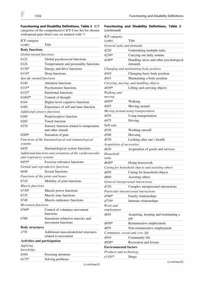
Functioning and Disability Definitions, Table 2 ICFcategories of the comprehensive ICF Core Set for chronicwidespread pain (brief core set marked with *)
ICF category(code) Title
Body functions
Global mental functions
b122 Global psychosocial functions
b126 Temperament and personality functions
b130* Energy and drive functions
b134* Sleep functions
Specific mental functions
b140 Attention functions
b147* Psychomotor functions
b152* Emotional functions
b1602* Content of thought
b164 Higher-level cognitive functions
b180 Experience of self and time function
Additional sensory functions
b260 Proprioceptive function
b265 Touch function
b270 Sensory function related to temperatureand other stimuli
b280* Sensation of pain
Functions of the haematological and immunologicalsystems
b430 Haematological system functions
Additional functions and sensations of the cardiovascularand respiratory systems
b455* Exercise tolerance functions
Genital and reproductive functions
b640 Sexual functions
Functions of the joints and bones
b710 Mobility of joint functions
Muscle functions
b730* Muscle power functions
b735 Muscle tone functions
b740 Muscle endurance functions
Movement functions
b760* Control of voluntary movementfunctions
b780 Sensations related to muscles andmovement functions
Body structures
s770 Additional musculoskeletal structuresrelated to movement
Activities and participation
Applyingknowledge
d160 Focusing attention
d175* Solving problems
(continued)
Functioning and Disability Definitions, Table 2(continued)
ICF category(code) Title
General tasks and demands
d220 Undertaking multiple tasks
d230* Carrying out daily routine
d240* Handling stress and other psychologicaldemands
Changing and maintaining body position
d410 Changing basic body position
d415 Maintaining a body position
Carrying, moving, and handling objects
d430* Lifting and carrying objects
Walking andmoving
d450* Walking
d455 Moving around
Moving around using transportation
d470 Using transportation
d475 Driving
Self-care
d510 Washing oneself
d540 Dressing
d570 Looking after one’s health
Acquisition of necessities
d620 Acquisition of goods and services
Householdtasks
d640* Doing housework
Caring for household objects and assisting others
d650 Caring for household objects
d660 Assisting others
General interpersonal interactions
d720 Complex interpersonal interactions
Particular interpersonal interactions
d760* Family relationships
d770* Intimate relationships
Work andemployment
d845 Acquiring, keeping and terminating ajob
d850* Remunerative employment
d855 Non-remunerative employment
Community, social and civic life
d910 Community life
d920* Recreation and leisure
Environmental factors
Products and technology
e1101* Drugs
(continued)
F 1332 Functioning and Disability Definitions
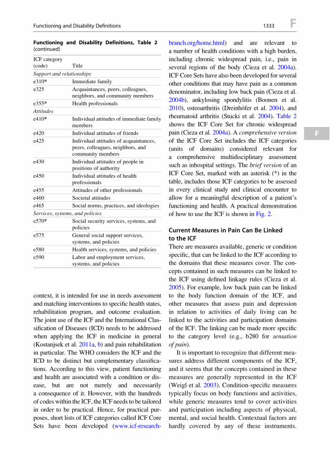
context, it is intended for use in needs assessment
andmatching interventions to specific health states,rehabilitation program, and outcome evaluation.
The joint use of the ICF and the International Clas-
sification of Diseases (ICD) needs to be addressedwhen applying the ICF in medicine in general
(Kostanjsek et al. 2011a, b) and pain rehabilitation
in particular. The WHO considers the ICF and theICD to be distinct but complementary classifica-
tions. According to this view, patient functioning
and health are associated with a condition or dis-ease, but are not merely and necessarily
a consequence of it. However, with the hundreds
of codeswithin the ICF, the ICF needs to be tailoredin order to be practical. Hence, for practical pur-
poses, short lists of ICF categories called ICF CoreSets have been developed (www.icf-research-
branch.org/home.html) and are relevant toa number of health conditions with a high burden,
including chronic widespread pain, i.e., pain inseveral regions of the body (Cieza et al. 2004a).
ICF Core Sets have also been developed for several
other conditions that may have pain as a commondenominator, including low back pain (Cieza et al.
2004b), ankylosing spondylitis (Boonen et al.
2010), osteoarthritis (Dreinhofer et al. 2004), andrheumatoid arthritis (Stucki et al. 2004). Table 2
shows the ICF Core Set for chronic widespread
pain (Cieza et al. 2004a). A comprehensive versionof the ICF Core Set includes the ICF categories
(units of domains) considered relevant for
a comprehensive multidisciplinary assessmentsuch as inhospital settings. The brief version of an
ICF Core Set, marked with an asterisk (*) in the
table, includes those ICF categories to be assessedin every clinical study and clinical encounter to
allow for a meaningful description of a patient’s
functioning and health. A practical demonstrationof how to use the ICF is shown in Fig. 2.
Current Measures in Pain Can Be Linkedto the ICFThere are measures available, generic or condition
specific, that can be linked to the ICF according tothe domains that these measures cover. The con-
cepts contained in such measures can be linked to
the ICF using defined linkage rules (Cieza et al.2005). For example, low back pain can be linked
to the body function domain of the ICF, and
other measures that assess pain and depressionin relation to activities of daily living can be
linked to the activities and participation domains
of the ICF. The linking can be made more specificto the category level (e.g., b280 for sensationof pain).
It is important to recognize that different mea-sures address different components of the ICF,
and it seems that the concepts contained in these
measures are generally represented in the ICF(Weigl et al. 2003). Condition-specific measures
typically focus on body functions and activities,
while generic measures tend to cover activitiesand participation including aspects of physical,
mental, and social health. Contextual factors are
hardly covered by any of these instruments.
Functioning and Disability Definitions, Table 2(continued)
ICF category(code) Title
Support and relationships
e310* Immediate family
e325 Acquaintances, peers, colleagues,neighbors, and community members
e355* Health professionals
Attitudes
e410* Individual attitudes of immediate familymembers
e420 Individual attitudes of friends
e425 Individual attitudes of acquaintances,peers, colleagues, neighbors, andcommunity members
e430 Individual attitudes of people inpositions of authority
e450 Individual attitudes of healthprofessionals
e455 Attitudes of other professionals
e460 Societal attitudes
e465 Social norms, practices, and ideologies
Services, systems, and policies
e570* Social security services, systems, andpolicies
e575 General social support services,systems, and policies
e580 Health services, systems, and policies
e590 Labor and employment services,systems, and policies
Functioning and Disability Definitions 1333 F
F

Nonetheless, there is a need to address all com-
ponents when assessing functioning and health in
patients with chronic health conditions (Ewertet al. 2004).
Finally, it is important to note that the perspec-
tive of functioning, disability, and health is differentwhen viewed from a medical versus functioning-
oriented perspective, e.g., in rehabilitation. From
the medical perspective, functioning and healthare seen primarily as a consequence of a disease
or condition. Accordingly, medical interventions
are targeted toward mitigation or elimination ofthe disease process. From a broader view, the mea-
surement of functioning, disability, and health is
required to evaluate patient-relevant outcomes ofan intervention. Hence, from a functioning-oriented
or rehabilitation perspective as depicted in the ICF,patients’ functioning and health are associated
with, but not merely a consequence of, a condition
or disease. Furthermore, functioning and health are
not only seen in association with a condition, butalso in associationwith personal and environmental
factors. Therefore, themeasurement of functioning,
disability, and health is relevant to evaluateintervention outcomes and for the diagnosis
(assessment) and overall clinical care. For the man-
agement of patients with pain conditions, consider-ation of the individual’s functioning is becoming
more and more critical than before.
Summary
In summary, the ICF can be used as a conceptual
framework and classification system to examine
and understand the impact and relation of pain
Activities
walking
standing
Participation
cannot go to university (study)
reduction of pain to enable resumption ofhigher education
Primary goal of rehabilitation:
cannot visit friends
cannot play soccer
sitting
bendingirritation of neuromeningealstructures
shortening of pelvic belt muscles
increased muscle tone of pelvic beltespecially of m. piriformis
iliosacral joint dysfunction
walking / standing
Environmental: lives in fourth floor flat; no elevator; lack of concentration due to medication
Personal: highly motivated for exercising
Body- Structures/Functions
reduced mobility of theleft leg
low back painpain in left leg
low back painMedical diagnosis:Name: Mr. Johnson
Age: 24
Hea
lth p
rofe
ssio
nal p
ersp
ectiv
e P
atie
nt p
ersp
ectiv
e
Contextualfactors:
Functioning and Disability Definitions,Fig. 2 Illustration of how the ICF components can beused to structure patient problems (listed in the uppersection “patient perspective”) as well as findings, andobservations by the rehabilitation team (listed in thelower section “health professional perspective”). Linesbetween the selected target problems from the patient’s
perspective (circled in the upper section, i.e., “walking/standing”), impaired body functions, and structures aswell as the given personal and environmental factors(in the lower section) denote their hypothesized associa-tive relationship. Note that the wording denotes patient’swords or special medical terms which can be linked backto specific domains of the ICF
F 1334 Functioning and Disability Definitions

with regard to the functioning and disability ofan individual. The ICF provides a holistic
biopsychosocial perspective that is beyond thetraditional biomedical model and, hence, can aid
in the thought process to guide clinical decision
making and clinical care in the context of painand pain-related conditions.
Acknowledgment Previous edition of this entry waswritten by Gerold Stucki and Thomas Ewert.
References
Boonen, A., Braun, J., van der Horst Bruinsma, I. E.,Huang, F., Maksymowych, W., Kostanjsek, N., Cieza,A., Stucki, G., & van der Heijde, D. (2010). ASAS/WHOICF Core Sets for ankylosing spondylitis (AS): How toclassify the impact of AS on functioning and health.Annals of the Rheumatic Diseases, 69(1), 102–107.
Cieza, A., Stucki, G., Weigl, M., Kullmann, L., Stoll, T.,Kamen, L., Kostanjsek, N., & Walsh, N. (2004a). ICFCore Sets for chronic widespread pain. Journal ofRehabilitation Medicine, (44 Suppl.), 63–68.
Cieza, A., Stucki, G.,Weigl, M., Disler, P., J€ackel,W., vander Linden, S., Kostanjsek, N., & de Bie, R. (2004b).ICF Core Sets for low back pain. Journal of Rehabil-itation Medicine, (44 Suppl.), 69–74.
Cieza, A., Geyh, S., Chatterji, S., Kostanjsek, N., Ust€un, B.,& Stucki, G. (2005). ICF linking rules: An update basedon lessons learned. Journal of Rehabilitation Medicine,37(4), 212–218.
Dreinhofer, K., Stucki, G., Ewert, T., Huber, E.,Ebenbichler, G., Gutenbrunner, C., Kostanjsek, N., &Cieza, A. (2004). ICF Core Sets for osteoarthritis. Jour-nal of Rehabilitation Medicine, (44 Suppl.), 75–80.
Ewert, T., Fuessl, M., Cieza, A., et al. (2004). Identifica-tion of the most common patient problems in patientswith chronic conditions using the ICF checklist. Jour-nal of Rehabilitation Medicine, 36, 186–188.
Kostanjsek, N., Escorpizo, R., Boonen, A., Walsh, N. E.,Ust€un, T. B., & Stucki, G. (2011a). Assessing theimpact of musculoskeletal health conditions using theinternational classification of functioning, disability
and health. Disability and Rehabilitation, 33(13–14),1281–1297.
Kostanjsek, N., Rubinelli, S., Escorpizo, R., Cieza, A.,Kennedy, C., Selb, M., Stucki, G., & €Ust€un, T. B.(2011b). Assessing the impact of health conditionsusing the ICF. Disability and Rehabilitation, 33(15–16), 1475–1482.
Nagi, S. Z. (1964). A study in the evaluation of disability andrehabilitation potential:Concepts,methods, andprocedures.The American Journal of Public Health, 54, 1568–1579.
Pope, A. M., & Tarlov, A. R. (1991). Disability inAmerica: Toward a national agenda for prevention.Washington, DC: National Academy Press.
Stucki, G., Ewert, T., & Cieza, A. (2002). Value andapplication of the ICF in rehabilitation medicine. Dis-ability and Rehabilitation, 24, 932–938.
Stucki, G., Cieza, A., Geyh, S., Battistella, L., Lloyd, J.,Symmons, D., Kostanjsek, N., & Schouten, J. (2004).ICF Core Sets for rheumatoid arthritis. Journal ofRehabilitation Medicine, (44 Suppl.), 87–93.
Weigl, M., Cieza, A., Harder, M., et al. (2003). Linkingosteoarthritis specific health status measures to the inter-national classification of functioning, disability andhealth (ICF). Osteoarthritis and Cartilage, 11, 1–5.
WHO. (1980). ICIDH. International Classification of Impair-ments, Disabilities and Handicaps. Geneva: WHO.
WHO. (2001). International classification of functioning,disability and health: ICF. Geneva: WHO.
Funiculus
Definition
Longitudinal subdivisions of the spinal whitematter that are named according to their location
within the spinal cord.
Cross-References
▶ Spinothalamic Projections in Rat
Funiculus 1335 F
F