f rita isabel costa cunha i a - repositorium.sdum.uminho.pt · escola de ciências rita isabel...
TRANSCRIPT
UM
inho 2
012
Rita Isabel Costa Cunha
Rita I
sabel
Cost
a C
unha
The mechanism mediating nuclear
translocation of the apoptosis
inducing factor (AIF)
Th
e m
ech
an
ism
me
dia
tin
g n
ucl
ea
r tr
an
slo
cati
on
of
the
ap
op
tosi
s in
du
cin
g f
act
or
(AIF
)

Universidade do Minho
Escola de Ciências
Rita Isabel Costa Cunha
The mechanism mediating nuclear
translocation of the apoptosis
inducing factor (AIF)
Tese de Mestrado em Genética Molecular
Trabalho efectuado sob a orientação de
Professora Doutora Manuela Côrte-Real
Doutora Susana Chaves
Outubro de 2012
DECLARAÇÃO
Nome: Rita Isabel Costa Cunha
Endereço eletrónico: [email protected]
Telefone: 917875769
Nº do Bilhete de Identidade: 13200058
Título da Tese de Mestrado:
The mechanism mediating nuclear translocation of the apoptosis inducing factor (AIF)
Orientadores:
Professora Doutora Manuela Côrte-Real
Doutora Susana Chaves
Instituição de Acolhimento:
Centro de Biologia Molecular e Ambiental (CBMA)
Ano de Conclusão: 2012
Designação do Mestrado:
Mestrado em Genética Molecular
1. É AUTORIZADA A REPRODUÇÃO INTEGRAL DESTA TESE, APENAS PARA EFEITOS
DE INVESTIGAÇÃO, MEDIANTE DECLARAÇÃO ESCRITA DO INTERESSADO, QUE A
TAL SE COMPROMETE.
Universidade do Minho, 31 de Outubro de 2012
_____________________________________________
Rita Isabel Costa Cunha
Agradecimentos
iii
Agradecimentos
Às minhas orientadoras, Professora Doutora Manuela Côrte-Real e Doutora Susana
Chaves. À Professora Manuela por toda a disponibilidade e carinho, pela partilha dos
conhecimentos e do rigor científico, pela dedicação e pelas palavras sempre sábias nos
momentos oportunos. À Susana por toda a preocupação e disponibilidade, por todas as
sugestões diárias na resolução dos problemas que foram ocorrendo ao longo deste trabalho.
Principalmente por ter feito que este último ano tenha sido de muita aprendizagem.
Um muito obrigado sincero às duas pela oportunidade.
Ao CBMA e ao Departamento de Biologia e a todos os seus funcionários e docentes.
Um especial à Dona Isabel e à Nádia por toda a boa disposição e favores prestados.
A todos os colegas da Micro I por me terem acolhido e proporcionado um excelente
ambiente de trabalho. Por todos os “cafés” recheados de gargalhadas e também pelas
sugestões na realização do trabalho experimental e pelo companheirismo. Por isso um muito
obrigado à Sara Alves, Dário Trindade, Helena Paula , Ana Marta Duarte, Jorge Rodrigues,
Flávio Azevedo, Tânia Fernandes, Rita Pacheco e Vera Martins.
E porque as frases mais ditas por mim neste ano foram: “Oh Andreiaaa sabes...?” ou “Oh
Ruizinhoo como é que…?”, um obrigado especial ao Rui Silva e Andreia Pacheco por
basicamente me ajudarem em tudo o que foi preciso, e também por me animarem sempre que
preciso.
Um agradecimento também aos arredores da Micro I: Filipa Pereira, Raúl Machado,
Joana Sá Pessoa, Filipa Vale, Manoel, Gabriel Rocha, Suellen Ferro, Lisandra Castro, Fábio
Faria e Paulo Geraldes. Obrigado pela ajuda e boa disposição.
E como nada se consegue sem o apoio dos amigos, agradeço aos meus amigos do
coração que tanto me aturaram este ano: Catherine Ferreira, Dulce Cunha, Filipe Pires, Joana
Tulha, André Charrua, Bruno Fernandes, Juliana Fernandes, Carla Sampaio, Bruno Freitas,
Cristina Real, Bruno Cunha, Joana Campos e Carina Rego. Obrigado pelo companheirismo,
incentivo, carinho, boa disposição e amizade constantes. Um obrigado especial ao meu
Pedrinho por me aguentar 24 sobre 24 horas por dia e mesmo assim ter sempre um gesto e
uma palavra de carinho! Obrigado Maninho!
Ao Touni por para além de ser companheiro de laboratório, amigo, ser o meu
companheiro para a VIDA! OBRIGADA por me aturares mais do que todos, por toda a ajuda,
carinho e apoio e por nunca desistires de mim!
E como os últimos são sempre os primeiros... À minha família (Papás, Maninho, Jú,
Madrinha e Padrinho, Pipa e Tios) por serem quem são! Obrigado pelo amor, alegria, atenção,
confiança, apoio e por me ensinarem a ser como sou.
Abstract
iv
The mechanism mediating nuclear translocation of the apoptosis inducing factor (AIF)
Abstract
Since the discovery that yeast cells can undergo programmed cell death in response to
several different stimuli, Saccharomyces cerevisiae has gained prominence in the cell death
field. Exposure of yeast cells to certain stimuli like acetic acid or hydrogen peroxide or even
heterologous expression of pro-apoptotic proteins can trigger cell death by apoptosis via a
mitochondrial pathway and exhibiting the typical hallmarks of apoptosis, such as chromatin
condensation, ROS (reactive oxygen species) accumulation, DNA fragmentation,
externalization of phosphatidylserine, mitochondrial dysfunction with release of cytochrome c
and of apoptosis inducing factor (Aif1p), among other pro-apoptotic proteins.
Aif1p is a flavoprotein that is involved both in cell survival and cell death. In healthy
cells, Aif1p is confined to the mitochondria, where it plays a role in bioenergetic and redox
metabolism due to its redox activity and its FAD and NADH domains. However, when cells are
exposed to an apoptotic stimulus, Aif1p is released from mitochondria to the cytosol and then
translocated to the nucleus, where it will induce DNA fragmentation and chromatin
condensation. Although the mechanism underlying this translocation is still poorly understood, it
is known that transport into the nucleus through the nuclear pore complexes is an energy-
dependent process for the majority of macromolecules, and normally mediated by the major
class of transport receptors, the karyopherins or Kaps. In this work, we aimed elucidate the
mechanism regulating yeast Aif1p import into the nucleus and to discover which Kap is
responsible for the nuclear import of Aif1p.
Apoptosis was triggered in cells expressing Aif1p tagged with GFP (by exposure to
acetic acid or hydrogen peroxide, chronological ageing, or heterologous expression of Bax c-
myc protein) and Aif1p localization was assessed by fluorescence microscopy and cellular
fractionation/western blot assays. However, we were not able to observe the release of Aif1p to
the cytosol or its nuclear import under our experimental conditions. We also attempted to map
the NLS domain of Aif1p by deletion of several domains with homology to previously mapped
domains in human AIF. Although we has not yet mapped the Aif1p NLS domain, we discovered
that deletion of the putative Hsp70p-binding domain led to aberrant localization of Aif1p,
suggesting that Hsp70p is important for Aif1p folding and stability. These results indicate that
Hsp70p may also be involved in the regulation of Aif1p localization in yeast.
Resumo
v
Mecanismo mediador da translocação nuclear do fator de indução de apoptose (AIF)
Resumo
Células de Saccharomyces cerevisiae são capazes de desencadear um processo de
morte celular programada em resposta a vários estímulos. Esta descoberta levou à utilização
extensiva deste organismo eucarionte unicelular como modelo no estudo de processos de
morte celular. Sabe-se que a exposição de células de levedura a certos estímulos, como o
ácido acético ou peróxido de hidrogénio, ou mesmo a expressão heteróloga de proteínas pró-
apoptóticas de mamífero, pode desencadear um tipo morte celular dependente de uma via
mitocondrial e exibindo características típicas da morte apoptótica, tais como a condensação
da cromatina, acumulação de espécies reativas de oxigénio (ROS), fragmentação do DNA,
externalização de fosfatidilserina e disfunção mitocondrial associada à libertação de citocromo
c e do fator indutor de apoptose (Aif1p).
Aif1p é uma flavoproteína que está envolvida tanto na sobrevivência como na morte
celular. Em células saudáveis, esta proteína está confinada à mitocôndria, onde desempenha
um importante papel no metabolismo bioenergético e estado redox. Este facto é devido à sua
atividade redox e aos seus domínios FAD e NADH. No entanto, em células expostas a um
estímulo apoptótico, ocorre libertação de Aif1p da mitocôndria para o citosol e
subsequentemente a sua translocação para o núcleo, onde induz fragmentação do DNA e
condensação da cromatina. Embora o mecanismo subjacente a esta translocação esteja ainda
pouco conhecido e estudado, sabe-se que para a maioria das macromoléculas o transporte
para o núcleo ocorre através dos complexos de poros nucleares e é um processo dependente
de energia, normalmente mediado por uma classe de recetores de transporte, as Kaps (do
inglês “karyopherins”, também conhecidas como importinas ou transportinas). Este trabalho
teve como objectivo elucidar a regulação do mecanismo de transporte de Aif1p para o núcleo e
identificar que/ais Kap(s) são responsáveis pelo importe do Aif1p para o núcleo na levedura.
A morte celular apoptótica foi induzida em células que expressam uma fusão da
proteína Aif1 com GFP (por exposição a ácido acético ou peróxido de hidrogénio,
envelhecimento cronológico, ou pela expressão heteróloga de proteínas Bax c-myc) e a
localização de Aif1p foi avaliada por microscopia de fluorescência e fracionamento celular
/western Blot. No entanto, não fomos capazes de observar libertação de Aif1p para o citosol ou
a sua importação nuclear nas condições experimentais utilizadas. Tentámos também mapear o
domínio NLS de Aif1p através da remoção de vários domínios homólogos com domínios
conhecidos de AIF humano. Embora não tenhamos ainda conseguido mapear o domínio NLS
do Aif1p, descobrimos que a deficiência no domínio putativo de ligação a Hsp70 resulta numa
localização aberrante de Aif1p, sugerindo que a Hsp70 é importante para a estabilidade desta
proteína e que esta proteína pode ainda estar envolvida na regulação da localização de Aif1p
na levedura.
Index
vi
Index
Agradecimentos ........................................................................................................................... iii
Abstract ........................................................................................................................................ iv
Resumo .......................................................................................................................................... v
Index.............................................................................................................................................. vi
Abbreviations ............................................................................................................................. viii
1. Introduction ............................................................................................................................... 1
1.1. Apoptosis ............................................................................................................................... 2
1.1.1. Mechanism of apoptosis ............................................................................................. 2
1.1.1.1. Extrinsic pathway ...................................................................................................... 3
1.1.1.2. Intrinsic pathway ...................................................................................................... 4
1.2. Apoptosis inducing factor (AIF) .......................................................................................... 7
1.2.1. The role of AIF in cells ................................................................................................ 8
1.2.2. The mechanism underlying AIF release from mitochondria ................................ 10
1.2.3. Heat shock protein 70 (Hsp70p) acts as a chaperone to AIF protein .................. 11
1.3. Yeast apoptosis .................................................................................................................. 12
1.3.1. Yeast apoptotic triggers ........................................................................................... 14
1.3.2. Yeast orthologue of AIF, Aif1p ................................................................................. 15
1.3.2.1. Stimuli that regulate Aif1p release ....................................................................... 15
1.4. Nucleocytoplasmic trafficking .......................................................................................... 16
1.4.1. Nuclear Pore Complex (NPC) ................................................................................... 16
1.4.2. Protein transport receptors and transport cycle ................................................... 18
1.4.3. Regulation of nucleocytoplasmic trafficking ......................................................... 21
1.4.4. Nuclear import of AIF ................................................................................................ 22
2. Aims and research plan ......................................................................................................... 24
3. Materials and methods ........................................................................................................... 27
3.1. Plasmids ............................................................................................................................... 28
Index
vii
3.2. Yeast strains and growth conditions................................................................................. 30
3.3. Hydrogen peroxide and acetic acid treatment ................................................................. 30
3.4. Heterologous expression of Bax c-myc ............................................................................ 30
3.5. Transformation of bacterial cells ....................................................................................... 31
3.6. Purification of Yeast DNA ................................................................................................... 32
3.7. Fluorescence Microscopy .................................................................................................. 32
3.8. Subcellular fractionation .................................................................................................... 32
3.8.1. Preparation of spheroplasts ..................................................................................... 32
3.8.2. Mitochondrial and cytosolic fraction preparation .................................................. 33
3.8.3. Nuclear fraction preparation ..................................................................................... 33
3.8.3.1. Protocol I .................................................................................................................. 33
3.8.3.2. Protocol II ................................................................................................................. 34
3.8.3.3. Protocol III ................................................................................................................ 34
3.8.4. SDS gel electrophoresis/Westernblot ...................................................................... 34
3.9. Immunoprecipitation ........................................................................................................... 35
3.9.1. SDS gel electrophoresis/Western Blot/Silver Staining .......................................... 35
4. Results ..................................................................................................................................... 38
4.1. Construction of pAIF1WT-GFP plasmid and localization AIF1p ..................................... 39
4.2. Acetic acid treatment and chronological ageing ............................................................. 40
4.3. Hydrogen peroxide treatment ............................................................................................ 41
4.4. Heterologous expression of Bax c-myc protein ............................................................... 42
4.5. Aif1p translocation to the nucleus and cytochrome c release ....................................... 43
4.6. Map the NLS of Aif1p........................................................................................................... 48
4.6.1. Construction of AIF1 fragments and localization of Aif1p .................................... 48
4.7. Protein-protein interactions of Aif1p ................................................................................. 51
5. Discussion and future perspectives ..................................................................................... 54
6. Literature cited ........................................................................................................................ 63
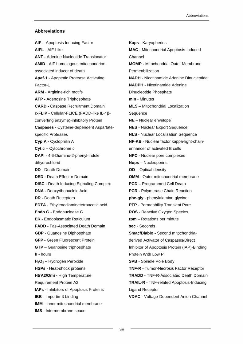
Abbreviations
viii
Abbreviations
AIF – Apoptosis Inducing Factor
AIFL - AIF-Like
ANT - Adenine Nucleotide Translocator
AMID - AIF homologous mitochondrion-
associated inducer of death
Apaf-1 - Apoptotic Protease Activating
Factor-1
ARM - Arginine-rich motifs
ATP - Adenosine Triphosphate
CARD - Caspase Recruitment Domain
c-FLIP - Cellular-FLICE (FADD-like IL-1β-
converting enzyme)-inhibitory Protein
Caspases - Cysteine-dependent Aspartate-
specific Proteases
Cyp A - Cyclophilin A
Cyt c – Cytochrome c
DAPI - 4,6-Diamino-2-phenyl-indole
dihydrochlorid
DD - Death Domain
DED - Death Effector Domain
DISC - Death Inducing Signaling Complex
DNA - Deoxyribonucleic Acid
DR - Death Receptors
EDTA - Ethylenediaminetetraacetic acid
Endo G - Endonuclease G
ER - Endoplasmatic Reticulum
FADD - Fas-Associated Death Domain
GDP - Guanosine Diphosphate
GFP - Green Fluorescent Protein
GTP – Guanosine triphosphate
h - hours
H2O2 – Hydrogen Peroxide
HSPs - Heat-shock proteins
HtrA2/Omi - High Temperature
Requirement Protein A2
IAPs - Inhibitors of Apoptosis Proteins
IBB - Importin-β binding
IMM - Inner mitochondrial membrane
IMS - Intermembrane space
Kaps - Karyopherins
MAC - Mitochondrial Apoptosis-induced
Channel
MOMP - Mitochondrial Outer Membrane
Permeabilization
NADH - Nicotinamide Adenine Dinucleotide
NADPH - Nicotinamide Adenine
Dinucleotide Phosphate
min - Minutes
MLS – Mitochondrial Localization
Sequence
NE – Nuclear envelope
NES - Nuclear Export Sequence
NLS - Nuclear Localization Sequence
NF-KB - Nuclear factor kappa-light-chain-
enhancer of activated B cells
NPC - Nuclear pore complexes
Nups – Nucleoporins
OD – Optical density
OMM - Outer mitochondrial membrane
PCD – Programmed Cell Death
PCR - Polymerase Chain Reaction
phe-gly - phenylalamine-glycine
PTP - Permeability Transient Pore
ROS - Reactive Oxygen Species
rpm – Rotations per minute
sec - Seconds
Smac/Diablo - Second mitochondria-
derived Activator of Caspases/Direct
Inhibitor of Apoptosis Protein (IAP)-Binding
Protein With Low Pi
SPB - Spindle Pole Body
TNF-R - Tumor-Necrosis Factor Receptor
TRADD - TNF-R-Associated Death Domain
TRAIL-R - TNF-related Apoptosis-Inducing
Ligand Receptor
VDAC - Voltage-Dependent Anion Channel


1. INTRODUCTION
Introduction
2
1.1. Apoptosis
Multicellular organisms often need to eliminate cells that are in excess, or
damaged cells that can put the organism at risk (Hengartner, 2000). To this end, they
use a highly regulated and complex process characterized by a group of endogenous
molecular events that culminates in “cell suicide” (Leist et al., 2001), designated by
programmed cell death (PCD). Several types of cell death have been associated with
PCD, but in eukaryotic cells the most common is apoptosis (Hengartner, 2000).
Apoptosis occurs during normal development of multicellular organisms and continues
through adult life. This process is as important as cell division or cell migration because
it allows the organism to control cell number and tissue size, as well as protect itself
from compromised cells that can threaten homeostasis, such as infected or damaged
cells (Gewies, 2003).
The term apoptosis derives from the Greek “apo” - from and “ptosis” – falling, an
analogy to the term used by Greeks to describe leaves falling from trees. This term was
used in 1972 by John Kerr and colleagues to describe a type of cell death with distinct
morphologic characteristics (Lawen, 2003). Indeed, apoptotic cells can easily be
recognized by several morphological features such as cell shrinking, chromatin
condensation and migration along the nuclear membrane, blebbing of the plasma
membrane and exposure of phosphatidylserine to the outer leaflet of the plasma
membrane. The final hallmark of apoptosis is cell fragmentation into compact
structures, called 'apoptotic bodies' that will be phagocyted by macrophages and
eliminated from the tissue without leading to an inflammatory response (Saraste et al.,
2000).
1.1.1. Mechanism of apoptosis
Apoptosis can be triggered by various stimuli and mechanisms, including virus
infection, cell stress and DNA damage. Cellular sensitivity to these stimuli can differ
depending on several factors, such as expression of anti- and pro-apoptotic proteins,
the harshness of the stimulus and the phase of the cell cycle (Gewies, 2003). The
apoptotic process consists of three consecutive steps: (i) a trigger by extracellular or
intracellular stimuli; (ii) execution by activation of intracellular proteases and (iii)
elimination of dead cells by engulfment of cell debris by neighboring cells or
macrophages (Saikumar et al., 1999). Deficient regulation of apoptosis can lead to
various pathologies (Rudin and Thompson, 1997); improper activation may cause or
contribute to several diseases such as ischemic strokes, AIDS (acquired
Introduction
3
immunodeficiency syndrome), and several neurodegenerative disorders (Raff et al.,
1993, Ameisen et al., 1995 and Smale et al., 1995). In contrast, a flawed activation of
this process can lead to some autoimmune diseases and to uncontrolled cell division
that culminates in the development of cancers (Tan, 1994; Lowe and Lin, 2000 and
Reed, 2003).
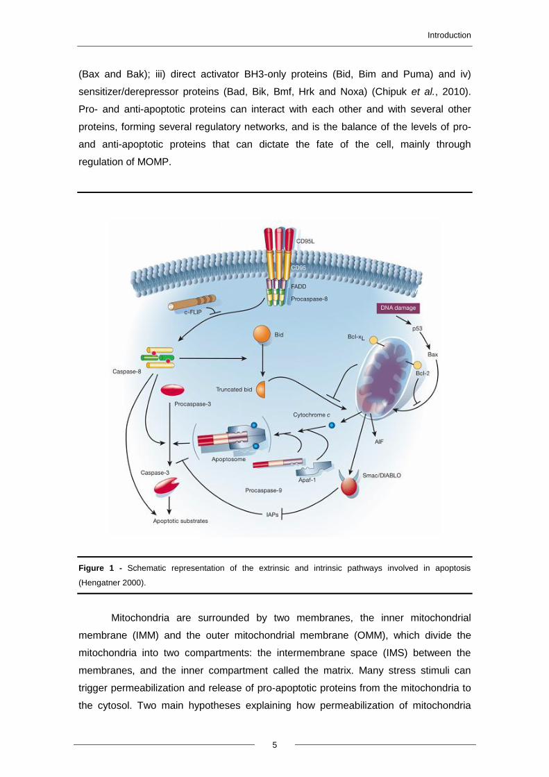
Two major apoptotic pathways have been identified in mammalian cells: the
extrinsic or death receptor pathway and the intrinsic or mitochondrial pathway (Figure
1) (Hengatner 2000 and Elmore, 2007).
1.1.1.1. Extrinsic pathway
In the extrinsic pathway, “death receptors” are activated to transmit apoptotic
signals from the exterior to the interior of cells. Examples of death receptors are TNFR-
1, Fas/CD95 and TRAIL receptors. Death receptors belonging to the tumor necrosis
factor receptor (TNFR) gene superfamily are present in the cell surface and transmit
the apoptotic signal after ligation of precise ligands such as Fas ligand, TNF alpha and
TRAIL. They have an important role in apoptosis because they can trigger a caspase
cascade within seconds of ligand binding and induce apoptosis very quickly (Sartorius
et al., 2001).
Caspases (Cysteine-dependent Aspartate-specific Proteases) are conserved
through evolution and can be found in several organisms such as insects, human,
hydra and nematodes (Cohen, 1997). They are synthesized as inactive zymogens,
named procaspases. They have a prodomain followed by a small and a large subunit
that are occasionally separated by a linker peptide. After maturation, procaspases are
proteolytically processed between the large and the small subunit. An active caspase is
a heterotetramer that consists of two large and two small subunits (Hengartner, 2000).
Their active site contains a cysteine residue critical for catalytic activity; it has high
affinity for aspartate residues, after which caspases cleave their substrates. So far,
about a hundred caspases substrates have been described (Fischer et al., 2003).
There are two different groups of caspases: (i) initiator caspases, which include
the procaspases -2, -8, -9 and –10, with long prodomains such as death effector
domains (DED) in procaspase-8 and -10 or caspase recruitment domains (CARD) in
procaspases-9 and -2; (ii) effector or executioner caspases, with only short
prodomains, which include procaspases -3, -6 and -7 (Chrowdhury et al., 2008).
In the extrinsic pathway, initiation of apoptosis requires both caspases and
death receptors. Apoptotic signaling is mediated by the death domain (DD) of the death
Introduction
4
receptor. Adapter molecules that possess their own DDs, as FADD or TRADD, are
then recruited to the DDs of the death receptor to form the death-inducing signaling
complex (DISC). With the help of FADD, procaspase 8 (initiator procaspase) is then
recruited to the DISC, and the amount of procaspase-8 molecules in the DISC leads to
their autocatalytic activation and consequent release of active caspase-8. Active
caspase-8 then activates the caspase signaling cascade; subsequently, effector
caspases cleave specific substrates, resulting in cell death (Stegh et al., 1998; Scaffidi
et al., 1998). It is possible to inhibit caspase-8 in order to protect cells from apoptosis.
One example is cellular-FLICE (FADD-like IL-1β-converting enzyme)-inhibitory protein
(c-FLIP), which can inhibit DISC signaling. This inhibition will lead to inactivation of
DISC and by consequence inhibition of the extrinsic pathway (Hengartner, 2000). In
some cases, the apoptotic signal produced by death receptors might not be capable of
inducing a caspase signaling cascade strong enough to lead to cell death on its own. In
other cases, there may be a bridge between the caspase signaling cascade and
mitochondria provided by Bid, a Bcl-2 family member (see below). Bid can be cleaved
into its truncated form (tBID) by caspase-8 and translocated to the mitochondria, where
it induces the release of pro-apoptotic proteins to the cytosol (Luo et al., 1998 and
Gustafsson et al., 2007).
1.1.1.2. Intrinsic pathway
The intrinsic pathway is triggered by “intrinsic” stresses such as DNA damage,
endoplasmic reticulum stress, lysosomal stress, and mitochondrial dysfunction. In
contrast with the extrinsic pathway, in which caspases are activated directly, the
intrinsic pathway requires the participation of mitochondria. Permeabilization of the
outer mitochondrial membrane (MOMP) and the release of pro-apoptotic factors are
crucial events of the intrinsic pathway and required for activation the downstream
caspase cascade. Mitochondria have thus gained great importance in the field of
mammalian apoptosis, since they are able to amplify the apoptotic signal from the
extrinsic pathway and also propagate the death signals generated within the cell, such
as oxidative stress, DNA damage and others (Wang, 2001).
The Bcl-2 family, a group of apoptotic regulators with both anti- and pro-
apoptotic members, plays a major role in the regulation of the intrinsic pathway. Bcl-2
family members share homologous regions known as BH domains, and can be divided
into four categories based on structural and functional similarities, namely: i) the anti-
apoptotic Bcl-2 proteins (A1, Bcl-2, Bcl-w, Bcl-xL and Mcl-1); ii) Bcl-2 effector proteins
Introduction
5
(Bax and Bak); iii) direct activator BH3-only proteins (Bid, Bim and Puma) and iv)
sensitizer/derepressor proteins (Bad, Bik, Bmf, Hrk and Noxa) (Chipuk et al., 2010).
Pro- and anti-apoptotic proteins can interact with each other and with several other
proteins, forming several regulatory networks, and is the balance of the levels of pro-
and anti-apoptotic proteins that can dictate the fate of the cell, mainly through
regulation of MOMP.
Figure 1 - Schematic representation of the extrinsic and intrinsic pathways involved in apoptosis
(Hengatner 2000).
Mitochondria are surrounded by two membranes, the inner mitochondrial
membrane (IMM) and the outer mitochondrial membrane (OMM), which divide the
mitochondria into two compartments: the intermembrane space (IMS) between the
membranes, and the inner compartment called the matrix. Many stress stimuli can
trigger permeabilization and release of pro-apoptotic proteins from the mitochondria to
the cytosol. Two main hypotheses explaining how permeabilization of mitochondria
Introduction
6
occurs have been put forth. The first hypothesis suggests that a transmembrane
channel named the permeability transition pore (PTP) is formed on the contact sites
between the IMM and the OMM. These pores are thought to be mainly constituted by
VDAC in the OMM and by adenine nucleotide translocator (ANT) in the IMM. PTP
opening results in loss of membrane potential, uptake of solutes and entry of water to
the matrix, leading to the rupture of the outer membrane and release of pro-apoptotic
proteins from the IMS to the cytoplasm (Lawen, 2003). Another hypothesis is based on
the ability of the pro-apoptotic Bcl-2 proteins to form pores. Indeed, the accepted model
proposes a stepwise structural reorganization of Bax leading to mitochondrial targeting
and homo-oligomerization. Bax is kept as a monomeric soluble cytosolic factor through
the engagement of its α9 helix in the dimerization pocket by the α1 helix. The activator
BH3s, tBID/BIM/PUMA, attack and expose the α1 helix of Bax, leading to a secondary
disconnection of the α9 helix and consequently mitochondrial insertion. Activator BH3s
stay associated with the N-terminally exposed Bax through the BH1 domain to drive
homo-oligomerization and activation of Bax (Ren et al., 2010).
Permeabilization of mitochondria results in the release of several proteins such
as cytochrome c, Omi/HtrA2 (High Temperature Requirement Protein A2) and
Smac/DIABLO (Second Mitochondria-derived Activator of Caspases/Direct Inhibitor of
Apoptosis Protein (IAP)-binding Protein With Low pI). Later, the apoptosis inducing
factor (AIF) and endonuclease G (EndoG) are also released. All these proteins are
located in the mitochondrial intermembrane space (IMS) and are released to the
cytosol in response to several apoptotic stimuli, such as extra- and intracellular
stresses like oxidative stress and treatment with cytotoxic drugs (Vaux, 2011). Proteins
of the first group activate the caspase-dependent mitochondrial pathway. After its
release to the cytosol, cytochrome c can interact with Apaf-1 (Protease Activating
Factor – 1), leading to the recruitment of pro-caspase 9, which together with dATP
forms a multi-protein complex, the apoptosome, which in turn activates caspase -9
leading to apoptosis. On the other hand, Smac and Omi/HtrA2 normally neutralize IAP
(Inhibitors of Apoptosis Proteins), which normally inhibit effector caspases. Pro-
apoptotic proteins of the second group are released only after the cell has committed to
die. Both AIF and Endo G are translocated to the nucleus (Joza et al., 2001). AIF
induces DNA fragmentation and chromatin condensation, whereas Endo G induces
DNA internucleossomal fragmentation (Wang, 2001).
Introduction
7
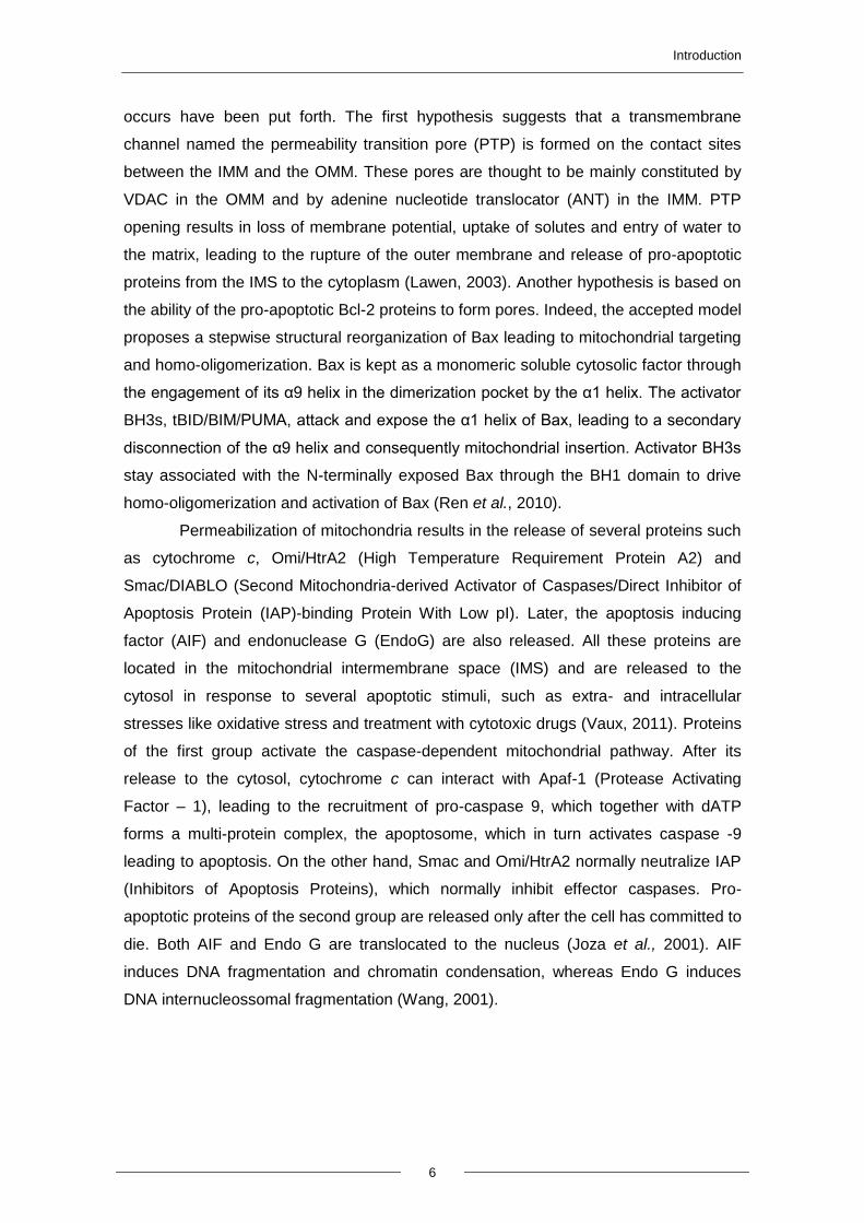
1.2. Apoptosis inducing factor (AIF)
The mammalian mitochondrial AIF is a flavoprotein that belongs to a larger
family of proteins with common structural and functional features, containing bacterial,
plant and fungal oxidoreductases. It is a protein with 613 aminoacids and is structurally
divided into three typical domains: a FAD and a NADH binding domain and a C-
terminal domain, where its pro-apoptotic activity resides. In addition, it also possesses
an N-terminal MLS (Mitochondria Localization Signal) (Figure 2) (Gurbuxani et al.,
2003). Besides AIF, AIF homologous mitochondrion-associated inducer of death
(AMID) and AIF-Like (AIFL) also belong to this family (Hangen et al., 2010).
Figure 2 – Representation of the three different AIF forms: the precursor, the mature and the truncated
AIF. AIF contains an N-terminal MLS domain (green), a FAD bipartite domain (yellow), an NADH binding
domain (violet) and a C-terminal domain (red). It also possesses an Hsp70p and a Cyclophilin A binding
domain. Cleavage at the MPP (Mitochondrial Processing Peptidase) cleavage site (blue dotted line)
generates mitochondrial mature AIF and cleavage by calpains (at the red dotted line) generates truncated
AIF (tAIF) (Yuste et al., 2007).
In healthy cells, AIF is confined to mitochondria, where it plays an important role
in bioenergetic and redox metabolism. However, after an apoptotic stimulus, AIF is
released from mitochondria to the cytosol and translocated into the nucleus
(Modjtahedi et al., 2006).
Introduction
8
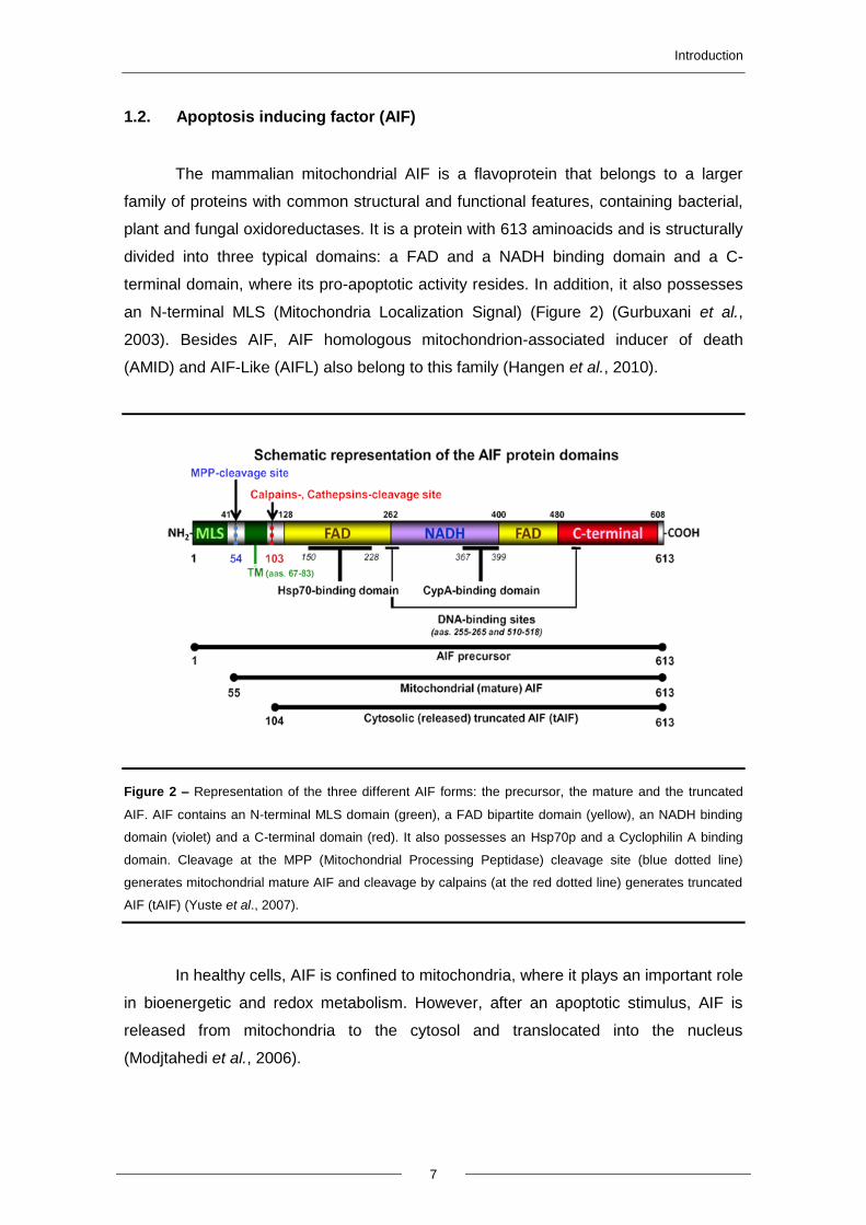
1.2.1. The role of AIF in cells
AIF has dual functions, in DNA fragmentation and in redox activity, and thus
acts both in cell death and cell survival. However, biochemical and mutational analysis
of AIF suggests that its apoptotic and redox functions can be separated (Lipton et al.,
2002). Three distinct functions have been associated with AIF, as seen in figure 3.
Figure 3 - Representation of the three different functions of AIF in cells. First, AIF has a role as an NADH
oxidase. Second, cytosolic AIF seems to promote mitochondrial membrane permeabilization and third, AIF
can promote chromatin condensation and DNA fragmentation (Candé et al., 2002).
The first function attributed to AIF is it NADH oxidase activity. AIF has a redox
potential of –308 mV ± 15 mV at pH 7.5. It has monodehydroascorbate reductase
activity and catalyses cytochrome c reduction in the presence of NADH. Results have
shown that the AIF NADH oxidase activity requires an electron donor (NADH), an
electron acceptor and a prosthetic FAD group for catalytic electron transfer (Miramar et
al., 2001).
The second function is permeabilization of the mitochondrial membrane and
consequent release of cytocrome c and additional AIF. This function is attributed to
cytosolic AIF, since studies revealed that when AIF is introduced into the cytosol it can
induce the release of AIF and cytochrome c from mitochondria (Daugas et al., 2000).
When cells commit to apoptosis, AIF translocates to the nucleus, where it exerts its
third function: to trigger cell death by promoting chromatin condensation and DNA
fragmentation (Candé et al., 2002). Several reports suggest that this role of AIF in the
Introduction
9
nucleus functions as a backup to caspase-dependent mechanisms. However, it was
also described that AIF-induced cell death occurs in the complete absence of caspases
and the oxireductase activation. AIF seems to play a role in caspase-independent PCD
in several organisms such as Caenorhabditis elegans, Dyctiostelium discoideum and
mammals (Arnoult et al., 2001, Wang et al., 2007, Wang et al., 2002 and Lorenzo et
al., 2007).
But how are these effects achieved at the molecular level? The crystal structure
of AIF shows that the surface of the protein has positively charged amino acids,
allowing it to form electrostatic connections with DNA. AIF binds with more affinity to
linearized forms than to intact circular plasmids, suggesting that it is introduced into the
DNA strand breaks. The AIF-DNA interaction is accompanied by DNA condensation
(shortening of the DNA), hairpin formation (intermolecular packageing) and DNA
oligomerization (Modjtahedi et al., 2006). Studies have shown that addition of AIF to
DNA does not cause DNA degradation. It was therefore suggested that after entering
the nucleus AIF can cooperate with several exo- and endonucleases, building up a so-
called “degradeosome” including cyclophilins, proteins belonging to the family of
peptidylprolyl cis–trans isomerases. Human AIF is able to interact with cyclophilin A
(CypA) and forms an active DNase, and several studies confirm that CypA is essential
for the apoptotic activity of AIF (Candé et al., 2004). These characteristics can be
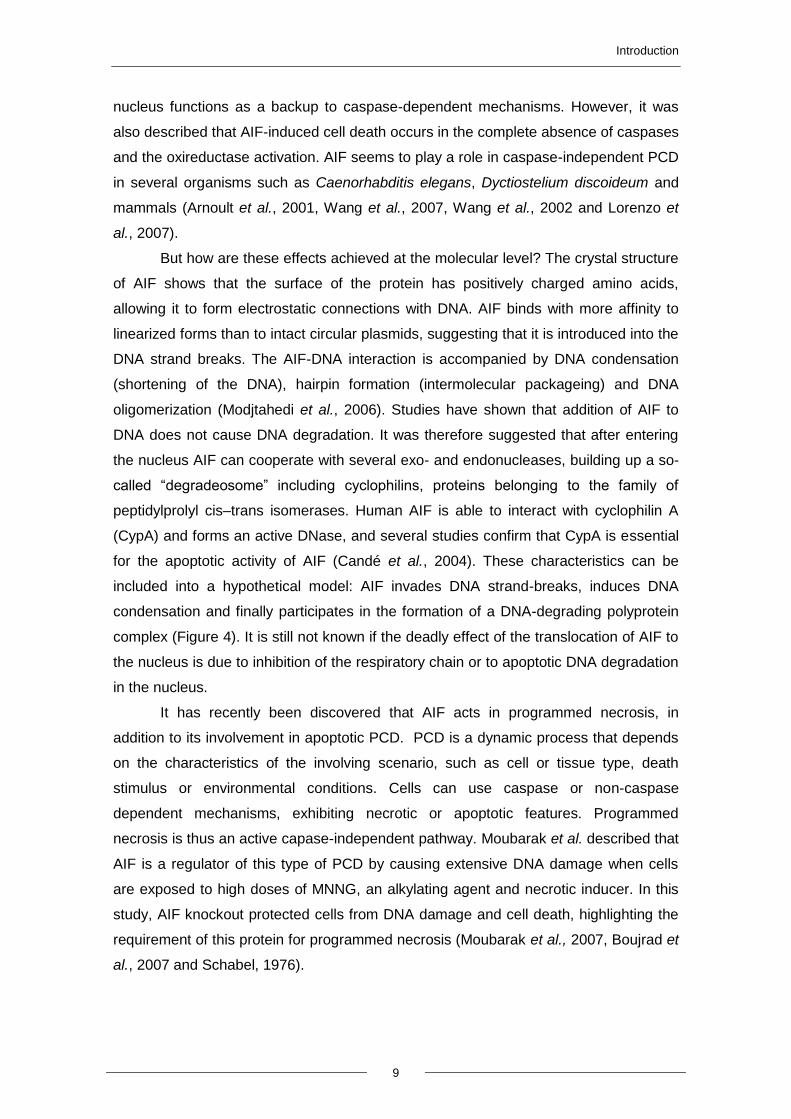
included into a hypothetical model: AIF invades DNA strand-breaks, induces DNA
condensation and finally participates in the formation of a DNA-degrading polyprotein
complex (Figure 4). It is still not known if the deadly effect of the translocation of AIF to
the nucleus is due to inhibition of the respiratory chain or to apoptotic DNA degradation
in the nucleus.
It has recently been discovered that AIF acts in programmed necrosis, in
addition to its involvement in apoptotic PCD. PCD is a dynamic process that depends
on the characteristics of the involving scenario, such as cell or tissue type, death
stimulus or environmental conditions. Cells can use caspase or non-caspase
dependent mechanisms, exhibiting necrotic or apoptotic features. Programmed
necrosis is thus an active capase-independent pathway. Moubarak et al. described that
AIF is a regulator of this type of PCD by causing extensive DNA damage when cells
are exposed to high doses of MNNG, an alkylating agent and necrotic inducer. In this
study, AIF knockout protected cells from DNA damage and cell death, highlighting the
requirement of this protein for programmed necrosis (Moubarak et al., 2007, Boujrad et
al., 2007 and Schabel, 1976).
Introduction
10
Figure 4 - Hypothesized model for the action of AIF on DNA (Modjtahedi et al, 2006).
1.2.2. The mechanism underlying AIF release from mitochondria
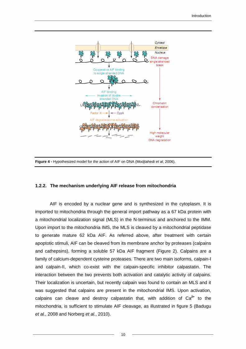
AIF is encoded by a nuclear gene and is synthesized in the cytoplasm. It is
imported to mitochondria through the general import pathway as a 67 kDa protein with
a mitochondrial localization signal (MLS) in the N-terminus and anchored to the IMM.
Upon import to the mitochondria IMS, the MLS is cleaved by a mitochondrial peptidase
to generate mature 62 kDa AIF. As referred above, after treatment with certain
apoptotic stimuli, AIF can be cleaved from its membrane anchor by proteases (calpains
and cathepsins), forming a soluble 57 kDa AIF fragment (Figure 2). Calpains are a
family of calcium-dependent cysteine proteases. There are two main isoforms, calpain-I
and calpain-II, which co-exist with the calpain-specific inhibitor calpastatin. The
interaction between the two prevents both activation and catalytic activity of calpains.
Their localization is uncertain, but recently calpain was found to contain an MLS and it
was suggested that calpains are present in the mitochondrial IMS. Upon activation,
calpains can cleave and destroy calpastatin that, with addition of Ca2+ to the
mitochondria, is sufficient to stimulate AIF cleavage, as illustrated in figure 5 (Badugu
et al., 2008 and Norberg et al., 2010).
Introduction
11
Figure 5 - Schematic representation of the mechanism behind the release of AIF from IMM to the
cytoplasm (Norberg et al., 2010).
1.2.3. Heat shock protein 70 (Hsp70p) acts as a chaperone to AIF protein
After permeabilization of the OMM, cleaved AIF can be released to the cytosol
and translocated to the nucleus. However, this translocation seems to be regulated by
cytoprotective proteins. The first proteins thought to play this role were heat-shock
proteins (HSPs). This group of proteins is found in most the living organisms, such as,
bacteria, yeast, humans, and others. They are involved in the folding and unfolding of
other proteins and their expression increases when cells are exposed to several
stresses but mostly when cells are exposed to elevated temperatures (Parsell and
Lindquist, 1993). This family of chaperones recognizes a broad spectrum of unfolded or
misfolded proteins (Chakrabart et al., 2011). Using a series of biochemical tests, it was
discovered that, within the family of HSPs proteins, Heat-shock protein 70 (Hsp70p)
was the only capable of physically interacting with AIF. Hsp70p is involved in the
inhibition of apoptosis by blocking Apaf-1 and by consequence the formation of the
apoptosome, (Ravagnan et al., 2001). It also binds to and neutralizes AIF, protecting
cells against translocation of AIF to the nucleus and by consequence protecting against
its apoptogenic effects, such as chromatin condensation and fragmentation. This is
corroborated by several studies, where the authors demonstrate that the interaction
between AIF and Hsp70p inhibits its translocation to the nucleus both in cultured cells
and in vivo (Ravagnan et al., 2001 and Ruchalski et al., 2001). However, the precise
Introduction
12
mechanism of AIF import into the nucleus is not known. Yeast Aif1p shows the same
localization and exhibits similar death executing pathways as mammalian AIF.
Therefore, this model system is particularly suitable to investigate the mechanism
involved in importing AIF to the nucleus.
1.3. Yeast apoptosis
Over the last decades, Saccharomyces cerevisiae has been a preferred
research tool in numerous areas of cell biology, due to its easy handling, technical
tractability, fast growth, small sequenced genome and the fact it is a eukaryote.
However, it was often questioned whether single-cell organisms need to commit
suicide or die by apoptosis. In fact, yeast cells tend to form colonies and communities,
and it is hypothesized that apoptosis may occur in yeast during chronological and
replicative ageing, leading to removal of virus-infected and damaged cells and
unsuccessful mating processes from colonies. This altruistic cell death gives younger
cells nutrients they can metabolize, contributing to the maintenance of the members of
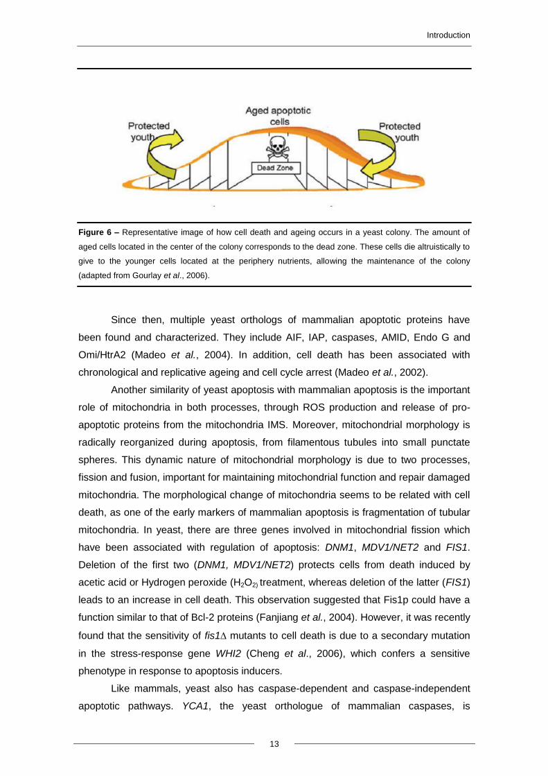
the community (Büttner et al., 2006) (Figure 6). It has now become clear that apoptosis
does not occur only in multicellular organisms, but can also be triggered in unicellular
organisms like yeast. It is believed that several apoptotic pathways are conserved in
yeast, making this organism attractive to study programmed cell death (Carmona-
Gutierrez et al., 2010).
The first observations of yeast apoptosis were made in a mutant strain of S.
cerevisiae with a mutation in the CDC48 gene, which encodes a protein necessary for
vesicle trafficking. The authors observed that dying cells of this mutant showed an
apoptotic phenotype with several characteristics of mammalian apoptosis, such as
phosphatidylserine exposure to the outer leaflet of the plasma membrane, DNA
fragmentation and margination, and condensation of chromatin (Madeo et al., 1997).
Introduction
13
Figure 6 – Representative image of how cell death and ageing occurs in a yeast colony. The amount of
aged cells located in the center of the colony corresponds to the dead zone. These cells die altruistically to
give to the younger cells located at the periphery nutrients, allowing the maintenance of the colony
(adapted from Gourlay et al., 2006).
Since then, multiple yeast orthologs of mammalian apoptotic proteins have
been found and characterized. They include AIF, IAP, caspases, AMID, Endo G and
Omi/HtrA2 (Madeo et al., 2004). In addition, cell death has been associated with
chronological and replicative ageing and cell cycle arrest (Madeo et al., 2002).
Another similarity of yeast apoptosis with mammalian apoptosis is the important
role of mitochondria in both processes, through ROS production and release of pro-
apoptotic proteins from the mitochondria IMS. Moreover, mitochondrial morphology is
radically reorganized during apoptosis, from filamentous tubules into small punctate
spheres. This dynamic nature of mitochondrial morphology is due to two processes,
fission and fusion, important for maintaining mitochondrial function and repair damaged
mitochondria. The morphological change of mitochondria seems to be related with cell
death, as one of the early markers of mammalian apoptosis is fragmentation of tubular
mitochondria. In yeast, there are three genes involved in mitochondrial fission which
have been associated with regulation of apoptosis: DNM1, MDV1/NET2 and FIS1.
Deletion of the first two (DNM1, MDV1/NET2) protects cells from death induced by
acetic acid or Hydrogen peroxide (H2O2) treatment, whereas deletion of the latter (FIS1)
leads to an increase in cell death. This observation suggested that Fis1p could have a
function similar to that of Bcl-2 proteins (Fanjiang et al., 2004). However, it was recently
found that the sensitivity of fis1 mutants to cell death is due to a secondary mutation
in the stress-response gene WHI2 (Cheng et al., 2006), which confers a sensitive
phenotype in response to apoptosis inducers.
Like mammals, yeast also has caspase-dependent and caspase-independent
apoptotic pathways. YCA1, the yeast orthologue of mammalian caspases, is
Introduction
14
considered a metacaspase and has a central role in yeast apoptosis. It is known that
yeast cells with a disruption of YCA1 are less susceptible to apoptotic cell death under
stress conditions like ageing and exposure to oxidative stress, salt, valproic acid, iron
and other metals. Recently, another caspase-like protease in yeast was found, Eps1p.
When released from the anaphase inhibitor Pds1p, Eps1p works as a caspase-like
protease and cleaves Mcd1p. Mcd1p is a yeast homolog of human cohesion Rad21
that is truncated after apoptotic stimuli and translocates from the nucleus to the
mitochondria, decreasing the mitochondrial membrane potential and thus promoting
the release of cytochrome c (Madeo et al., 2009). However, not all yeast apoptotic cell
death is dependent on YCA1 or on another caspase-like protease.
1.3.1. Yeast apoptotic triggers
In yeast, apoptosis can be triggered by three different strategies: by
heterologous expression of pro-apoptotic proteins, by environmental stress or drug-
induced stress, or by yeast endogenous apoptotic inducers.
Heterologous expression of proteins consists in expressing one or various
proteins in an organism that does not possess these proteins in its own genetic
background. It is described that yeast cells do not possess obvious orthologous of
proteins from the Bcl-2 family. However, heterologous expression of these human pro-
apoptotic proteins can trigger an active cell death. While some studies showed that
when Bax or Bak proteins are expressed in yeast, they can trigger cell death with
apoptotic characteristics, such as release of cytochrome c and generation of ROS,
others demonstrated that Bax induces a cell death process associated with activation
of autophagy (Kissova et al., 2007). These facts suggest that these proteins have their
function conserved in this organism (Eisenberg et al., 2007 and Madeo et al., 2004).
It is also known that several exogenous agents such as hyperosmotic stress,
heavy metals, amiodarone, ethanol, elevated temperatures, oxidative stress, UV
radiation, various pharmacological agents (such as aspirin), osmotin, viral "killer"
toxins, HOCl, pheromones and sometimes sugar can efficiently induce apoptosis in
yeast (Liang et al., 2008; Silva et al., 2005; and Carmona Gutierrez et al., 2010).
However, the best characterized and the most used exogenous inducers of apoptosis
in yeast are H2O2 and acetic acid (Madeo et al., 1999 and Ludovico et al., 2001).
Finally, several endogenous factors that trigger yeast apoptosis have been
described, such as defects in cellular processes like chromatid cohesion, N-
glycosylation, mRNA stability (Carmona-Gutierrez et al., 2010). Other examples of
Introduction
15
endogenous apoptosis triggers are DNA damage resultant from oxygen metabolism
and ROS generation and failed replication. Chronological and replicative ageing are
additional examples, and apoptosis also occurs during the development of colonies on
solid media (Herker et al., 2004 and Vachova and Palkova, 2005). Other endogenous
triggers are some pro-apoptotic proteins that are released from the mitochondria to the
cytosol, such as AIF, AIF-homologous mitochondrion-associated inducer of death
(AMID) and EndoG orthologues, which once in the cytosol are translocated to the
nucleus.
1.3.2. Yeast orthologue of AIF, Aif1p
Aif1p, the yeast orthologue of AIF, was first described in 2004 by Wissing et al.
It was shown that Aif1p, as mammalian AIF, is mitochondrial in normal cells and
relocalizes to the nucleus in the presence of apoptotic stimuli. It was also found that the
role of yeast Aif1p is dependent on cyclophilin A and partially on caspase activity.
Therefore in yeast, in contrast with mammalian cells, Aif1p-mediated apoptosis is not
completely independent on the yeast metacaspase Yca1p. Moreover, in contrast to
mammalian AIF, purified Aif1p degrades yeast nuclei and plasmid DNA (Wissing et al.,
2004).
AIF1 deletion led to increased survival to treatment with H2O2 and acetate, as
well as to decreased chronological ageing. Like in mammalian cells, Aif1p also has a
vital role in respiration via its NADH oxidase domain, and AIF1-deficient S. cerevisiae
have decreased growth on non-fermentable carbon sources. This redox function is
important for an effective anti-oxidant defense and oxidative phosphorylation (Madeo et
al., 2009).
1.3.2.1. Stimuli that regulate Aif1p release
Ludovico (2002) was the first to observe translocation of Aif1p to the nucleus in
cells undergoing chronological ageing or treated with camptothecin, an S phase-
specific anticancer drug that inhibits the action of the enzyme DNA topoisomerase-I.
Then Wissing et al. (2004) also observed translocation of Aif1p to the nucleus, in cells
treated with H2O2. Since then, few additional studies have addressed this issue and the
role of Aif1p in yeast apoptosis. Morton et al. described that antimicrobial peptides, the
dermaseptins obtained from amphibians, are capable of inducing yeast cell death. The
yeast cell death described by this group is independent of the metacaspase Yca1p but
Introduction
16
depends on Aif1p for nuclear fragmentation (Morton et al., 2007). Bostrycine is an
anthracenedione with antimicrobial and phytotoxic activity that belongs to the quinone
family, which inhibits cell proliferation and induces a decrease in the mitochondrial
membrane potential, leading to mitochondrial disruption. When yeast cells were treated
with this compound, cell death with hallmarks of apoptosis was observed. However,
bostrycin-induced cell death was promoted in yca1Δ. In contrast, this death phenotype
was partially rescued in aif1Δ cells (Chunlingku et al., 2009). Another substance that
can induce apoptosis is allicin, an antimicrobial extracted from garlic that is capable of
inducing apoptosis through its oxidative properties; both Aif1p and Yca1p seem to be
involved in this process, suggesting that allicin induces apoptosis through an
alternative mechanism (Gruhlke et al., 2010).
So far only H2O2 and chronological ageing have been shown to trigger Aif1p
release to the cytosol and translocation to the nucleus (Wissing et al., 2004). However,
there have been no studies addressing the mechanism underlying the import of yeast
Aif1p into the nucleus, which, like for mammalian AIF, is most likely an active and
highly regulated process.
1.4. Nucleocytoplasmic trafficking
The nucleus is a defining characteristic of eukaryotic cells and is physically
separated from the cytosol by the nuclear envelope (NE), a double membrane
structure. Most genetic information is confined to the nucleus, where most functions of
the cell are thus governed. Non-cytosolic proteins synthesized in the cytoplasm have to
be directed to their specific locations, where they exert their functions. For that they
contain a signal sequence that will direct them to a specific location. For example,
proteins containing a nuclear localization sequence (NLS) are imported into the
nucleus (Macara, 2001).
1.4.1. Nuclear Pore Complex (NPC)
The NE is crossed by multiple supramolecular structures named nuclear pore
complexes (NPCs), structures specialized in transport of small molecules, ions and
macromolecules between the cytoplasm and the nucleus. The number of NPCs in each
cell is variable and depends on cell size and transcriptional activity, and differs between
species. Human cells may contain about 5x10³ – 5x10⁷ NPCs per nucleus and these
NPCs have about 60 MDa of molecular weight. The yeast NPC is much smaller than
Introduction
17
that of higher eukaryotes, with a molecular weight of approximately 44 MDa, and there
are about 200 NPCs per nucleus (Freitas et al., 2009).
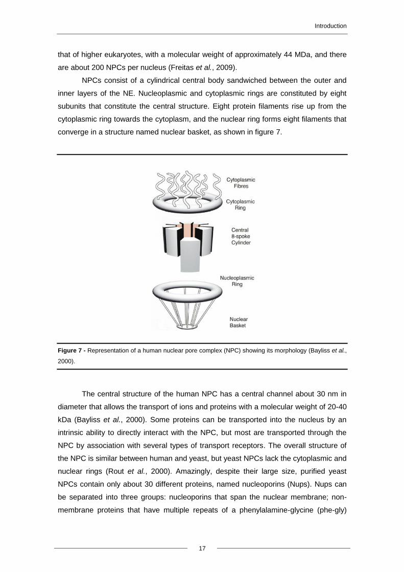
NPCs consist of a cylindrical central body sandwiched between the outer and
inner layers of the NE. Nucleoplasmic and cytoplasmic rings are constituted by eight
subunits that constitute the central structure. Eight protein filaments rise up from the
cytoplasmic ring towards the cytoplasm, and the nuclear ring forms eight filaments that
converge in a structure named nuclear basket, as shown in figure 7.
Figure 7 - Representation of a human nuclear pore complex (NPC) showing its morphology (Bayliss et al.,
2000).
The central structure of the human NPC has a central channel about 30 nm in
diameter that allows the transport of ions and proteins with a molecular weight of 20-40
kDa (Bayliss et al., 2000). Some proteins can be transported into the nucleus by an
intrinsic ability to directly interact with the NPC, but most are transported through the
NPC by association with several types of transport receptors. The overall structure of
the NPC is similar between human and yeast, but yeast NPCs lack the cytoplasmic and
nuclear rings (Rout et al., 2000). Amazingly, despite their large size, purified yeast
NPCs contain only about 30 different proteins, named nucleoporins (Nups). Nups can
be separated into three groups: nucleoporins that span the nuclear membrane; non-
membrane proteins that have multiple repeats of a phenylalamine-glycine (phe-gly)
Introduction
18
motif, and non-membrane proteins that don’t have the phe-gly motif (Macara et al.,
2001).
1.4.2. Protein transport receptors and transport cycle
For the majority of macromolecules, transport into the nucleus and through the
NPCs is an energy-dependent process. This process can be mediated by the major
class of transport receptors, which includes different soluble proteins (karyopherins or
Kaps, also named importins, exportins or transportins). They mediate import into or
export from the nucleus of proteins that cannot be transported by simple diffusion
through the NPCs, as well as mediate the transport of non-coding RNAs. Karyopherins
are acidic proteins with a molecular weight of 90 to 145 kDa. About 10 of the 20
karyopherins identified were demonstrated to participate in the nuclear import of
proteins in eukaryotes (Tran et al., 2006). Sequence analysis identified 13 predicted
importin-β proteins in the yeast S. cerevisiae.
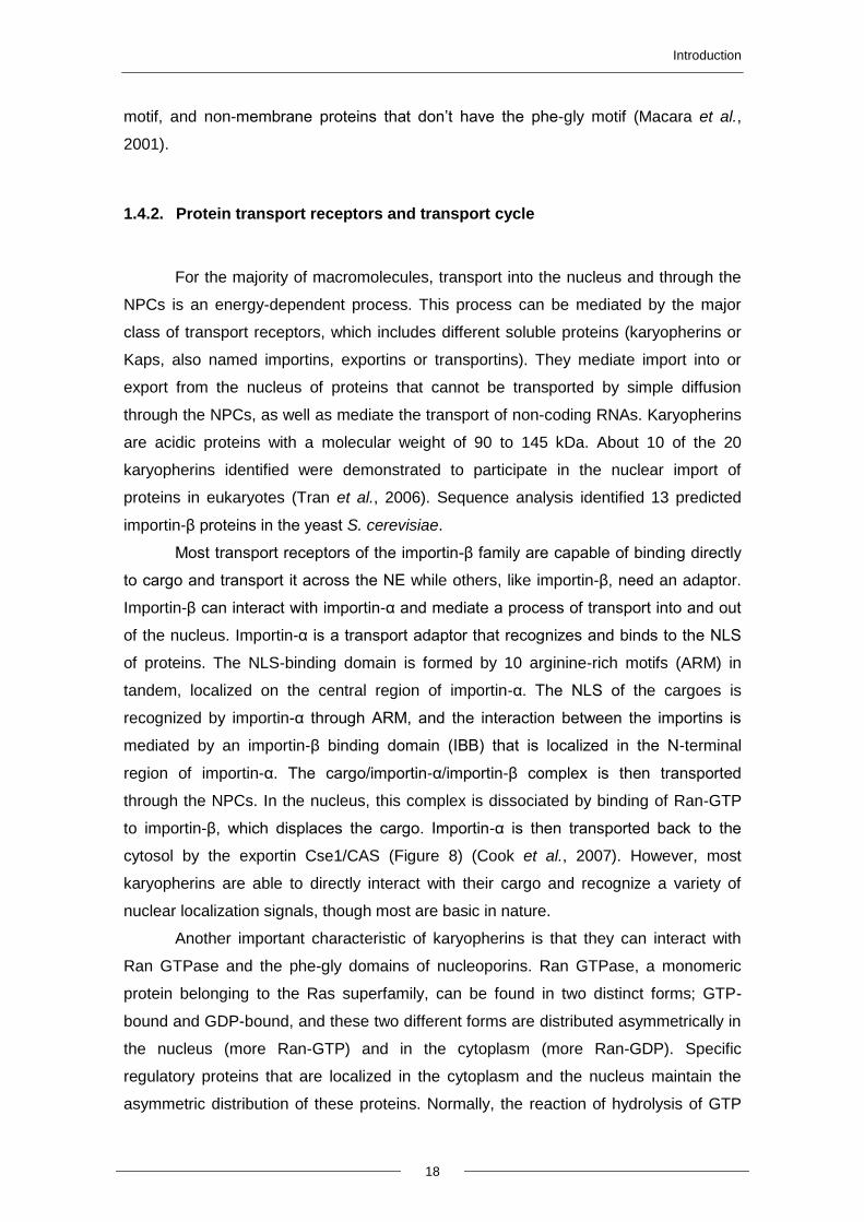
Most transport receptors of the importin-β family are capable of binding directly
to cargo and transport it across the NE while others, like importin-β, need an adaptor.
Importin-β can interact with importin-α and mediate a process of transport into and out
of the nucleus. Importin-α is a transport adaptor that recognizes and binds to the NLS
of proteins. The NLS-binding domain is formed by 10 arginine-rich motifs (ARM) in
tandem, localized on the central region of importin-α. The NLS of the cargoes is
recognized by importin-α through ARM, and the interaction between the importins is
mediated by an importin-β binding domain (IBB) that is localized in the N-terminal
region of importin-α. The cargo/importin-α/importin-β complex is then transported
through the NPCs. In the nucleus, this complex is dissociated by binding of Ran-GTP
to importin-β, which displaces the cargo. Importin-α is then transported back to the
cytosol by the exportin Cse1/CAS (Figure 8) (Cook et al., 2007). However, most
karyopherins are able to directly interact with their cargo and recognize a variety of
nuclear localization signals, though most are basic in nature.
Another important characteristic of karyopherins is that they can interact with
Ran GTPase and the phe-gly domains of nucleoporins. Ran GTPase, a monomeric
protein belonging to the Ras superfamily, can be found in two distinct forms; GTP-
bound and GDP-bound, and these two different forms are distributed asymmetrically in
the nucleus (more Ran-GTP) and in the cytoplasm (more Ran-GDP). Specific
regulatory proteins that are localized in the cytoplasm and the nucleus maintain the
asymmetric distribution of these proteins. Normally, the reaction of hydrolysis of GTP
Introduction
19
into GDP is slow; however, this reaction can be accelerated by two proteins, Ran-GAP
and Ran-BP1. On the other hand, the nuclear protein RCC1 promotes the reverse
reaction. It is the combined action of Ran regulatory proteins that creates and
maintains the Ran-GTP gradient across the NE, and it is this gradient that establishes
the directionality of nucleocytoplasmic transport (Freitas et al., 2009).
.
Figure 8 - Overview of the transport cycle of the classical nuclear localization signal (NLS) transport with
the ligation of importin-β and importin-α (Cook et al., 2007).
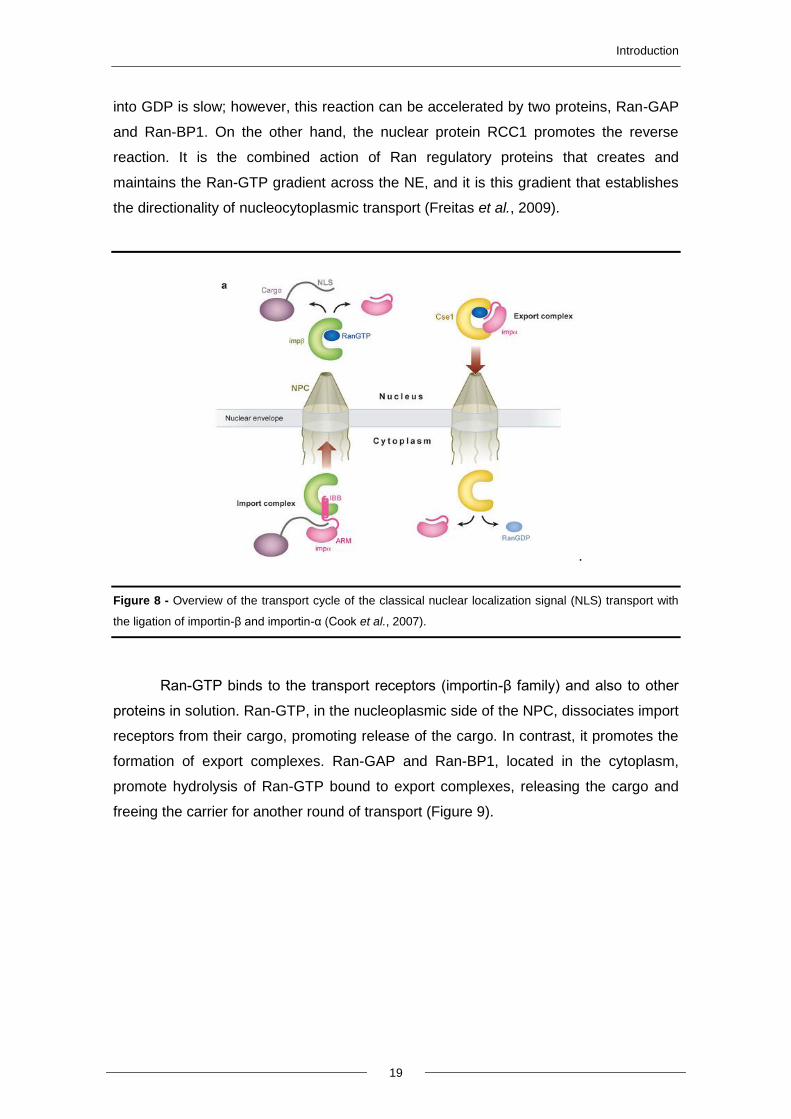
Ran-GTP binds to the transport receptors (importin-β family) and also to other
proteins in solution. Ran-GTP, in the nucleoplasmic side of the NPC, dissociates import
receptors from their cargo, promoting release of the cargo. In contrast, it promotes the
formation of export complexes. Ran-GAP and Ran-BP1, located in the cytoplasm,
promote hydrolysis of Ran-GTP bound to export complexes, releasing the cargo and
freeing the carrier for another round of transport (Figure 9).
Introduction
20
Figure 9 - Schematic representation of processes underlying nuclear import and export mediated by β-
karyopherins (Importin-β) (Bayliss et al., 2000).
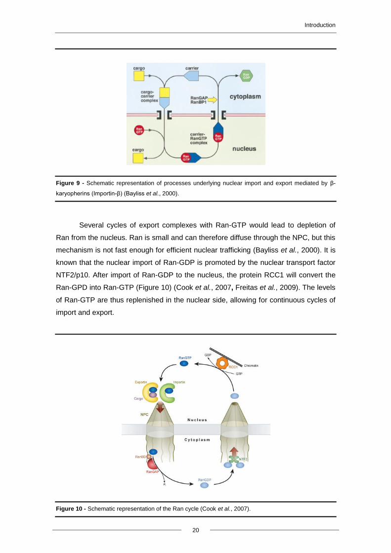
Several cycles of export complexes with Ran-GTP would lead to depletion of
Ran from the nucleus. Ran is small and can therefore diffuse through the NPC, but this
mechanism is not fast enough for efficient nuclear trafficking (Bayliss et al., 2000). It is
known that the nuclear import of Ran-GDP is promoted by the nuclear transport factor
NTF2/p10. After import of Ran-GDP to the nucleus, the protein RCC1 will convert the
Ran-GPD into Ran-GTP (Figure 10) (Cook et al., 2007, Freitas et al., 2009). The levels
of Ran-GTP are thus replenished in the nuclear side, allowing for continuous cycles of
import and export.
Figure 10 - Schematic representation of the Ran cycle (Cook et al., 2007).
Introduction
21
1.4.3. Regulation of nucleocytoplasmic trafficking
As mentioned above, the majority of proteins are synthesized in the cytosol and
then transported to their final localization in order to exert their function. This process is
often an important regulatory step in cellular pathways. Indeed, there are several
examples of proteins whose function is regulated by nuclear localization. One example
is NF-kB (nuclear factor kappa-light-chain-enhancer of activated B cells), a
transcription factor found in almost all types of mammalian cells. It is involved in the
cellular response to several stimuli, such as several types of stress, cytokines,
ultraviolet radiation, free radicals and LDL oxidation. This transcription factor is bound
to an inhibitory protein (IkB) in unstimulated cells and therefore is latent. However,
upon activation by extracellular agents, IkB is phosphorylated by a protein kinase and
then ubiquitinated and degraded by the proteasome, allowing NF-kB translocation to
the nucleus. There, it binds to the kB consensus sequence generally leading to an
increase in the expression of the target gene (O’Neill et al., 1997).
Another example is the yeast transcription factor Yap1p, which regulates
expression of target genes containing a binding site named Yap1p-response element
on their promoter. Normally Yap1p is located in the cytosol and is translocated into the
nucleus under stress conditions. In the nucleus, nuclear export of Yap1p is inhibited,
which leads to its accumulation in the nucleus and consequently inducing the
expression of several genes encoding antioxidant proteins. Exposure to H2O2 induces
the formation of disulfide bonds between the C-terminal cysteine-rich domain (C-CRD)
and the N-terminal cysteine-rich domain (N-CRD) of Yap1p. The C-CRD possesses a
NES, which ordinarily leads to export of Yap1p from the nucleus, but it is masked by
this dually disulfide-bonded structure. Therefore, Yap1p export is inhibited, promoting
Yap1p nuclear accumulation (Gulshan et al., 2005).
Another example of regulated transport is the regulation of protein localization
by phosphorylation and desphosphorylation of the yeast transcription factor Pho4p.
Pho4p localization is regulated in response to modifications in the concentration of
inorganic phosphate in the media. Pho4p is imported into the nucleus through the
importin-β family member Pse1p/Kap121p. It is known that inhibition of the
Pho4p/Pse1p interaction is mediated by phosphorylation and that Pho4p translocation
requires Pse1p. This suggests that this import is regulated by phosphorylation in vivo.
In yeast cells grown in phosphate-rich medium, Pho4p is phosphorylated by the Pho80-
Pho85 cyclin-CDK complex and import into the nucleus is therefore inhibited.
Consequently, there is no transcription of phosphate starvation-specific genes.
However, in yeast grown with limited phosphate, the CDK inhibitor Pho81p inhibits
Introduction
22
Pho80-Pho85, leading to accumulation of unphosphorylated Pho4p. Unphosphorylated
Pho4p can then interact with Pse1p/Kap121p, translocate into the nucleus and induce
transcription of phosphate-responsive genes (Kaffman et al., 1998).
1.4.4. Nuclear import of AIF
AIF function is regulated at different levels including through its subcellular
localization. As mentioned above, AIF is transported to the mitochondria due to the N-
terminal MLS. After mitochondrial membrane permeabilization, truncated AIF is
released into the cytosol and then translocated to the nucleus.
The crystal structure of AIF indicates that this protein has to be re-localized to
the nucleus to exert its apoptotic activity (Ye et al., 2002). To discover which region of
this gene is responsible for the apoptotic activity of this protein, Gurbuxani and
colleagues mapped functional regions of AIF by deleting several regions and observing
the resultant phenotype. The authors determined that the C-terminal domain (beyond
residue 567) is responsible for AIF-induced chromatin condensation (once AIF is in the
nucleus). Deletion of the Hsp70p-binding region (residues 150 to 228) leads to a gain
of function phenotype, i.e., facilitates nuclear translocation in response to an apoptotic
stimulus. One region of AIF contains a consensus NLS (residues 367 to 459), normally
involved in protein translocation to the nucleus. However, two NLS domains are
described for AIF, one more N-terminal (residues 277 to 301) and another closer to the
C-terminus (residues 445–451) (Susin et al., 1999). In this study, the authors showed
that the C-terminal NLS is functionally more important for AIF translocation to the
nucleus than the N-terminal NLS domain. However, deletion of the region containing
the C-terminal NLS only partially inhibited AIF translocation, which suggests that there
are other domains involved in AIF import (Gurbuxani et al., 2003). However, the
mechanism mediating this nuclear translocation of AIF is still unclear.


2. AIMS AND
RESEARCH PLAN
Aims and research plan
25
Apoptosis-inducing factor is a flavoprotein with oxidoreductase activity localized
in the mitochondrial intermembrane space. Upon apoptosis induction, AIF translocates
to the nucleus, where it leads to chromatin condensation and DNA degradation. AIF
has been suggested to control a caspase-independent pathway of apoptosis, important
for neurodegeneration and normal development. However, it remains unknown how
AIF translocates to the nucleus. Yeast Aif1p shows the same localization and exhibits
similar death executing pathways as mammalian AIF, though it mediates a partially
caspase- dependent pathway (Wissing et al., 2004). Therefore, our aim was to use the
yeast model system to investigate the mechanism involved in importing Aif1p to the
nucleus.
Aim 1. Determine which soluble import factor is necessary to import Aif1p
Transform mutants in each of the yeast soluble transport receptors (Kaps) with
Aif1p-GFP and assess the localization of Aif1p after apoptosis induction by
fluorescence microscopy. Mutation of the Kap mediating Aif1p import will prevent its
nuclear accumulation.
Aim 2. Map the NLS of Aif1p
Clone different fragments of Aif1p in frame with GFP and assess their
localization by fluorescence microscopy. Identify the smallest sequence that is both
necessary and sufficient for transport into the nucleus.


3. MATERIALS AND
METHODS
Materials and methods
28
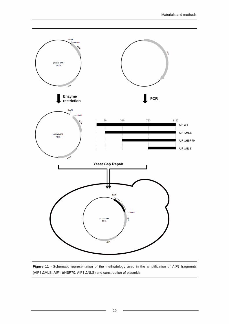
3.1. Plasmids
All the plasmids and oligonucleotides used in this work are listed in Table I and
ll, respectively. Plasmids were amplified in the Escherichia coli XL1Blue strain (as
described below) and purified using a Miniprep kit (GenElute Plasmid Miniprep kit,
Sigma-Aldrich) according to manufacturer’s instructions. The identity of the plasmids
was confirmed by digestion with specific restriction enzymes and by PCR.
pAIF1WT-GFP, pAIF1ΔMLS-GFP, pAIF1ΔHSP70-GFP, pAIF1ΔNLS-GFP were
constructed by Gap Repair (Figure 11). Briefly, four fragments of the AIF1 gene (Wild-
type AIF1, AIF1 ΔMLS, AIF1 ΔHSP70, AIF1 ΔNLS) were amplified by Polymerase
Chain Reaction (PCR) using the plasmid pAIF1 from Euroscarf as a template and
oligonucleotides forward 1-4, respectively, and the reverse oligonucleotide 5 (Table II).
Saccharomyces cerevisiae strain DF5a was transformed with pYX242GFP vector
digested with EcoRI and HindIII and each of the four fragments (Figure 11). Correct
integration of the AIF1 fragments in pYX242GFP was confirmed by colony PCR using
oligonucleotides that anneal upstream and downstream of the insertion (6 and 7,
respectively). After confirmation, the plasmids were purified from yeast cells (described
below in 3.6), amplified in E. coli and sequenced.
Table I – List of the plasmids used in this work.
Plasmid Description References/Sources
pAIF1 AIF1 ORF Euroscarf (Germany)
pYX242GFP LEU2, AmpR Rosenblum et al., 1998
pAIF1WT-GFP AIF1 1-1137 cloned in pYX242GFP This study
pAIF1ΔMLS-GFP AIF1 Δ1-78 cloned in pYX242GFP This study
pAIF1ΔHSP70-GFP AIF1 Δ1-336 cloned in pYX242GFP This study
pAIF1ΔNLS-GFP AIF1 Δ1-723 cloned in pYX242GFP This study
pBAX c-myc Bax c-myc cloned in PCM189, URA3 Priault et al., 1999
Table II – List of the oligonucleotides used in this work.
Number Name Oligonucleotide sequence (5’ - 3’)
1 AIF1WT Fw ATCTATAACTACAAAAAACACTATCAGGAATTCGGGCCCATGACA
2 AIF1ΔMLS Fw ATCTATAACTACAAAAAACACTATCAGGAATTCGGGCCCATGAGGGAACTGGGT
3 AIF1ΔHSP70 Fw ATCTATAACTACAAAAAACACTATCAGGAATTCGGGCCCATGCCA
4 AIF1ΔNLS Fw ATCTATAACTACAAAAAACACTATCAGGAATTCGGGCCCATGGGT
5 AIF1 Rev GACAACAACAGTGAATAATTCTTCACCTTTAGACATCCGGGG
6 YX Fw ATCTATAACTACAAAAAACACATACAGGAATTCGGGCCCATGACA
7 GFP Rev AGCGTCGACGTTACCTTATTTGTACAATTCATCCATACCATGGG
8 YX Fw CTTTTTAATTCTAAATCAATCTTTTTCAA
9 GFP Rev AACATCACCATCTAATTCAAC
Materials and methods
29
Figure 11 - Schematic representation of the methodology used in the amplification of AIF1 fragments
(AIF1 ΔMLS, AIF1 ΔHSP70, AIF1 ΔNLS) and construction of plasmids.
Materials and methods
30
3.2. Yeast strains and growth conditions
All S. cerevisiae strains used in this work and respective genotypes are shown
in Table III. Strains were transformed with the indicated plasmids using the Lithium
acetate/Single Stranded carrier DNA/Polyethylene Glycol (PEG) method previously
described in (Gietz and Woods, 2006). Transformants were selected on Synthetic
Complete (SC) medium [SC containing 0.17% (w/v) Yeast nitrogen base without
aminoacids and ammonium sulfate, 0.5% (w/v) ammonium sulfate, 0.14% (w/v), drop-
out mixture lacking histidine, leucine, tryptophan and uracil, 0.008% (w/v) Histidine,
0.04% (w/v) Leucine, 0.008% (w/v) Tryptophan and 0.008% (w/v) Uracil] lacking the
appropriate aminoacids plus 2% (w/v) of carbon source and 2% agar. Yeast strains
were maintained on solid YPD or SC medium (lacking the appropriate amminoacids),
grown at 30°C for 48 h, stored at 4°C, and refreshed every 2 weeks. Yeast cultures
were grown aerobically in SC medium with 2% Glucose or Galactose as a carbon
source. Strains transformed with plasmids were grown in the same medium lacking the
appropriate amino acids. Cells were incubated at 30°C with orbital shaking (200 rpm)
and a liquid/air ratio of 1:5.
3.3. Hydrogen peroxide and acetic acid treatment
Cells were grown overnight until exponential phase (OD600 = 0.5-0.6) on SC
Glu, collected by centrifugation and resuspended in new medium (medium with pH 3 in
the case of acetic acid treatment) to a final concentration of 107 cells/mL
(approximately OD600 = 0.2) and incubated with H2O2 (1 mM, 2 mM and 3 mM), or
acetic acid (140 mM, 160 mM, 180 mM) for up to 360 min at 30°C. At specific time
points, serial dilutions (1:10) were spotted onto YPD plates and colony growth was
scored after 2 days of incubation at 30°C. Viability was determined in relation to time 0
(100%). In parallel, 500 µL of cells were harvested and processed for fluorescence
microscopy.
3.4. Heterologous expression of Bax c-myc
Strains harboring the plasmid pBAX c-myc were grown overnight in SC Glucose
lacking uracil and supplemented with doxycycline (10 µg/ml) to repress Bax c-myc
expression. Cells were centrifuged, washed three times with sterilized water,
ressuspended in the same medium without doxycycline (to induce Bax c-myc
expression) or with doxycycline (as a negative control). At specific time points, serial
Materials and methods
31
dilutions (1:10) were spotted onto YPD plates and colony growth was scored after 2
days of incubation at 30°C. Viability was determined in relation to time 0 (100%). In
parallel, 500 µL of cells were harvested and processed for fluorescence microscopy.
Table III – List of S. cerevisiae strains used in this work.
Strain Genotype Reference/Source
DF5a Mat a; ura3-52, leu2-3, 112 lys2-801, trp1-1,
his3Δ200 Finley et al., 1987
BY4741 Mat a; his3Δ 1, leu2Δ 0, met15Δ 0, ura3Δ 0 Euroscarf (Germany)
BY4742 Mat α; leu2, lys2, ura3, his3 Euroscarf (Germany)
W303a Mat a; ade2; ura3; his3; trp1; leu2 Costa V., IBMC
ssa1Δ BY4741 YAL005C :: KanMX4 Euroscarf (Germany)
DF5a pYX242GFP DF5a harboring pYX242GFP This study
DF5a pAIF1WT-GFP DF5a harboring pAIF1WT-GFP This study
DF5a pAIF1ΔMLS-GFP DF5a harboring pAIF1ΔMLS-GFP This study
DF5a pAIF1ΔHSP70-GFP DF5a harboring pAIF1ΔHSP70-GFP This study
DF5a pAIF1ΔNLS-GFP DF5a harboring pAIF1ΔNLS-GFP This study
W303 pAIF1WT-GFP W303 harboring pAIF1WT-GFP This study
W303 pAIF1ΔMLS-GFP W303 harboring pAIF1ΔMLS-GFP This study
W303 pAIF1ΔHSP70-GFP W303 harboring pAIF1ΔHSP70-GFP This study
W303 pAIF1ΔNLS-GFP W303 harboring pAIF1ΔNLS-GFP This study
W303 pAIF1WT-GFP pBAX c-myc
W303 harboring pAIF1WT-GFP and pBAX c-
myc This study
BY4741 pAIF1WT-GFP BY4741 harboring pAIF1WT-GFP This study
BY4741 pAIF1ΔMLS-GFP BY4741 harboring pAIF1ΔMLS-GFP This study
BY4741 pAIF1ΔNLS-GFP BY4741 harboring pAIF1ΔNLS-GFP This study
BY4741 pAIF1WT-GFP pBAX c-myc
BY4741 harboring pAIF1WT-GFP and pBAX c-myc
This study
BY4741 pAIF1ΔMLS-GFP pBAX c-myc
BY4741 harboring pAIF1ΔMLS-GFP and pBAX c-myc
This study
ssa1Δ pAIF1WT-GFP ssa1Δ harboring pAIF1WT-GFP This study
ssa1Δ pAIF1ΔMLS-GFP ssa1Δ harboring pAIF1ΔMLS This study
ssa1Δ pAIF1WT-GFP pBAX c-myc
ssa1Δ harboring pAIF1WT-GFP and pBAX c-myc
This study
ssa1Δ pAIF1ΔMLS-GFP pBAX c-myc
ssa1Δ harboring pAIF1ΔMLS-GFP and pBAX c-myc
This study
3.5. Transformation of bacterial cells
Super Optimal Broth medium (250 ml) [SOB; 2% (w/v) Tryptone peptone; 0.5%
(w/v) Yeast extract; 2.5 mM KCl, 10 mM NaCl; 10 mM MgSO4, 10 mM MgCl2] was
inoculated with 2 colonies of XL1Blue E. coli strain and grown at 18°C, 200 rpm with a
ratio of flask liquid/air of 1:5 until OD600 = 0.6, about 4-5 days. Then the culture was
placed on ice for 10 min and the cells pelleted for 10 min at 2500g, at 4°C. The pellet
was suspended in 80 mL ice-cold TB buffer [10 mM Pipes; 15 mM CaCl2; 250 mM KCl;
55 mM MnCl2] and left on the ice for 10 min. Cells were centrifuged for 10 min at 2500
g and gently suspended in 20 mL ice-cold TB buffer. DMSO was added to a final
concentration of 7% and cells incubated on ice for 10 min. Finally, competent cells

Materials and methods
32
were aliquoted, frozen with liquid nitrogen and stored at -80°C. E. coli transformation
was performed by a standard protocol for chemically competent cells and
transformants selected on Luria Bertani medium [LB; 1% (w/v) Tryptone, 0.5% (w/v)
Yeast extract, 1% (w/v) NaCl and 2% (w/v) Agar] with 100 μg/mL of Ampicillin.
3.6. Purification of Yeast DNA
Plasmid DNA from yeast cells was purified using the GenElute Plasmid
Miniprep kit (Sigma-Aldrich) according to manufacturer’s instructions, with
modifications. Cells from a 10 mL overnight culture were centrifuged and the pellet
resuspended in 200 µL of Resuspension solution. About 200 µL of glass beads and 10
µL of lyticase were added to the cells and the tubes incubated for 10-20 min at 37°C.
Next, tubes were vortexed for 15 min. Cells were lysed by adding 200 µL of Lysis
solution and gentle inversions of the tubes. 350 µL of Neutralization solution was
added and the tubes inverted 5-6 times, and the lysate spun at 15000 rpm for 10 min.
The columns from the kit were washed with 500 µL of Column Preparation solution,
and the supernatant from the neutralization step was loaded into the columns after
centrifugation at 15000 rpm for 1 min, columns were washed with 500 µL of Wash
Optional solution, then with 750 µL of Wash solution and dried. The columns were
changed to new tubes, and DNA eluted with 50 µL of Elution buffer.
3.7. Fluorescence Microscopy
Samples (200 µL) were harvested and centrifuged for 2 min at 10000 rpm, and
the pellet was resuspended in 100 µL of Phosphate-Buffered Saline [PBS; 80 mM
Na2HPO4, 20 mM NaH2PO4 and 100 mM NaCl], fixed in 100% (v/v) ethanol and 4 µL of
DAPI (2 µg/mL) added. After 5 min of incubation, cells were washed twice with PBS
and resuspended in 10 µL of PBS. Cells were visualized in a Leica Microsystems DM-
5000B epifluorescence microscope using appropriate filter settings with a 100x oil
immersion objective. Images were acquired with a Leica DCF350FX digital camera and
processed with LAS AF Leica Microsystems software.
3.8. Subcellular fractionation
3.8.1. Preparation of spheroplasts
Cells were grown in 2.4 L of SC medium without appropriate aminoacids and
using Galactose as the carbon source, to obtain more mitochondrial mass. For acetic
Materials and methods
33
acid treatment, cells were grown until late exponential phase (OD600 = 1) and the
culture divided in two: one culture, without treatment, was used as a negative control
and the other was treated with 120 mM of acetic acid (pH 3,0) for 120 min. For Bax c-
myc expression, cells were treated as described above, in the presence and absence
of doxyclyclin. Cells were collected and incubated in Pre-incubation buffer [0.5 M β-
mercaptoethanol, 0.1 M Tris, pH 9.3] for 15 min at 30°C and washed 2 times with
Tris/KCl buffer [10 mM Tris, 0,5 M KCl, pH 7]. Spheroplasts were obtained by
incubation in Digestion buffer [1.35 M Sorbitol, 10 mM Citric acid, 30 mM Na2PO4, 1
mM EDTA, pH 5.8] containing 10 mg or 50 mg of zymoliase /1 O.D for untreated and
treated cells. Digestion of cell wall was performed during 30 and 50 min for untreated
and treated cells, respectively.
3.8.2. Mitochondrial and cytosolic fraction preparation
Spheroplasts were washed with Washing buffer [0.750 M Sorbitol, 0.4 M
Mannitol, 10 mM Tris Maleate, pH 6.8] and disrupted by osmotic shock in
Homogenization buffer [0.6 M Mannitol, 10 mM Tris Maleate, pH 6.8] and mild
homogenization in a hand-potter, to preserve the outer mitochondrial membrane. The
homogenate was centrifuged at 2500 rpm to remove cell debris and nuclei, and the
supernatant centrifuged at 15000 rpm for 15 min. The pellet corresponds to the
mitochondrial fraction and the supernatant to the cytosolic fraction. Mitochondria were
then resuspended in Recuperation buffer [0.6 M Mannitol, 2 mM EDTA, 10 mM Tris
Maleate, pH 6.8].
3.8.3. Nuclear fraction preparation
3.8.3.1. Protocol I
Crude nuclei resulting from the 2500 rpm centrifugation were washed 3 times
with Recuperation buffer (3000 g for 5 min) and the final washed pellet resuspended in
Recuperation buffer and loaded onto a Ficoll 400 step gradient in Ultra-Clear tubes (30,
40 and 50% (w/v)). The gradient was centrifuged at 18000 rpm for 60 min at 2°C. The
nuclei band in the 40% layer was collected. Tubes containing the nuclear extract were
stored at -80°C.
Materials and methods
34
3.8.3.2. Protocol II
Spheroplasts were resuspended in half volume of Nuclei buffer [1 M Sorbitol, 20
mM PIPES pH 6.3, 0.5 mM CaCl2, 1 mM DTT, 0.1 M EDTA, 1 M PMSF] and added
dropwise to an erlenmeyer containing Ficoll buffer [18% (w/v) Ficoll 400, 20 mM PIPES
pH 6.3, 0.5 mM CaCl2, 1 mM DTT, 0.1 M EDTA, 1 M PMSF] with continuous agitation,
in a ice-cold bath to prevent protease activity. The Ficoll suspension was centrifuged at
5000 rpm for 5 min at 4°C, and this step was repeated until the supernatant become
clear. The supernatant was then transferred to new tubes and the tubes centrifuged at
13000 rpm for 20 min at 4°C. The pellet was resuspended in 5-10 volumes of Nuclei
buffer and the suspension centrifuged again at 9000 rpm for 10 min at 4°C. The final
pellet (nuclear fraction) was resuspended in Storage buffer [20 mM Tris-HCl pH 7.5,
0.1 mM EDTA, 100 mM KCl, 10% (v/v) Glycerol] and stored at -80°C.
3.8.3.3. Protocol III
The third protocol used was based on protocol II, but only serial centrifugations
were performed. The protocol started with two washes at 500 rpm of the crude nuclei
resultant from the 2500 rpm centrifugation, followed by centrifugations at 1000, 2000,
6000, 9000 and 13000 rpm of the supernatants. All centrifugations were performed
during 10 min and all the washes and resuspensions were made with recuperation
buffer. Samples of the sedimented pellets in each centrifugation were collected to
determine the sedimentation pattern of the different organelles.
3.8.4. SDS gel electrophoresis/Westernblot
Protein concentration of the fractions was estimated through the Bradford
method using BSA as standard (Bradford, 1976). Proteins (50 µg) of the subcellular
fractions were separated electrophoretically on a 15% SDS polyacrylamide gel at 20
mA and transferred to a Hybond-P Polyvinylidene Difluoride Membrane (PVDF)
(Hybond-ECL, GE Healthcare) at 60 mA for 1 h and 30 min. Membranes were blocked
for 1 h in PBS-T [PBS with 0.05% (v/v) Tween-20] containing 5% (w/v) non-fat dry milk,
washed in PBS-T and then cut into strips and incubated with the primary antibodies;
mouse monoclonal anti-yeast phosphoglycerate kinase (PGK1) antibody (1:5000,
Molecular Probes), mouse monoclonal anti-yeast porin (POR1) antibody (1:5000,
Molecular Probes), rabbit polyclonal anti-yeast cytochrome c (CYC1) antibody (1:1000,
custom-made by Millegen), mouse monoclonal anti-green fluorescent protein (GFP)
Materials and methods
35
antibody (1:3000; Roche Applied Science), rabbit polyclonal anti-histone H4 (H4B)
antibody (1:2000, abcam), rabbit polyclonal anti-human Bax (BAX) antibody (1:10000,
Sigma). Then membranes were incubated with secondary antibodies against mouse or
rabbit IgG-peroxidase (1:10000; Sigma Aldrich). Pgk1p and Por1p were used as
controls for cytosolic and mitochondrial fractions, respectively. Immunodetection of
bands was revealed by chemiluminescence (Immobilon, Millipore).
3.9. Immunoprecipitation
Immunoprecipitation was performed using protein G-coupled Dynabeads
(Invitrogen). Briefly, cytosol was prepared as described above (See 3.8.2),
supplemented with a mixture of protease inhibitors (0.4 μL/mL Aprotinin, 1 μg/mL
Leupeptin, 1 μg/mL Pepstatin, 1 mM Phenylmethylsulfonyl fluoride) and with 100 mM
NaCl and 0.01% NP-40. 1, 2 or 5 μg of monoclonal anti-GFP antibody (Roche Applied
Science) were added and the lysate was incubated overnight at 4°C with agitation.
Protein G-coupled Dynabeads (30 µL) were then added and incubated for 6 h at 4°C
with agitation. Beads were washed 6 times for 2 min with IP buffer [50 mM Tris-HCl, pH
7.5; 100 mM NaCl; 2 mM EDTA, 0.01% (v/v) NP-40] and bound proteins eluted with
Laemmli sample buffer for 5 min at 95°C. The sample buffer was collected to new
tubes.
3.9.1. SDS gel electrophoresis/Western Blot/ Silver Staining of
immunoprecipitated proteins
Immunoprecipitated samples were loaded in duplicate onto a 4-20% Mini-
PROTEAN TGX Precast Gel (BioRad) to guarantee a good separation of proteins. One
half of the gel was transferred to PVDF as described above and Aif1p-GFP detected
using mouse monoclonal anti-green fluorescent protein (GFP) antibody as the primary
antibody (1:3000; Roche Applied Science).
The other half of the gel was stained by silver staining. The gel was fixed by
washing at least twice with Fixing Solution [30% (v/v) Ethanol, 10% (v/v) Acetic Acid]
for 30 min. After fixing, the gel was washed with Washing Solution [30% (v/v) Ethanol]
for 10 min and with deionized water for 10 min. The gel was then soaked with
Sensitizing Solution [0.02% (w/v) Sodium Thiosulfate] and carefully agitated for 1 min
and rinsed three times with deionized water for 30 sec. The gel was then incubated
with gentle agitation with freshly made Staining Solution [0.15% (w/v) Silver Nitrate] for
at least 30 min, washed 3 times with deionized water and transferred to Developing

Materials and methods
36
Solution [0.05% (v/v) Formaldehyde, 3% (w/v) Sodium carbonate]. When an adequate
degree of staining was achieved, gels were transferred to Stop Solution [5% (v/v)
Acetic acid] for 1 min and then placed in deionized water.


4. RESULTS
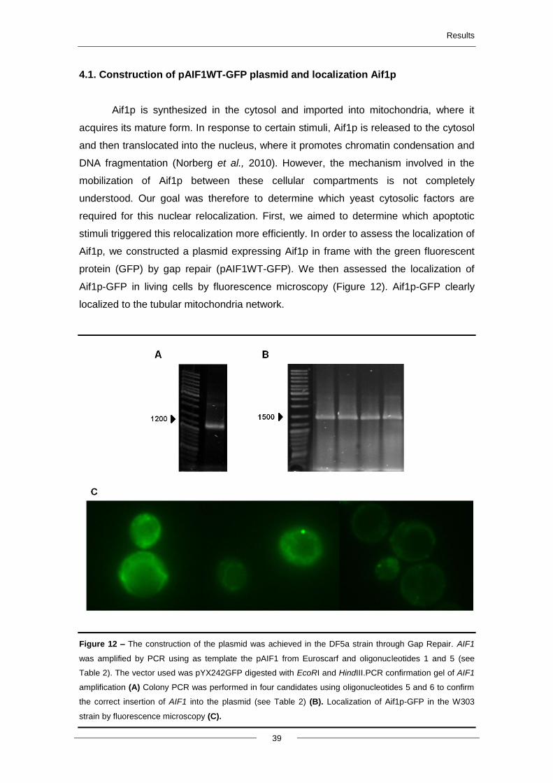
Results
39
4.1. Construction of pAIF1WT-GFP plasmid and localization Aif1p
Aif1p is synthesized in the cytosol and imported into mitochondria, where it
acquires its mature form. In response to certain stimuli, Aif1p is released to the cytosol
and then translocated into the nucleus, where it promotes chromatin condensation and
DNA fragmentation (Norberg et al., 2010). However, the mechanism involved in the
mobilization of Aif1p between these cellular compartments is not completely
understood. Our goal was therefore to determine which yeast cytosolic factors are
required for this nuclear relocalization. First, we aimed to determine which apoptotic
stimuli triggered this relocalization more efficiently. In order to assess the localization of
Aif1p, we constructed a plasmid expressing Aif1p in frame with the green fluorescent
protein (GFP) by gap repair (pAIF1WT-GFP). We then assessed the localization of
Aif1p-GFP in living cells by fluorescence microscopy (Figure 12). Aif1p-GFP clearly
localized to the tubular mitochondria network.
Figure 12 – The construction of the plasmid was achieved in the DF5a strain through Gap Repair. AIF1
was amplified by PCR using as template the pAIF1 from Euroscarf and oligonucleotides 1 and 5 (see
Table 2). The vector used was pYX242GFP digested with EcoRI and HindIII.PCR confirmation gel of AIF1
amplification (A) Colony PCR was performed in four candidates using oligonucleotides 5 and 6 to confirm
the correct insertion of AIF1 into the plasmid (see Table 2) (B). Localization of Aif1p-GFP in the W303
strain by fluorescence microscopy (C).
Results
40
It has been described that yeast Aif1p is translocated from the mitochondria to
the nucleus in response to several stimuli, including exogenous stimuli such as
chemical stresses (H2O2 or acetic acid), or endogenous stimuli triggered by
chronological ageing (Gutierrez et al., 2010). Therefore, cells expressing pAIF1WT-
GFP plasmid were exposed to these different apoptosis-inducing conditions.
Heterologous expression of human pro-apoptotic proteins, such as the pro-apoptotic
member of the Bcl-2 family Bax, triggers permeabilization of the outer mitochondrial
membrane (MOMP) and the subsequent release of cytochrome c. Therefore, we also
addressed whether under this latter condition of MOMP induction, Aif1p is also
released and translocated to the nucleus.
4.2. Acetic acid treatment and chronological ageing
Exposure of S. cerevisiae to acetic acid at pH 3 leads to cell death with features
of mammalian apoptosis, namely exposure of phosphatidylserine, DNA strand breaks
and chromatin condensation (Ludovico et al., 2001). Moreover, cell death induced by
acetic acid is associated with the mitochondrial pathway and results in cytochrome c
release, ROS production and other mitochondrial dysfunctions (Ludovico et al., 2002).
The length of time that a yeast cell can survive in a non-dividing state is called yeast
chronological lifespan (Fabrizio and Longo, 2003). Chronological ageing has been
described as a physiological trigger for apoptosis in yeast (Herker et al., 2004; Fabrizio
and Longo, 2003). Consistently, Wissing et al. observed nuclear localization of Aif1p in
chronologically aged cells.
We first attempted to observe Aif1p translocation to the nucleus using these two
stimuli to induce apoptosis. Acetic acid treatment was performed as previously
described. Cells expressing pAIF1WT-GFP were treated with 140 mM, 160 mM, 180
mM of acetic acid for up to 360 min or grown for 3-4 days for chronological ageing
studies, as described by Wissing et al. (2004) and cells were observed by fluorescence
microscopy. However, we could observe that there was a complete loss of
fluorescence in cells after acetic acid treatment and chronological ageing, in
comparison with cells from T0 (data not shown). This may be due to the intracellular
acidification of cells, which occurs in both cases (Sokolov et al., 2006; Fabrizio et al.,
2004). As accumulation of Aif1p-GFP in the nucleus had been previously observed in
cells undergoing acetic acid-induced cell death and in aged cells (Wissing et a.l, 2004),
we hypothesize that the GFP variant used in that study might have been less sensitive
to pH changes.
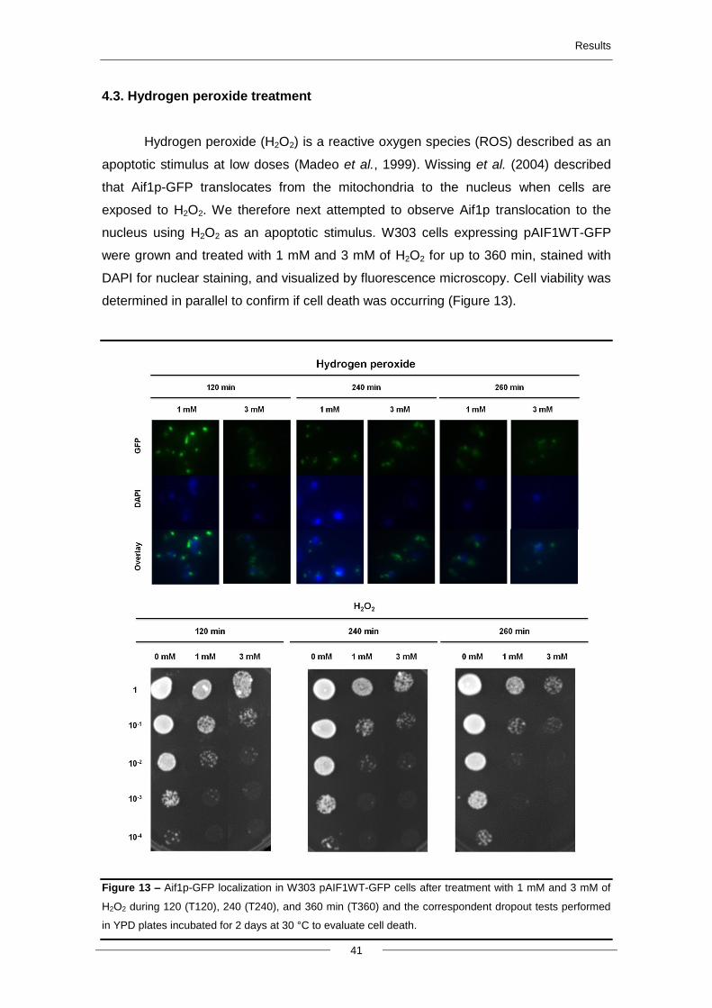
Results
41
4.3. Hydrogen peroxide treatment
Hydrogen peroxide (H2O2) is a reactive oxygen species (ROS) described as an
apoptotic stimulus at low doses (Madeo et al., 1999). Wissing et al. (2004) described
that Aif1p-GFP translocates from the mitochondria to the nucleus when cells are
exposed to H2O2. We therefore next attempted to observe Aif1p translocation to the
nucleus using H2O2 as an apoptotic stimulus. W303 cells expressing pAIF1WT-GFP
were grown and treated with 1 mM and 3 mM of H2O2 for up to 360 min, stained with
DAPI for nuclear staining, and visualized by fluorescence microscopy. Cell viability was
determined in parallel to confirm if cell death was occurring (Figure 13).
Figure 13 – Aif1p-GFP localization in W303 pAIF1WT-GFP cells after treatment with 1 mM and 3 mM of
H2O2 during 120 (T120), 240 (T240), and 360 min (T360) and the correspondent dropout tests performed
in YPD plates incubated for 2 days at 30 °C to evaluate cell death.
Results
42
Exposure to H2O2 resulted in dose-dependent cell death. However, even after
360 minutes of incubation with different concentrations of H2O2, Aif1p-GFP still did not
re-localize to the nucleus. It is likely that Aif1p-GFP was not released from
mitochondria under these conditions, and by consequence not translocated to the
nucleus. As the mode of H2O2-induced cell death depends greatly on its concentration
and on growth conditions of the culture, we decided to seek a different stimulus to
induce mitochondrial outer membrane permeabilization.
4.4. Heterologous expression of Bax c-myc protein
Bax, a protein from the Bcl-2 family, is the best-studied member of this group
and has important functions in mitochondrial morphogenesis and cell death (Karbowski
et al., 2006). Over-expression of Bax in yeast leads to morphological features similar to
mammalian apoptosis namely plasma membrane blebbing, DNA fragmentation and
others (Ligr et al., 1998). In yeast, several studies highlighted the role of mitochondria
in Bax-induced cell death, though as referred in the introduction there is some
controversy concerning the type of cell death induced by Bax protein. However, it is
well established that a c-myc tagged version of human Bax triggers mitochondrial outer
membrane permeabilization and release of cytochrome c from the mitochondria to the
cytosol. However, is referred before t is not known if it also induces the release of
Aif1p. The W303 strain expressing pAIF1WT-GFP was therefore transformed with a
plasmid expressing Bax c-myc, under the control of a tetracyclin-regulatable promoter.
Expression of Bax- c-myc was inhibited in negative control cells by addition of 1 µg/ml
of doxycyclin, an antibiotic member of the tetracycline antibiotics group that represses
the promoter. Cells were therefore grown in the presence or absence of doxycyclin to
repress or induce expression of Bax (protocol described in 3.5) (Priault et al., 1999).
Nuclei were then stained with DAPI to observe if Aif1p-GFP translocates into the
nucleus in response to Bax c-myc expression (Figure 14).
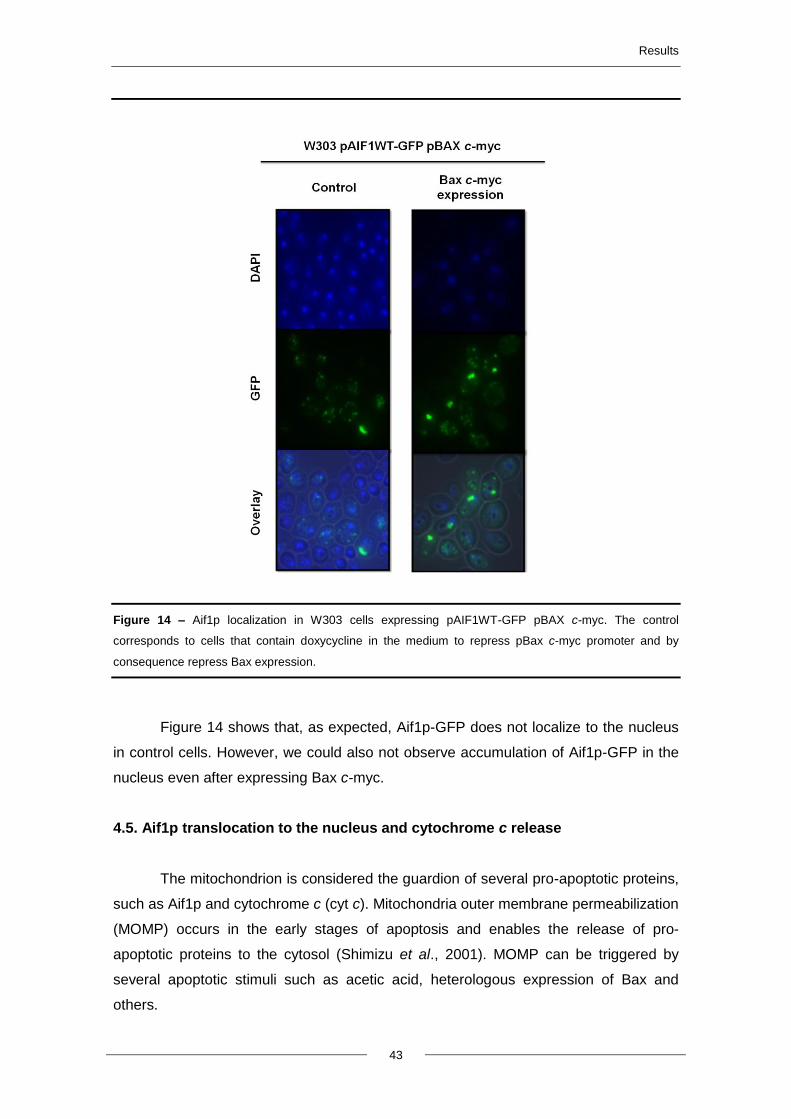
Results
43
Figure 14 – Aif1p localization in W303 cells expressing pAIF1WT-GFP pBAX c-myc. The control
corresponds to cells that contain doxycycline in the medium to repress pBax c-myc promoter and by
consequence repress Bax expression.
Figure 14 shows that, as expected, Aif1p-GFP does not localize to the nucleus
in control cells. However, we could also not observe accumulation of Aif1p-GFP in the
nucleus even after expressing Bax c-myc.
4.5. Aif1p translocation to the nucleus and cytochrome c release
The mitochondrion is considered the guardion of several pro-apoptotic proteins,
such as Aif1p and cytochrome c (cyt c). Mitochondria outer membrane permeabilization
(MOMP) occurs in the early stages of apoptosis and enables the release of pro-
apoptotic proteins to the cytosol (Shimizu et al., 2001). MOMP can be triggered by
several apoptotic stimuli such as acetic acid, heterologous expression of Bax and
others.
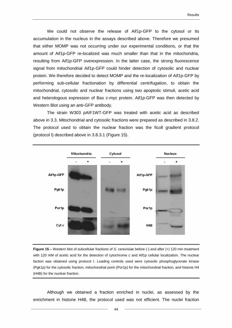
Results
44
We could not observe the release of Aif1p-GFP to the cytosol or its
accumulation in the nucleus in the assays described above. Therefore we presumed
that either MOMP was not occurring under our experimental conditions, or that the
amount of Aif1p-GFP re-localized was much smaller than that in the mitochondria,
resulting from Aif1p-GFP overexpression. In the latter case, the strong fluorescence
signal from mitochondrial Aif1p-GFP could hinder detection of cytosolic and nuclear
protein. We therefore decided to detect MOMP and the re-localization of Aif1p-GFP by
performing sub-cellular fractionation by differential centrifugation, to obtain the
mitochondrial, cytosolic and nuclear fractions using two apoptotic stimuli, acetic acid
and heterologous expression of Bax c-myc protein. Aif1p-GFP was then detected by
Western Blot using an anti-GFP antibody.
The strain W303 pAIF1WT-GFP was treated with acetic acid as described
above in 3.3. Mitochondrial and cytosolic fractions were prepared as described in 3.8.2.
The protocol used to obtain the nuclear fraction was the ficoll gradient protocol
(protocol l) described above in 3.8.3.1 (Figure 15).
Figure 15 – Western blot of subcellular fractions of S. cerevisiae before (-) and after (+) 120 min treatment
with 120 mM of acetic acid for the detection of cytochrome c and Aif1p cellular localization. The nuclear
faction was obtained using protocol I. Loading controls used were cytosolic phosphoglycerate kinase
(Pgk1p) for the cytosolic fraction, mitochondrial porin (Por1p) for the mitochondrial fraction, and histone H4
(H4B) for the nuclear fraction.
Although we obtained a fraction enriched in nuclei, as assessed by the
enrichment in histone H4B, the protocol used was not efficient. The nuclei fraction
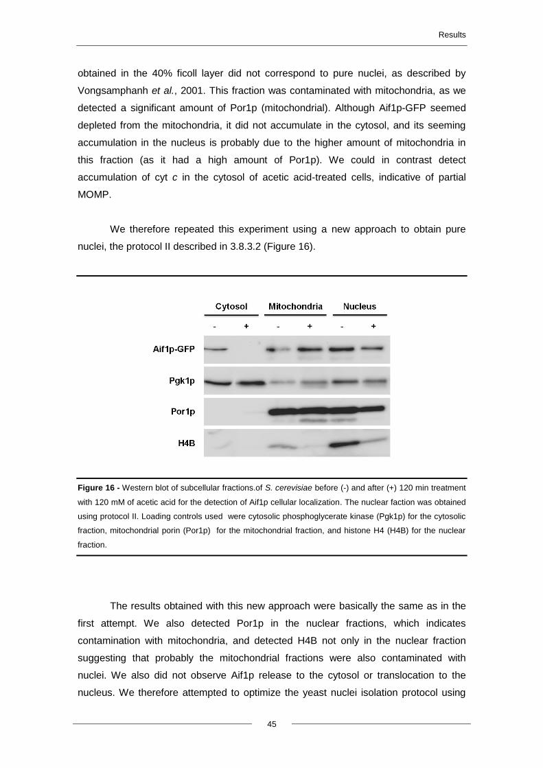
Results
45
obtained in the 40% ficoll layer did not correspond to pure nuclei, as described by
Vongsamphanh et al., 2001. This fraction was contaminated with mitochondria, as we
detected a significant amount of Por1p (mitochondrial). Although Aif1p-GFP seemed
depleted from the mitochondria, it did not accumulate in the cytosol, and its seeming
accumulation in the nucleus is probably due to the higher amount of mitochondria in
this fraction (as it had a high amount of Por1p). We could in contrast detect
accumulation of cyt c in the cytosol of acetic acid-treated cells, indicative of partial
MOMP.
We therefore repeated this experiment using a new approach to obtain pure
nuclei, the protocol II described in 3.8.3.2 (Figure 16).
Figure 16 - Western blot of subcellular fractions.of S. cerevisiae before (-) and after (+) 120 min treatment
with 120 mM of acetic acid for the detection of Aif1p cellular localization. The nuclear faction was obtained
using protocol II. Loading controls used were cytosolic phosphoglycerate kinase (Pgk1p) for the cytosolic
fraction, mitochondrial porin (Por1p) for the mitochondrial fraction, and histone H4 (H4B) for the nuclear
fraction.
The results obtained with this new approach were basically the same as in the
first attempt. We also detected Por1p in the nuclear fractions, which indicates
contamination with mitochondria, and detected H4B not only in the nuclear fraction
suggesting that probably the mitochondrial fractions were also contaminated with
nuclei. We also did not observe Aif1p release to the cytosol or translocation to the
nucleus. We therefore attempted to optimize the yeast nuclei isolation protocol using
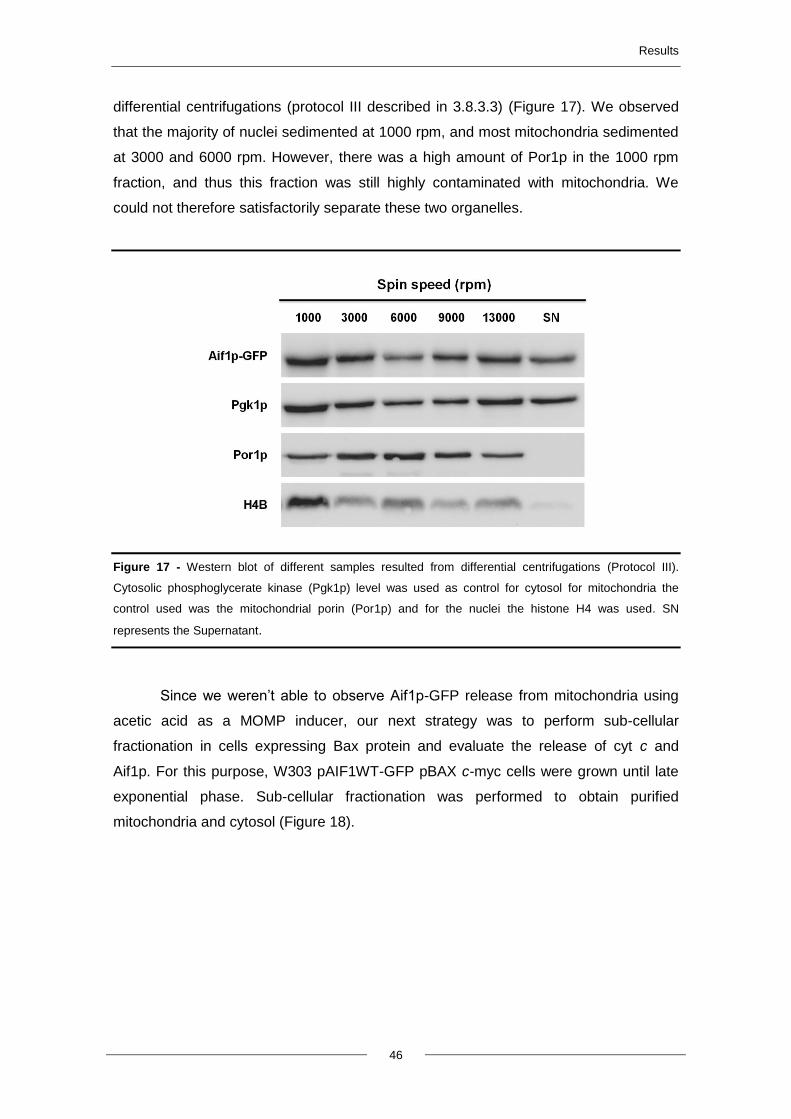
Results
46
differential centrifugations (protocol III described in 3.8.3.3) (Figure 17). We observed
that the majority of nuclei sedimented at 1000 rpm, and most mitochondria sedimented
at 3000 and 6000 rpm. However, there was a high amount of Por1p in the 1000 rpm
fraction, and thus this fraction was still highly contaminated with mitochondria. We
could not therefore satisfactorily separate these two organelles.
Figure 17 - Western blot of different samples resulted from differential centrifugations (Protocol III).
Cytosolic phosphoglycerate kinase (Pgk1p) level was used as control for cytosol for mitochondria the
control used was the mitochondrial porin (Por1p) and for the nuclei the histone H4 was used. SN
represents the Supernatant.
Since we weren’t able to observe Aif1p-GFP release from mitochondria using
acetic acid as a MOMP inducer, our next strategy was to perform sub-cellular
fractionation in cells expressing Bax protein and evaluate the release of cyt c and
Aif1p. For this purpose, W303 pAIF1WT-GFP pBAX c-myc cells were grown until late
exponential phase. Sub-cellular fractionation was performed to obtain purified
mitochondria and cytosol (Figure 18).
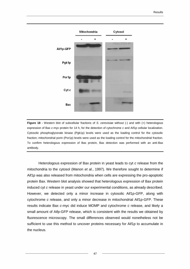
Results
47
Figure 18 - Western blot of subcellular fractions of S. cerevisiae without (-) and with (+) heterologous
expression of Bax c-myc protein for 14 h, for the detection of cytochrome c and Aif1p cellular localization.
Cytosolic phosphoglycerate kinase (Pgk1p) levels were used as the loading control for the cytosolic
fraction; mitochondrial porin (Por1p) levels were used as the loading control for the mitochondrial fraction.
To confirm heterologous expression of Bax protein, Bax detection was performed with an anti-Bax
antibody.
Heterologous expression of Bax protein in yeast leads to cyt c release from the
mitochondria to the cytosol (Manon et al., 1997). We therefore sought to determine if
Aif1p was also released from mitochondria when cells are expressing the pro-apoptotic
protein Bax. Western blot analysis showed that heterologous expression of Bax protein
induced cyt c release in yeast under our experimental conditions, as already described.
However, we detected only a minor increase in cytosolic Aif1p-GFP, along with
cytochrome c release, and only a minor decrease in mitochondrial Aif1p-GFP. These
results indicate Bax c-myc did induce MOMP and cytochrome c release, and likely a
small amount of Aifp-GFP release, which is consistent with the results we obtained by
fluorescence microscopy. The small differences observed would nonetheless not be
sufficient to use this method to uncover proteins necessary for Aif1p to accumulate in
the nucleus.
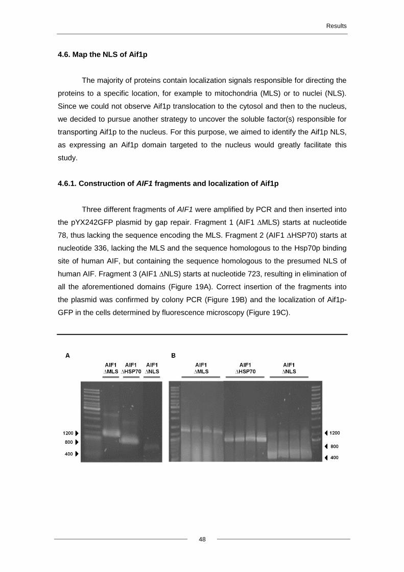
Results
48
4.6. Map the NLS of Aif1p
The majority of proteins contain localization signals responsible for directing the
proteins to a specific location, for example to mitochondria (MLS) or to nuclei (NLS).
Since we could not observe Aif1p translocation to the cytosol and then to the nucleus,
we decided to pursue another strategy to uncover the soluble factor(s) responsible for
transporting Aif1p to the nucleus. For this purpose, we aimed to identify the Aif1p NLS,
as expressing an Aif1p domain targeted to the nucleus would greatly facilitate this
study.
4.6.1. Construction of AIF1 fragments and localization of Aif1p
Three different fragments of AIF1 were amplified by PCR and then inserted into
the pYX242GFP plasmid by gap repair. Fragment 1 (AIF1 ∆MLS) starts at nucleotide
78, thus lacking the sequence encoding the MLS. Fragment 2 (AIF1 ∆HSP70) starts at
nucleotide 336, lacking the MLS and the sequence homologous to the Hsp70p binding
site of human AIF, but containing the sequence homologous to the presumed NLS of
human AIF. Fragment 3 (AIF1 ∆NLS) starts at nucleotide 723, resulting in elimination of
all the aforementioned domains (Figure 19A). Correct insertion of the fragments into
the plasmid was confirmed by colony PCR (Figure 19B) and the localization of Aif1p-
GFP in the cells determined by fluorescence microscopy (Figure 19C).
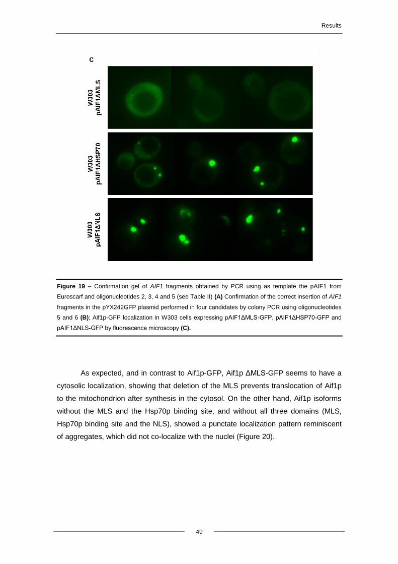
Results
49
Figure 19 – Confirmation gel of AIF1 fragments obtained by PCR using as template the pAIF1 from
Euroscarf and oligonucleotides 2, 3, 4 and 5 (see Table II) (A) Confirmation of the correct insertion of AIF1
fragments in the pYX242GFP plasmid performed in four candidates by colony PCR using oligonucleotides
5 and 6 (B); Aif1p-GFP localization in W303 cells expressing pAIF1ΔMLS-GFP, pAIF1ΔHSP70-GFP and
pAIF1ΔNLS-GFP by fluorescence microscopy (C).
As expected, and in contrast to Aif1p-GFP, Aif1p ΔMLS-GFP seems to have a
cytosolic localization, showing that deletion of the MLS prevents translocation of Aif1p
to the mitochondrion after synthesis in the cytosol. On the other hand, Aif1p isoforms
without the MLS and the Hsp70p binding site, and without all three domains (MLS,
Hsp70p binding site and the NLS), showed a punctate localization pattern reminiscent
of aggregates, which did not co-localize with the nuclei (Figure 20).
(b)
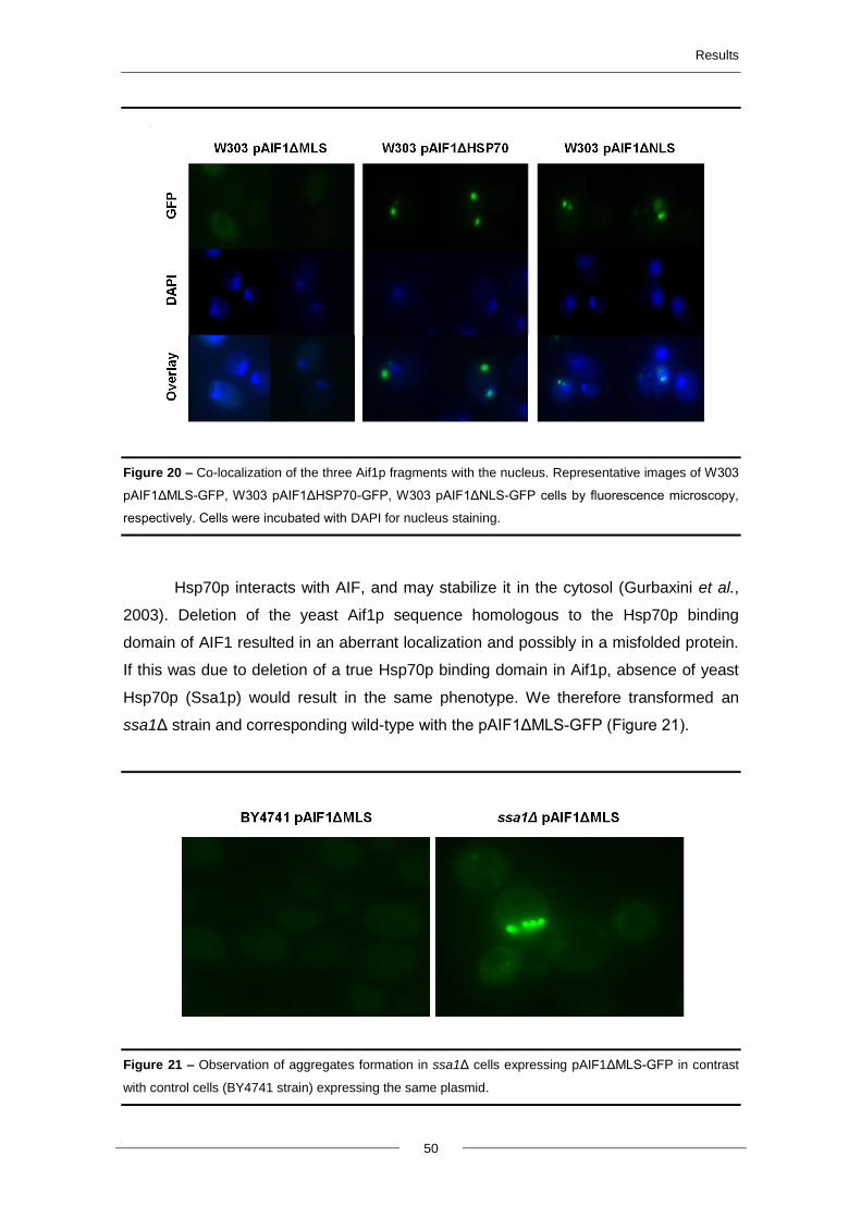
Results
50
Figure 20 – Co-localization of the three Aif1p fragments with the nucleus. Representative images of W303
pAIF1ΔMLS-GFP, W303 pAIF1ΔHSP70-GFP, W303 pAIF1ΔNLS-GFP cells by fluorescence microscopy,
respectively. Cells were incubated with DAPI for nucleus staining.
Hsp70p interacts with AIF, and may stabilize it in the cytosol (Gurbaxini et al.,
2003). Deletion of the yeast Aif1p sequence homologous to the Hsp70p binding
domain of AIF1 resulted in an aberrant localization and possibly in a misfolded protein.
If this was due to deletion of a true Hsp70p binding domain in Aif1p, absence of yeast
Hsp70p (Ssa1p) would result in the same phenotype. We therefore transformed an
ssa1Δ strain and corresponding wild-type with the pAIF1ΔMLS-GFP (Figure 21).
Figure 21 – Observation of aggregates formation in ssa1Δ cells expressing pAIF1ΔMLS-GFP in contrast
with control cells (BY4741 strain) expressing the same plasmid.
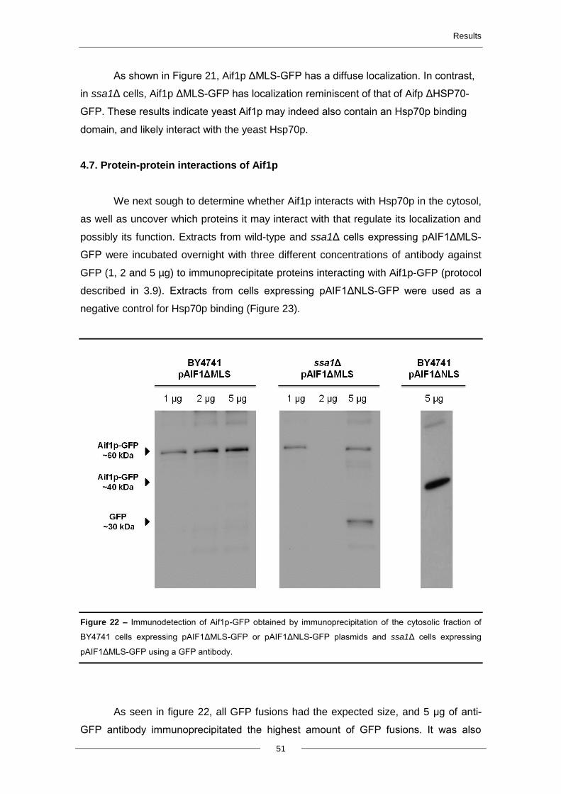
Results
51
As shown in Figure 21, Aif1p ΔMLS-GFP has a diffuse localization. In contrast,
in ssa1Δ cells, Aif1p ΔMLS-GFP has localization reminiscent of that of Aifp ΔHSP70-
GFP. These results indicate yeast Aif1p may indeed also contain an Hsp70p binding
domain, and likely interact with the yeast Hsp70p.
4.7. Protein-protein interactions of Aif1p
We next sough to determine whether Aif1p interacts with Hsp70p in the cytosol,
as well as uncover which proteins it may interact with that regulate its localization and
possibly its function. Extracts from wild-type and ssa1Δ cells expressing pAIF1ΔMLS-
GFP were incubated overnight with three different concentrations of antibody against
GFP (1, 2 and 5 µg) to immunoprecipitate proteins interacting with Aif1p-GFP (protocol
described in 3.9). Extracts from cells expressing pAIF1ΔNLS-GFP were used as a
negative control for Hsp70p binding (Figure 23).
Figure 22 – Immunodetection of Aif1p-GFP obtained by immunoprecipitation of the cytosolic fraction of
BY4741 cells expressing pAIF1ΔMLS-GFP or pAIF1ΔNLS-GFP plasmids and ssa1Δ cells expressing
pAIF1ΔMLS-GFP using a GFP antibody.
As seen in figure 22, all GFP fusions had the expected size, and 5 µg of anti-
GFP antibody immunoprecipitated the highest amount of GFP fusions. It was also
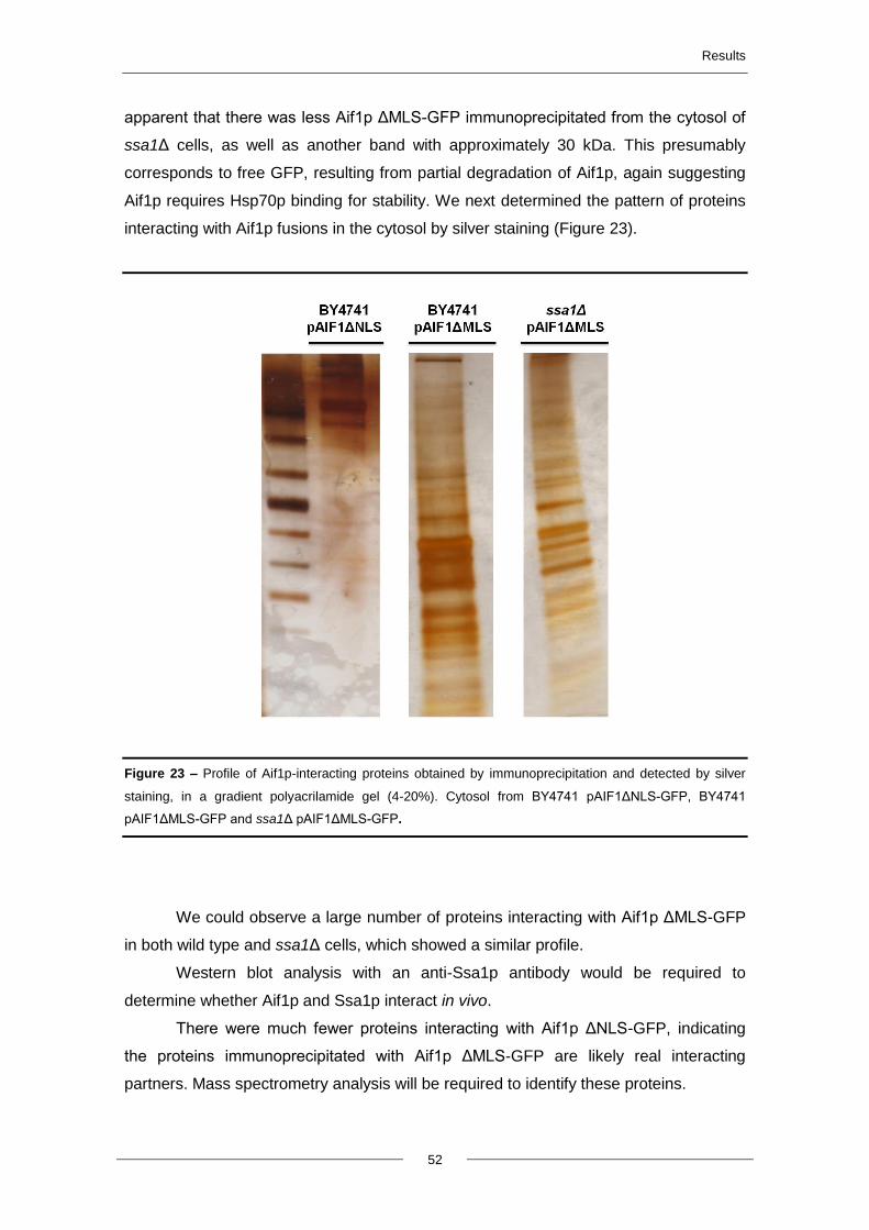
Results
52
apparent that there was less Aif1p ΔMLS-GFP immunoprecipitated from the cytosol of
ssa1Δ cells, as well as another band with approximately 30 kDa. This presumably
corresponds to free GFP, resulting from partial degradation of Aif1p, again suggesting
Aif1p requires Hsp70p binding for stability. We next determined the pattern of proteins
interacting with Aif1p fusions in the cytosol by silver staining (Figure 23).
Figure 23 – Profile of Aif1p-interacting proteins obtained by immunoprecipitation and detected by silver
staining, in a gradient polyacrilamide gel (4-20%). Cytosol from BY4741 pAIF1ΔNLS-GFP, BY4741
pAIF1ΔMLS-GFP and ssa1Δ pAIF1ΔMLS-GFP.
We could observe a large number of proteins interacting with Aif1p ΔMLS-GFP
in both wild type and ssa1Δ cells, which showed a similar profile.
Western blot analysis with an anti-Ssa1p antibody would be required to
determine whether Aif1p and Ssa1p interact in vivo.
There were much fewer proteins interacting with Aif1p ΔNLS-GFP, indicating
the proteins immunoprecipitated with Aif1p ΔMLS-GFP are likely real interacting
partners. Mass spectrometry analysis will be required to identify these proteins.


5. DISCUSSION AND
FUTURE
PERSPECTIVES
Discussion and future perspectives
55
Since the discovery that yeasts can commit “cell suicide”, S. cerevisiae has
been extensively used to study apoptotic cell death, with the objective of gaining insight
into poorly understood mechanistic aspects of mammalian apoptosis. In both
mammalian and yeast apoptosis, mitochondria have gained great importance due to
their ability of propagate the death signals generated within the cell, such as oxidative
stress, DNA damage and others (Wang, 2001). Many stress stimuli can trigger MOMP
and the release of pro-apoptotic proteins from the mitochondria to the cytosol, such as
cytochrome c and Aif1p.
Cytochrome c exerts its lethal function in the cytosol, which has been well
characterized in higher eukaryotes, but mammalian AIF needs to be translocated into
the nucleus in order to exert its pro-apoptotic function. The mechanism underlying this
translocation is still poorly understood, though it involves a regulated interaction with
Hsp70p in the cytosol (Ye et al., 2002). The yeast orthologue of mammalian AIF also
plays a role in cell death, and pure Aif1p can degrade yeast nuclei and plasmid DNA.
Aif1p, as mammalian AIF, has a mitochondrial localization in normal cells and is
translocated to the nucleus after an apoptotic stimulus. However, to our knowledge the
mechanism mediating translocation of Aif1p into the nucleus has not yet been studied.
For the majority of macromolecules, transport into the nucleus through nuclear pore
complexes is an energy-dependent process and normally mediated by the major class
of transport receptors, the karyopherins or Kaps. We therefore aimed to elucidate the
mechanism regulating yeast Aif1p import into the nucleus, and determine which Kap is
responsible for the nuclear accumulation of Aif1p. However, we were unable to
establish a robust system to detect accumulation of Aif1p-GFP in the nucleus, and thus
we were not able to screen the collection of yeast kapΔ mutant strains to determine
which were defective in this process.
Several stimuli are described as capable of triggering apoptosis in yeast leading
to MOMP and the release of pro-apoptotic proteins such as cyt c and Aif1p (Ludovico
et al., 2002, Vachova and Palkova, 2005, Khoury et al., 2008, Wissing et al., 2004 and
Priault et al., 1999). Wissing and colleagues were able to observe the translocation of
Aif1p from the mitochondria to the nucleus by fluorescence microscopy, using a
plasmid containing the AIF1 gene under the control of a promoter repressed by
methionine. Unfortunately, expressing Aif1p-GFP from this plasmid requires removing
methionine from the medium, and minimum levels of fluorescence were observed using
the microscope (D. Trindade, personal communication). For this reason, we cloned
Aif1p-GFP in pYX242GFP, a multi-copy plasmid, under the control of the strong TPI
promoter (pAIF1WT-GFP). This allows the constitutive expression of Aif1p-GFP at high

Discussion and future perspectives
56
levels. Indeed, we could observe that cells expressing this plasmid had a strong GFP
fluorescence with mitochondrial morphology.
Localization of Aif1p-GFP was studied in cells expressing pAIF1WT-GFP after
exposure to acetic acid or H2O2, or in cells undergoing chronological ageing or
expressing Bax c-myc using fluorescence microscopy and sub-cellular
fractionation/western blot. Chronological ageing and acetic acid treatment resulted in a
total loss of Aif1p green fluorescence levels, presumably because both lead to an
increase of intracellular acidification affecting fluorescence emission (not shown). For
that reason, the fluorescence microscopy assays were only performed in cells exposed
to H2O2 and expressing Bax c-myc and the cellular fractioning assays in cells exposed
to acetic acid and expressing Bax c-myc protein. By fluorescence microscopy, we were
not able to observe the release of Aif1p-GFP from the mitochondria or its translocation
to the nucleus either in response to H2O2 exposure or to heterologous expression of
Bax c-myc. These results suggested that MOMP was not occurring under our
experimental conditions, or that the amount of Aif1p-GFP re-localized was much
smaller than that remaining in the mitochondria. The signal from mitochondrial Aif1p-
GFP resulting from its high overexpression could therefore hinder detection of cytosolic
and nuclear protein. To overcome this problem, we are in the process of tagging
endogenous Aif1p with GFP in the genome by homologous recombination. Disrupting
AIF1 using short flanking sequences has proven difficult (not shown), probably
because this gene resides in a poorly accessible chromatin region, near the telomere.
We have therefore amplified AIF1 tagged with GFP flanked by homology sequences for
pAG61 (an integrative plasmid), and cloned it into this plasmid by Gap Repair. We are
also in the process of cloning the 3’ UTR region of AIF1 downstream of the URA3
marker in the same plasmid. After digestion with appropriated enzymes, this plasmid
will work as a cassette with 1kb of homology up- and downstream of the AIF1 gene, to
allow for more effective homologous recombination in the genome of any yeast strain.
This strategy will allow the observation of endogenous Aif1p levels in the different
organelles by fluorescence microscopy, without Aif1p overexpression.
As mentioned above, it may have been that we could not observe the release of
Aif1p-GFP by fluorescence microscopy because MOMP was not occurring under our
experimental conditions, or because the amount of Aif1p-GFP localizing to the nucleus
was bellow our detection limits. For that reason, we tried to detect MOMP and the
localization of Aif1p-GFP by sub-cellular fractionation in purified mitochondrial,
cytosolic, and nuclear fractions. The results of the cellular fractioning and western blot
demonstrated that there was some release of cyt c both in cells treated with acetic acid
and in cells expressing Bax c-myc. On the other hand, Aif1p-GFP release into the
Discussion and future perspectives
57
cytosol was not observed, suggesting that MOMP is occurring allowing the release of
cyt c but not of Aif1p-GFP, or that the levels of MOMP achieved were very low, and
only the earlier release of cyt c was observed. The accumulation of Aif1p-GFP in the
nucleus was evaluated by detecting Aif1-GFP in the nuclear fraction. Yeast nuclei were
purified using three different methods; however, it was not possible to obtain purified
nuclei by differential centrifugation, because it was always contaminated with
mitochondria. Consequently, the levels of Aif1p-GFP detected in the nucleus are
probably due to the mitochondrial contaminations. It has now been reported that Aif1p
is translocated to the nucleus after exposure to hydrogen peroxide in only 25% of cells
(Gruhlke et al., 2012). Therefore, we expect that detecting an enrichment of 25% at
best of Aif1p-GFP in the nuclear fraction after treatment would not be reliable, and
using this method to confirm accumulation of Aif1p-GFP in the nucleus would only be
possible in case there were no traces of mitochondrial contamination in the nuclear
fraction. Since this would be very technically challenging, we abandoned this
methodology.
The major goal of this work consisted in determining which Kap is responsible
for the transport of Aif1p to the nucleus. Our intention was to transform mutants in each
of the yeast soluble transport receptors (Kaps) with pAIF1WT-GFP and assess the
localization of Aif1p-GFP by fluorescence microscopy after apoptosis induction. The
mutant with less accumulation in the nucleus would lack the Kap responsible for Aif1p
import. However, since it is described that with an apoptotic stimulus, at best 25% of
cells show Aif1p translocation to the nucleus, and due to the functional redundancy of
this family, there would be the risk of not being able to observe a significant decrease
in Aif1p-GFP accumulation in the nucleus in individual Kaps mutants. Mapping the NLS
of Aif1p would thus overcome the need for the use of apoptotic stimuli to observe Aif1p
translocation to the nucleus, and greatly facilitate this study.
The human AIF protein possesses five typical domains: an N-terminal
Mitochondrial Localization Signal (MLS) domain; a FAD and NADH binding domain
responsible for its oxireductase activity; an Hsp70p binding domain; a C-terminal end
domain; and the Nuclear Localization Signal (NLS) that is located between the
aminoacids 367 and 459 (Modjtahedi et al., 2006, Daugas et al., 2000 and Gurbuxani
et al., 2003) (Figure 2). Taking this information into account and the homology of hAIF
with yeast Aif1p, we cloned different AIF1 isoforms in frame with GFP where different
domains were deleted, in order to determine the location of the Aif1p NLS (AIF1 ΔMLS,
AIF1 ΔHSP70 and AIF1 ΔNLS). Cells transformed with the different plasmids were
visualized by fluorescence microscopy in order to evaluate the localization of the
different Aif1p isoforms. As expected, deletion of the MLS domain led to a change of
Discussion and future perspectives
58
Aif1p localization from the mitochondria to the cytosol. On the other hand, deletion of
the Hsp70p binding domain (AIF1 ΔHSP70 and AIF1 ΔNLS isoforms) led to aberrant
localization of GFP, possibly in aggregates. Since deletion of the MLS domain did not
result in the nuclear import of Aif1p-GFP, our results suggest that Aif1p translocation to
the nucleus is regulated at a step subsequent to Aif1p release from the mitochondria.
Our results also suggested that interaction with Hsp70p in the cytosol seems to be
important for the correct folding of Aif1p, though additional experiments are required.
Since none of the Aif1p fragments had a nuclear localization, we could not map
the NLS of Aif1p. It would be interesting to construct several deletions from the C-
terminal end of the gene to map the NLS. Using this strategy, deletion of the Hsp70p
binding domain would not occur and Aif1p would not be destabilized. It would still be
necessary to delete the MLS domain to guarantee that Aif1p would have a cytosolic
localization, and possible an apoptotic stimulus. The location of the hAIF NLS is known,
and directed our choice of domains to be deleted. Using further analysis of the
homology between the two aminoacids sequences, it would be interesting to delete the
corresponding aminoacids in Aif1p and assess if the nuclear accumulation of Aif1p is
abolished (once we establish a solid system), in order to discover if the NLS domain is
conserved between these organisms.
We cannot discard that AIF does not have a true NLS domain and that the
transport of this protein into the nucleus is not dependent on this domain for direct
interaction with a transport receptor, but instead is involved in a regulated mechanism
involving other proteins, for example the Hsp70p protein. As mentioned above, Hsp70p
protein binds to cytosolic hAIF protein and possibly to Aif1p in yeast (see below), acting
as chaperone that helps in the folding of the protein and in it stabilization. The NLS
domain of a protein is a necessary and sufficient domain to promote translocation of
the protein to the nucleus. Since deletion of the MLS did not lead to nuclear import of
Aif1p, we hypothesize that either Aif1p has an NLS that is masked and/or regulated by
additional proteins, or that Aif1p does not have this domain. It is therefore likely that
AIF1 ΔMLS only translocates to the nucleus after apoptotic stimuli. Aif1p and hAIF
proteins possess several similarities in terms of structure, function and mode of action,
It would thus be interesting to test whether heterologous expression of hAIF protein in
yeast complements the phenotype of an aif1Δ mutant and whether hAIF-GFP
translocates to the nucleus after an apoptotic stimulus (Candé et al., 2002). It would be
also interesting to express other hAIF isoforms (described by Gurbuxani et al., 2003) to
determine if they have the same localization and phenotype when expressed in yeast
cells. Once the Kap(s) responsible for Aif1p nuclear accumulation is identified, it would
Discussion and future perspectives
59
be interesting to determine if the same Kap is responsible for hAIF import to the
nucleus.
Hsp70p is described as being capable of regulating apoptosis. This protein is
involved mostly in the inhibition of apoptosis by blocking Apaf-1/formation of the
apoptosome or by stabilization of AIF protein in the cytosol, inhibiting nuclear
accumulation of this protein, and thus leading to inhibition of DNA degradation in the
nucleus. This protein has been associated with a cytoprotective role against the
apoptogenic effects of AIF targeted to the extramitochondrial compartment, as its
down-regulation facilitates the induction of apoptosis, whereas its up-regulation leads
to inhibition of apoptosis (Ravagnan et al., 2001). Deletion of Hsp70p binding domain
(residues 150 to 228) leads to a gain of function phenotype, i.e., facilitates the nuclear
translocation of AIF. As mentioned previously, in the course of trying to map the NLS
domain of Aif1p, we observed the formation of green fluorescent aggregate-like
structures in Aif1p constructs lacking the sequence homologous to the hAIF Hsp70-
binding domain, suggesting that yeast Aif1p interacts with Hsp70p. We also observed
that cytosolic Aif1p with an intact Hsp70p binding domain localized to aberrant green
fluorescent spots in yeast cells lacking Hsp70p protein. This corroborates the idea that
binding of Hsp70p protein to Aif1p is required for stabilization and folding of Aif1p and
suggests another role of the yeast Hsp70p in the function of Aif1p. We hypothesize
that, in the absence of Hsp70p protein or when its Hsp70p binding domain is deleted,
cytosolic Aif1p becomes misfolded or unfolded, probably loosing its normal function.
Several stressors lead to misfolded or unfolded proteins, but cells normally
ensure that proteins are folded correctly using a variety of chaperones, foldases or
lectins. When proteins cannot be restored, misfolded proteins are targeted for
processing or degradation (Chakrabarti et al., 2011). In yeasts, there are two major
pathways described for protein degradation; the vacuolar pathway and the proteasome
pathway. Since AIF1 ∆HSP70 forms spots, possible aggregates, it is possible that
Aif1p ΔHSP70-GFP is misfolded and should therefore be partly degraded by one of the
degradation pathways. Degradation by the vacuolar pathway does not seem to be
occurring in these cells, because the green fluorescent aggregates do not seem to
have a vacuolar localization. However, to confirm the exact location of the aggregates,
cells expressing the pAIF1ΔHSP70-GFP and pAIF1ΔNLS-GFP plasmids should be
stained with Arg-CMAC, a label for the lumen of the vacuole. On the other hand,
aggregates could correspond to Aif1p-GFP inside of autophagosomes formed during
autophagy. Autophagy is the mechanism by which cells use parts of themselves to
survive starvation and stress, limiting cell death by recycling energy and substrates,
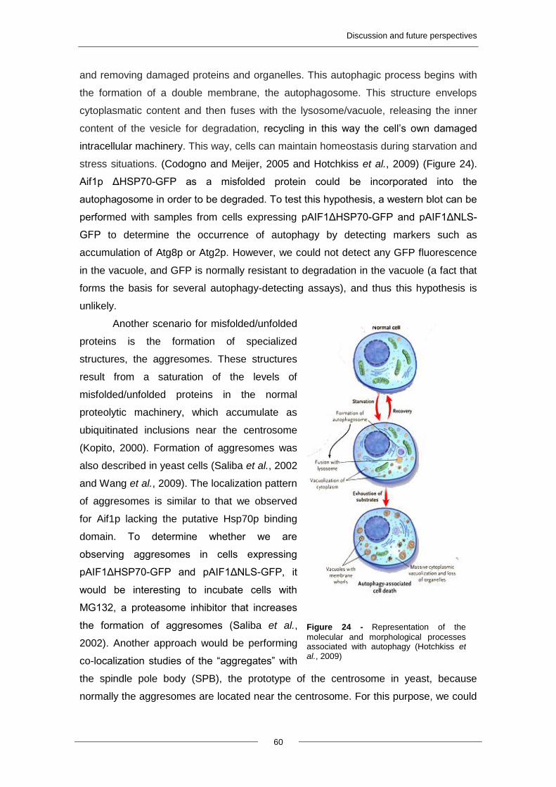
Discussion and future perspectives
60
and removing damaged proteins and organelles. This autophagic process begins with
the formation of a double membrane, the autophagosome. This structure envelops
cytoplasmatic content and then fuses with the lysosome/vacuole, releasing the inner
content of the vesicle for degradation, recycling in this way the cell’s own damaged
intracellular machinery. This way, cells can maintain homeostasis during starvation and
stress situations. (Codogno and Meijer, 2005 and Hotchkiss et al., 2009) (Figure 24).
Aif1p ΔHSP70-GFP as a misfolded protein could be incorporated into the
autophagosome in order to be degraded. To test this hypothesis, a western blot can be
performed with samples from cells expressing pAIF1ΔHSP70-GFP and pAIF1ΔNLS-
GFP to determine the occurrence of autophagy by detecting markers such as
accumulation of Atg8p or Atg2p. However, we could not detect any GFP fluorescence
in the vacuole, and GFP is normally resistant to degradation in the vacuole (a fact that
forms the basis for several autophagy-detecting assays), and thus this hypothesis is
unlikely.
Another scenario for misfolded/unfolded
proteins is the formation of specialized
structures, the aggresomes. These structures
result from a saturation of the levels of
misfolded/unfolded proteins in the normal
proteolytic machinery, which accumulate as
ubiquitinated inclusions near the centrosome
(Kopito, 2000). Formation of aggresomes was
also described in yeast cells (Saliba et al., 2002
and Wang et al., 2009). The localization pattern
of aggresomes is similar to that we observed
for Aif1p lacking the putative Hsp70p binding
domain. To determine whether we are
observing aggresomes in cells expressing
pAIF1ΔHSP70-GFP and pAIF1ΔNLS-GFP, it
would be interesting to incubate cells with
MG132, a proteasome inhibitor that increases
the formation of aggresomes (Saliba et al.,
2002). Another approach would be performing
co-localization studies of the “aggregates” with
the spindle pole body (SPB), the prototype of the centrosome in yeast, because
normally the aggresomes are located near the centrosome. For this purpose, we could
Figure 24 - Representation of the
molecular and morphological processes associated with autophagy (Hotchkiss et al., 2009)
Discussion and future perspectives
61
use cells expressing BFP-tagged components of the SPB (Wang et al., 2009 and
Moens et al., 1971).
To uncover which proteins interact with Aif1p-GFP in the cytosol, we performed
an immunoprecipitation of the cytosol from cells expressing the pAIF1ΔMLS and
pAIF1ΔNLS (control) (Figure 23). Our results show that the Aif1 ΔMLS protein
specifically interacts with multiple proteins in the cytosol (as the most
immunoprecipitated proteins do not interact with the Aif1 ΔNLS protein). Taken
together, our results thus suggested that Aif1p interacts specifically with several
proteins in the cytosol, including with the Hsp70p protein. To confirm the interaction of
Aif1p with Hsp70p protein, we will next detect the presence of Hsp70p in the
immunoprecipitated fraction with an antibody against Ssa1 (Hsp70p). We will next
optimize this protocol in order to identify Aif1p-interacting proteins by mass
spectrometry, both in cytosolic extracts from cells without and with an apoptotic
stimulus. We will first elute interacting proteins with a salt gradient to distinguish which
bands represent proteins with a strong interaction with cytosolic Aif1p, and to
determine the salt concentration for which we eliminate most unspecific bands. Bands
and/or the elution fractions can then be analyzed by mass spectrometry for protein
identification. Identification of proteins that interact with cytosolic Aif1p before and after
an apoptotic stimulus should provide valuable insight into the regulation of this protein
and its pro-apoptotic effect.


6. LITERATURE
CITED
Literature cited
64
Ameisen JC, Estaquier J, Idziorek T, De Bels F. (1995) Programmed cell death and
AIDS: significance, perspectives and unanswered questions. Cell Death Differ, 2(1): 9-
22.
Arnoult D, Tatischeff I, Estaquier J, Girard M, Sureau F, Tissier JP. (2001). On the
evolutionary conservation of the cell death pathway: mitochondrial release of an
apoptosis-inducing factor during Dictyostelium discoideum cell death. Mo. Biol Cell, 12:
3016–3030.
Badugu R, Garcia M, Bondada V, Joshi A, Geddes JW. (2008) N terminus of calpain 1
is a mitochondrial targeting sequence. The Journal of Biological Chemistry,
283(6):3409-3417.
Bayliss R, Corbett AH, Stewart M. (2000) The Molecular Mechanism of Transport of
Macromolecules Through Nuclear Pore Complexes. Traffic, 1(6): 448-456.
Boujard T, Ramezi J, Vandeputte M, Labbé L, Mambrini M. (2007) Group feeding
behavior of brown trout is a correlated response to selection for growth shaped by the
environment. Behav Genet, 37(3): 525-34.
Bradford MM. (1976) A rapid and sensitive method for the quantitation of microgram
quantities of protein utilizing the principle of protein-dye binding. Anal Biochem, 7; 72:
248-54.
Burton TR, Eisenstat DD, Gibson SB. (2009) BNIP3 (Bcl-2 19 kDa Interacting Protein)
Acts as Transcriptional Repressor of Apoptosis-Inducing Factor Expression Preventing
Cell Death in Human Malignant Gliomas. Journal of Neuroscience, 29(13): 4189-4199.
Buttner S, Eisenberg T, Herker E, Carmona-Gutierrez D, Kroemer G, Madeo F. (2006)
Why yeast cells can undergo apoptosis: death in times of peace, love, and war. J Cell
Biol, 175: 521-5.
Candé C, Cohen I, Daugas E, Ravagnan L, Larochette N, Zamzami N, Kroemer G.
(2002) Apoptosis-inducing factor (AIF): a novel caspase-independent death effector
released from mitochondria. Biochimie, 84(2-3): 215-222.
Candé C, Vahsen N, Kouranti I, Schmitt E, Daugas E, Spahr C, Luban J, Kroemer RT,
Giordanetto F, Garrido C, Penninger JM, Kroemer G. (2004) AIF and cyclophilin A
cooperate in apoptosis-associated chromatinolysis. Oncogene, 23(8): 1514-1521.
Carmona-Gutierrez D, Eisenberg T, Büttner S, Meisinger C, Kroemer G, Madeo F.
(2010) Apoptosis in yeast: triggers, pathways, subroutines. Cell Death and
Literature cited
65
Differentiation, 17(5): 763-773.
Chakrabarti A, Chen A, Varner J. (2011) A Review of the Mammalian Unfolded Protein
Response. Biotechnol Bioeng, 108(12): 2777-93.
Cheng W, Teng X, Park HK, Tucker CM, Dunham MJ, Hardwick JM. (2008) Fis1
deficiency selects for compensatory mutations responsible for cell death and growth
control defects. Cell Death Differ, 15(12): 1838–1846.
Chipuk JE, Moldoveanu T, Llambi F, Parsons MJ, Green DR. (2010) The BCL-2 family
reunion. Molecular Cell, 37(3): 299-310.
Chowdhury I., Tharakan B., Bhat G.K. (2008) Caspases - An update. Comparative
Biochemistry and Physiology, 151(1): 10-27.
Codogno P, Meijer AJ. (2005). Autophagy and signaling: their role in cell survival and
cell death. Cell Death and Differentiation, 12 Suppl 2: 1509-18.
Cohen GM. (1997) Caspases: the executioners of apoptosis. The Biochemical Journal,
326 (Pt 1): 1-16.
Cook A, Bono F, Jinek M, Conti E. (2007) Structural Biology of Nucleocytoplasmic
Transport. Annual Review of Biochemistry, 76: 647-671.
Daugas E, Nochy D, Ravagnan L, Loeffler M, Susin SA, Zamzami N, Kroemer G.
(2000) Apoptosis-inducing factor (AIF): a ubiquitous mitochondrial oxidoreductase
involved in apoptosis. FEBS Letters, 476(3): 118-123.
Eisenberg T, Büttner S, Kroemer G, Madeo F. (2007) The mitochondrial pathway in
yeast apoptosis. Apoptosis, 12(5): 1011-1023.
Elmore S. (2007) Apoptosis: a review of programmed cell death. Toxicologic
Pathology, 35(4): 495-516.
Fabrizio P, Pletcher SD, Minois N, Vaupel JW, Longo VD. (2004) Chronological ageing
in dependent replicative life span regulation by Msn2/Msn4 and Sod2 in
Saccharomyces cerevisiae, FEBS Lett, 557 136–142.
Fabrizio P, Longo VD. (2003) The chronological life span of Saccharomyces
cerevisiae. Ageing Cell, 2: 73-81.
Fabrizio P, Longo VD. (2008) Chronological ageing-induced apoptosis in yeast.
Biochim Biophys Acta,1783: 1280-5.
Finley D, Ozkaynak E, Varshavsky A. (1987) The yeast polyubiquitin gene is essential
for resistance to high temperatures, starvation, and other stresses. Cell, 27;48(6):
1035-46.
Literature cited
66
Fannjiang Y, Cheng WC, Lee SJ, Qi B, Pevsner J, McCaffery JM, Hill RB, Basañez G,
Hardwick JM. (2004) Mitochondrial fission proteins regulate programmed cell death in
yeast. Genes & Development, 18(22): 2785-97.
Fischer UTE, Schulze-Osthoff K. (2005) New approaches and therapeutics targeting
apoptosis in disease. Pharmacological Reviews, 57(2): 187-215.
Freitas N, Cunha C. (2009) Mechanisms and Signals for the Nuclear Import of
Proteins. Current Genomics, 10(8): 550-557.
Fröhlich KU, Fussi H, Ruckenstuhl C. (2007) Yeast apoptosis – from genes to
pathways. Seminars in Cancer Biology, 17(2): 112-121.
Gewies A. (2003) Introduction to Apoptosis. ApoReview, pp.1-26.
Gietz RD, Woods RA. (2006) Yeast transformation by the LiAc/SS Carrier DNA/PEG
method. Methods Mol Biol, 313: 107-20.
Gourlay C, Du W, Ayscough K. (2006) Apoptosis in yeast – mechanisms and benefits
to a unicellular organism. Molecular Microbiology, 62(6): 1515–1521
Gruhlke M, Portz D, Stitz M, Anwar A, Schneider T, Jacob C, Schlaich NL, Slusarenko
AJ. (2010) Allicin disrupts the cell's electrochemical potential and induces apoptosis in
yeast. Free Radical Biology & Medicine, 49: 1916–1924.
Gulshan K, Rovinsky SA, Coleman ST, Moye-Rowley WS. (2005) Oxidant-specific
folding of Yap1p regulates both transcriptional activation and nuclear localization. The
Journal Biological Chemistry, 280(49): 40524-33.
Gurbuxani S, Schmitt E, Cande C, Parcellier A, Hammann A, Daugas E, Kouranti I,
Spahr C, Pance A, Kroemer G, Garrido C. (2003) Heat shock protein 70 binding
inhibits the nuclear import of apoptosis-inducing factor. Oncogene, 22(43): 6669-78.
Gustafsson AB, Gottlieb RA. (2007) Bcl-2 family members and apoptosis, taken to
heart. American Journal of Cell Physiology, 292(1): C45-51.
Gruhlke MCH, Noll U, Schlaich NL, Slusarenko AJ. (2012) Protein-Kinase A is a central
regulator of allicin-induced cell-death in Saccharomyces cerevisiae. 9th IMYA, Rome,
16-20/09.
Hangen E, Blomgren K, Bénit P, Kroemer G, Modjtahedi N. (2010) Life with or without
AIF. Trends in Biochemical Sciences, 35(5): 278-287.
Hengartner MO. (2000) The biochemistry of apoptosis. Nature, 407(6805): 770-6.
Literature cited
67
Herker E, Jungwirth H, Lehmann KA, Maldener C, Frohlich KU, Wissing S, Buttner S,
Fehr M, Sigrist S, Madeo F. (2004) Chronological ageing leads to apoptosis in yeast. J
Cell Biol, 164: 501-7.
Hotchkiss RS, Strasser A, McDunn JE, Swanson PE. (2009) Cell death. The New
England Journal of Medicine, 361(16): 1570-83.
Joza N, Pospisilik JA, Hangen E, Hanada T, Modjtahedi N, Penninger JM, Kroemer G.
(2009) AIF: Not Just an Apoptosis-Inducing Factor. Annals of the N ew York Academy
of Sciences, 1171: 2-11.
Kaffman A, Rank NM, O'Shea EK. (1998) Phosphorylation regulates association of the
transcription factor Pho4 with its import receptor Pse1/Kap121. Genes & Development,
12(17): 2673-83.
Karbowski M, Norris KL, Cleland MM, Jeong SY, Youle RJ. (2006). Role of Bax and
Bak in mitochondrial morphogenesis. Nature, 443: 658-62.
Khoury CM, Greenwood MT. (2008) The pleiotropic effects of heterologous Bax
expression in yeast. Biochim Biophys Acta,1783(7): 1449-65.
Kopito RR. (2000) Aggresomes, inclusion bodies and protein aggregation. Trends Cell
Biol, 10(12): 524-30.
Lawen A. (2003) Apoptosis - An introduction. Bioessays, 25(9): 888-96.
Leist M, Jaattela M. (2001) Four deaths and a funeral: from caspases to alternative
mechanisms. Nature Reviews Molecular Cell Biology, 2: 589–598.
Li W, Sun L, Liang Q, Wang J, Mo W, Zhou B. (2006) Yeast AMID Homologue Ndi1p
Displays Respiration-restricted Apoptotic Activity and Is Involved in Chronological
Ageing. Molecular Biology of the Cell, 17(4): 1802-1811.
Liang Q, Li W, Zhou B. (2008) Caspase-independent apoptosis in yeast. Biochimica et
Biophysica Acta, 1783(7): 1311-1319.
Ligr M, Madeo F, Frohlich E, Hilt W, Frohlich KU, Wolf DH. (1998). Mammalian Bax
triggers apoptotic changes in yeast. FEBS Lett, 438: 61-5.
Lipton SA, Bossy-Wetzel E. (2002) Dueling Activities of AIF in Cell Death versus
Survival: DNA Binding and Redox Activity. Cell, 111(2): 147-150.
Lorenzo HK, Susin SA. (2007) Therapeutic potential of AIF-mediated caspase-
Literature cited
68
independent programmed cell death. Drug Resistance Updates, 10(6): 235-255.
Lowe, SW, Lin, AW. (2000) Apoptosis in cancer. Carcinogenesis, 21: 485-95.
Ludovico P. (2002) Endogenous programmed cell death of Saccharomyces cerevisiae
Master Thesis, Departamento de Biologia, Universidade do Minho.
Ludovico P, Rodrigues F, Almeida A, Silva MT, Barrientos A, Côrte-Real M. (2002)
Cytochrome c release and mitochondria involvement in Programmed Cell Death
induced by acetic acid in Saccharomyces cerevisiae. Molecular Biology of the Cell,
13(8): 2598-606.
Ludovico P, Sousa MJ, Silva MT, Leão C, Côrte-Real M. (2001) Saccharomyces
cerevisiae commits to a programmed cell death process in response to acetic acid.
Microbiology (Reading, England), 147(Pt 9): 2409-15.
Luo X, Budihardjo I, Zou H, Slaughter C, Wang X. (1998) Bid, a Bcl2 interacting
protein, mediates cytochrome c release from mitochondria in response to activation of
cell surface death receptors. Cell, 94(4): 481-490.
Macara IG. (2001) Transport into and out of the Nucleus. Microbiology and Molecular
Biology Reviews, 65(4): 570-594.
Madeo F, Carmona-Gutierrez D, Ring J, Büttner S, Eisenberg T, Kroemer G. (2009)
Caspase-dependent and caspase-independent cell death pathways in yeast.
Biochemical and Biophysical Research Communications, 382(2): 227-231.
Madeo F, Engelhardt S, Herker E, Lehmann N, Maldener C, Proksch A, Wissing S,
Fröhlich KU. (2002) Apoptosis in yeast: a new model system with applications in cell
biology and medicine. Current Genetics, 41(4): 208-216.
Madeo F, Frohlich E, Frohlich KU. (1997) A yeast mutant showing diagnostic markers
of early and late apoptosis. J Cell Biol, 139: 729-34.
Madeo F, Fröhlich E, Ligr M, Grey M, Sigrist SJ, Wolf DH, Fröhlich KU. (1999) Oxygen
stress: a regulator of apoptosis in yeast. The Journal of Cell Biology, 145(4): 757.
Madeo F, Herker E, Wissing S, Jungwirth H, Eisenberg T, Fröhlich KU. (2004)
Apoptosis in yeast. Current Opinion in Microbiology, 7(6): 655-660.
Manon S, Chaudhuri B, Guerin M. (1997). Release of cytochrome c and decrease of
cytochrome c oxidase in Bax-expressing yeast cells, and prevention of these effects by
Literature cited
69
co expression of Bcl-xL. FEBS Lett, 415: 29-32.
Miramar MD, Costantini P, Ravagnan L, Saraiva LM, Haouzi D, Brothers G, Penninger
JM, Peleato ML, Kroemer G, Susin SA. (2001) NADH-oxidase activity of mitochondrial
apoptosis-inducing factor (AIF). The Journal of Biological Chemistry, 276(19): 16391-
16398.
Modjtahedi N, Giordanetto F, Madeo F, Kroemer G. (2006) Apoptosis-inducing factor:
vital and lethal. Trends in Cell Biology, 16(5): 264-272.
Moens PB, Rapport E. (1971) Spindles, spindle plaques, and meiosis in the yeast
Saccharomyces cerevisiae. J. Cell Biol, 50: 344–361
Morton C, Santos S, Coote P. (2007) An amphibian-derived, cationic, a-helical
antimicrobial peptide kills yeast by caspase-independent but AIF-dependent
programmed cell death. Molecular Microbiology, 65(2): 494–507.
Moubarak RS, Yuste VJ, Artus C, Bouharrour A, Greer PA, Menissier-de Murcia J,
Susin SA. (2007) Sequential activation of poly (ADP-ribose) polymerase 1, calpains,
and Bax is essential in apoptosis-inducing factor-mediated programmed necrosis. Mol
Cell Biol, 27(13): 4844-62.
Norberg E, Orrenius S, Zhivotovsky B. (2010) Mitochondrial regulation of cell death:
Processing of apoptosis-inducing factor (AIF). Biochemical and Biophysical Research
Communications, 396(1): 95-100.
O'Neill LA, Kaltschmidt C. (1997) NF-kappa B: a crucial transcription factor for glial and
neuronal cell function. Trends in Neurosciences, 20(6): 252-8.
Parrino J, Hotchkiss RS, Bray M. (2007) Prevention of immune cell apoptosis as
potential therapeutic strategy for severe infections. Emerging Infectious Diseases,
13(2): 191-198.
Parsell DA, Lindquist S. (1993) The function of heat-shock proteins in stress tolerance:
degradation and reactivation of damaged proteins. Annu Rev Genet, 27: 437-96.
Priault M, Chaudhuri B, Clow A, Camougrand N, Manon S. (1999) Investigation of bax-
induced release of cytochrome c from yeast mitochondria permeability of mitochondrial
membranes, role of VDAC and ATP requirement. Eur J Biochem, 260: 684-91.
Raff MC, Barres BA, Burne JF, Coles HS, Ishizaki Y, Jacobson MD. (1993)
Programmed cell death and the control of cell survival: lessons from the nervous
Literature cited
70
system. Science, 262(5134): 695-700.
Ravagnan L, Gurbuxani S, Susin SA, Maisse C, Daugas E, Zamzami N, Mak T,
Jäättelä M, Penninger JM, Garrido C, Kroemer G. (2001) Heat-shock protein 70
antagonizes apoptosis-inducing factor. Nature Cell Biology, 3(9): 839-43.
Reed, JC. (2003) Apoptosis-targeted therapies for cancer. Cancer Cell, 3: 17-22.
Ren D, Tu HC, Kim H, Wang GX, Bean GR, Takeuchi O, Jeffers JR, Zambetti GP,
Hsieh JJ, Cheng EH. (2010) BID, BIM, and PUMA are essential for activation of the
BAX- and BAK-dependent cell death program. Science, 330(6009): 1390-3.
Rout MP, Aitchison JD, Suprapto A, Hjertaas K, Zhao Y, Chait BT. (2000) The Yeast
Nuclear Pore Complex: Composition, Architecture, and Transport Mechanism. Journal
of Cell Biology, 148(4): 635-651.
Rosenblum JS, Pemberton LF, Bonifaci N, Blobel G. (1998) Nuclear import and the
evolution of a multifunctional RNA-binding protein. J Cell Biol, 6;143(4): 887-99.
Ruchalski K, Mao H, Li Z, Wang Z, Gillers S, Wang Y, Mosser DD, Gabai V, Schwartz
JH, Borkan SC. (2001) Heat shock protein 70 antagonizes apoptosis-inducing factor.
The Journal of Biological Chemistry, 281(12): 7873-80.
Rudin CM, Thompson CB. (1997) Apoptosis and disease: regulation and clinical
relevance of programmed cell death. Annu Rev Med, 48: 267-81.
Saikumar P, Dong Z, Mikhailov V, Denton M, Weinberg JM, Venkatachalam MA.
(1999) Apoptosis: Definition, Mechanisms and relevance to disease. The American
Journal of Medicine, 107(5): 489-506.
Saliba RS, Munro PMG, Luthert PJ, Cheetham ME. (2002) The cellular fate of mutant
rhodopsin: quality control, degradation and aggresome formation. Journal of Cell
Science, 115(14).
Saraste A, Pulkki K. (2000) Morphologic and biochemical hallmarks of apoptosis.
Cardiovascular Research, 45(3): 528-37.
Sartorius U, Schmitz I, Krammer PH. (2001) Molecular mechanisms of death-receptor-
mediated apoptosis. Chembiochem, 2(1): 20-9.
Scaffidi C, Fulda S, Srinivasan A, Friesen C, Li F, Tomaselli KJ, Debatin KM, Krammer
PH, Peter ME. (1998) Two CD95 (APO-1/Fas) signaling pathways. The EMBO Journal,
17(6): 1675-1687.
Literature cited
71
Schabel, FM, Jr. (1976). Nitrosoureas: a review of experimental antitumor activity.
Cancer Treat Rep, 60: 665–698.
Sheridan C, Martin SJ. (2010) Mitochondrial fission/fusion dynamics and apoptosis.
Mitochondrion, 10(6): 640-648.
Shimizu S, Matsuoka Y, Shinohara Y, Yoneda Y, Tsujimoto Y. (2001) Essential role of
voltage-dependent an ion channel in various forms of apoptosis in mammalian cells. J
Cell Bio, 152: 237-50.
Smale, G, Nichols, N.R, Brady, DR, Finch, CE, Horton, WE, Jr. (1995) Evidence for
apoptotic cell death in Alzheimer's disease. Exp Neurol, 133: 225-30.
Sokolov S, Knorre D, Smirnova E, Markova O, Pozniakovsky A,Skulachev V, Severin F
(2006) Ysp2 mediates death of yeast induced by amiodarone or intracellular
acidification. Biochim Biophys Acta, 1757: 1366–1370.
Stegh AH, Schickling O, Ehret A, Scaffidi C, Peterhänsel C, Hofmann TG, Grummt I,
Krammer PH, Peter ME. (1998) DEDD, a novel death effector domain-containing
protein, targeted to the nucleolus. The EMBO Journal, 17(20): 5974-5986.
Susin SA, Lorenzo HK, Zamzami N, Marzo I, Snow BE, Brothers GM, Mangion J,
Jacotot E, Costantini P, Loeffler M, Larochette N, Goodlett DR, Aebersold R,
Siderovski DP, Penninger JM and Kroemer G. (1999).Molecular characterization of
mitochondrial apoptosis-inducing factor. Nature, 397: 441–446.
Tan, EM. (1994) Autoimmunity and apoptosis. J Exp Med, 179: 1083-6.
Tran EJ, Wente SR. (2006) Dynamic Nuclear Pore Complexes: Life on the Edge. Cell,
125(6): 1041-1053.
Ye H, Cande C, Stephanou NC, Jiang S, Gurbuxani S, Larochette N, Daugas E,
Garrido C, Kroemer G, Wu H. (2002) DNA binding is required for the apoptogenic
action of apoptosis inducing factor. Nat. Struct. Biol, 9: 680–684.
Yuste VJ, Lorenzo HK, Susin SA. (2007) AIFM1 (apoptosis-inducing factor,
mitochondrion-associated, 1). Atlas Genet Cytogenet Oncol Haematol.
Vachova L, Palkova Z. (2005) Physiological regulation of yeast cell death in
multicellular colonies is triggered by ammonia. J Cell Biol. 169: 711-7.
Vaux DL. (2011) Apoptogenic factors released from mitochondria. Biochimica et
Biophysica Acta, 1813(4): 546-550.
Literature cited
72
Vongsamphanh R, Fortier PK, Ramotar D. (2001) Pir1p Mediates Translocation of the
Yeast Apn1p Endonuclease into the Mitochondria To Maintain Genomic Stability.
Molecular and cell biology, 1655: 1647–1655.
Wang Y, Meriin AB, Zaarur N, Romanova NV, Chernoff YO, Costello CE, Sherman MY.
(2009) Abnormal proteins can form aggresome in yeast: aggresome-targeting signals
and components of the machinery. The FASEB journal, 0892-6638/09/0023-0451.
Wang X. (2001) The expanding role of mitochondria in apoptosis. Genes &
Development, 15(22): 2922-2933.
Wang X, Wang J, Gengyo-Ando K, Gu L, Sun CL, Yang C. (2007). C. elegans
mitochondrial factor WAH-1 promotes phos-phatidylserine externalization in apoptotic
cells through phospholipid scramblase SCRM-1. Nat. Cell Biol, 9, 541–549.
Wang X, Yang C, Chai J, Shi Y, Xue D, (2002). Mechanisms of AIF-mediated apoptotic
DNA degradation in Caenorhabditis elegans. Science, 298, 1587–1592.
Wissing S, Ludovico P, Herker E, Büttner S, Engelhardt SM, Decker T, Link A, Proksch
A, Rodrigues F, Corte-Real M, Fröhlich KU, Manns J, Candé C, Sigrist SJ, Kroemer G,
Madeo F. (2004) An AIF orthologue regulates apoptosis in yeast. Journal of Cell
Biology, 166(7): 969-974.
Xu C, Wang J, Gao Y, Lin H, Du L, Yang S, Long S, She Z, Cai X, Zhou S, Lu Y.
(2009) The anthracenedione compound bostrycin induces mitochondria-mediated
apoptosis in the yeast Saccharomyces cerevisiae. FEMS Yeast Res, 10: 297–308.