f r nesthesia www continuous femoral … › files › 2013 › pdf › (v11p41-47...continuous...
TRANSCRIPT

BOEZAART AP
© 2006 THE JOURNAL OF THE NEW YORK SCHOOL OF REGIONAL ANESTHESIA (WWW.NYSORA.COM) CONTINUOUS FEMORAL NERVE BLOCK
JOURNAL OF NEW YORK SCHOOL OF REGIONAL ANESTHESIA http://www.nysora.com VOLUME 11 41
CONTINUOUS FEMORAL NERVE BLOCK BY ANDRÉ P BOEZAART MD, PHD
Author Affiliation: Department of Anesthesia, University of Iowa, Iowa City, IA
INTRODUCTION
Due to the multiplicity and divergence of the nerve supply to the joints of the lower extremity, a femoral nerve block per se is almost never adequate as the sole anesthetic for lower limb surgery. It is almost always necessary to block the other three major peripheral nerves to the lower extremity as well. A femoral nerve block is therefore usually performed in conjunction with a sciatic, and/or obturator, and/or lateral cutaneous nerve of the thigh block.
The sensory distribution of the femoral nerve supply is outlined in Figure 1 [1]). The anterior approach to the femoral nerve is similar for “single shot” or continuous nerve blocks. This communication will outline the technique for continuous femoral nerve block only. For “single shot” block a 50 mm 22 G Stimuplex needle is typically used and the local anesthetic agent is injected after location of the nerve with the nerve stimulator set at 0.4 – 0.6 mA and 200 - 300µs.
Most continuous catheter techniques that were developed after the initial attempts of Ansbro in 1946 [2] were hampered by inaccurate catheter placement or catheter dislodgement. In order to provide reliable analgesia for lower extremity surgery and prevent readmission due to failed catheter placement, it was necessary to develop a method to ensure real-time catheter positioning (i.e., during placement). This can now be done at insertion with all continuous peripheral nerve blocks (rather than hours later when the initial block has worn off), by stimulating the nerves via both the needle through which the catheter is placed and via the catheter itself [3]. This accuracy of catheter placement is combined with a method to secure the catheter that prevents dislodgement.
Figure 1a. Somatic neurotomal distribution of the lower extremity. (Copied with permission from Brown DL. Atlas of Regional Anesthesia.)

BOEZAART AP
© 2006 THE JOURNAL OF THE NEW YORK SCHOOL OF REGIONAL ANESTHESIA (WWW.NYSORA.COM) CONTINUOUS FEMORAL NERVE BLOCK
JOURNAL OF NEW YORK SCHOOL OF REGIONAL ANESTHESIA http://www.nysora.com VOLUME 11 42
INDICATIONS The femoral nerve block is mainly indicated for the pain control associated with unilateral anterior knee surgery. It is important to note that the posterior obturator nerve gives off an articular branch that supplies the posterior aspect of the knee and this nerve may be responsible for the pain experienced in the posterior aspect of the knee following knee surgery despite an effective femoral nerve block. It was originally thought that this pain was due to the absence of a sciatic nerve block, but work done by the Virginia Mason group [4] disputed this notion. Allen and co- workers demonstrated that there was no difference in postoperative pain if a sciatic nerve block was done in conjunction with a femoral nerve block. It is, however, necessary to perform a sciatic nerve block additionally if surgery distal or posterior to the knee joint (for example
anterior or posterior cruciate ligament repair) is done. It is often necessary to block the obturator and/or sciatic nerve separately in addition to the femoral nerve after total knee replacement surgery. The pain experienced in the posterior aspect of the knee is often short-lived and effectively controlled with “single-shot” blocks. Continuous femoral nerve blocks have been demonstrated to improve the outcome of total knee arthroplasty [5]. Outcome with continuous femoral nerve block was better than “single shot” femoral block and continuous epidural anesthesia.
EQUIPMENT A sheathed insulated 17-gauge Tuohy needle (StimuCath, Arrow International, Reading, PA, USA) (Figure 2a) and a catheter with an inner steel spring capable of conducting electrical impulses to its distal uncovered “bullet-tip” end –
Figure 1b. Somatic neurotomal distribution of the lower extremity. (Copied with permission from Brown DL. Atlas of Regional Anesthesia.)

BOEZAART AP
© 2006 THE JOURNAL OF THE NEW YORK SCHOOL OF REGIONAL ANESTHESIA (WWW.NYSORA.COM) CONTINUOUS FEMORAL NERVE BLOCK
JOURNAL OF NEW YORK SCHOOL OF REGIONAL ANESTHESIA http://www.nysora.com VOLUME 11 43
a “stimulating catheter” (Figures 2b) – are used in the methods described here (Arrow StimuCath™, Arrow International, Reading, PA, USA)
Figure 2a: StimuCath needle. The StimuCath needle is an insulated Tuohy needle with a bare tip and a bare proximal area. PNS = Peripheral Nerve Stimulator.
Figure 2b: StimuCath catheter. The StimuCath catheter has an inner spring reinforcement, which is electrically conductive and continues from its proximal
ANATOMY The femoral nerve is situated lateral to the femoral artery and is deep to the iliaca fascia, which in turn is deep to the fascia lata. The femoral artery and vein, again, is in a separate fascia compartment (Figure 5). The femoral nerve is one nerve bundle near the inguinal crease, but a short distance more distally; it divides into its seven branches.
ANATOMIC LANDMARKS Identify and mark the femoral artery in the groin. The femoral nerve is situated 1 – 2 cm lateral to the femoral artery (Figure 4).
Figure 3. Anatomy. Reprinted from Grants Atlas of Anatomy
Figure 4. Anatomical landmarks and needle placement A = line marking femoral artery B = Inguinal crease C = Intended path for tunneling catheter Needle entry is 1 – 2 cm lateral of the femoral artery and aimed

BOEZAART AP
© 2006 THE JOURNAL OF THE NEW YORK SCHOOL OF REGIONAL ANESTHESIA (WWW.NYSORA.COM) CONTINUOUS FEMORAL NERVE BLOCK
JOURNAL OF NEW YORK SCHOOL OF REGIONAL ANESTHESIA http://www.nysora.com VOLUME 11 44
TECHNIQUE PATIENT POSITION The patient lies supine with a clear view of the patella. NEEDLE PLACEMENT
After generous skin and subcutaneous tissue infiltration of local anesthetic agent (beware not to block the femoral nerve in the process!), the needle is inserted aiming approximately 45 degrees cephalad (Figure 4). The
point of needle entry is just inferior to the inguinal crease. The nerve stimulator is clipped to the proximal bare area of the needle and two definite “pops” are felt when the needle penetrates first the fascia lata and then the iliaca fascia. It is very important to penetrate both these layers of fascia, since the electrical current may well cross the fascia layer and cause muscle twitching, but the local anesthetic agent will not cross the fascia layer if deposited superficial to it (Figure 5). This is a common mistake when performing femoral nerve blocks.
In our experience femoral blocks are predictably successful if the needle is placed deep to the fascia iliaca. Using a nerve stimulator, this will be the case when brisk twitches of the quadriceps muscles are evoked that move the patella while being able to dial the nerve stimulator output down to between 0.3 and 0.6 mA at a pulse width of 200 - 300µs.
Another common mistake is to stimulate the nerve to the sartorius muscle, which is situated superficial to the fascia iliaca. The local anesthetic agent will then not reach the femoral nerve. Aim the bevel of the needle in the direction in which the catheter is intended to go – cephalad in this instance. CATHETER PLACEMENT • The nerve stimulator clip is now removed from the
needle and attached to the proximal end of the stimulating catheter (Figure 6).
• Introduce the stimulating catheter into the needle. • The muscle twitches should resume again and should
be unchanged in character and intensity. The catheter is then gradually advanced beyond the tip of the needle for a distance of approximately 3 to 5 centimeters. The muscle twitches (patella movement) should continue unchanged over the entire distance of the catheter advancement.
• If stimulation ceases during catheter advancement, the catheter should be carefully withdrawn to inside the shaft of the needle, the needle position changed by a combination of rotation (Figure 7), angulation or depth adjustment until the catheter advances easily while the muscle twitches are maintained unchanged throughout.
• The catheter is now correctly placed near the femoral nerve but will most likely dislodge over time unless secured.
Figure 5. Anatomy. The femoral nerve lies deep to the fascia iliaca, which in turn lies deep to the fascia lata. Note that the femoral artery and vein are situated in a separate fascia sheath. Reprinted from Grants Atlas of Anatomy

BOEZAART AP
© 2006 THE JOURNAL OF THE NEW YORK SCHOOL OF REGIONAL ANESTHESIA (WWW.NYSORA.COM) CONTINUOUS FEMORAL NERVE BLOCK
JOURNAL OF NEW YORK SCHOOL OF REGIONAL ANESTHESIA http://www.nysora.com VOLUME 11 45
Figure 6: Catheter placement. The needle is held steady; the nerve stimulator is now clipped to the proximal end of the catheter and the catheter in advanced through the needle. There should be no change in the quality of the muscle (quadriceps) twitches. Be sure that the same
TUNNELING TO SECURE THE CATHETER • Penetrate the skin with the inner steel stylet of the
needle 1 – 3 mm from the catheter entry site and advance the stylet subcutaneously in a lateral direction to exit the skin 8 – 10 cm laterally (Figure 8).
Figure 8. Tunneling. Insert the inner stylet of the needle 2 – 3 mm from the catheter exit wound and advance subcutaneously to exit the skin 6 – 10 cm laterally.
• “Rail-road” the needle over the stylet (Figure 9).
Figure 9. Tunneling. Insert the inner stylet of the needle 3 – 5 mm from the catheter exit site and advance subcutaneously to exit the skin approximately 8 – 10 cm laterally and “railroad the needle retrogradely back over the stylet. See text.
• Remove the stylet and feed the catheter retrogradely
through the needle (Figure 9). After passage of the catheter, remove the needle (Figure 9) and observe the skin bridge (figure 10).
Figure 10. Tunnelling. Remove the stylet from the needle and feed the proximal end of the catheter through the needle.
BOEZAART AP
© 2006 THE JOURNAL OF THE NEW YORK SCHOOL OF REGIONAL ANESTHESIA (WWW.NYSORA.COM) CONTINUOUS FEMORAL NERVE BLOCK
JOURNAL OF NEW YORK SCHOOL OF REGIONAL ANESTHESIA http://www.nysora.com VOLUME 11 46
Figure 11. Tunneling. Remove the needle and secure the catheter with sterile dressings. Observe the skin bridge. If the skin bridge is undesirable, allow the needle to exit through the same hole in the skin as the catheter. Be careful not to damage the catheter with the needle
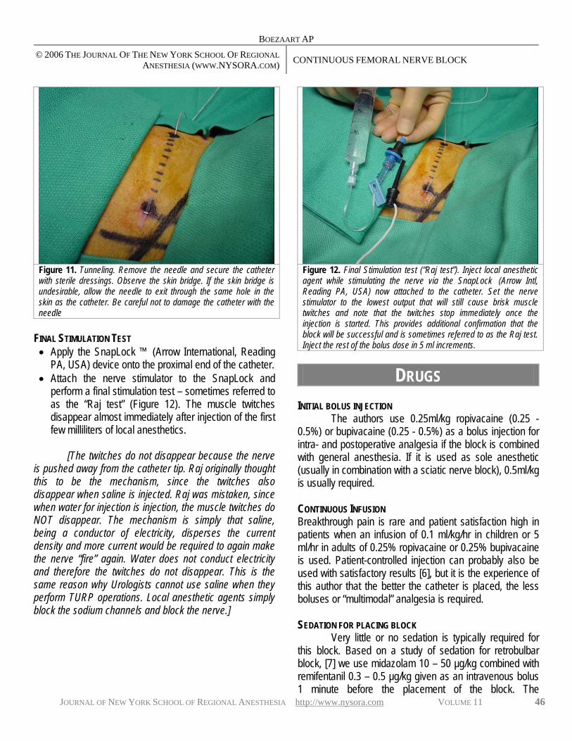
FINAL STIMULATION TEST • Apply the SnapLock ™ (Arrow International, Reading
PA, USA) device onto the proximal end of the catheter. • Attach the nerve stimulator to the SnapLock and
perform a final stimulation test – sometimes referred to as the “Raj test” (Figure 12). The muscle twitches disappear almost immediately after injection of the first few milliliters of local anesthetics.
[The twitches do not disappear because the nerve
is pushed away from the catheter tip. Raj originally thought this to be the mechanism, since the twitches also disappear when saline is injected. Raj was mistaken, since when water for injection is injection, the muscle twitches do NOT disappear. The mechanism is simply that saline, being a conductor of electricity, disperses the current density and more current would be required to again make the nerve “fire” again. Water does not conduct electricity and therefore the twitches do not disappear. This is the same reason why Urologists cannot use saline when they perform TURP operations. Local anesthetic agents simply block the sodium channels and block the nerve.]
Figure 12. Final Stimulation test (“Raj test”). Inject local anesthetic agent while stimulating the nerve via the SnapLock� (Arrow Intl, Reading PA, USA) now attached to the catheter. Set the nerve stimulator to the lowest output that will still cause brisk muscle twitches and note that the twitches stop immediately once the injection is started. This provides additional confirmation that the block will be successful and is sometimes referred to as the Raj test. Inject the rest of the bolus dose in 5 ml increments.
DRUGS INITIAL BOLUS INJECTION
The authors use 0.25ml/kg ropivacaine (0.25 - 0.5%) or bupivacaine (0.25 - 0.5%) as a bolus injection for intra- and postoperative analgesia if the block is combined with general anesthesia. If it is used as sole anesthetic (usually in combination with a sciatic nerve block), 0.5ml/kg is usually required. CONTINUOUS INFUSION Breakthrough pain is rare and patient satisfaction high in patients when an infusion of 0.1 ml/kg/hr in children or 5 ml/hr in adults of 0.25% ropivacaine or 0.25% bupivacaine is used. Patient-controlled injection can probably also be used with satisfactory results [6], but it is the experience of this author that the better the catheter is placed, the less boluses or “multimodal” analgesia is required. SEDATION FOR PLACING BLOCK
Very little or no sedation is typically required for this block. Based on a study of sedation for retrobulbar block, [7] we use midazolam 10 – 50 µg/kg combined with remifentanil 0.3 – 0.5 µg/kg given as an intravenous bolus 1 minute before the placement of the block. The

BOEZAART AP
© 2006 THE JOURNAL OF THE NEW YORK SCHOOL OF REGIONAL ANESTHESIA (WWW.NYSORA.COM) CONTINUOUS FEMORAL NERVE BLOCK
JOURNAL OF NEW YORK SCHOOL OF REGIONAL ANESTHESIA http://www.nysora.com VOLUME 11 47
remifentanil injection can be repeated when necessary if painful conditions such as fractures are present. Alfentanil (10 micrograms per kg) or fentanyl is also appropriate alternatives in this situation.
Blocks are usually performed in non-anesthetized patients, but under certain circumstances they may be performed in anesthetized patients. Examples include children, very painful conditions such as fractures, or very anxious patients. The skin and subcutaneous tissue should always be properly anesthetized for blocks as well as for the intended tunneling path of the catheter.
SPECIAL PRECAUTIONS AND PRACTICAL POINTS
• The catheter should always be withdrawn entirely into
the needle before the needle is repositioned. Catheter withdrawal should be done carefully to prevent damage to the catheter.
• The presence of significant paresthesia during catheter advancement should be carefully evaluated before advancement of the catheter.
• Be suspicious of sub-perineural needle or catheter placement if brisk muscle twitches are present with nerve stimulator settings less than 0.2mA (except in children).
• Since an indwelling catheter is left in situ for some time, formal sterile procedures are necessary. The entry site of the catheter should be inspected daily for early signs of infection.
• Sensation should be allowed to return to the limb before the catheter is removed. Catheters should never be cut while being removed. If the surgical pain is still intolerable, a bolus of the local anesthetic agent should be injected and the infusion initiated again. If surgical pain is tolerable or manageable with simple analgesics, the catheter may be removed by gently pulling on it in the direction of the tunneling or by removing the part distal to the skin bridge first. Radiating pain experienced during removal may indicate that the catheter has curled around a nerve root. Surgical removal of catheters has never been reported to be necessary but should probably be
considered if radiating pain persists during attempted removal. The skin bridge makes removal easier.
• Do not inject LA agent or saline through the needle prior to catheter placement. This will make catheter placement impossible. The “opening up” of the sheath with saline or LA agent is a fallacy.
• NEVER cut a catheter that has a spring wire inside, such as the StimuCath. This will most certainly result in the catheter “falling apart”, since there is a wire inside the catheter that anchors the distal end of the catheter to the proximal end. This situation is bound to attract medico-legal attention, since parts of the catheter may break off and may be left behind.
.
REFERENCES 1. Brown DL. Atlas of Regional Anesthesia. 2nd ed.
Philadelphia, PA: W.B. Saunders Company; 1999. 2. Ansbro P. A method of continuous brachial plexus
block. American Journal of Surgery 1946; 121: 716 – 722
3. Boezaart AP, de Beer JF, du Toit C, van Rooyen K. A new technique of continuous interscalene nerve block. Canadian Journal of Anesthesia 1999; 46(3): 275 – 281.
4. Allen HW, Lui SS, Ware PD, Nairn CS, Owens BD. Peripheral nerve blocks improve analgesia after total knee replacement surgery. Anesth Analg 1998; 93: 93 – 97.
5. Capdevilla X, Barthelet Y, Biboulet P, Ryckwaert Y, et al. Effects of perioperative technique on surgical outcome and duration of rehabilitation after major knee surgery. Anesthesiology 1999; 91: 8 – 15
6. Borgeat A, Schäppi B, Biasca N, Gerber C. Patient-controlled analgesia after major shoulder surgery: Patient-controlled interscalene analgesia versus patient-controlled analgesia. Anesthesiology 1997; 87: 1343 – 1347.
7. Boezaart AP, Berry AR, Nell ML, van Dyk AL. A comparison of propofol and remifentanil for sedation and limitation of movement during peri-retrobulbar block. Journal of Clinical Anesthesia 2001; 13: 422 – 426.