f. 01 oj - national institutes of health · 366 d. l. kacian et al. materials whatman microgranular...
TRANSCRIPT

D. L. KALJA?;. K. F. WPITSO?;, A. BUKK'I- AND S . SPIEGELMAN
instituie 01 Cnncw Hcseaych. C d e g t o j Pnysiiaans and Surgeons, Columbia Unaversaty, iVew York.
(Received June 1st rq ; r )
Y.Y. r0 : : jz IG.S,A.!
SIJMMARY
DNA polymerase fwm avian rryeiobla?tos?s virm lid\ been pwified by a com- bination of rulumn rliromatographv and gel filtration methods The iaolated enzyme sediments xi apprwmately 6 S and consists of two subunits of molecular weiglds IIO ooo and 64 ooo It is fret. oi RS-4 and DNA endonuclease activity. The enzyme pos5esspi the RSA-, DXA-, and hybrid directed polymeraic activities found in the virior,
iNTRODUCTlO>
The discovery1,2 of s ribonuclease-sensitive DNA polymerase activity in on- cogenic RSA viruses was quickly extended3-' to x wide .variety of oncornavirusess. I: wzs filrrher shown tha t the product DNA was complementary to the RNA uf the virion sed as the source of the enzvme preparatiorr3, $-I1. The.e findings were promptly foliowed bq- experiments that establsiied the existence in these viruses of polymerase activitics that respond ti: doubie-stranded DNAY*12-15 and, with a very high effi- ciency. to synthetic honiopolymeri- dupiexes13*16 composed of pdyribonucleotides poiydeoxyri tmnucii.oticieJ. and hybrid structures of the tw(,, The size of the DNA product synthesized was generally much less than that of the template empl0yed3*~9,11.
In aaditim to tiit; DNA paivnierase activities, evidence was also found for DNA endo- and exonu*cleases13s 17, l i g a s ~ ~ ~ . and a nucleosidetriphosphate phospho- transferase's in tlie vmon
Ik i i ica t ion of the RNA dependent DNA polymerase is a necessary prerequi- site to an unambiquous analysis of the reaction mechanism. Purity is also requi- red to delineate tlie relation of this polymerase activity to the others observed with DNA ana synthetic homopolymeric duplexes as templates. Finally, pure enzyme should permit a decision on whether the nuclease, ligase and phosphotransferase ac- tivities are coliectively or individually inherent and necessary components of the DNA polymerase function.
We report here the purification and characterization of the DNA polymerase activity from avian myeloblastosis virus.
Abbreviations: AMV, avian myeloblastosis virus; BBOT, z,~-bis-z-(~-tert.-butylbenzoxa- zolyl) thiophene.
Biochim. Biophys. Acta, 246 (1971) 365-383

366 D. L. KACIAN et al .
MATERIALS
Whatman microgranular DEAE-cellulose, DE52, 1.0 mequiv/g dry weight, and phosphocellulose, PII, 7.4 mequiv/g dry weight were obtained from Reeve Angel. Sephadex G-zoo and CM-Sephadex C-50 were purchased from Pharmacia Fine Che- micals. Clarkson Chemical Company provided hydroxylapatite. Unlabeled nucleoside triphosphates and dithiothreitol came from P-L Biochemicals. Miles Laboratories supplied polynucleotides and Micrococcus lysodeikticzts DNA. Tritiated nucleoside triphosphates were obtained from New England Nuclear, Schwarz BioResearch, and Amersham-Searle. Nucleic acid polymers were the generous gifts of Drs. F. Bollum (University of Kentucky), A. N. Nussbaum (Hoffman-LaRoche), and L. A. Under- kofler (Miles Laboratories). Acrylamide and methylene bisacrylamide came from Bio-Rad Laboratories. Nonidet P-40 was a product of Shell Chemical CO.
METHODS
( I ) Purif ication of avian myeloblastosis virus Avian myeloblastosis virus (AMV), BAI strain A, was obtained by methods
previously described19 from the blood of chicks in the terminal stage of myeloblastic leukemia20 and from infected myeloblasts suspended in tissue culture. Virus from blood plasma was purified essentially as described by CARNECIE et aLZ1. As a final step, the virus suspension was sedimented at 27 ooo rev./min in the Spinco SW27 rotor through 12 ml of 20 yo glycerol in 0.01 M Tris-HC1 (pH 8.5), 0.15 M NaC1, I mM EDTA (Tris-NaC1-EDTA buffer) onto a 6-ml pad of glycerol. The virus was removed from the pad, suspended in the same buffer without glycerol, and stored at -70~.
Myeloblastosis virus produced in tissue culture was supplied by Dr. J. W. Beard, Duke University. The culture fluid had been concentrated 50-fold by centrifugation and contained from 2.5 1o12 to 5 - 10l2 virus particles per m P . After centrifugation at 3000 xg for IO min, the virus was concentrated against a 6-ml pad of glycerol at 27 ooo rev./min for I h in the Spinco SW 27 rotor. Further purification was as pre- viously described3.
(2) Isolation of AMV RNA Purified virus from blood plasma was lysed by adding sodium dodecyl sulfate
to 0.5 %. The suspension was extracted twice with phenokresol solution (prepared according to K I R B Y ~ ~ and equilibrated with Tris-NaC1-EDTA buffer) and the RNA was precipitated by addition of 0.1 vol. of 3 M NaCl and 2 vol. of 95 Yo ethanol. After a second alcohol precipitation, the RNA was layered onto a IO to 30 04 glycerol gradient containing 0.01 M Tris-HC1 (pH 7.4), 0.1 M NaC1, and I mM EDTA in the Spinco 5W 41 rotor. After centrifugation at 41 ooo rev./min and 5" for 3 h, fractions were collected dropwise from the bottom of the tube and those containing the 70-5 RNA component were pooled and alcohol precipitated.
(3) Preparation of polynucleotide duplexes Polynucleotide duplexes were formed by annealing equimolar amounts of two
complementary homopolymers at concentrations of approximately IOO pg/ml each in 0.01 M Tris-HC1 (pH 7.4), 0.2 M NaCl a t room temperature for 15 min.
Biochim. Biophys. Acta, 246 (1971) 365-383
(4 j Polyacrylamide gel electrojhoresis of proteins Polyacrylaniide gel electrophoresis in the presence of sodium dodecyl sulfate
was performed by a modification of the method of SHAPIRO e,? ai.24. Protein samples were precipitated with an equal volume of IO yo trichloroacetic
acid, allowed to stand a t oo for 15 min, and centrifuged at 16 oooxg for 30 min. Recovery was greater than 95 "/I. The pellet was thoroughly drained, and thepre- cipitated protein was dissolved in 25-50 p l of 0.01 M sodium phosphate (pH 7.8), I yo sodium dodecyl sulfate, I "/o 2-mercaptoethanol. After 30 min at bo", glycerol was added to 13 7; and the sample was layered onto the gei. Gels contained 5 7; acrylamide, 0.25 methylene bisacrylamide, 0.1 sodium dodecyl sulfate, and 0.1 M sodium phosphate (pH 7.8). Electrophoresis was performed at IO rnR per gel for 15 min and then at 15 mA per gel for 75 min.
Gels were stained for z h in 2 0.25 "/o solution of Coomassie brilliant biue in IO "/b acetic acid, 50 '7; meihanoi. They were destained by diffusion in 7 "/6 acetic acid, 5 7; methanol arid stored in the same solvent.
Molecuiar weights of polypeptide chains were determined as described by WOLF et aLZ5 using as molecular weight markers polymers of ribonuclease A prepared ith diethyl pyrocarbonate. Electrophoresis of proteins ar: pH 8.9 in Tris-glycine buffer was performed as described by DAVIP .
(5) Protein determination
albumin (Fraction Vi as standard. Protein was measured by the method of LQWRY et aLZ7 using crystalline bovine
(6 j Polymerase assay The assay- mixture for homopolymer templated reactions (total volume 0.1 ml)
contained the foliowing in pmoles: Tris-HCl (pH 8.3), 5.0; MgCi,, 0.6; 0.02 each of the required labeled and unlabeled deoxynucleoside triphosphates; and double-stran- ded homopolvmer template, 1.2 . 103 pmoles polymer phosphate in each strand. Reactions were incubated ai 37' for IO min and terminated by the addition of coid 5 "/I trichloroacetic acid.
After IO min, the acid-precipitable radioactivity was collected on nitrocellu- lose filters and counted in 0.4 yo 2,5-bis-z- (5-tert.-butylbenzoxazolyl)thiophene(BBOT) in toluene.
Assays using natural RNA and DNA templates were prepared identically ex- cept that they contained 0.02 pmoles each of three unlabeled nucleoside triphos- phates and 4nmoles of the fourth labeled triphosphate. Templates were used at levels from I to 2 pg per 0.1 mi assay.
Specific activities of the 3H-labeled triphosphates were 35-50 counts/min per pniole for honiopolymer-templated reactions and 350-500 counts/min per pmole for those using natural RNA or DNA templates.
17) Preparataow of AMV DIVA $olymerase The procedure is described for 60 mg of purified viral protein. Larger amounts
have been handled successfully by scaling up the various steps proportionately. 12 ml of AMV (5 mg/ml in 0.01 M Tris-HC1 (pH 8.5), 0.15 M NaC1, I mM
EDTA) were mixed in order with 1.2 ml Nonidet P-40, 1.2 ml IO yo sodium deoxy-
Biochim. Biophys. Acta. 246 (1971) 365-383

D. L. KACIAN et al. 368
cholate, and 3.6 ml 4 M KC1 until homogeneous. The mixture was kept at oo for 15 min and then was centrifuged at 16 ooo xg for IO min. The pellet was discarded and the supernatant diluted to IO times its volume with 0.01 M potassium phosphate (pH 7.2), z mM dithiothreitol, IO "/o glycerol.
The solution was applied to a 1.2 cm x 11.0 cm column of DEAE-cellulose carefully equilibrated with the same buffer. The column was washed with 80 ml of 0.05 M potassium phosphate (pH 7.2), z mM dithiothreitol, IO yo glycerol, and eluted with 40 ml 0.3 M potassium phosphate (pH 7.2), z mM dithiothreitol, IO yo glycerol. The flow rate was about 36 ml/h.
The peak activity fractions from the DEAE-cellulose column were pooled and diluted to 3 times their volume with 0.01 M potassium phosphate (pH 8.0), z mM dithiothreitol, IO "/b glycerol. The material was loaded onto a 0.9 cm x 8.0 cm column of CM-Sephadex C-50 previously equilibrated with the same buffer. The column was washed with 8 ml of 0.1 M potassium phosphate (pH 8.0), 2 mM dithiothreitol, IO yo glycerol and eluted with 12 ml of 0.3 M potassium phosphate (pH 8.0), 2 mM dithiothreitol, IO yo glycerol. A flow rate of 15 ml/h was maintained. The peak fractions were pooled, glycerol was added to 50 SA, and the enzyme stored at -20'.
(8) Phosphocellulose column chromatography of A M V DNA polymerase The peak fractions from a DEAE-cellulose column (about 5 mg protein) were
pooled and diluted 6-fold with 0.01 M potassium phosphate (pH 8.0), 2 mM dithio- threitol, IO vh glycerol and applied to a 0.9 cm x 9.0 cm column of phosphocellulose equilibrated with the same buffer.
The column was eluted with a 150-ml gradient from 0.05 M potassium phos- phate (pH 8.0) to 0.5 M potassium phosphate (pH 8.0) containing z mM dithio- threitol and IO yo glycerol. The flow rate was maintained at about 0.4 ml/min and about 1.5-ml fractions were collected.
(9) Hydroxylapatite column chromatography of AMV DNA polymerase The peak fractions from a phosphocellulose column were pooled and diluted
5-fold with 0.01 M potassium phosphate (pH 7.2), 2 mM dithiothreitol, IO "/o glycerol and loaded onto a 0.9 cm x 9.0 cm column of hydroxylapatite equilibrated with the same buffer.
The column was eluted with a 150-ml gradient from 0.05 to 0.5 M potassium phosphate (pH 7.2) containing 2 mM dithiothreitol and IO "/b glycerol. The flow rate was maintained at about 0.2 ml/min and about 2.0-ml fractions were collected.
( I O ) DNA cellulose chromatography of AMV D N A polymerase DNA cellulose was prepared essentially as described by ALBERTS AND HER-
RICK^^. Clean cellulose (Munktell 410) was washed several times with boiling ethanol and distilled water to remove remaining pyridine. It was then pre-cycled with base and acid (0.1 M NaOH, water, 0.01 M HCI) and washed to neutrality with water. The cellulose was then thoroughly dried, first in air and then by lyophilization.
Calf thymus DNA was dissolved in 0.01 M Tris-HC1 (pH 7.4), I mM EDTA at a concentration of I mg/ml. The DNA solution was poured into petri dishes and mixed with the cellulose to form a slurry (approximately I g cellulose to 3 ml DNA solution). The material was extensively air dried, ground to a powder, and lyophi-
Bzochim. Biophys. Acta, 246 (1971) 365-383
369 PCRIFICATION OF -4MV DNA POLYMERASE
iized. Slow, complete drying seems tu be essential for good adsorption of the DNA. Tlte powder w-as resuspended in 0.01 M Tris-HC1 (pH 7.4),1 mM EDTA. 0.15 M NaCI, washed twice with the same buffer, and checked for DNA adsorption by measuring optically the amount of DNA reieased by boiling. About 30-40 s, of the input DNA was taken Lip by the celluiose
For chromatography ut AMV DNA polymerase, a 0.5 cm x IO cm column of DNA cellulosc wits exhaustively equilibrated with 0.01 M potassium phosphate (pH 8.01, 2 mM dithiothreito!, 10 0; glycerol. Approximateiy G o pg of AMV DNA polymerase (piiosphocelluiose iractionj was applied to the column in about 0.03 M potassium phosphate buffer. The column was eluted with a 32-ml linear gradient from 0.01 to 0.5 M potassium phosphate (pH 8.oj contaning 2 mM dithiothreitol, IO 7; glycerol. The flow rate was maintained at 8 mi/h and 0.5 ml fractions were collected.
( II) Glycerol gradtent centrzfugation of AMV D N A Poiymerase AMV DNA polymerase (phosphocellulose fraction, approximately 0.7 mgj was
layered over a I n to 30 yo iv/v) glycerol gradient in 0.2 M potassium phosphate p H k o ) , 2 mM dithiothreitol in the Spinco SW j0.I rotor. Bovine serum albumin was run on a parallel gradient to serve as marker. The proteins were sedimented at 50 000 rev.imin and T for 9.5 11 and Io-drop fractions were collected dropwise from the bottoms of the tubes through a 20-G needle.
(12 ) Assay of contaminatzng nuclease activities Kibonuciease activity In the CM-Sephadex enzyme was measured by following
the breakdown of 3H-labeled Escherichia coli 4-S and 5-S RNA. on polyacrylamide gels. The RNA (15pgj and enzyme (0.35pg) were incubated in 0.025 ml of the standard assay mixture iacking cieoxyriboside triphosphates and template. After o rnin and 60 min of incubation at 37', sodium dodecy! sulfate was added to I 04, and the samples subjected to electrophoresis on polyacrylamide gels as described by H i s H o ~ e t a l . ~ ~ . The gels were frozen. cut into ~ - m m slices, dried on filter paper strips, and counted in 0.4 96 BBOT in toluene.
Deoxyribonuclease activity was measured by following the breakdown of 3H-
labeled E. coli DNA by alkaline sucrose gradient centrifugation. Two standard reaction mixtures were prepared omitting the deox-yribonucleo-
tides and including the iabeled DNA (approximately 0.30 pg). Purified A m i DNA polymerase (1.4 .fig) was added to one, and both were incubated at 37". After 30 min, EDTA was added to 5 mM and sodium dodecyl sulfate to 0.5 "/b and the samples were layered onto j to 20 7; sucrose gradients containing 0.1 M NaOH, 0.9 M NaC1, and I inM EDTA in the Spinco SW 50.1 rotor. After centrifugation at 50 900 rev./ rnin for 3 h at IO', fractions were collected dropwise from the bottom of the tube and precipitated with trichloroacetic acid. Insoluble material was collected on nitro- cellulose filters and counted in BBOT-toluene scintillation fluid.
RESULTS
Extraction of the DNA Polymerase from virions Solubilization of the AMV DNA polymerase was effected by treatment of the
Baocham. Biophys. Acta, 246 (1971) 365-383

D. L. KACI.4N E t al. 370
virus particles with detergent (0.7 yo deoxycholate; 7 % Nonidet P-40) and salt (0.8 M KC1) a t 0'. Glycerol gradient analysis in 0.1 M potassium phosphate showed that over 95 yo of the enzyme activity sedimented at 8.4 S or less after treatment. Assays were performed using poly (rA) . poly (rU), AMV 70-S RNA, and M . lyso- deikticus DNA as templates. Specific activities and recoveries were determined with poly (rA) - poly (rU). Lower concentrations of detergent or salt, while capable of releasing the enzyme from the virion, left much of the activity attached to material that seclimented at higher s values. After release the enzyme showed an absolute requirement for added template.
The extract was centrifuged a t low speed to remove a small amount of material that reduced the flow rate of the DEAE-cellulose column. The pellet contained a negligible amount (< 3 $6) of the activity.
Chromatography of AMV DNA polymerase on DEA E-cellulose The solubilized enzyme was diluted Io-fold to reduce the concentrations of
salt and detergent and loaded onto a column of DEAE-cellulose. The column was then exhaustively washed with 0.05 M potassium phosphate buffer, which removes the detergents and much of the protein. Virtually all the activity was retained through- out loading and washing. The enzyme was then eluted with 0.3 M potassium phos- phate buffer. About 5 yo of the protein is recovered from the column, together with most of the activity.
Fig. I shows a DEAE-cellulose column assayed, respectively, with three diffe- rent templates: the homopolymer duplex poly (rA) - poly (rU), 70-S AMV RNA, and M . lysodeikticus DNA. With each template, the activity is found generally to coincide with the protein peak. The slight displacement of activity observed with various templates appears to be due to their differential sensitivity to contaminating proteins and salts.
Generally, greater than 90 yo of the starting activity is eluted with the 0.3 M potassium phosphate. The DEAE-cellulose column step yields about a 20-fold en- richment of the enzyme.
CM-Sephadex chromatcgraphy of AMV DNA polymerase The peak activity fractions from the DEAE-cellulose column were pooled,
diluted 3-fold with low salt buffer, and loaded onto a column of CM-Sephadex. The column was washed with 0.1 M potassium phosphate and eluted with 0.3 M potas- sium phosphate. A411 of the enzyme activity is retained by the column during loading and washing. Fig. 2 shows the profiles obtained by assaying the column with three different templates. About 0.5-1 "/o of the total protein is eluted with the 0.3 M buffer.
The degree of purification after CM-Sephadex chromatography and the amount of activity recovered have varied with different batches of virus, but highly reprodu- cible results are obtained when the same starting material is used. In every case, the column effectively removes all the acidic protein contaminants as measured by polyacrylamide g d electrophoresis at pH 8.9. Preparations using different batches of virus produced in tissue culture have yielded enzyme of 30-60-fold higher specific activity than the crude extract. Variability in yield is probably due to the instability of the enzyme. The history of the virus preparation may be of paramount importance
Bzochrm. Baophys. Acta, 246 (1971) 365-383
37= PURIFICATION O F AMv DNA POLYMEXASE
in obtaining high yields and specific activities. It appears that certain Treatments (e.g. rnultinle freezing and thawing) adversely affect the stability of the enzyme.
An actual enzyme preparation carried through the Chf-Sephadex step is surn- marized i~ Tablc I
The CM-Sephadex enzyme was assayed as described in METHODS for RNA and DXA endonmiease activities. As can be seen in Figs. 3 and 4, no detectable breab-
Fractii
i4
41-
Froctlon
Fig. I. DEAE-cellulose chromatography of AMV DNA polymerase. Avian myeloblastosis virus (600 mg) was solubilized as described in METHODS and chromatographed on a 2.5 cm x 25 cm column. Fractions of 4.4 ml were collected from the 0.3 M phosphate eluent. Assays using poly (rA) . poly (rU), AMV 70-S RNA, and M . lysodeikticus DNA were performed using 2 pl, 5 pl and 5 pl, respectively, from each fraction.
Biochim. Biophys. Acta, 246 (1971) 365-383

372 D. L. KACIAN et al.
down of the nucleic acids occurred when incubated with purified enzyme under stand- ard conditions, minus the deoxyriboside triphosphates.
Enzyme stored in 50 yo glycerol a t - zoo has retained greater than go yo of its activity for more than 5 weeks.
Froclian
Fig. 2 . CM-Sephadex chromatography of AMV DNA polymerase. The pcak fractions (19-24) of thc DEXE-cellulose column shown in Fig. I were pooled and chromatographed on a I . j cm i 17.5 cm column. Fractions of 1.5 ml were collected from the 0.3 M phosphate eluent. I pl, 5 1'1, and j pl were used, respectively, to assay activity wlth poly (rA) . poly (rU), AMV I<NX. a n d DNX.
Biochzm. Riophys. Acta, 246 ( 1 9 7 1 ) 365-383
PURIFICATION O F ,4MV DNA POLYMERASE 373
TABLE I
PURIFICATION O F DN-4 POLYMERASE FROM AVIAN MYELOBLASTOSIS VIRUS
-1ssayed in the standard assay mixture using poly (rA) . poly (rC) as template. Specific activity expressed as pmoles dTMP incorporated per IO min per pg of protein.
Fraction Total Specific Total Yield protein activity (unitslpg) activity (units) ( % I (mg)
( I j Solubilized virus '55.4 26.4 4.1 . 106 IOO ( 2 ) DEAE-cellulose column pool ( 3 ) CM-Sephadex pool
8.0 487.5 3 . 9 . I 0 6 95 1.5 909.0 1 . 4 . 106 34
4 r
N
0
6
4
2
5 IO 15 20 25 DISTANCE (rnm) FRACTION
Fig. 3 . Assay of ribonuclease activity in CM-Sephadex enzyme. 3H-labeled E. coli 4-S and 5-S RNA (30 ooo disinf./min per pg) were incubated with CM-Sephadex enzyme as described in METHODS and resolved on 4.8 yb pre-swollen polyacrylamide gels at IO mA per gel for 60 min.
Fig. 4. Assay of deoxyribonuclease activity in CM-Sephadex enzyme. 3H-labeled E . coli DNA (69 ooo disint./min per pg) was incubated with CM-Sephadex enzyme as described in METHODS and examined by alkaline sucrose density gradient centrifugation.
Evidence for two comfionents an the A M V DNA polymerase Peak fractions from several col\lmns were pooled, denatured with sodium
dodecyl sulfate and mercaptoethanol and analyzed on 5 "/o sodium dodecyl sulfate- polyacrylamide gels. Fig. 5 shows the band patterns a t various stages of purification. After DEAE cellulose chromatography there are still over 25 different polypeptide chains present. The second gel reveals that after CM-Sephadex two bands become very prominent suggesting that they may be responsible for enzyme activity. This is further supported by the third gel of material passed through a Sephadex G-200 column. Fig. 6 shows the activity profile from the Sephadex G-zoo column and shows that the enzyme is separated from smaller ultraviolet-absorbing material.
It is of interest to note (Table 11) that the Sephadex G-200 protein, which shows only two bands, retains the ability to respond to all the templates that were active with the crude detergent-disrupted virion preparations. Too much weight should not be given to the relative responses to the synthetic templates since they vary from one polymer preparation to another.
Bzochzm. Bzophys. Acta, 246 (1971) 365-383

374
'4 D. L. KACIAN et al.
- .00 -
- .06 -
D
- .04 ' - - .02
Fraclion
Fraction
Fig. 5. Sodium dodecyl sulfate-poiyacrylamide gels of DEAE-cellulose, CM-Sephadex and Se- phadex G-zoo enzyme fractions. Sodium dodecyl sulfate gels of AMV DNA polymerase were r u n after purification by DEAE-cellulose chromatography (left), CM-Sephadex chromatography (middle), and Sephadex G-zoo chromatography. They contain, respectively, 32, 30, and 28 ,ug of protein.
Fig. 6. Sephadex G-zoo chromatography of AMV DNA polymerase. The peak fractions (475 pg protein in I ml) from a CM-Sephadex column were pooled and loaded onto a 0.9 cm x 54 cm column of Sephadex G-zoo equilibrated with 0.3 M potassium phosphate (pH 8.0), 2 mM di th iv threitol, 5 yo glycerol. The column was eluted at a flow rate of 7 ml/h and 0.6 ml fractions were collected. IO-pl aliquots from each fraction were used to assay with each template.
To gain further information on the association of the two principal bands with the polymerase activity, the behavior of the protein on other columns was examined.
Biochim. Biophys. Acta, 246 (1971) 365-383
PURIFICATION OF AMV DNA POLYMERASE 375
TABLE I1 ACTIVIT'Y OF PURIFIED AMV DNA POLYMERASE WITH VARIOUS TEMPLATES
Asskys were performed as described in METHODS except that all reactions were incubated a t 37" for I O min. Each contained 0.6 pg of Sephadex G-zoo enzyme. /3H]dTTP was used in all reactions except those with poly (dC) . poly (dG) and poly (rI) . poly (rC) where the label was in dGTP. Homopolymer duplex templated reactions contained 1 . 2 . 1 0 3 pmoles polymer phosphate in each strand. XMV RNA and M . Iysodeikticus DK.1 were present at 1 .2 pg and 2 p g per reaction, respectively.
Template Labeled base incorporated (pmoles)
Poly (dT) . poly (rA) 1294.6 .\MV 1 w . i 8 .9 M. l ymie i k f zcus DNA 7.6 Poly (d(') . poly (dG) 235.6 Poly ( r Z ) . poly ( r l - ) 440.5 Poly ( r l ) . poly (rC) 311 4
To this end, the DEAE-cellulose enzyme was put on phosphocellulose instead of CM-Sephadex and eluted with a phosphate gradient. As shown in Fig. 7, the enzyme activity elutes as a single sharp peak. The inset on Fig. 7 shows the sodium dodecyl sulfate gel analysis of the peak region and reveals the same two principal bands along with some minor contaminants. Fractions 32-39 of the active region from Fig. 7 were pooled and chromatographed on hydroxylapatite. The elution profile of the activity with a phosphak gradient is shown in Fig. 8 along with the sodium dodecyl sulfate gel of the peak region. Here we see the two principal bands as virtually the only detectable components. Enzyme eluted from another phosphocellulose column was then separated by affinity chromatography on DNA cellulose as described in JIETHOI)S. Once again (Fig. 9) the enzyme was recovered in a single sharp peak which, when analyzed by gel electrophoresis, was seen to contain the same two polypeptide c1iain.s.
0 1. U s P n
Fraction Fig. 7. Phosphocellulose column chromatography of AMV DNA polymerase. r\MV DNA\ poly- merase (DEAE-cellulose fraction) was chromatographed on phosphocellulose as described in METHODS. z pl from each fraction were used to assay poly (dT) . poly (rX) ternplated activity and 20 pl from each for AMV RNA activity. A sodium dodecyl sulfate-polyacrylamide gel of the peak fraction is shown.
Biochim. Biophys. Acta, 246 (1971) 365-383
376 D. L. KACIAN et al.
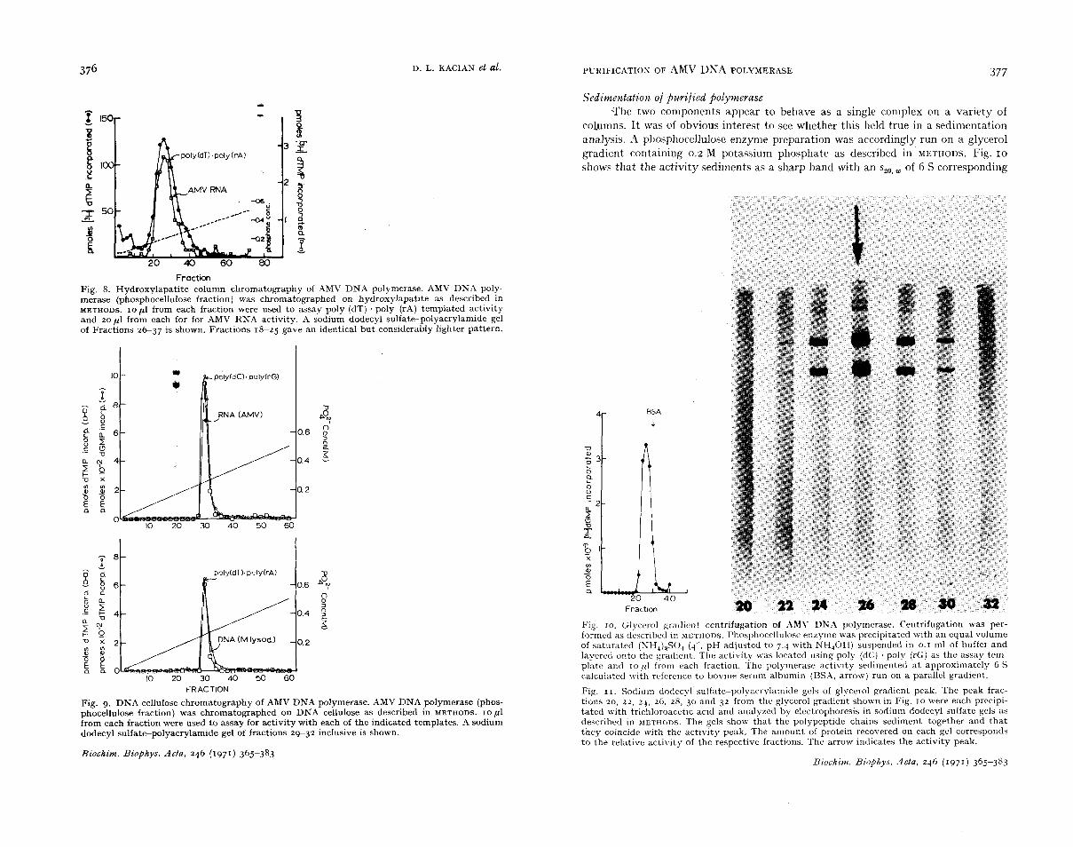
Fraction Fig. 8. Hydroxylapatite column chromatography of AMV DNA polymerase. AMV DNA poly- merase (phosphocellulose fraction) was chromatographed on hydroxylapatite as described in METHODS. IO p1 from each fraction were used to assay poly (dT) . poly (rAj templated activity and zop l from each for for AMV RNA activity. A sodium dodecyl sulfate-polyacrylamide gel of Fractions 2 6 3 7 is shown. Fractions 18-25 gave an identical but conslderably lighter pattern.
v VI I
4
2
0 IO x) 30 40 50 60
k-RACTION
Fig. 9. DNA cellulose chromatography of AMV DNA polymerase. AMV DNA polymerase (phos- phocellulose fraction) was chromatographed on DNA cellulose as described in METHODS. I O pl from each fraction were used to assay for activity with each of the indicated templates. -4 sodium dodecyl sulfate-polyacrylamide gel of fractions 29-32 inclusive is shown.
Biochim. Biophys. Acta, 246 (1971) 365-383
Sedimentation of purified polymerase -The two components appear to behave as a single complex on a variety of
columns. I t was of obvious interest to see whether this held true in a sedimentation analysis. A phosphocellulose enzyme preparation was accordingly run on a glycerol gradient containing 0.2 M potassium phosphate as described in METHODS. Fig. IO
shows that the activity sediments as a sharp band with an szo, of 6 S corresponding
a 20 4 0
Fraction
Fig. IO. Glycerol px l i cn t centrifugation of AMY DNA polymerase. Centrifugation was per- formed as described in METHODS. Phosphocellulose enzyme was precipitated with an equal volume of saturatcd (NH,)2S0, (q'., pH adjusted to 7.4 with NH,OH) suspended in 0.1 ml of buffer and layered onto the gradient. The activity was located using poly (dC) . poly (rGj as the assay tem- plate and I O 1'1 from each fraction. The polymerase activity sedimented a t approximately 6 S calculated with rvfcrencc to bovine serum albumin (BSX. arrow) run on a parallel gradient.
Fig. I I . Sodium dodecyl sulfate-polyacrylamide gels of glycerol gradient peak. The peak frac- tions 20, 21, 24, 26, 28, 30 and 3 2 from the glycerol gradient shown in Fig. I O were each precipi- tated with trichloroacctic acid and analyzed by electrophoresis in sodium dodecyl sulfate gels as described in METHODS. The gels show that the polypeptide chains sediment together and that they coincide with the activity peak, The amount of protein recovered on each gel corresponds to the relative activity of the respective fractions. The arrow indicates the activity peak.
Biochim. Biophys. Acta, 246 (1971 j 365-383

D. L. KACIAN et al.
to a molecular weight of IIO 000. It may be noted that a similar sedimentation value has been reported30 for the DNA polymerase of the Rous sarcoma virus. The yeak fractions (20-32 inclusive) were precipitated and analyzed electrophoretically in sodium dodecyi sulfate-acrylamide gels. As may be seen from Fig. 11, the same two bands are observed as the principal components in the peak region. It is evident that they are sufficiently tightly complexed to behave as a single physical entity under these conditions.
378
Mole ratios and molecular weights of the two components The molecular weights were determined on sodium dodecyl sulfate-acrylamide
gels using polymers of ribonuclease A as molecular weight markers. As may be seen from Fig. 12, the two chains have molecular weights of IIO ooo and 69 ooo
If these two polypeptides are in fact subunits of the same enzyme. then the relative number of each present in an enzyme unit should be proportional to their
DISTANCE (cm)
Fig. 12. Determination of molecular weights of subunits. The molecular weights of the polypep- tide chains present in AMV DNA polymerase extracts were determined on sodium dodecyl sul- fate-polyacrylamide gels as described in METHODS. The circles show the positions of polymers of ribonuclease A (mol. wt. 13 700) run as markers. The arrows mark the positions of the two major bands run on a parallel gel, indicating molecular weights of 110 ooo and 69 000. Molecular weights of marker polymers range from 13 700 to 123 300.
Fig. I 3. Scan of sodium dodecyl sulfate-polyacrylamide gel of CM-Sephadex enzyme. Sodium dodecyl sulfate-polyacrylamide gels were scanned, using a 0.05 mm slit, in the Gilford Model 2400 spectrophotometer equipped with linear transport. The pattern shows that the enzyme is approximately 90 yo pure assuming that all proteins are stained equally by Coomassie blae.
Biochim. Biophys. Acta, 246 (1971) 365-383
379 PURIFICATION OF AMV DNA POLYMERASE
molecular weights. I t has been shown31 that the amount of Coomassie brilliant blue bound to various proteins differed by less than IO %. By scanning stained sodium dodecyl sulfate gels, the amount of each protein present can be A typical scan is shown in Fig. 13 from which the ratio of the two components is readily obtained.
Gels containing various amounts of enzyme from different sources were scanned and measured with the results shown in Table 111. It is evident that the weight ratio of the chains found in the various preparations is that expected if the two components are present in equal numbers.
TABLE I11
WEIGHT RATIO O F CHAINS
Gels were scanned as described by B E R C ~ ~ . The area under each peak was estimated by multi- plying the peak height by the peak width a t half-height.
Gel Enzyme preparation IIO ooo mol. wt. No. polypeptide : 69 ooo
mol. wt. polypeptide
34 DEAE-cellulose enzyme 1.3
64 Sephadex G-zoo enzyme 1.5
69 Sephadex G-200 enzyme 1.8 75 Phosphocellulose enzyme 1.4
1.9 1.7
I .5
53 CM-Sephadex enzyme 63 CM-Sephadex enzyme
67 CM-Sephadex enzyme
1.3 83 Hydroxylapatite enzyme 1.7 88 Glycerol gradient enzyme
89 Glycerol gradient enzyme I .6
90 Glycerol gradient enzyme 1.6
(Fraction 24, Fig. 1 1 )
(Fraction 26, Fig. 1 1 )
(Fraction 28, Fig. 11)
Average of I I gels: Expected value (assuming one of each polypeptide
r .6
1.6 chain per enzyme molecule) :
TABLE IV
REQUIREMENTS O F AMV DNA POLYMERASE REACTION
Complete system: 50 mM Tris-HC1, pH 8.3; 6 mM MgC1,; 0.2 mM each dATP, dCTP, dGTP; 0.04 mM [8H]dTTP, 665 counts/min per pmole; 0.7 pg AMV DNA polymerase, phosphocellu- lose fraction; 2 ,ug AMV RNA; 0.4 mM dithiothreitol; roo mM KCl; incubated 20 min a t 37'.
System [ 8 H ] d T T P incorporated (pmoles)
Complete
-MgCl, + 0.6 mM MnCl, -MgCl,
-dATP -dCTP -dGTP -AMV RNA -dithiothreitol - KCl
12.4
3.5 0.4 0.3 0.5
8 . 6 5.3
0.0
0. I
Biochim. Biophys. Acta, 246 (1971) 365-383

380 E. L. KACIAN et al.
Pro9erties and reguirements of the reactzon with Purif ied A MV DNA polvmerasc The availabie information on the optimal conditions and requirements of RNA-
dependent DNA polymerases have thus far been derived from studies with deter- gent-disrupted virion preparations. Table T V summarizes a reexamination with a purified AM\.' polymerase. It will be noted that the reaction is dependent an RNA, the presence of all four deoxyri.boside triphosphates, and a divalent cation, Mg2+ being superior to MnZ+ over a wide range of concentrations. The addition of dithio- threitoi and KCi lead to marked improvement. Fig. 14 shows that the effect of the dithiothreitol is most readiiy apparent in the later stages of prolonged syntheses.
MINUTES
Fig. 14. Effect of dithiothreitol on AMV DN.4 polymerase activity. Reaction kinetics in the presence ( A ) and absence (e ) of 0.4 mM dithiothreitol (DTT) are shown. The template is AMV 70-S RNA. The curves show that dithiothreitoi is necessary for long-term synthesis. The exact tlme varies a t which the reactioil without dithiothreito! ceases.
DISCUSSION
The two-column procedure described for preparing purified DNA polymerase from avian myeloblastosis virus is a rapid and convenient procedure for obtaining reasonably pure enzyme in good yield. At the CM-Sephadex stage, enzyme from tissue culture virus is 90 06 pure or better. Substitution of phosphocellulose for CM-Sepha- dex chromatography gives similar results. If one starts with virus isolated from the plasma of infected birds, enzyme oi even higher purity is obtained at corresponding stages of purification. Rernovai of residual contamination can be achieved by (NH,) SO, concentration followed by either Sephadex G-zoo or hydroxylapatite chromato- graphy, or centrifugation through glycerol gradients.
Two polypeptide chains are found to be associated with the activity in I : I ratio throughout purification by DEAE-cellulose, CM-Sephadex, Sephadex G-zoo, phosphocellulose, hydroxylapatite and DNA cellulose affinity chromatography as well as by glycerol gradient centrifugation. This suggests that they are subunits of the polymerase. However, proof that the two polypeptides are in fact subunits will require examination of the activities of the separated and reconstituted proteins. The possibility that the subunits may each have one or more of the activities associat- ed with them individually cannot be ruled out at this time.
There is a 1.6-fold discrepancy between the molecular weight of the active form as determined by glycerol gradient centrifugation (110 000) and the sum of the molecular weights of the two polypeptide chains determined on sodium dodecyl sulfate-polyacrylamide gels (110 ooo and 69 000). Assuming that the molecular
Biochim. Biophys. Acta, 246 (1971) 365-383
PURIFICATION OF AMV DNA POLYMERASE 381
weight determinations are not in error, the fact that the ratio of the chains is con- stant across the activity peak (Table 111) in the glycerol gradient suggests that the discrepancy is due to differences in molecular asymmetry of the subunits as compared with assembled proteins. A quantitatively similar situation obtains with QB repli- case in which the sum of the molecular weights of the subunits determined on so- dium dodecyl sulfate gels is 1.7 times that determined by sedimentation for the native en~yme3~93~.
The value obtained for the sedimentation coefficient of the enzyme would seem to exclude the possibility that the molecule contains more than one of each subunit.
It is worth emphasizing that association of the two major polypeptides, even through an extensive series of purification steps, does not constitute proof that they represent the desired enzyme. As with all such enzyme purifications, minor con- taminants (e.g. I yo or less) that might not be detectable could conceivably be res- ponsible for the activity. We cannot prove rigorously at this time that the two prin- cipal polypeptide chains constantly observed in our active preparations compose the polymerase. However, in addition to their invariant presence and constant I : I ratio, several additional considerations argue for their being the enzyme components.
Throughout purification, there have been no minor bands that are consistently present in all preparations. The most frequently observed contaminant is a pair of bands representing approximately 4 of the total protein. From their gel mobilities, they have molecular weights of 190 ooo and 210 ooo and theiefore would be expected to sediment much more rapidly, either individually or as a complex, than the pol- merase. They are not enriched relative to the two major bands during the course of purification of the activity. It seems likely that they represent aggregates of the major bands due to the relativelv large amount of protein applied to the gels.
Were the activity due to any contaminant present as a few percent of the protein, the specific activity of the purified enzyme would be many-fold greater than that observed. The most active preparations of E. coli DNA polymerase36 assayed with poly [d(A-T)], QB replicase3' assayed with QB RNA, and E. coli transcriptase assayed with poly [d(A-T)]38 and calf thymus DNAss have specific activities', respectively, of 1.2, 3.2, 4.5, and 1.2 moles nucleoside monophosphate incorporated per sec per mole enzyme. The most active preparations of AMV DNA polymerase incor- porate 1.2 moles dXMP per sec per mole enzyme. This value is in excellent agreement with those for the other purified polymerases and argues that the principal protein components are responsible for activity. These calculations encompass both initiation and chain elongation. They are not based on any assumptions concerning the numbers of enzyme molecules participating, but rather on the reported enzyme specific activities.
If in fact the enzyme is essentially pure, the amount of protein recovered should correlate with the number of enzyme molecules expected per virion. I t i s reasonable to assume that there is at least one polymerase molecule per virus particle. Since the virion contains 3.0 . IO* daltons of protein40 and the molecular weight of the AMV DNA polymerase is 1.8. 105 daltons, one enzyme molecule per virion
* The highest value obtained for enzyme fractions described as homogenous was used. The reported specific activities were converted from arbitrary units to moles of nucleoside mono- phosphate per sec per mole of enzyme.
Biochim. Biophys. Acta, 246 (1971) 365-383

D. L. KACIAN et al. 382
would represent 0.067~ of the total protein. Normally, 0.3-1 yo of the starting protein is recovered, which would correspond to 5-17 enzyme molecules per virus particle. Were the enzyme a 2 yo contaminant of our preparation, each virion would contain only 0.1-0.3 polymerase molecule.
The availability of pure enzyme makes possible a re-examination of the nature of the intermediates and the final product uncomplicated by possibly irrelevant or interfering enzyme activities.
ACKNOWLEDGMENTS
We would like to express our deep appreciation to Dr. A. J. Langiois, Dr. Dorothy Beard and Dr. J. W. Beard. Without their continuous and generous COO-
peration in supplying virus, these experiments would not have been possibie. We would also like to acknowledge the assistance of Dr. R. Chuang in the latter phases of this investigation. This work was supported in part by the National Institutes of Health, National Cancer Institute, Special Virus Cancer Progran Contract 70-2049 Training Grant CA-05011, Research Grant CA-02332. and Postdoctoral Fellowship CA-38924.
REFERENCES
I H. M. TEMIN AND s. MIZUTANI, Nature, 226 (1970) 1211. 2 D. BALTIMORE, Nature, 226 (1970) 1209. 3 S. SPIEGELMAN, A. BURNY, M. R. DAS, J . KEYDAR, J. SCHLOM, PI. TRAVNICEK, AND K. WATSON
4 M. GREEN, M. ROKUTANDA, I(. FUJINAGA, R. K. RAY, H. ROKUTANDA AND C. GURGO, Proc.
5 M. HATANAKA, R. J . HUEBNER AND R. 1'. GILDEN, Proc. Natl. Acad. Sci. U S . , 67 (1970) 143. 6 E. M. SCOLNICK, S. A. AARONSON AND G. J . TODARO, Proc. Nati. Acad. Sci. U.S., 67 (1970)
7 A. GARAPIN, J. P. MCDONNELL, W. LEVINSON, N. QUINTRELL, L. FANSHIER AND J . M. BISHOP,
8 R. C. NOWINSKI, L. J . OLD, N. H. SARKAR AND D. H. MOORE, Virology, 42 (1970) 1152. 9 K. FUJINAGA, J. T. PARSONS, J. W. BEARD, D. BEARD AND M. GREEN, Proc. Natl. Acad.
Nature, 227 (1970) 563.
Natl. Acad. Sci. U.S., 67 (1970) 385.
1034.
J. Virol., 6 (1970) 589.
SCZ. U.S., 67 (1970) 1432. I O P. H. DUESBERG AND E. CANAANI, virology, 42 (1970) 783. 11 M. ROKUTANDA, H. ROKUTANDA, M. GREEN, K. FUJINAGA, R. K. RAY AND C. GURGO, Nature
12 S. SPIEGELMAN, A. BURNY, M. R. DAS, J. KEYDAR, J. SCHLOM, M. TRAVNICEK AND K. WATSON.
13 S. MIZUTANI, D. BOETTIGER AND H. M. TEMIN, Nature, 228 (1970) 424. 14 J. RIMAN AND G. BEAUDREAU, Nature, 228 (1970) 427. 15 J. P. MCDONNELL, A. GARAPIN, W. E. LEVINSON, N. QUINTRELL, L. FANSHIER AND J. M.
16 S. SPIEGELMAN, A. BURNY, M. K. DAS, J. KEYDAR, J. SCHLOM, M. TRAVNICEK AND K. WATSON
17 S. MIZUTANK, H. M. TEMIN, M. KODAMA AND R. T. WELLS, Nature, 230 (1971) 232. 18 P. ROY AND D. H. L. BISHOP, Biochim. Biophys. Acta, 235 (1971) 191. 19 A. J. LANGLOIS, R. A. BONAR, P. R. RAO, D. P. BOLOGNESI, I). BEARD AND J . W. BEARD,
20 E. A. ECKERT, D. G. SHARP, E. B. MOMMAERTS, R. H. REEVE, D. BEARD AND J. W-. BEARD,
21 J. W. CARNEGIE, A. O'C. DEENEY, K. C. OLSON AND G. S. BEAUDREAU, Biochim. Biophys.
22 G. S. BEAUDREAU AND C. BECKER, J. Natl. Cancer Inst., 20 (1958) 339.
Biochim. Biophys. Acta, 246 (1971) 365-383
227 (1970) 1026.
Nature, 227 (1970) 1029.
BISHOP, Nature, 228 (1970) 433.
Nature, 228 (1970) 430.
Proc, Exp. Biol. Med., 123 (1966) 286.
J. Natl. Cancer Inst., 14 (1954) 1039.
Acta, 190 (1969) 274.
PURIFICATION O F AMV DNA POLYMERASE 383
23 K. S. KIRBY in L. GROSSMAN AND K. MOLDAVE (eds.), Melhods iw Enzymology, Vol. IZB,
24 A. L. SHAPIRO, E. VINUELA AND J. V. MAIZEL, JR., Biochem. Biophys. Res. Commun., 28 (1967)
25 B. WOLF, P. M. LAUSAROT, J. A. LESNAW AND M. E. REICHMANN, Btochim. Biophys. Acta,
26 B. J. DAVIS, Ann. N . Y . Acad. Sci., I Z I (1964) 404. 27 0. H. LOWRY, IC'. J. ROSEBROUGH, A. L. FARR AND R. J. RANDALL, J. Biol. Chem., 193 (1951)
28 B. ALBERTS AND G. HERRICK, Methods in Enzymology, Vol. 21, Academic Press, New York,
29 D. H. L. BISHOP, J . R. CLAYBROOK AND S. SPIEGELMAN, J. Mol. Biol., 26 (1967) 373. 30 P. DUESBERG, K. V. D. HELM AND E. CANAANI, Proc. Natl. Acad. Sci. U.S., 68 (1971) 747. 31 S. FAZEKAS DE. ST. GROTH, R. G. WEBSTER AND A. DATYNER, Biochim. Biophys. Acta, 71
32 H. C. BERG, Biochim. Biophys. Acta, 183 (1969) 65. 33 R. R. BURGESS, J. Biol. Chem., 244 (1969) 6168. 34 M. KONDO, R. GALLERANI, C. WEISSMANN, Nature, 228 (1970) 525. 35 R. KAMEN, Nature, 228 (1970) 527. 36 C. C. RICHARDSON, in G. L. CANTONI AND D. R. DAVIES, Procedures in Nucleic Acid Research,
37 L. EOYANG AND J. T. AUGUST, Methods in Enzymology, Vol. IZB, Academic Press, New York,
38 D. BERG, K. BARRETT AND M. CHAMBERLIN, Methods in Enzymology, VoI. 21, Academic Press,
39 R. R. BURGESS, J. Biol. Chem., 244 (1969) 6160. 40 R. A. EONAR AND J. W. BEARD, J. Natl. Cancer Inst., 23 (1959) 183.
Academlc Press, New York, 1968, p. 87.
815.
200 (1970) 180.
265.
1971. p. 198.
(1963) 377.
Harper and Row, New York and London, 1966, p. 263.
1968, P. 530.
New York, 1971, p. 506.
Biochim. Biophys. Acta, 246 (1971) 365-383