eyeblink conditioning as models of declarative ... - j-pbs.org
TRANSCRIPT

Review
1
emory is one of the mental faculties generated by the brain and it acts as the glue that binds our mental life.
Some memory afflicted diseases such as Alzheimer’s disease have devastating effects on patients especially when the diseases progress to advanced states that the patients start to forget their names or their close relative’s names, or to lose their way home, basically they are losing themselves. Moreover, when we talk about memory, we always have to talk about learning as well, because they are connected processes. Kandel et al. (2000) stated that “learning is the process by which we acquire knowledge about the world, whereas memory is the process by which that knowledge is encoded, stored and later retrieved.”1 Moreover, not only are learning and memory important for our normal functions, but they can also lead to abnormal behaviors such as post-traumatic stress disorders. Therefore, the study of learning and memory is very important for the understanding of the behaviors of human being. In this area of study, there are many learning paradigms that have been developed such as fear conditioning, maze learning, vestibulo-ocular reflex. Each has been the major learning paradigm and has great benefit in its own arena. However, one of the learning paradigms that has an extensive understanding in both neurobiological and behavioral science and has a promising potential for clinical applications is eyeblink conditioning.2
Memory system Before we delve into the details, the system of memory will be introduced briefly. The memory can be classified into two major forms: declarative (explicit) memory and non-declarative (implicit) memory. The declarative (explicit) memory is the memory that you can use to describe what and where something is or how and when it happened, and it requires conscious awareness when it is recalled; for example memories about personal experiences, knowledge, fact, or directions. More importantly, this kind of memory depends on the integrity of the hippocampus during the acquisition period. On the other hand, the non-declarative (implicit) memory does not require conscious awareness to recall. Usually, the implicit memory is recalled or is expressed in terms of performance; for example memories of how to perform some activities e.g. driving a car, playing tennis. This kind of memory does not require an intact hippocampus. The non-declarative memory is a collective term of many memory abilities, and it is comprised of skills and habits, the phenomenon of priming, nonassociative forms of memory like sensitization and habituation, and simple forms of classical conditioning including emotional conditioning (e.g. fear conditioning) and somatic condi-tioning (e.g. delay eyeblink conditioning). Each kind of memories requires diverse brain areas during the memory processes.1 Eyeblink conditioning paradigm Eyeblink conditioning learning paradigm initially has performed in rabbits,3 and then progressed to include rats4 and mice.5 Eyeblink conditioning has also been done in human for a long time and has become more active nowadays.2,6 Eyeblink conditioning is a form of Pavlovian conditioning in which the training trial consists of a paired presentation of two stimuli – a neutral conditioned stimulus (CS) and an eyeblink-eliciting unconditioned stimulus (US) where the CS precedes the US. During the learning trials, animals or subjects learn about the relationship between
Eyeblink Conditioning As Models of Declarative and Non-declarative Memories Narawut Pakaprot
Abstract Learning and memory are the crucial cognitive functions, which equip animals for the ability to preserve our experiences of both knowledge and motor skills that we have learned. Eyeblink conditioning is a basis form of associative learning that conditions the eyeblink reflex to a neutral stimulus that predicts an aversive stimulus. With small manipulations, eyeblink conditioning procedures have been used as learning models of both declarative and non-declarative memories. The procedures can then be used to examine the basic mechanisms of memory and the pathophysiology of memory afflicted diseases, and to test new or alternative treatments of memory disorders. This paper introduced the methods of eyeblink conditioning as well as the memory circuitry responsible for mediating the memory, and the clinical applications of the procedures were also reviewed.
J Physiol Biomed Sci. 2011; 24(2): 1-12 Keywords: delay eyeblink conditioning, trace eyeblink conditioning, hippocampus, cerebellum, declarative memory, non-declarative memory
M
From the Department of Physiology, Faculty of Medicine Siriraj Hospital, Mahidol University, Bangkok, Thailand. Corresponding author: Narawut Pakaprot, MD, PhD Department of Physiology, Faculty of Medicine Siriraj Hospital, Mahidol University, Bangkok, Thailand E-mail: [email protected] © 2011 Journal of Physiological and Biomedical Sciences Available online at www.j-pbs.org

J
twnear
liecdbdmsginanbmrmruopnoocsUooth(thu
Physiol Biomed
Figure 1 Eyebefore, during a
wo stimuli. Tneutral stimulueliciting uncona strong and sresponse.
In eyeblinkight whereas t
eyes or a pconditioning depending on between the tdifferent neurmemory procesame. When tgiven to animanstead respond
and moving nictitating memboth eyes of membrane andrecorded from musculus orbrecorded. Theunconditioned over time, thproducing thenictitating memonset of the USor an electricconditioned resee Figure 1. TUR but it is tonset, and occonset. The peahe US presum8). In general,he temporal re
use the CS t
Sci. 2011; 24(2)
eblink responseand after trainin
The conditioneus, which elicinditioned stimusignificant stim
k conditioningthe US is an a
periorbital eleparadigm vathe durations
two stimuli. ral circuitry esses, the logthe paired preals, the animald to the US asthe nictitatinmbrane is a prabbits. The
d the eyelid, omusculus levicularis oculi
e elicited respresponse (UR
he animals lee same movembrane after tS, to protect thcal shock. Thsponse (CR) bThe CR sharetypically smacurs after the ak of the CR umably in order , during the traelationship beto predict the
: 1-12
e: comparing eng.
ed stimulus (Cits no responsulus (US), on
mulus that pro
g paradigm,7 thairpuff pumpedectrical shockaries into mof and the ti
Although eacresponsible f
gic behind thesentation of sls do not respos a reflex by bng membraneprotective me
movement oor the electro
vator labii supei (MOO) arponse to an
R). With the coearn to respement of thethe onset of thhe eye from rehis later respbecause it is es many charaller than the CS presentati
usually falls arto maximize taining period,etween the CSe onset of th
eyeblink amplitu
CS) typically ses. The eyeblthe other han
oduces a reflex
he CS is a tond directly intok. The eyebmany procediming relationch procedure for learning e learning is stimuli is initiond to the CS,blinking the eye backward. embrane coveof the nictita
omyogram (EMerior (MLLS) re measured US is called
ontinuous trainpond to the e eyelid and he CS, before
eceiving an airponse is calleelicited by the cteristics withUR, has a faon before theround the onsethe eye protec the animals l
S and the US, he US, and t
2
ude
is a link-
nd, is xive
ne or o the blink dures nship
has and the
ially , but yelid The
ering ating MG)
and and
d an ning CS, the
e the rpuff ed a
CS, h the aster e US et of ction earn and
then
accresp
depthe maning proUSbraeac Del
ProIn bmentracbetwdelain Fprousethe US stimendbetwcallrelatheyperprostimareacontraithe highdiffin min hpro
Figrab
cordingly proponses (CR) toAs mentione
pend on the dstimuli. The t
ny experimenprocedures.
ocedures is the. This gap surin learns thes
ch brain area du
lay vs trace ey
ocedure both procedurntioned, the ce eyeblink cween the CS ay and trace eyFigure 2. The
ocedure is 250ed in mouse an
CS lasts for 3onset is at t =
muli are overlad of the trialween the CS led “inter-stimationship betwy are physiciod of both
ocedure, on themuli, resultingas that are
nditioning traiining. Those b
forebrain. Thher brain invoferent mammamice11 and rathumans.14,15 T
ocedure used i
gure 2 Delay abbits.
oduce well-to the CS.
ed before, thedurations of antwo procedurents are delay a
The major de stimulus freerprisingly affese two tasks, uring the mem
yeblink condi
res, the CS prmajor differe
conditioning pand the US. Tyeblink condite example of 0-ms standard nd rabbit expe350 ms from t = 250 ms withapped for 100l. The term onset and the
mulus interval”ween the two ally associate
h stimuli. Tre other hand, hg in the invol
required dining, not durain areas are he lengths of olvement are alian species. Tts,12 over 300
The example oin rabbit expe
and trace eyeb
time adaptiv
e variants ofnd the relation
es that have beand trace eyebdifference bete gap between ects not only th
but also the mory processes
itioning proce
recedes the Uence between procedures is The essential tioning proceddelay eyeblindelay conditi
eriments.9,10 In = 0 ms to t = 3h a duration o
0 ms and co-te250-ms signi
e US onset; th”. Thus, in this
stimuli is aped through thrace eyeblinkhas a silent galvement of th
during the turing the dela
such as the hipf the gap thatalso critical aThat critical lems in rabbits
of trace eyeblineriments is ca
link conditioning
N. Pakapr
ve conditione
f the paradigmnships betwee
een employed iblink conditiontween the twthe CS and th
he way that thcontribution o
s.
edures
US; however, athe delay anthe silent gacomponents o
dures are shownk conditioninioning which this procedur
350 ms, and thof 100 ms. Boterminated at thifies the dela
his delay is alss procedure, thpparent becaushe overlappink conditioninap between twhe higher braitrace eyeblinay conditioninppocampus ant determine thand vary amonength is 250 m13 and 1000 mnk conditioninalled “500-ms
g procedures in
rot
ed
m en in n-
wo he he of
as nd ap of
wn ng is
re, he th he ay so he se ng ng wo in nk ng nd he ng ms ms ng s
n

Eyeblink conditioning J Physiol Biomed Sci. 2011; 24(2): 1-12
3
trace conditioning”, which is derived from the 500-ms gap interval.16 The procedure starts with a 250-ms of CS, then followed by a 500-ms of a stimulus free gap, and ends with a 100 ms of US. To learn the trace eyeblink conditioning procedure, the brains have to overcome the gap in order to make the association between both stimuli. Delay eyeblink conditioning as a model of non-declarative memory In delay conditioning in which there is no stimulus-free interval between the stimuli, the brain areas that are necessary for the acquisition and retention of the memory are confined only to the brain stem nuclei and the cerebellum.17 Decerebrated animals that were transected above the red nucleus still showed significant levels of learning in the delay conditioning training.18 Along with the results of other studies that lesioned other nuclei in the brain stem and the cerebellum, the evidence revealed that the brain stem and the cerebellar circuit are necessary and sufficient to acquire and retain the temporal relationship of two stimuli that are temporally overlapped, and to use the association for the expression of the learned adaptive behavioral response.17,19 Higher brain areas such as the prefrontal cortex, the motor cortex, and the hippocampus showed activity-related changes during delay eyeblink conditioning training although they did not play essential roles during the acquisition and retention of the memory. The lesion made in the hippocampus did not impair the acquisition of the delay eyeblink conditioning memory,20 causing the delay eyeblink conditioning memory a hippocampal independent form of memory. Moreover, the availability of conscious awareness, which depends on the higher brain functions, of the CS-US contingency did not affect the ability of subjects to learn delay eyeblink conditioning.14 These data suggest that delay eyeblink conditioning can be considered as a learning model of non-declarative memory.21 However, the higher brain areas might help in the facilitation of the memory acquisition.22 For the cerebellum, it is widely accepted that the deep cerebellar interpositus nucleus is essential for the acquisition and retention of delay eyeblink conditioning and the cerebellar cortex plays a facilitory role in acquiring the normal CR in terms of the rate of acquisition and the timing of appropriately timed response.10,19,23 Trace eyeblink conditioning as a model of declarative memory Trace eyeblink conditioning, on the other hand, requires higher brain areas during the learning and memory processes. The presence of the gap in trace eyeblink conditioning is surprisingly very significant, and determines which brain areas are necessary in the acquisition, retention, and consolidation processes of the memory. One of the essential areas involved in the acquisition and short-term retention of the memory is the hippocampus, leading to the consideration that the trace eyeblink conditioning memory is a hippocampus-dependent form of memory.24-26 More-over, conscious awareness plays a significant role in the
acquisition of the memory. Clark and Squire showed that subjects who were aware of the contingency of CS and US learned better in trace eyeblink conditioning. This procedure thus is considered an experimentally controllable model of declarative memory in which the declarative knowledge of the stimulus contingencies are acquired and consolidated over time.14,21 Other areas such as the prefrontal cortex, the anterior cingulated gyrus, and the splenial cortex showed the involvement in trace eyeblink conditioning as well. It has been proposed that the forebrain circuitry plays a major role in bridging the temporal gap in trace conditioning through pontine-cerebellar nuclear connections.22,27 This connection can then bypass the cerebellar cortex as it appears that the cerebellar cortex play much less role in trace than delay eyeblink conditioning.8 The detailed circuitry responsible for trace eyeblink conditioning has been investigated systematically and extensively in the recent years. The cerebellar circuitry seems to be involved in trace eyeblink conditioning as it is in delay eyeblink conditioning. The deep cerebellar interpositus nuclei have been shown to be crucial for the acquisition, and the short- and long-term retentions of the trace conditioning memory.16,28 The cerebellar cortex, on the other hand, seems to play differential roles between delay and trace eyeblink conditioning.28 Reports from mouse and rat studies demonstrated that functional disruptions of the cerebellar cortex impaired delay, but not trace, eyeblink conditioning.9,29-32 The effect of cerebellar cortex lesions on human patients in eyeblink conditioning training pointed toward the same conclusions as those found in animal studies. In patients who had cerebellar cortex lesions, delay eyeblink conditioning was more strongly impacted than was trace eyeblink conditioning training.6 The patterns of activation in the cerebellum were investigated using a metabolic mapping technique.33 The authors demonstrated that there was more widespread activation in the cerebellar cortex in the delay eyeblink conditioning group than in the trace conditioning or the unpaired groups, and different regions of the deep cerebellar interpositus nuclei were activated in each training procedure. The anterior interpositus nucleus was activated more in the delay conditioning group than in the unpaired group whereas the posterior interpositus nucleus was activated more in the trace conditioning groups than in the delay conditioning and the unpaired groups. The evidence indicating less involvement of the cerebellar cortex in trace than in delay conditioning suggests that the cerebellar cortex plays a less significant role in trace eyeblink conditioning than in delay conditioning. Moreover, it seems that different areas in interpositus nuclei are recruited during each paradigm, suggesting a complicated interaction within the cerebellar structures and also between the cerebellum and other brain areas.27 Woodruff-Pak and Disterhoft (2008) concluded that temporal gaps in trace eyeblink conditioning training were responsible for forebrain dependency and for the activation of different cerebellar circuits during the memorization of conditioned responses.8

J Physiol Biomed Sci. 2011; 24(2): 1-12 N. Pakaprot
4
Circuitry of delay eyeblink conditioning The circuitry of the eyeblink conditioning procedure has been investigated extensively for several decades. Because of the well-organized neuronal arrangement in the cerebellum, most of the circuitry essential for delay eyeblink conditioning has been determined.19 Figure 3 shows the essential circuitry of delay eyeblink conditioning, which localizes with in the brain stem and the cerebellum. US pathway The US pathway begins at the trigeminal sensory neurons receiving the sensory signals from the eye that is trained by either an airpuff or a periorbital shock as a US, and the unconditioned response (UR) is produced through the reflex pathway as shown in Figure 3b. The UR occurs after the onset of US, and it is not adaptive, but rather reflexive in response to the US. The sensory neurons convey the signals via a direct projection to cranial motor nuclei which are the accessory abducens and abducens nuclei, and the facial nucleus,34,35 and an indirect projection via the brain stem reticular formation to the facial nucleus.36,37 These cranial motor nuclei are on the same side of the trained eye. In the adaptive pathway, which is involved in the production of the conditioned response (CR) as shown in Figure 3a, the trigeminal sensory neurons ipsilateral to the trained eye send the efferent axons across the midline and form synapses with the inferior olivary neurons particularly in the dorsal accessory olive. The inferior olivary neurons then convey the airpuff signals through the climbing fibers. The climbing fibers cross the midline again and form excitatory synapses onto the proximal dendrites of the cerebellar Purkinje cells ipsilateral to the trained eye.19 Each Purkinje cell forms synapses with only a single climbing fiber, producing the most powerful excitatory synapse in the nervous system.38 The action potentials from the climbing fibers produce the complex spikes in the Purkinje cells.
CS pathway The most common sensory modality for being used as a CS in eyeblink conditioning study has been sound.22 However, other sensory modalities such as light and somatosensory stimulation can also act as a CS. In general, the CS signals are conveyed through the pontine nuclei to be later processed in the cerebellar circuit.27 The pontine nuclei receive signals from many sensory cortices including auditory, visual, somatosensory cortices, as well as from prefrontal areas.39-43 Although showing some overlap, the terminations of these projections are localized in specific regions of the pons, and appropriate lesions of the pontine nuclei could selectively impair the CR performance elicited by a particular type of CS.19 For example, the inactivation of the medial pontine nuclei severely impaired the retention of CR performance elicited by the light CS, but not the tone CS, whereas the inactivation of the lateral pontine nuclei had a greater effect on the tone CS elicited CR performance.19,44,45 In case of a tone CS, the signal is initially processed by the auditory nuclei in the brain stem. The auditory nuclei on the same side of the trained eye send the efferent axons across the midline and the axons form excitatory synapses with the lateral pontine nuclei contralateral to the trained eye.19 On the other hand, the visual nuclei – the lateral geniculate nucleus (LGN) and nucleus of the optic tract – contralateral to the trained eye receives the visual CS input from the side of the trained eye. The efferent fibers from the visual nuclei then project ipsilaterally to the medial pontine nuclei contralateral to the trained eye.44 Recently, the activation of somatosensory modality has been employed as a CS by the use of the mystacial vibrissae stimulation.46,47 The vibrissae stimula-tion CS signals reach the cerebellum also via the pontine nuclei contralateral to the trained eye.22,27,48 However, the detailed circuitry carrying this CS signals is still needed some more investigation. After receiving the CS signals,
Figure 3 Essential circuitry of delay eyeblink conditioning (Modified from Thompson and Krupa, 1994)
E
thinfwssinfs MInmpblepEpd1oth
sipdamthPRlocpmmaasfatrin
Eyeblink condition
he pontine nucnto the cerebe
fibers end in cwhose axons synapses with stimulation of n the Purkinje
fibers – also sesynapses with t
Mechanisms ofn the quest fo
memory, twopotentiation (Lbelieved to plaearning and m
persistent enhEPSP) followpresynaptic adiscovered in 1996.49 LTD, oof synaptic effihe presynaptic
In the cersignals, Purkipsilateral to t
deep cerebellaare the only oumany investigahe Purkinje
Purkinje cell-pRawson and Tong-term dep
cell synapses prior model mechanism hamain learning are other mecha role in the states that whfollowed by thappropriate timraining, the conduces a long
ning
clei, send the ellum ipsilateracontact with th
– parallel fthe distal de
the parallel fibe cells. Both pend collateral the deep cereb
of memory storfor the mechao major proLTP) and lonay major rolememory. LTP hancement in wing high-freafferent (repe
the rabbit hipon the other h
ficacy followinc afferent.50 rebellum, afteinje cells inthe trained eyar interpositusutput signal oations focusincell-climbing
parallel fiber Tilokskulchai.5
ression (LTDin the cerebeof Marr (19
as been hypothmechanism of
hanisms at othecerebellar lea
hen the paralhe activation me period foro-activation ofg-term input-s
mossy fibers aal to the trainehe dendrites offibers – formendrites of Pubers produces
pathways – clibranches to fo
bellar interposi
rage in the ceanisms underlycesses which
ng-term depres in the informis a phenomesynaptic str
equency stimeated learninppocampus bhand, is a longng low-frequen
er receiving bn the HVI ye send inhibits nuclei. This of the cerebellng on the synag fiber synap
synapses we51-53 In 1982, I
D) at the paralellar cortex.54 969) and Albhesized by Itof the cerebelluer synaptic sitarning as welllel fibers areof the climb
r an extendedf these two Puspecific depre
across the mided eye. The mof the granule cm the excitaurkinje cells. the simple spmbing and moorm the excitaitus nuclei.
erebellum ying learning h are long-tession (LTD) rmation storagenon defined rength (amplitmulation of ng) and it y Terje Lomog-lasting decrncy stimulatio
both CS and cerebellar cotory axons toinhibitory ax
lar cortex. Earaptic responsepses and at ere performedIto discoveredllel fiber-Purk
Building on bus (1971),
o (1984) to beum although thtes that might pll.55-57 The thee stimulated
bing fibers in d time duringurkinje cell inession of syna
5
dline ossy cells atory The
pikes ossy
atory
and term
are ge in as a tude the
was o in ease
on of
US ortex o the xons rlier, es at
the d by d the kinje
the this
e the there play eory and the
the nputs aptic
stresyndecreduintealloincrfromsignin tto mrespandconproare neumoincrCS outpmoappareaare inteeyethe eyeanimthatnoxcoinfromconthatmiccon
Purdep(LTincrneu
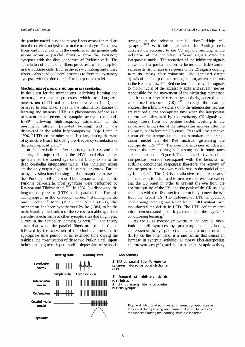
Figthe mec
ength at thenapses.58,59 Wcrease the respuction of therpositus nucleows the interprease its firingm the mossynals of the intethe Red nucleumotor nuclei oponsible for thd the external nditioned respocess, the inhib
reduced at thurons are stimssy fibers frorease of firingonset, but bef
put of the intor nuclei v
propriate CRs.as in the circudemonstrated
erpositus neueblink conditio
interpositus neblink CR.23 Tmals learn to t the US onsxious quality oncides with thm the airpuffnditioning leart showed the ce demonstranditioning learn
As the LTD rkinje cell sypression of theTP), on the otrease in syna
uron synapses
ure 4 Neuronacircuit during re
chanisms during
J Phy
e relevant pWith this dep
ponse to the he inhibitory ei. The reductositus neurons
g rates in respoy fiber collateerpositus neurus. The Red nuof the accessohe movement eyelid closureponse (CR).1
bitory signals he appropriate
mulated by thom the pontig rates of the ifore the US onnterpositus nuvia the Red.17,19,27 The neuit during bot
d in Figure 4. Turons correspooned responseneurons was coThe CR is anadapt and to
set in order tof the US, andhe US onset in f US. The infrning was testdeficit in LT
ated the imrning.5
mechanism ynapses by e synaptic actither hand, is aptic activities
(60), and the
al activities at desting and learng the learning st
ysiol Biomed Sci
parallel fiberpression, the
CS signals, refferent sig
tion of the inhs to be more eonse to the CS erals. The inrons, in turn, aucleus then reory sixth and of the nictita
e, respectively17,19 Throughonto the interpe time when the excitatory ine nuclei, reinterpositus nenset. This wel
ucleus stimulad nucleus, geuronal activitth resting andThe increased ond with thes; therefore, onsidered as thn adaptive resproduce the ro prevent thed the peak of order to fullyfluence of LTted by mGluRTD. The LTD
mpairment in
works at theproducing th
ivities, long-tea mechanism
s at mossy fib increase in sy
different synaptining states. Thetate are include
i. 2011; 24(2): 1-
r-Purkinje cePurkinje cel
resulting in thgnals onto thhibitory signaexcitable and tsignals comin
ncreased outpuactivate neuronelays the signa
seventh nerveating membran, generating th
h the learninpositus neuronthe interposituCS signals vesulting in theurons after thll-time adaptivates the cranigenerating thties at differen
d learning stateactivities of the behavior othe activity o
he model of thsponse becausresponse earlie eye from ththe CR usuall
y protect the eyTD in eyeblinR1 mutant mic
D deficit mutanthe eyeblin
parallel fibehe long-lastinerm potentiatio
that causes aber-interposituynaptic activit
c sites in e possible ed.
12
ell lls he he als to ng ut ns als es ne he ng ns us
via he he ve ial he nt es he of of he se er he ly ye nk ce nt nk
er-ng on an us ty

J Physiol Biomed Sci. 2011; 24(2): 1-12 N. Pakaprot
6
due to the LTP is important of the learning of eyeblink conditioning.61 Anatomical evidence looking for the changes of ultrastructures in the synapses has shown that there was an significant increase in the length of excitatory synapses at the interpositus neurons, supporting the influence of LTP at the interpositus nucleus synapses after eyeblink conditioning training.62 Therefore, when the LTD reduces the parallel fiber-Purkinje cell synaptic activities, removing the interpositus neurons from inhibitory efferent signals of the cerebellar cortex, the LTP, on the other hand, enhances the synaptic activities of the interpositus neurons, allowing the neurons to be efficiently stimulated by the mossy fibers. The efferent signals of the interpositus neurons then transmit to the Red nucleus and then to the cranial motor nuclei, producing the conditioned response.19 The cerebellar circuitry that is required for the generation of conditioned responses is shown in Figure 3a. The interpositus nuclei have been shown to be critical for the acquisition, short- and long-term retention of both delay and trace eyeblink conditioning.16,28 Moreover, several lines of evidence supported the role of the interpositus nuclei as a short- and long-term storage site of both delay and trace eyeblink conditioning memory.16,19,62-65 Circuitry of trace eyeblink conditioning In delay eyeblink conditioning, only the cerebellum and the brain stem are essential and are required for the acquisition and retention of the memory. In trace eyeblink conditioning as a model of declarative memory, the brain stem and the cerebellum are still required for the CR generation, especially for the cerebellum because it is the site where the basic association of CS and US is formed and permanently stored.16,65 However, many other higher brain areas are critically involved in the memory processes of trace conditioning. The higher brain areas such as the hippocampus, the prefrontal cortex, the anterior cingulate gyrus, the thalamus, the striatum and the sensory cortex showed the involvement in eyeblink conditioning and played different roles at each stage of the memory processes.19,22 Clark et al. (2011) hypothesized that it may be the declarative knowledge of the stimulus contingencies that is stored and is consolidated in the higher brain areas during the memory processes.21 Nonetheless, the true mechanisms of the higher brain involvement in trace
eyeblink conditioning are the challenging tasks. Weiss and Disterhoft (1996, 2011) proposed the interaction of the higher brain areas with the cerebellum as shown in Figure 5, and the actions of the forebrain and the hippocampus are to potentiate the effect of the CS signals at the pontine nuclei through the cortico-pontine projections. The result of the potentiation will effectively expand the duration of the CS signals such that the CS signals overlap with the US signals even though physically there are no overlap of the CS and the US due to the stimulus free gap of trace eyeblink conditioning.27,66 The involvement of some major brain areas during the trace eyeblink conditioning is reviewed here. Hippocampus For many years, the hippocampus has been the center of intensive studies for learning and memory. Its anatomical connections are very intricate and complex, and it plays a significant role in the acquisition of declarative memory. Although the contribution of the hippocampus in eyeblink conditioning has been intensively investigated, it is a challenging task to define the true nature of hippocampal-cerebellar communications since there is no direct connection between these two areas.16 The hippocampus receives inputs from and sends outputs to a wide range of neocortical areas such as the posterior parietal cortex (area 7), the inferior temporal association area (area 20, 37), and the retrosplenial cortex and subcortical areas such as the basal forebrain and hypothalamic nuclei. The neocortical signals reach the hippocampus through the entorhinal cortex, whereas the subcortical signals mainly reach the hippocampus through the fimbria-fornix pathway.67,68 However, it appears that there is no direct connection between the hippocampus and the cerebellum. Weiss and Disterhoft (1996, 2011) proposed that these two regions could be functionally connected via the frontal cortex and the pons. The possible indirect connections consist of the pathway from the hippocampus to the retrosplenial cortex which conveys through the entorhinal cortex and the pathway from the hippocampus to the mamillary nucleus and thalamic nuclei which passes through the fimbria-fornix pathways. Both pathways eventually connect with the cerebellum through the pontine nuclei, which relay and send the signals via mossy fibers into the cerebellar
Figure 5 The circuitry of trace eye-blink conditioning. AV thalamus, anteroventral thalamus; MD thala-mus, dorsomedial thalamus; VA thalamus, ventral anterior thala-mus; dlPFC, dorsolateral prefrontal cortex; AC: anterior cingulate gyrus. (Modified from Weiss & Disterhoft, 2011)

Eyeblink conditioning J Physiol Biomed Sci. 2011; 24(2): 1-12
7
circuit.27,66 The influence of the hippocampus on eyeblink
conditioning has been demonstrated in many studies. Hippocampal lesions prevented the acquisition of trace eyeblink conditioned response,12 and abolished a recently learned trace conditioned responses.13,28,69 However, this effect of the hippocampal lesions was limited only to the recently acquired trace eyeblink conditioned responses.24-26 The electrophysiology studies showed an increase in the firing rate of CA1 of the hippocampus during the acquisition of eyeblink conditioning both in delay eyeblink procedure70 and in trace eyeblink procedure.71,72 This conditioned hippocampal pyramidal neuron activity was then abolished or prevented by lesioning the deep cerebellar interpositus nucleus.73,74 These bidirectional lesion effects of the hippocampus and the cerebellum are evidence that these two regions interact during the learning process of eyeblink conditioning, and this interaction is especially required for mediating trace eyeblink conditioning. The increase of hippocampal activities has very specific characteristics. The activities increase very early during the training in the US period. The increased activities then shift forward into the CS period about the time point when conditioned responses appear. The increased activities eventually decline with the continued training. Generally, the increase of the activities in a given hippocampal neuron represents only some limited time period of conditioned responses during the trial. The summation of such increase models the learned conditioned responses, suggesting the distributive behavioral representation over space and time in the hippocampus.19 Although the hippocampus shows synaptic changes during the acquisition of the memory, it is not critical for the acquisition of delay eyeblink conditioning. The hippocampus, instead, seems to play a modulatory role because disruption of the septo-hippocampal system retarded the acquisition of delay eyeblink conditioning,75 but lesions of the hippocampus itself did not affect the acquisition of the delay eyeblink conditioning.20 When the interstimulus interval (ISI) between the CS onset and the US onset is extended for 1400 ms to match in difficulty to a short trace procedure, the task becomes a very-long-delay eyeblink conditioning. Hippocampal lesions in rats impair the acquisition of the memory, but the animals can learn significant amount of memory with extensive training.76 It indicates that the hippocampus is important, but not essential, in acquiring delay conditioning, especially when the ISI is very long. In trace eyeblink conditioning if the trace interval exceeds some species-specific length (300 ms in rabbit), the hippocampus becomes essential in acquisition and short-term retention of trace eyeblink conditioning.24 When the critical trace time interval in trace conditioning is exceeded this critical length, the cerebellum alone can no longer form an association between the CS and the US. Therefore, the hippocampus may be recruited in order to help in forming the association between two stimuli.32 These behavioral data concur with the previous physiological data, emphasizing the involvement of the hippocampus in both delay and trace eyeblink conditioning.
Prefrontal cortex In primates, the dorsolateral prefrontal cortex (dlPFC) is a plausible area to mediate the trace interval during trace eyeblink conditioning because of its involvement in working memory processes.22 Studies of dlPFC areas showed neurons that had sustained activity during the delay period in delayed matching to sample tasks.77-79 In anatomical studies, dlPFC was discovered to receive input from the cerebellum via the mediodorsal (MD) thalamus, and lesions of the MD thalamus impaired trace eyeblink conditioning learning, but not delay conditioning learning.80 The rabbit homologue of the primate dlPFC was identified by injection of HRP or WGA-HRP into the MD thalamus, and it was shown to be located at the midline regions of the prefrontal cortex (prelimbic area).81 The medial prefrontal cortex (mPFC) was shown to be involved in trace eyeblink conditioning. Lesions of mPFC lesions retarded trace, but not delay, eyeblink conditioning learning in rabbits.82 When the mPFC lesion was made one day after the learning, the animals exhibited a slight decline in performance compared to the severely impaired performance in the hippocampal lesion group and the cerebellar lesion group. When the lesions were made one month after the learning, only the mPFC and cerebellar lesion groups showed a deficit in retention whereas the hippocampal lesion group showed no deficit.25 This result concurs with the study by Oswald et al. (2008) in which the mPFC lesion made one week after learning produced deficits in the expression of trace eyeblink conditioning.83 The study of the single-unit activity of neurons in the mPFC during eyeblink conditinonig showed different subpopulations of neurons exhibiting both mono and biphasic responses to the simuli.84 These studies demonstrated that trace eyeblink conditioning essentially required the hippocampus and to a lesser degree the mPFC during the acquisition phase. During the consolidation period, the memory was reorganized and the mPFC critical for the long-term retention of the memory, suggesting the critical involvement of the mPFC in trace eyeblink conditioning.21 Caudal anterior cingulate gyrus (cAC) Studies have shown that the cAC to be involved in trace eyeblink conditioned learning. Lesions in the cAC prevented the acquisition of a trace conditioned response.85,86 The in-vivo electrophsiological study recorded from this area showed learning-related changes in neuronal activity very early prior to the exhibition of a conditioned response (during the first 10 CS-US pairings), even earlier than the onset of neuronal changes during the acquisition of trace conditioning in other regions such as the hippocampus.87 The CS elicited an increase in the neuronal firing rate throughout the paired training sessions, with the greatest increase during the first two training sessions, whereas the CS-elicited response declined rapidly to baseline levels in the unpaired training sessions. The US-elicited response in the paired training trials was also greater than the response in the unpaired trials. With these characteristics, Weiss et al. (2006) proposed an attentional role for cAC during conditioning learning.22 This notion
J Physiol Biomed Sci. 2011; 24(2): 1-12 N. Pakaprot
8
was supported by the later study on the anatomical connections of cAC with neuronal tract tracing techniques.43 The cAC was shown to be anatomically connected with the basal forebrain cholinergic system: the horizontal limb of the diagonal band of Broca (HDB) and the nucleus basalis magnocellularis (NBM). The basal forebrain cholinergic system is involved in the control of attention and also eyeblink conditioning learning.88,89 Moreover, the cAC was demonstrated to have projections to lateral pontine nuclei,43 providing possible direct communications with the cerebellar circuit via the pontine nuclei.
Sensory cortex The ideas that primary sensory cortex takes part in the storage of long-term memory have been around for a long time.22 In eyeblink conditioning, the multiple unit recording from the somatosensory cortex showed the increase in firing rates corresponding to the time-course of the conditioned response during delay eyeblink conditioning training with tone CS and airpuff US.90 Disterhoft and colleagues have investigated the involvement of sensory cortex in eyeblink conditioning by using the stimulation of the mystacial vibrissae. Because of the well defined somatotopic patterns in the sensory cortex of the neurons sensitive to whisker stimulations in rabbits, the changes of activities in the sensory cortex during the eyeblink conditioning training were assessed conveniently.46,47 Tactile information from each whisker travels to cortex in a one-to-one orientation from the trigeminal nerve to medullary barrelets, to thalamic barreloids, and eventually to somatosensory barrel cortex.91,92 The involvement of the sensory cortex in trace eyeblink conditioning has been investigated by Galvez et al. (2006). The expansion of sensory cortex after trace eyeblink conditioning were demonstrated by cytochrome-oxidase-staining.47 The sensory cortex is critical for the acquisition and the retention of trace eyeblink conditioning. Galvez et al. (2007) found that pretraining lesions of the sensory cortex sensitive to whisker stimulation prevented the acquisition of the trace eyeblink conditioning memory whereas the lesions made after 10 days of acquisition period significantly reduced the expression of previously learned conditioned responses, suggesting that sensory cortex was the site for long-term storage of whisker trace eyeblink conditioning.93 Weiss et al. (2011) proposed that sensory cortex is the site that represent the declarative memory about the behavioral significance of the CS.27 Advantages and Applications Eyeblink conditioning procedures play a significant role in learning and memory research because of its advantages in many aspects. Some of the important aspects are that the procedures have the excellent controlled environment, and provide a wealth of data available for the comprehension of learning and memory mechanisms.65,66 For example, UR offers an independent measure of performance which can be compare with the effects of other parameters acting on CR, and the behavioral CR is robust, reliable, and discrete,
and the exact amplitude-time course of the response is measurable.17 Moreover, it is powerful and useful because of its simplicity, reliability and repeatability, especially in rabbits, as their learning develops very uniformly and depends directly on the amount of training trials the animal receives. Owing to the extensive knowledge of the neurobiology and psychobehavior of eyeblink conditioning especially the delay procedure, the procedures can be used to thoroughly investigate the mechanisms of memory formation during all periods – acquisition, consolidation, and retention – and the mechanisms of memory extinction (forgetting). Trace eyeblink conditioning as a model of declarative memory is a powerful tool for studying the interaction of multiple brain memory system, the involvement of higher cognitive functions e.g. conscious awareness, or system consolidation – a characteristic of declarative memory.21 With only small adjustments in the configuration of the CS and the US, eyeblink conditioning procedures can be used to examine the mechanisms of both declarative and non-declarative memories and the interaction between both kinds of the memories as well. Nowadays with modern molecular techniques, mouse eyeblink conditioning increasingly becomes popular and important because of its subcellular specificity that can target small populations of neurons or only some molecules involved in memory mechanisms as demonstrated in the studies mentioned earlier. Therefore, molecular techniques significantly promote in-depth examination of memory mechanisms. Moreover, many genetically engineered mice were linked with many diseases. These mice were used as the models in order to examine the pathophysiology of the diseases that affect the learning and memory capability of both declarative and non-declarative memories such as Alzheimer’s disease.94-96
Not only has eyeblink conditioning been employed in animal studies, but also used in human studies as well. The history of human eyeblink conditioning has begun over hundred years ago.2 Recently, human eyeblink conditioning has been under active investigations for both basic mechanisms of learning and the use of this learning paradigm in memory afflicted disorders,15,97-99 and the evidence from the human studies corresponds with the data from animal studies that has been review previously. For example, by using the fMRI, Cheng et al. (2008) found that the strong and comparable activities in the cerebellum of subjects during both delay and trace eyeblink conditioning training, whereas the greater activities in the hippocampus during trace than during delay eyeblink conditioning training.98 Therefore, human eyeblink conditioning would be a potential tool for investigating the memory disorders in the future. For example, the deficit in the expression of type-I metabotropic glutamate receptors (mGlu I) in the cerebellar Purkinje cell has been found in multiple sclerosis patients.100 Delay eyeblink conditioning was used for the investigation of the cerebellar impairment in multiple sclerosis patients.101 Also the impairments in eyeblink conditioning learning were also being examined in many diseases e.g. schizophrenia, attention deficit hyperactive disorder.102-104 Furthermore, not only can the eyeblink

Eyeblink conditioning J Physiol Biomed Sci. 2011; 24(2): 1-12
9
conditioning procedures help to enhance our understanding of the diseases, but they could also be applied to assess the potential of new treatments. For example, Alzheimer’s patients showed the deficit in declarative memory due to the impairment in the hippocampus during the early stage of the disease.105 A new approach for the treatment of Alzheimer’s disease conducted by Wang et al. (2010) was tested. In this experiment, the benefit of allopregnanolone, a metabolite of progesterone, was examined in 3xTgAD mice, an animal model of Alzheimer’s disease, and it was found that allopregnanolone induced the survival of neural progenitor cells in the hippocampus, and increased memory performances of the mice in trace eyeblink conditioning procedures.106
Conclusion Eyeblink conditioning paradigm can be learning models of both declarative and non-declarative memories. Delay conditioning procedure as the model of non-declarative memory depends only on the cerebellum and the brain stem nuclei during the acquisition and the retention periods. Trace conditioning procedure, on the other hand, as the model of declarative memory requires the cerebellum, the brain stem nuclei, and other higher brain areas during the memory processes. The cerebellum is always required for both delay and trace eyeblink conditioning because it is the location where the basic association of CS and US is formed and stored. Higher brain areas such as the hippocampus showed activity related changes during delay eyeblink conditioning, but they are not necessary for the acquisition and the retention of the memory. In turn, higher brain areas play very significant and differential roles during each stage of the memory processes in trace eyeblink conditioning. Although, eyeblink conditioning paradigm has been studies exhaustively and extensively, there are still lots to be learned from this potential learning paradigm such as the basic knowledge of memory mechanisms. By utilizing the eyeblink conditioning procedures, animal models and patients were investigated for the pathophysiology of the memory afflicted diseases and the new therapeutic approaches can be tested. For example, some alternative substances such as some potential herbal medicines can be tested for their therapeutic effects. Moreover, the advancement in molecular techniques and the advent of new techniques such as optogenetics will drastically increase our understanding of the memory processes and might be able to shed some light to the cure of the memory disorders such as Alzheimer’s disease.
Acknowledgement The author is grateful to Prof. Richard F. Thompson, Dr. Ka Hung Lee and Dr. Andrew M. Poulos, the Neuroscience Program at The University of Southern California, and the Faculty of Medicine Siriraj Hospital, Mahidol University.
Conflict of Interest None to declare.
References 1. Kandel ER, Kupfermann I, Iverson S. Learning and
Memory. In: Kandel ER, Kupfermann I, Iverson S, editors. Principles of Neural Science. New York: McGraw-Hill Companies; 2000. p. 1227-46.
2. Woodruff-Pak DS, Steinmetz JE. Eyeblink classical conditioning. Boston: Kluwer Academic Publishers; 2000.
3. Gormezano I, Kehoe EJ, Marshall BS. Twenty years of classical conditioning research with the rabbit. In: Sprague JM, Epstein AN, editors. Progress in learning and motication. San Diego, CA: Academic Press; 1983. p. 197-275.
4. Schmajuk NA, Christiansen BA. Eyeblink conditioning in rats. Physiol Behav. 1990;48(5):755-8.
5. Aiba A, Kano M, Chen C, Stanton ME, Fox GD, Herrup K, et al. Deficient cerebellar long-term depression and impaired motor learning in mGluR1 mutant mice. Cell. 1994;79(2):377-88.
6. Gerwig M, Haerter K, Hajjar K, Dimitrova A, Maschke M, Kolb FP, et al. Trace eyeblink conditioning in human subjects with cerebellar lesions. Exp Brain Res. 2006;170(1):7-21.
7. Lavond DG, Steinmetz JE. The classical conditioning paradigm. In: Lavond DG, Steinmetz JE, editors. Handbook of classical conditioning Boston: Kluwer Academic Publishers; 2003. p. 1-35.
8. Woodruff-Pak DS, Disterhoft JF. Where is the trace in trace conditioning? Trends Neurosci. 2008;31(2):105-12.
9. Kishimoto Y, Fujimichi R, Araishi K, Kawahara S, Kano M, Aiba A, et al. mGluR1 in cerebellar Purkinje cells is required for normal association of temporally contiguous stimuli in classical conditioning. Eur J Neurosci. 2002;16(12):2416-24.
10. Lavond DG, Steinmetz JE. Acquisition of classical conditioning without cerebellar cortex. Behav Brain Res. 1989;33(2):113-64.
11. Tseng W, Guan R, Disterhoft JF, Weiss C. Trace eyeblink conditioning is hippocampally dependent in mice. Hippocampus. 2004;14(1):58-65.
12. Weiss C, Bouwmeester H, Power JM, Disterhoft JF. Hippocampal lesions prevent trace eyeblink conditioning in the freely moving rat. Behav Brain Res. 1999;99(2):123-32.
13. Moyer JR, Jr., Deyo RA, Disterhoft JF. Hippocampectomy disrupts trace eye-blink conditioning in rabbits. Behav Neurosci. 1990;104(2):243-52.
14. Clark RE, Squire LR. Classical conditioning and brain systems: the role of awareness. Science. 1998;280 (5360):77-81.
15. McGlinchey-Berroth R, Carrillo MC, Gabrieli JD, Brawn CM, Disterhoft JF. Impaired trace eyeblink conditioning in bilateral, medial-temporal lobe amnesia. Behav Neurosci. 1997;111(5):873-82.
16. Pakaprot N, Kim S, Thompson RF. The role of the cerebellar interpositus nucleus in short and long term memory for trace eyeblink conditioning. Behav Neurosci. 2009;123(1):54-61.
17. Thompson RF. In search of memory traces. Annu Rev Psychol. 2005;56:1-23.
18. Mauk MD, Thompson RF. Retention of classically conditioned eyelid responses following acute decerebration. Brain Res. 1987;403(1):89-95.
19. Christian KM, Thompson RF. Neural substrates of eyeblink conditioning: acquisition and retention. Learn Mem. 2003;10(6):427-55.
20. Schmaltz LW, Theios J. Acquisition and extinction of a classically conditioned response in hippocampectomized rabbits (Oryctolagus cuniculus). J Comp Physiol Psychol. 1972;79(2):328-33.

J Physiol Biomed Sci. 2011; 24(2): 1-12 N. Pakaprot
10
21. Clark RE. Eyeblink conditioning and systems consolidation: an ironic yet powerful pairing. Neurobiol Learn Mem. 2011;95(2):118-24.
22. Weiss C, Weible AP, Galvez R, Disterhoft JF. Forebrain-cerebellar interactions during learning. Cell science Reviews. 2006;3:200-30.
23. McCormick DA, Thompson RF. Neuronal responses of the rabbit cerebellum during acquisition and performance of a classically conditioned nictitating membrane-eyelid response. J Neurosci. 1984;4(11): 2811-22.
24. Kim JJ, Clark RE, Thompson RF. Hippocampectomy impairs the memory of recently, but not remotely, acquired trace eyeblink conditioned responses. Behav Neurosci. 1995;109(2):195-203.
25. Takehara K, Kawahara S, Kirino Y. Time-dependent reorganization of the brain components underlying memory retention in trace eyeblink conditioning. J Neurosci. 2003;23(30):9897-905.
26. Takehara K, Kawahara S, Takatsuki K, Kirino Y. Time-limited role of the hippocampus in the memory for trace eyeblink conditioning in mice. Brain Res. 2002;951(2): 183-90.
27. Weiss C, Disterhoft JF. Exploring prefrontal cortical memory mechanisms with eyeblink conditioning. Behav Neurosci. 2011;125(3):318-26.
28. Woodruff-Pak DS, Lavond DG, Thompson RF. Trace conditioning: abolished by cerebellar nuclear lesions but not lateral cerebellar cortex aspirations. Brain Res. 1985;348(2):249-60.
29. Kishimoto Y, Hirono M, Sugiyama T, Kawahara S, Nakao K, Kishio M, et al. Impaired delay but normal trace eyeblink conditioning in PLCbeta4 mutant mice. Neuroreport. 2001;12(13):2919-22.
30. Kishimoto Y, Kawahara S, Fujimichi R, Mori H, Mishina M, Kirino Y. Impairment of eyeblink conditioning in GluRdelta2-mutant mice depends on the temporal overlap between conditioned and unconditioned stimuli. Eur J Neurosci. 2001;14(9): 1515-21.
31. Kishimoto Y, Kawahara S, Suzuki M, Mori H, Mishina M, Kirino Y. Classical eyeblink conditioning in glutamate receptor subunit delta 2 mutant mice is impaired in the delay paradigm but not in the trace paradigm. Eur J Neurosci. 2001;13(6):1249-53.
32. Woodruff-Pak DS, Green JT, Levin SI, Meisler MH. Inactivation of sodium channel Scn8A (Na-sub(v)1.6) in Purkinje neurons impairs learning in Morris water maze and delay but not trace eyeblink classical conditioning. Behav Neurosci. 2006;120(2):229-40.
33. Plakke B, Freeman JH, Poremba A. Metabolic mapping of the rat cerebellum during delay and trace eyeblink conditioning. Neurobiol Learn Mem. 2007;88(1):11-8.
34. Berthier NE, Desmond JE, Moore JW. Brainstem control of the nictitating membrane response. In: Gormezano I, Prokasy WF, Thompson RF, editors. Classical conditioning. Hillsdale, NJ: Lawrence Erlbaum Associates; 1987. p. 275-86.
35. Cegavske CF, Harrison TA, Torigoe Y. Identification of the substrates of the unconditioned response in the classically conditioned rabbit, nictitating-membrane preparation. In: Gormezano I, Prokasy WF, Thompson RF, editors. Classical conditioning. Hillsdale, NJ: Lawrence Erlbaum Associates; 1987. p. 65-90.
36. Hiraoka M, Shimamura M. Neural mechanisms of the corneal blinking reflex in cats. Brain Res. 1977;125(2): 265-75.
37. Tamai Y, Iwamoto M, Tsujimoto T. Pathway of the blink reflex in the brainstem of the cat: interneurons between the trigeminal nuclei and the facial nucleus. Brain Res. 1986;380(1):19-25.
38. Sacchetti B, Scelfo B, Strata P. Cerebellum and emotional behavior. Neuroscience. 2009;162(3):756-62.
39. Brodal P, Bjaalie JG. Organization of the pontine nuclei. Neurosci Res. 1992;13(2):83-118.
40. Glickstein M, Cohen JL, Dixon B, Gibson A, Hollins M, Labossiere E, et al. Corticopontine visual projections in macaque monkeys. J Comp Neurol. 1980;190(2):209-29.
41. Schmahmann JD, Pandya DN. Anatomical investigation of projections to the basis pontis from posterior parietal association cortices in rhesus monkey. J Comp Neurol. 1989;289(1):53-73.
42. Schmahmann JD, Pandya DN. Projections to the basis pontis from the superior temporal sulcus and superior temporal region in the rhesus monkey. J Comp Neurol. 1991;308(2):224-48.
43. Weible AP, Weiss C, Disterhoft JF. Connections of the caudal anterior cingulate cortex in rabbit: neural circuitry participating in the acquisition of trace eyeblink conditioning. Neuroscience. 2007;145(1):288-302.
44. Halverson HE, Freeman JH. Ventral lateral geniculate input to the medial pons is necessary for visual eyeblink conditioning in rats. Learn Mem. 2010;17(2):80-5.
45. Halverson HE, Freeman JH. Medial auditory thalamic input to the lateral pontine nuclei is necessary for auditory eyeblink conditioning. Neurobiol Learn Mem. 2010;93(1):92-8.
46. Das S, Weiss C, Disterhoft JF. Eyeblink conditioning in the rabbit (Oryctolagus cuniculus) with stimulation of the mystacial vibrissae as a conditioned stimulus. Behav Neurosci. 2001;115(3):731-6.
47. Galvez R, Weiss C, Weible AP, Disterhoft JF. Vibrissa-signaled eyeblink conditioning induces somatosensory cortical plasticity. J Neurosci. 2006;26(22):6062-8.
48. Ward RL, Weiss C, Disterhoft JF, editors. Responses of pontine nucleus neurons during whisker-signaled trace eyeblink conditioning in rabbits. Society for Neuroscience; 2008; Washington, DC.
49. Lomo T. The discovery of long-term potentiation. Philos Trans R Soc Lond B Biol Sci. 2003;358(1432): 617-20.
50. Ito M. The molecular organization of cerebellar long-term depression. Nat Rev Neurosci. 2002;3(11):896-902.
51. Rawson JA, Tilokskulchai K. Suppression of simple spike discharges of cerebellar Purkinje cells by impulses in climbing fibre afferents. Neurosci Lett. 1981;25(2): 125-30.
52. Rawson JA, Tilokskulchai K. Repetitive firing of cerebellar Purkinje cells in response to impulse in climbing fibre afferents. Neurosci Lett. 1981;25(2):131-5.
53. Rawson JA, Tilokskulchai K. Climbing fibre modification of cerebellar Purkinje cell responses to parallel fibre inputs. Brain Res. 1982;237(2):492-7.
54. Ito M, Kano M. Long-lasting depression of parallel fiber-Purkinje cell transmission induced by conjunctive stimulation of parallel fibers and climbing fibers in the cerebellar cortex. Neurosci Lett. 1982;33(3):253-8.
55. Albus JS. A theory of cerebellar function. Mathematical Biosciences. 1971;10:25-61.
56. Ito M. The modifiable neuronal network of the cerebellum. Jpn J Physiol. 1984;34(5):781-92.
57. Marr D. A theory of cerebellar cortex. J Physiol. 1969; 202(2):437-70.
58. Ito M. Long-term depression. Annu Rev Neurosci. 1989;12:85-102.
59. Ito M. Cerebellar long-term depression: characterization, signal transduction, and functional roles. Physiol Rev. 2001;81(3):1143-95.
60. Thompson RF, Bao S, Chen L, Cipriano BD, Grethe JS, Kim JJ, et al. Associative learning. Int Rev Neurobiol. 1997;41:151-89.
61. Freeman JH, Steinmetz AB. Neural circuitry and plasticity mechanisms underlying delay eyeblink conditioning. Learn Mem. 2011;18(10):666-77.
Eyeblink conditioning J Physiol Biomed Sci. 2011; 24(2): 1-12
11
62. Weeks AC, Connor S, Hinchcliff R, LeBoutillier JC, Thompson RF, Petit TL. Eye-blink conditioning is associated with changes in synaptic ultrastructure in the rabbit interpositus nuclei. Learn Mem. 2007;14(6):385-9.
63. Hu B, Chen H, Yang L, Tao ZF, Yan J, Zhang YH, et al. Changes of synaptic ultrastructure in the guinea pig interpositus nuclei associate with response magnitude and timing after trace eyeblink conditioning. Behav Brain Res. 2012;226(2):529-37.
64. Krupa DJ, Thompson RF. Reversible inactivation of the cerebellar interpositus nucleus completely prevents acquisition of the classically conditioned eye-blink response. Learn Mem. 1997;3(6):545-56.
65. Thompson RF, Steinmetz JE. The role of the cerebellum in classical conditioning of discrete behavioral responses. Neuroscience. 2009;162(3):732-55.
66. Weiss C, Disterhoft JF. Eyeblink conditioning, motor control, and the analysis of limbic-cerebellar interactions. Behavioral and Brain Sciences. 1996;19:479-81.
67. Daitz HM, Powell TP. Studies of the connexions of the fornix system. J Neurol Neurosurg Psychiatry. 1954;17(1): 75-82.
68. Powell TP, Guillery RW, Cowan WM. A quantitative study of the fornixmamillo-thalamic system. J Anat. 1957;91(4):419-37.
69. Solomon PR, Vander Schaaf ER, Thompson RF, Weisz DJ. Hippocampus and trace conditioning of the rabbit's classically conditioned nictitating membrane response. Behav Neurosci. 1986;100(5):729-44.
70. Berger TW, Thompson RF. Neuronal plasticity in the limbic system during classical conditioning of the rabbit nictitating membrane response. I. The hippocampus. Brain Res. 1978;145(2):323-46.
71. Green JT, Arenos JD. Hippocampal and cerebellar single-unit activity during delay and trace eyeblink conditioning in the rat. Neurobiol Learn Mem. 2007;87(2):269-84.
72. McEchron MD, Disterhoft JF. Sequence of single neuron changes in CA1 hippocampus of rabbits during acquisition of trace eyeblink conditioned responses. J Neurophysiol. 1997;78(2):1030-44.
73. Clark GA, McCormick DA, Lavond DG, Thompson RF. Effects of lesions of cerebellar nuclei on conditioned behavioral and hippocampal neuronal responses. Brain Res. 1984;291(1):125-36.
74. Sears LL, Steinmetz JE. Acquisition of classically conditioned-related activity in the hippocampus is affected by lesions of the cerebellar interpositus nucleus. Behav Neurosci. 1990;104(5):681-92.
75. Salvatierra AT, Berry SD. Scopolamine disruption of septo-hippocampal activity and classical conditioning. Behav Neurosci. 1989;103(4):715-21.
76. Beylin AV, Gandhi CC, Wood GE, Talk AC, Matzel LD, Shors TJ. The role of the hippocampus in trace conditioning: temporal discontinuity or task difficulty? Neurobiol Learn Mem. 2001;76(3):447-61.
77. Bodner M, Kroger J, Fuster JM. Auditory memory cells in dorsolateral prefrontal cortex. Neuroreport. 1996;7(12): 1905-8.
78. Funahashi S. Prefrontal cortex and working memory processes. Neuroscience. 2006;139(1):251-61.
79. Fuster JM. Behavioral electrophysiology of the prefrontal cortex of the primate. Prog Brain Res. 1990;85:313-23; discussion 23-4.
80. Powell DA, Churchwell J. Mediodorsal thalamic lesions impair trace eyeblink conditioning in the rabbit. Learn Mem. 2002;9(1):10-7.
81. Buchanan SL, Powell DA, Thompson RH. Prefrontal projections to the medial nuclei of the dorsal thalamus in the rabbit. Neurosci Lett. 1989 Nov 20;106(1-2):55-9.
82. McLaughlin J, Skaggs H, Churchwell J, Powell DA. Medial prefrontal cortex and pavlovian conditioning: trace
versus delay conditioning. Behav Neurosci. 2002;116(1): 37-47.
83. Oswald BB, Maddox SA, Powell DA. Prefrontal control of trace eyeblink conditioning in rabbits: role in retrieval of the CR? Behav Neurosci. 2008;122(4):841-8.
84. McLaughlin J, Powell DA, White JD. Characterization of the neuronal changes in the medial prefrontal cortex during jaw movement and eyeblink Pavlovian conditioning in the rabbit. Behav Brain Res. 2002;132(2): 117-33.
85. Kronforst-Collins MA, Disterhoft JF. Lesions of the caudal area of rabbit medial prefrontal cortex impair trace eyeblink conditioning. Neurobiol Learn Mem. 1998;69(2):147-62.
86. Weible AP, McEchron MD, Disterhoft JF. Cortical involvement in acquisition and extinction of trace eyeblink conditioning. Behav Neurosci. 2000;114(6): 1058-67.
87. Weible AP, Weiss C, Disterhoft JF. Activity profiles of single neurons in caudal anterior cingulate cortex during trace eyeblink conditioning in the rabbit. J Neurophysiol. 2003;90(2):599-612.
88. Sarter M, Parikh V. Choline transporters, cholinergic transmission and cognition. Nat Rev Neurosci. 2005;6(1): 48-56.
89. Solomon PR, Solomon SD, Schaaf EV, Perry HE. Altered activity in the hippocampus is more detrimental to classical conditioning than removing the structure. Science. 1983;220(4594):329-31.
90. Wikgren J, Ruusuvirta T, Korhonen T. Activity in the rabbit somatosensory cortex reflects the active procedural memory trace of a classically conditioned eyeblink response. Neurosci Lett. 2003;341(2):119-22.
91. Woolsey TA. Barrels: 25 years later. Somatosens Mot Res. 1996;13(3-4):181-6.
92. Woolsey TA, Van der Loos H. The structural organization of layer IV in the somatosensory region (SI) of mouse cerebral cortex. The description of a cortical field composed of discrete cytoarchitectonic units. Brain Res. 1970;17(2):205-42.
93. Galvez R, Weible AP, Disterhoft JF. Cortical barrel lesions impair whisker-CS trace eyeblink conditioning. Learn Mem. 2007;14(1):94-100.
94. Disterhoft JF, Oh MM. Pharmacological and molecular enhancement of learning in aging and Alzheimer's disease. J Physiol Paris. 2006;99(2-3):180-92.
95. Ewers M, Morgan DG, Gordon MN, Woodruff-Pak DS. Associative and motor learning in 12-month-old transgenic APP+PS1 mice. Neurobiol Aging. 2006;27(8): 1118-28.
96. Weiss C, Venkatasubramanian PN, Aguado AS, Power JM, Tom BC, Li L, et al. Impaired eyeblink conditioning and decreased hippocampal volume in PDAPP V717F mice. Neurobiol Dis. 2002;11(3):425-33.
97. Blaxton TA, Zeffiro TA, Gabrieli JD, Bookheimer SY, Carrillo MC, Theodore WH, et al. Functional mapping of human learning: a positron emission tomography activation study of eyeblink conditioning. J Neurosci. 1996;16(12):4032-40.
98. Cheng DT, Disterhoft JF, Power JM, Ellis DA, Desmond JE. Neural substrates underlying human delay and trace eyeblink conditioning. Proc Natl Acad Sci U S A. 2008;105(23):8108-13.
99. Daum I, Channon S, Canavan AG. Classical conditioning in patients with severe memory problems. J Neurol Neurosurg Psychiatry. 1989;52(1):47-51.
100.Fazio F, Notartomaso S, Aronica E, Storto M, Battaglia G, Vieira E, et al. Switch in the expression of mGlu1 and mGlu5 metabotropic glutamate receptors in the cerebellum of mice developing experimental autoimmune encephalomyelitis and in autoptic cerebellar samples from patients with multiple sclerosis. Neuropharmacology. 2008;55(4):491-9.

J Physiol Biomed Sci. 2011; 24(2): 1-12 N. Pakaprot
12
101.Rampello L, Casolla B, Pignatelli M, Battaglia G, Gradini R, Orzi F, et al. The conditioned eyeblink reflex: a potential tool for the detection of cerebellar dysfunction in multiple sclerosis. Mult Scler. 2011;17(10):1155-61.
102.Bolbecker AR, Steinmetz AB, Mehta CS, Forsyth JK, Klaunig MJ, Lazar EK, et al. Exploration of cerebellar-dependent associative learning in schizophrenia: effects of varying and shifting interstimulus interval on eyeblink conditioning. Behav Neurosci. 2011;125(5): 687-98.
103.Forsyth JK, Bolbecker AR, Mehta CS, Klaunig MJ, Steinmetz JE, O'Donnell BF, et al. Cerebellar-Dependent Eyeblink Conditioning Deficits in Schizophrenia Spectrum Disorders. Schizophr Bull. 2010 Dec 9.
104.Green JT, Chess AC, Conquest CJ, Yegla BA. Conditioned inhibition in a rodent model of attention-deficit/hyperactivity disorder. Behav Neurosci. 2011; 125(6):979-87.
105.Csernansky JG, Wang L, Swank J, Miller JP, Gado M, McKeel D, et al. Preclinical detection of Alzheimer's disease: hippocampal shape and volume predict dementia onset in the elderly. Neuroimage. 2005;25(3): 783-92.
106.Wang JM, Singh C, Liu L, Irwin RW, Chen S, Chung EJ, et al. Allopregnanolone reverses neurogenic and cognitive deficits in mouse model of Alzheimer's disease. Proc Natl Acad Sci U S A. 2010;107(14): 6498-503.