extracellular atp induces cytokine expression and ... atp induces cytokine expression and apoptosis...
TRANSCRIPT

of June 11, 2018.This information is current as
Receptor in Murine Mast Cells7P2XExpression and Apoptosis through Extracellular ATP Induces Cytokine
Adam and Silvia Bulfone-PausMartina Hein, Frank Petersen, Lutz Thon, Dieter Elena Bulanova, Vadim Budagian, Zane Orinska,
http://www.jimmunol.org/content/174/7/3880doi: 10.4049/jimmunol.174.7.3880
2005; 174:3880-3890; ;J Immunol
References
http://www.jimmunol.org/content/174/7/3880.full#ref-list-1at:
, 28 of which you can access for freecites 58 articlesThis article
average* publication
4 weeks from acceptance toFast Publication! • scientists
Every submission reviewed by practicingNo Triage! • decision
from submission to initialRapid Reviews! 30 days* •
Submit online. ?The JIWhy
Subscription
http://jimmunol.org/subscriptiononline at:
isThe Journal of ImmunologyInformation about subscribing to
Permissionshttp://www.aai.org/About/Publications/JI/copyright.htmlSubmit copyright permission requests at:
Email Alerts
http://jimmunol.org/alertsup at: Receive free email-alerts when new articles cite this article. Sign
Print ISSN: 0022-1767 Online ISSN: 1550-6606. Immunologists All rights reserved.Copyright © 2005 by The American Association of1451 Rockville Pike, Suite 650, Rockville, MD 20852The American Association of Immunologists, Inc.,
is published twice each month byThe Journal of Immunology
by guest on June 11, 2018http://w
ww
.jimm
unol.org/D
ownloaded from
by guest on June 11, 2018
http://ww
w.jim
munol.org/
Dow
nloaded from
by guest on June 11, 2018http://w
ww
.jimm
unol.org/D
ownloaded from

Errata
/content/186/4/2683.full.pdf or: next page
An erratum has been published regarding this article. Please see
Print ISSN: 0022-1767 Online ISSN: 1550-6606. Immunologists All rights reserved.Copyright © 2005 by The American Association of1451 Rockville Pike, Suite 650, Rockville, MD 20852The American Association of Immunologists, Inc.,
is published twice each month byThe Journal of Immunology
by guest on June 11, 2018http://w
ww
.jimm
unol.org/D
ownloaded from
by guest on June 11, 2018
http://ww
w.jim
munol.org/
Dow
nloaded from

Extracellular ATP Induces Cytokine Expression and Apoptosisthrough P2X7 Receptor in Murine Mast Cells
Elena Bulanova,1,2* Vadim Budagian,1* Zane Orinska,* Martina Hein,* Frank Petersen,*Lutz Thon,† Dieter Adam,† and Silvia Bulfone-Paus*
Extracellular ATP and other nucleotides act through specific cell surface receptors and regulate a wide variety of cellular re-sponses in many cell types and tissues. In this study, we demonstrate that murine mast cells express several P2Y and P2X receptorsubtypes including P2X7, and describe functional responses of these cells to extracellular ATP. Stimulation of bone marrow-derived mast cells (BMMC), as well as MC/9 and P815 mast cell lines with millimolar concentrations of ATP, resulted in Ca2�
influx across the cellular membrane and cell permeabilization. Moreover, brief exposures to ATP were sufficient to induceapoptosis in BMMCs, MC/9, and P815 cells which involved activation of caspase-3 and -8. However, in the time period betweencommitment to apoptosis and actual cell death, ATP triggered rapid but transient phosphorylation of multiple signaling moleculesin BMMCs and MC/9 cells, including ERK, Jak2, and STAT6. In addition, ATP stimulation enhanced the expression of severalproinflammatory cytokines, such as IL-4, IL-6, IL-13, and TNF-�. The effects of ATP were mimicked by submillimolar concen-trations of 3-O-(4�-benzoyl)-benzoyl-benzoyl-ATP, and were inhibited by pretreatment of mast cells with a selective blocker ofhuman and mouse P2X7 receptor, 1[N,O-bis(5-isoquinolinesulphonyl)-N-methyl-L-tyrosyl]-4-phenylpiperazine, as well as oxidizedATP. The nucleotide selectivity and pharmacological profile data support the role for P2X7 receptor as the mediator of theATP-induced responses. Given the importance of mast cells in diverse pathological conditions, the ability of extracellular ATP toinduce the P2X7-mediated apoptosis in these cells may facilitate the development of new strategies to modulate mast cellactivities. The Journal of Immunology, 2005, 174: 3880–3890.
M ast cells are derived from CD34� hemopoietic pro-genitor cells and circulate in blood and the lymphaticsystem before homing to tissues and acquiring their
final effector characteristics under the influence of local tissue mi-croenvironmental factors (1–3). The expansion, homing, and mat-uration of mast cell progenitors are influenced by several cyto-kines, most important of which are stem cell factor and IL-3 (1).Mast cells are recognized as the key cells of allergic inflammatoryreactions, but they are also implicated in the pathogenesis of anumber of chronic inflammatory diseases, in wound healing, infibrosis, and in native immunity (2). They are described as long-living cells that keep relatively constant numbers in tissues underphysiologic conditions, which depends both on the rate of produc-tion of mast cell precursors from the bone marrow and the lengthof survival of mature mast cells within tissues (2). Given, however,their pivotal role in the acute allergic reaction, mast cell numbersneed to be tightly controlled by a balance between cell prolifera-tion, development, and death. A link between mast cell activationduring early stages of the allergic process and triggering of anti-apoptotic signaling pathways has been suggested as an importantfactor that contributes to the extended life of mast cells (2, 3).Antiapoptotic mechanisms limit the initiation of programmed cell
death, thereby contributing to the multiple pathological conditionsthat involve mast cell activities.
Mast cells contain cytoplasmic granules that store several pre-formed mediators, such as TNF-�, IL-4, histamine, heparin, sero-tonin, kinins, and proteases, which are released immediately uponactivation of mast cell after cross-linkage of Fc�RI (1, 2). Newlysynthesized mediators include IL-1 through IL-8, TNF-�, IL-12,IL-13, IL-15, and IL-16, chemokines, prostaglandins and leukotri-enes, and growth and angiogenesis factors, such as vascular endo-thelial factor and platelet-derived growth factor (1–3). Despite thewell-characterized role of Fc�RI in mast cell activation, a varietyof other agonists can activate mast cells. These include comple-ment, lipid mediators, neuropeptides, cytokines, chemokines, mi-crobial products, and extracellular nucleotides (2–6). In particular,rat mast cells migrate toward adenine nucleotides (7), whereasextracellular ATP has been demonstrated to induce pore formationin the plasma membrane of rat peritoneal mast cells (8). Further-more, ATP induced release of hexosaminidase, an elevation inCa2� level and protein tyrosine phosphorylation in MC/9 mast cellline (9), and stimulated release of histamine and leukotriene frommurine bone marrow-derived mast cells (BMMCs)3 (10–12). Incontrast, no histamine release in response to ATP or adenosine wasobserved in human lung mast cells, although ATP was able tomodulate anti-IgE-induced release of this compound (13), thus em-phasizing functional heterogeneity of mast cells from differentsources.
*Department of Immunology and Cell Biology, Research Center Borstel, Borstel,Germany; and †Institute of Immunology, Christian-Albrechts-University, Kiel, Ger-many
Received for publication June 25, 2004. Accepted for publication January 15, 2005.
The costs of publication of this article were defrayed in part by the payment of pagecharges. This article must therefore be hereby marked advertisement in accordancewith 18 U.S.C. Section 1734 solely to indicate this fact.1 E.B. and V.B. contributed equally to this work.2 Address correspondence and reprint requests to Dr. Elena Bulanova, Department ofImmunology and Cell Biology, Research Center Borstel, Parkallee 22, D-23845 Bor-stel, Germany. E-mail address: [email protected]
3 Abbreviations used in this paper: BMMC, bone marrow-derived mast cell; oATP,oxidized ATP; KN-62, 1[N,O-bis(5-isoquinolinesulphonyl)-N-methyl-L-tyrosyl]-4-phenylpiperazine; BzATP, 3-O-(4�-benzoyl)-benzoyl-benzoyl-ATP; pTyr, phospho-tyrosine; PARP, poly(ADP-ribose) polymerase; PI, propidium iodide; AFC, 7-amido-4(trifluoromethyl)coumarin; CaMKII, Ca2�/calmodulin-dependent protein kinase II.
The Journal of Immunology
Copyright © 2005 by The American Association of Immunologists, Inc. 0022-1767/05/$02.00
on Decem
ber 22, 2010w
ww
.jimm
unol.orgD
ownloaded from
by guest on June 11, 2018http://w
ww
.jimm
unol.org/D
ownloaded from

Extracellular ATP and other nucleotides act through specific cellsurface receptors and can regulate a broad range of cellular re-sponses, such as platelet aggregation, smooth muscle contractility,neurotransmission, vascular tone, mucociliary clearance, mito-genic stimulation, or induction of cell death (reviewed in Refs. 14and 15). ATP is known to be released in a Ca2�-dependent mannerfrom storage compartments in nerve terminals, chromaffin cells,circulating platelets, and mast cells (6). Relatively large amountsof ATP and UTP are liberated from epithelial and endothelial cells,smooth muscle, glial cells, fibroblasts, and hepatocytes upon me-chanical stimulation, including shear stress, hypotonic swelling,stretch, and vascular injury (14). In addition, ATP can be releasedfrom exercising muscle, ischemic cells, inflammatory cells, andnecrotic/apoptotic cells (6, 14). The presence of ATP in the extra-cellular space leads to a robust activation of purinergic receptors(6, 14, 15). Two primary classes of purinoreceptors, P1 and P2,mediate biological effects of extracellular nucleotides. The P2 re-ceptors are subdivided in two mechanistically distinct subclasses,the metabotropic G protein-coupled P2Y receptors and the iono-tropic ligand-gated channel P2X receptors (6, 14). Each purinore-ceptor is defined by its relative response to different purinergicligands. Adenosine has been demonstrated to be a selective agonistof P1 receptors (13). UTP serves as a high potency agonist forhuman P2Y2 and P2Y4 receptors, whereas at P2Y1 and P2Y11 it isinactive (14, 15). ADP activates P2Y12 and P2Y13, and was re-ported to be equipotent or even more potent as ATP for P2Y1,while for P2Y11 ATP is more potent than ADP (14). UDP selec-tively activates P2Y6 (14). The unique naturally occurring agonistof P2X receptors is ATP (14, 15–17).
P2X7 receptor shares similar to other P2X receptors overallmembrane topology of two membrane-spanning domains, a largeextracellular loop, and intracellular N- and C-terminal domains,but its COOH-terminal intracellular chain is �200 amino acidslonger (17, 18). P2X7 has a pharmacological profile similar to thereceptor previously designated as P2Z, with prominent expressionin many immune cells, particularly lymphocytes, monocytes, mac-rophages, bone marrow, dendritic, mast, mesangial, and microglialcells (6, 14, 17), as well as on a limited number of other cell typesincluding parotid acinar cells, testis, and fibroblasts (14). P2X7
receptor requires millimolar levels of ATP in the presence of di-valent cations to achieve activation (19, 20), which leads to theformation of a nonselective cationic channel with low affinity forATP and increased permeability to Ca2�, intracellular depolariza-tion, and equilibration of sodium and potassium gradients (21, 22).In addition, P2X7 receptor may also induce the opening of a non-selective pore permeable to large organic molecules up to 900 Da(6, 14). Continuous activation of the receptor and the formation ofa large transmembrane pore can cause perturbations in ion ho-meostasis and finally result in cell death (14).
A number of studies have implicated P2X7 in mediating ATP-induced apoptosis in macrophages, mesangial, dendritic, and mi-croglial cells (14, 22–25). Zanovello et al. (26) demonstrated thatextracellular ATP caused apoptosis in P815 mastocytoma cell line.However, the ability of extracellular ATP to induce apoptosis inBMMCs as well as in MC/9 and P815 mast cell lines through P2X7
receptor has not been investigated yet. In this report, we provideexperimental evidence that extracellular ATP in millimolar rangeinduces the P2X7-mediated apoptosis in BMMCs and MC/9 mastcells. In agreement with earlier findings (26), ATP triggers apo-ptosis in P815 mastocytoma cell line, and this effect is also P2X7-dependent. Finally, in the time lag between the commitment toapoptosis and actual cell death, extracellular ATP stimulates thephosphorylation of ERK1/2, Jak2, and STAT6 in mast cells, and
transitorily up-regulates expression of several proinflammatory cy-tokines, such as IL-4, IL-6, IL-13, and TNF-�.
Materials and MethodsReagents and Abs
ATP; 1[N,O-bis(5-isoquinolinesulphonyl)-N-methyl-L-tyrosyl]-4-phenyl-piperazine (KN-62), and oxidized ATP (oATP), both antagonists of P2X7
receptor (27, 28); 3-O-(4�-benzoyl)-benzoyl-benzoyl-ATP (BzATP), ago-nist of P2X7 receptor (14); and EGTA were purchased from Sigma-Aldrich. Anti-phosphotyrosine (anti-pTyr) Abs (RC20H) were obtainedfrom BD Transduction Laboratories. Abs against STAT6 (M-20), phospho-STAT6 (pSTAT6 (Tyr641)), and P2X7 (L-20) were purchased from SantaCruz Biotechnology, Abs against Jak2 phosphorylated at Tyr1007/1008 werefrom BioSource International, and Abs against intact poly(ADP-ribose)polymerase (PARP) and cleaved PARP (7C9) from Cell Signaling Tech-nology. Concentration of IL-4, IL-6, IL-13, and TNF-� in cell supernatantswas detected by standard ELISA procedure using DuoSet kits from R&DSystems.
Cell culture and stimulation
BMMCs were obtained from femoral bone marrow of 6-wk-old C57BL/6mice as previously described (29), and cultured in IMDM supplementedwith 10% FCS (PAA Laboratories), 50 �M 2-ME, 2 mM L-glutamine(Sigma-Aldrich), 0.1 mM nonessential amino acids, 100 U/ml penicillin,100 �g/ml streptomycin, 1 mM sodium pyruvate (all from Invitrogen LifeTechnologies) and 5 ng/ml IL-3 (PeproTech). Cells were collected after 4wk of culture. The purity of mast cells was assessed by morphological(toluidine blue staining) and FACS analysis (98% of the cells were c-Kit-and Fc�RI positive). MC/9 (American Type Culture Collection (ATCC)CRL 8306) is a murine mast cell line derived from fetal liver cells of a(B6 � A/J)F1 mouse. MC/9 cells were cultured in DMEM supplementedwith 10% FCS, 4.5 g/L glucose, 2 mM L-glutamine, 100 U/ml penicillin,100 �g/ml streptomycin, 1 mM sodium pyruvate, 1.5 g/L sodium carbon-ate, 116 mg/L L-arginine, 36 mg/L L-asparagine, 6 mg/L folic acid, and 0.1mM nonessential amino acids (all components from Invitrogen Life Tech-nologies), 10% WEHI-3 conditioned medium, and 40% conditioned me-dium from Con A-activated splenocytes obtained from BALB/c mice. Themastocytoma cell line P815 (ATCC TIB-64) derived from DBA/2 micewas cultured in RPMI 1640 supplemented with 10% FCS, 2 mM L-glu-tamine, 100 U/ml penicillin, and 100 �g/ml streptomycin.
Before stimulation, cells were washed twice with Dulbecco’s PBS. Foreach assay, 5 � 106 cells/ml were stimulated with ATP (100 �M, 1 or 3mM) or BzATP (100 �M) for different time intervals at 37°C. For inhib-itory experiments, cells were pretreated with 1 �M KN-62 for 5 min or300 �M oATP for 30 min followed by ATP stimulation.
RT-PCR
RNA was extracted from cells using TRIzol reagent (Invitrogen Life Tech-nologies). cDNA was synthesized from 5 �g of total RNA using randomoligonucleotides and SuperScriptII kit (Invitrogen Life Technologies). Se-quences of the primers used are shown in Table I. All primers were pur-chased from Metabion. cDNA was amplified by standard PCR procedure asdescribed previously (30). To evaluate mRNA expression semiquantita-tively, in addition to the PCR product from 35 cycles, 15 �l of the PCRproduct from the 25 cycles and the 30 cycles were run simultaneously.�-Actin message was used to normalize the cDNA amount to be used. Amock PCR (without cDNA) was included to exclude contamination in allexperiments.
Western blotting
Cell pellets were lysed for 15 min on ice in 1% Nonidet P-40 cell extractionbuffer: 20 mM Tris-HCl (pH 8.0), 15 mM NaCl, 2 mM EDTA, 10 mMsodium fluoride, 1 �g/ml pepstatin A, 1 �g/ml leupeptin, 10 mM PMSF,and 100 �M sodium vanadate (all reagents from Sigma-Aldrich). The de-tergent-insoluble material was removed by centrifugation at 13,000 rpm for15 min at 4°C. Samples were resuspended in SDS-PAGE loading buffer,boiled for 5 min, and analyzed by 10% SDS-PAGE. The resolved proteinswere transferred onto nitrocellulose (Bio-Rad). Blots were blocked for 1 hin PBS with 0.05% Tween 20 and 3% BSA (Sigma-Aldrich). After incu-bations with first and second Abs and washing with PBS with 0.05%Tween 20, visualization of specific proteins was conducted by an ECLmethod using ECL Western blotting detection reagents (Amersham Phar-macia) according to the manufacturer’s instructions.
3881The Journal of Immunology
on Decem
ber 22, 2010w
ww
.jimm
unol.orgD
ownloaded from
by guest on June 11, 2018http://w
ww
.jimm
unol.org/D
ownloaded from

Apoptosis assay and flow cytometric analysis
For apoptosis induction cells were cultured at 1 � 106 cells/ml in thepresence or absence of ATP or BzATP. The percentage of apoptotic cellswas evaluated by ApoTarget Annexin-VFITC Apoptosis kit (BioSource In-ternational) according to the manufacturer’s protocol. Cell viability wasdetermined by propidium iodide (PI) exclusion. Cells were analyzed byflow cytometry using FACSCalibur (BD Biosciences) and CellQuestsoftware.
Changes in plasma membrane permeability
ATP-dependent increase in the plasma membrane permeability was mea-sured using the extracellular fluorescent tracer Lucifer yellow (MolecularProbes) as described earlier (31). For Lucifer yellow uptake, cells wereincubated for 15 min at 37°C in PBS containing 250 �M sulfinpyrazoneand 1 mg/ml Lucifer yellow, and stimulated with 3 mM ATP. After severalwashings to remove the extracellular dye, cells were analyzed with aninverted fluorescence microscope (Nikon Diaphot 300) using a �40 ob-jective and a fluorescein filter.
Intracellular Ca2� measurement
Cells were preincubated with 2 �M membrane-permeable fura 2-AM (Mo-lecular Probes) for 30 min at 37°C and Ca2� influx was measured in PTI-RF-M2001 spectrophotometer (Photon Technology International) using340/380 excitation filters and emission at 510 nm. After recording of back-ground for 20 s, cells were stimulated with 3 mM ATP. Maximal andminimal fluorescence ratios were determined by lysing the cells in 0.05%reduced Triton X-100 and subsequent addition of EGTA (final concentra-tion 10 mM) and concentration of Ca2� was calculated using equation ofGrynkiewicz et al. (32).
Fluorogenic substrate assay for caspase activity
The enzymatic activity of caspase-3 and -8 was measured essentially asdescribed (33). Cells were collected and lysed in a buffer containing 10mM HEPES (pH 7.4), 142 mM KCl, 5 mM MgCl2, 1 mM EGTA, 0.2% v/vNonidet P-40, 1 mM DTT, and 2 mM Pefabloc. To measure caspase ac-tivity, 100 �l of caspase buffer (20 mM PIPES, 100 mM NaCl, 10 mMDTT, 1 mM EDTA, 0.1% w/v CHAPS, 10% w/v sucrose (pH 7.2)) con-taining 100 �M zDEVD-7-amido-4(trifluoromethyl)coumarin (AFC) or zI-ETD-AFC were added to cytosolic extracts (25 �g protein) and incubatedat 37°C. The release of AFC was measured as emission at 505 nm uponexcitation at 405 nm using a Labsystems Fluoroscan II fluorometerequipped with a thermostat plate reader.
Data analysis
All experiments were performed in at least three independent assays, whichyielded highly comparable results. Data are summarized as mean � SD.Statistical analysis of the results was performed by Student’s t test forunpaired samples. A p-value of �0.05 was considered statisticallysignificant.
ResultsMast cells express purinoreceptors of the P2X and P2Y subtypes
Extracellular nucleotides can reportedly induce a number of bio-logically relevant responses in mast cells, including the release ofhistamine, morphological changes, and chemotaxis (6, 7). To an-alyze the expression pattern of P2 receptors in BMMCs, MC/9,and P815 cells, a panel of mouse purinoreceptors was tested byRT-PCR using specific primers. The amplified bands were purified
Table I. Sequences of primers
Name of Primer Sequence Size (bp)
IL-4 sense: 5�-ACCCCCAGCTAGTTGTCATC-3� 307anti-sense: 5�-CGAAAAGCCCGAAAGAGTC-3�
IL-6 sense: 5�-CACGGCCTTCCCTACTTCA-3� 355anti-sense: 5�-TCTGGCTTTGTCTTTCTTGTTATC-3�
IL-13 sense: 5�-CATGGCGCTCTGGGTGACTGC-3� 325anti-sense: 5�-TTTGGTATCGGGGAGGCTGGAGAC-3�
TNF-� sense: 5�-AGCCCACGTCGTAGCAAACCACCAA-3� 446anti-sense: 5�-ACACCCATTCCCTTCACAGAGCAAT-3�
P2X1 sense: ACTCCCCGGATGGTGCTGGTA-3� 376anti-sense: CTTGGGCTTTCCTTTCTGCTTTTC-3�
P2X2 sense: 5�-CCGGAGGCTCGACCCCAAGTATGA-3� 393anti-sense: 5�-GGTGGCTGGTCCTGGGAGTAGTGA-3�
P2X3 sense: 5�-GCCCCATTTTGCCCCATCTTGA-3� 258anti-sense: 5�-GGGGTACTCGCTGCCATTCTCCAT-3�
P2X4 sense: 5�-GCGGGCCACAGCTTTCAGGAGATG-3� 322anti-sense: 5�-GGAGCGCCAAGCCAGAGCCAACA-3�
P2X5 sense: 5�-TTGGGGAACGACTCTGGGATGTG-3� 451anti-sense: 5�-ATGGGGCAGTAGAGATTGGTGGAG-3�
P2X6 sense: 5�-CTGTGGGATGTGGCTGACTT-3� 485anti-sense: 5�-TCAAAGTCCCCTCCAGTCAT-3�
P2X7 sense: 5�-ATATCCACTTCCCCGGCCAC-3� 227anti-sense: 5�-TCGGCAGTGATGGGACCAG-3�
P2Y1 sense: 5�-ACT CCC CGG ATG GTG CTG GTA-3� 410anti-sense: 5�-ACCGTGCTCGCAAATTCATCGTT-3�
P2Y2 sense: 5�-CTGGGAGGGGGACGAACTGGGATACA-3� 151anti-sense: 5�-CCAGGTTTTGAGGCGGCATAGGAAGATA-3�
P2Y4 sense: 5�-GACTCATGGCCCGGCGACTGTAT-3� 385anti-sense: 5�-GCCCACCTGCTGATGCTTTCTTC-3�
P2Y6 sense: 5�-CCTGGCACTGGCGGACCTGAT-3� 452anti-sense: 5�-GGCGGGCCATGCGACAATAAC-3�
P2Y12 sense: 5�-ACCTCAGCCAATACCACCTTCTCC-3� 266anti-sense: 5�-GGCCCGGCTCCCAGTTTAG-3�
P2Y13 sense: 5�-GCTCGGGACAATCAACACCACT-3� 393anti-sense: 5�-AACGGCATGATGATCTTGAGGAAC-3�
P2Y14 sense: 5�-TCTACACGGCCATCACGAGGAA-3� 248anti-sense: 5�-AATGGCAGCCGAGAGTAGCAGAGT-3�
�-actin sense 5�-GTGGGGCGCCCCAGGCACCA-3� 500anti-sense: 5�-CTCCTTAATGTCACGCACGATTTC-3�
3882 ATP INDUCES APOPTOSIS OF MAST CELLS
on Decem
ber 22, 2010w
ww
.jimm
unol.orgD
ownloaded from
by guest on June 11, 2018http://w
ww
.jimm
unol.org/D
ownloaded from

from gel and sequenced to prove the identity of the products (datanot shown). This approach revealed that primary mast cells andmast cell lines express varying amounts of mRNA coding for sev-eral members of the P2X family (P2X1, P2X2, P2X3, P2X4, P2X6,and P2X7). Notably, the expression of P2X3 was absent in MC/9cells, whereas message for P2X5 was detected only in P815 cells(Fig. 1A). Interestingly, all mast cells exhibited a prominent ex-pression of P2X7 receptor. This fact was further corroborated byWestern blotting analysis using Abs directed against P2X7 recep-tor, which confirmed the presence of P2X7 protein in the cell ly-sates (Fig. 1B). Furthermore, mast cells expressed varying amountsof mRNA for P2Y1, P2Y4, P2Y6, P2Y12, P2Y13, and P2Y14. How-ever, P2Y4 and P2Y12 were absent in MC/9 cells, whereas P2Y2
was found only in P815 (Fig. 1A). Thus, primary mast cells andmast cell lines express several P2 receptors subtypes, includingP2X7.
Extracellular ATP induces cell membrane permeability in mastcells
It has been repeatedly reported that signal transduction throughP2X7 receptor is associated with Ca2� influx across the cellularmembrane (14, 31), and may also open a nonselective pore capableof allowing uptake of low molecular mass hydrophilic solutes (upto 900 Da), such as Lucifer yellow and ethidium bromide (14). Inaddition, the ability of ATP to induce pore formation in the plasmamembrane of rat peritoneal mast cells or mobilization of calciumin IL-dependent cultured mast cells has already been documented(8, 12). Thus, we tested whether these effects could be observed inBMMCs, MC/9, and P815 cells treated with various concentra-tions of ATP (0.1–3 mM). Indeed, concentrations of ATP in therange of 1–3 mM rendered the cells permeable for Lucifer yellow(Fig. 2A) or ethidium bromide (data not shown). The fluorescentdyes exhibited diffuse distribution in the cytoplasm of ATP-treatedmast cells, indicating membrane permeabilization. Phase-contrastmicroscopy analysis demonstrated that mast cells did not becomepermeable to trypan blue (molecular mass 961 Da) after ATP stim-ulation, showing a molecular mass limitation for the phenomenonof permeabilization (data not shown). Furthermore, we also ob-served an increase in intracellular Ca2�, although this effect wasrather weak in P815 cells (Fig. 2B). The stimulation of the cells inCa2�-free medium in the presence of Ca2� chelator EGTA (2mM) abrogated ATP- or BzATP-induced Ca2� uptake (data notshown), indicating that changes in the amount of intracellularCa2� are due to Ca2� influx across the cellular membrane ratherthan release from the intracellular stores. The fact that only ratherhigh concentrations of ATP (1–3 mM) were able to induce the cellmembrane permeability and influx of calcium ions supports theinvolvement of P2X7 receptor (20, 22).
To assess the role of P2X7 receptor more rigorously, BMMCsand mast cell lines were treated with BzATP, KN-62, and oATP.BzATP is a more potent agonist for P2X7 receptor than ATP andcan induce the P2X7-mediated responses at lower concentrations(14, 22). In contrast, an isoquinoline derivative KN-62 is widelyused as a most potent and selective antagonist of both human andmouse P2X7 receptor due to its species-specific action, because itis inactive at rat receptor (27, 34). In addition, oATP is consideredas an effective antagonist of P2X7 because it covalently and irre-versibly binds to the receptor and inhibits its effects (28), althoughdata are available indicating that this agent also blocks currents atP2X1 and P2X2 receptors (22). In fact, treatment of mast cells withBzATP (100 �M) mimicked ATP action (Fig. 2A). In contrast, thepretreatment of cells with KN-62 (1 �M) for 5 min before ATPstimulation was sufficient to prevent the permeabilization of thecell membrane in BMMCs and P815 cells, and significantly atten-uate this response in MC/9 cells (Fig. 2A). Furthermore, the ATP-induced Ca2� influx was significantly inhibited in the presence of1 �M KN-62 (Fig. 2B), which is in agreement with earlier findings(34). In addition, the pretreatment of mast cells with oATP at con-centrations �300 �M for 30 min at 37°C before ATP stimulationalso prevented the permeabilization of the cell membrane andCa2� uptake, further corroborating the involvement of P2X7 (datanot shown). The fact that KN-62 was not able to fully abrogatemembrane permeability in MC/9 cells may reflect differences inthe amount and/or kinetics of activation of functional P2X7 recep-tors upon their surface. Taken together, these results show thatATP in millimolar range induces Ca2� influx and permeabilization
FIGURE 1. Expression of purinoreceptors of P2X and P2Y subtypes inmurine mast cells. A, RT-PCR analysis of P2X and P2Y receptors expres-sion in BMMCs, MC/9, and P815 mast cells. Total RNA was extractedfrom BMMC, MC/9, and P815 cells, and RT-PCR was performed as de-scribed in Materials and Methods. The amplified products were electro-phoresed on 1.5% agarose gel. RNAs derived from BMMCs of at least 10mice were tested separately: BMMCs #1 or BMMCs #2 represent the high-est or the lowest level of expression of each purinoreceptor, respectively.The amount of cDNA analyzed was similar in different samples, as shownby PCR amplification of �-actin cDNA. A mock PCR (without cDNA) wasincluded to exclude contamination. B, Expression of P2X7 protein in mu-rine mast cells was analyzed by Western blotting in cell lysates usingspecific anti-P2X7 Abs.
3883The Journal of Immunology
on Decem
ber 22, 2010w
ww
.jimm
unol.orgD
ownloaded from
by guest on June 11, 2018http://w
ww
.jimm
unol.org/D
ownloaded from
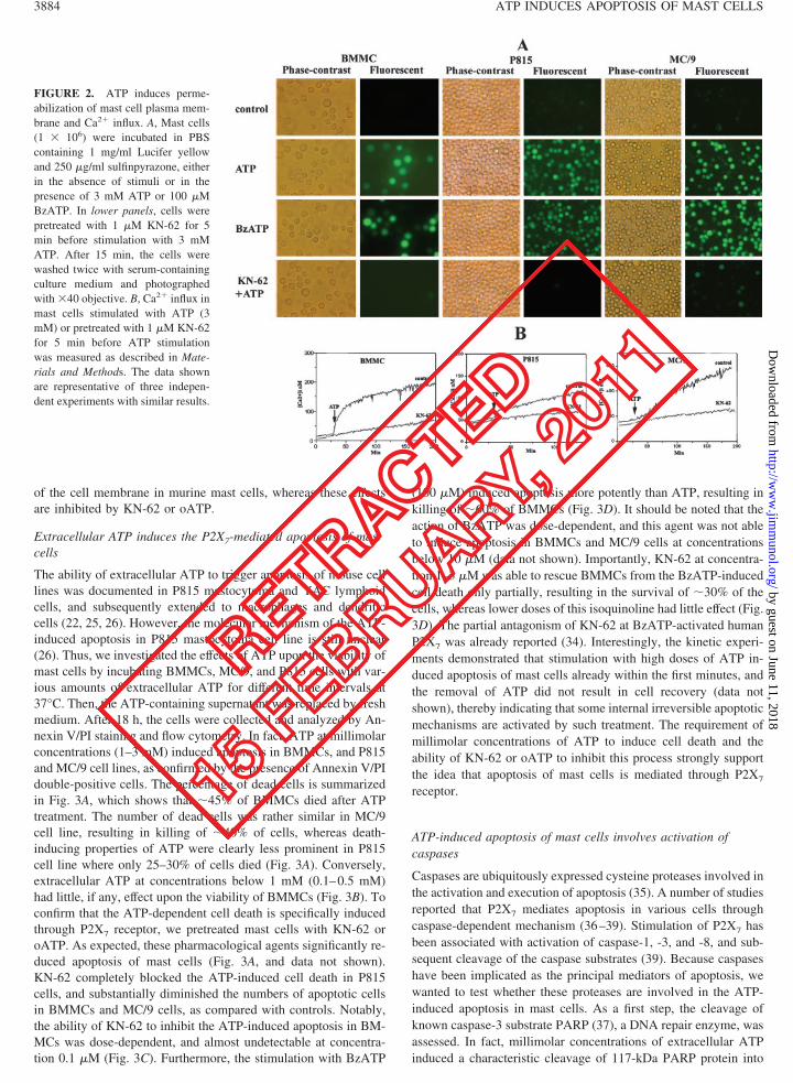
of the cell membrane in murine mast cells, whereas these effectsare inhibited by KN-62 or oATP.
Extracellular ATP induces the P2X7-mediated apoptosis of mastcells
The ability of extracellular ATP to trigger apoptosis of mouse celllines was documented in P815 mastocytoma and YAC lymphoidcells, and subsequently extended to macrophages and dendriticcells (22, 25, 26). However, the molecular mechanism of the ATP-induced apoptosis in P815 mastocytoma cell line is still unclear(26). Thus, we investigated the effects of ATP upon the viability ofmast cells by incubating BMMCs, MC/9, and P815 cells with var-ious amounts of extracellular ATP for different time intervals at37°C. Then, the ATP-containing supernatant was replaced by freshmedium. After 18 h, the cells were collected and analyzed by An-nexin V/PI staining and flow cytometry. In fact, ATP at millimolarconcentrations (1–3 mM) induced apoptosis in BMMCs, and P815and MC/9 cell lines, as confirmed by the presence of Annexin V/PIdouble-positive cells. The percentage of dead cells is summarizedin Fig. 3A, which shows that �45% of BMMCs died after ATPtreatment. The number of dead cells was rather similar in MC/9cell line, resulting in killing of �40% of cells, whereas death-inducing properties of ATP were clearly less prominent in P815cell line where only 25–30% of cells died (Fig. 3A). Conversely,extracellular ATP at concentrations below 1 mM (0.1–0.5 mM)had little, if any, effect upon the viability of BMMCs (Fig. 3B). Toconfirm that the ATP-dependent cell death is specifically inducedthrough P2X7 receptor, we pretreated mast cells with KN-62 oroATP. As expected, these pharmacological agents significantly re-duced apoptosis of mast cells (Fig. 3A, and data not shown).KN-62 completely blocked the ATP-induced cell death in P815cells, and substantially diminished the numbers of apoptotic cellsin BMMCs and MC/9 cells, as compared with controls. Notably,the ability of KN-62 to inhibit the ATP-induced apoptosis in BM-MCs was dose-dependent, and almost undetectable at concentra-tion 0.1 �M (Fig. 3C). Furthermore, the stimulation with BzATP
(100 �M) induced apoptosis more potently than ATP, resulting inkilling of �60% of BMMCs (Fig. 3D). It should be noted that theaction of BzATP was dose-dependent, and this agent was not ableto induce apoptosis in BMMCs and MC/9 cells at concentrationsbelow 10 �M (data not shown). Importantly, KN-62 at concentra-tion 1–3 �M was able to rescue BMMCs from the BzATP-inducedcell death only partially, resulting in the survival of �30% of thecells, whereas lower doses of this isoquinoline had little effect (Fig.3D). The partial antagonism of KN-62 at BzATP-activated humanP2X7 was already reported (34). Interestingly, the kinetic experi-ments demonstrated that stimulation with high doses of ATP in-duced apoptosis of mast cells already within the first minutes, andthe removal of ATP did not result in cell recovery (data notshown), thereby indicating that some internal irreversible apoptoticmechanisms are activated by such treatment. The requirement ofmillimolar concentrations of ATP to induce cell death and theability of KN-62 or oATP to inhibit this process strongly supportthe idea that apoptosis of mast cells is mediated through P2X7
receptor.
ATP-induced apoptosis of mast cells involves activation ofcaspases
Caspases are ubiquitously expressed cysteine proteases involved inthe activation and execution of apoptosis (35). A number of studiesreported that P2X7 mediates apoptosis in various cells throughcaspase-dependent mechanism (36–39). Stimulation of P2X7 hasbeen associated with activation of caspase-1, -3, and -8, and sub-sequent cleavage of the caspase substrates (39). Because caspaseshave been implicated as the principal mediators of apoptosis, wewanted to test whether these proteases are involved in the ATP-induced apoptosis in mast cells. As a first step, the cleavage ofknown caspase-3 substrate PARP (37), a DNA repair enzyme, wasassessed. In fact, millimolar concentrations of extracellular ATPinduced a characteristic cleavage of 117-kDa PARP protein into
FIGURE 2. ATP induces perme-abilization of mast cell plasma mem-brane and Ca2� influx. A, Mast cells(1 � 106) were incubated in PBScontaining 1 mg/ml Lucifer yellowand 250 �g/ml sulfinpyrazone, eitherin the absence of stimuli or in thepresence of 3 mM ATP or 100 �MBzATP. In lower panels, cells werepretreated with 1 �M KN-62 for 5min before stimulation with 3 mMATP. After 15 min, the cells werewashed twice with serum-containingculture medium and photographedwith �40 objective. B, Ca2� influx inmast cells stimulated with ATP (3mM) or pretreated with 1 �M KN-62for 5 min before ATP stimulationwas measured as described in Mate-rials and Methods. The data shownare representative of three indepen-dent experiments with similar results.
3884 ATP INDUCES APOPTOSIS OF MAST CELLS
on Decem
ber 22, 2010w
ww
.jimm
unol.orgD
ownloaded from
by guest on June 11, 2018http://w
ww
.jimm
unol.org/D
ownloaded from

the 89-kDa fragment both in BMMCs and MC/9 cells, while pre-treatment with KN-62 abrogated this process (Fig. 3E). The cleav-age of PARP was also observed in P815 cells (data not shown).Furthermore, ATP induced activation of caspase-3 and -8 in BM-MCs after 1 h of treatment, increasing the basal activity ofcaspase-3 to 134% and caspase-8 to 162% (Fig. 3F). Similar re-sults were observed in MC/9 and P815 cells (data not shown).Importantly, lower doses of ATP (below 1 mM) or BzATP (below10 �M) were not able to induce caspase activation (data notshown). Thus, ATP induces the P2X7-mediated apoptosis of mastcells, which involves activation of caspase-3 and -8.
Extracellular ATP induces the phosphorylation of signalingmolecules in mast cellsOne of the early effects of P2X7 receptor activation is Ca2� influxacross the plasma membrane and the equilibration of membraneK� and Na� gradients (14). Such ionic perturbations can triggeractivation of a number of intracellular signaling molecules, includ-ing MAPKs (40). It has been shown that ATP can induce phos-phorylation of ERK in PC12 cells and fetal astrocytes (40, 41), andof JNK in BAC1 murine macrophages (22). In addition, we haverecently demonstrated that stimulation with extracellular ATPmediates activation of p56lck, ERK, and JNK in Jurkat cells (42).
FIGURE 3. ATP induces caspase-dependent apoptosis of murine mast cells. A, Mast cells were incubated with ATP (3 mM) for 30 min at 37°C orpretreated with 1 �M KN-62 for 5 min before ATP treatment, washed twice with medium, and incubated for 18 h at 37°C. Percentage of apoptotic cellswas analyzed by Annexin VFITC/PI staining and FACS. The results (mean � SD) are based on three independent experiments. ��, Data from mast cellstreated with ATP were significantly different from control (p � 0.01). �, Data from mast cells treated with KN-62 � ATP were significantly different fromATP-treated mast cells (p � 0.05). B, BMMCs were incubated with different ATP concentrations (0.1–5 mM) for 30 min at 37°C, washed twice withmedium, and incubated for 18 h at 37°C. Percentage of apoptotic cells was analyzed by Annexin VFITC/PI staining and FACS. The ability of extracellularATP to induce mast cell apoptosis is dose-dependent, because ATP at concentrations below 1 mM (0.1–0.5 mM) did not affect the viability of BMMCs.C, BMMCs were incubated with ATP (3 mM) for 30 min at 37°C or pretreated with different concentrations of KN-62 (0.1–3 �M) for 5 min before ATPtreatment to prevent apoptosis. After 18 h, the percentage of apoptotic cells was analyzed by Annexin VFITC/PI staining followed by FACS analysis. D,BMMCs were incubated with BzATP (100 �M) or pretreated with different concentrations of KN-62 (0.1–3 �M) for 5 min before BzATP treatment toprevent apoptosis, and numbers of apoptotic cells were counted after Annexin VFITC/PI staining by flow cytometry. Stimulation with BzATP inducedapoptosis in BMMCs more potently than ATP, whereas KN-62 had only partial protective effect. E and F, BMMCs and MC/9 were treated with 3 mMATP for 1 h or pretreated with KN-62 for 5 min before ATP treatment. Cell lysates were analyzed by 10% SDS-PAGE using Abs recognizing intact (117kDa) and cleaved (89 kDa) PARP (E). Activity of caspase-3 and -8 in BMMCs was analyzed after 1 h of ATP treatment as described in Materials andMethods (F). Basal activity of caspases was considered as 100%. The data shown are representative of three independent experiments with similar results.
3885The Journal of Immunology
on Decem
ber 22, 2010w
ww
.jimm
unol.orgD
ownloaded from
by guest on June 11, 2018http://w
ww
.jimm
unol.org/D
ownloaded from

Based on these findings, our next goal was to determine whetherATP is able to induce activation of intracellular signaling mole-cules in mast cells. For this purpose, BMMCs, MC/9, and P815cells were pulse-stimulated with extracellular ATP for relativelybrief periods ranging from 5 to 15–30 min. We considered suchpulses to represent a more “physiologic” stimulus, as mast cells arelikely exposed to high ATP concentrations only for short timeintervals due to the ubiquitous expression of ecto-ATPases/ecto-nucleotidases (43) which enzymatically degrade extracellularATP. In the experiments illustrated in Fig. 4, mast cells were stim-ulated with 3 mM ATP. At the indicated time points, the cells werecollected, lysed, and assessed for patterns of tyrosine phosphory-lation by Western blotting. Immunoprecipitation and probing withanti-pTyr Abs showed that a number of proteins were phosphor-ylated upon the exposure of cells to extracellular ATP. These in-cluded 10–18, 20–22, 27, 30, 33, 38, 45, 55–60, and 80–85 kDaproteins (Fig. 4, upper panel). Such phosphorylation pattern wasalready detectable within the first 5 min of ATP administration,reaching its maximum after 15 min, and returning to near basallevel within 30 min (Fig. 4, and data not shown).
Notwithstanding, ATP stimulation induced rather weak changesin the pattern of tyrosine phosphorylation in P815 cells, as com-pared with untreated control cells (Fig. 4). The probing of themembranes with phosphospecific Abs allowed us to identifyamong the phosphorylated proteins STAT6, ERK1 (p44), ERK2(p42), as well as weak phosphorylation of Jak2 (Fig. 4A). Kinetic
experiments confirmed that ATP induced a rapid and transientphosphorylation of these signaling molecules. In contrast, thephosphorylation state of other members of Jak (Jak1, Jak3, Tyk2)and STAT family (STAT1, STAT3, STAT5), as well as p38 ki-nase, JNK1 (p54), and JNK2 (p52) was not affected, remaining atthe same level throughout the time course of the experiment (datanot shown). For loading control, membranes were stripped andreprobed with anti-STAT5, anti-ERK, or anti-Jak2 Abs, respec-tively. In contrast, these effects were abolished when cells werepretreated with KN-62 (Fig. 4A) or oATP (Fig. 4B) before ATPstimulation, suggesting that the observed phosphorylation is me-diated through P2X7 receptor. Taken together, these results showthat extracellular ATP induces activation of multiple signalingmolecules via P2X7 receptor in murine mast cells.
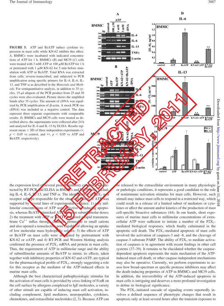
Extracellular ATP induces cytokine expression in mast cells
Mast cells are efficient producers of many key inflammatory cy-tokines in response to stimulation with a variety of stimuli (1–3).To understand the biological significance of the observed activa-tion of signaling molecules after ATP stimulation, a number ofcytokines (e.g., IL-1�, IL-2, IL-4, IL-6, IL-7, IL-13, IL-15, IL-18,and TNF-�) were tested by semiquantitative RT-PCR using spe-cific primers to detect changes in the level of transcription. To thisend, BMMCs or MC/9 cells were stimulated with ATP at concen-trations ranging from 100 �M to 3 mM, harvested for RNA prep-aration, and assessed for the level of cytokine transcription. Fig. 5,A and B, show that ATP in millimolar range was a potent stimulusfor the transcription of IL-4, IL-6, IL-13, and TNF-� in BMMCs.However, only messages for IL-4 and IL-13 were up-regulated inMC/9 cells (Fig. 5C). Notably, the expression of IL-6 and TNF-�was already rather high in MC/9 cells, which might account for theinability of ATP to further enhance mRNA level of these cyto-kines. The increase in the transcription was most prominent after3 h of ATP stimulation and undetectable after 12 h (data notshown), presumably due to the cell apoptosis. Furthermore, thestimulation of BMMCs with BzATP (100 �M) mimicked ATPaction (3 mM), whereas the increase in the cytokine transcriptionwas significantly inhibited by the pretreatment of cells with 1 �MKN-62 (Fig. 5B) or oATP (data not shown), further supporting theinvolvement of P2X7. Notably, KN-62 at concentrations below 0.5�M had little, if any, effect on the transcription of these cytokines(data not shown).
Next, we wanted to test whether mast cells may release thesecytokines to the culture medium in response to the stimulation withATP or BzATP. To this end, BMMCs and MC/9 cells were incu-bated with these agents essentially as described above, and the cellsupernatants were harvested for ELISA after 24 h. These experi-ments showed the ability of ATP or BzATP to up-regulate pro-duction of IL-6 and IL-13 in BMMCs and MC/9 cells, whereasKN-62 inhibited most of these effects (Fig. 5D). Interestingly,KN-62 had almost no effect upon the BzATP-induced release ofIL-13 in MC/9 cells. In contrast, no changes in the release ofTNF-� and IL-4 from BMMCs and MC/9 cells were observed(data not shown). Taken together, these experiments demonstratethe ability of ATP or BzATP to up-regulate transcription and pro-duction of several proinflammatory cytokines in murine mast cells.
DiscussionThe present work demonstrates that murine mast cells express afunctional P2X7 receptor capable of initiating apoptosis after stim-ulation with millimolar concentrations of extracellular ATP. Con-comitantly, we observed a rapid but transient phosphorylation ofmultiple intracellular proteins, such as ERK1/2, Jak2, and STAT6.Notwithstanding the development of apoptosis, an up-regulation in
FIGURE 4. ATP induces tyrosine phosphorylation of intracellular pro-teins and activation of Jak2, STAT6, and ERKs. A, BMMCs, MC/9, andP815 cells were stimulated with 3 mM ATP for 5 or 15 min or pretreatedwith 1 �M KN-62 for 5 min before ATP stimulation. After treatment, thecells were washed and lysed in 1% Nonidet P-40 buffer. Protein lysateswere analyzed by 10% SDS-PAGE using anti-pTyr and anti-phospho-specific Abs to Jak2, STAT6, and ERK1/2. To prove that theequal amount of proteins was loaded in each sample, the blots werestripped and reprobed with anti-Jak2, STAT6, and ERK Abs. B, Mast cellswere pretreated with 300 �M oATP for 30 min before ATP stimulation orleft untreated. Then, the cells were washed, stimulated with 3 mM ATP for5 or 15 min, washed again, and lysed in 1% Nonidet P-40 buffer. Proteinlysates were analyzed by 10% SDS-PAGE using anti-pTyr Abs.
3886 ATP INDUCES APOPTOSIS OF MAST CELLS
on Decem
ber 22, 2010w
ww
.jimm
unol.orgD
ownloaded from
by guest on June 11, 2018http://w
ww
.jimm
unol.org/D
ownloaded from
the expression level of several proinflammatory cytokines was de-tected by RT-PCR and ELISA in BMMCs and MC/9 cells, includ-ing IL-4, IL-6, IL-13, and TNF-�. The identification of P2X7 as areceptor subtype responsible for the observed effects of ATP wassupported by several lines of experimental evidence: 1) only mil-limolar concentrations of ATP were effective in inducing apopto-sis, whereas BzATP mimicked ATP action at submillimolar doses;2) the treatment with ATP or BzATP triggered a rapid transmem-brane Ca2� influx through channels permeable to small cationsand also opened a nonselective pore capable of allowing an uptakeof low molecular mass hydrophilic solutes; 3) the effects of ATPor BzATP on mast cells were abrogated by pretreatment withKN-62 or oATP; and 4) RT-PCR and Western blotting analysisconfirmed the presence of P2X7 mRNA and protein in mast cells.Thus, the requirement of ATP in millimolar range and the abilityof submillimolar amounts of BzATP to mimic its effects, takentogether with inhibitory properties of KN-62 and oATP, are typicalfor the pharmacological profile of P2X7, strongly suggesting a rolefor this receptor as the mediator of the ATP-induced effects inmurine mast cells.
Although the best characterized pathophysiologic stimulus forthe activation of mast cells is provided by cross-linking of Fc�R onthe cell surface by allergens complexed to IgE molecules, a varietyof other stimuli are capable of inducing mast cell activation, in-cluding complement, lipid mediators, neuropeptides, cytokines,chemokines, and extracellular nucleotides (2, 3). Because ATP can
be released to the extracellular environment in many physiologicor pathologic conditions, it represents a good candidate to the roleof nonimmune activation stimulus for mast cells. However, suchstimuli may induce mast cells to respond in a restricted way, whichcould result in a release of a limited subset of mediators or cyto-kines or affect the amount and/or kinetics of the production of mastcell-specific bioactive substances (44). In our hands, short expo-sures of murine mast cells to millimolar concentrations of extra-cellular ATP were sufficient to initiate a number of the P2X7-mediated biological responses, which finally culminated in theapoptotic cell death. The P2X7-mediated apoptosis of mast cellsinvolved the activation of caspases-3 and -8, and the cleavage ofcaspase-3 substrate PARP. The ability of P2X7 to mediate activa-tion of caspases is in agreement with recent findings in other cellsystems (37–39). It remains to be elucidated whether the caspase-dependent apoptosis represents the main mechanism of the ATP-induced mast cell death, or other caspase-independent mechanismsmay also be operative. Currently, we perform experiments to as-sess how broad-spectrum or specific protease inhibitors may affectthe death-inducing properties of ATP in BMMCs and MC/9 cells.In addition, the irreversibility of the ATP-induced apoptosis inmast cells is intriguing and deserves a more profound investigationto define its biological significance.
The P2X7-initiated cascade of signaling events reportedly in-volves a defined sequence of phenotypic changes that result inapoptosis only at least several hours after the transient exposure to
FIGURE 5. ATP and BzATP induce cytokine ex-pression in mast cells while KN-62 inhibits this effect.A, BMMCs were incubated with indicated concentra-tions of ATP for 1 h. BMMCs (B) and MC/9 (C) cellswere treated with 3 mM ATP or 100 �M BzATP for 1 hor pretreated with 1 �M KN-62 for 5 min before stim-ulation with ATP or BzATP. Total RNA was extractedfrom cells, reverse-transcribed, and subjected to PCRamplification using specific primers for IL-4, IL-6, IL-13, and TNF-� as described in the Materials and Meth-ods. For semiquantitative analysis, in addition to 35 cy-cles, 15-�l aliquots of the PCR product from 25 and 30cycles were also evaluated. Picture shows the amplifiedbands after 35 cycles. The amount of cDNA was equal-ized by PCR amplification of �-actin. A mock PCR (nocDNA) was included as a negative control. The datarepresent three separate experiments with comparableresults. D, BMMCs and MC/9 cells were treated as de-scribed above, the supernatants were collected after 24 hand analyzed for IL-6 and IL-13 by ELISA. Results rep-resent mean � SD of three independent experiments (�,p � 0.05 vs control, and ��, p � 0.05 vs ATP andBzATP, respectively).
3887The Journal of Immunology
on Decem
ber 22, 2010w
ww
.jimm
unol.orgD
ownloaded from
by guest on June 11, 2018http://w
ww
.jimm
unol.org/D
ownloaded from

ATP (22). It has been suggested that in the time period betweencommitment to apoptosis and actual cell death, agonistic stimula-tion of P2X7 receptor also activates additional signaling pathways,which may lead to cytokine production in case of proteolytic pro-cessing and release of IL-1� from LPS-primed macrophages thatprecedes cell death (45), and activation of various transcriptionfactors like NFAT or NF-�B (46), thus modulating the overallresponse to ATP. Accordingly, a rapid ATP-induced phosphory-lation of ERK1/2, Jak2, and STAT6 was observed in murine BM-MCs and MC/9 cells, which returned to nearly basal levels after 30min, whereas the phosphorylation state of other members of Jakand STAT families, p38 kinase, and JNK was not altered (data notshown). Additional experiments are underway to determine theidentity of other tyrosine-phosphorylated proteins in BMMCs andMC/9 cells upon ATP stimulation. Furthermore, we observed thatATP induced in a dose-dependent manner a significant increase inthe transcription level of IL-4 and IL-13, and to a lesser extent, ofIL-6 and TNF-�. Furthermore, ATP stimulation enhanced the re-lease of IL-6 and IL-13 to the culture medium from BMMCs andMC/9 cells. Moreover, BzATP, which is a more potent agonist forP2X7 receptor than ATP, mimicked the effects of ATP at lowerconcentration. Notwithstanding, only �40–45% of BMMCs orMC/9 cells died via apoptosis after 18 h of ATP treatment. Thus,not all mast cells are equally susceptible to ATP, and the observedphosphorylation pattern as well as the increase in the cytokineexpression may predominantly take place in the apoptosis-resistantmast cells, although additional experiments are required to confirmor reject this suggestion.
Gargett and Wiley (27) first reported the fact that an isoquino-line derivative KN-62 can act as a potent antagonist of P2X7 re-ceptor. Although KN-62 is also a potent inhibitor of Ca2�/calmod-ulin-dependent protein kinase II (CaMKII), an inactive analog ofKN-62, KN-04, also blocked the P2X7 responses, indicating thatthe inhibition of P2X7 receptor by KN-62 is not mediated byCaMKII (27). Importantly, the action of KN-62 upon P2X7 doesnot involve its inhibitory properties on CaMKII in short-term stud-ies, whereas prolonged exposures to KN-62 require cautious in-terpretation because of concomitant inhibition of this kinase (14,27). Humphreys and colleagues (22, 34) have demonstrated thehigh selectivity of KN-62, which inhibits both human and mouseP2X7 but is inactive at rat receptor, thus providing a very usefultool for identifying the P2X7-mediated functional responses. How-ever, the molecular mechanism(s) that underlie the ability ofKN-62 to inhibit human and mouse P2X7 receptor remain unclear(22, 34). It has been suggested that KN-62 elicits its inhibitoryaction on human P2X7 receptor through direct binding to the ami-no-terminal half, which contains the large extracellular loop. Cor-respondingly, the introduction of the first 335 amino acids of thehuman receptor sequence conferred KN-62 sensitivity to rat P2X7
receptor (34). The pretreatment of mast cells with KN-62 or an-other irreversible P2X7 antagonist, oATP, inhibited the effects ofextracellular ATP such as membrane permeabilization, proteinphosphorylation, and cytokine expression. Moreover, these chem-ical agents significantly reduced the numbers of apoptotic BM-MCs, MC/9, and P815 cells as compared with untreated controls.Notably, the antagonists were more effective in preventing theATP-induced apoptosis in P815 cells, whereas KN-62 was able toantagonize the BzATP-induced effects only partially, which is inaccord with previous findings (34). It is noteworthy that theamount and/or kinetics of activation of functional P2X7 receptorsupon the cell surface of BMMCs and these two mast cell lines maypresumably vary, contributing to slightly different functional re-sponses to ATP and the ability of the antagonists to inhibit them.Importantly, ATP induced transmembrane Ca2� influx, which was
most prominent in BMMCs and MC/9 but rather weak in P815cells, whereas KN-62 or oATP abrogated this effect. The ability ofP2X7 receptor to trigger a long-lasting transmembrane Ca2� influxis in agreement with earlier studies (14, 31).
The fact that brief exposures to millimolar concentrations ofextracellular ATP result in the irreversible P2X7 receptor-mediatedapoptosis raises the issue of the relevance of this process to mastcell physiology. Mast cells play critical roles in a variety of aller-gic, autoimmune, and inflammatory diseases (1, 3, 6, 47). Theexpression of an active P2X7 receptor capable of mediating apo-ptosis offers a possibility to quickly eliminate unwanted mast cellsunder circumstances which favor accumulation of extracellularATP in rather high concentrations. Although it is still not clearwhether such high levels of ATP could be achieved in the extra-cellular space, amounts of ATP in the protected compartments atthe level of the cell membrane could easily reach concentrationssufficient to activate the low-affinity P2X7 receptor (14). Recentfindings implicate mast cells in a variety of neuroinflammatorydiseases, especially those worsened by stress (6), thereby ques-tioning the role of neuronal mast cell activation in the developmentof migraines and multiple sclerosis (6). It remains to be elucidatedwhether extracellular ATP can contribute through the P2X7-medi-ated apoptosis to the elimination of unwanted mast cells in suchpathological conditions, and how an activation of antiapoptoticsignaling pathways may oppose its action.
Given that many cell types including mast cells can releaseATP, this chemical agent might be able to alter function of thesame or nearby cells by autocrine/paracrine mechanism. In fact,the amount of ATP released from one mast cell was shown to besufficient to diffuse several tens of micrometers and elicit rises inintracellular Ca2� in surrounding cells (6, 48). It has been sug-gested that the operation of an ATP-based autocrine/paracrine loopcan support the P2X7-mediated lymphoid cell growth in the ab-sence of serum-derived growth factors (49). Many studies havedocumented that P2X7 receptor/pore participates in diverse mono-cyte, macrophage, microglia, lymphocyte, and dendritic cells re-sponses such as cell membrane permeabilization, cytokine release,multinucleated giant cell formation, cell proliferation or apoptosis(6, 14, 15). For example, stimulation of P2X7 receptor by ATPinduces posttranslational processing of IL-1� and IL-18 precursorsin human monocytes (50, 51), altered cytokine production in micelacking this receptor (52), stimulation of JNK activity and induc-tion of apoptosis in murine macrophages (22), inhibition of oste-oclastic resorption (53), and activation of transcription factorNF-�B (54) and NFAT (46) in microglial cell lines. Data from ourlaboratory suggest that ATP can trigger proliferation of Jurkat Tlymphoblastoid cell line and mediate p56lck-dependent phosphor-ylation of ERK and JNK, and activation of AP-1 transcriptionfactor, simultaneously down-regulating p50/p65 NF-�B het-erodimers (42). It is believed that in T and B lymphocytes, ATPacting over P2X7 generates a pore smaller than that seen in othercell types, such as macrophages (14). In regard to mast cell func-tion, extracellular ATP has been demonstrated to stimulate degran-ulation and release of histamine from these cells (10, 11), and toinduce cell membrane permeabilization (9, 12). Notably, ATP wasshown to trigger apoptosis in P815 mastocytoma cell line, althoughthe mechanism of the ATP-mediated cell death remained obscure(26). Further, ATP stimulated the release of hexosaminidase fromMC/9 mast cell line through a mechanism that was distinct fromthe activation induced by the cross-linking of Fc receptors (9), andinduced the release of histamine and leukotriene from BMMCs(10–12). Several studies have reported a direct Ca2�-dependent
3888 ATP INDUCES APOPTOSIS OF MAST CELLS
on Decem
ber 22, 2010w
ww
.jimm
unol.orgD
ownloaded from
by guest on June 11, 2018http://w
ww
.jimm
unol.org/D
ownloaded from
histamine release and a potentiation of Ag- or ionophore A23187-induced histamine release from rat peritoneal mast cells in re-sponse to ATP stimulation (55, 56). The ability of extracellularATP to induce degranulation of murine mast cells is in accordancewith our findings (data not shown). Conversely, ATP only modu-lated anti-IgE-induced histamine release from human lung mastcells without having the ability to stimulate the release of thiscompound (13). Thus, mast cells derived from different body dis-tricts or distinct species exhibit a functional heterogeneity, whichinvites a cautious interpretation of mast cell responses in vitro withrespect to their relevance in vivo.
ATP is found at a concentration 5–10 mM in the cytosol of mosteukaryotic cell types, whereas its amount in platelets is signifi-cantly higher and can reach a concentration of �1 M (14). Releaseof ATP to extracellular space by both lytic and nonlytic mecha-nisms has been observed from virtually all cell types and tissuesunder conditions of hypoxia, ischemia, inflammation, injury, andcell necrosis or apoptosis (14, 15). However, extracellular ATPconcentrations are generally maintained at extremely low levels bya sequential action of ubiquitous cell surface enzymes such asecto-ATPases/ectonucleotidases, which rapidly degrade it to ADP,AMP, and adenosine (6, 14). Conversely, ATP can be copackagedwith serotonin in platelet granules and released locally in signifi-cant amounts during platelet activation (14, 57). Thus, plateletsthemselves are a major source of ATP, whereas cytosolic ATPstores can also be liberated by the sudden breakage of intact cells,as might occur during the rupture of blood vessels and other tissueinjury (57). These two sources suggest that significant amounts ofextracellular ATP may be generated at vascular sites of thrombusformation and infection/inflammation. The key role of extracellu-lar ATP and P2 receptors in hemostasis has already been empha-sized (14). On the other hand, an emerging concept implicatesmast cells as repair cells, which provide antithrombotic and/or pro-fibrinolytic mediators to prevent thrombus formation or help todissolve thrombotic material in the course of vascular repair pro-cess (58, 59). The fibrinolytic potential of a cell is determined bythe relative abundance of either generated plasminogen activatorsor plasminogen activator inhibitors. Mast cells act fibrinolyticthrough expression of a key fibrinolytic enzyme, tissue-type plas-minogen activator, without coexpression of plasminogen activatorinhibitors, and supernatants as well as cell lysates from culturedmast cells are capable of lysing a fibrin clot in a manner similar torecombinant tissue-type plasminogen activator (58). The recogni-tion of mast cell as a profibrinolytic cell raises a question of itspotential pathophysiologic role in the prevention or repair of vas-cular thrombosis and tissue repair following fibrinogen extravasa-tion. Mast cells can accumulate around thrombosed vessels andprovide a number of important repair molecules including anti-thrombotic heparin, and other profibrinolytic substances. Thus,mast cell recruitment and activation in the sites of vascular injurymay result in local thrombolysis and prevention of coagulation,which may interfere with the healing of a vascular rupture at theinitial stages of injury. Therefore, the apoptosis of mast cells inresponse to rather high concentrations of extracellular ATP whichmay be achieved at the sites of acute vascular injury due to itsrelease from damaged blood cells, endothelial cells, and activatedplatelets appears to be physiologically relevant, because it presum-ably prevents their function as antithrombotic and/or profibrino-lytic cells at initial stages of vascular rupture.
In summary, our results demonstrate that ATP induces a numberof the P2X7-mediated cell responses in murine mast cells, includ-ing the phosphorylation of signaling molecules, transcription ofcytokines, membrane permeabilization, and induction of apopto-sis. The high sensitivity of mast cells to the cytotoxic effects of
extracellular ATP suggests that this process might have a biolog-ical relevance in certain physiologic or pathologic conditions. Thefact that extracellular ATP can play an important role in maintain-ing mast cell numbers through the P2X7-mediated apoptosis mayprovide helpful insights to the development of new therapeuticapproaches to intervene and limit excessive and/or undesired mastcell activities.
AcknowledgmentsWe are grateful to Manuel Fohlmeister and Katrin Streeck for excellenttechnical assistance and Renate Bergmann for the help with ELISA.
DisclosuresThe authors have no financial conflict of interest.
References1. Benoist, C., and D. Mathis. 2002. Mast cells in autoimmune disease. Nature
420:875.2. Piliponsky, A. M., and F. Levi-Schaffer. 2000. Regulation of apoptosis in mast
cells. Apoptosis 5:435.3. Robbie-Ryan, M., and M. A. Brown. 2002. The role of mast cells in allergy and
autoimmunity. Curr. Opin. Immunol. 14:728.4. Wedemeyer, J., M. Tsai, and S. J. Galli. 2000. Roles of mast cells and basophils
in innate and acquired immunity. Curr. Opin. Immunol. 12:624.5. Theoharides, T. C., and D. E. Cochrane. 2004. Critical role of mast cells in
inflammatory diseases and the effect of acute stress. J. Neuroimmunol. 146:1.6. North, R. A. 2002. Molecular physiology of P2X receptors. Physiol. Rev.
82:1013.7. McCloskey, M. A., Y. Fan, and S. Luther. 1999. Chemotaxis of rat mast cells
toward adenine nucleotides. J. Immunol. 163:970.8. Tatham, P. E., and M. Lindau. 1990. ATP-induced pore formation in the plasma
membrane of rat peritoneal mast cells. J. Gen. Physiol. 95:459.9. Sudo, N., K. Tanaka, Y. Koga, Y. Okumura, C. Kubo, and K. Nomoto. 1996.
Extracellular ATP activates mast cells via a mechanism that is different from theactivation induced by the cross-linking of Fc receptors. J. Immunol. 156:3970.
10. Nakamura, H., H. Sato, and Y. Ikura. 1989. The stimuli releasing histamine frommurine bone marrow-derived mast cells. 1. The presence of P2X7-purinoceptors.Arerugi 38:1359.
11. Sato, H., N. Sakaguchi, M. Ebisawa, K. Matsumoto, A. Akasawa, and Y. Ikura.1991. The stimuli releasing histamine from murine bone marrow-derived mastcells. II. Mechanisms involved in histamine release induced by extracellular ATPand its metabolites. Arerugi 40:680.
12. Saito, H., M. Ebisawa, D. C. Reason, K. Ohno, K. Kurihara, N. Sakaguchi,A. Ohgimi, E. Saito, A. Akasawa, K. Akimoto, et al. 1991. Extracellular ATPstimulates interleukin-dependent cultured mast cells and eosinophils through cal-cium mobilization. Int. Arch. Allergy Appl. Immunol. 94:68.
13. Schulman, E. S., M. C. Glaum, T. Post, Y. Wang, D. C. Raible, J. Mohanty,J. H. Butterfield, and A. Pelleg. 1999. ATP modulates anti-IgE-induced release ofhistamine from human lung mast cells. Am. J. Respir. Cell Mol. Biol. 20:530.
14. Di Virgilio, F., P. Chiozzi, D. Ferrari, S. Falzoni, J. M. Sanz, A. Morelli,M. Torboli, G. Bolognesi, and O. R. Baricordi. 2001. Nucleotide receptors: anemerging family of regulatory molecules in blood cells. Blood 97:587.
15. North, R. A., and A. Suprenant. 2000. Pharmacology of cloned P2X receptors.Annu. Rev. Pharmacol. Toxicol. 40:563.
16. Fredholm, B. B., M. P. Abbracchio, G. Burnstock, J. W. Daly, T. K. Harden,K. A. Jakobson, P. Left, and M. Williams. 1994. Nomenclature and classificationof purinoceptors. Pharmacol. Rev. 46:143.
17. Surprenant, A., F. Rassendren, E. Kawashima, R. A. North, and G. Buell. 1996.The cytolytic P2Z receptor for extracellular ATP identified as a P2X receptor(P2X7). Science 272:735.
18. Wiley, J. S., and G. R. Dubyak. 1989. Extracellular adenosine triphosphate in-creases cation permeability of chronic lymphocytic leukemic lymphocytes. Blood73:1316.
19. Greenberg, S., F. Di Virgilio, T. H. Steinberg, and S. C. Silverstein. 1988. Ex-tracellular nucleotides mediate Ca2� fluxes in J774 macrophages by two distinctmechanisms. J. Biol. Chem. 263:10337.
20. Steinberg, T. H., A. S. Newman, J. A. Swanson, and S. C. Silverstein. 1987.ATP4- permeabilizes the plasma membrane of mouse macrophages to fluorescentdyes. J. Biol. Chem. 262:8884.
21. Rassendren, F., G. N. Buell, C. Virginio, G. Collo, R. A. North, andA. Surprenant. 1997. The permeabilizing ATP receptor, P2X7. Cloning and ex-pression of a human cDNA. J. Biol. Chem. 272:5482.
22. Humphreys, B. D., J. Rice, S. B. Kertesy, and G. R. Dubyak. 2000. Stress-activated protein kinase/JNK activation and apoptotic induction by the macro-phage P2X7 nucleotide receptor. J. Biol. Chem. 275:26792.
23. Ferrari, D., M. Villalba, P. Chiozzi, S. Falzoni, P. Riccardi-Castagnoli, andF. Di Virgilio. 1996. Mouse microglial cells express a plasma membrane poregated by extracellular ATP. J. Immunol. 156:1531.
24. Nihei, O. K., A. C. Campos de Carvalho, W. Salvino, and L. A. Alves. 2000.Pharmacologic properties of P2Z/P2X7 receptor characterized in murine dendriticcells: role in the induction of apoptosis. Blood 96:996.
3889The Journal of Immunology
on Decem
ber 22, 2010w
ww
.jimm
unol.orgD
ownloaded from
by guest on June 11, 2018http://w
ww
.jimm
unol.org/D
ownloaded from

25. Countiho-Silva, R., P. M. Persechini, R. Da Cunha Bisaggio, J.-L. Perfettini,A. C. Torres de Saneto, J. M. Kanellopoulos, I. Motta-Ly, A. Dautry-Varsat, andD. M. Ojcius. 1999. P2Z/P2X7 receptor-dependent apoptosis of dendritic cells.Am. J. Physiol. 276:C1139.
26. Zanovello, P., V. Bronye, A. Rosato, P. Pizzo, and F. Di Virgilio. 1990. Re-sponses of mouse lymphocytes to extracellular ATP. J. Immunol. 145:1545.
27. Gargett, C. E., J. S. Wiley. 1997. The isoquinoline derivative KN-62 a potentantagonist of the P2Z-receptor of human lymphocytes. Br. J. Pharmacol. 120:1483.
28. Murgia, M., S. Hanau, P. Pizzo, M. Rippa, and F. Di Virgilio. 1993. OxidizedATP: an irreversible inhibitor of the macrophage purinergic P2Z receptor. J. Biol.Chem. 268:8199.
29. Huff, T. F., and C. S. Lantz. 1997. Culture of mast cells. In Immunology MethodsManual. I. Lefkovits, ed. Academic Press, London, p. 1393.
30. Bulanova, E., V. Budagian, Z. Orinska, H. Krause, R. Paus, and S. Bulfone-Paus.2003. Mast cells express novel functional interleukin-15 receptor � isoforms.J. Immunol. 170:5045.
31. Mutini, C., S. Falzoni, D. Ferrari, P. Chiozzi, A. Morelli, O. R. Boricordi,G. Collo, R. Ricciardi-Castanoli, and F. Di Virgilio. 1999. Mouse dendritic cellsexpress the P2X7 purinergic receptor: characterization and possible participationin antigen presentation. J. Immunol. 163:1958.
32. Grynkiewicz, G., M. Poenie, and R. Y. Tsien. 1985. A new generation of Ca2�
indicators with greatly improved fluorescence properties. J. Biol. Chem.260:3440.
33. Stennicke, H. R., and G. S. Salvesen. 1997. Biochemical characteristics ofcaspases-3, -6, -7, and –8. J. Biol. Chem. 272:25719.
34. Humphreys, B. D., C. Virginio, A. Suprenant, J. Rice, and G. R. Dubyak. 1998.Isoquinolines as antagonists of the P2X7 nucleotide receptor: high selectivity forthe human versus rat receptor homologues. Mol. Pharmacol. 54:22.
35. Earnshaw, W. C., L. M. Martins, and S. H. Kaufman. 1999. Mammaliancaspases: structure, activation, substrates, and functions during apoptosis. Annu.Rev. Biochem. 68:383.
36. Donnelly-Roberts, D. L., M. T. Namovic, C. R. Faltynek, and M. F. Jarvis. 2004.Mitogen-activated protein kinase and caspase signaling pathways are required forP2X7 receptor (P2X7R)-induced pore formation in human THP-1 cells. J. Phar-macol. Exp. Ther. 208:1053.
37. Wen, L. T., C. C. Caldwell, and A. F. Knowles. 2002. Poly(ADP-ribose) poly-merase activation and changes in Bax protein expression associated with extra-cellular ATP-mediated apoptosis in human embryonic kidney 293-P2X7 cells.Mol. Pharmacol. 63:706.
38. Hillman, K. A., H. Harada, C. M. Chan, A. Townsend-Nocholson, S. E. Moss,K. Miyamoto, Y. Suketa, G. Burnstock, R. J. Unwin, and P. M. Dunn. 2003.Chicken DT40 cells stably transfected with the rat P2X7 receptor ion channel: asystem suitable for the study of purine receptor-mediated cell death. Biochem.Pharmacol. 66:415.
39. Ferrari, D., M. Los, M. K. A. Bauer, P. Vandenabeele, S. Wesselborg, andK. Schulze-Osthoff. 1997. P2Z purinoreceptor ligation induces activation ofcaspases with distinct role in apoptotic and necrotic alterations of cell deacth.FEBS Lett. 447:71.
40. Swanson, K. D., C. Reigh, and G. E. Landreth. 1998. ATP-stimulated activationof the mitogen-activated protein kinases through ionotrophic P2X2 purinorecep-tors in PC12 cells. Difference in purinoreceptor sensitivity in two PC12 cell lines.J. Biol. Chem. 273:19965.
41. Neary, J. T., M. McCarthy, Y. Kang, and S. Zuniga. 1998. Mitogenic signalingfrom P1 and P2 purinergic receptors to mitogen-activated protein kinase in hu-man fetal astrocyte cultures. Neurosci. Lett. 242:159.
42. Budagian, V., E. Bulanova, L. Brovko, Z. Orinska, R. Fayad, R. Paus and S.Bulfone-Paus. 2003. Signaling through P2X7 receptor in human T cells involvesp56lck, MAP kinases, and transcription factors AP-1 and NF-�B. J. Biol. Chem.278:1549.
43. Vassort, G. 2001. Adenosine 5�-triphosphate: a P2-purinergic agonist in the myo-cardium. Physiol. Rev. 81:767.
44. Galli, S. J., M. Maurer, and C. S. Lantz. 1999. Mast cells as sentinels of innateimmunity. Curr. Opin. Immunol. 11:53.
45. Ferrari, D., P. Chiozzi, S. Falzoni, M. Dal Susino, L. Melchiorri, O. R. Baricordi,and F. Di Virgilio. 1997. Extracellular ATP triggers IL-1� release by activatingthe purinergic P2Z receptor of human macrophages. J. Immunol. 159:1451.
46. Ferrari, D., C. Stroh, and K. Schulze-Osthoff. 1999. P2X7/P2Z purinoreceptor-mediated activation of transcription factor NFAT in microglial cells. J. Biol.Chem. 274:13205.
47. Seman, M., S. Adriouch, F. Scheuplein, C. Krebs, D. Freese, G. Glowacki,P. Deterre, F. Haag, and F. Koch-Nolte. 2003. NAD-induced T cells death: ADP-ribosylation of cell surface proteins by ART2 activates the cytolytic P2X7 puri-noceptor. Immunity 19:571.
48. Osipchuk, Y., and M. Cahalan. 1992. Cell-to-cell spread of calcium signals me-diated by ATP receptors in mast cells. Nature 359:241.
49. Baricordi, O. R., L. Melchiorri, E. Adinolfi, S. Falzoni, P. Chozzi, G. Buell, andF. Di Virgilio. 1999. Increased proliferation rate of lymphoid cells transfectedwith P2X7 ATP receptor. J. Biol. Chem. 274:33206.
50. Perregaux, D. G., P. McNiff, R. Laliberte, M. Conklyn, and C. A. Gabel. 2000.ATP acts as an agonist to promote stimulus-induced secretion of IL-1� and IL-18in human blood. J. Immunol. 165:4615.
51. Mehta, V. B., J. Hart, and M. D. Wewers. 2001. ATP-stimulated release of in-terleukin (IL)-1� and IL-18 requires priming by lipopolysaccharide and is inde-pendent of caspase-1 cleavage. J. Biol. Chem. 276:3820.
52. Solle, M., J. Labasi, D.G. Perregaux, E. Stam, N. Petrushova, B. H. Koller,R. J. Griffiths, and C. A. Gabel. 2001. Altered cytokine production in mice lack-ing P2X7 receptors. J. Biol. Chem. 276:125.
53. Naemsch, L. N., S. J. Dixon, and S. M. Sims. 2001. Activity-dependent devel-opment of P2X7 current and Ca2� entry in rabbit osteoclasts. J. Biol. Chem.276:39107.
54. Ferrari, D., S. Wesselborg, M. K. Bauer, and K. Schulze-Osthoff. 1997. Extra-cellular ATP activates transcription factor NF-�B through the P2Z purinoreceptorby selectively targeting NF-�B p65. J. Cell Biol. 137:1635.
55. Chakravarty, N. 1980. The role of plasma membrane Ca2�-Mg2� activated aden-osine triphosphate or rat mast cells on histamine release. Acta Pharmacol. Toxi-col. 47:223.
56. Cockroft, S., and B. D. Gomperts. 1979. Activation and inhibition of calcium-dependent histamine secretion by ATP ions applied to rat mast cells. J. Physiol.(London) 296:229.
57. Beigi, R., E. Kobatake, M. Aisawa, and G. R. Dubyak. 1999. Detection of localATP release from activated platelets using cell surface-attached firefly luciferase.Am. J. Physiol. 276:C267.
58. Bankl, H. C., and P. Valent. 2002. Mast cells, thrombosis, and fibrinolysis: theemerging concept. Thrombosis Res. 105:359.
59. Valent, P., M. Baghestanian, H. C. Bankl, C. Sillaber, W. R. Sperr, J. Wojta,B. R. Binder, and K. Lechner. 2002. New aspects in thrombosis research: possiblerole of mast cells as profibrinolytic and antithrombotic cells. Thromb. Haemo-stasis 85:786.
3890 ATP INDUCES APOPTOSIS OF MAST CELLS
on Decem
ber 22, 2010w
ww
.jimm
unol.orgD
ownloaded from

The Journal of Immunology
Letter of Retraction
We wish to retract the article titled “Extracellular ATP Induces Cytokine Expression and Apoptosis through P2X7 Receptor in MurineMast Cells” by Elena Bulanova, Vadim Budagian, Zane Orinska, Martina Hein, Frank Petersen, Lutz Thon, Dieter Adam, and SilviaBulfone-Paus, The Journal of Immunology, 2005, 174: 3880–3890.
This retraction follows a formal investigation by the Research Center Borstel into scientific misconduct. The main findings of theinvestigation were the following:
1) Fig. 3: Although in principle the reported results are correct, the error bars could not be verified on the basis of the raw data.2) Fig. 4: The figure seems to contain several severe manipulations. Various raw data are not available. Independent repetitions of the
experiments shown in Fig. 4B regarding BMMC and MC/9 (left and middle panels) confirmed the original data in principle.
The first two authors declined to sign this Letter of Retraction. All the other authors wish to retract the article. We deeply regret theseirregularities and apologize to the scientific community for any inconvenience this might cause.
Zane OrinskaMartina HeinFrank PetersenSilvia Bulfone-PausResearch Center BorstelBorstel, Germany
Lutz ThonFerring GmBHKiel, Germany
Dieter AdamUniversity of KielKiel, Germany
Copyright� 2011 by The American Association of Immunologists, Inc. 0022-1767/11/$16.00
www.jimmunol.org/cgi/doi/10.4049/jimmunol.1090144