extracellular acid proteases of wine microorganisms: gene
TRANSCRIPT

Extracellular acid proteases of wine microorganisms: gene identification, activity
characterization and impact on wine
by
Vernita Jennilee Reid
Thesis presented in partial fulfilment of the requirements for the degree of
Master of Science
at Stellenbosch University
Institute for Wine Biotechnology, Faculty of AgriSciences
Supervisor: Dr BT Divol
Co-supervisor: Prof M duToit
March 2012

ii
Declaration
By submitting this thesis electronically, I declare that the entirety of the work contained therein is my own, original work, that I am the sole author thereof (save to the extent explicitly otherwise stated), that reproduction and publication thereof by Stellenbosch University will not infringe any third party rights and that I have not previously in its entirety or in part submitted it for obtaining any qualification.
Date: 14/12/2011
Copyright © 2012 Stellenbosch University All rights reserved
Stellenbosch University http://scholar.sun.ac.za

iii
Summary
Non-Saccharomyces yeasts of oenological origin have previously been associated with spoilage or
regarded as undesired yeasts in wine. However, these yeasts have recently come under investigation for
their positive contribution towards wine aroma especially when used in sequential or co-inoculated
fermentations with Saccharomyces cerevisiae. These yeasts are also known to secrete a number of
enzymes that could be applicable in wine biotechnology. Amongst these enzymes are aspartic proteases.
The secreted proteases from some non-Saccharomyces yeast may play a role in protein haze reduction,
as demonstrated by some authors, while simultaneously increasing the assimilable nitrogen content of
the wine for the utilization and growth of fermentative microorganisms. Moreover, the proteases may have
an indirect effect on wine aroma by liberating amino acids that serve as aroma precursors. Although
many screenings have been performed detecting protease activity in non-Saccharomyces yeasts, no
attempts have been made to characterize these enzymes. This study set out to isolate and characterize
genes encoding extracellular aspartic proteases from non-Saccharomyces yeasts.
An enzymatic activity screening of a collection of 308 Saccharomyces and non-Saccharomyces yeasts,
isolated from grape must, was performed. The aspartic protease-encoding genes of two non-
Saccharomyces yeasts, which showed strong extracellular proteolytic activity on plate assays, were
isolated and characterized by in silico analysis. The genes were isolated by employing degenerate and
inverse PCR. One gene was isolated from Metschnikowia pulcherrima IWBT Y1123 and named MpAPr1.
The other putative gene was isolated from Candida apicola IWBT Y1384 and named CaAPr1. The
MpAPr1 gene is 1137 bp long, encoding a 378 amino acid putative protein with a predicted molecular
weight of 40.1 kDa. The CaAPr1 putative gene is 1101 bp long and encodes a 367 amino acid putative
protein with a predicted molecular weight of 39 kDa. These features are typical of extracellular aspartic
proteases. The deduced protein sequences showed less than 40% homology to other yeast extracellular
aspartic proteases. By heterologous expression of MpAPr1 in S. cerevisiae, it was confirmed that the
gene encodes an extracellular acid protease. The expression of MpAPr1 was shown to be induced in
media containing proteins as sole nitrogen source and repressed when a preferred nitrogen source was
available. The gene was expressed in the presence of casein, bovine serum albumin (BSA) and grape
juice proteins and repressed in the presence of ammonium sulphate. Expression was most induced in the
presence of grape juice proteins, which was expected since these proteins are present in the natural
habitat of the yeast. A genetic screening confirmed the presence of the MpAPr1 gene in 12 other
M. pulcherrima strains isolated from grape juice. The extracellular protease activity of the strains was also
visualized on plates. As far as we know, this is the first report on the genetic characterization of secreted
aspartic proteases from non-Saccharomyces yeasts isolated from grape must and provides the
groundwork for further investigations.
Stellenbosch University http://scholar.sun.ac.za

iv
Opsomming
Nie-Saccharomyces giste is voorheen met wynbederf geassosieer en hul teenwoordigheid in wyn is
ongewens. Hierdie giste is onlangs ondersoek vir hulle positiewe bydrae tot wyn aroma, in veral
sekwensiële en ko-inokulerings met Saccharomyces cerevisiae. Sommige van die nie-Saccahromyces
giste skei ‘n verskeidenhied ensieme af wat moontlik vir die wynmaker van nut kan wees. Een groep van
hierdie ensieme is die aspartiese suurproteases. Soos deur sommige navorsers aangetoon word, kan die
proteases die vorming van proteïenwaasverlaging, terwyl dit terselfdertyd die assimilerende
stikstofinhoud van die wyn vir die gebruik en groei van fermentasie-mikroörganismes verhoog. Die
proteases kan moontlik ook ‘n indirekte uitwerking op die aromaprofiel van die wyn hê deur die vrystelling
van aminosure wat as aromavoorlopers dien. Alhoewel baie studies gedoen is wat die ekstrasellulêre
teenwoordigheid van proteases bevestig in nie-Saccharomyces giste wat van druiwesap/wyn afkoms is,
is daar geen dokumentasie oor die genetiese karakterisering van hierdie ensieme beskikbaar nie. Die
doel van hierdie studie was om gene wat aspartiese proteases in nie-Saccharomyces giste enkodeer, te
isoleer en gedeeltelik te karakteriseer.
‘n Versameling van 308 Saccharomyces en nie-Saccharomyces giste wat uit druiwe sap geïsoleer is, is
gesif vir ensiematiese aktiwiteit deur plaattoetse uit te voer. Twee gene wat aspartiese protease
enkodeer, is geïsoleer van twee nie-Saccharomyces giste. Dit hetpositief gedurende die aktiwiteitstoetse
getoets en is deur in silico–analise gekarakteriseer. Die gene is deur die uitvoering van gedegenereerde
en inverse PKR geïdentifiseer. Een geen is vanaf Metschnikowia pulcherrima IWBT Y1123 geïsoleer en
is MpAPr1 genoem, terwyl die ander van Candida apicola IWBT Y1384 geïsoleer en CaAPr1 genoem is.
Die MpAPr1-geen is 1137 bp lank en enkodeer ‘n proteïen wat uit 378 aminosure bestaan met ‘n
voorspelde molekulêre massa van 40.1 kDa. Daar teenoor is die CaAPr1-geen 1101 bp lank en enkodeer
vir ‘n proteïen wat uit 367 aminosure met ‘n molekulêre massa van 39 kDa bestaan. Hierdie eienskappe
is kenmerkend van aspartiese protease. Die afgeleide proteïenvolgorde het minder as 40% homologie
met ander ekstrasellulêre aspartiese proteases vertoon, wat dui op die nuwigheid van hierdie ensieme.
Die MpAPr1-geen is heterologies in S. cerevisiae YHUM272 uitgedruk en dit het bevestig dat die geen
inderdaad ‘n ekstrasellulêre aspartiese protease enkodeer. Die MpAPr1-geen is uitgedruk in media wat
alleenlik proteïen as stikstofbron bevat het, terwyl dit onderdruk is in gevalle waar ‘n verkose stikstofbron
beskikbaar was. Die geen is uitgedruk in die teenwoordigheid van kaseïen, BSA en proteïene afkomstig
vanaf druiwesap en in die teenwoordigheid van ammoniumsulfaat onderdruk. Die hoogste uitdrukking
was in die teenwoordigheid van druifproteïene. Hierdie proteïene is teenwoordig in die natuurlike habitat
van die gis en is dus dalk ‘n bekende stikstofbron vir die gis. ‘n Genetiese sifting het die teenwoordigheid
van die MpAPr1-geen in 12 ander M. pulcherrima–rasse, wat ook van wynkundige oorsprong is, bevestig.
Die aspartiese protease-aktiwiteit van die 12 rasse is ook op agarplate waargeneem. Na ons wete, is dit
die eerste verslag oor die genetiese karakterisering van afgeskeide aspartiese proteases van nie-
Saccharomyces giste van wynkundige oorsprong en verskaf die grondslag vir verdere ondersoek.
Stellenbosch University http://scholar.sun.ac.za

v
This thesis is dedicated to
My Mother
Stellenbosch University http://scholar.sun.ac.za

vi
Biographical sketch
Vernita Reid was born in Bloemfontein, South Africa on the 12th of November 1984. She
attended Heide Primary School and completed her matriculation at Oranje Girls’ School in 2002.
She obtained a BSc degree in Food Biotechnology in 2007 and a BSc Honours degree in Food
Science in 2008 from the University of the Free State. She enrolled at Stellenbosch University in
2010 for an MSc in Wine Biotechnology.
Stellenbosch University http://scholar.sun.ac.za

vii
Acknowledgements
I wish to express my sincere gratitude and appreciation to the following persons and institutions:
God, for wisdom and understanding, to God be the glory
Dr Benoit Divol for acting as my supervisor, for his patience, guidance and constructive
criticism throughout this study
Prof Maret du Toit for acting as my co-supervisor, for her advice and guidance
throughout this study
Dr Evodia Setati and Mr Alexis Eschstruth for their invaluable technical guidance
Lab colleagues for their assistance, support and encouragement
My family for their constant enthusiasm and support and for always being there for me
Friends for their support and encouragement
The Institute for Wine Biotechnology, Winetech and the THRIP funding programme
of the National Research Foundation, the Harry Crossley Foundation and
Stellenbosch University (Sub-committee B) for financial support
Stellenbosch University http://scholar.sun.ac.za

viii
Preface
This thesis is presented as a compilation of four chapters.
Chapter 1 General Introduction and project aims
Chapter 2 Literature review
Aspartic proteases and non-Saccharomyces yeasts and their potential
application in wine biotechnology
Chapter 3 Research Results
Identification and characterization of extracellular aspartic protease genes
from Metschnikowia pulcherrima IWBT Y1123 and Candida apicola IWBT
Y1384
Chapter 4 General discussion and conclusions
Stellenbosch University http://scholar.sun.ac.za

ix
Contents
Chapter 1. General introduction and project aims 1
1.1 Introduction 2
1.2 Rationale and scope of the study 3
1.3 References 3
Chapter 2. Literature review: Aspartic proteases of non-Saccharomyces yeasts and their potential applications in wine biotechnology 5
2.1 General introduction 6
2.2 Proteolytic enzymes 6
2.2.1 Definition and characterization of proteolytic enzymes 6
2.2.2 Aspartic proteases 8
2.2.2.1 General description 8
2.2.2.2 Structure of aspartic proteases 10
2.2.2.3 Catalytic mechanism of aspartic proteases 11
2.2.2.4 Secretion pathway and expression in yeasts 14
2.2.3 Model systems of yeast proteases used in the food industry 16
2.3 Oenological importance of non-Saccharomyces yeasts 17
2.3.1 Wine microbial diversity: spontaneous and inoculated fermentations 17
2.3.2 Growing interest in non-Saccharomyces wine yeasts 21
2.3.3 Non-Saccharomyces yeasts with extracellular enzyme activity 22
2.4 The role of aspartic proteases in wine 23
2.4.1 Production and the risk of protein haze formation 23
2.4.2 Increase in available assimilable nitrogen and wine aroma 26
2.5 References 27
Chapter 3. Research results: Identification and partial characterization of extracellular aspartic protease genes from Metschnikowia pulcherrima IWBT Y1123 and Candida apicola IWBT Y1384 36
3.1 Introduction 37
3.2 Materials and Methods 39
Stellenbosch University http://scholar.sun.ac.za

x 3.2.1 Strains, plasmids and culture conditions 39
3.2.2 Molecular biology and Bioinformatics techniques 40
3.2.2.1 Nucleic acid extraction 40
3.2.2.2 In silico analyses 41
3.2.2.3 PCR methods 41
3.2.3 Cloning and heterologous expression in S. cerevisiae YHUM272 42
3.2.4 DNA sequencing 43
3.2.5 Protein work 43
3.2.5.1 SDS-PAGE and zymography 43
3.2.5.2 Protein sequencing 43
3.3 Results 44
3.3.1 Protease activity screening and strain selection 44
3.3.2 Isolation and cloning of protease-encoding genes 44
3.3.3 In silico analysis of the putative gene and deduced protein sequences 45
3.3.4 Putative identification based on homology studies 45
3.3.5 Heterologous expression of the protease-encoding gene of M. pulcherrima IWBT Y1123 in S. cerevisiae YHUM272 46
3.3.6 Induction and substrate specificity investigation 47
3.3.7 Genetic screening of 12 M. pulcherrima strains for the presence of MpAPr1 49
3.4 Discussion 50
3.5 Acknowledgements 54
3.6 References 54
Chapter 4. General discussion and conclusions 79
4.1 Results and general discussion 80
4.2 Conclusions and future prospects 82
4.3 References 83
Stellenbosch University http://scholar.sun.ac.za

Chapter 1
General introduction and project aims
Stellenbosch University http://scholar.sun.ac.za

2
General introduction and project aims
1.1 Introduction
The production of wine is a complex biochemical transformation facilitated by a large pool of
enzymes of plant and microbial origin (Pretorius et al., 1999; Fleet, 2003). The yeast
Saccharomyces cerevisiae plays the predominant role in the transformation of grape juice to
wine, whether the juice is inoculated with commercially available S. cerevisiae strains or left to
ferment spontaneously with the microorganisms present in the grape must (Fleet et al., 1984;
Bisson, 2004). This yeast has high ethanol tolerance and fermentation capacity and releases
secondary metabolites which plays a role in enhancing the aroma and flavour of wine. The
metabolic activities of this yeast are very well characterized (Fleet, 2003). Besides S. cerevisiae,
a range of other yeast species are also present in spontaneously fermenting must and some
may also be present in wine. These yeasts, classified as non-Saccharomyces yeasts, were
thought to be detrimental to wine flavour and aroma and were mostly categorized as wine
spoilage yeasts (Du Toit and Pretorius, 2000; Loureiro and Malfeito-Ferreira, 2003). These
include yeasts of the genera Candida, Metschnikowia, Debaryomyces, Zygosaccharomyces,
Kluyveromyces, and Kloeckera, to name a few (Fleet et al., 1984; Heard and Fleet, 1987).
However, it has been demonstrated that some of these yeasts can confer desirable aroma
nuances to wine when used in conjunction with S. cerevisiae in co-inoculated fermentations
(Ciani and Comitini, 2011; Domizio et al., 2011). It has also been reported by a number of
authors that some non-Saccharomyces yeasts are good secretors of extracellular enzymes e.g.
pectinases, glucosidases and proteases, that could be of interest to the winemaker
(Charoenchai et al., 1997; Fernandez et al., 2000; Strauss et al., 2001). Of particular interest
are the extracellular proteases produced by some non-Saccharomyces wine yeasts.
It has already been reported in literature that the addition of proteases to wine is efficient for
reducing protein haze formation without being detrimental to wine quality (Lagace and Bisson,
1990; Pocock et al., 2003). Protein haze formation in white wine is usually due to the
denaturation of wine proteins during bottle storage (Hsu et al., 1987; Ferreira et al., 2001;
Pocock and Waters, 2006; Marangon et al., 2011). The presence of haze reduces the
commercial value of the wine, making it unacceptable for consumers as it may be perceived as
microbial spoilage (Pocock and Waters, 2006). Winemakers usually add bentonite to their white
wine in order to precipitate the proteins down before bottling. The disadvantages are that such a
treatment is expensive, reduces product yield and may have a negative effect on wine aroma
(Waters et al., 2005). Besides the potential to reduce unsightly protein haze in white wine,
proteases may also liberate peptides and amino acids thereby increasing the assimilable
nitrogen content of wine for the growth of fermentation (and spoilage) microorganisms, which is
essential for efficient fermentation. An increase in assimilable nitrogen may also lead to an
Stellenbosch University http://scholar.sun.ac.za

3 increase in the formation of aroma compounds such as ethyl acetate, acetic acid and other
volatile acids (Bell and Henschke, 2005).
1.2 Rationale and scope of the study
Wine is a unique environment that is characterized by a low pH (2.8 – 4.2) (Somers, 1971), low
temperature (15 - 25˚C), and the presence of inhibitors such as SO2 (160 mg/l), ethanol (10 –
25%) and low sugar content (2.5 – 12 g/l). Organisms and their secretome that are able to
survive or even flourish under these conditions are highly adapted. Certain non-Saccharomyces
yeasts that are able to survive in wine also have the ability to secrete enzymes into the wine
matrix (Bossi et al., 2006). Investigations have been conducted demonstrating the production of
extracellular acid proteases by wine non-Saccharomyces yeasts (Charoenchai et al., 1997;
Fernández et al., 2000; Strauss et al., 2001) but none have focused on characterizing these
enzymes on genetic level or the mechanism involved in the secretion (and regulation) of these
enzymes. The wealth of knowledge and potential regarding non-Saccharomyces yeasts with
hidden potential for oenology is largely untapped.
The aim of this study is to identify and characterize extracellular acid protease encoding genes
from non-Saccharomyces yeast isolated from grape must. Part of the focus of this work is to
better understand the adaptation and the interactions of these microorganisms in the particular
life medium that wine is. It would contribute to the global knowledge of the potential certain wine
microorganisms might possess to survive in wine. The study will provide further insight into
these enzymes on genetic and activity levels.
Specific objectives of the study
1. To identify and isolate new genes encoding aspartic proteases from non-Saccharomyces
yeasts isolated from grape must
2. To characterize the genes and the proteins that they encode
3. To explore the potential applicability of these enzymes in winemaking
1.3 References
Bell, S-J., Henschke P.A., 2005. Implications of nitrogen nutrition for grapes, fermentation and wine. Australian Journal of Grape and Wine Research 11, 242–295. Bisson, L. 2004. The biotechnology of wine yeast. Food Biotechnology 18, 63–96. Bossi, A., Bonizzato, L., Zapparoli, G., 2006. Acidic extracellular proteases from microrganisms of fairly acidic niche. Protein & Peptide Letters 13, 737-741. Charoenchai, C., Fleet, G.H., Henschke, P.A., Todd, B.E.N.T., 1997. Screening of non-Saccharomyces wine yeasts for the presence of extracellular hydrolytic enzymes. Australian Journal of Grape and Wine Research 3, 2-8.
Stellenbosch University http://scholar.sun.ac.za

4 Ciani, M., Comitini, F., 2011. Non-Saccharomyces wine yeasts have a promising role in biotechnological approaches to winemaking. Annals in Microbiology 61, 25–32. Domizio, P., Romani, C., Comitini, F., Gobbi, M., Lencioni, L., Mannazzu, I., Ciani, M., 2011. Potential spoilage non-Saccharomyces yeasts in mixed cultures with Saccharomyces cerevisiae. Annals of Microbiology 61, 137–144. Du Toit, M., Pretorius, I.S., 2000. Microbial spoilage and preservation of wine: Using weapons from nature’s arsenal. A review. South African Journal of Enology and Viticulture 21, 74-96. Fernández, M., Ubeda, J.F., Briones, A.I., 2000. Typing of non-Saccharomyces yeasts with enzyme activities of interest in winemaking. International Journal of Food Microbiology 59, 29-36. Ferreira, R.B., Picarra-Pereira, M.A., Monteiro, S., Loureiro, V.B., Teixeira, A.R., 2001. The wine proteins. Trends in Food Science & Technology 12, 230–239. Fleet, G.H., Lafon-Lafourcade, S., Ribéreau-Gayon, P., 1984. Evolution of yeasts and lactic acid bacteria during fermentation and storage of Bordeaux wines. Applied and Environmental Microbiology 48,1034-1038. Fleet, G.H., 2003. Yeast interactions and wine flavour. International Journal of Food Microbiology 86, 11-22. Heard, G.M., Fleet, G.H., 1987. Occurrence and growth of yeast species during the fermentation of some Australian wines. Food Technology in Australia 38, 22-25. Hsu, J.C., Heatherbell, D. A., Flores, J.H., Watson B.T., 1987. Heat-Unstable Proteins in Grape Juice and Wine. II. Characterization and Removal by Ultrafiltration. American Journal of Enology and Viticulture 38, 17-22. Lagace, L.S., Bisson, L.F., 1990. Survey of yeast acid proteases for effectiveness of wine haze reduction. American Journal of Enology and Viticulture 41, 147-155. Loureiro, V., Malfeito-Ferreira, M., 2003. Spoilage yeasts in the wine industry. International Journal of Food Microbiology 86, 23– 50. Marangon, M., Van Sluyter, S.C., Neilson, K.A., Chan, C., Haynes, P.A., Waters, E.J., Falconer, R.J., 2011. Roles of grape thaumatin-like protein and chitinase in white wine haze formation. Journal Agricultural and Food Chemistry 59, 733–740. Pocock, K.F., Høj, P.B., Adams, K.S., Kwiatkowski, M.J., Waters, E.J., 2003. Combined heat and proteolytic enzyme treatment of white wines reduces haze forming protein content without detrimental effect. Australian Journal of Grape and Wine Research 9, 56-63. Pocock K.F., Waters, E.J., 2006. Protein haze in bottled white wines: How well do stability tests and bentonite fining trials predict haze formation during storage and transport? Australian Journal of Grape and Wine Research 12, 212–220. Pretorius, I.S., Van der Westhuizen, T.J., Augustyn, O.P.H., 1999. Yeast biodiversity in vineyards and wineries and its importance to the South African wine industry. South African Journal of Enology and Viticulture 20, 61-74. Somers, T. C., 1971. The polymeric nature of wine pigments. Phytochemistry 10, 2175-2186. Strauss, M.L.A., Jolly, N.P., Lambrechts, M.G., Van Rensburg, P., 2001. Screening for the production of extracellular hydrolytic enzymes by non-Saccharomyces wine yeasts. Journal of Applied Microbiology 91, 182-190. Waters, E.J., Alexander, G., Muhlack, R., Pocock, K.F., Colby, C., O’Neill, B.K., Høj, P.B. Jones, P., 2005. Preventing protein haze in bottled white wine. Australian Journal of Grape and Wine Research 11, 215–225.
Stellenbosch University http://scholar.sun.ac.za

5
Chapter 2
Literature review
Aspartic proteases of non-Saccharomyces
yeasts and their potential application in wine
biotechnology
Stellenbosch University http://scholar.sun.ac.za

6
2. Literature review
2.1 General introduction
Winemaking involves the biochemical conversion of grape juice to wine where microorganisms,
mainly yeasts, in the juice convert glucose to ethanol, carbon dioxide and a range of other
secondary metabolites (Fleet, 2003). The conversion is facilitated by a large pool of enzymes of
both plant and microbial origin. Winemakers reinforce this pool of indigenous enzymes by
adding a variety of industrially produced enzymes such as pectinases, hemicellulases,
glucanases and glycosidases in order to help enhance clarification, juice yield, as well as the
release of aroma compounds, tannins and colour.
The yeast Saccharomyces cerevisiae is the predominant yeast responsible for fermentation
(Fleet et al., 1984). In recent years however, researchers have been investigating the impact
non-Saccharomyces yeasts have on wine production. Some of the non-Saccharomyces yeasts
have been shown to secrete hydrolytic enzymes including proteases that might be of interest to
the winemaker (Esteve-Zarzoso et al., 1998; Dizy and Bisson, 2000; Fernández et al., 2000).
This review consists of three main sections. The first will focus on aspartic proteases with a
short introduction into proteolytic enzymes followed by a more detailed discussion into aspartic
proteases, i.e. their structure, catalytic mechanisms and the secretion of extracellular aspartic
proteases in yeasts. The last two sections will discuss the oenological importance and the role
of non-Saccharomyces yeasts in winemaking, and the potential of aspartic proteases in
winemaking, respectively.
2.2 Proteolytic enzymes
Proteolytic enzymes catalyse the cleavage of peptide bonds within peptides and proteins. They
are encoded by about 2% of genes in all kinds of organisms. These enzymes regulate most
physiological processes (Tyndall et al., 2005). Some of the important medical roles that
proteolytic enzymes fulfil include food digestion, protein turnover, blood coagulation, embryonic
development and cell division. Approximately 14% of the five hundred human peptidases are
under investigation as drug targets and include the β-secretase that play a role in Alzheimer’s
disease. The human immunodeficiency virus (HIV) protease is another well-known drug target.
They are thus an important group of enzymes in scientific, medical research and biotechnology
(Rawlings et al., 2009).
2.2.1 Definition and categorization of proteolytic enzymes
Proteolytic enzymes are also known as proteinases or proteases, however the Enzyme
Commission (EC) and the Nomenclature Committee of the International Union of Biochemistry
and Molecular Biology (NC-IUBMB) prefer the term peptidases be used for all enzymes that
Stellenbosch University http://scholar.sun.ac.za

7 hydrolyse peptide bonds (subclass E.C.3.4). Nevertheless, proteolytic enzymes are perhaps the
most generally understood term in the current usage. Exopeptidases cleave one or a few amino
acids from the N- or C-terminus while endopeptidases act internally in the polypeptide chains.
Exopeptidases that hydrolyse at a free N-terminus to release a single amino acid residue are
called aminopeptidases, while those that release dipeptides and tripeptides are named
dipeptidyl-peptidases and tripeptidyl-peptidases, respectively. Those hydrolysing at a free C-
terminus to release a single residue are named carboxypeptidases and those releasing
dipeptides are named peptidyl-dipeptidases. Other exopeptidases remove terminal residues that
are substituted, cyclized or linked by isopeptide bonds (peptide linkages other than those of α-
carboxyl to α-amino groups) (Barrett et al., 1998).
Proteases are categorized based on their catalytic mechanism, the amino acid residues present
in the catalytic site and their three-dimensional structure. According to the NC-IUBMB,
proteases can be categorized into four mechanistic classes which include the serine
endopeptidases, cysteine endopeptidases, aspartic endopeptidases and metallo-
endopeptidases. Each type of protease is specific in its ability to break a certain peptide bond
and exhibits a characteristic set of functional amino acid residues arranged in a specific
configuration to produce its catalytic site (Barrett et al., 2004; Tyndall et al., 2005). Table 2.1
shows the different protease families, some common examples and the amino acid residues
present in each catalytic domain. Proteases commonly recognize the extended or α-strand
backbone conformation in substrates, inhibitors, and products (Tyndall et al., 2005).
The MEROPS database is a manually curated information resource dedicated solely to
peptidases, their substrates and inhibitors. It can be found at http://merops.sanger.ac.uk. The
MEROPS database divides peptidases into protein species which are then sub-divided into
families according to statistically significant similarities in their amino acid sequences.
Homologous families are then grouped into clans.
The protein species are the Aspartic peptidases, Cysteine peptidases, Glutamic peptidases,
Metallopeptidases, Asparagine peptidases, Serine peptidases, and Threonine peptidases.
The Serine proteases have the catalytic triad aspartic acid, histidine, and serine and play
important roles in digestion. They have one of two structural folds: the trypsin-like type (serine
protease l) which is made up of two β-barrels and the subtilisin-like type (serine protease ll)
made up of a three-layer αβα sandwich fold.
The Cysteine proteases have similar folds as the serine type but are more V-shaped and have
the catalytic dyad histidine and cysteine or triad with an aspartic acid residue. A common
example is papain which is used as a meat tenderiser.
Stellenbosch University http://scholar.sun.ac.za

8 The Metalloproteases have a characteristic divalent zinc metal ion in their catalytic site and are
important for wound healing and tissue morphogenesis (Rao et al., 1998).
The Aspartic proteases, which will be the main focus of this review and in particular those
secreted by non-Saccharomyces yeasts, have a tertiary structure consisting of two
approximately symmetric lobes with each lobe carrying an aspartic acid residue to form the
catalytic site. Unlike the other types of proteases, the activity of the aspartic proteases is
dependent on low pH conditions (Northrop, 2001; Cascella et al., 2005; Borelli et al., 2008).
Threonine proteases contain a threonine nucleophile at their N-terminus and sometimes a
serine residue as well. Glutamic proteases, which were formerly known as pepstatin-insensitive
carboxyl proteases, have a glutamic acid and a glutamine residue in their catalytic sites. They
are also active at acidic pH and are found in some bacterial and fungal species (Tanokura et al.,
1992; Fujinaga et al., 2004; Tyndall et al., 2005).
Asparagine proteases were recently discovered and are found in certain pathogenic viruses and
bacteria (e.g. Escherichia coli) (Rawlings et al., 2011). The catalytic site may consist of a single
residue, asparagine or may contain asparagine with serine, asparagines or cysteine.
Besides these families there have been discoveries of proteases with unidentified catalytic
mechanism. This indicates that novel types of proteases may exist (Tanokura et al., 1992;
Tyndall et al., 2005; Rawlings et al., 2009).
2.2.2 Aspartic proteases
2.2.2.1 General description
Aspartic endopeptidases (E3.4.23.x) are widely distributed in living organisms from vertebrates
to fungi, plants and retroviruses. Most of these enzymes are composed of approximately 323 to
340 amino acid residues, with molecular weights ranging between 35 000 to 50 000 Daltons
(Da) and isoelectric points (pI) ranging between 3 and 4.5 because of the high percentage of
acidic amino acid residues (about 13%) in the proteins. They have optimum function at pH 3 to
4. They show substrate specificity towards extended peptide substrates and residues with large
hydrophobic side chains on either side of the scissile bond (Barrett et al., 1998; Rawlings et al.,
2009).
Stellenbosch University http://scholar.sun.ac.za

9 Table 2.1 Families of proteolytic enzymes.
Family Representative protease(s)
Characteristic active site residues
Optimal pH Inhibitors
Serine protease I
Chymotrypsin Trypsin Elastase
Asp102
, Ser195
, His57
Neutral and alkaline (7-11)
PMSF
Serine protease II
Subtilisin Asp32
, Ser221
, His64
Neutral and alkaline (7-11)
PMSF
Cysteine proteases
Papain Actinidin Cathepsins B and H
Cys25
, His159
, Asp158
Neutral Sulfhydryl agents (PCMB)
Aspartic proteases
Penicillopepsin Rhizopus chineses acid proteases Rennin
Asp11
, Asp 213
Acid to neutral (2.5 – 7)
Pepstatin, EPNP, DAN
Metallo – proteases I
Bovine carboxypeptidase A
Zn,Glu 270
, Try248
Neutral to alkaline (7 -9)
EDTA
Metallo – proteases II
Thermolysin Zn, Glu143
, His231
Neutral to alkaline (7 -9)
EDTA
Threonine proteases
Polycystin-1 Thr3049 Neautral DON,
Glutamic proteases
Scytalidoglutamic peptidase
Gln107, Glu190 Acid (2 – 6) EPNP
Asparagine Nodavirus peptidase Asp75,Asn363 Neutral Unknown
DAN, diazoacetylnorleucinemethyl; DON, 5-diazo-4-oxonorvaline; PMSF, phenylmethylsulfonyl fluoride; PCMB, (p-
chloromercuribenzoic acid; EPNP, 1,2-epoxy-3-(p-nitrophenoxy)propane); EDTA Ethylenediaminetetraacetic acid.
(Beynon and Bond, 1990; Rao et al., 1998).
As stated previously, these enzymes are characterized by the presence of two aspartic acid
side chains in the catalytic site. They are inhibited by pepstatin, a hexapeptide from
Streptomyces which contains the unusual amino acid statine (Davies, 1990; Dunn, 2002).
Examples of aspartic proteases (APs) include rennet which has been used for thousands of
years in cheese making, cathepsin D, a major lysozomal enzyme and rennin which plays an
important role in blood pressure. Pepsin, a gastric enzyme, is probably the most studied AP and
was the second protein structure to be analyzed by X-ray diffraction (Bernal and Crowfoot,
1934). Pepsin has often been used as a model for the study of APs. The APs of retroviruses
such as Rouse Sarcoma and HIV have also been studied extensively and their crystal
structures have been determined as early as 1989 (Navia et al., 1989). APs play an important
role in sporulation of fungi (Davies, 1990). According to the MEROPS and Protein Data Bank
(PDB), there are eight sub-families within the Aspartic proteases with the sequence Asp-
Thr(Ser)-Gly at their active site. The subfamilies differ according to the specific residues in the
active site, the position of the catalytic aspartic acid residues in the peptide chains, substrate
Stellenbosch University http://scholar.sun.ac.za

10 specificity, the number of disulfide bridges in their structure (Rawlings and Bateman, 2009;
Rawlings et al., 2009) and the optimal pH at which the enzymes function.
2.2.2.2 Structure of aspartic proteases
The pepstatin-sensitive aspartic protease family can be divided into two fold families: the
eukaryote pepsin-like type and the retroviral type (Figure 2.1) (Dunn, 2002). In pepsin-like
aspartic proteases the tertiary structure consists of two asymmetric lobes formed by α/β
monomers. A catalytic aspartic dyad is formed at the lobe interface with an aspartic residue
contributed by each lobe or domain. A flap made up of a β-hairpin covers the catalytic site. The
interface or bridge between the two lobes is a six-stranded, antiparallel β-sheet. The active site
cleft within the lobes is large enough to accommodate approximately seven amino acid
residues. The retroviral types on the other hand are β homodimers; aspartates are located on
two loops at the monomer interface and two β-hairpins cover the active site (Sielecki et al.,
1991; Dunn, 2002). The amino acid residues in eukaryotes are generally Asp-Thr-Gly-Ser/Asp-
Ser-Gly-Thr and Asp-Thr-Gly-Ala in retroviral proteases. The two families are evolutionary
related and it appears that the eukaryotic APs evolved from the prokaryotic APs as a result of
gene duplication: the cleavage site lobes are homologous, the aspartic dyad is situated at the
interface region of the lobes for both families, and the viral subunits are structurally similar to the
N-terminal lobes of the pepsin-like enzymes. A water molecule forms part of the catalytic site
and is located between the aspartic residues, binding them (Blundell et al., 1990; Andreeva et al
2006; Cascella et al., 2005).
As far as sequence and domain similarities are concerned, Cascella and co-workers (2005)
investigated the structures and amino acid sequences of aspartic proteases and found that
approximately 20% of the protein sequences of the pepsin family are conserved or show highly
conserved mutations, and approximately 80% of these residues fall within three regions. The
first region is the catalytic site region made up of the two aspartate-containing loops and β-
sheets located at the lobes’ interface. The second region comprises four anti-parallel β-sheets
located opposite the substrate binding site. This is the only structural region that cross-links the
two lobes of the protein. The third conserved region is located on the surface of the N-terminal
lobe. It is mostly comprised of polar residues (particularly serine, threonine and aspartate)
involved in specific contacts on the protein surface.
The enzyme fold is very well maintained; the core is rigid whereas the solvent-exposed loops
are mobile. A fully conserved tyrosine residue is located opposite the active site which is
believed to be responsible for clamping the substrate. The number and position of disulfide
bridges may vary from sequence to sequence and may impact more strongly on native-state
stability (Cascella et al., 2005; Friedman and Caflisch, 2010).
Stellenbosch University http://scholar.sun.ac.za

11 There are seventeen conserved water molecules within the structure of aspartic proteases of
higher organisms (Chitpinityol et al., 1997; Andreeva et al., 2006). They facilitate stabilization of
the fold and enable the rigidity of the active site conformation.
2.2.2.3 Catalytic mechanism of aspartic proteases
The vertebrate aspartic proteases, and it is believed most of the fungal aspartic proteases, are
synthesized as inactive zymogens, and contain an additional N-terminal segment approximately
45 amino acid residues long, that gets cleaved and separated upon activation (Davies, 1990).
The pro-enzyme of pepsin is comprised of one β-strand and three α-helices (Dunn, 2002) and
contains a number of basic amino residues including nine lysine, two arginine and two histidine
residues (James and Sielecki, 1986). It occupies the active site cleft at the interface where the
two lobes meet. Autocatalytic activation, i.e. self-cleavage of the pre-pro-segment, occurs upon
exposure to an acidic environment. As the pH is decreased, acidic residues get protonated and
this disturbs the electrostatic interactions with positively charged amino acid residues on the
propeptide. Activation can be done by intermolecular or intramolecular activation. Between pH 4
and 5, intermolecular cleavage dominate, while at lower pH, activation tends to be
intramolecular (Campos and Sancho, 2003). The flap residues undergo major displacements
upon ligand binding.
Two catalytic mechanisms have been proposed. The first catalytic mechanism is a general acid-
general base mechanism where the aspartate carboxyl groups act alternately as general acid
and general base. Following exposure to low pH, cleavage events occur that lead to a
conformational rearrangement. Optimal pH is between 3 and 4. A water molecule is hydrogen
bonded to the two aspartic residues and acts as the nucleophile that attacks the carbonyl
carbon of a scissile peptide bond arranged in the active site. Simply, the reaction proceeds in
two chemical steps of similar free energy barriers (kcat ~ 18 kcal/ mol). Using pepsin as example,
in step 1 Asp215 acts as a general base to remove one proton from the water molecule followed
by nucleophilic attack of the water molecule to the carbonyl carbon of the substrate scissile
bond while Asp32 donates a proton to the carbonyl oxygen atoms of the scissile peptide bond. A
tetrahedral intermediate is formed with Asp215 being hydrogen bonded to the attacking oxygen
atom, while the hydrogen remaining on that oxygen is hydrogen bonded to the inner oxygen of
Asp32. During step 2, a reversal of configuration occurs around the nitrogen atom of the scissile
bond with the transfer of the hydrogen from Asp215 to that nitrogen atom. At the same time a
proton is transferred from the inner oxygen of Asp32 to the carbonyl oxygen on the peptide bond
being cleaved. Hereafter the C-N bond breaks releasing the two products. Asp215 is negatively
charged at this stage and ready for the next round of catalysis (Dunn, 2002; Jiang et al., 2005;
Coates et al., 2008). The mechanism is illustrated in Figure 2.2.
Stellenbosch University http://scholar.sun.ac.za

12
Figure 2.1 The pepsin-like and retroviral protease folds. The structures of β-secretase (BACE) and
human immunodeficiency virus (HIV-1) APs are shown in complex with model substrates (orange sticks).
The conserved regions are drawn as spheres: (a) Eukaryotic β-secretase. Blue represents the β-hairpin,
green represents the β-monomer and red represents the α-monomer. The black arrow indicates the flap
position. (b) Retroviral HIV-1 AP. Green represents β-monomers and blue represents β-hairpins
(Cascella et al., 2005).
Stellenbosch University http://scholar.sun.ac.za

13
Figure 2.2. Catalytic mechanism of aspartic proteases. (1) The water molecule tightly bound to the
aspartic residues nucleophilically attacks the scissile carbonyl bond. (2) The tetrahedral intermediate
stabilized by hydrogen bonds to the negatively charged carboxyl of Asp32. (3) and (4) Fission of the
scissile C-N bond accompanied by transfer of a proton to the leaving amino group. Dashed lines indicate
hydrogen bonds or charge sharing (as appropriate). (Coates et al., 2008).
The second mechanism has been proposed by Northrop (2001) who suggested that a low-
barrier hygrogen bond (LBHB), not present in the first proposed mechanism, binds the two
catalytic aspartic residues in the catalytic site (illustrated in Figure 2.3). In this mechanism,
species E is the free enzyme poised for catalysis. Step 1 is the binding of substrate to form a
loose complex (ES). Step 2 is the closing of the flap down upon the substrate to squeeze all
components into the correct geometry and distances for the catalytic process to begin. Step 3
includes the removal of a proton from the bound water molecule to stimulate attack on the
carbonyl carbon (FT). Step 4 involves a proton transfer to the nitrogen of the peptide bond (ET’).
Step 5 is the bond cleavage event (EP’Q). Step 6 is the opening of the flap to free the products
(FPQ) and step 7 is release of the products (FQ). Step 8 includes a loss of one proton (EQ’) and
step 9 involves binding of a new water molecule and re-formation of the low-barrier hydrogen
Stellenbosch University http://scholar.sun.ac.za

14 bond (E). This mechanism also differs from the previous one in that a final isomerisation step
(step 9) is included which is not mentioned in the former mechanism. Some authors have
disagreed with this proposal based on the wide angle between the two inner oxygens of the
aspartic residues being too wide for a hydrogen bond to be formed (Andreeva and Rumsh,
2001; Dunn, 2002).
Figure 2.3 The low-barrier hydrogen bond (LBHB) catalytic mechanism of aspartic proteases proposed
by Northrop (2001).
2.2.2.4 Secretion pathway and expression in yeasts
The secretion pathway and processing of the secreted aspartyl proteases (also known as Saps)
of the yeast genera Candida have been studied most extensively. Species C. albicans,
C. tropicalis and C. parapsilosis are common human pathogens and cause oral and vaginal
candidiasis. The Saps are virulence factors because they assist in penetration and invasion of
the pathogen, provide nutrition to the cells and evade immune responses of the host (Naglik et
al., 2003a). Ten SAP genes (SAP1 – SAP10) have been identified in the C. albicans genome
(Naglik et al., 2004).
The processing of the Saps is initiated with mRNA being transcribed in the nucleus and
transferred to the cytoplasm followed by translation of a pre-pro-peptide on the rough
endoplasmic reticulum (ER). The pre-pro-enzymes of C. albicans Saps is approximately 60
amino acids larger than the mature enzyme. A hydrophobic signal peptide on the N-terminus is
recognized by signal recognition particles and receptors on the ER membrane and directs the
protein into the secretion pathway (Naglik et al., 2003b; Cheng et al., 2008). The N-terminal
signal peptide (pre-peptide sequence) is cleaved in the rough ER lumen by a signal peptidase
Stellenbosch University http://scholar.sun.ac.za

15 complex (von Heijne, 1985). After cleavage the signal peptide is rapidly degraded in the ER
lumen. Once in the ER, the proteins are modified and folded through glycosylation and the
formation of disulfide bonds. Glycosylation is the addition of glycans to the amino (N-
glycosylation) or hydroxyl (O-glycosylation) groups of specific amino acid residues which aid in
protein stability and function (Lehle et al., 2006). Thereafter, the pro-enzyme is transferred via
vesicles to the Golgi apparatus where the pro-peptide region is cleaved by the Kex2 subtilisin-
like endoproteinase which specifically cleaves peptides after a conserved lysine-arginine
sequence (Togni et al., 1996; Newport and Agabian, 1997; Punt et al., 2003). Propeptides are
about 20 amino acids long and carry one or two basic amino acid residues in their C-terminus
and a few non-basic residues (Conesa et al., 2001; Naglik et al., 2003b). A simplified illustration
of the pathway is shown in Figure 2.4. Alternative but less efficient processing pathways for
Saps are thought to exist (Begga et al., 2000). Once activated, the enzyme is packaged into
secretory vesicles and transported to the plasma membrane and either remains attached to the
cell membrane, or is released into the extracellular space depending on the nature of the Sap.
Figure 2.4 Secretory pathway of secreted aspartyl proteases (Saps) of Candida spp.. S: Secretion signal
peptide. P: Pro-peptide region. M: Mature protein. Џ indicates glycosylation. Adapted from Togni et al.
(1996).
Because the Sap enzymes are encoded as pre-pro-enzymes, regulation of proteinase
expression is either controlled at mRNA transcription level or at protein level but occurs
predominantly at the mRNA level (White and Agabian, 1995).
The maturation pathway of the extracellular alkaline protease, Aep of the yeast Yarrowia
lipolytica has also been documented (Beckerich et al., 1998; Swennen and Beckerich, 2007).
The pathway is relatively similar to that found in Candida spp. The acid protease of Y. lipolytica,
Stellenbosch University http://scholar.sun.ac.za

16 Axp however does not have a lysine-arginine signal site and is believed to follow a different
maturation pathway yet to be elucidated (Beckerich et al., 1998; McEwen and Young, 1998).
2.2.3 Model systems of yeast proteases used in the food industry
Y. lipolytica is a yeast species known to secrete high amounts of a number of enzymes such as
proteases, RNases and lipases (Peters and Nelson 1948; Tsugawa et al., 1969). It is a high
producer of citric acid and has been used in enzyme secretion/expression studies as
recombinant host because of its suitability for analysis by a range of molecular markers and
molecular tools (Madzak et al., 2004; Yu et al., 2010). It is generally regarded as safe (GRAS)
and has been used for biotechnological applications such as citric acid production, peach
flavour production, and single cell protein production (Swennen and Beckerich, 2007). The acid
proteases of Y. lipolytica have been investigated for use in beer brewing to reduce beer chill
haze (Ogrydziak, 1993). Depending on the pH of the medium, the yeast either produces an
alkaline or acid protease at the end of the exponential phase (Young et al., 1996; Swennen and
Beckerich, 2007).
The protein and gene sequences of an acid protease secreted by the dimorphic
hemiascomycetous yeast Y. lipolytica were determined by Young and co-workers (1996) and
named Axp and AXP1, respectively. Axp is expressed upon carbon, nitrogen and sulphur
starvation under acidic conditions (Young et al., 1996; Gonzalez-Lopez et al., 2001).
The Axp acid protease is secreted when the medium is at a pH range of 2.0 to 5.5 and has
activity at pH 2-5. The enzyme has an optimum activity at pH 3.2. It is secreted as a precursor
with a molecular weight of 42 kDa and undergoes autocatalytic activation at acidic pH. The
mature form results from the cleavage of the bond located between phenylalanine-44 and
alanine-45 between the C-terminus of the proregion and the N-terminus of the mature enzyme.
The enzyme is non-glycosylated. As stated previously, Axp does not have a lysine-arginine
signal site and it is believed that maturation occurs autocatalytically. Similar self-processing
mechanisms have been described for C. tropicalis Sapt1, Mucor miehei, M. pusillus and
Aspergillus niger var. macrospores aspartic proteases. It is known that Axp translocation occurs
co-translationally (McEwen and Young, 1998).
The structural gene encodes a 397 residue long polypeptide including a 17 amino acid long
signal peptide, a 27 amino acid long pro-region and the 353 amino acid long mature enzyme.
According to the MEROPS database, the Axp enzyme belongs to the pepstatin-sensitive APs in
family A1 and clan AA and has two potential disulfide bonds. Axp expression is regulated at
transcriptional level by external pH (Beckerich et al., 1998; McEwen and Young, 1998;
Gonzalez-Lopez et al., 2001; Rawlings et al., 2009).
Stellenbosch University http://scholar.sun.ac.za

17 Aspartic acid proteases are one of the most studied protease families. Because these enzymes
are active under acidic conditions it is particularly useful for the food industry e.g. in cheese
manufacturing and beer brewing. The aspartic acid proteases from a number of yeasts have
been identified and are relatively well characterised e.g. the Saps of C. albicans. The same
cannot be said for yeasts isolated from wine environments. Certain yeasts isolated from
fermenting wine have been shown to have extracellular proteolytic activity which could be of use
to the wine industry (Lagace and Bisson, 1990). The non-Saccharomyces yeasts found in grape
juice have long been categorized as spoilage or unwanted yeasts in winemaking. However, in
recent years these yeasts have come under investigation for their ‘wine quality enhancing’
potential, including the secretion of enzymes such as proteases (Charoenchai et al., 1997;
Fernández et al., 2000; Jolly et al., 2006; Mendoza and Farίas, 2010). These topics will be
discussed in the following sections.
2.3 Oenological importance of non-Saccharomyces wine yeasts
2.3.1 Wine microbial diversity: Spontaneous and inoculated fermentations
During natural (spontaneous) fermentation of grape juice (or must) to wine, fermentable sugars
are converted into alcohol, carbon dioxide and other by-products by microorganisms which
include fungi, yeasts and bacteria, with the yeasts playing a predominant role (Pretorius et al.,
1999; Fleet, 2003). The fermentation conversion occurs by a sequence of enzymatic reactions,
both intra- and extracellularly of the different microbes. Enzymes originating from the grape
itself are also involved. Grape must impose strong selective pressure on microorganisms due to
its low pH, ~3.5, and high sugar content, typically 200-350 g/L, so that only certain microbial
species are able to grow and survive in it (Ribereau-Gayon et al., 2006). The yeasts present
during spontaneous fermentation may be divided into two groups, the Saccharomyces yeasts,
particularly S. cerevisiae and the non-Saccharomyces yeasts which include yeasts of the
genera Rhodotorula, Pichia, Candida, Metschnikowia, Debaryomyces, Zygosaccharomyces,
Kluyveromyces, Kloeckera (Hanseniaspora uvarum), and Hansenula (anomala), amongst
others (Fleet et al., 1984; Henick-Kling et al., 1998; Pretorius et al., 1999; Lambrechts and
Pretorius, 2000). Low levels of lactic acid bacteria (Pediococcus spp., Leuconostoc
mesenteroides, and Lactobacillus spp.) are present in grape musts but their concentrations
decrease dramatically with most of them dying off during alcoholic fermentation. Filamentous
fungi found on grape skins include Botrytis, Uncinula, Alternaria, Plasmopara, Aspergillus,
Penicillium, Rhizopus, Oidium and Cladosporum spp. but these also do not survive fermentation
(Henick-Kling et al., 1998; Du Toit and Pretorius, 2000; Fleet, 2003).
Up to 15 different culturable yeast species are found on the skins of ripe grapes reaching
numbers of up to 104-106 cfu/g (Fleet, 2003; Jolly et al., 2003a). Rhodotorula spp.,
Cryptococcus spp, Hanseniaspora/Kloeckera spp. and Metschnikowia/Candida spp. are the
Stellenbosch University http://scholar.sun.ac.za

18 Table 2.2 Anamorphs, teleomorphs and synonyms of some of the non-Saccharomyces yeasts in the
Ascomycetous genera encountered in wine fermentations (Kurtzman & Fell, 1998).
Anamorphic form Teleomorphic form Synonyms1
Brettanomyces bruxellensis Dekkera bruxellensis
Candida colliculosa Torulaspora delbrueckii Saccharomyces rosei
Candida famata Debaryomyces hansenii
Candida globosa Citeromyces matritensis
Candida guilliermondii Pichia guilliermondii
Candida hellenica Zygoascus hellenicus
Candida lambica Pichia fermentans
Candida pelliculosa Pichia anomala Hansenula anomala
Candida pulcherrima Metschnikowia pulcherrima Torulopsis pulcherrima
Candida reukaufii Metschnikowia reukaufii
Candida sorbosa Issatchenkia occidentalis
Candida stellata (now Starmerella bombicola or Candida zemplinina)
Torulopsis stellata
Candida valida Pichia membranifaciens
Kloeckera africana Hanseniaspora vineae
Kloeckera apiculata Hanseniaspora uvarum
Kloeckera apis Hanseniaspora guilliermondii
Kloeckera corticis Hanseniaspora osmophila
Kloeckera javanica Hanseniaspora occidentalis
3 Issatchenkia terricola Pichia terricola
3 Kluyveromyces thermotolerans (now Lachancea thermotolerans)
3 Saccharomyces kluyveri
3 Saccharomycodes ludwigii
3 Zygosaccharomyces bailii Saccharomyces bailii
3 Pichia farinosa
1 Names sometimes found in older literature. 2 No teleomorphic form. 3 No anamorphic form.
Stellenbosch University http://scholar.sun.ac.za

19 most prevalent yeasts isolated from grape skins. S. cerevisiae are usually found on winery
equipment and in fermenting must, and in much lower numbers on grape skins (Vaughan-
Martini and Martini, 1995). Non-Saccharomyces species that have been isolated from winery
equipment and cellar surfaces include Candida spp., Cryptococcus spp., Pichia anomala, Pichia
membranifaciens, Rhodotorula spp., K. apiculata, Metschnikowia pulcherrima and
Debaryomyces hansenii (Loureiro and Malfeito-Ferreira, 2003). Table 2.2 lists some of the non-
Saccharomyces yeasts that have been isolated from wine environments.
Spontaneous fermentation is initiated by the non-Saccharomyces yeasts, in particular the
apiculate lemon-shaped Kloeckera apiculata (teleomorph Hanseniaspora uvarum) yeasts
(Martinand and Rietsch, 1891) which dominate the fermentation for the first 3-4 days. Species
of Rhodotorula, Candida, Pichia and Cryptococcus are also present but in lower levels. The
apiculate yeasts produce low amounts of ethanol (<4% v/v), and higher amounts of secondary
compounds such as acetic acid (Müller-Thurgau, 1896). The non-Saccharomyces yeasts are
sensitive to ethanol and start to die-off approximately three to four days into alcoholic
fermentation as the concentration of ethanol increases, pH decreases and conditions in the
fermenting must become more anaerobic (Fleet, 2003; Romano et al., 2003). Some are
however able to survive and remain in the wine after alcoholic fermentation is completed (Fleet,
1984; Pardo et al., 1989) e.g. C. stellata (now C. zemplinina) and Dekkera bruxellensis,
originating from cellar equipment. The elliptical, oval-shaped S. cerevisiae, known for its high
ethanol tolerance, takes over from the non-Saccharomyces yeasts and completes the
fermentation (Fleet, 1984; Romano et al., 2003). S. cerevisiae releases thermal energy during
fermentation (William, 1982) causing a rise in fermentation temperature of up to 6˚ C (Goddard,
2008). It is believed that this temperature elevation plays an important role in the ability of
S. cerevisiae to outcompete other yeasts: S. cerevisiae performs better at higher temperatures
(>15˚C) (Arroyo-López et al., 2009) compared to other yeasts for example Hanseniaspora
uvarum, Torulaspora delbrueckii, Candida zemplinina, Pichia fermentans and Kluyveromyces
marxianus (Charoenchai et al., 1998; Salvadó et al., 2011a). Another advantage of
S. cerevisiae is that it is able to respire the created ethanol in the presence of oxygen at a later
stage of the fermentation (Piškur et al., 2006). S. cerevisiae constructs a niche for itself in the
fermenting must via its metabolic activity which includes vigorous sugar metabolism, ethanol
production and temperature elevation (Goddard, 2008; Salvadó et al., 2011b). S. cerevisiae has
a high fermentation capacity and produces lower concentrations of secondary products. Figure
2.5 illustrates the concentrations and sequential succession of Saccharomyces and non-
Saccharomyces yeasts during spontaneous wine fermentation. Wines produced by
spontaneous fermentation are regarded as having improved complexity, mouth-feel and
integration of flavours (Heard and Fleet, 1985).
Stellenbosch University http://scholar.sun.ac.za

20
Figure 2.5 Generalized growth of yeasts during spontaneous fermentation of wine (Dittrich and
Grossmann, 2005).
When the aim is to deliver a particular wine style to a certain market segment, spontaneous
fermentation may aid in producing distinctive wine styles that represent the yeast diversity of a
specific region. Nevertheless, spontaneous fermentation poses a high risk of wine spoilage by
undesired yeasts and bacteria, has a longer duration and the resultant wine can be highly
unpredictable. In order to control fermentations and to obtain wines of reproducible and uniform
quality, most winemakers inoculate their grape must with commercially available strains of
S. cerevisiae to an initial concentration of 3x10⁶ cells/mL (Fleet, 2003; Bisson, 2004). This
controlled fermentation process also leads to faster fermentations. S. cerevisiae out-competes
other microbes for nutrients due to the high initial inoculum concentration, thereby granting
greater microbial stability of the final wine product. S. cerevisiae dominates the fermentation
and the influences of other microbes, especially the non-Saccharomyces yeasts, are so to
speak eliminated. Certain non-Saccharomyces yeasts e.g. Pichia and Hansenula may produce
off-odours such as acetic acid and acetaldehyde in wine (Du Toit and Pretorius, 2000). Selected
S. cerevisiae strains are commercially available with known and desired genetic characteristics
e.g. producing fruity flavours or low ethanol, giving winemakers greater control and predictability
of the end-product (Jolly et al., 2006).
Stellenbosch University http://scholar.sun.ac.za

21 2.3.2 Growing interest in non-Saccharomyces wine yeasts
In the past, the influence of the non-Saccharomyces yeasts on wine was restricted and even
eliminated by inoculation with pure S. cerevisiae cultures, because they were considered as
undesired or spoilage yeasts (Amerine and Cruess, 1960). During spontaneous fermentation
non-Saccharomyces yeasts can produce metabolites like esters, higher alcohols, acetic acid
and acetoin that may result in a negative sensorial profile of the wine (Amerine and Cruess,
1960). However, in the past 3 decades great interest has grown in the potential beneficial role of
non-Saccharomyces yeasts in wine biotechnology (Esteve-Zarzoso et al., 1998; Mendes
Ferreira et al., 2001). It has been shown that some of the metabolites that these yeasts produce
may be beneficial and contribute to the complexity of the wine when they are used in mixed
fermentations with S. cerevisiae cultures (Zironi et al., 1993; Ciani and Maccarelli, 1998; Jolly et
al., 2003; Romano et al., 2003; Mendoza et., 2007; Kim et al., 2008; Ciani et al., 2010; Mendoza
and Farίas, 2010; Rodrίguez et al., 2010; Ciani and Comitini, 2011; Domizio et al., 2011). It is
believed that when pure non-Saccharomyces yeasts are cultured with S. cerevisiae strains,
their negative metabolic activities may not be expressed, or could be modified by the metabolic
activities of the S. cerevisiae strains (Ciani and Comitini, 2011).
Various combinations of mixed and sequential fermentations of selected non-Saccharomyces
yeasts with S. cerevisiae species have been investigated and have shed light on the positive
albeit negative influence certain non-Saccharomyces yeasts may have on wine. Only some of
the major findings will be highlighted here. Candida stellata (recently reclassified as Starmerella
bombicola or as C. zemplinina depending on the strains) has a positive interaction with
S. cerevisiae (Ciani and Ferraro, 1996; Ciani and Ferraro, 1998). It was shown to produce high
amounts of glycerol with a production average of 11.76 g/l .The presence of C. stellata may
improve the analytical profile of wine (Ciani and Maccarelli, 1998). In a substituted fermentation
of C. stellata followed by inoculation with S. cerevisiae, decreased acetic acid and higher
alcohols, and increased glycerol and succinic acid was observed (Ciani and Ferraro, 1998).
Torulaspora delbrueckii is a good producer of succinic acid that contributes positively to the total
acidity of wine with insufficient acidity and also produces ethanol to a concentration above 4%
(v/v) (Ciani and Maccarelli, 1998). It contributes thus advantageously to the complexity of the
wine aroma. Bely et al. (2008) reported that two T. delbrueckii strains produced low amounts of
acetic acid in high sugar fermentations when used in mixed culture fermentations with
S. cerevisiae, with a ratio of 20:1 T. delbrueckii/S. cerevisiae. The result was wine with 53% less
volatile acidity and 60% less acetaldehyde than the wine inoculated with only S. cerevisiae. This
co-inoculation leads to the improvement of the analytical profile of the sweet wine.
Candida pulcherrima is a high producer of flavour enhancing esters and produced no
undesirable volatiles during mixed fermentations with S. cerevisiae (Zohre and Erten, 2002;
Jolly et al., 2003). Delayed fermentations were observed with sequential fermentations of
C. pulcherrima followed by S. cerevisiae due to an antagonistic effect against S. cerevisiae
Stellenbosch University http://scholar.sun.ac.za

22 (Nguyen and Panon, 1998). The antagonistic effect was ascribed to the C. pulcherrima pigment
produced by the C. pulcherrima. K. apiculata showed a large variability and significantly inverse
correlations between either acetic acid and ethyl acetate formation and ethanol production,
which could be profitably used for selection of yeast strains for industrial purposes (Ciani and
Maccarelli, 1998). Mixed fermentations of Pichia fermentans with S. cerevisiae resulted in an
increase in aromatic compounds such as acetaldehyde, ethyle acetate, 1-propanol, n-butanol,
1-hexanol, ethyl caprylate, 2,3-butanediol and glycerol (Clemente-Jimenez, 2005). During a
mixed fermentation of Lachancea thermotolerans and S. cerevisiae, a significant decrease in
acetic acid production was observed (Mora et al., 1990).
Certain non-Saccharomyces yeasts produce natural antimicrobial agents, called killer toxins,
which may play important roles in the control of spoilage microflora in wine and other food
products (Ciani and Comitini, 2011). After crushing the grapes winemakers add sulphur dioxide
to their juice as antimicrobial agent and antioxidant to control the growth of undesirable
oxidative microbes (Du Toit and Pretorius 2000). The concentration of sulphur dioxide in dry
wines is approximately 160 g/L and can be up to 300-400 g/L for sweet wines. Sulphur dioxide
negatively affects the respiratory systems of humans and animals and can damage vegetation.
Killer toxins pose an alternative to the use of sulphur dioxide and other chemicals in order to
control and prevent food spoilage (Heard and Fleet, 1987). Tetrapisispora phaffi produces a
killer toxin, KpKt which can be used to combat the growth of apiculate yeasts present in the
early stages of fermentation (Ciani and Fatichenti, 2001). The killer toxins of Kluyveromyces
wickerhamii (Kwkt) and Pichia anomala (Pikt) are active against Brettanomyces bruxellensis
species (Comitini et al., 2004).
Various non-Saccharomyces yeasts produce higher alcohols (n-propanol, isobutanol, isoamyl
alcohol, active amyl alcohol) at lower levels than S. cerevisiae (Romano et al., 1993;
Lambrechts & Pretorius, 2000) that can contribute to wine complexity.
2.3.3 Non-Saccharomyces yeasts with extracellular enzyme activity
Besides the attributes and features of non-Saccharomyces yeasts discussed above, quite a
large number of them secrete a range of enzymes that may be of biotechnological use to the
wine industry. This ability of these yeasts has drawn the interest of a number of researchers
(Ogrydziak 1993; Charoenchai et al., 1997; Esteve-Zarzoso et al 1998; Dizy and Bisson, 2000;
Fernandez et al., 2000; Van Rensburg and Pretorius, 2000; Strauss et al., 2001). Species that
produce the greatest number of extracellular enzymes are Starmerella bombicola, H. uvarum
(K. apiculata) and M. pulcherrima (C. pulcherrima). S. cerevisiae, the major wine yeast, is
known as a poor producer of extracellular enzymes. The enzymes that non-Saccharomyces
yeasts secrete include esterases, glycosidases, lipases, glucanases, pectinases, β-glucosidase,
proteases and cellulases.
Stellenbosch University http://scholar.sun.ac.za

23
The enzyme β-glycosidase hydrolyses glycosidic precursors particularly from grapes to liberate
e.g. odourous terpenes such as geraniol and linalool. Yeasts with extracellular β-glycosidase
activity include Debaryomyces spp., Candida spp., K. apiculata and P. anomala and could
potentially contribute to the production of different aromas to wine (Fernández et al. 2000;
Mendes Ferreira et al., 2001; Fernández-Gonzáles et al., 2003; Rodriguez et al., 2004).
M. pulcherrima and Pichia membranaefaciens strains demonstrated proteolytic activity against
casein (Fernandez et al., 2000). Charoenchai et al. (1997) confirmed that some strains of
Candida species and K. apiculata produced extracellular proteolytic or lipolytic activities.
Strauss et al. (2001) screened 245 non-Saccharomyces isolates of oenological origin for the
presence of 9 extracellular hydrolytic enzymes including proteases, pectinases, β-glucanases,
lichenases, β-glucosidases cellulases, xylanases, amylases and sulphite reductase activity. Ten
isolates of C. stellata, C. pulcherrima, and K. apiculata and one strain of Debaryomyces
hansenii showed proteolytic activity. Nine isolates represented by C. stellata, C. oleophila,
C. pulcherrima, C. valida and K. apiculata showed pectolytic activity. Isolates of C. stellata,
C. pulcherrima and K. apiculata isolates showed lichenase activity. Cellulase activity was found
in 11 isolates of C. stellata, C. pulcherrima and K. apiculata. Isolates with glucanase activity
included C. stellata, C. hellenica, K. apiculata, C. sorbosa, C. lambica, C. pulcherrima. Yet
again isolates of C. stellata, C. pulcherrima and K. apiculata had xylanase activity. It is clear that
the extracellular enzymes produced by non-Saccharomyces yeasts hold strong potential in wine
biotechnology. However, the screenings were performed mostly by plate assays and some
liquid assays, and it is thus necessary to evaluate the extracellular hydrolytic activities of these
yeasts in wine or wine simulating conditions to have a true reflection of the wine
biotechnological applications that these enzymes hold.
The rest of this review will focus particularly on the proteases and how secretory aspartic
proteases of non-Saccharomyces yeasts may be applied and contribute to the winemaking
process.
2.4 The role of aspartic proteases in winemaking
2.4.1 Reduction in the risk of haze formation
One of the most important technical challenges for wine makers producing white wines is that of
protein haze. This phenomenon occurs in white wine low in polyphenol content as a result of
coagulation of proteins in the wine from unfavourable storage conditions, resulting in their
aggregation. The denatured proteins can either precipitate to form an amorphous sediment or
deposit, or can flocculate producing a suspended unstable and unsightly haze in bottled wine
(Pocock and Waters, 2006). The presence of haze reduces the commercial value of the wine
making it unacceptable for consumers as it may be perceived as microbial spoilage (Waters et
al., 2005).
Stellenbosch University http://scholar.sun.ac.za

24
The total concentration of proteins in wines varies generally from 15 to 230 mg/L (Ferreira et al.,
2002) and is mainly from the grapes but may also originate from yeast autolysis. The
grape/wine proteins are essentially glycosylated (Moreno-Arribas et al., 2002). The addition of
extrinsic enzymes e.g. pectinases to aid in juice extraction and lysozyme as antimicrobial agent
adds slightly to protein content. The proteins responsible for haze formation originate mainly
from the grape berries and have been classified as pathogenesis-related (PR) proteins although
they are expressed in grape berries irrespective of pathogenic attack on the plant (Tattersall et
al., 1997; Pocock et al., 2000). They are divided into two main classes, the thaumatin-like
proteins (18 kDa-24 kDa) and the chitinases (30 kDa) (Waters et al., 1996; Van Sluyter 2009;
Le Bourse et al., 2011). Recently however it has been suggested that the chitinases are mainly
responsible for haze formation (Marangon et al., 2011). The PR proteins have low isoelectric
point values (4.1 – 5.8) (Dawes et al., 1994), are acid-stable and are resistant to proteolytic
hydrolysis. Their resistance to proteolysis is not because of inhibitors but due to their
conformation and compact globular structure preventing proteases from having access to
peptide bonds (Waters et al., 1992; Conterno and Delfini, 1993; Tattersall et al., 2001). Non-
protein factors such as polyphenols, the wine pH and the presence of polysaccharides in the
wine also contribute to wine turbidity (Ferreira et al., 2002). Proteins are also important for foam
formation and foam stability in sparkling wines. Approximately half of all wine proteins may be
bound to grape phenolics (Somers and Ziemelis, 1973; Manteau et al., 2003).
Removal of protein haze thus forms an integral part of the white wine making process and has
also enjoyed much attention as a research topic (Waters et al., 2005; Gonzalez-Ramos et al.,
2008; De Bruijn et al., 2009; Marangon et al., 2011). Typically in industry, the haze caused by
proteins is removed from the wine by bentonite fining. Bentonite, a cation exchanger, is a
montmorillonite clay that carries a net negative charge and interacts electrostatically with and
adsorbs to proteins, which carry a net positive charge at wine pH (Blade and Boulton, 1988;
Ferreira et al., 2002). The bentonite settles out from the wine, removing the proteins that are
absorbed to it. The wine is then removed by clarification. Wines usually require approximately 1
g/l bentonite to remove haze and ensure stability; however different protein fractions may
require distinct bentonite concentrations (Hsu et al., 1987; Dawes et al., 1994; Pocock et al.,
2003). Bentonite is non-specific and can bind to positively charged compounds in the wine other
than proteins. Under certain conditions, bentonite fining may have an adverse effect on the
quality of wine because some colour, flavour and aroma compounds may be removed together
with the proteins (Voilley et al., 1990; Waters et al., 2005). Another disadvantage of bentonite
fining is that up to 20% of the wine volume can be lost as bentonite lees resulting in major
economic losses for the wine industry (Lagace and Bisson, 1990).
Stellenbosch University http://scholar.sun.ac.za

25 Because of the drawbacks presented by bentonite fining, alternative treatments to remove
haze-causing proteins have been investigated, amongst these the application of proteolytic
enzymes (Bakalinsky and Boulton, 1985; Rosi et al., 1987). Extracellular proteolytic activities
produced by C. olea, C. lipolytica, Cryptococcus flavus, K. apiculata and C. pulcherrima could
be correlated with their ability to reduce wine haze (Lagace and Bisson, 1990). Dizy and Bisson
(2000) demonstrated that strains of Kloeckera and Hanseniaspora produced the most
proteolytic activity in grape juice, and affected the protein profile of the finished wines. Van
Rensburg and Pretorius (2000) confirmed some degradation of wine proteins in wines incubated
with proteases from K. apiculata. However, the haze forming potential of the wines was not
significantly reduced by the protease activity produced (Dizy and Bisson, 2000). On the
contrary, in some cases the haze level formed was greater in the fermentations with high
proteolytic activity. Most commercial protease preparations have failed as they did not work
under wine-making conditions (low pH and temperature) (Waters et al., 2005).
Pocock et al. (2003) demonstrated that the combined treatment of heat (90˚C for 1 minute or
45˚C for 24 h) and proteolysis (using Trenolin blank) reduced bentonite requirements
significantly. Trenolin blank is a commercially available Aspergillopepsin enzyme with pectolytic
and proteolytic activity. A 44% – 50% reduction in protein content was observed after heat and
Trenolin blank treatment as well as reduced bentonite requirements and SO2 content of the
wine. No detrimental effect was observed on the organoleptic properties of the wine. Because of
the compact and globular structure of thaumatin-like PR proteins, it has few exposed loops
accessible to proteases causing them to be highly resistant to proteolysis and making heat
treatment necessary for effective proteolysis (Tattersall et al., 2001). Modification of winemaking
procedures to induce protein unfolding should be investigated.
Besides proteolytic treatments, a range of alternatives to bentonite treatment have been
investigated. These include ultrafiltration (Hsu et al., 1987) which could unfortunately also lead
to losses in aroma compounds. The addition of certain polysaccharides has also been
proposed. Arabinoglucans in wine derived from grapes and mannoproteins from fermenting
yeasts reduce visible haziness by decreasing the particle size of the haze, known as the haze-
protective factor (Waters et al., 1993). Mannoproteins are also released when wine is left on
yeast lees, which stabilizes the wine against haziness. Mannoproteins comprised between 25%
and 34% of the yeast cell walls (Nguyen et al., 1998). Mannoproteins released by S. cerevisiae
are found in significant amounts in the wine. They have interesting oenological ability, e.g.
inhibit tannin aggregation in wine, enhance the complexity and balance of aromas in wine
(Bautista et al., 2007), and adsorb ochratoxin A (Caridi et al., 2006). Fining has been practiced
with other proteins such as casein, albumin and gelatine which also reduce wine astringency
(Ferreira et al., 2002). Flash pasteurization, using zirconium dioxide as alternative adsorbent
(Marangon et al., 2011), and immobilised phenolic compounds have also been suggested
Stellenbosch University http://scholar.sun.ac.za

26 (Waters et al., 2005). Despite these investigations bentonite still remains the only industrially
applied method for wine protein stabilization.
2.4.2 Increase in available assimilable nitrogen and wine aroma
Besides the potential to aid in haze reduction, the extracellular proteolytic activity of non-
Saccharomyces yeasts of oenological origin may also hold potential to increase the assimilable
nitrogen sources for the growth of microorganisms during fermentation.
Wine contains approximately 100 to 600 mg/L nitrogen mainly composed of peptides and free
amino acids. Proteins may account for up to 2% of the total nitrogen content (Feuillat et al.,
1998). The nitrogen-containing compounds in grape juice and wine are made up of an ammonia
component and a more complex amino acid-based nitrogen component, e.g. amino acids,
oligopeptides, polypeptides, proteins, amide nitrogen, bioamines, nucleic acids, amino sugar
nitrogen, pyrazines, vitamins and nitrate (Henschke and Jiranek, 1993; Cramer et al., 2002). In
wine, concentrations of these compounds are found in a broad range. Yeasts use a mechanism
called nitrogen catabolite repression (NCR), which mediates the selection of good nitrogen
sources by the expression of appropriate transport system (permeases) and the degradation of
non appropriate permeases (Bell and Henschke, 2005). S. cerevisiae, the principal yeast used
for fermentation, preferentially utilizes simple nitrogen sources such as ammonium ions and free
alpha amino nitrogen compounds present in the form of primary amino acids such as glutamine
and asparagine (Henschke and Jiranek, 1993). Arginine is quantitatively the most important
amino acid utilizable by Saccharomyces in grapes and, subsequently unfermented juice. This
amino acid is rapidly incorporated by the yeast at the start of fermentation and subsequently
released back into the wine during autolysis. Secondary amino acids, such as proline and
hydroxyproline are not metabolised to any great extent under winemaking conditions. Only low
molecular weight peptides can also be utilized but grape proteins cannot be used as a source of
nitrogen since S. cerevisiae lacks significant extracellular proteolytic enzymes to hydrolyse
these proteins during fermentation. Proteinase A, a vacuolar protease of S. cerevisiae, is only
secreted during autolysis following fermentation (Ogrydziak 1993; Alexandre et al., 2001).
Recently however, Younes et al. (2011) identified proteolytic activity in S. cerevisiae PlR1
secreted during fermentation. Proteases secreted by non-Saccharomyces yeasts that are able
to hydrolyse grape proteins under wine making conditions may increase the assimilable
nitrogen sources by liberating peptides and possibly amino acids. On the other hand, certain by-
products considered detrimental to health such as biogenic amines and ethyl carbamate can be
produced by degradation of nitrogen compounds. Ethyl carbamate (urethane) is formed by the
reaction of urea and ethanol. It is an undesirable compound of wine since it is considered as
carcinogen and mutagen (Bell and Henschke, 2005).
Stellenbosch University http://scholar.sun.ac.za

27 An inadequacy of nitrogen-containing compounds of grape juices for wine fermentation has
often been reported. Insufficient initial assimilable nitrogen sources, amongst other causes, may
lead to stuck or sluggish fermentations. Sluggish or stuck fermentations, refers to those
fermentations that commence normally but become slow or stop before must sugar
concentrations are depleted (Henschke and Jiranek, 1993). Most winemakers therefore
supplement their must with additional nitrogen sources such as diammonium phosphate (DAP)
or ammonium sulphate (Hernandez-Orte et al., 2006). DAP added at the initial stage during the
yeast growth phase increases the size of the yeast population, but has little effect on population
size when added at later stages. Amino acid mixtures are also used to supplement grape must
nitrogen concentrations. The greater efficiency of amino acid mixtures, especially balanced
mixtures, compared with single nitrogen sources is linked to the ability of yeast to directly
incorporate amino acids into protein, thereby minimising the need to maintain an energetically
expensive amino acid synthetic capability (Bell and Henschke, 2005). A supplement of amino
acids in grape juice could shorten fermentation time (Hermández-Orte et al., 2006). Utilization of
nitrogen-containing compounds by yeasts is strain-dependent and the fermentation conditions
also play a role (Valero et al., 1999), e.g. yeasts consume less nitrogen at low temperature and
ethanol inhibits the uptake of most amino acids (Bisson, 1991).
Esters, higher alcohols, volatile fatty acids such as γ-butyrolactone, isobutanol and isobutyric
acid and carbonyls are important contributors to the fermentation bouquet of wine (Fleet, 2003).
These compounds principally arise as primary metabolites of yeast sugar and amino acid
metabolism (Henschke and Jiranek 1993, Swiegers et al. 2005). Higher alcohols may be
produced by catabolic transformation of branched-chain amino acids via the Ehrlich pathway.
Therefore, the production of these flavour-active compounds during fermentation is influenced
by the amino acid composition of the must. When concentrations of higher alcohols are low they
contribute to the aroma complexity but an excess of higher alcohols has a negative impact on
wine quality. These alcohols, together with organic acids, provide substrates for ester formation.
Most esters confer pleasant flavours to wine, e.g. fruity and floral notes (Lambrechts and
Pretorius 2000). Nitrogen compounds also regulate the formation of other volatiles, such as
hydrogen sulfide, thiols/mercaptans and monoterpenes (Henschke and Jiranek, 1993). At high
initial nitrogen content in must, the concentrations of total higher alcohols are at their lowest.
Wines with high concentrations of esters, e.g. ethyl esters, and low higher alcohol
concentrations, are associated with must with higher concentrations of amino acids (Hernández-
Orte et al., 2006).
In summary, proteases can liberate peptides and amino acids contributing to the yeast’s
nitrogen pool required for coordinating amino acids, purine and pyrimidine synthesis (Bell and
Henschke, 2005) needed for cell growth, flavour-active metabolites and also fermentation
activity. However, nitrogenous compounds influence clarification and microbial stability.
Stellenbosch University http://scholar.sun.ac.za

28 Therefore careful nitrogen management is necessary as to control the growth of spoilage or
undesired fungi and bacteria.
2.5 References
Alexandre, H., Heintz, D., Chassagne, D., Guilloux-Benatier, M.,Charpentier, C., Feuillat, M., 2001. Protease A activity and nitrogen fractions released during alcoholic fermentation and autolysis in enological conditions. Journal of Industrial Microbiology and Biotechnology 26, 235–240. Amerine, M.A., Cruess, W.V., 1960. The technology of winemaking. The AVI Publishing Company Inc., Connecticut. Andreeva, N. S., Gurskaya, G. V., 2006. Interdomain interactions in aspartic proteases of higher organisms and their analogs in retroviral enzymes. Molecular Biology 40, 427–432. Andreeva, N.S., Rumsh, L.D., 2001. Analysis of crystal structures of aspartic proteinases: On the role of amino acid residues adjacent to the catalytic site of pepsin-like enzymes. Protein Science 10, 2439–2450. Arroyo-López F.N., Orlić S., Querol A., Barrio, E., 2009. Effects of temperature, pH and sugar concentration on the growth parameters of Saccharomyces cerevisiae, S. kudriavzevii and their interspecific hybrid. International Journal of Food Microbiology 131, 120–127. Bakalinsky, A. T., Boulton, R., 1985. The study of an immobilized acid protease for the treatment of wine proteins. American Journal of Enology and Viticulture 36, 23-29. Barrett, A.J., Rawlings, N.D., Woessner, J.F., 1998. Handbook of Proteolytic Enzymes, first ed. Academic Press, London. Barrett, A.J., Rawlings, N.D., Woessner, J.F., 2004. Handbook of Proteolytic Enzymes, second ed. Academic Press, London. Bautista, R., Fernández, E., Falqué, E. 2007. Effect of the contact with fermentation-lees or commercial-lees on the volatile composition of white wines. European Food Research Technology 224, 405–413. Beckerich, J-M., Boisramé-Baudevin, A., Gaillardin, C., 1998. Yarrowia lipolytica: a model organism for protein secretion studies. International Microbiology 1, 123–130. Beggah, S., Léchenne, B., Reichard, U., Foundling, S. Monod, M., 2000. Intra- and intermolecular events direct the propeptide-mediated maturation of the Candida albicans secreted aspartic proteinase Sap1p. Microbiology 146, 2765–2773. Bell, S-J., Henschke P.A., 2005. Implications of nitrogen nutrition for grapes, fermentation and wine. Australian Journal of Grape and Wine Research 11, 242–295. Bely M., Stoeckle, P., Masneuf-Pomarède, I., Dubourdieu, D., 2008. Impact of mixed Torulaspora delbrueckii–Saccharomyces cerevisiae culture on high-sugar fermentation. International Journal of Food Microbiology 122, 312–320. Bernal, J. D., Crowfoot, D., 1934. X-ray photographs of crystalline pepsin. Nature 1, 794-95. Beynon, R. J., Bond, J. S. 1990. Proteolytic enzymes: a practical approach. Oxford University Press, Oxford. Bisson, L. F.,1991. Influence of nitrogen on yeast and fermentation of grapes. International symposium on Nitrogen in Grapes and Wine, (pp. 78-89). Davis: American society for Enology and viticulture. Bisson, L. 2004. The biotechnology of wine yeast. Food Biotechnology 18, 63–96. Blade, W.H., and R. Boulton. 1988. Adsorption of protein by bentonite in a model wine solution. American Journal of Enology and Viticulture 39,193-199.
Stellenbosch University http://scholar.sun.ac.za

29 Blundell, T.L., Jenkins, J.A., Sewell B.T., Pearl, L.H. Cooper, J. B., Tickle, I.J., Veerapandian B., Wood S.P., 1990. X-ray analyses of aspartic proteinases. The three-dimensional structure at 2.1 Ǻ resolution of endothiapepsin. Journal of Molecular Biology 211, 919-941.
Borelli, C., Ruge, E., Lee, J.H., Schaller, M., Vogelsang, A., Monod, M., Korting, H.C., Huber, R., Maskos, K.,2008. X-ray structures of Sap1 and Sap5: Structural comparison of the secreted aspartic proteinases from Candida albicans. Proteins: Structure, Function, and Bioinformatics 72, 1308-1319.
Campos, L.A., Sancho, J., 2003. The active site of pepsin is formed in the intermediate conformation dominant at mildly acidic pH. FEBS Letters 538, 89-95. Cascella, M., Micheletti, C., Rothlisberger, U., Carloni, P., 2005. Evolutionarily conserved functional mechanics across pepsin-like and retroviral aspartic proteases. Journal of the American Chemical Society 127, 3734-3742. Caridi, A., Galvano, F., Tafuri, A., Ritieni, A., 2006. Ochratoxin A removalduring winemaking. Enzyme and Microbial Technology 40, 122–126. Charoenchai, C., Fleet, G.H., Henschke, P.A., Todd, B.E.N.T., 1997. Screening of non-Saccharomyces wine yeasts for the presence of extracellular hydrolytic enzymes. Australian Journal of Grape and Wine Research 3, 2-8. Charoenchai, C., Fleet, G. H., Henschke, P. A., 1998. Effects of temperature, pH and sugar concentration on the growth rates and cell biomass of wine yeasts. American Journal of Enology and Viticulture 49, 283–288. Cheng, Y., Avis., T.J., Bolduc, S., Zhao, Y., 2008. Recombinant secretion in Pseudozyma flocculosa and Pseudozyma antartica with a novel signal peptide. Bioscience, Biotechnology and Biochemistry 72, 3158-3166. Chitpinityol, S., Goode, D., James, M., Crabbe, C., 1997. Studies on the binding of α-crystallin to recombinant prochymosins and chymosin. Molecular Vision 4. Ciani, M., Ferraro, L. 1996. Enhanced glycerol content in wines made with immobilized Candida stellata cells. Applied and Environmental Microbiology 62, 128-132. Ciani, M., Ferraro, L., 1998. Combined use of immobilized Candida stellata cells and Saccharomyces cerevisiae to improbe the quality of wines. Journal of Applied Microbiology 85, 247–254. Ciani, M., Fatichenti, F., 2001. Killer toxin of Kluyveromyces phaffii DBVPG 6076 as a biopreservative agent to control apiculate wine yeasts. Applied and Environmental Microbiology 67, 3058–3063. Ciani, M., Maccarelli, F., 1998. Oenological properties of non-Saccharomyces yeasts associated with winemaking. World Journal of Microbial Biotechnology 14, 199–203. Ciani, M., Comitini, F., Mannazzu, I., Domizio, P., 2010. Controlled mixed culture fermentation: a new perspective on the use of non-Saccharomyces yeasts in winemaking. FEMS Yeast Research 10, 123–133. Ciani, M., Comitini, F., 2011. Non-Saccharomyces wine yeasts have a promising role in biotechnological approaches to winemaking. Annals in Microbiology 61, 25–32. Clemente-Jimenez, J.F., Mingorance-Cazorla, L., Martínez-Rodríguez, S., LasHeras-Vázquez, F.J., Rodríguez-Vico, F., 2004. Molecular characterization and oenological properties of wine yeasts isolated during spontaneous fermentation of six varieties of grape must. Food Microbiology 21, 149-155. Coates, L., Tuan, H-F., Tomanicek, S., Kovalevsky, A, Mustyakimov, M., Erskine, P., Cooper, J., 2008. The catalytic mechanism of an aspartic proteinase explored with neutron and X-ray diffraction. Journal of American Chemical Society 130, 7235–7237.
Stellenbosch University http://scholar.sun.ac.za

30 Comitini, F., De Ingeniis, J., Pepe, L., Mannazzu, I., Ciani, M., 2004. Pichia anomala and Kluyveromyces wickerhamii killer toxins as new tools against Dekkera/Brettanomyces spoilage yeasts. FEMS Microbiology Letters 238, 235–240. Conesa, A., Weelink, G., van den Hondel, C.A., Punt, P.J., 2001. C-terminal propeptide of the Caldariomyces fumago chloroperoxidase: an intramolecular chaperone? FEBS Letters 503, 117–120. Conterno, L., Delfini, C., 1993. Peptidase activity and the ability of wine yeasts to utilise grape must proteins as sole nitrogen source. Journal of Wine Research 5, 113-126. Cramer, A.C., Vlassides,S., Block, D.E., 2002. Kinetic model fornitrogen-limited wine fermentations. Biotechnology and Bioengineering 77, 49–60. Davies, D.R., 1990. The structure and function of aspartic proteases. Annual Reviews of Biophysics and Biophysical Chemistry 19, 189-215. Dawes, H., Boyes, S., Keene, J., Heatherbell, D., 1994. Protein instability of wines: influence of protein isoelectric point. American Journal of Enology and Viticulture 45, 319-326. De Bruijn, J.C., Loyola, A., Flores, F., Hevia, P., Melìn, and I. Serra. 2009. Protein stabilisation of Chardonnay wine using trisacryl and bentonite: A comparative study. Internitional Journal Food Science and Technology 44, 330-336.
Dittrich, H.H., Grossmann, M., 2005. Mikrobiologie des Weines. Struttgart, Ulmer.
Dizy, M. and Bisson, L.F. 2000. Proteolytic activity of yeast stains during grape juice fermentation. American Journal of Enology and Viticulture 51, 155–167. Domizio, P., Romani, C., Comitini, F., Gobbi, M., Lencioni, L., Mannazzu, I., Ciani, M., 2011. Potential spoilage non-Saccharomyces yeasts in mixed cultures with Saccharomyces cerevisiae. Annals of Microbiology 61, 137–144. Dunn, B.M., 2002. Structure and mechanism of the pepsin-like family of aspartic peptidases. Chemical Reviews 102, 4431-4458. Du Toit, M., Pretorius, I.S., 2000. Microbial spoilage and preservation of wine: Using weapons from nature’s arsenal. A review. South African Journal of Enology and Viticulture 21, 74-96. Esteve-Zarzoso, B., Paloma Manzanares, P., Ramón, D., Querol, A., 1998. The role of non-Saccharomyces yeasts in industrial winemaking. International Microbiology 1, 143–148. Fernández, M., Ubeda, J.F., Briones, A.I., 2000. Typing of non-Saccharomyces yeasts with enzyme activities of interest in winemaking. International Journal of Food Microbiology 59, 29-36. Fernández-González, M., Di Stefano, R., Briones, A., 2003. Hydrolysis and transformation of terpene glycosides from muscat must by different yeast species. Food Microbiology 20, 35-41. Ferreira, R.B., Picarra-Pereira, M.A., Monteiro, S., Loureiro, V.B., Teixeira, A.R., 2001. The wine proteins. Trends in Food Science & Technology 12, 230–239. Feuillat, M., Charpentier, C., Maujean, A., 1998. Les composés azotés. In: Flanzy, C. ed. Oenologie: fondements scientifiques et techniques. Lavoisier, Paris, 94–116. Fleet, G.H., Lafon-Lafourcade, S., Ribéreau-Gayon, P., 1984. Evolution of yeasts and lactic acid bacteria during fermentation and storage of Bordeaux wines. Applied and Environmental Microbiology 48,1034-1038. Fleet, G.H., 2003. Yeast interactions and wine flavour. International Journal of Food Microbiology 86, 11-22 Friedman, R., Caflisch, A., 2010. On the orientation of the catalytic dyad in aspartic proteases. Proteins 78, 1575–1582.
Stellenbosch University http://scholar.sun.ac.za

31 Fujinaga, M., Cherney, M.M., Oyama, H., Oda, K., James, M.N.G., 2004. The molecular structure and catalytic mechanism of a novel carboxyl peptidase from Scytalidium lignicolum. The Proceedings of the National Academy of Sciences USA 101, 3364–3369. Goddard, M. R., 2008. Quantifying the complexities of Saccharomyces cerevisiae’s ecosystem engineering via fermentation. Ecology 89, 2077–2082. Gonzalez-Lopez, C.I., Szabo, R., Blanchin-Roland, S., Gaillardin, C., 2001. Genetic control of extracellular protease synthesis in the yeast Yarrowia lipolytica. Genetics 160, 417–427. Gonzalez-Ramos, D., Cebollero, E., Gonzalez, R., 2008. A recombinant Saccharomyces cerevisiae strain overproducing mannoproteins stabilizes wine against proteinhaze. Applied and Environmental Microbiology 74, 5533–5540. Heard, G.M., Fleet, G.H. 1985. Growth of natural yeast during the fermentation of inoculated wines. Applied and Environmental Microbiology 50, 727-728. Heard, G.M., Fleet, G.H., 1987. Occurrence and growth of yeast species during the fermentation of some Australian wines. Food Technology in Australia 38, 22-25. Henick-Kling, T., Ediger W., Daniel, P., Monk, P., 1998. Selective effects of sulfur dioxide and yeast starter culture addition on indigenous yeast populations and sensory characteristics of wine. Journal of Applied Microbiology 84, 865–876. Henschke, P. A,, Jiranek, V., 1993. Hydrogen sulfide formation during fermentation: effect of nitrogen composition in model grape must. International symposium on nitrogen in grapes and wine 172–184, Seattle, Davis: American Society for Enology and Viticulture. Hernandez-Orte, P., Bely, M., Cacho, J., Ferrreira, V., 2006. Impact of ammonium additions on volatile acidity,ethanol, and aromatic compound production by different Saccharomyces cerevisiae strains during fermentation incontrolled synthetic media. Australian Journal of Grape and Wine Research 12, 150–160. Hsu, J.C., Heatherbell, D. A., Flores, J.H., Watson B.T., 1987. Heat-unstable proteins in grape juice and wine. II. Characterization and removal by ultrafiltration. American Journal of Enology and Viticulture 38, 17-22. James M.N.G., Sielecki, A.R., 1986. Molecular structure of an aspartic proteinase zymogen, porcine pepsinogen, at 1.8 Å resolution. Nature 319, 33-38. Jolly, N.P., Augustyn, O.P.H. & Pretorius, I.S., 2003a. The occurrence of non-Saccharomyces yeast strains over three vintages in four vineyards and grape mustsfrom four production regions of the Western Cape, South Africa. South African Journal of Enology and Viticulture 24, 35-42. Jolly, N.P., Augustyn, O.P.H., Pretorius, I.S., 2003b. The use of Candida pulcherrima in combination with Saccharomyces cerevisiae for the production of Chenin blanc wine. South African Journal of Enology and Viticulture 24, 63-69. Jolly, N.P., Augustyn, O.P.H., Pretorius, I.S., 2006. The role and use of non-Saccharomyces yeasts in wine production. South African Journal of Enology and Viticulture 27, 15–38. Kim, D-H., Hong, Y-A., Park, H-D., 2008. Co-fermentation of grape must by Issatchenkia orientalis and Saccharomyces cerevisiae reduces the malic content in wine. Biotechnology Letters 30,1633–1638. Kurtzman, C.P. & Fell, J.W., 1998. The yeasts, a taxonomic study, fourth ed. Elsevier Science Publishers, Amsterdam. Lagace, L.S., Bisson, L.F., 1990. Survey of yeast acid proteases for effectiveness of wine haze reduction. American Journal of Enology and Viticulture 41, 147-155. Lambrechts, M.G., Pretorius I.S., 2000. Yeast and its importance to wine aroma. South African Journal of Enology and Viticulture 21, 97–129.
Stellenbosch University http://scholar.sun.ac.za

32 Le Bourse, D., Conreux A., Villaume, S., Lameiras, P., Nuzillard, J.-M., Jeandet, P., 2011. Quantification of chitinase and thaumatin-like proteins in grape juices and wines. Analytical and Bioanalytical Chemistry 401, 1541–1549. Lehle, L., Strahl, S., Tanner, W., 2006. Protein glycosylation, conserved from yeast to man: A model organism helps elucidate congenital human diseases. Angewandte Chemie International Edition 45, 6802–6818. Loureiro, V., Malfeito-Ferreira, M., 2003. Spoilage yeasts in the wine industry. International Journal of Food Microbiology 86, 23– 50. Madzak, C., Gaillardin, C., Beckerich, J-M., 2004. Heterologous protein expression and secretion in the non-conventional yeast Yarrowia lipolytica: a review. Journal of Biotechnology 109, 63–81. Manteau S., Lambert B., Jeandet P., Legendre L., 2003. Changes in chitinase and thaumatin-like pathogenesis-related proteins of grape berries during the Champagne winemaking process. Americam Journal of Enology and Viticulture 54, 267-272. Marangon, M., Van Sluyter, S.C., Neilson, K.A., Chan, C., Haynes, P.A., Waters, E.J., Falconer, R.J., 2011. Roles of grape thaumatin-like protein and chitinase in white wine haze formation. Journal Agricultural and Food Chemistry 59, 733–740. Martinand, V., Rietsch, M., 1891. Des microorganismes que l'onrencontre sur les raisins murs et de leur développement pendant la fermentation. Comptes Rendus de l' Académie des Sciences de Paris 112, 736-749.
McEwen, R.K., Young, T.W., 1998. Secretion and pH-dependent self-processing of the pro-form of the Yarrowia lipolytica acid extracellular protease. Yeast, 14, 1115-1125. Mendes Ferreira, A., Clίmaco, M.C., Mendes Faia, A., 2001. The role of non-Saccharomyces species in releasing glycosidic bound fraction of grape aroma components – a preliminary study. Journal of Applied Microbiology 91, 67-71. Mendoza, L.M., Manca de Nadra, M.C. Farías, M.E., 2007. Kinetics and metabolic behaviour of a composite culture of Kloeckera apiculata and Saccharomyces cerevisiae wine related strains. Biotechnology Letters 29, 1057–1063. Mendoza, L., Farías, M.E., 2010. Improvement of wine organoleptic characteristics by non-Saccharomyces yeasts. Current Research, Technology and Education Topics in Applied Microbiology and Microbial Biotechnology 2, 908-919. Mora, J., Barbas J.I., Mulet, A., 1990. Growth of yeast species during the fermentation of musts inoculated with Kluyveromyces thermotolerans and Saccharomyces cerevisiae. American Journal of Enology and Viticulture 41, 156–159. Moreno-Arribas, M.V., Pueyo, E., Polo, M.C., 2002. Analytical methods for the characterization of proteins and peptides in wines. Analytica Chimica Acta 458, 63–75. Müller-Thurgau, L. 1896. Das Zusammenwirken verschiedener Heferassen bei der Weingarung - Unsere bisherigen Erfahzungenueber die Anwendung der Reinhefen bei der Weingarung.Jahresberuft der deustchschweizerischen, Versuchstationin Wadesweil 1894-1895, 76-83. Naglik, J.R., Rodgers, C.A., Shirlaw, P.J., Dobbie, J.L. Fernandes-Naglik, L.L. Greenspan, D., Agabian, N., Challacombe, S.J., 2003a. Differential expression of Candida albicans secreted aspartyl proteinase and phospholipase B genes in humans correlates with active oral and vaginal infections. The Journal of Infectious Diseases 188, 469–79. Naglik,J.R. ,Challacombe, S.J., Hube, B., 2003b. Candida albicans secreted aspartyl proteinases in virulence and pathogenesis. Microbiology and Molecular Biology Reviews 67, 400–428. Naglik, J., Albrecht, A., Bader, O., Hube, B., 2004. Candida albicans proteinases and host/pathogen interactions. Cellular microbiology 6, 915-926.
Stellenbosch University http://scholar.sun.ac.za

33 Navia, M. A., Fitzgerald, P.M.D., McKeever, M., Leu, C-T., Heimbach, J.C., 1989. Three dimensional structure of aspartyl proteases from human immunodeficiency virus HIV-1. Nature 337, 615-620. Nguyen, H-V., Panon, G., 1998. The yeast Metschnikowia pulcherrima has an inhibitory effect against various yeast species. Sciences des Aliments 18, 515-526. Newport, G., Agabian, N., 1997. KEX2 Influences Candida albicans proteinase secretion and hyphal formation. The Journal of Biological Chemistry 272, 28954–28961. Northrop, D.B., 2001. Follow the protons: A low-barrier hydrogen bond unifies the mechanisms of the aspartic proteases. Accounts of Chemical Research 34, 790-797. Ogrydziak, D. M., 1993. Yeast extracellular proteases. Critical Reviews in Biotechnology 13, 1-55. Pardo, I., Garcia, M.I., Zuniga, M., Uruburu, F., 1989. Dynamics of microbial populations during fermentation of wines from the Utiel-Requena region of Spain. Applied and Environmental Microbiology 50, 539-541. Peters, I.I., Nelson, F.E., 1948. Preliminary characterization of the lipase of Mycotorula lipolytica. Journal Paper J-1481 of the Iowa Agricultural Experiment Station, Project 895. Piškur, J., Rozpędowska, E., Polakova, S., Merico, A., Compagno, C., 2006. How did Saccharomyces evolve to become a good brewer? Trends in Genetics 22, 183-186. Pocock, K.F., Hayasaka,Y., McCarthy, M.G., Waters, E.J., 2000. Thaumatin-like proteins and chitinases, the haze-forming proteins of wine, accumulate during ripening of grape (Vitis vinifera) berries and drought stress does not affect the final levels per berry at maturity. Journal of Agricultural Food Chemistry 48, 1637-1643. Pocock, K.F., Høj, P.B., Adams, K.S., Kwiatkowski, M.J., Waters, E.J., 2003. Combined heat and proteolytic enzyme treatment of white wines reduces haze forming protein content without detrimental effect. Australian Journal of Grape and Wine Research 9, 56-63. Pocock K.F., Waters, E.J., 2006. Protein haze in bottled white wines: How well do stability tests and bentonite fining trials predict haze formation during storage and transport? Australian Journal of Grape and Wine Research 12, 212–220. Pretorius, I.S., Van der Westhuizen, T.J., Augustyn, O.P.H., 1999. Yeast biodiversity in vineyards and wineries and its importance to the South African wine industry. South African Journal of Enology and Viticulture 20, 61-74. Punt, P.J., Drint-Kuijvenhoven A., Lokman B.C., Spencer, J.A., Jeenes D., Archer D.A. van den Hondel, C.A.M.J.J., 2003. The role of the Aspergillus niger furin-type protease gene in processing of fungal proproteins and fusion proteins. Evidence for alternative processing of recombinant (fusion-) proteins. Journal of Biotechnology 106, 23–32. Rao, M.B., Tanksale, A.M., Ghatge, M.S., Deshpande, V.V., 1998. Molecular and biotechnological aspects of microbial proteases. Microbiology and Molecular Biology Reviews 62, 597-635. Rawlings, N.D., Bateman, A., 2009. Pepsin homologues in bacteria. BMC Genomics 10, 437-448. Rawlings, N.D., Barrett, A.J., Bateman, A., 2009, MEROPS: the peptidase database. Nucleic Acids Research 38, Database issue D227–D233. Rawlings, N.D., Barrett, A.J., Bateman, A., 2011. Asparagine peptide lyases. A seventh catalytic type of proteolytic enzymes. The Journal of Biological Chemistry 286, 38321–38328. Ribereau-Gayon, P., D. Dubourdieu, B. Doneche, and A.Lonvaud. 2006. Handbook of Enology, second ed. John Wiley and Sons, Chichester, UK. Rodriguez, M.E., Lopez, C.A., Broock, M., Valles, S., Ramon, D., Caballero, A.C., 2004. Screening and typing of Patagonian wine yeasts for glycosidase activities. Journal of Applied Microbiology 96, 84-95.
Stellenbosch University http://scholar.sun.ac.za

34 Rodríguez, M.E., Lopes, C.A., Barbagelata, R.J., Barda, N.B., Caballero, A.C., 2010. Influence of Candida pulcherrima Patagonian strain on alcoholic fermentation behaviour and wine aroma. International Journal of Food Microbiology 138, 19-25. Romano, P., Suzzi, G., Comi, G. Zironi, R., 1993. Higher alcohol and acetic acid production by apiculate wine yeasts. Journal of Applied Bacteriology 73, 126-130. Romano, P., Fiore, C., Paraggio, M., Caruso, M., Capece, A., 2003. Function of yeast species and strains in wine flavour. International Journal of Food Microbiology 86,169-180. Rosi, I., Costamagna, L., 1987. Screening for extracellular acid protease(s) production by wine yeasts. Journalfor the Institute of Brewing, 93, 322-324. Salvadó, Z., Arroyo-López F.N., Barrio E., Querol A., Guillamón J.M, 2011a. Quantifying the individual effects of ethanol and temperature on the fitness advantage of Saccharomyces cerevisiae. Food Microbiology 28, 1155-1161. Salvadó, Z., Arroyo-López F.N., Guillamón J.M., Salazar, G., Querol, A., Barrio E., 2011b. Temperature adaptation markedly determines evolution within the genus Saccharomyces. Applied and Environmental Microbiology 77, 2292–2302. Sielecki, A.R., Fujinaga, M., Read, R.J., James, M.N.G., 1991. Refined structure of porcine pepsinogen at 1.8 Å resolution. Journal of Molecular Biology 219, 671-692. Somers, T. C., Ziemelis, G., 1973. Direct determination of wine proteins. American Journal of Enology and Viticulture, 24, 47–50. Swennen, D., Beckerich, J-M., 2007. Yarrowia lipolytica vesicle-mediated protein transport pathways. BMC Evolutionary Biology 7, 219-238. Swiegers, J.H., Bartowsky, E.J., Henschke, P.A. Pretorius, I.S., 2005. Yeast and bacterial modulation of wine aroma and flavour. Australian Journal Grape and Wine Research 11, 139-173.
Tanokura, M., Matsuzaki, H., Iwata, S., Nakagawa, A., Hamaya, T., Takizawa, T., Takahashi, K.,1992. Crystallization and preliminary X-ray investigation of proteinase A, a non-pepsin-type acid proteinase from Aspergillus niger var. macrosporus. Journal of Molecular Biology 223, 373-375.
Tattersall, D.B., Heeswijck, R., Hoj, P.B., 1997. Identification and characterization of a fruit-specific, thaumatin-like protein that accumulates at very high levels in conjunction with the onset of sugar accumulation and berry softening in grapes. Plant Physiology 114, 759–769. Tattersall, D.B., Pocock, K.F., Hayasaka, Y., Adams, K., van Heeswijck,R., Waters, E.J. and Høj, P.B., 2001. Pathogenesis related proteins –their accumulation in grapes during berry growth and their involvementin white wine heat instability. Current knowledge and futureperspectives in relation to winemaking practices. In: MolecularBiology and Biotechnology of the Grapevine. Ed. K.A. Roubelakis-Angelakis. Kluwer Academic Publishers: Dordrecht, Netherlands. pp. 183–201. Togni, G., Sanglard, D., Quadroni, M., Foundling, S.I., Monod, M., 1996. Acid proteinase secreted by Candida tropicalis: Functional analysis of preproregion cleavages in C. tropicalis and Saccharomyces cerevisiae. Microbiology 142, 493-503. Tsugawa, R., Nakase, T., Koyabashi, T., Yamashita, K., Okumura, S., 1969. Fermentation of n-paraffins by yeast. Part III. α-Ketoglutarate productivity of various yeasts. Agricultural Biolology and Chemistry (Tokyo) 33, 929–938. Tyndall, J.D.A., Nall, T., Fairlie, D.P., 2005. Proteases universally recognize β-strands in their active sites. Chemical Review 105, 973-1000. Valero, E., Mauricio J. C., Milán, M. C., Ortega, J. M., 1999. Changes in the urea content of wine under different fermentation and aging conditions by two Saccharomyces cerevisiae races. Biotechnology Letters, 21, 555-559.
Stellenbosch University http://scholar.sun.ac.za

35 Vaughan-Martini, A., Martini, A., 1995. Facts, myths and legends on the prime industrial microorganism. Journal of Industrial Microbiology 14, 514-522. Van Rensburg, P., Pretorius, I.S., 2000. Enzymes in winemaking: harnessing natural catalysts for efficient biotransformations: a review. South African Journal of Enology and Viticulture 21, 52-73. Van Sluyter, S.C., Marangon, M., Stranks, S.D., Neilson, K. A., Hayasaka, Y., Haynes, P. A., Menz, R. I., Waters, E.J., 2009. Two-step purification of pathogenesis-related proteins from grape juice and crystallization of thaumatin-like proteins. Journal of Agricultural and Food Chemistry 57, 11376-11382. Voilley, A., Lamer, C., Dubois, P., Feuillat, M., 1990. Influence ofmacromolecules and treatments on the behavior of aroma compounds in a model wine. Journal of Agricultural and Food Chemistry 38, 248–251. Von Heijne, G., 1985. Signal Sequences. The limits of variation. Journal of Molecular Biology 184, 99-105. Waters, E.J., Wallace, W., Williams, P.J., 1992. Identification of heat–unstable wine proteins and their resistance to peptidases. Journal of Agricultural and Food Chemistry 40, 1514-1519. Waters, E. J., Wallace, W., Tate, M. E., Williams, P. J., 1993. Isolation and partial characterization of a natural haze protectivefactor from wine. Journal of Agricultural and Food Chemistry 41, 724–730 Waters, E.J., Shirley, N.J., Williams, P.J., 1996. Nuisance proteins of wine are grape pathogenesis-related proteins. Journal of Agricultural and Food Chemistry 44, 3–5. Waters, E.J., Alexander, G., Muhlack, R., Pocock, K.F., Colby, C., O’Neill, B.K., Høj, P.B. Jones, P., 2005. Preventing protein haze in bottled white wine. Australian Journal of Grape and Wine Research 11, 215–225. White, T.C., Agabian, N., 1995. Candida albicans secreted aspartyl proteinases: Isoenzyme pattern is determined by cell type, and levels are determined by environmental factors. Journal of Bacteriology 177, 5215–5221. Williams, L.A., 1982. Heat release in alcoholic fermentation: a critical reappraisal. American Journal of Enology and Viticulture 33, 149-153. Younes, B., Cilindre, C., Villaume, S., Parmentier,M., Jeandet, P., Yann Vasserot, Y., 2011. Evidence for an extracellular acid proteolytic activity secreted by living cells of Saccharomyces cerevisiae PlR1. Impact on grape proteins. Journal of Agricultural and Food Chemistry 59, 6239–6246. Young, T.W., Wadeson, A., Glover, D.J., Quincey, R.V., Butlin, M.J., Kamei, E.A., 1996. The extracellular acid protease gene of Yarrowia lipolytica: sequence and pH-regulated transcription. Microbiology 142, 2913-2921. Yu, X., Li, H., Li, J., Chi, Z., 2010. Overexpression of acid Protease of Saccharomycopsis fibuligera in Yarrowia lipolytica and characterization of the recombinant acid protease for skimmed milk clotting. Biotechnology and Bioprocess Engineering 15, 467-475. Zironi, R., Romano, P., Suzzi, G., Battistutta, F., Comi, G., 1993. Volatile metabolites produced in wine by mixed and sequential cultures of Hanseniaspora guilliermondii or Kloeckera apiculata and Saccharomyces cerevisiae. Biotechnology Letters 15, 235–238. Zohre, D.E., Erten, H., 2002. The influence of Kloeckera apiculata and Candida pulcherrima yeasts on wine fermentation. Process Biochemistry 38, 319-324.
Stellenbosch University http://scholar.sun.ac.za

36
Chapter 3
Research results
Identification and partial characterization of
extracellular aspartic protease genes from
Metschnikowia pulcherrima IWBT Y1123 and
Candida apicola IWBT Y1384
Stellenbosch University http://scholar.sun.ac.za

37
Identification and partial characterization of extracellular aspartic protease genes from
Metschnikowia pulcherrima IWBT Y1123 and Candida apicola IWBT Y1384
Abstract
By using degenerate primers and Inverse-PCR, two extracellular aspartic protease encoding
genes were identified and sequenced from two yeast species of oenological origin:
Metschnikowia pulcherrima IWBT Y1123 named MpAPr1 and Candida apicola IWBT Y1384
named CaAPr1. MpAPr1 is 1137 bp long and the mature protein consists of 362 amino acids
with a molecular weight of 39.2 kDa. The prepeptide had a predicted pI of 4.22 and one
potential N-glycosylation site. The gene sequence of MpAPr1 shared significant homology to
only one nucleotide sequence, a hypothetical protein of Clavispora lusitaniae ATCC 42720 with
52% coverage and 65% identity scores. This is an indication of the novelty of the gene. The
putative CaAPr1 gene is 1101 bp long encoding a 367 amino acid long protein with a predicted
molecular weight of 39.1 kDa and a pI of 4.33. It is thought that the putative protein follows a
non-classical translocation process because no signal peptide could be predicted for the
protein. Three potential N-glycosylation sites were predicted for the putative protein. Both
MpAPr1 and CaAPr1 putative proteins showed homology to proteases of yeast genera.
Heterologous expression of MpAPr1 in S. cerevisiae YHUM272 confirmed that it encodes an
aspartic protease. MpAPr1 production and secretion was shown to be induced in the presence
of casein, grape juice proteins and to a lesser extent BSA. The MpAPr1 gene was found to be
present in 12 other M. pulcherrima strains; however plate assays revealed that the degree of
protease activity was strain dependent.
Keywords: Extracellular aspartic proteases; Metchnikowia pulcherrima; Candida apicola;
MpAPr1; CaAPr1; wine
Abbreviations: IPTG, isopropyl β-D-1-thiogalactopyranoside; SDS-PAGE, sodium dodecyl
sulfate polyacrylamide gel electrophoresis; YPD, yeast extract peptone dextrose; YNB, yeast
nitrogen base; X-gal, 5-bromo-4-chloro-indolyl-galactopyranoside.
3.1 INTRODUCTION
Aspartic proteases [EC 3.4.23], also known as acid proteases (or APs), have been isolated from
a range of organisms including retroviruses, bacteria, fungi, insects and vertebrates. Some
common examples include pepsin, cathepsin D, chymosin, and the microbial penicillopepsin,
with pepsin being the most studied AP. They are divided into three main families, A1 pepsin, A2
retropepsin, and A3 pararetroviruses. These enzymes may be intracellular or extracellular, they
are active under acidic conditions, pH 2 – 5, have molecular weights ranging from 35 kDa to
50 kDa and isoelectric point (pI) values of 3.9 - 4.9 (Davies, 1990; Barrett et al., 1998; Rao et
al., 1998). The APs have two reactive aspartic acid residues in their catalytic sites that are
essential for their functioning. The two aspartic acid residues are found within two characteristic
Stellenbosch University http://scholar.sun.ac.za

38 hydrophobic sequences, Asp32-Thr-Gly-Ser in the N-terminal domain, and a corresponding
Asp215-Thr-Gly-Ser/Thr in the C-terminal domain (according to pepsin numbering) (Dunn,
2002). The enzymes are inhibited by pepstatin A, a hexapeptide from Streptomyces (Davies,
1990) and are also sensitive to diazoacetylnorleucinemethyl (DAN), and 1,2-epoxy-3-(p-
nitrophenoxy)propane (EPNP) in the presence of copper ions (http:/merops.sanger.ac.uk).
These proteases are monomers and have a bilobal structure with the active site cleft located at
the interface where the two lobes meet. Each lobe contributes an aspartic acid residue to the
active site. The enzymes also have a flap region that closes over the active site cleft. The
retroviral APs are smaller homodimers formed by two identical domains (Tyndall et al., 2005).
APs are endopeptidases that cleave peptides of at least 6 residues long with hydrophobic
residues in the scissile bond. The mechanism of action is a general acid-general base
mechanism where one of the aspartic acid residues in the active site act as an acid, while the
other function as a base. A water molecule, hydrogen bonded between the two aspartic acid
residues, plays a central role in the catalytic reaction (Dunn, 2002).
The extracellular acid proteases of fungi and yeast have been studied extensively, for example
those of Aspergillus, Penicillium and Candida spp. They show specificity against aromatic or
bulky amino acid residues on both sides of the scissile peptide bond, and their action is less
stringent than that of e.g. pepsin (Ogrydziak, 1993; Rao et al., 1998). The pathogenic yeast,
Candida albicans has 10 secreted aspartic proteases (Saps), which are encoded by 10 genes
(SAP1 – SAP10) (Naglik et al., 2004). The secretory pathway of the APs secreted by
C. albicans and other Candida spp. has been investigated. The process is similar to that found
in Saccharomyces cerevisiae. The SAP genes are translated as pre-pro-enzymes on the rough
endoplasmic reticulum (ER). The pre-peptide (signal peptide) is cleaved in the (ER) by a signal
peptidase complex followed by glycosylation and formation of disulfide bonds. The Saps
undergo further maturation during transportation via the Golgi apparatus, where processing is
performed by a Kex2 protease (Togni et al., 1996; Newport and Agabian, 1997; Naglik et al.,
2004). At the end of the secretory pathway, Saps are either incorporated to the cell wall via a
GPI anchor or released into the extracellular space (Albrecht et al., 2006).
Most commercial winemakers inoculate their grape juice with commercially available
Saccharomyces cerevisiae strains (inoculated at an initial concentration of 3x10⁶ cells/ml) in
order to obtain a uniform and predictable product, and also to ‘minimize’ the growth of other
microbes present in the grape must (Fleet, 2003). Recently however, the role and contribution
of the non-Saccharomyces yeasts (mostly present during the initial stages of spontaneous wine
fermentation) in the final wine product have been investigated (Lambrechts and Pretorius, 2000;
Jolly et al., 2006; Ciani and Comitini, 2011). These include yeasts from the genera Rhodotorula,
Pichia, Candida, Metschnikowia, Kloeckera and Hansenula, amongst others. Some of the non-
Stellenbosch University http://scholar.sun.ac.za

39 Saccharomyces yeasts produce metabolites like esters, higher alcohols, acetic acid and acetoin
that may contribute positively or negatively to the flavour complexity of the wine (Ciani and
Maccarelli, 1998; Mendoza and Farίas, 2010). They also secrete enzymes, e.g. pectinases, β-
glucosidases and proteases that might be of interest to the wine maker. The proteases cannot
only increase the concentration of assimilable nitrogen sources for the growth of desirable (and
spoilage) microbes, but also improve clarification and possibly reduce wine protein haze
(Lagace and Bisson, 1990; Pocock et al., 2003; Jolly et al., 2006). Treatments such as
bentonite fining and ageing on total yeast lees to reduce the risk of haze formation can be
expensive, amongst other disadvantages (Waters et al., 2005). Successful protease treatment
prior to fining may reduce costs. Although many non-Saccharomyces wine yeasts have been
shown to have extracellular proteolytic activity, the characteristics of these enzymes have not
been studied at genetic level. This study describes the isolation and characterization of acid
protease-encoding genes from two wine associated yeasts, Metschnikowia pulcherrima IWBT
Y1123 and Candida apicola IWBT Y1384. The deduced protein sequences were characterized
by in silico investigations and the gene isolated from M. pulcherrima IWBT Y1123 was
expressed in S. cerevisiae YHUM272. Protease induction studies were also performed.
3.2 MATERIALS AND METHODS
3.2.1 Strains, plasmids and culture conditions
The strains used in this study as well as their sources are listed in Table 3.1. Yarrowia lipolytica
UOFS Y1698 was provided by Prof. Lodewyk Kock, University of the Free State, South Africa.
Metschnikowia pulcherrima FOEB L0642 was provided by Prof. Isabelle Masneuf-Pomarède,
ENITA de Bordeaux, France. The yeast strains were maintained on YPD agar (Biolab
diagnostics, Wadenville, South Africa) and freshly cultured prior to use in experiments. Yeast
strains were grown at 30°C in YPD broth (Biolab diagnostics). Plasmids were constructed and
grown in Escherichia coli DH5α grown at 37°C on a rotary shaker at 150 rpm in Luria–Bertani
medium (Biolab diagnostics, Wadenville, South Africa), supplemented with 100 mg/l ampicillin
(Ampicillin sodium salt, Sigma-Aldrich, Johannesburg, South Africa), 0.5 mM IPTG (Sigma-
Aldrich) and 80 μg/ml X-gal (Sigma-Aldrich) when appropriate. Chemically competent E. coli
DH5α cells were transformed according to the Promega Technical Manual (TM042, Promega,
Whitehead Scientific, Cape Town, South Africa). Solid media contained 20 g/l agar. All strains
were stored at −80°C in broth containing 40% (v/v) glycerol. All plasmids used in this study are
listed in Table 3.2.
Induction of protease secretion. To induce protease secretion, the method of Lagace and
Bisson (1990) was followed with modifications. The yeast strain M. pulcherrima IWBT Y1123
was grown in 10 ml MYGP medium (0.3% malt, 0.3% yeast extract, 0.5% peptone and 2% (w/v)
Stellenbosch University http://scholar.sun.ac.za

40 glucose) for 24h at 30°C on a rotating wheel. The cells were harvested by centrifugation at 5000
rpm for 5 minutes at 4°C and washed twice with 0.9% physiological water. The cells were then
added to a base medium containing 1% glucose, 0.1% Difco YNB (without amino acids and
ammonium sulphate), and 0.066% ammonium sulphate. The pH was adjusted to 5.5 with 1N
HCl and the medium was filter sterilized. The culture was grown for 24h at 30°C with shaking at
160 rpm. After the 24h incubation period, the cultures were spiked with different nitrogen
sources which included 0.250 mg/ml grape juice proteins, 0.250 mg/ml bovine serum albumin
(Fraction V, Roche, Mannheim Germany), 0.250 mg/ml casein (Sigma-Aldrich), and 0.250
mg/ml ammonium sulphate (Merck, Wadeville, South Africa). As negative control, one flask was
not spiked with any nitrogen source. The cultures were grown for a further 48h at 30°C and 160
rpm. At the end of induction the cultures were centrifuged at 5000 rpm for 10 minutes at 4°C.
The cell pellet was frozen at –80°C for RNA extraction. The crude supernatant was filtered
through a 0.45 μm filter and concentrated 6 times by filtration using Amicon centrifugal filter
units (Millipore, Davies Diagnostics, Randburg, South Africa) with a 10 kDa cut-off. The
retentate was used as the crude protease preparation and stored at 4°C. Total protein
concentration was determined using the Bio-Rad Protein Assay kit (Bio-Rad Labs., Hercules,
CA, USA) according to the manufacturer’s protocol. Experiments were performed in triplicate.
Grape juice proteins were extracted from Chardonnay grape juice by acetone precipitation.
Grape juice was obtained from the Nietvoorbij Experimental cellar (Agricultural Research
Council, Stellenbosch, South Africa) from grapes harvested during the 2011 harvest season.
One volume of ice cold 100% acetone (Merck, Wadeville, South Africa) was added to the grape
juice and incubated overnight at −20°C. The proteins were recovered by centrifugation at 10000
rpm for 30 minutes at 4˚C. The protein pellet was washed twice with 4:1 acetone to water and
dried overnight at −20°C. The proteins were re-suspended in 0.05 M citrate phosphate buffer,
pH 3.5.
Acid protease activity determination: Acid protease activity was determined by spotting of the
cells on skim milk plates at pH 3.5 according to Charoenchai et al. (1997). Pure colonies were
suspended in 10 μl of mQ water (Millipore) and spotted on plates. Plates were incubated at
30˚C for 3 days. Enzymatic activity was visualised by a zone of clearance of at least 1 mm
around the edges of the yeast colony. All assays were performed in triplicate.
3.2.2 Molecular biology and Bioinformatics techniques
3.2.2.1 Nucleic acid extraction
Genomic DNA was isolated from 24h YPD cultures grown at 30˚C with shaking at 160 rpm.
Genomic DNA and RNA were extracted from yeast cultures using the methods described in
Current Protocols in Molecular Biology (2008) according to Hoffman and Winston (1987) and
Stellenbosch University http://scholar.sun.ac.za
41 Collart and Oliviero (1993), respectively. Plasmid DNA was recovered from E. coli cultures using
the QIAprep Spin Miniprep Kit (Qiagen, Southern CrossBiotechnology, Cape Town, South
Africa) according to the manufacturer’s instructions. Genomic DNA and RNA concentrations
were quantified using the NanoDrop® ND-1000 Spectrophotometer (Wilmington, USA).
3.2.2.2 In silico analyses
Homology searches of nucleotide sequences and database searches were carried out using the
Basic Local Alignment Search Tool (BLAST) service provided by the National Centre for
Biotechnology Information (NCBI) website (www.ncbi.nlm.nih.gov/BLAST). Multiple sequence
alignments and comparisons of DNA sequences were performed using the programs of MultAlin
(http://multalin.toulouse.inra.fr/multalin/) and ClustalW alignment software provided by the
European Biotechnology Institute (EBI) (www.ebi.ac.uk/clustalw). The nucleotide sequences
were translated into amino acid sequences and alignments were performed using Transeq and
Showalign from the EMBOSS software suite (http://www.ebi.ac.uk) (Rice et al., 2000).
Degenerate primers were designed using the following program
http://hcgs.unh.edu/protocol/basic/pcrdegenpri.html. The secretion signal peptide of the putative
protein was detected by the use of the software (http://cbs.dtu.dk/services/SignalP/) (Version
3.0) on the CBS website. For the calculation of the pI and molecular weight of the putative
proteins the software of the Expasy website was utilised (http://web.expasy.org) and for
prediction of N-glycosylation sites, http://www.cbs.dtu.dk/services/NetNGlyc/. To investigate and
compare the conserved regions and motifs in the protein sequences with other proteins the
conserved domain database (CDD) (http://www.ncbi.nlm.nih.gov/Structure/cdd/wrpsb.cgi) at
NCBI was used.
3.2.2.3 PCR methods
The primers and PCR programmes used in this study are listed in Table 3.3 and Table 3.4,
respectively. Primer oligonucleotide sequences were synthesised by Integrated DNA
Technologies (IDT, Whitehead Scientific). All PCRs were performed using the Phusion High-
Fidelity DNA Polymerase System (Finnzymes, Vantaa, Finland) unless otherwise specified.
PCR programmes were run with an Applied Biosystems 2720 Thermal Cycler (California, USA).
In order to generate sticky ends, PCR products were incubated for a further 10 min at 72˚C with
addition of dATP nucleotides (Takara Bio Inc., Separations, Randburg, South Africa), when
necessary. Fragments were purified from agarose gel using the Zymoclean Gel DNA Recovery
Kit (Zymo Research, California, USA). PCR products were cloned with pGEM®-T Easy Vector
Systems (Promega).
Yeast identification: In order to identify the yeasts, gDNA were amplified with the primers ITS1
and ITS4 (Table 3.3) using the PCR programme (Table 3.4) previously described (White et al.,
1990; Lott et al., 1998).
Stellenbosch University http://scholar.sun.ac.za

42 Degenerate PCR. To obtain the partial gene sequences of the acid proteases of M. pulcherrima
IWBT Y1123, A. pullulans IWBT Y1008 and C. apicola IWBT Y2384 a PCR based approach
was followed. The amino acid sequence of the aspartic protease of Y. lipolytica CLIB122 strain
(Accession number XP_504725.1; Dujon et al., 2004) was BLASTed and aligned with aspartic
proteases of other yeast species. The conserved sequences, which are also identified as the
active site regions of the enzymes, were used to design degenerate primers. An alignment of
the acid protease of CLIB122 with those of three other yeasts is shown in Figure 3.1.
Inverse PCR (IPCR). Genomic DNA (gDNA) was digested separately with 5 different restriction
enzymes, EcoRI, EcoRV, DraI, HpaI and XbaI (Roche Diagnostics, Randburg, South Africa).
These enzymes were selected based on (a) they do not cut within the sequence obtained from
amplification with degenerate primers and (b) would result in fragments between 2000 and 4000
bps. This was determined by using the genome sequence of Candida glabrata (Koszul et al.,
2002), which is closely related to M. pulcherrima. In short, 200 ng genomic DNA was digested in
50 μl reaction mix for 2h at 37˚C followed by inactivation of the restriction enzymes at 65˚C for
20 minutes. Self-ligation proceeded in 200 μl reactions at 16˚C overnight using T4 ligase
(Promega). The IPCR was performed with 2 μl of the ligation reaction mix in 50 μl reactions
using Elongase (Invitrogen, USA). After amplification, the reaction mix was diluted 100 times
and 2 μl thereof was used as template for a nested IPCR. The fragments were cloned and
sequenced as previously described. After in silico analysis, new primers were designed based
on the sequences obtained from the IPCR. These were used to amplify the full length genes
encoding the acid proteases from gDNA.
Reverse transcription-PCR. Reverse transcription was performed using the ImPromIITM Reverse
Transcription System of Promega according to the manufacturer’s instructions. The Oligo(dT)15
primer of the kit was used to initiate reverse transcription of poly(A)+ mRNA molecules. PCR on
the obtained cDNA was carried out as described above. cDNA was either amplified with the
MpAPr1 encoding primers (MpAPr1-F/MpAPr1-R) or the NL4/NL1 primer set encoding the
constitutively expressed 26S rDNA gene (Kurtzman and Robnett, 1998).
3.2.3 Cloning and heterologous expression in S. cerevisiae YHUM272
After amplification of the protease encoding gene from gDNA, the PCR product was cloned into
pJET1.2 CloneJet™PCR Cloning Kit (Fermentas) followed by transformation of E. coli DH5α.
After plasmid extraction, the gene was excised from the pJET1.2 plasmid by restriction
digestion with BamHI and XhoI (Roche Diagnostics) and ligated into the corresponding sites of
the pCEL13 expression vector using T4 DNA ligase (Fermentas) and transformed into
S. cerevisiae YHUM272. Yeasts were transformed using an electroporation method by
Volschenk et al. (2004). Positive transformants were selected on minimal medium (0.17% Difco
Stellenbosch University http://scholar.sun.ac.za

43 YNB without amino acids and ammonium sulphate, 0.5% ammonium sulphate, 2% glucose)
plates supplemented with 40 μg/ml tryptophan, 60 μg/ml leucine and 20 μg/ml histidine.
To confirm successful transformation of the yeast with pCEL13 containing the gene, colony
PCR with positive transformants as template, were performed. The primers 5′-KPNPGK-631,
which recognizes the promoter sequence on the plasmid, and Mpulch_IPCR_R2 (nested) were
used.
3.2.4 DNA sequencing
DNA strands were sequenced in an ABI 3130XL Genetic Analyzer at the Central Analytical
Facility (Stellenbosch University) using the SP6 and T7 primers (Promega) or pJET1.2 specific
primers (Fermentas, Inqaba Biotec, South Africa).
3.2.5 Protein work
3.2.5.1 SDS-PAGE and zymography
SDS-PAGE analysis was performed on concentrated culture supernatants as previously
described by Laemmli (1970) with 12.5% bis-acrylamide gel on a Bio-Rad Mini-PROTEAN®
Tetra Cell System (Bio-Rad Labs., Hercules, CA, USA). Zymography was performed according
to Folio et al. (2008) at pH 3.5 and using gelatine as co-polymerized substrate. To visualize
protein bands, gels were stained with Coomassie blue R-250.
3.2.5.2 Protein sequencing
Selected protein bands were excised from bis-acrylamide gels and, following trypsin digestion,
were sequenced by Nano-LC and LC-MS/MS at the Central Analytical Facility of Stellenbosch
University (Bellville, South Africa). Experiments were performed on a Thermo Scientific EASY-
nLC II connected to a LTQ Orbitrap Velos mass spectrometer (Thermo Scientific, Bremen,
Germany) equipped with a nano-electropsray source. Thermo Proteome Discoverer 1.2.0.208
(Thermo Scientific, Bremen, Germany) were used to identify proteins via automated database
searching (Mascot, Matrix Science, London, UK) of all tandem mass spectra against the
deduced protein sequence using a decoy database search with FDR 0.01. Two missed tryptic
cleavages were allowed. Proteins were considered positively identified when they were
identified with at least 2 tryptic peptides per proteins, a Mascot score of more that p<0.05 and
peptides of high and medium confidence as determined by Proteome Discoverer with a false
discovery rate of 0.01.
Stellenbosch University http://scholar.sun.ac.za

44 3.3 RESULTS
3.3.1 Protease activity screening and strain selection
A collection of 308 yeast strains were isolated from grape juice which was pressed from
Chardonnay grapes harvested during the 2009 harvest season in Stellenbosch, South Africa.
The strains were screened for extracellular protease activity by performing plate assays under
acidic conditions (pH 3.5). Three strains displayed strong protease activity at pH 3.5 namely
IWBT Y1123, IWBT Y1008 and IWBT Y1384 (data not shown). These isolates were identified to
the species level by PCR amplification of the internal transcribed spacers (ITS1 and ITS2) and
5.8S rRNA gene regions, sequencing of the amplicons and performing BLAST searches. The
strains were named Metschnikowia pulcherrima for IWBT Y1123, Aureobasidium pullulans for
IWBT Y1008 and Candida apicola for IWBT Y1384, according to closest related species with
identity scores of more than 98%.
3.3.2 Isolation and cloning of the aspartic protease encoding genes
Degenerate primers were designed based on amino acid sequence similarities (the conserved
active site regions) of aspartic proteases from various yeast species, mainly Y. lipolytica and
some Candida spp. PCR was performed on gDNA of Y. lipolytica UOFS-Y1698, used as
reference strain, A. pullulans IWBT Y1008, M. pulcherrima IWBT Y1123 and FOEB L0642 (of
South African and French origin respectively), and C. apicola IWBT Y1384. The amplicons
generated by PCR were run on an agarose gel and the fragments corresponding to the
expected size (~570 bp based on the length of the sequence between the active site encoding
regions of the Y. lipolytica CLIB122 gene) were excised and used as template for a second
amplification with the same primers (Figure 3.2). The fragments were excised from the agarose
gel, ligated into the pGEM®-T Easy vector and transformed into E. coli DH5α. After extraction
the fragments in the plasmids were sequenced. Homology searches were performed with the
deduced protein sequences of the PCR fragments. The deduced sequences of M. pulcherrima
IWBT Y1123 and C. apicola IWBT Y1384 yielded hits with known aspartic proteases. However,
that of A. pullulans IWBT Y1008 displayed no homology to acid proteases. Experiments were
thus continued with the sequences of the two positive strains. New primers specific for each
strain were designed from the putative partial gene sequences to perform nested-IPCR as
described in the Materials and Methods section. The PCR products from the nested-IPCR are
illustrated in Figure 3.3. The fragments obtained from amplification with the nested primers from
the digestion with EcoRI, DraI, EcoRV and HpaI for IWBT Y1123, and EcoRI, DraI, EcoRV and
XbaI for IWBT Y1384 were cloned and sequenced as before followed by performance of BLAST
searches of the deduced amino acid sequences in order to confirm acid protease homology.
Positive hits were obtained. Finally, primers were designed to amplify the full length genes
encoding putative aspartic proteases (Figure 3.4). According to the sequences obtained from
these new primers the putative gene of M. pulcherrima IWBT Y1123 is 1137 bp long and that of
Stellenbosch University http://scholar.sun.ac.za

45 C. apicola IWBT Y1384 is 1101 bp long. The nucleotide and their deduced amino acid
sequences were analysed in silico.
3.3.3 In silico analysis of the putative gene and deduced protein sequences
Alignments of the sequences obtained, nucleotide and amino acid, are illustrated in Figures 3.5
and 3.7. Specific features and different characteristics of the sequences are listed in Table 3.5.
The putative genes were named MpAPr1 from the M. pulcherrima strain and CaAPr1 from the
C. apicola strain, respectively. The length of the putative genes differ only by 36 bp with the
MpAPr1 being 1137 bp long and CaAPr1 being 1101 bp long. The molecular weight of the
putative preproenzymes deduced from the gene sequences are 40.9 kDa (MpAPr1) and
39.1 kDa (CaAPr1). The predicted secretion signal peptide cleavage site for the preproprotein of
M. pulcherrima IWBT Y1123 is between Gly16 and Met17 with the first 16 amino acids being the
secretion signal peptide. Conflicting results were obtained from different on-line databases and
software programmes with regard to the prediction of the signal peptide of the enzyme of
CaAPr1. The protein was found not to have a signal peptide, but was still considered to be a
secreted protein that follows a non-classical secretion pathway according to the CBS
Secretome 2.0 Server, http://www.cbs.dtu.dk/services/SecretomeP/. Only one potential N-
glycosylation site could be identified for the MpAPr1 protein while three potential sites were
predicted for the CaAPr1 protein. Both putative proteins were classified as eukaryotic aspartic
proteases (A1 family, EC3.4.23) according to the conserved catalytic motifs (DTGS/DSGT) and
active site flap regions, which are typically conserved motifs of this family of proteases. Figures
3.6 and 3.8 illustrate the major conserved domains of aspartic proteases identified in the
putative proteins, which includes the catalytic residue motifs, inhibitor binding sites, catalytic
motifs and the active site flap regions.
3.3.4 Putative identification based on homology studies
The putative gene sequence of MpAPr1 was compared to other nucleotide sequences by
performing homology searches against the NCBI database. Only one significant hit was
observed; Clavispora lusitaniae ATCC 42720 hypothetical protein, mRNA (Accession number
XM_002615870.1) with 52% coverage and 65% identity. This is an indication of the novelty of
the gene sequence. The rest of the hits shared between 86% - 96% identity, but only 2% - 3%
coverage with the gene sequence. These included identity scores with short coding regions of
Dermanyssus gallinae (red mite) (HE565350.1), Aspergillus terreus (XM_001211527.1), the
plant pathogen Albugo laibachii (FR824182.1), Drosophila mojavensis (fruit fly)
(XM_002004513.1), Yarrowia lipolytica (XM_500144.1) and even Homo sapiens (AC106053.6).
The hits showed coverage of the gene sequences that encode one of the active site regions of
the putative acid protease indicating the potential acid protease nature of MpAPr1.
Stellenbosch University http://scholar.sun.ac.za
46 Seven of the proteins in the NCBI database that share similarities with the putative protein of
MpAPr1 are listed in Table 3.6. The best score was with the aspartyl protease of Clavispora
lusitaniae ATCC 42720, the same as with the nucleotide alignment. Unlike with the nucleotide
sequences, all the matches are to fungal proteins, particularly of the Candida genus with much
higher coverage and identity scores.
Similar results were obtained for the gene sequence of CaAPr1. Very low coverage scores
(between 2% and 7%) with relatively high identity scores (80% to 90%) were observed for the
region corresponding to the active site encoding gene sequences. The best score was given
with the aspartyl proteinase (PAPA) gene of the soil fungus Trichoderma asperellum
(AY611632.1). Although most of the matches were of fungal species, others included aspartic
peptidases and unidentified proteins from a variety of species such as Ajellomyces dermatitidis
mRNA prepropenicillopepsin XM_002625219.1, the house mouse Mus musculus (JN950245.1)
and the fish Dicentrarchus labrax (AM943112.1). Again, it is clear that the active site encoding
regions are highly conserved throughout different species. Table 3.7 shows seven of the
proteins that share similarities with the putative protein of CaAPr1. All the proteins are from
fungal species.
A phylogenetic tree of the two putative proteins with the proteins listed in Tables 3.6 and 3.7 is
shown in Figure 3.9. It is clear that the CaAPr1 protein is more closely related to the aspartic
proteases of Y. lipolytica than the MpAPr1 protein is. The MpAPr1 protein on the other hand, is
closely related to the Candida spp.
The role of M. pulcherrima strains has been investigated in fermentation studies (Jolly et al.,
2003 and Rodrίguez et al., 2010) and is known to secrete a number of enzymes (Charoenchai
et al., 1997; Strauss et al., 2001 and Jolly et al., 2006), and therefore holds great
biotechnological potential, especially for the wine industry. However, the proteolytic enzyme(s)
of M. pulcherrima has not been studied at a genetic level. It was subsequently decided to
continue studies only with the putative MpAPr1 gene.
3.3.5 Heterologous expression of the protease encoding gene of M. pulcherrima IWBT
Y1123 in S. cerevisiae YHUM272
The MpAPr1 putative gene sequence of M. pulcherrima IWBT Y1123 was cloned into the
shuttle vector pCEL13 for expression in the laboratory strain S. cerevisiae YHUM272. The yeast
was also transformed with the vector not containing the gene. Successful transformation was
confirmed by PCR with primers 5′-KPNPGK-631 and Mpulch_ IPCR_ R2 (nested). Colony PCR
was performed on the yeast transformed with MpAPr1, the yeast transformed with the empty
vector as well as with the untransformed yeast. The presence of the putative gene fragment
Stellenbosch University http://scholar.sun.ac.za

47 was detected only in the yeast transformed with MpAPr1 and not in the other strains (Figure
3.10A).
The extracellular acid protease activity of the transformed strains was investigated by plate
assays. Only the recombinant strain and the IWBT Y1123 strain showed extracellular protease
activity as indicated by the zone of clearance around the colonies (Figure 3.10B). No activity
was observed from the strain that contained the empty plasmid or from the untransformed
yeast. The results confirmed that the MpAPr1 gene from M. pulcherrima IWBT Y1123 indeed
encodes an extracellular acid protease enzyme.
3.3.6 Induction and substrate specificity investigation
The induction and substrate specificity of the MpAPr1 gene of M. pulcherrima IWBT Y1123
were studied upon exposure to different nitrogen sources by simultaneously investigating gene
expression and the presence of the protease in the extracellular medium. After pre-culturing
M. pulcherrima IWBT Y1123, cells were transferred to minimal medium and incubated for 1 day,
after which cultures were spiked with different nitrogen sources and incubated for 2 more days,
as explained in Material and Methods. The different nitrogen sources included ammonium
sulphate, BSA, casein and grape juice proteins. As a control, a fifth culture received no nitrogen
source addition for the final two-day incubation. The experiments were performed in triplicate.
The cells were harvested by centrifugation and the culture supernatants were concentrated by
ultra-filtration and used as the crude protease preparations. Total RNA was extracted from the
harvested cells. Reverse transcription was performed on mRNA and cDNA was used as
template for PCR. The 28S rDNA gene was used as constitutively expressed (housekeeping)
gene and was present in all the samples. The amplification showed a similar expression level of
the housekeeping gene in all the samples (Figure 3.11). Thus the amount of MpAPr1 transcript
could be compared between the cultures containing different nitrogen sources. The results
indicated that expression was induced in the presence of casein and grape juice proteins and
only slightly in the presence of BSA. No amplicon, and thus no expression, was observed either
in the presence of ammonium sulphate or in the absence of any nitrogen source.
In order to assess the presence of the protease and its activity in the extracellular medium, the
concentrated culture supernatants representing total extracellular proteins were analysed by
SDS-PAGE. The protease activity was visualized by zymography at pH 3.5 using gelatine as
protease substrate. Figure 3.12 shows the extracellular proteins of M. pulcherrima IWBT Y1123
grown in the presence of casein. Lane (a) shows the profile of a commercially available casein
preparation alone before incubation with M. pulcherrima IWBT Y1123. The bands between
35 kDa and 25 kDa correspond to the molecular weights of two of the four subunits of casein.
Lane (b) shows the total extracellular proteins of the culture supernatant after incubation with
casein. The casein bands were still clearly visible but another band between 35 kDa and 40 kDa
(at approximately 39 kDa) was also visible. This band was excised from the gel and sequenced.
Stellenbosch University http://scholar.sun.ac.za

48 After 3 days of incubation in the medium without the presence of the yeast, no degradation was
observed of the casein (lane c). No protease activity was observed in the zymogram.
The analysis of the extracellular proteins of M. pulcherrima IWBT Y1123 after incubation with
BSA as nitrogen source is shown in Figure 3.13. The first blank sample, lane (a), shows the
profile of BSA alone before incubation with M. pulcherrima IWBT Y1123. The band at
approximately 68 kDa corresponds to the molecular weight of BSA. The profile in lane (b)
illustrates the proteins in the culture supernatant after incubation with M. pulcherrima IWBT
Y1123. An evident degradation of BSA is observed by a decrease in intensity of the band at
68 kDa. A number of low molecular weight bands are present which may be a combination of
hydrolysis artefacts of BSA and proteins secreted by the yeast. Lane (c) shows the profile of
BSA after 3 days incubation without the yeast. No degradation of BSA was observed indicating
that the protein is stable under the incubation conditions and hydrolysis must therefore be as a
result of proteolysis. No proteolytic activity could be observed by zymography either in the BSA
or the blank sample.
The analysis of the extracellular proteins of M. pulcherrima IWBT Y1123 grown on grape juice
proteins is illustrated in Figure 3.14. Lane (a) shows the profile of the grape proteins after
extraction from grape juice by acetone precipitation. The band at ~60 kDa may be grape
invertase (Marangon et al., 2009 and Le Bourse et al., 2011) and the intense band just beneath
25 kDa may be the common grape proteins, thaumatin-like (22 kDa) and/or chitinase (25 kDa)
(Waters et al., 1992 and Waters et al., 1996). These proteins are known to be very stable due to
their conformation thus degradation due to incubation conditions was not expected (Waters et
al., 2005). Lane (b) shows the extracellular protein profile of M. pulcherrima IWBT Y1123 grown
in the presence of grape juice proteins. The band at ~35 kDa disappeared and those at 10 kDa,
25 kDa and 60 kDa were fainter. New faint bands could be seen between 25 kDa and 40 kDa.
The arrow points to the band that was excised from the gel and sequenced. In lane (c) very faint
bands could be seen and a slightly more intense band was visible for the culture grown on
ammonium sulphate. Faint bands were also seen for the blank sample where no nitrogen
source was added. These could be proteins secreted by the yeast or released from early
autolysis due to starvation of the yeast. Activity could be visualized for the culture supernatant
grown with grape juice proteins but not with ammonium sulphate or the blank sample. This
indicated the presence of the protease. A clear zone was visible at the top of the lane and
another clear zone was observed at just above 40 kDa making it hard to estimate the molecular
weight of the protease from the zymogram activity.
In order to identify whether the protease was indeed present in the culture supernatants, protein
bands corresponding to the expected size as predicted by prediction software were manually
excised from lanes in SDS-PAGE gels from casein-induced and grape protein-induced protein
profiles. The bands were trypsin digested and Nano-LC-MS/MS analysis was performed. The
Stellenbosch University http://scholar.sun.ac.za

49 obtained peptide sequences were processed against the MASCOT database and the deduced
amino acid sequence of the MpAPr1 gene. The protein bands were positively identified to the
deduced protein sequence both from casein-induced and grape protein-induced cut-outs. For
the band from casein-induced culture identification of the protein resulted in 21.27% sequence
coverage of the protein with 6 peptides identified by the MASCOT search engine, and for the
band from the grape protein-induced, culture identification resulted in 19.9% sequence
coverage with 5 peptides identified.
A positive correlation was therefore found between the expression of the gene at transcription
level and protein production by the presence of the protease in the extracellular matrix of the
yeast grown in casein and grape juice proteins. However, activity could not be visualized in the
zymogram with casein. This could be because the concentration of the protease in the culture
supernatant was too low.
3.3.7 Genetic screening of 12 M. pulcherrima strains for the presence of MpAPr1
The identity of twelve M. pulcherrima strains previously isolated from grape juice of different
harvest seasons was confirmed by amplifying and sequencing the ITS-5.8S rDNA locus and
performing BLAST searches as well as by performing RFLP analysis on the amplicons. RFLP
analysis was performed with two restriction endonucleases, CfoI and HaeIII. The results in
Figure 3.15 confirm that the strains were all M. pulcherrima.
In order to evaluate the presence of MpAPr1, PCR was performed on the gDNA of the twelve
strains with the MpAPr1 amplifying primers (MpAPr1-F/MpAPr1-R). The gene appears to be
present in all the strains as seen in Figure 3.16. Extracellular acid protease activity of the twelve
M. pulcherrima strains was assessed using a plate assay. Yeasts, grown to stationary growth
phase in YPD broth, were spotted on skim milk plates at pH 3.5. Enzymatic activity was
visualised by a clear halo of at least 1 mm around the edges of the yeast colony. Assays were
performed in triplicate. Activity could be visualized in all the strains; however strain IWBT Y1123
had a greater activity than the rest of the strains indicated by a wider halo (Figure 3.16). VIN13,
used as negative control, displayed no activity at all.
The results showed a positive correlation between the presence of the gene, confirmed by PCR,
and protease activity screening on plates. However, activity was not equal amongst the strains
as illustrated by plate assays. The PCR fragments of seven of the putative genes were
sequenced and aligned with the sequence of IWBT Y1123 (data not shown). Only the sequence
of one strain, IWBT Y1065 showed distinguishable polymorphism with 1.2% nucleotide
differences compared to the other sequences. However, the deduced protein products revealed
only one amino acid difference, Val11 → Ile.
Stellenbosch University http://scholar.sun.ac.za

50 3.4 DISCUSSION
In this study, the putative gene and amino acid sequences of two aspartic proteases were
retrieved and characterized by in silico analysis. The first putative gene was from
M. pulcherrima IWBT Y1123 and the other from C. apicola IWBT Y1384. The putative proteins
were predicted to have molecular weights of 40.8 kDa and 39.1 kDa, respectively. The
molecular weight of the protein from strain IWBT Y1123 without the signal peptide (mature
protease) was predicted to be 39.2 kDa. These values are in line with what has been published
previously with the sizes of aspartic proteases ranging from 35 kDa to 50 kDa (Davies, 1990;
Rao et al., 1998). The putative genes were named MpAPr1 isolated from IWBT Y1123 and
CaAPr1 isolated from IWBT Y1384. The predicted pI values of the two putative proteins were
4.22 and 4.33 which are also in line with what has been reported in literature (Rao et al., 1998;
Tyndall et al., 2005).
By performing homology searches, it was found that both gene sequences shared homology
with genes from various species, including fish, insects and humans. The coverage scores,
although low, revealed high identity with the database sequences. These conserved regions
correspond to the active site-encoding regions on the putative protein sequences. It appears
that the active site-encoding regions, especially that which is located on the N-terminal end are
highly conserved throughout different species and indicates how the enzymes may have
evolved throughout different species but have most probably retained their function. This is
expected seeing that aspartic acid proteases are found in almost all living organisms: viruses,
bacteria, plants, mammals etc. (Davies 1990).
No signal peptide could be detected in the CaAPr1 protein and it was predicted that the putative
protein is also not a glycosylphosphatidylinositol(GPI)-anchored protein (data not shown).
Although a signal peptide was detected for the DNA deduced Axp aspartic protease of
Y. lipolytica 148, the prepro-region of the protein shared no homology with other extracellular
proteins and the secretory motif of the protein was distinct from the common motif for yeast
extracellular protease processing (Young et al.; 1996). McEwen and Young (1998) could not
confirm whether the Axp precursor contains the signal peptide but it was confirmed that Axp
translocation occurs co-translationally. Thus the signal peptide could have been cleaved co-
translationally. Nonetheless the translocation process of Axp is still unknown. Because of the
close homology of the CaAPr1 putative protein to the aspartic protease of Y. lipolytica, it may be
suggested that the two proteins could follow a similar secretion and maturation pathway which
is yet to be elucidated (Beckerich et al., 1998), or because of the lack of an identifiable signal
peptide, the putative CaAPr1 protein may present a novel translocation process.
Stellenbosch University http://scholar.sun.ac.za

51 The putative gene MpAPr1 was ligated into pCEL13, carrying the constitutive PGK1 promoter,
and expressed in S. cerevisiae YHUM272. Activity assays were performed on plates
supplemented with skim milk. A zone of clearance confirmed positive activity. The activity
assays confirmed that the MpAPr1 gene encodes an extracellular aspartic protease. The zone
of clearance was much wider in the IWBT Y1123 strain (native host) compared to the
transformed YHUM272 strain, indicating weaker activity in the recombinant strain. This could be
due to a number of factors: the differences in metabolic machinery of S. cerevisiae YHUM272
and M. pulcherrima IWBT Y1123. S. cerevisiae YHUM272 may not be systematically
recognizing the (unfamiliar) secretion signal peptide of the protein, or the signal peptide is
cleaved improperly. Expression may be lower under the PGK1 promoter of the plasmid. PGK1
gene expression is activated when yeast cells are grown on glucose, while PGK1 mRNA levels
are low when growth is on lactate (Chambers et al., 19898; Moore et al., 1991). The skim milk
media contained lactate and had a glucose concentration of 0.8%. Due to the presence of
lactate from the skim milk powder and the low glucose concentration, decreased activation of
the PGK1 promoter is possible. In 1987, Mellor et al. suggested that the absence of a
downstream activating sequence (DAS) in expression vectors can explain in part the low yield
of foreign proteins expressed under the control of the PGK1 promoter when compared to the
endogenous levels of 3-phosphoglycerate kinase (PGK). Further investigation is needed to
elucidate the decreased expression in the recombinant strain. Figure 3.10 shows that the
colonies of the recombinant S. cerevisiae YHUM272 carrying the MpAPr1 gene and that of
IWBT Y1123 was slightly bigger than the colonies of the strains not carrying the gene. In order
to assess the role of MpAPr1 expression on nitrogen source utilization, transformed and
untransformed YHUM272 strains were spotted on skim milk media not containing the essential
amino acids tryptophan, leucine and histidine. The cells not carrying the MpAPr1 gene did not
grow at all while the MpAPr1-transformed strain grew slightly and displayed minimal protease
activity (results not shown). This indicates the important role extracellular proteolytic activity can
play in the survival of yeasts under poor carbon and/or preferred nitrogen conditions.
M. pulcherrima IWBT Y1123 was grown in media containing different nitrogen sources (casein,
BSA, grape juice proteins and ammonium sulphate) in order to evaluate induced expression of
the MpAPr1 gene. RT-PCR established that the gene is strongly expressed in the presence of
casein and grape juice proteins, and only slightly in the presence of BSA. The strongest
expression of the gene appeared to be in the presence of grape proteins. Quantitative real-time
PCR could be performed in future in order to quantify MpAPr1 expression under different growth
conditions, particularly different nitrogen sources. The presence and activity of the MpAPr1
protease was assessed by performing SDS-PAGE analysis and zymography on cell-free
concentrated supernatants. Proteolysis was observed in the SDS-PAGE analysis of the BSA
induced culture and activity was observed in the grape protein-induced culture by zymography.
Sequencing of the protein bands corresponding to the expected size of the mature enzyme from
Stellenbosch University http://scholar.sun.ac.za
52 casein-induced and grape protein-induced cultures further confirmed the presence of the
MpAPr1 protein in the extracellular media. According to literature, protease secretion in yeasts
is induced by the presence of proteins as sole nitrogen source in the extracellular medium
(Dabas and Morschhäuser, 2008). It was not a surprise that the production and secretion of the
acid protease is induced by grape proteins seeing that these are present in the (natural)
environment from which the yeast was isolated, and may be a familiar source of nitrogen for the
yeast. Casein is a commonly used inducer for proteases from yeasts and fungi (Banerjee et al.,
1991; Gotoh et al., 1994), and BSA has also been used in a number of studies to induce the
production and secretion of proteases (Lagace and Bisson, 1990; Togni et al., 1996). Proteins
are considered alternative or secondary nitrogen sources for yeasts whereas amino acids,
ammonium, glutamine and urea are preferred nitrogen sources (Banerjee et al., 1991).
Therefore, the presence of the protease was expected in the cultures containing BSA, casein
and grape juice proteins and not with ammonium sulphate. The genes encoding proteases are
repressed when high concentrations of the preferred nitrogen sources are available (Dabas and
Morschhäuser, 2008). The GATA transcription factors, which control the use of alternative
nitrogen sources, have been studied extensively in S. cerevisiae and C. albicans (Marzluf et al.,
1997; Morschhäuser, 2011). Investigating the presence and role of these transcription factors in
M. pulcherrima would broaden our understanding of the expression of MpAPr1.
It appears that the MpAPr1 protease gene has no introns seeing that the predicted size of the
mature enzyme correspond to the size (~39 kDa) of the protein band excised from the SDS-
PAGE gel. This is also an indication of the low level of N-glycosylation of the protein hence the
ease of migration through the gel. Only one potential N-glycosylation site was predicted for the
deduced protein sequence.
In 1994, Gotoh and co-workers reported on the purification of an acid protease from
C. pulcherrima KSY 188-5 (teleomorph Metschnikowia) with a molecular weight of 36.5 kDa
estimated by SDS-PAGE and gel filtration, and a pI of 4.7 determined by isoelectric focusing,
which is similar to what has been found in this study for the protein of M. pulcherrima IWBT
Y1123, 39.2 kDa and pI 4.22, respectively. The enzyme also had a wide substrate specificity
hydrolysing casein, BSA, haemoglobin and collagen. Further characterization of MpAPr1 is
needed to confirm whether it is identical to the enzyme described by Gotoh et al. (1994).
Sequencing the gene encoding the protease from C. pulcherrima KSY 188-5 would also enable
one to better compare the two proteases. An acid protease gene of M. reukaufii W6b has also
been characterized (Li et al., 2009). This gene is 1527 bp long without any introns and encodes
a 508 amino acid long protein with an estimated molecular weight of 53.5 kDa and a predicted
pI of 4.2. The molecular weight of this protein is somewhat higher than the usual molecular
weight of yeast aspartic proteases, but had the same pI of the MpAPr1 protease.
Stellenbosch University http://scholar.sun.ac.za

53 The presence of MpAPr1 and extracellular protease activity were confirmed in 12
M. pulcherrima strains isolated from grape juice. As stated previously, activity was not equal
amongst the strains as illustrated by plate assays. An alignment of seven of the putative
protease encoding genes with that of IWBT Y1123 revealed that the sequence from strain IWBT
Y1065 had 1.2% nucleotide differences compared to the other sequences. The deduced amino
acid sequence of the putative gene had only one difference, Val11 → Ile, compared with the
other sequences. These two amino acids, valine and isoleucine, are both aliphatic and non-
polar and a substitution between the two is unlikely to affect protein activity. No correlation was
thus found between protein sequence and the degree of activity displayed on plates.
Sequencing and examining the nucleotide regions upstream and downstream of the gene-
encoding sequence may explain the varying degrees of activity.
Future work should include purification of the MpAPr1 enzyme and determination of its
biochemical properties such as optimum pH and temperature of the enzyme activity. Another
important aspect is to test the activity of the acid protease in wine, and whether it is able to
hydrolyse wine proteins and reduce protein haze formation under oenological conditions, as
potential inhibitors such as phenolic compounds and ethanol in wine may affect the activity of
the protease. This study showed that MpAPr1 can digest some grape juice proteins and thus
has potential to be tested for haze reduction abilities, its impact on aroma profile and whether
the protease activity will lead to a non-negligible increase in available assimilable nitrogen
content to be used by S. cerevisiae and lactic acid bacteria during alcoholic and malolactic
fermentations. An increase in assimilable nitrogen content will certainly have an impact on the
fermentation kinetics. Pollock et al. (2003) suggested heat treatment of the wine together with
protease treatment could effectively reduce white wine haze. Other potential biotechnological
applications of the enzyme include cheese manufacturing (Rao et al., 1998) and beer haze chill-
proofing (Ormrod et al., 1991).
Our work has provided insight into how some non-Saccharomyces yeasts may survive in wine
by extracellular proteolytic activity (Bossi et al., 2006). To our knowledge this the first report on
the extracellular aspartic protease encoding genes of M. pulcherrima and C. apicola.
3.5 Acknowledgements
The authors would like to thank Winetech, the THRIP funding programme of the National
Research Foundation, Stellenbosch University (Sub-Committee B) and the Harry Crossley
Foundation for financial support.
3.6 References
Albrecht, A., Felk, A., Pichova, I., Naglik, J.R., Schaller, M., de Groot, P., MacCallum, D., Odds, F.C., Schäfer, W., Klis, F., Monod, M., Hube, B., 2006. Glycosylphosphatidylinositol-anchored proteases of
Stellenbosch University http://scholar.sun.ac.za

54 Candida albicans target proteins necessary for both cellular processes and host-pathogen interactions. Journal of Biological Chemistry 281, 688-694. Banerjee, A., Ganesan, K., Datta, A., 1991. Induction of secretory acid proteinase in Candida albicans. Journal of General Microbiology 137, 2455-2461. Barrett, A.J., Rawlings, N.D., O'Brien, E.A., 2001. The MEROPS Database as a Protease Information System. Journal of Structural Biology 134, 95-102. Beckerich, J-M., Boisramé-Baudevin, A., Gaillardin, C., 1998. Yarrowia lipolytica: a model organism for protein secretion studies. International Microbiology 1, 123-130. Bossi, A., Bonizzato, L., Zapparoli, G., 2006. Acidic extracellular proteases from microrganisms of fairly acidic niche. Protein & Peptide Letters 13, 737-741. Charoenchai, C., Fleet, G.H., Henschke, P.A., Todd, B.E.N.T., 1997. Screening of non-Saccharomyces wine yeasts for the presence of extracellular hydrolytic enzymes. Australian Journal of Grape and Wine Research 3, 2-8. Chambers, A., Tsang, J.S.H., Stanway, C., Kingsman, A.J., Kingsman, S.M., 1989. Transcriptional control of the Saccharomyces cerevisiae PGK gene by RAPI. Molecular and Cellular Biology 9, 5516-5524. Ciani, M., Maccarelli, F., 1998. Oenological properties of non-Saccharomyces yeasts associated with wine-making. World Journal of Microbiology & Biotechnology 14, 199-203. Ciani, M., Comitini, F., 2011. Non-Saccharomyces wine yeasts have a promising role in biotechnological approaches to winemaking. Annals of Microbiology 61, 25-32. Collart, M.A., Oliviero, S., 1993. Preparation of yeast RNA. In: Ausubel, F.M., Brent, R., Kingston, R.E., Moore, D.D., Seidman J.G., Smith., J.A., Struhl, K. (Eds), Current protocols in molecular biology 5. John Wiley, New York, unit 13.12. Dabas, N., Morschhäuser, J., 2008. A transcription factor regulatory cascade controls secreted aspartic protease expression in Candida albicans. Molecular Microbiology 69, 586-602. Davies, D.R., 1990. The structure and function of aspartic proteases. Annual Reviews of Biophysics and Biophysical Chemistry 19, 189-215. Dujon, B., Sherman, D., Fischer, G., Durrens, P., Casaregola, S., Lafontaine, I., De Montigny, J., Marck, C., Neuveglise, C., Talla, E., Goffard, N., Frangeul, L., Aigle, M., Anthouard, V., Babour, A., Barbe, V., Barnay, S., Blanchin, S., Beckerich, J., Beyne, E., Bleykasten, C., Boisrame, A., Boyer, J., Cattolico, L., Confanioleri, F., De Daruvar, A., Despons, L., Fabre, E., Fairhead, C., Ferry-Dumazet, H., Groppi, A., Hantraye, F., Hennequin, C., Jauniaux, N., Joyet, P., Kachouri, R., Kerrest, A., Koszul, R., Lemaire, M., Lesur, I., Ma, L., Muller, H., Nicaud, J., Nikolski, M., Oztas, S., Ozier-Kalogeropoulos, O., Pellenz, S., Potier, S., Richard, G., Straub, M., Suleau, A., Swennen, D., Tekaia, F., Wesolowski-Louvel, M., Westhof, E., Wirth, B., Zeniou-Meyer, M., Zivanovic, I., Bolotin-Fukuhara, M., Thierry, A., Bouchier, C., Caudron, B., Scarpelli, C., Gaillardin, C., Weissenbach, J., Wincker, P., Souciet, J., 2004. Genome evolution in yeasts. Nature 430, 35-44. Dunn, B.M., 2002. Structure and mechanism of the pepsin-like family of aspartic peptidases. Chemical Reviews 102, 4431-4458. Feuillat, M., Charpentier, C., Maujean, A., 1998. Les composés azotés. In: Flanzy, C. (ed.),Oenologie: fondements scientifiques et techniques. Lavoisier, Paris, pp. 94–116. Folio, P., Ritt, J., Alexandre, H., Remize, F., 2008. Characterization of EprA, a major extracellular protein of Oenococcus oeni with protease activity. International journal of food microbiology 127, 26-31. Fleet, G.H., 2003. Yeast interactions and wine flavour. International Journal of Food Microbiology 86, 11-22. Gotoh, T., Kikuchi, K-I., Kodama, K., Konno, H., Kakuta, T., Koizumi, T., Nojiro, K., 1995. Purification and properties of extracellular carboxyl proteinase secreted by Candida pulcherrima. Bioscience, Biotechnology, and Biochemistry 59, 367-371.
Stellenbosch University http://scholar.sun.ac.za

55 Gundllapalli, S., Cordero Otero, R., Pretorius, I.S., 2006. Development of a screening method for the identification of a novel Saccharomyces cerevisiae mutant over-expressing Trichoderma reesei cellobiohydrolase II. Annals of Microbiology 56, 143–150. Hoffman, C.S., Winston, F., 1987. A ten-minute DNA preparation from yeast efficiently releases autonomous plasmids for transformation of Escherichia coli. Gene 57, 267-272. Jolly, N.P., Augustyn, O.P.H., Pretorius, I.S., 2003. The use of Candida pulcherrima in combination with Saccharomyces cerevisiae for the production of Chenin blanc wine. South African Journal of Enology and Viticulture 24, 63-69. Jolly, N.P., Augustyn, O.P.H., Pretorius, I.S., 2006. The role and use of non-Saccharomyces yeasts in wine production. South African Journal of Enology and Viticulture, 27, 15-39. Koszul, R., Malpertuy, A., Frangeul, L., Bouchier, C., Wincker, P., Thierry, A., Duthoy, S., Ferris, S., Hennequin, C., Dujon, B., 2003. The complete mitochondrial genome sequence of the pathogenic yeast Candida (Torulopsis) glabrata. FEBS letters 534, 39-48. Kurtzman, C.P., Robnett, C.J., 1998. Identification and phylogeny of ascomycetous yeasts from analysis of nuclear large subunit (26S) ribosomal DNA partial sequences. Antonie van Leeuwenhoek 73, 331–371. Laemmli, U.K., 1970. Cleavage of structural proteins during the assembly of the head of Bacteriophage T4. Nature, 277, 680-685. Lagace, L.S., Bisson, L.F., 1990. Survey of yeast acid proteases for effectiveness of wine haze reduction. American Journal of Enology and Viticulture 41, 147-155. Lambrechts, M.G., Pretorius I.S., 2000. Yeast and its importance to wine aroma. South African Journal of Enology and Viticulture 21, 97–129. Le Bourse, D., Conreux, A., Villaume, S., Lameiras, P., Nuzillard, J-M., Jeandet, P., 2011. Quantification of chitinase and thaumatin-like proteins in grape juices and wines. Analytical and Bioanalytical Chemistry, 401, 1541-1549. Li, J., Chi, Z., Liu, Z., Yue, L., Peng, Y., Wang, L.,2009. Cloning and characterization of a novel aspartic protease gene from marine-derived Metschnikowia reukaufii and its expression in E. coli. Applied Biochemistry and Biotechnology 159, 119-132. Lott, T.J., Burns, B.M., Zancope-Oliveira, R., Elie, C.M., Errol Reiss, E., 1998. Sequence analysis of the internal transcribed spacer 2 (ITS2) from yeast species within the genus Candida. Current Microbiology 36, 63–69. Marangon, M., Van Sluyter, S.C., Haynes, P.A., Waters, E.J., 2009. Grape and wine proteins: their fractionation by hydrophobic interaction chromatography and identification by chromatographic and proteomic analysis. Journal of Agricultural and Food Chemistry, 57, 4415–4425. Marzluf, G.A., 1997. Genetic regulation of nitrogen metabolism in the fungi. Microbiology and Molecular Biology Reviews 61, 17-32. McEwen, R.K., Young, T.W., 1998. Secretion and pH-dependent self-processing of the pro-form of the Yarrowia lipolytica acid extracellular protease. Yeast, 14, 1115-1125. Mendoza, L., Farías, M.E., 2010. Improvement of wine organoleptic characteristics by non-Saccharomyces yeasts. Current Research, Technology and Education Topics in Applied Microbiology and Microbial Biotechnology 2, 908-919. Moore, P.A., Sagliocco, F.A., Wood, R.M.C., Brown, A.J.P., 1991. Yeast glycolytic mRNAs are differentially regulated. Molecular and Cellular Biology 11, 5330-5337. Morschhäuser, J., 2011. Nitrogen regulation of morphogenesis and protease secretion in Candida albicans. International Journal of Medical Microbiology 301, 390-394. Naglik, J., Albrecht, A., Bader, O., Hube, B., 2004. Candida albicans proteinases and host/pathogen interactions. Cellular microbiology 6, 915-926.
Stellenbosch University http://scholar.sun.ac.za

56 Newport, G., Agabian, N., 1997. KEX2 influences Candida albicans proteinase secretion and hyphal formation. Journal of Biological Chemistry 272, 28954-28961. Ogrydziak, D.M., 1993. Yeast Extracellular Proteases. Critical Reviews in Biotechnology 13, 1-55. Ormrod, I. H. L., Lalor, E. F., Sharpe, F. R., 1991.The release of yeast proteolytic enzymes into beer. Journal of Institute of Brewing 97, 441-443. Pocock, K.F., Høj, P.B., Adams, K.S., Kwiatkowski, M.J., Waters, E.J., 2003. Combined heat and proteolytic enzyme treatment of white wines reduces haze forming protein content without detrimental effect. Australian Journal of Grape and Wine Research 9, 56-63. Rao, M.B., Tanksale, A.M., Ghatge, M.S., Deshpande, V.V., 1998. Molecular and biotechnological aspects of microbial proteases. Microbiology and Molecular Biology Reviews 62, 597-635. Rodríguez, M.E., Lopes, C.A., Barbagelata, R.J., Barda, N.B., Caballero, A.C., 2010. Influence of Candida pulcherrima Patagonian strain on alcoholic fermentation behaviour and wine aroma. International Journal of Food Microbiology 138, 19-25. Strauss, M.L.A., Jolly, N.P., Lambrechts, M.G., Van Rensburg, P., 2001. Screening for the production of extracellular hydrolytic enzymes by non-Saccharomyces wine yeasts. Journal of Applied Microbiology 91, 182-190. Togni, G., Sanglard, D., Quadroni, M., Foundling, S.I., Monod, M., 1996. Acid proteinase secreted by Candida tropicalis: Functional analysis of preproregion cleavages in C. tropicalis and Saccharomyces cerevisiae. Microbiology 142, 493-503. Tyndall, J.D.A., Nall, T., Fairlie, D.P., 2005. Proteases universally recognize β-strands in their active sites. Chemical Review 105, 973-1000. Van Dyk, D., Pretorius, I.S., Bauer, F.F., 2005. Mss11p is a central element of the regulatory network that controls FLO11 expression and invasive growth in Saccharomyces cerevisiae. Genetics 169, 91-106. Volschenk, H., Viljoen-Bloom, M., Van Staden, J., Husnik, J., Van Vuuren, H.J.J., 2004. Genetic engineering of an industrial strain of Saccharomyces cerevisiae for L-malic acid degradation via an efficient malo-ethanolic pathway. South African Journal of Enology and Viticulture 25, 63–73. Waters, E.J., Wallace, W., Williams, P.J., 1992. Identification of heat-unstable wine proteins and their resistance to peptidases. Journal of Agricultural and Food Chemistry 40, 1514-1519. Waters, E.J., Shirley, N.J., Williams P.J., 1996. Nuisance proteins of wine are grape pathogenesis-related proteins. Journal of Agricultural and Food Chemistry 44, 3-5. Waters, E.J., Alexander, G., Muhlack, R., Pocock, K.F., Colby, C., O’Neill, B.K., Høj, P.B., Jones, P., 2005. Preventing protein haze in bottled white wine. Australian Journal of Grape and Wine Research 11, 215–225. White, T.J., Bruns, T., Lee, S., Taylor, J., 1990. Amplification and direct sequencing of fungi ribosomal RNA genes for phylogenetics. In: Innis, M.A., Gelfand, D.H., Sninsky, J.J., White, T.J (Eds), PCR protocols. A guide to methods and applications. Academic Press, San Diego, pp 315-322. Younes, B., Cilindre, C., Villaume, S., Parmentier,M., Jeandet, P., Yann Vasserot, Y., 2011. Evidence for an extracellular acid proteolytic activity secreted by living cells of Saccharomyces cerevisiae PlR1. Impact on grape proteins. Journal of Agricultural and Food Chemistry 59, 6239–6246. Young, T.W., Wadeson, A., Glover, D.J., Quincey, R.V., Butlin, M.J., Kamei, E.A., 1996. The extracellular acid protease gene of Yarrowia lipolytica: sequence and pH-regulated transcription. Microbiology 142, 2913-2921.
Stellenbosch University http://scholar.sun.ac.za

57
Table 3.1 List of strains used in this study.
Microbe Strain Description/Genotype Collection/Reference
Saccharomyces cerevisiae VIN13 Wine yeast Anchor Yeasta
Yarrowia lipolytica Y1698 Dairy yeast UOFSb
Saccharomyces cerevisiae YHUM272 Σ1278b MATα ura3-52 Δtrp1::hisG Δleu2::hisG Δhis3::hisG Van Dyk et al. 2005
Aureobasidium pullulans Y1008 Wine yeast-like fungus IWBTc
Candida apicola Y1384 Wine yeast IWBT
Metschnikowia pulcherrima L0642 Wine yeast FOEBd
Metschnikowia pulcherrima Y1123 Wine yeast IWBT
Metschnikowia pulcherrima Y1072 Wine yeast IWBT
Metschnikowia pulcherrima Y1065 Wine yeast IWBT
Metschnikowia pulcherrima Y1063 Wine yeast IWBT
Metschnikowia pulcherrima Y1094 Wine yeast IWBT
Metschnikowia pulcherrima Y1325 Wine yeast IWBT
Metschnikowia pulcherrima Y1213 Wine yeast IWBT
Metschnikowia pulcherrima Y1207 Wine yeast IWBT
Metschnikowia pulcherrima Y1125 Wine yeast IWBT
Metschnikowia pulcherrima Y1120 Wine yeast IWBT
Metschnikowia pulcherrima Y1108 Wine yeast IWBT
Metschnikowia pulcherrima Y1115 Wine yeast IWBT
Metschnikowia pulcherrima Y1337 Wine yeast IWBT
Escherichia coli DH5α [F-j80lacZΔM15Δ(lacZYA-argF) U169 deoR recA1 endA1 hsdR17(rk_, mk1)
phoA supE44 thi-1 gyrA96 relA1l] GIBCO-Invitrogen Life Technologies, Mowbray, South Africa
aAnchor Yeast: Anchor Yeast (Cape Town, South Africa) bUOFS: UFS MIRCEN Yeast culture collection (Bloemfontein, South Africa) cIWBT: Institute for Wine Biotechnology (Stellenbosch, South Africa) dFOEB: Faculté d’Oenologie de Bordeaux (Bordeaux, France)
Stellenbosch University http://scholar.sun.ac.za

58
Table 3.2 List of plasmids used in this study
Vector Description Reference
pGEM®-T Easy ApR LacZ Promega (Whitehead Scientific, CapeTown, South Africa)
pJET1.2 ApR rep(pMB1) eco47IR Fermentas (Inqaba Biotec, South Africa)
pCEL13 ApR URA3 PGK1P–PGK1T Gundllapalli et al., 2006
Table 3.3 List of primers used in this study
Primer name Primer sequence (5’-3’)* Source
ITS1 TCCGTAGGTGAACCTGCGG White et al., 1990
ITS4 TCCTCCGCTTATTGATATGC White et al., 1990
Degenerate primers:
AXP1 forward GAYACNGGNTCNTCNGAY This study
AXP1 reverse NGANGTNGCNGARTCNAR This study
Inverse primers:
Mpulch_ IPCR_ F1 CAGATCTCGGCAAGGGCTCGTTGAT This study
Mpulch_ IPCR_ R1 TGCGCAAACTGGAAATTGGAAAGCA This study
Mpulch_ IPCR_ F2 nested
GGTGAACGCCCCAGTTCT This study
Mpulch_ IPCR_ R2 nested
AGCTGGAACTGTCTGGGCTA This study
Capi_IPCR_F1 CAGTGCCCAGAATGGAAGCAGTGTG This study
Capi_IPCR_R1 ACACCGGCAGTTTCGCCCTGAGTAG This study
Capi_IPCR_F2 nested
TGGTAACAAGTCGAATGGTGTG This study
Capi_IPCR_R2 nested
CCTTGAAGTTTGTCTGATTTGTG This study
Primers for full gene :
MpAPr1-F GGATCCATGCAATTCCTCACTCTTCTTTC This study
MpAPr1-R CTCGAGTTAAGCACTTATGATGTTTGACGA This study
CaAPr1-F GGA TCCATGGTACTAGCTAAGAACTATGTT CATTTA
This study
CaAPr1-R CTCGAGTTAGTTGACAGATCCGGGAAT This study
5′-KPNPGK-631 GGGGTACCCTTTATTTTGGCTTCACCC Volschenk et al., 2004
NL4 GGTCCGTGTTTCAAGACGG Kurtzman and Robnett, 1998
NL1 GCATATCAATAAGCGGAGGAAAAG Kurtzman and Robnett, 1998
Underlined sequences indicate restriction sites. GGATCC = BamHI, CTCGAG = XhoI, GGTACC = KpnI
Stellenbosch University http://scholar.sun.ac.za
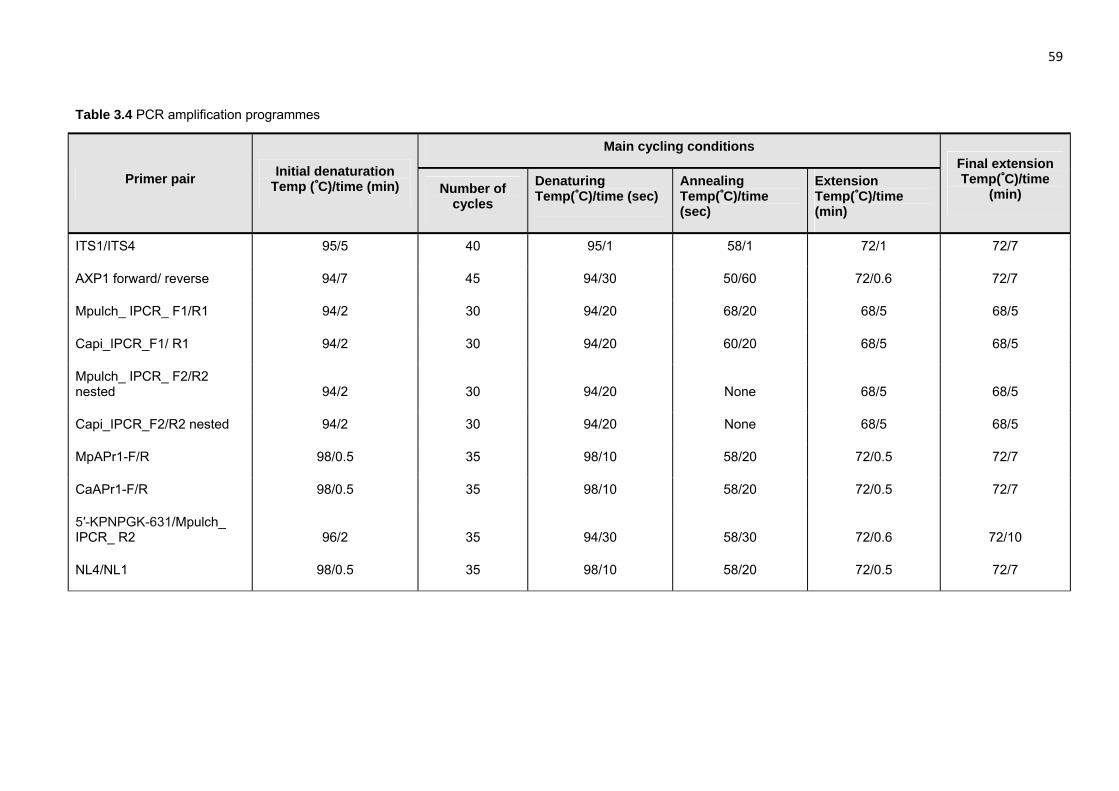
59
Table 3.4 PCR amplification programmes
Primer pair Initial denaturation
Temp (˚C)/time (min)
Main cycling conditions Final extension Temp(˚C)/time
(min) Number of cycles
Denaturing Temp(˚C)/time (sec)
Annealing Temp(˚C)/time (sec)
Extension Temp(˚C)/time (min)
ITS1/ITS4 95/5 40 95/1 58/1 72/1 72/7
AXP1 forward/ reverse 94/7 45 94/30 50/60 72/0.6 72/7
Mpulch_ IPCR_ F1/R1 94/2 30 94/20 68/20 68/5 68/5
Capi_IPCR_F1/ R1 94/2 30 94/20 60/20 68/5 68/5
Mpulch_ IPCR_ F2/R2 nested 94/2 30 94/20 None 68/5 68/5
Capi_IPCR_F2/R2 nested 94/2 30 94/20 None 68/5 68/5
MpAPr1-F/R 98/0.5 35 98/10 58/20 72/0.5 72/7
CaAPr1-F/R 98/0.5 35 98/10 58/20 72/0.5 72/7
5′-KPNPGK-631/Mpulch_ IPCR_ R2 96/2 35 94/30 58/30 72/0.6 72/10
NL4/NL1 98/0.5 35 98/10 58/20 72/0.5 72/7
Stellenbosch University http://scholar.sun.ac.za

60
Table 3.5 Characteristics of the putative gene and amino acid sequences of the aspartic proteases of M.
pulcherrima IWBT Y1123, MpAPr1 and C. apicola IWBT Y1384, CaAPr1
Characteristic IWBT Y1123 IWBT Y1384
Gene length (base pairs) 1137 1101
Protein sequence length (amino acids)
378 367
Theoretical molecular weight (Dalton)
40885.61 39139.06
Theoretical pI 4.22 4.33
Signal peptide Position 1 – 16 None (Non-classical secretion)
Cleavage site Between 16 and 17 (Glycine-
Methionine) None
N-glycosylation sites Position 43 Positions 73, 188, 226
Catalytic motifs Positions 90–102, 257–269 Positions 59-70, 230-241
Fully conserved catalytic regions
93-96 (DTGS), 260-263 (DSGT) 62-65 (DTGS), 233-236 (DSGT)
Active site flap Positions 122 - 133 Positions 97 – 107
Protease family Eukaryotic aspartyl protease (A1
family) EC3.4.23 Eukaryotic aspartyl protease (A1 family) EC3.4.23
Stellenbosch University http://scholar.sun.ac.za

61 Table 3.6 Proteins from the NCBI database showing similarity to the deduced protein sequence of MpAPr1
Accession number
Description Coverage
(%) Identities
(%) Expect value
Maximum score
Gaps
XP_002615916.1 Clavispora lusitaniae ATCC 42720, Eukaryotic aspartyl protease
100 56 8e-127 377 17/395 (4%)
XP_002548135.1 Candida tropicalis MYA-3404, Candidapepsin precursor 97 39 7e-63 213 40/395 (10%)
1J71_A Candida tropicalis, Structure Of Extracellular Aspartic Proteinase
79 41 7e-58 198 29/325 (9%)
XP_002421073.1 Candida dubliniensis CD36, Putative secreted aspartyl protease,
97 36 3e-55 193 43/399 (11%)
XP_711061.1 Candida albicans SC5314, Secretory aspartyl proteinase SAP2p
97 38 6e-55 192 52/405 (13%)
EHA18836.1 Aspergillus niger ATCC 1015, Aspartic protease 97 34 2e-46 170 54/407 (13%)
XP_501603.1 Yarrowia lipolytica CLIB122, Eukaryotic aspartyl protease 85 33 1e-37 146 42/354 (12%)
Stellenbosch University http://scholar.sun.ac.za

62 Table 3.7 Proteins from the NCBI database showing similarity to the deduced protein sequence of CaAPr1
Accession number
Description Coverage
(%) Identities
(%) Expect value
Maximum score
Gaps
XP_500342.1 Yarrowia lipolytica CLIB122, Eukaryotic aspartyl protease 92 37 2e-54 190 34/364 (9%)
XP_499671.1 Yarrowia lipolytica CLIB122, Eukaryotic aspartyl protease 94 37 5e-53 188 36/372 (10%)
XP_002548135.1 Candida tropicalis MYA-3404, Candidapepsin precursor 89 40 5e-53 187 35/351 (10%)
XP_001398592.1 Aspergillus niger CBS 513.88, Aspartic-type endopeptidase opsB
93 36 5e-51 183 44/379 (12%)
XP_720071.1 Candida albicans SC5314, Secretory aspartyl proteinase SAP8p
87 36 3e-43 161 47/348 (14%)
XP_456485.2 Debaryomyces hansenii CBS767, Eukaryotic aspartyl protease 83 34 2e-38 149 74/369 (20%)
EEU06988.1 Saccharomyces cerevisiae JAY291, Yps1p Eukaryotic aspartyl protease
78 37 2e-37 149 41/317 (13%)
Stellenbosch University http://scholar.sun.ac.za

63
Figure 3.1 Alignment of the protein sequences of the extracellular acid protease of Y. lipolytica CLIB122
and those of three other yeasts. Y_lipolyti, Yarrowia lipolytica; C_tropical, Candida tropicalis; C_albicans,
Candida albicans; G_zeae, Gibberella zeae. Boxed sequences indicate the active site regions used to
design degenerate primers. The arrows indicate the aspartic acid residues of the active sites. Residues in
red represent high consensus, residues in blue low consensus and residues in black, neutral. All the
characters in upper case in the consensus sequence are identical with the consensus sequence, and
those in lower case are either the most likely residue or a homologous symbol. The small symbols
represent different homologies according to the Blosum62-12-2 symbol comparison table. The different
symbols that are homologous are: (!) for I and V; ($) for L and M; (%) for F and Y; (#) for B, D, E, N, Q
and Z. The point (.) represents the residue identical to the first sequence at the same position.
1 100Y_lipolyti MQFSLAAVA SFACLALAAP ANPGFLAVPM SRAP-----A PDGQKGISFA AGGGINVEMI NEVMAYNVDIC_tropical MVWVIGLMKQ VFISIVFGLT VEGLAISKLN DRGAAPGLMG NFFDKRSTPG VLSLELYVNR NHDDSNFTIG PHFVVNEYSK RDDYISVELY NEQVTYSANIC_albicans MLLKN ILSVLAFALL IDAAPVKRSP GFVTLDFNVK RSLVDPDDPT VESKRSPLFL DLDPTKIPVD DTGRNDGVDK RGP-VAVKLD NEIITYSADIG_zeae MKPTYST LFLSLLASTE AISLHKREHG LEPRVMSVPI QRRQIDNPLA HD--RKRLNR RAGTVNVGID NEQSLYFLNAConsensus .......... .......... .......... .....la.v. .f........ ..p..l.vp. .r.p.....a pdg....... rgg.!nVe.. NE...Y..#i
101 200Y_lipolyti KVGG--QKVT VQIDTGSSDL WVFSSDSIYC EN----DPSL CTKYGSYNPK KSQNSKDTGE HFDIQYGIGD -ASGNYYKDD ASLGEAKVTD FQFGVSTGDKC_tropical TVGSNSQKQN VIVDTGSSDL WVVDSSANCQ EK-SGYSSDY CFSGGTYDPS SSSTIQELGK SFNIRYGDGS SSSGTWVKDT VGINGAIILN QQFG--DVNSC_albicans TVGSNNQKLS VIVDTGSSDL WIPDSKAICI PKWRGDRGDF CKNNGSYSPA ASSTSKNLNT RFEIKYADGS YAKGNLYQDT VGIGGASVKN QLFA--NVWSG_zeae SIGTPPQNFR LHLDTGSSDL WVNSVNSELC DT----HANI CAESGLYNAN KSSTYEYVNS GFNISYADGS GASGDYVTDT FRMGEVSIKD LQFG--IGYIConsensus .!G...Qk.. v..DTGSSDL W!.ss.si.c e......... C...GsYnp. kSstsk..g. .F#I.YgdGs .asGnyykDt ...gea.!.# .qFg...g..
201 300Y_lipolyti SSGDVSVFGI GPLADEAT-- ---RSHYPNF PALLKEQGVI KKNVYGMAFG LPGDKQNSEI TFGAYNSGRY SGSLKTVPIT TQG----HFG IKCDKAKIGDC_tropical TSVSQGILGI GLDTNEST-- ---DTIYENF PINLKEQGFI NTNAYSLYLN AP-SATSGTI IFGGIDHAKY TGSLTTLPLT SNR----EFT IQTNSATVGTC_albicans TSAHKGILGI GFQANEAT-- ---RTPYDNL PISLKKQGII AKNAYSLFLN SP-EASSGQI IFGGIDKAKY SGSLVELPIT SDR----TLS VGLRSVNVMGG_zeae TSDNEGVIGI GYTSNEAVVD QPDPEFYKNM PARLASDGVI ASNAYSLYLD DL-ESATGKI LFGGVDEQHF IGDLVTVPIM KINDEYSEFY VKLQSINSGSConsensus tS...g!.GI G..a#Eat.. ...r..Y.Nf Pa.Lke#GvI .kNaYs$.l. .p.....g.I .FGg.#...% sGsL.tvPit ........f. !k..sa..g.
301 400Y_lipolyti KTILEGED-- VVLDSGTSLT YLTKSNYDAV INQVQAAGIK LTDAGQGISA LPCSDIGKLS MEYTFSGKTI KVSGKDMTIP ATYLDSSDTS GL-----CIFC_tropical STID--INTG LLLDSGTTLT YLPQSVVDSI ANAI-GGDIT YNRPIG-AYI WSCNRNGKVT YNFP-QGLNI DIPYSDLAVP LYYSNGAVAG F------CALC_albicans RNVN--VNAG VLLDSGTTIS YFTPSIARSI IYAL-GGQVH FDSAGNKAYV ADCKTSGTVD FQFD-KNLKI SVPASEFLYQ LYYTNGKPYP K------CEIG_zeae EIVGEGLDLG VVLDSGSTLT YLPSSVTDSI YQLV-GADYE EGQTTAYVPC DLANQGGNLT FKFT-SPAEI TVPLSELILD FTDITGRQMS FTNGQAACSFConsensus .t!.eg.#.g vvLDSGttlt Ylt.S..ds! in.v.ga.i. ...ag..... ..c...Gkl. ..%t.sg..I .!p.s#...p .ty..g...s .......C.f
401 500Y_lipolyti GITESTGQLS NLNLFGDTFL RAIYSVYDLD NNQVSIAQAA SGQEDKYVEI TGPLPNC_tropical GILYG----E NFNILGDNFL RHAYVVYNLD ALTISLAPVV Y-TSDSNVTI V C_albicans RVRES----E D-NILGDNFM RSAYIVYDLD DKKISMAQVK Y-TSESNIVA IN G_zeae GIAPST---S QVSILGDTFL RSAYVVFDLD NNEISLAQSN FEATGSHILE ISKGKNAVPS ATGSDGPQSS GSENAAGSLS PLGSTGAVSI LAGAMALTFAConsensus g!.est...s #.nilGDtF$ R.aY.V%#LD nn.!S.Aq.. ....ds.!.i .....n.... .......... .......... .......... ..........
501 Y_lipolytiC_tropicalC_albicansG_zeae WFLIConsensus ....
Stellenbosch University http://scholar.sun.ac.za

64
Figure 3.2 PCR amplification with degenerate primers. (A) First round of amplification. (B) Amplification
of the excised bands at approximately 570 bp. Y. lipolytica UOFS-Y1698 used as reference strain (RS).
Arrows indicate excised and sequenced bands. M, molecular weight marker, GeneRulerTM 100bp DNA
ladder (Fermentas).
Stellenbosch University http://scholar.sun.ac.za

65
Figure 3.3 Inverse-PCR amplification. PCR performed on gDNA which was digested with different
restriction enzymes. (A) Amplification with external primers. (B) Amplification with nested primers. Lanes
2, 8, 15, 21 was performed with gDNA digested with EcoRI; Lanes 3, 9, 16, 22 gDNA digested with DraI;
Lanes 4, 10, 17, 23 gDNA digested with EcoRV; Lanes 5, 11, 18, 24 gDNA digested with Hpa I; Lanes 6,
12, 19, 25 gDNA digested with XbaI. Lane 26 is the PCR negative control. Lane 1,7,14, 20 are molecular
weight markers, GeneRulerTM 100bp DNA ladder Plus (Fermentas).
Stellenbosch University http://scholar.sun.ac.za

66
Figure 3.4 PCR amplifications indicating the sizes of the full genes. (-ve) PCR negative control. M is the
molecular weight marker, GeneRulerTM 100bp DNA ladder Plus (Fermentas).
Stellenbosch University http://scholar.sun.ac.za

67 1 ATGCAATTCCTCACTCTTCTTTCTCTTGCAGTCGCTCTCGTCAACGGCATGGCCATCCCTGGGCTTGACACA 72
1 M Q F L T L L S L A V A L V N G‡ M A I P G L D T 24
73 CGCGACGAGGCGGCGCCTTTGCAGCTTGACTTCACCGTGCTGAAAACAGTTGGCAACACGACGGCAAAGGAG 144
25 R D E A A P L Q L D F T V L K T V G N T T A K E 48
145 TTCTGGGCGAAATACGGATCAAAAAATAAGAAGCGTGACGCGTACCCGGAAGTGATCACGGACTATCGCGAC 216
49 F W A K Y G S K N K K R D A Y P E V I T D Y R D 72
217 TTGAGCTACCAGATCGATGTGTACTTGGGCGCAGATAAACAGAAGAATACCGTTTCCTTGGATACGGGCTCG 288
73 L S Y Q I D V Y L G A D K Q K N T V S L D T G S 96
289 TCGGATCTTTGGGTGCCCAGCAGCGGCTATAGCCCAGACAGTTCCAGCTCTGCACAGGACACCGGTGAAGCA 360
97 S D L W V P S S G Y S P D S S S S A Q D T G E A 120
361 TTCAAAATTGGATATTTGGATGGAAGCGGTGCACTGGGTGAATATTACAAGGACAAGTTCCAGTTCAGCACA 432
121 F K I G Y L D G S G A L G E Y Y K D K F Q F S T 144
433 GCCAAGCCAGTGCTTTCCAATTTCCAGTTTGCGCAGACTAGCGACGAGGCGGGCATGGGGATTCTAGGCATT 504
145 A K P V L S N F Q F A Q T S D E A G M G I L G I 168
505 GCCGACCGGAACCAGGAAGCGTCCGATTCGGTATATGATAATCTCCCTTGGGCATTGCAAAAAGCCGGAATC 576
169 A D R N Q E A S D S V Y D N L P W A L Q K A G I 192
577 ACACCCAAGGCGTCATACTCGTTATTCTTGGGCCCAGATCTCGGCAAGGGGTCGTTGATTTTCGGCGGCATT 648
193 T P K A S Y S L F L G P D L G K G S L I F G G I 216
649 GACACAGACAAGTATACTGGAGAGTTGCAGCTGTACCCGATTGACCTGTCCAGCGGTGGCTTGGCCGTCGAT 720
217 D T D K Y T G E L Q L Y P I D L S S G G L A V D 240
721 GTGCAGTCAGTGAATTTCAACGGGAAAATCATCTCGGTGAACGCCCCAGTTCTTTTGGACTCTGGAACCTCT 792
241 V Q S V N F N G K I I S V N A P V L L D S G T S 264
793 TTGGGGCTTCTCAGCCTGGACCTTATTGAGGAGTTGGACACGATATTCGACTCTCAAACTGTGAAACAGGGT 864
265 L G L L S L D L I E E L D T I F D S Q T V K Q G 288
865 GGGATCGAGTACAAAATTGTCAGCTGTGACCAGCCTTCAGACAAGAGCCTCGACTTTGATTTTGGCGACAAC 936
289 G I E Y K I V S C D Q P S D K S L D F D F G D N 312
937 ACGATCTCAATTCCGTTTTCTGAGGCCATTATCAAGCAAGACGACGGCACGTGTTTGTTAGGGTTCGGATAC 1008
313 T I S I P F S E A I I K Q D D G T C L L G F G Y 336
1009 TATAATGACATTCAGATCTTCGGCGACGTGTTTTTGAGACAGGCATACGTGTACTACGATCTCACCGATAAG 1080
337 Y N D I Q I F G D V F L R Q A Y V Y Y D L T D K 360
1081 ACGATTTCGCTTGCACAGGCGTCTTATTCTAGCTCGTCAAACATCATAAGTGCTTAA 1137
361 T I S L A Q A S Y S S S S N I I S A * 378
Figure 3.5 The DNA and amino acid sequences of M. pulcherrima IWBT Y1123 putative secreted
aspartic protease. The putative secretion signal peptide is underlined. The cleavage site is indicated by
(‡). The active site amino acid sequences and active site flap amino acid sequence are shaded in yellow
and turquoise, respectively. The putative N-glycosylation site is boxed.
Stellenbosch University http://scholar.sun.ac.za

68
Figure 3.6 The conserved domains within the putative aspartic protease of M. pulcherrima IWBT Y1123
analysed in this study, indicating that the protein is a secreted aspartic protease-like (SAP-like) enzyme.
Stellenbosch University http://scholar.sun.ac.za
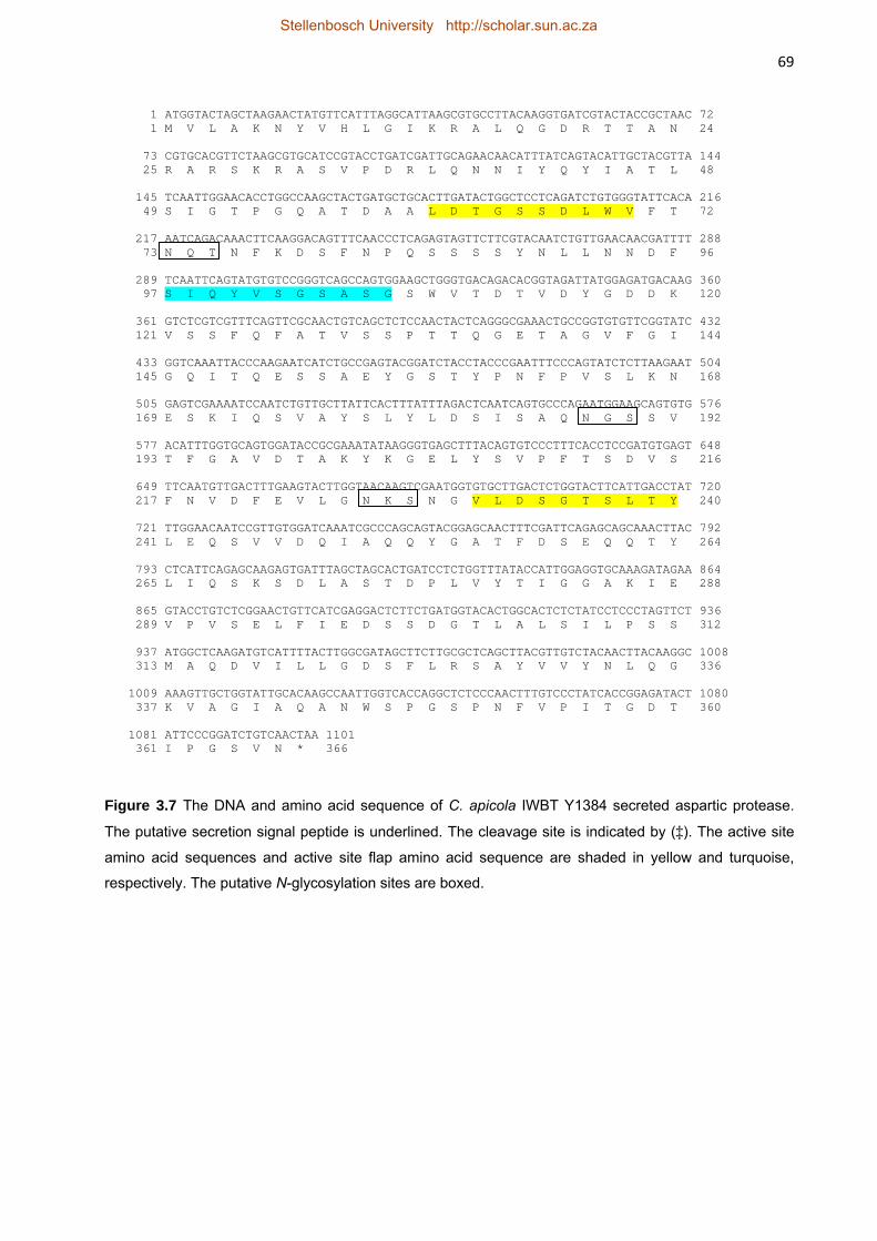
69
Figure 3.7 The DNA and amino acid sequence of C. apicola IWBT Y1384 secreted aspartic protease.
The putative secretion signal peptide is underlined. The cleavage site is indicated by (‡). The active site
amino acid sequences and active site flap amino acid sequence are shaded in yellow and turquoise,
respectively. The putative N-glycosylation sites are boxed.
1 ATGGTACTAGCTAAGAACTATGTTCATTTAGGCATTAAGCGTGCCTTACAAGGTGATCGTACTACCGCTAAC 72 1 M V L A K N Y V H L G I K R A L Q G D R T T A N 24
73 CGTGCACGTTCTAAGCGTGCATCCGTACCTGATCGATTGCAGAACAACATTTATCAGTACATTGCTACGTTA 144 25 R A R S K R A S V P D R L Q N N I Y Q Y I A T L 48
145 TCAATTGGAACACCTGGCCAAGCTACTGATGCTGCACTTGATACTGGCTCCTCAGATCTGTGGGTATTCACA 216 49 S I G T P G Q A T D A A L D T G S S D L W V F T 72
217 AATCAGACAAACTTCAAGGACAGTTTCAACCCTCAGAGTAGTTCTTCGTACAATCTGTTGAACAACGATTTT 288 73 N Q T N F K D S F N P Q S S S S Y N L L N N D F 96
289 TCAATTCAGTATGTGTCCGGGTCAGCCAGTGGAAGCTGGGTGACAGACACGGTAGATTATGGAGATGACAAG 360 97 S I Q Y V S G S A S G S W V T D T V D Y G D D K 120
361 GTCTCGTCGTTTCAGTTCGCAACTGTCAGCTCTCCAACTACTCAGGGCGAAACTGCCGGTGTGTTCGGTATC 432 121 V S S F Q F A T V S S P T T Q G E T A G V F G I 144
433 GGTCAAATTACCCAAGAATCATCTGCCGAGTACGGATCTACCTACCCGAATTTCCCAGTATCTCTTAAGAAT 504 145 G Q I T Q E S S A E Y G S T Y P N F P V S L K N 168
505 GAGTCGAAAATCCAATCTGTTGCTTATTCACTTTATTTAGACTCAATCAGTGCCCAGAATGGAAGCAGTGTG 576 169 E S K I Q S V A Y S L Y L D S I S A Q N G S S V 192
577 ACATTTGGTGCAGTGGATACCGCGAAATATAAGGGTGAGCTTTACAGTGTCCCTTTCACCTCCGATGTGAGT 648 193 T F G A V D T A K Y K G E L Y S V P F T S D V S 216
649 TTCAATGTTGACTTTGAAGTACTTGGTAACAAGTCGAATGGTGTGCTTGACTCTGGTACTTCATTGACCTAT 720 217 F N V D F E V L G N K S N G V L D S G T S L T Y 240
721 TTGGAACAATCCGTTGTGGATCAAATCGCCCAGCAGTACGGAGCAACTTTCGATTCAGAGCAGCAAACTTAC 792 241 L E Q S V V D Q I A Q Q Y G A T F D S E Q Q T Y 264
793 CTCATTCAGAGCAAGAGTGATTTAGCTAGCACTGATCCTCTGGTTTATACCATTGGAGGTGCAAAGATAGAA 864 265 L I Q S K S D L A S T D P L V Y T I G G A K I E 288
865 GTACCTGTCTCGGAACTGTTCATCGAGGACTCTTCTGATGGTACACTGGCACTCTCTATCCTCCCTAGTTCT 936 289 V P V S E L F I E D S S D G T L A L S I L P S S 312
937 ATGGCTCAAGATGTCATTTTACTTGGCGATAGCTTCTTGCGCTCAGCTTACGTTGTCTACAACTTACAAGGC 1008 313 M A Q D V I L L G D S F L R S A Y V V Y N L Q G 336
1009 AAAGTTGCTGGTATTGCACAAGCCAATTGGTCACCAGGCTCTCCCAACTTTGTCCCTATCACCGGAGATACT 1080 337 K V A G I A Q A N W S P G S P N F V P I T G D T 360
1081 ATTCCCGGATCTGTCAACTAA 1101 361 I P G S V N * 366
Stellenbosch University http://scholar.sun.ac.za

70
Figure 3.8 The conserved domains within the putative aspartic protease of C. apicola IWBT Y1384
analysed in this study, indicating that the protein is a secreted aspartic protease-like (SAP-like) enzyme.
Stellenbosch University http://scholar.sun.ac.za

71
Figure 3.9 Phylogenetic tree of the deduced protein sequences of MpAPr1 of M. pulcherrima IWBT Y1123 and CaAPr1 of C. apicola IWBT Y1384 with aspartic
proteases from other yeasts found on the NCBI website. The phylogenetic tree was constructed by using the neighbour joining method (PHYLIP 3.56). The scale bar
represents the number of base substitutions per site.
Stellenbosch University http://scholar.sun.ac.za
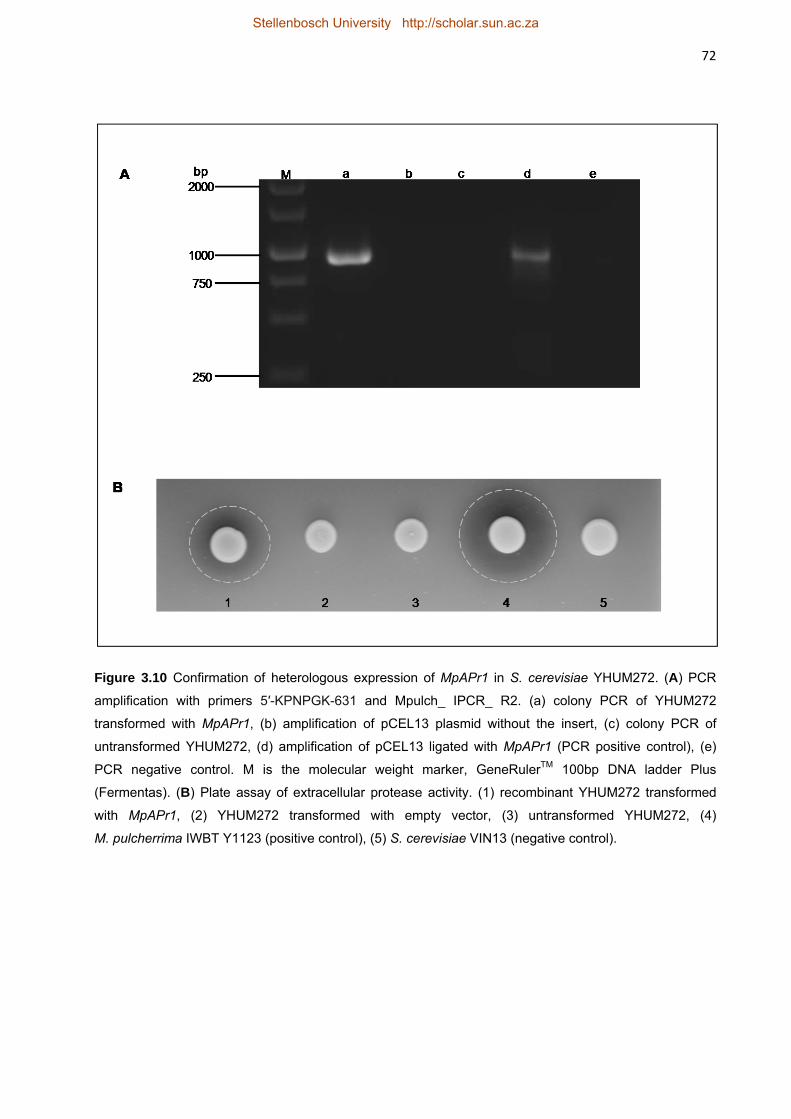
72
Figure 3.10 Confirmation of heterologous expression of MpAPr1 in S. cerevisiae YHUM272. (A) PCR
amplification with primers 5′-KPNPGK-631 and Mpulch_ IPCR_ R2. (a) colony PCR of YHUM272
transformed with MpAPr1, (b) amplification of pCEL13 plasmid without the insert, (c) colony PCR of
untransformed YHUM272, (d) amplification of pCEL13 ligated with MpAPr1 (PCR positive control), (e)
PCR negative control. M is the molecular weight marker, GeneRulerTM 100bp DNA ladder Plus
(Fermentas). (B) Plate assay of extracellular protease activity. (1) recombinant YHUM272 transformed
with MpAPr1, (2) YHUM272 transformed with empty vector, (3) untransformed YHUM272, (4)
M. pulcherrima IWBT Y1123 (positive control), (5) S. cerevisiae VIN13 (negative control).
Stellenbosch University http://scholar.sun.ac.za
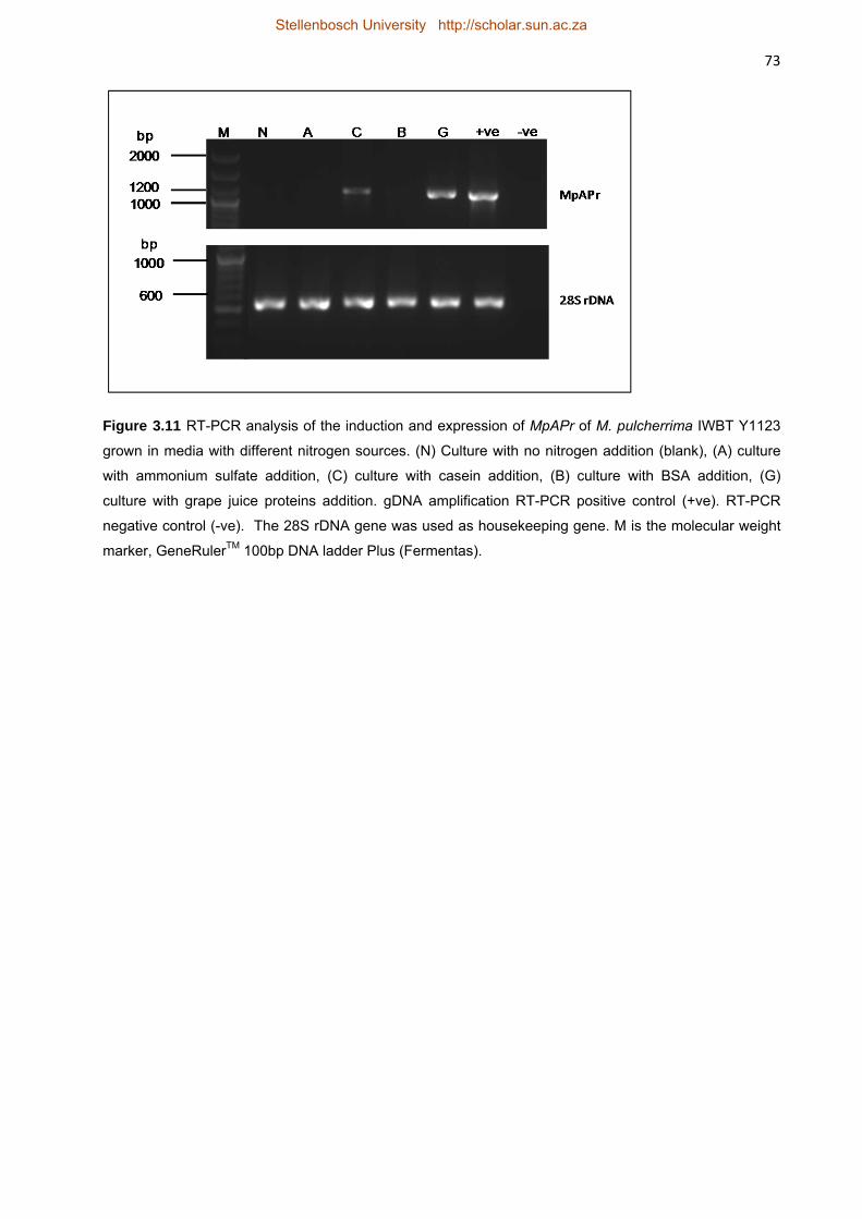
73
Figure 3.11 RT-PCR analysis of the induction and expression of MpAPr of M. pulcherrima IWBT Y1123
grown in media with different nitrogen sources. (N) Culture with no nitrogen addition (blank), (A) culture
with ammonium sulfate addition, (C) culture with casein addition, (B) culture with BSA addition, (G)
culture with grape juice proteins addition. gDNA amplification RT-PCR positive control (+ve). RT-PCR
negative control (-ve). The 28S rDNA gene was used as housekeeping gene. M is the molecular weight
marker, GeneRulerTM 100bp DNA ladder Plus (Fermentas).
Stellenbosch University http://scholar.sun.ac.za

74
Figure 3.12 Assessment of extracellular proteins of M. pulcherrima IWBT Y1123 grown in the presence
of casein (A) SDS-PAGE analysis. Lanes: (a) casein without culture supernatant, (b) culture supernatant
with casein, (c) casein after 3 day incubation without culture supernatant. (B) Zymograms. (d) Casein
without culture supernatant, (e) Culture supernatant with casein, (f) casein after 3 day incubation without
culture supernatant. MW is the molecular weight standard proteins, PageRulerTM Prestained Ladder (Bio-
Rad). The arrow points to the band excised and sequenced. (Estimated according to theoretical MW is
39.2 kDa).
Stellenbosch University http://scholar.sun.ac.za

75
Figure 3.13 Analysis of extracellular proteins of M. pulcherrima IWBT Y1123 cultured in the presence of
BSA. (A) SDS-PAGE analysis. Lanes: (a) BSA without culture supernatant, (b) culture supernatant with
BSA, (c) BSA after 3 day incubation without culture supernatant, (d) no nitrogen blank sample. (B)
Zymogram. (e) BSA without culture supernatant, (f) culture supernatant with BSA, (g) BSA after 3 days
incubation without culture supernatant, (h) no nitrogen (blank) sample. MW is the molecular weight
standard proteins, PageRulerTM Prestained Ladder (Bio-Rad).
Stellenbosch University http://scholar.sun.ac.za

76
Figure 3.14 Analysis of the extracellular proteins of M. pulcherrima IWBT Y1123 cultured on grape juice
proteins and ammonium sulphate. (A) SDS-PAGE analysis. Lanes: (a) grape juice proteins without
culture supernatant, (b) culture supernatant on grape juice proteins-induced, (c) culture supernatant with
ammonium sulphate-induced, (d) no nitrogen (blank) sample. (B) Zymogram. (e) grape juice proteins
culture supernatant, (f) culture supernatant with ammonium sulphate-induced, (g) no nitrogen (blank)
sample. MW is the molecular weight standard proteins, PageRulerTM Prestained Ladder (Bio-Rad). The
arrow points to the band excised and sequenced. (Estimated according to theoretical MW is 39.2 kDa).
Stellenbosch University http://scholar.sun.ac.za

77
Figure 3.15 Identification of 12 M. pulcherrima strains by ITS and ITS-RFLP. (A) PCR amplification of the
ITS regions of M. pulcherrima strains. M, molecular weight marker, GeneRulerTM 100bp DNA ladder
(Fermentas). (B) Digestion of the ITS amplifications with restriction endonuclease CfoI. (C) Digestion of
the ITS amplifications with restriction endonuclease HaeIII. (+ve) PCR positive control in this case strain
M. pulcherrima IWBT Y1123, (-ve) PCR negative control. M is the molecular weight marker, FastRulerTM
Low Range DNA Ladder (Fermentas).
Stellenbosch University http://scholar.sun.ac.za

78
Figure 3.16 Protease activity screening of M. pulcherrima strains. (A) PCR amplifications with primers
MpAPr1-F/MpAPr1-R showing the presence of the MpAPr1 protease-encoding gene. M is the molecular
weight marker, GeneRulerTM 100bp DNA ladder Plus (Fermentas). (B) An example of a plate assay
showing the different degrees of activity with a positive result indicated by a clear halo around the colony;
(+) low activity, diameter of the halo from the edge of the colony 2mm, (++) medium activity, diameter of
the halo from the edge of the colony 3 mm, (+++) high activity, diameter of the halo from the edge of the
colony 5 mm, (-) no activity. Strain M. pulcherrima IWBT Y1123 was used as positive control (displaying
high activity) and S. cerevisiae VIN13 as negative control (displaying no activity). Strains IWBT Y1072
and IWBT Y1063 have low activity while strains IWBT Y1065 and IWBT Y1094 show medium activity.
Stellenbosch University http://scholar.sun.ac.za

79
Chapter 4
General discussion and conclusions
Stellenbosch University http://scholar.sun.ac.za
80
4. General discussion and conclusions
4.1 Results and general discussion
The enzymes of the aspartic endoprotease family are defined by the presence of two aspartic
acid residues in their catalytic sites that are critical for the functioning of the enzymes. These
enzymes function under acidic conditions. Literature reports that some non-Saccharomyces
yeasts of oenological origin have extracellular proteolytic activity under acidic conditions, e.g.
Candida stellata, Kloeckera apiculata and Candida pulcherrima (Charoenchai et al., 1997).
These poorly characterised enzymes could potentially fulfil a number of roles in wine production
such as the reduction of protein haze in white wines (Lagace and Bisson, 1990; Dizy and
Bisson, 2000). The amino acids and peptides liberated from the action of the proteases could
be used as sources of nitrogen by the fermentative microorganisms. Some amino acids can be
metabolised by yeasts to form aromatic compounds such as higher alcohols (Swiegers and
Pretorius, 2005), thus the proteolytic activity could have an indirect impact on the aroma profile
of the wine. The main aim of the study was to isolate and characterize extracellular aspartic
proteases at a genetic level from non-Saccharomyces yeasts that are of oenological origin.
The genes encoding extracellular aspartic proteases from two non-Saccharomyces yeasts were
isolated and characterized by in silico analysis. The yeasts were Metschnikowia pulcherrima
IWBT Y1123 and Candida apicola IWBT Y1384. These yeasts were isolated from grape must
and showed strong proteolytic activity during an enzymatic screening of 308 other yeasts.
The genes were retrieved by following a PCR approach using degenerate primers and inverse-
PCR. The gene from M. pulcherrima IWBT Y1123 was named MpAPr1. The gene is 1137 bp
long. The mature form of the protein encoded by the gene consists of 362 amino acids with a
predicted Mr of 39.2 kDa and one possible N-glycosylation site. The gene also encodes a
putative 16 amino acid signal peptide. The putative gene from C. apicola IWBT Y1384 was
named CaAPr1, is 1101 bp long and encodes a putative protein with Mr 39.1 kDa. This enzyme
had no predictable signal peptide and is thought to follow a non-classical secretion pathway and
had 3 possible N-glycosylation sites. The predicted pI values of the putative proteins were 4.22
for MpAPr1 and 4.33 for CaAPr1, respectively. These characteristics correspond to previously
published data on the extracellular proteases of yeasts. The AXP gene of Yarrowia lipolytica
148 encodes a mature acid protease of 353 amino acids with a predicted Mr of 39.2 kDa. The
gene also encodes a putative pre-peptide that is 17 amino acids long (Young et al., 1996).
Cryptococcus sp. S-2 possesses a CAP1 gene with an open reading frame of 1254 bp encoding
a protein of 417 amino acids long. The mature protein had an estimated Mr of 34 kDa on SDS-
PAGE (Rao et al., 2011).
Stellenbosch University http://scholar.sun.ac.za
81 From homology searches of the putative gene sequences, it was confirmed that the sequences
encoding the conserved active sites are indeed highly conserved throughout different species
(Davies, 1990; Dunn, 2002). Both putative protein sequences, MpAPr1 and CaAPr1, showed
homology to only fungal aspartic proteases with average identity scores of less than 40%,
indicating the novelty of the putative proteins. The CaAPr1 protein showed close homology to
the aspartic proteases of Y. lipolytica, which appears to follow a translation pathway other than
that described for Candida spp. (McEwen and Young, 1998; Naglik et al., 2004). CaAPr1 and
the acid proteases from Y. lipolytica may have similar processing pathways.
The putative MpAPr1 gene was cloned and expressed in Saccharomyces cerevisiae YHUM272.
Activity plate assays confirmed that MpAPr1 indeed codes for an extracellular aspartic protease.
The recombinant strain demonstrated weaker activity than the native host. A few explanations
may exist for this occurrence: differences in promoters could affect expression, metabolic
differences between the native and the recombinant strain and the signal peptide of the protein
may not be systematically recognized by the S. cerevisiae.
M. pulcherrima IWBT Y1123 was grown in media containing different nitrogen sources which
included casein, BSA, grape juice proteins and ammonium sulphate. Reverse transcription-PCR
(RT-PCR) confirmed that the expression of MpAPr1 in IWBT Y1123 was induced when casein,
grape juice proteins and BSA were used as sole nitrogen sources. The gene was not expressed
in the presence of ammonium sulphate. This result was expected since the secretion of
proteases in yeasts is induced when protein is the only available nitrogen source (Banerjee et
al., 1991; Dabas and Morschhäuser, 2008). Expression of the gene was the highest when grape
proteins were the sole nitrogen sources, as demonstrated by RT-PCR. The accurate profiling of
the MpAPr1 gene expression by qRT-PCR in the presence different nitrogen sources will shed
some light on the effect these nitrogen sources have on the growth of the yeast. Protease
activity in the presence of grape juice proteins was also confirmed by zymography, but not with
casein or BSA. A possible explanation for this could be that the concentration of the protease in
the gel was too low for activity to be detected, which correlates to the lower expression of the
gene in the presence of casein and BSA compared to the expression induced by the grape juice
proteins. Protein bands corresponding to the expected size of the putative protein was excised
from SDS-PAGE gels and analysed by nano-LC-MS/MS. The bands from the casein-induced
culture and the grape protein-induced culture had 21.3% and 20% identity with the putative
MpAPr1 protein sequence.
The presence of MpAPr1 was confirmed in 12 other M. pulcherrima strains by PCR. Plate
assays revealed that the strains did not have equal activity, as indicated by the size of the halo
surrounding the colonies. No correlation could be made between putative protein sequences
and the level of activity displayed on plates. Investigating the sequences upstream and
Stellenbosch University http://scholar.sun.ac.za

82 downstream of the gene-encoding sequences may provide some insight into the differences in
expression.
4.2 Conclusions and future prospects
The aim to isolate and characterize aspartic proteases encoding genes from non-
Saccharomyces yeasts isolated from grape must, as set out in Chapter 1, has been achieved.
The results obtained correspond well to existing data regarding aspartic proteases secreted by
yeasts (Rao et al., 1998). Some of the grape juice proteins have been hydrolysed when cultured
with M. pulcherrima IWBT Y1123. This illustrates the potential applicability of this yeast and /or
its secreted proteases. However, further investigations are needed to test whether the protease
is able to reduce haze formation and also whether the proteolytic activity will impact on the
assimilable nitrogen content and aroma profile of the wine. Some wine proteins are indeed
known to be insensitive to proteolytic attack due to their globular structure (Waters et al., 1992).
It has been suggested that partially ‘opening’ up the protein structure will expose peptide bonds
to be hydrolysed by proteases. One such proposal was suggested by Pocock and co-workers
(2006), who demonstrated that heat treatment together with proteolysis may reduce protein
haze.
This study has extended our understanding of how some yeasts may survive and even flourish
in wine which is characterized by low pH, low temperatures and the presence of a number of
inhibitors such as ethanol and polyphenols. Extracellular proteolytic activity that is adapted to
the acidic environment may aid in nutrient supply for the yeasts.
Future prospects should include purification of the enzymes, MpAPr1 and CaAPr1, e.g. by
anion-exchange chromatography and gel filtration, and biochemical characterization. This
should include determination of the activity range with regard to pH and temperature. The effect
of different potential inhibitors on the activity of the enzymes should also be tested, e.g. ethanol,
sulphur dioxide and polyphenols. The activity of the enzymes should also be tested in wine to
determine its applicability in wine biotechnology.
Mixed fermentations of M. pulcherrima IWBT Y1123 with S. cerevisiae may also be envisaged
to determine its influence on protein stability, wine aroma, and the interaction between the two
yeast species. By releasing proteases able to breakdown proteins present in grape must,
M. pulcherrima may advantageously affect the fermentation kinetics by increasing the available
assimilable nitrogen content for the growth of S. cerevisiae during alcoholic fermentation.
Sequential fermentations of M. pulcherrima species with S. cerevisiae have shown positive
results with regards to an increase in fruity and floral aroma in wine (Jolly et al., 2003;
Rodrίguez et al., 2010). The wines produced from sequential fermentations had higher
concentrations of aromatic compounds such as higher alcohols, esters and terpenols compared
Stellenbosch University http://scholar.sun.ac.za

83 to fermentations where only S. cerevisiae was inoculated in the grape must. Commercial
cultures of combined S. cerevisiae and non-Saccharomyces yeasts are already available on the
market. Such an example is of S. cerevisiae with Torulaspora delbrueckii and Kluyveromyces
thermotolerans from Viniflora® HARMONY.nsac. This combined culture is supposed to produce
a wine with a richer and rounder flavour with enhanced fruity aroma notes (Jolly et al., 2006).
Further investigations are required to determine the potential commercial value of
M. pulcherrima.
4.3 References
Banerjee, A., Ganesan, K., Datta, A., 1991. Induction of secretory acid proteinase in Candida albicans. Journal of General Microbiology 137, 2455-2461. Charoenchai, C., Fleet, G.H., Henschke, P.A., Todd, B.E.N.T., 1997. Screening of non-Saccharomyces wine yeasts for the presence of extracellular hydrolytic enzymes. Australian Journal of Grape and Wine Research 3, 2-8. Dabas, N., Morschhäuser, J., 2008. A transcription factor regulatory cascade controls secreted aspartic protease expression in Candida albicans. Molecular Microbiology 69, 586-602. Davies, D.R., 1990. The structure and function of aspartic proteases. Annual Reviews of Biophysics and Biophysical Chemistry 19, 189-215. Dizy, M. and Bisson, L.F. 2000. Proteolytic activity of yeast stains during grape juice fermentation. American Journal of Enology and Viticulture 51, 155–167. Dunn, B.M., 2002. Structure and mechanism of the pepsin-like family of aspartic peptidases. Chemical Reviews 102, 4431-4458. Jolly, N.P., Augustyn, O.P.H., Pretorius, I.S., 2003. The use of Candida pulcherrima in combination with Saccharomyces cerevisiae for the production of Chenin blanc wine. South African Journal of Enology and Viticulture 24, 63-69. Jolly, N.P., Augustyn, O.P.H., Pretorius, I.S., 2006. The role and use of non-Saccharomyces yeasts in wine production. South African Journal of Enology and Viticulture 27, 15–38. Lagace, L.S., Bisson, L.F., 1990. Survey of yeast acid proteases for effectiveness of wine haze reduction. American Journal of Enology and Viticulture 41, 147-155. McEwen, R.K., Young, T.W., 1998. Secretion and pH-dependent self-processing of the pro-form of the Yarrowia lipolytica acid extracellular protease. Yeast, 14, 1115-1125. Naglik, J., Albrecht, A., Bader, O., Hube, B., 2004. Candida albicans proteinases and host/pathogen interactions. Cellular microbiology 6, 915-926. Pocock, K.F., Høj, P.B., Adams, K.S., Kwiatkowski, M.J., Waters, E.J., 2003. Combined heat and proteolytic enzyme treatment of white wines reduces haze forming protein content without detrimental effect. Australian Journal of Grape and Wine Research 9, 56-63. Rao, M.B., Tanksale, A.M., Ghatge, M.S., Deshpande, V.V., 1998. Molecular and biotechnological aspects of microbial proteases. Microbiology and Molecular Biology Reviews 62, 597-635. Rao S, Mizutani O, Hirano T, Masaki K, Iefuji H., 2011. Purification and characterization of a novel aspartic protease from basidiomycetous yeast Cryptococcus sp. S-2. Journal of Bioscience and Bioengineering 112, 441-446.
Stellenbosch University http://scholar.sun.ac.za

84 Rodríguez, M.E., Lopes, C.A., Barbagelata, R.J., Barda, N.B., Caballero, A.C., 2010. Influence of Candida pulcherrima Patagonian strain on alcoholic fermentation behaviour and wine aroma. International Journal of Food Microbiology 138, 19-25. Swiegers, J.H., Pretorius, I.S., 2005. Yeast modulation of wine flavour. Advances in Applied Microbiology. 57, 131-175. Young, T.W., Wadeson, A., Glover, D.J., Quincey, R.V., Butlin, M.J., Kamei, E.A., 1996. The extracellular acid protease gene of Yarrowia lipolytica: sequence and pH-regulated transcription. Microbiology 142, 2913-2921. Waters, E.J., Wallace, W., Williams, P.J., 1992. Identification of heat–unstable wine proteins and their resistance to peptidases. Journal of Agricultural and Food Chemistry 40, 1514-1519.
Stellenbosch University http://scholar.sun.ac.za