extensor mechanism - neoligaments · the range of motion and tissue tension is assessed. if ... •...
TRANSCRIPT
Extensor Mechanism
Surgical Technique Manual
Quadriceps Tendon ReconstructionPatellar Tendon ReconstructionPost Patellectomy Reconstruction
0086

The QuadsTape SystemTM and the PatellarTape SystemTM each comprise a Poly-Tape implant with associated instrumentation to repair a ruptured quadriceps or patellar tendon. Both the QuadsTape System and the PatellarTape System include:• a 30 mm wide by 800 mm long Poly-Tape implant
with an open weave mesh structure;• a 20 cm rigid probe and a 20cm malleable probe,
either of which may be used at the discretion of the surgeon.
In addition, the PatellarTape System includes a 4.5 mm diameter drill bit with a plain shank.
QuadsTape and PatellarTape Systems
Introduction
We would like to thank Mr. A. D. Toms, Consultant Orthopaedic Surgeon, Royal Devon and Exeter Hospital, Exeter, UK, and Mr. S. H. White, Consultant Orthopaedic Surgeon, Robert Jones and Agnes Hunt Orthopaedic Hospital, Shropshire, UK, for their work in developing this technique1.
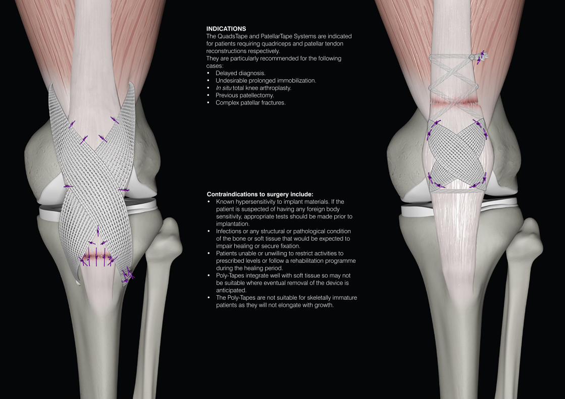
INDICATIONSThe QuadsTape and PatellarTape Systems are indicated for patients requiring quadriceps and patellar tendon reconstructions respectively.They are particularly recommended for the following cases:• Delayed diagnosis.• Undesirable prolonged immobilization.• In situ total knee arthroplasty.• Previous patellectomy.• Complex patellar fractures.
Contraindications to surgery include:• Known hypersensitivity to implant materials. If the
patient is suspected of having any foreign body sensitivity, appropriate tests should be made prior to implantation.
• Infections or any structural or pathological condition of the bone or soft tissue that would be expected to impair healing or secure fixation.
• Patients unable or unwilling to restrict activities to prescribed levels or follow a rehabilitation programme during the healing period.
• Poly-Tapes integrate well with soft tissue so may not be suitable where eventual removal of the device is anticipated.
• The Poly-Tapes are not suitable for skeletally immature patients as they will not elongate with growth.
Product Overview
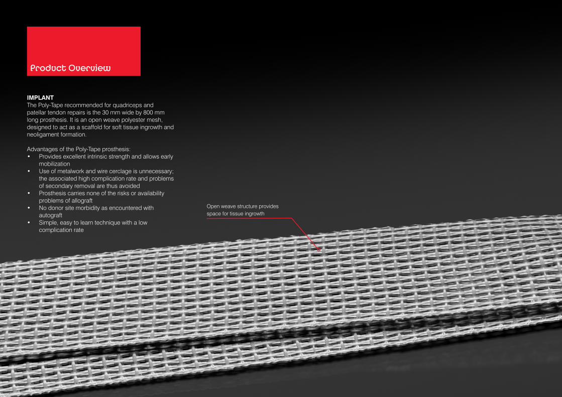
Open weave structure provides space for tissue ingrowth
IMPLANTThe Poly-Tape recommended for quadriceps and patellar tendon repairs is the 30 mm wide by 800 mm long prosthesis. It is an open weave polyester mesh, designed to act as a scaffold for soft tissue ingrowth and neoligament formation.
Advantages of the Poly-Tape prosthesis:• Provides excellent intrinsic strength and allows early
mobilization• Use of metalwork and wire cerclage is unnecessary;
the associated high complication rate and problems of secondary removal are thus avoided
• Prosthesis carries none of the risks or availability problems of allograft
• No donor site morbidity as encountered with autograft
• Simple, easy to learn technique with a low complication rate

Parallel fibres provide high strength
INSTRUMENTATIONThe following single use instruments are packaged with the QuadsTape implant set:• 20 cm malleable probe and rigid probe with eye for
passing the Poly-Tape through soft tissue.
The following single use instruments are packaged with the PatellarTape implant set:• 20 cm malleable probe and rigid probe with eye for
passing the Poly-Tape through soft tissue.• 4.5 mm diameter drill bit (plain shank)

PREPARATION AND INSPECTIONThe patient is positioned supine with the knee supported appropriately. A side support and sandbag are useful to facilitate knee positioning. The leg is prepared and draped using aseptic technique. Broad spectrum antibiotic prophylaxis is administered intravenously.
RECOMMENDED APPROACHA midline approach is recommended. Adequate exposure is essential and should provide sufficient access for the proximal and distal placement of the prosthesis. The ends of the tendon or ligament are identified as well as any additional pathology.
NOTE: It is necessary particularly in chronic cases to free up any adhesions involving the quadriceps mechanism. This will facilitate optimal postoperative rehabilitation.
The Poly-Tape is threaded through the eyelet of the selected probe. The probe is used to pass the Poly-Tape transversely through the proximal end of the patellar tendon, just distal to the patella. In order to avoid abrasion of the prosthesis it should be passed through the mid substance of the patellar tendon.
Both ends are then taken proximally crossing anterior to the patella, ensuring it is laid flat.
The ends of the ruptured tendon are approximated.
The probe is used to pass the Poly-Tape through the distal end of the quadriceps tendon.
The tension is carefully adjusted to remove any slackness from the Poly-Tape where it enters and exits the tissue.
Small tacking sutures are used to stabilize the Poly-Tape at soft tissue entry and exit points.
QuadsTape System
1 2

The probe is used to weave the Poly-Tape through the proximal end of the quadriceps tendon and musculature in a Bunnell fashion.
The tension is carefully adjusted to draw the quadriceps mechanism together and remove any slackness from the Poly-Tape where it enters and exits the tissue in the proximal tendon stump.
The range of motion and tissue tension is assessed. If this is satisfactory, the proximal ends of the Poly-Tape are knotted. Each surplus end of the Poly-Tape is cut with scissors at right angles to its length. This will minimize the generation of loose fibres. A short tail is left when cutting each end.
IMPORTANT:• Any loose fibres created when trimming the Poly-Tape
to length must be carefully removed from the incision site
• After trimming to length it may be necessary to restrain the cut ends by stitching them back to the Poly-Tape
A fibrous tissue envelope is recruited from surrounding tissue and closed over the prosthesis with small tacking sutures. This is vital to ensure the knot is covered with, and remains buried in, tissue. This also encourages fibrous ingrowth, reduces abrasion to the graft, and distances the prosthesis from the superficial wound.
WOUND CLOSUREThe wound is irrigated and a vacuum drain is put in place. Haemostasis is achieved. The dead space is closed with absorbable sutures before skin closure with a subcuticular, absorbable suture. The wound is covered with a suitable dressing.
3 4 5

PREPARATION AND INSPECTIONThe patient is positioned supine. Broad spectrum antibiotic prophylaxis is administered intravenously. A side support and sandbag are useful to facilitate knee positioning. The leg is prepared and draped using aseptic technique.
RECOMMENDED APPROACHA midline approach is recommended. Adequate exposure is essential and should provide sufficient access for the proximal and distal placement of the prosthesis. The ends of the tendon or ligament are identified as well as any additional pathology.
NOTE: It is necessary particularly in chronic cases to free up any adhesions involving the quadriceps mechanism. This will facilitate optimal postoperative rehabilitation.
A transverse bone tunnel is positioned at the level of the tibial tuberosity. A 4.5 mm diameter drill bit is used to make the tunnel from the lateral side to the medial.
NOTE: Where possible, round the tunnel edges to prevent abrasion of the Poly-Tape.
The Poly-Tape is threaded through the eyelet of the selected probe. The probe is used to pass the Poly-Tape transversely through the distal quadriceps tendon at its patellar insertion.
The Poly-Tape is brought distally, crossing over itself on the anterior aspect of the patella. The medial end of the Poly-Tape is passed through the bone tunnel.
The ends of the Poly-Tape are pulled tight with the knee in 20º of flexion. The tape is then secured with stay sutures to allow the range of motion and tissue tension to be assessed.
PatellarTape System
1 2
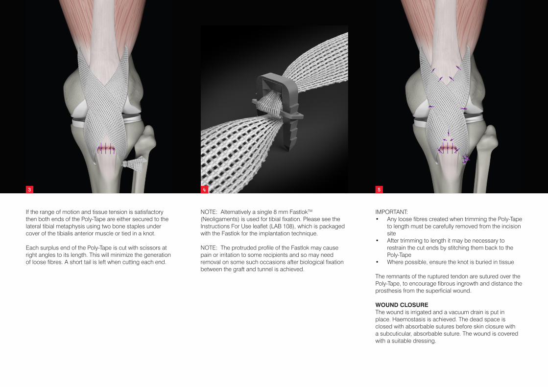
If the range of motion and tissue tension is satisfactory then both ends of the Poly-Tape are either secured to the lateral tibial metaphysis using two bone staples under cover of the tibialis anterior muscle or tied in a knot.
Each surplus end of the Poly-Tape is cut with scissors at right angles to its length. This will minimize the generation of loose fibres. A short tail is left when cutting each end.
NOTE: Alternatively a single 8 mm FastlokTM
(Neoligaments) is used for tibial fixation. Please see the Instructions For Use leaflet (LAB 108), which is packaged with the Fastlok for the implantation technique.
NOTE: The protruded profile of the Fastlok may cause pain or irritation to some recipients and so may need removal on some such occasions after biological fixation between the graft and tunnel is achieved.
4
IMPORTANT:• Any loose fibres created when trimming the Poly-Tape
to length must be carefully removed from the incision site
• After trimming to length it may be necessary to restrain the cut ends by stitching them back to the Poly-Tape
• Where possible, ensure the knot is buried in tissue
The remnants of the ruptured tendon are sutured over the Poly-Tape, to encourage fibrous ingrowth and distance the prosthesis from the superficial wound.
WOUND CLOSUREThe wound is irrigated and a vacuum drain is put in place. Haemostasis is achieved. The dead space is closed with absorbable sutures before skin closure with a subcuticular, absorbable suture. The wound is covered with a suitable dressing.
3 4 5

TECHNIQUE FOR RECONSTRUCTION OF EXTENSOR MECHANISM RUPTURE POST PATELLECTOMYA 4.5mm drill is used to create a bone tunnel at the level of the tibial tuberosity from lateral to medial. Bone tunnel edges are rounded where possible. The selected probe is used to pass the Poly-Tape transversley through the tunnel.
The Poly-Tape is crossed over the torn extensor mechanism. It is passed through the remnant of the quadriceps tendon and muscle in a Bunnell fashion. The free ends are transversely knotted and sutured to the surrounding extensor mechanism.
The patellar retinaculum is repaired and the Poly-Tape is sutured to the fibrous tissue envelope.
Extensor Mechanism Rupture Post Patellectomy
1

POSTOPERATIVE MANAGEMENTThe rehabilitation programmes shown provide only an outline of the prescribed regimes. For a full description refer to the documents entitled “QuadsTape System Rehabilitation Programme for Quadriceps Tendon Reconstruction” (LAB 132) and “PatellarTape System rehabilitation Programme for Patellar Tendon Reconstruction”(LAB 133).
Any rehabilitation programme should be supervised by a specialist physiotherapist. All mobilization and exercises should be performed within the pain free range of movement.
As in any implant surgery, satisfactory wound healing is of paramount importance.
The patient should be warned not to exceed the prescribed activity levels or to overload the repair before complete healing has occurred.
These rehabilitation programmes were developed in conjunction with Ian Horsley MSc, MCSP, Clinical Lead Physiotherapist, English Institute of Sport (EIS) North West, of BackinAction Physiotherapy and Sports Injury Clinic, Wakefield, UK.
QuadsTape SystemWeeks 0-1• The patient may fully weight bear using crutches for
stability (3 point gait moving towards reciprocal gait).
• A brace or splint is used to allow the patient to mobilize between physiotherapy sessions.
• The rehabilitation programme is commenced.
Weeks 1-3• Rehabilitation is continued with increasing repetitions
and pool work is commenced.
• Sutures are typically removed at this stage. Weeks 3-6• Rehabilitation is continued with increasing range of
knee flexion. • Balance exercises and static cycling are
commenced.
Weeks 6-12• The brace is discarded when full terminal knee
extension control is achieved.
• Crutches are discarded when the patient has a reciprocal gait pattern.
• Rowing machine/stepper/cross trainer are commenced with low resistance.
Week 12 Onwards• Resistance to exercises is increased (avoid leg
extension machine).
• On agreement with the physiotherapist, functional training and return to activity is allowed.
PatellarTape SystemWeeks 0-1• The patient may fully weight bear using crutches for
stability (3 point gait moving towards reciprocal gait).
• A brace or splint is used to allow the patient to mobilize between physiotherapy sessions.
• The rehabilitation programme is commenced with active extension. Do not aggressively push flexion.
Weeks 1-3• Rehabilitation is continued with increasing repetitions
and static cycling and pool work are commenced.
• Crutches are discarded when the patient has a reciprocal gait pattern.
• The brace is discarded when full terminal knee extension control is achieved.
• Sutures are typically removed at this stage.
Weeks 3-6• Balance exercises and proprioceptive training are
commenced. Weeks 6-12• Elliptical trainer and functional training are
commenced. Week 12 onwards• Jog-walk and jog-run exercises are commenced.
• On agreement with the physiotherapist, return to activity is allowed.
REFERENCE 1. Toms AD, Smith A, White SH. Analysis of the
Leeds-Keio ligament for extensor mechanism repair: favourable mechanical and functional outcome. Knee. 2003;10(2):131-134 .
For further references on this procedure please refer to the document “Neoligaments Scientific Articles” (LAB 144). This may be obtained from the Neoligaments™ Sales Department, or downloaded from www.neoligaments.com
Ordering Information
Please refer to the Instructions for Use leaflet packaged with the Poly-Tape for essential information about Poly-Tapes, including Use, Sterility, Indications, Contraindications, Warnings and Precautions, Potential Adverse Effects and Storage. Additional copies may be obtained from the Neoligaments™ Sales Department, or downloaded from www.neoligaments.com
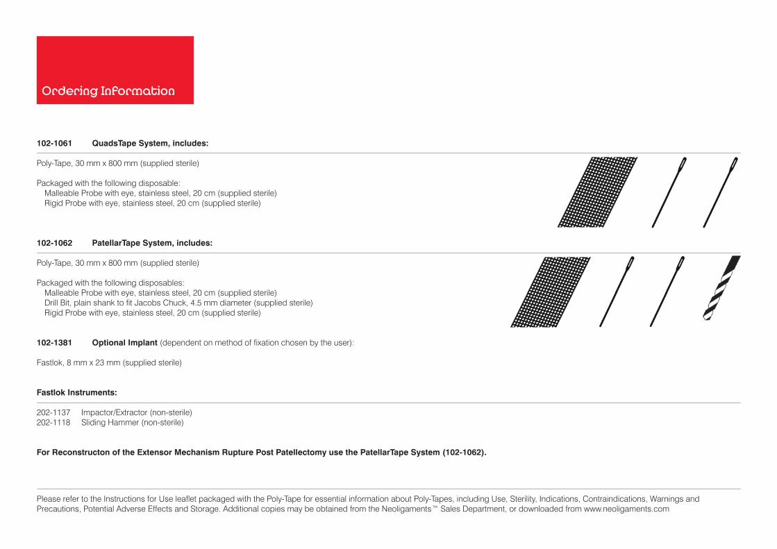
102-1061 QuadsTape System, includes:
Poly-Tape, 30 mm x 800 mm (supplied sterile) Packaged with the following disposable:
Malleable Probe with eye, stainless steel, 20 cm (supplied sterile) Rigid Probe with eye, stainless steel, 20 cm (supplied sterile)
102-1062 PatellarTape System, includes:
Poly-Tape, 30 mm x 800 mm (supplied sterile) Packaged with the following disposables:
Malleable Probe with eye, stainless steel, 20 cm (supplied sterile) Drill Bit, plain shank to fit Jacobs Chuck, 4.5 mm diameter (supplied sterile) Rigid Probe with eye, stainless steel, 20 cm (supplied sterile)
102-1381 Optional Implant (dependent on method of fixation chosen by the user):
Fastlok, 8 mm x 23 mm (supplied sterile)
Fastlok Instruments:
202-1137 Impactor/Extractor (non-sterile) 202-1118 Sliding Hammer (non-sterile)
For Reconstructon of the Extensor Mechanism Rupture Post Patellectomy use the PatellarTape System (102-1062).

Neoligaments™ A division of Xiros™ Springfield House Whitehouse Lane Leeds LS19 7UE
Tel. +44 (0) 113 238 7202 Fax. +44 (0) 113 238 7201 [email protected] www.neoligaments.com
Xiros Limited, Registered in England No. 1664824.
All rights reserved. © Neoligaments™ 2017. Worldwide patents and patents pending. Neoligaments, QuadsTape System, PatellarTape System, Fastlok and Xiros are trademarks of Xiros.
Developed and manufactured by
LAB 249 2.00