expression of retinoid-responsive genes occurs in colorectal carcinoma-derived cells irrespective of...
TRANSCRIPT

Expression of Retinoid-Responsive GenesOccurs in Colorectal Carcinoma-Derived
Cells Irrespective of the Presence ofResistance to All-Trans Retinoic Acid
PRZEMYSLAW WALISZEWSKI, MD, PhD,1,2,4* MIROSLAWA K. WALISZEWSKA, MSC,4
MANJUELA GUPTA, PhD,3 JEFFREY W. MILSOM, MD,1 AND ROBERT E. HURST, PhD4
1Department of Colorectal Surgery, The Cleveland Clinic Foundation, Cleveland, Ohio2Department of Cancer Biology, The Cleveland Clinic Foundation, Cleveland, Ohio
3Department of Immunopathology, The Cleveland Clinic Foundation, Cleveland, Ohio4Department of Urology, The University of Oklahoma Health Sciences Center,
Oklahoma City, Oklahoma
Background and Objectives:Retinoids are metabolized in human intes-tinal epithelial cells to all-trans retinoic acid; however, it is unknownwhether these cells express retinoid receptors, and whether sensitivity orresistance to the hormone is associated with a particular pattern of expres-sion of retinoid-responsive genes.Methods: Northern blot analysis and reverse transcriptase polymerasechain reaction (RT-PCR) were used to identify mRNAs for retinoid re-ceptors. Both Relative RT-PCR and transfection of retinoid-inducibleplasmid were applied to test functionality of the pathway in a modelsystem for colorectal carcinoma progression (primary SW480, all-transretinoic acid-sensitive cells vs. metastatic SW620, -insensitive cells).Results: Three colorectal carcinoma-derived cell lines were inhibited bythe hormone. Retinoic acid receptor typea (hRARa) and retinoid Xreceptor typea (hRXRa) mRNAs were detected in normal enterocytes,colonocytes, and in all colorectal carcinoma-derived cells studied. Primarycarcinomas and metastatic lesions expressed high amounts of hRARareceptor protein, showing no simple correlation between the amounts ofmRNA and receptor protein. No pattern of expression of the retinoid-responsive genes was associated with sensitivity or resistance to the ret-inoid. Expression of the genes occurred irrespective of resistance to thehormone or inactivity of the pathway.
Abbreviations: atRA, all-trans retinoic acid; CAT, chloramphenicol acetyltransferase; CRBP I , cellular retinol binding protein type I; CRABPI, II, cellular retinoic acid binding protein types I and II; EGF-R, epidermal growth factor receptor; hRAR, retinoic acid receptor; hRXR, retinoidX receptor; MK, angiogenic cytokine; RARE, retinoic acid responsive element; TG, transglutaminase; DEPC, diethylpyrocarbonate; DTT,dithiothreitol; dNTP; deoxynucleotidetriphosphate; PMSF, phenylmethylsulfonyl fluoride.These results were presented in part at the 86th Annual Meeting of the American Association for Cancer Research, March 15–20, 1995, Toronto,Canada.Contract grant sponsor: Fulbright Foundation (through the Office for Polish-U.S. Scientific Exchange, Warsaw).*Correspondence to: Przemyslaw Waliszewski, MD, PhD, Os. Jana III Sobieskiego 41 m. 21, 60-688 Poznan, Poland.E-mail: [email protected] 4 August 1997
Journal of Surgical Oncology 1997;66:156–167
© 1997 Wiley-Liss, Inc.

Conclusions:Colonocytes possess a molecular system for transduction ofthe retinoid signal. All-trans retinoic acid modifies gene expression andinhibits proliferation of these cells. Therefore, retinoids are likely to beeffective in chemoprevention of colorectal carcinoma.J. Surg. Oncol. 1997;66:156–167. © 1997 Wiley-Liss, Inc.
KEY WORDS: retinoids; retinoid receptors; retinoid-responsive genes; complexity;colorectal neoplasms
INTRODUCTIONThe traditional view of retinoids, vitamin A and its
derivatives, either as growth factors (vitamin A) or achromophore involved in light absorption during the vi-sion process (11-cis-retinal), changed when their activityin modifying expression of several genes important forcellular proliferation and differentiation was discovered[1, 2]. This finding provided a background for chemo-prevention, a relatively new approach to cancer control,in which cellular differentiation, proliferation, or induc-tion of apoptotic death are manipulated in initiated cellsprogressing toward malignancy, transformed cells, andadjacent cells comprising the epithelial and stromal field[3].
All-trans retinoic acid, actively synthesized in varioustissues as a result of enzymatic transformation of vitaminA andb-carotene [4, 5], was found to be one of the mostbiologically active retinoids in exerting an antitumori-genic effect in a variety of transformed epithelial cells [2,3]. The signaling pathway involves binding to specifichigh-affinity nuclear receptors [1, 6]. The retinoid recep-tors are classified into the retinoic acid receptor (hRAR)and retinoid X receptor (hRXR) subfamilies. The hRARfamily is comprised ofa, b, andg subtypes [7, 8] whileonly a andb subtypes have been identified for the hRXRsubfamily to date [9–11]. The highest affinity ligands forthe hRAR and hRXR subfamilies are all-trans retinoicacid and 9-cis retinoic acids, respectively [12]. ThehRAR and hRXR proteins form heterodimers that bindmuch more efficiently to DNA retinoid-responsive ele-ments than do hRAR-hRAR homodimers [11]. The li-gand-receptor complexes bind to retinoid-responsive el-ements on gene promoters and modulate gene transcrip-tion in a ligand- and dose-dependent manner. Expressionof the retinoid-responsive genes is believed to determinecellular sensitivity to retinoids [1].
Retinoids are also bound in the cytoplasm by a high-affinity binding protein, cellular retinol binding proteintype I (CRBP I), which binds retinol, and cellular retinoicacid binding protein types I and II (CRABP I andCRABP II), which bind all-trans retinoic acid [13–16].These latter two proteins seem to modulate the activity ofall-trans retinoic acid, at least in part, by inhibiting in-teraction with the nuclear receptors.
Vitamin A andb-carotene are absorbed and enzymati-
cally metabolized in enterocytes in ileum [17]. Recently,the enzymatic systems converting vitamin A to the bio-logically active all trans-retinoic acid were discovered inthe human colon carcinoma-derived cells, Caco 2 [18].Epidemiological data relating vitamin A and colon car-cinoma are contradictory; some investigators reportedthat intestinal mucosa was protected by retinoids duringcolorectal tumorigenesis [19] while others reported nocorrelation between risk of colorectal carcinoma and theamount of vitamin A in the diet [20, 21]. In animal stud-ies of experimental tumorigenesis, retinoids unequivo-cally decreased the frequency of colorectal carcinomas[22, 23]. No chemoprevention trials of retinoids in hu-man colorectal carcinoma have been reported; however,it was demonstrated that retinoids had clear antiprolifer-ative effect on some established human colorectal carci-noma cell lines [24–26]. Given the finding of the reti-noid-metabolizing enzymes in Caco-2 cells, the questionarises whether the all-trans retinoic acid signaling path-way is defective and whether loss of responsiveness toall-trans retinoic acid plays a role in human colorectaltumorigenesis. Although the presence of retinoic acid-binding activity in the cytosol of colonic mucosa andtumor specimens has been demonstrated [27], no reportshave appeared concerning the presence of elements of thesignaling pathway in human colonocytes. Also, no dataexist concerning when in the process of tumorigenesisretinoid sensitivity might be lost or what mechanisms forloss of sensitivity would be responsible.
This study was undertaken to identify elements of theall-trans retinoic acid signaling pathway in intestinal ep-ithelial cells, and test whether resistance or susceptibilityto the proliferation-inhibiting activity of this retinoid wasassociated with a particular pattern of expression of ret-inoid-responsive genes. A series of seven colorectal car-cinoma cell lines was surveyed to identify cell lines sen-sitive to proliferation inhibition by all-trans retinoic acid.The SW 480 cells derived from a primary tumor weresensitive to the hormone while the SW 620 cells derivedfrom a lymph node metastasis from the same patient wereresistant. Therefore, these cell types form an in vitromodel of human colorectal carcinoma in which influ-ences of retinoids on gene expression in relation to sen-sitivity in genetically related cells can be investigated.Northern blot analysis and RT-PCR were used to identify
Retinoid-Responsive Genes in Human Colonocytes 157

mRNAs encoding hRARa and hRXRa receptor proteins.Expression of a series of retinoid-responsive genes—hRARa, hRARb, CRBP I, CRABP I, and CRABP II [1],epidermal growth factor receptor (EGF-R)[1, 28], trans-glutaminase (TG)[29], angiogenic cytokine (MK) [30],as well as hRXRa, and hRXRb was investigated in themodel. Subsequently, both cell types were transfectedwith a plasmid containing the chloramphenicol acetyltransferase (CAT) gene under control of the retinoic acidresponsive element (RARE) from the hRARb gene andfunctionality of the pathway was evaluated. In addition,and for the first time, these results in vitro were com-pared to studies performed with clinical material, inwhich constitutive levels of retinoid receptor mRNAsand proteins were determined on cells derived from pri-mary tumors and liver metastases, and enterocytes andcolonocytes obtained from normal mucosa from the samepatient.
MATERIALS AND METHODSSurgical Specimens
Normal-appearing ileal and colonic mucosa was ob-tained from one patient with primary adenocarcinoma(HCC 111). Four primary tumor samples and four livermetasteses were also obtained. Tissue samples were rap-idly transported from the operating room to the surgicalpathology suite of the Cleveland Clinic Foundation andwere immediately processed as described below. In somecases, corresponding samples from the same patient wereobtained. With the case HCC 111 corresponding entero-cytes, colonocytes and primary tumor cells were ana-lyzed by Northern blot analysis or ordinary RT-PCR (notRelative RT-PCR) and the enhanced chemoluminescence(ECL) assay for hRARa receptor protein. Two liver me-tastases (HCC 134 and HCC 139) were analyzed byNorthern blot analysis, ordinary RT-PCR and ECL assay.In the case of HCC 139, corresponding primary tumorwas included in the ECL assay.
Cell Culture and Isolation of IntestinalEpithelial Cells
Established colorectal carcinoma cell lines, Caco 2,Colo 205, DLD 1, HT 29, SW 480, SW620, SW 837, SW1463, representing different stages of the disease, werepurchased from the American Type Culture Collection(Rockville, MD). Cells were cultured in red phenol-freeRPMI-1640 medium with 10% fetal calf serum (GibcoBRL, Gaithersburg, MD)[31]. For isolation of intestinalepithelial cells, mucosa was separated mechanically fromthe smooth muscle layer and cut into pieces 5 × 5 mm.The pieces were washed for 30 minutes in DTT/RPMI-1640 solution (1.5 mg/ml) at 37°C with shaking, fol-lowed by incubation three times for 30 minutes withDispase (neutral protease from Bacillus polimyxa, Boe-hringer, Mannheim, Germany) dissolved in RPMI-1640medium (3 mg/ml) with vigorous shaking (225 rpm). Red
blood cells were lysed by incubation 15 minutes at roomtemperature in hypotonic solution (7 mg NH4CO3 and700 mg NH4Cl in 100 ml H2O) followed by washingtwice in cold PBS. The suspension contained 97% viablecells as demonstrated by staining with Trypan Blue. Thecells were easily identified as epithelial cells on the basisof morphology and size. No red blood cells were found.Also, stromal cells were not present. Single lymphocyteswere identified (1.8% ± 0.3%) as demonstrated by indi-rect immunostaining of the air-dried alcohol-fixed cellu-lar smear using anti-keratin monoclonal antibody AE1/AE3 (Boehringer Mannheim, Germany), polyclonal an-tibodies directed to a desmin and a vimentin (Dako,Copenhagen, Denmark) and a monoclonal antibody di-rected to a leukocyte common antigen CD45RB (Dako,Carpinteria, CA)[32].
Effect of All-Trans Retinoic Acid on Proliferation
Cells of each cell line but SW 837 were seeded onto 96well plates at a concentration of 6,000 cells/ml in 200mlof RPMI-1640 medium/well, and cultured with and with-out 5mM all-trans retinoic acid (Sigma, St. Louis, MO).MCF-7 breast carcinoma cells, which are known to ex-press retinoic acid receptors and to be sensitive to all-trans retinoic acid treatment, were used as a positivecontrol [33]. Fetal calf serum was omitted from theRPMI-1640 medium used for the assay. Media and li-gand were changed every 24 hours. After 6 days of treat-ment, the cell number was measured by 3-(4,5-dimethylthiazol-2-yl)-2,5-diphenyltetrazolium bromide(MTT) assay [34].
Northern Blot Analysis
Established human colorectal carcinoma cells wereseeded onto 15 cm plates with 200,000 cells/plate andcultured until 90% confluent. The RPMI-1640 mediumwas changed and cells were incubated 48 hours withoutor with all-trans retinoic acid at a final concentration of5 mM. Total RNA was isolated by the guanidinum thio-cyanate-phenol-chloroform extraction [35]. Total RNAwas isolated from both normal intestinal epithelial cellsobtained from surgical specimens, colorectal carcinoma-derived cell lines, four primary colorectal carcinomas,and four liver metastases of colorectal carcinoma.
Approximately 15mg of total RNA isolated from eachsample was electrophoresed through 1% agarose gelscontaining 0.66 M formaldehyde, blotted overnight ontonylon membranes (Magna Graph, MSI, Westboro, MA)in 10 × SSC buffer, and baked for 2 hours at 80°C.Hybridization of RNA to the specific probes was per-formed with Denhardt’s reagent and 50% Formamide[36]. Blots were exposed to X-ray film at −80°C for 1, 2,and 4 weeks.
The 0.83 kb cDNA probe for hRARa was obtained bydigestion of the complete hRARa sequence by the re-
158 Waliszewski et al.

striction enzyme, Eco RI. The 0.6 kb and 0.9 kb cDNAprobes for hRARb were obtained by digestion of thecomplete hRARb coding sequence by Eco RI/Bam HI.The cDNA probe for hRARg was a full coding sequenceof the gene. The 0.74 kb probe for hRXRa was obtainedby digestion of the complete hRXRa coding sequence byApa I/Hinc II. Probes were radiolabeled with32P-dATPby T7QuickPrime Kit (Pharmacia, Sweden). Specific ra-dioactivity of each probe was about 2 × 10⁹ cpm/mgDNA [36]. Quality and amount of the RNA was assessedby probing the blots with a cDNA forb-tubulin.
RT-PCRPrimers were prepared in the Department of Molecular
Biology at the Cleveland Clinic Foundation and in theCenter for Molecular Medicine, University of Oklahoma,and purified by high pressure liquid chromatography(HPLC) prior to use. First strand cDNA was preparedusing Superscript II Reverse Transcriptase (Gibco BRL,Gaithersburg, MD) from total RNA. Briefly, 2mg of totalRNA in 2 ml of diethylpyrocarbonate (DEPC) water wasmixed with 1ml of 10 × DNase I buffer, 6ml of DEPC-treated water, and 1ml of DNase I. The mixture wasdigested (25°C, 15 minutes) to remove any genomicDNA contaminating the RNA samples. DNase I was in-activated (1ml 20 mM EDTA, 65°C, 10 minutes) and 1ml oligo (dT)12–18 (Gibco BRL, 500mg/ml) was added.Subsequently, the samples were heated (70°C, 10 min-utes) and quick-chilled on ice. After centrifugation, 4mlof 5 × First Strand Buffer (Gibco BRL), 2ml 0.1 Mdithiothreitol (DTT), 1 ml 10 mM deoxynucleotidetri-phosphate (dNTP), and 1ml Superscript II Reverse Tran-scriptase (Gibco BRL) were added. The mixture was in-cubated for 1 hour at 37°C.
The PCR reaction mixtures contained 10ml reversetranscriptase reaction product, 10ml 10 × PCR buffer(Gibco BRL), 1ml 10 mM dNTP, 3ml 50 mM MgCl2, 5ml each sense and antisense primer (10 mM), 1ml TaqDNA polymerase (Gibco BRL, 5 U/ml), and sterile waterto a final volume of 100ml. PCR reaction mixtures wereoverlayed with 35ml mineral oil in a 0.5 ml microfugetube, then placed into a Perkin-Elmer thermocycler. PCRreaction products were separated by electrophoresis on a2% agarose gel at 60 V for 2 hours, and the ethidiumbromide-stained bands were visualized by UV transillu-mination.
Relative RT-PCRThis approach is based upon the modified protocol of
the Arbitrarily Primed PCR [37, 38]. This techniqueyields relative concentrations of PCR products and al-lows the amplification of test and marker cDNAs each tobe within the linear portion of the sigmoid PCR curveand for optimization of the reaction conditions withoutthe need for a marker with the same concentration andsimilar physico-chemical sequence parameters as the test
cDNA. This technique differs from ordinary RT-PCRdescribed above and should not be considered strictlyquantitative. The technique can be considered semi-quantitative and is not designed to measure absoluteamounts of either mRNA or cDNA. First, 10mg of totalRNA isolated from SW 480 and SW 620 cells untreatedand treated for 48 hours with 5mM all-trans retinoic acidwas dissolved in 10ml of DEPC water, mixed with 1mlof 10 × DNase buffer, and incubated with 1ml DNase I(25°C, 15 minutes). DNase I was inactivated (1ml 20mM EDTA, 65°C, 10 minutes). RNA was extractedtwice by phenol-chlorophorm and ethanol/2 M sodiumacetate (22:1 v/v) precipitated. First strand cDNA wassynthesized in the similar way as for regular PCR exceptthat 100 ng of p(N)6 random primer was used (Boe-hringer) per reaction per 5mg of total RNA. After thereverse transcriptase reaction, the samples were heatedup (95°C, 10 minutes), diluted up to 100ml, and frozenat −20°C. To perform Relative RT-PCR, the first-strandcDNA samples to be compared were normalized simul-taneously to the sameb-Actin cDNA concentration byperforming 20 cycles of PCR withb-Actin primers.Based upon the normalization, exactly the same amountof first strand cDNA was aliquoted from each sample,and 32 cycles of PCR were performed with each pair ofprimers. Normalization was checked each time by simul-taneously amplifying in a separate tube for 20 cycles.The difference of gene expression between all-trans ret-inoic acid-treated and control cells was determined semi-quantitatively under ultraviolet (UV) light. The RelativeRT-PCR was repeated using independent samples of to-tal RNA. Table I contains the list of primers, the expectedmolecular weights of the RT-PCR products, and PCRconditions used. The identity of the RT-PCR productswas confirmed by restriction enzyme digestion or se-quencing.
Dot Blot ECL Assay With Murine MonoclonalAntibody Against hRARa
Normal human enterocytes, normal human colono-cytes, colonic fibroblasts CD-Co 18, intestinal smoothmuscle cells HISM, human colorectal carcinoma-derivedcells, Colo 205, DLD 1, and HT 29 as well as surgicalspecimens of primary colorectal carcinomas, HCC 111,HCC 124, HCC 139 and surgical specimens of liver me-tastases of colorectal carcinomas, HCC 133LM, HCC134LM, HCC 139 LM were suspended in 10 ml of bufferA [20 mM Tris-HCl, 1 mM EDTA, 0.6 M NaCl, pH 7.8with protease inhibitors (1 mM phenylmethylsulfonylfluoride (PMSF), 1 mM leupeptin, 1 mM antipain, 0.1mM bacitracin, 1 TIU/ml aprotinin, 1 mM aminobenza-midine, 10mM pepstatin A)] and washed twice. MCF-7cells and hRARa-positive breast carcinoma were used aspositive controls. Rat spleen was used as a negative con-trol. After transfer to a glass homogenizer, each sample
Retinoid-Responsive Genes in Human Colonocytes 159

was homogenized on ice in 2 ml of the buffer and cen-trifuged 15 minutes at 50,000 rpm and 4°C. Fat wasremoved from the sample and supernatant was transferedto the plastic tubes and the protein concentration wasdetermined by Bio-Rad Protein Assay kit (BioRad, SanDiego, CA). The plastic tubes with the supernatant werecovered with gauze, frozen for 15 seconds in liquid ni-trogen and lyophilized overnight at −60°C. After lyophi-lization, the protein concentration was equalized to 5.0mg/ml, and 5ml of the solution was mixed with 50ml ofdistilled water. The entire volume was transfered ontonitrocellulose in a vacuum. Hybridization with murineanti hRARa monoclonal antibodies was performed usingan ECL kit (Amersham, Little Chalfont, UK). The mem-brane was transfered to a plastic bag and exposed toKodak film for 30, 60, and 120 minutes at room tem-perature.
Transfection of Chloramphenicol AcetylTransferase Gene Under Control of Retinoic Acid
Responsive Element Into SW480/SW620 Cellsand CAT Assay
The pBLCAT2 reporter plasmid containing the chlor-amphenicol acetyltransferase (CAT) gene linked to a thy-midine kinase promoter on the 58 end to allow for a lowbasal activity and containing the SV40 polyadenylationsignal attached to the 38 end of the CAT gene to allowstabilization of the message was used as a control for thebasal level of activity. The same plasmid containing theretinoic acid responsive element from the RARb gene(pRARE-CAT) was used to test for functionality of theall-trans retinoic acid signaling pathway. Cells of each
line were seeded onto twelve 10 cm plates with 50,000cells/plate. Cells were cultured until 90% confluent. TheRPMI-1640 medium containing 10% fetal calf serumwas changed every 24 hours. The serum was analyzed byHPLC and confirmed to contain negligible amounts ofvitamin A and all-trans retinoic acid. The plasmids weretransfected by the calcium phosphate method with theglycerol shock [36]. Six plates of control cells weretransfected with the pBLCAT2 plasmid. Effectiveness ofthe transfections was verified by PCR. After transfection,washing with PBS, and refeeding with medium, threeplates were treated with 5mM all-trans retinoic acid for48 hours. Then, CAT activity was measured with a CATELISA kit (Boehringer-Mannheim, Indianapolis, IN) andcompared to the CAT activity in the non-treated cells(Ratio C). Similarly, six plates of cells of the same linewere transfected with the pRARE-CAT plasmid. Threeof them were treated with the retinoid, and then CATactivity was measured, and compared to the CAT activityin the non-treated pRARE-CAT cells (Ratio R). Trans-formed uroepithelial cells HUC-PC and T24 were usedas a negative control and a positive control for the trans-fection, respectively. The assay was performed in dupli-cates. The R/C ratio is the ratio of absorbances of ELISAfor CAT activity in the absence and presence of 5mMall-trans retinoic acid in cells transfected with the plas-mid pRARE-CAT (R) to the ratio of absorbances ofELISA for CAT activity in the absence and presence ofthe hormone in cells transfected with the control plasmidpBLCAT2 (C). The R/C ratio close to 1 indicates that nosignificant induction of CAT gene occurred due to the
TABLE I. List of Primers, PCR Conditions, and the Expected Molecular Weights of RT-PCR Products
Primer Primer sequence PCR conditions MW (bp)
hRARa S 58-TTG TCT GTC AGG ACA AGT CC 32 cycles (94°C, 60 s; 188hRARa A 58-CGT CTT CAC GAA ACT TCA CC 55°C, 60 s; 72°C, 120 s)hRARb S 58-TCG TCT GCC AGG ACA AAT CA 32 cycles (94°C, 60 s; 189hRARb A 58-CGT CTT CAC GAA ACT TCA CC 55°C, 60 s; 72°C, 120 s)hRXRa S 58-CAG GCA AGC ACT ATG GAG TGT 32 cycles (94°C, 60 s; 165hRXRa A 58-TG GCC AGG CAC TTC TGG TA 60°C, 60 s; 72°C, 120 s)hRXRb S 58-CTT TGT GCA ATC TGC GGG GAC 32 cycles (94°C, 60 s; 192hRXRb A 58-TGG CCA GGC ACT TCT GAT ATC 60°C, 60 s; 72°C, 120 s)CRBP I S 58-GTC GAC TTC ACT GGG TAC TGG A 32 cycles (94°C, 60 s; 441CRBP I A 58-TTG AAT ACT TGC TTG CAG ACC ACA 60°C, 60 s; 72°C, 120 s)CRABP I S 58-CGG CAC CTG GAA GAT GCG CA 32 cycles (94°C, 60 s; 370CRABP I A 58-CCA CGT CAT CGG CGC CAA AC 60°C, 60 s; 72°C, 120 s)CRABP II S 58-CCC AAC TTC TCT GGC AAC TGG A 32 cycles (94°C, 60 s; 410CRABP II A 58-CTC TCG GAC GTA GAC CCT GGT 60°C, 60 s; 72°C, 120 s)EGF-R S 58-GT CTG CCG CAA ATT 33 cycles (94°C, 45 s; 544EGF-R A 58-CC GCG TAT GAT TTC 54°C, 45 s; 72°C, 90 s)MK S 58-CTG CAA CTG GAA GAA GGA 32 cycles (94°C, 60 s; 206MK A 5 8-TTC CCT TCC CTT TCT TGG 60°C, 60 s; 72°C, 120 s)TG S 58-GGG CGA ACC ACC TGA ACA AA 33 cycles (94°C, 45 s; 300TG A 58-GGT CAC TAC CTA GCA TGT TGT 54°C, 45 s; 72°C, 90 s)b-Actin S 58-GCG CTC GTC GTC GAC AAC GGC 20 cycles, times and temperatures 220b-Actin A 58-CAT GGG GTA GCT CGT GCC GTA GCA GT as for the primers tested
S denotes sense sequence; A denotes antisense sequence.
160 Waliszewski et al.

interaction of the retinoid receptors with RARE of theplasmid under the retinoid treatment, and, therefore, thesignaling pathway is not functional.
RESULTSEffects of All-Trans Retinoic Acid on Proliferation
All cell lines studied grew well in RPMI 1640 mediumsupplemented with nonessential aminoacids and 10% fe-tal calf serum. All cell lines but two, grew well in theabsence of serum; Colo 205 and SW 1463 grew muchslower (data not shown) as compared to the cells culturedin medium with 10% fetal calf serum. Other than theHT-29 cells, all other lines exhibited the same morphol-ogy in the presence of serum or all-trans retinoic acid asin their absence. HT 29 cell morphology changed fromthe usual polygonal shape during the treatment with all-trans retinoic acid to a more rounded morphology andcontinued to exclude Trypan Blue.
Results of the MTT assay for the antiproliferative ef-fect of all-trans retinoic acid are shown in Table II. Asignificant inhibition of proliferation (P < 0.05, Student’st-test) was observed in the populations of Colo 205 cells(threefold), HT 29 cells (fivefold) as compared to thenon-treated cells. In addition, a statistically significantinhibition was observed in case of SW 480 cells (one-fold). No significant difference was observed in case ofCaco 2, DLD 1, SW 620, and SW 1463 cells.
Northern Blot Analysis and RT-PCR Study ofReceptor mRNA Expression
Northern blot analysis identified hRARa mRNA asthe expected 2.8 kb and 3.8 kb bands in total RNA iso-lated from cultured colorectal carcinoma cell lines(Fig.1). In two of the primary carcinomas and all fourcases of colon cancer metastases to liver, bands corre-sponding to hRARa mRNA were not seen, even thoughthe b-tubulin band was visible (Fig. 1). The more sensi-tive technique of RT-PCR with primers for hRARa li-gand binding domain was applied with results shown in
Figure 2A. In all instances, a single band correspondingto the expected 188 bp RT-PCR product of the hRARaligand binding domain was present, including normal en-terocytes and colonocytes, which were not shown on theNorthern blot, as well as in the liver metastases thatfailed to show a band by Northern blot analysis. Becausethe RT-PCR method was more sensitive and was vali-dated by the Northern blot analysis, it was used to in-vestigate the presence of hRXRa transcripts, and in allinstances, this gene was found to be expressed as well(Fig. 2B). A single band corresponding to the expected165 bp product was seen in each sample. Northern blotanalysis confirmed the presence of hRXRa transcripts at5.6 kb (data not shown). Northern probing for hRARband hRARg was inconclusive even at the longest expo-sure times.
hRARa Protein Expression
Figure 3 demonstrates the results of ECL analysis forthe hRARa receptor protein. The densitometrically de-termined relative (to MCF-7) amounts of receptor proteinare shown in Table III together with comparativeamounts of mRNA determined densitometrically fromthe Northern blots. The range of mRNA amounts wassimilar for normal colonocytes and for established celllines. Other than the DLD 1 line, the HT 29 and SW 620lines contained almost the same amount of receptor pro-tein as did normal colonocytes. In contrast, the primary
TABLE II. Results of MTT Assay for Antiproliferative Effect of5 µM All-Trans Retinoic Acid (atRA) in Human ColorectalCarcinoma-Derived Cell Lines
Cell line Control S.D. 5mM atRA S.D.
Caco 2 2.71 0.33 2.56 0.19Colo 205 0.52 0.12 0.19 0.08DLD 1 1.67 0.29 1.53 0.72HT 29 1.72 0.26 0.32 0.16SW 480 0.50 0.10 0.34 0.07SW 620 0.39 0.11 0.32 0.10SW 1463 0.31 0.14 0.28 0.17MCF-7 1.41 0.05 0.62 0.04
Results are the mean absorbances at 570 nm ± standard deviation from32 wells. The italic type indicates the difference between control andtest is statistically significant atP < 0.05.
Fig. 1. A: Northern blot analysis of hRARa mRNA expression incolorectal carcinoma-derived cell lines, primary colorectal carcino-mas, and liver metastases of colorectal carcinoma. Eco RI fragment ofhRARa cDNA (0.83 kb) was used as a probe.B: Amounts of totalRNA on the blot were normalized againstb-tubulin cDNA. hRARamRNA was expressed as 2.8 kb and 3.8 kb bands. Colorectal carci-noma-derived cells, lanes 1–5: Colo 205, DLD 1, HT 29, SW 620, SW837; primary colorectal carcinomas, lanes 6–10: HCC 147, HCC 148,HCC 111, HCC 124, HCC 134; liver metastases of colorectal carci-nomas, lanes 11–14: HCC 104LM, HCC 133LM, HCC 134LM, HCC139 LM.
Retinoid-Responsive Genes in Human Colonocytes 161

tumors and liver metastases contained up to 40-foldhigher amounts of receptor protein. Comparison of re-ceptor protein amounts in the sensitive HT 29 line andthe insensitive DLD 1 and SW 620 lines shows that allthe three had similar mRNA amounts, but the amount ofreceptor protein was roughly 10 times as high in the DLD1 line as in the HT 29 and SW 620 lines. Of interest wasthe general lack of correlation between the amount ofmRNA and the amount of receptor protein seen in thecells, even among the normal cells. This discrepancy wasparticularly striking for the liver metastases, in which themRNA was not detectable by Northern blot analysis, butwhich corresponded to high amounts of receptor protein.
Relative Expression of Retinoid-Responsive Genesin SW 480/SW 620 Cell Lines
The results of Relative RT-PCR analysis of the SW480 and SW 620 cell lines are summarized in Table IV.These two cell lines were derived from the primary tumorand a lymph node metastatic lesion of the same patient,but the SW 480 line was sensitive to all-trans retinoicacid while the SW 620 line was not. The two cell linesdiffered markedly in constitutive and stimulated expres-sion of several of the retinoid responsive genes, selectedgels of which are shown in Figure 4. In the presence of5 mM all-trans retinoic acid, hRARa was down-regulatedin the SW 480 cells but was marginally up-regulated inSW 620 cells (Fig. 4A). As found using Northern blotanalysis in the other sensitive cell line, HT 29, hRARa
mRNA was up-regulated as expected (data not shown).CRBP I was strongly up-regulated, as expected, in bothcell lines (Fig. 4B). The MK angiogenic cytokine genewas sharply down-regulated in the SW 480 cells, butstrongly up-regulated in the SW 620 cell line (Fig. 4C).EGF-R was twofold up-regulated in SW480 cells, butwas constitutively low and further down-regulated inSW620 cells (Fig. 4D). The following genes were alsomeasured with results listed in Table IV, but gels are notshown. The hRARb gene was not present constitutivelyin either cell line, but was induced in both, as expected.CRABP I was down-regulated in the SW 480 cells asexpected but up-regulated in SW 620. CRABP II wasdown-regulated about twofold in SW 480 cells, in con-trast to the expected up-regulation. In the SW 620 cells,CRABP II was undetectable after treatment. TG genewas down-regulated in SW 480 cells, an unexpectedfinding, but was strongly up-regulated in SW 620 cells.
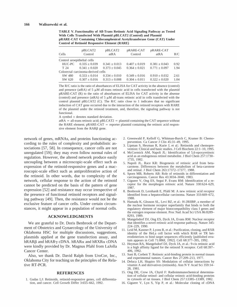
Plasmid Transfection and CAT Activity
Plasmid transfection was successful in all cases. Asexpected, the R/C ratio for HUC-PC cells, a negativecontrol, was close to 1, indicating that the signaling path-way is not active. In contrast, the R/C ratio for T 24 cellsdenotes an almost twofold increase of CAT activity, in-dicating that the signaling pathway is functional. CATactivity in SW480 cells transfected with the plasmidpRARE-CAT was significantly increased (almost three-fold) under treatment with 5mM all-trans retinoic acid ascompared to controls and SW 620 cells transfected withthe same plasmid; the R/C ratio was 2.61. CAT activityin SW620 cells transfected with the pRARE-CAT plas-mid remained in the same range as in the control cellstransfected with the plasmid pBLCAT2; the R/C ratiowas close to 1 (Table V).
DISCUSSION
Chemoprevention is an attractive option in cancer con-trol because the risks of metastasis and cancer therapy arecompletely avoided. A number of factors have beentested as chemopreventive agents in colorectal tumori-genesis, including nonsteroidal anti-inflammatory drugs(aspirin, sulindac), difluoromethylornithine, calcium, vi-tamin D3, and organoselenium compounds [39–42]. Al-though retinoids have been used successfully in animalstudies to inhibit colorectal tumorigenesis, no clinicaltrials have been reported in humans. This study providesa molecular background for chemoprevention in humanlarge intestine; however, we emphasize here that retinoidchemotherapy of advanced colorectal carcinomas willmost likely be less successful. This is because a numberof random molecular defects increases during tumor pro-gression. As shown in the in vitro model, even geneti-cally related tumor cells may differ significantly in re-sponse to the retinoid treatment, in functionality of the
Fig. 2. Qualitative identification of hRARa (A) and hRXRa (B)cDNAs in normal colonocytes, colorectal carcinoma-derived cells, andliver metastases of colorectal carcinomas using primers for ligandbinding domains of the receptors by ordinary RT-PCR. Identity of theproducts was confirmed by restriction enzyme digestion of the radio-labeled RT-PCR products and visualization on the 6% polyacrylamidegel. The first strand cDNAs were not normalized againstb-Actin.Lane 1: molecular weight standards, 100 bp DNA ladder. Lane 2:normal human enterocytes (ileum). Lane 3: normal human colonocytes(sigmoid). Colorectal carcinoma-derived cells, lane 4: Caco 2; lane 5:Colo 205; lane 6: DLD-1; lane 7: HT 29; lane 8: SW 480; lane 9: SW620; lane 10: SW 837; lane 11: SW 1463. Liver metastases of colo-rectal carcinoma, lane 12: HCC 133LM; lane 13: HCC 134LM; lane14: HCC 139LM.
162 Waliszewski et al.

pathway, or in the patterns of expression of retinoid-responsive genes. For the first time, we demonstratedthat the key elements of all-trans retinoic acid signalingpathway are expressed in human intestinal epithelialcells, and that expression of the retinoid-responsivegenes is modified whether the specific pathway is func-tional or not. In addition, results with cultured cells arecompared with clinical specimens.
The antiproliferative assay showed that 3/7 of the celllines were inhibited by 5mM all-trans retinoic acid. Thisconcentration is nonphysiological; however, no cytotoxiceffect was observed. Such value remains within the av-erage range of biologically active concentrations of theretinoid in in vitro cellular systems, i.e., between 1mMand 10mM [33, 43–47]. Thus, it appears that transformedintestinal epithelial cells tolerate the retinoid in a similarmanner as the other transformed epithelial cell types do.Furthermore, according to Gudas (personal communica-tion), with cancer cells, the antiproliferative effect of the
retinoid may not occur at physiological concentrations,and to observe such effect in vitro, the extracellular con-centration of the retinoid must be raised to distinctlynonphysiological levels. Indeed, one of the challenges ofretinoid research is to discover retinoid compounds withless toxicity than all-trans retinoic acid so the dose can beincreased sufficiently high to inhibit cancer or premalig-nant cells [43].
The major elements for all-trans retinoic acid signaltransduction are hRARa, hRXRa, CRABP I, andCRABP II [1, 48]. As shown in Figures 1 and 2, hRARaand hRXRa mRNAs were expressed in all cells exam-ined, and, in the case of hRARa, the protein was iden-tified as well (Fig. 3). Thus, failure of some cell lines toshow an antiproliferative response is not due to absenceof these elements [49]. Interestingly, the observed modu-lation of expression of a number of retinoid-responsivegenes under 5mM all-trans retinoic acid stimulation sug-gests indirectly that signal transduction to the nucleus
Fig. 3. Results of ECL dot blot with murine monoclonal antibody against hRARa receptor protein. Lane 1: primary colorectal carcinoma HCC139. Liver metastases of colorectal carcinomas, lanes 2–4: HCC 139LM, HCC 134LM, HCC 133LM. Lane 5: normal human enterocytes (ileum).Lane 6: normal human colonocytes (sigmoid). Lane 7: human colonic smooth muscle cells. Lane 8: human colonic fibroblasts. Colorectalcarcinoma-derived cells, lanes 9–11: DLD-1, SW 620, HT 29. Primary colorectal carcinomas, lanes 12, 13: HCC 111, HCC 124. Positivecontrols, lane 14: MCF-7; lanes 15, 16: human primary breast carcinoma (dilutions 1:8 and 1:4). Negative control, lane 17: rat spleen.
TABLE III. Densitometric Analysis of hRAR a mRNA and Receptor Protein
SampleRelative amount
of hRARa mRNARelative amount ofthe receptor protein
MCF-7 (positive control) 1.00 1.00Normal cells
Colonocytes 1.10 1.51Smooth muscle cells 0.14 7.12Colonic fibroblasts 0.09 0.06
Colorectal carcinoma-derived cellsHT 29 (atRA-sensitive) 0.78 0.65DLD 1 (atRA-insensitive) 0.72 7.01SW 620 (atRA-insensitive) 0.81 0.75
Primary carcinomasHCC 111 2.42 26.64HCC 124 0.18 34.98HCC 139 ND 42.79
Liver metastasesHCC 133 LM ND 6.69HCC 134 LM ND 3.69HCC 139 LM ND 10.28
Results are expressed relative to amounts found in MCF-7 cells. ND denotes that mRNAwas not detected by Northern blot analysis.atRA 4 all-trans retinoic acid; hRARa 4 retinoic acid receptor typea.
Retinoid-Responsive Genes in Human Colonocytes 163
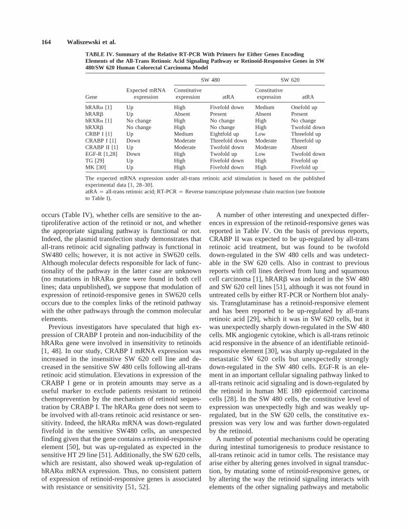
occurs (Table IV), whether cells are sensitive to the an-tiproliferative action of the retinoid or not, and whetherthe appropriate signaling pathway is functional or not.Indeed, the plasmid transfection study demonstrates thatall-trans retinoic acid signaling pathway is functional inSW480 cells; however, it is not active in SW620 cells.Although molecular defects responsible for lack of func-tionality of the pathway in the latter case are unknown(no mutations in hRARa gene were found in both celllines; data unpublished), we suppose that modulation ofexpression of retinoid-responsive genes in SW620 cellsoccurs due to the complex links of the retinoid pathwaywith the other pathways through the common molecularelements.
Previous investigators have speculated that high ex-pression of CRABP I protein and non-inducibility of thehRARa gene were involved in insensitivity to retinoids[1, 48]. In our study, CRABP I mRNA expression wasincreased in the insensitive SW 620 cell line and de-creased in the sensitive SW 480 cells following all-transretinoic acid stimulation. Elevations in expression of theCRABP I gene or in protein amounts may serve as auseful marker to exclude patients resistant to retinoidchemoprevention by the mechanism of retinoid seques-tration by CRABP I. The hRARa gene does not seem tobe involved with all-trans retinoic acid resistance or sen-sitivity. Indeed, the hRARa mRNA was down-regulatedfivefold in the sensitive SW480 cells, an unexpectedfinding given that the gene contains a retinoid-responsiveelement [50], but was up-regulated as expected in thesensitive HT 29 line [51]. Additionally, the SW 620 cells,which are resistant, also showed weak up-regulation ofhRARa mRNA expression. Thus, no consistent patternof expression of retinoid-responsive genes is associatedwith resistance or sensitivity [51, 52].
A number of other interesting and unexpected differ-ences in expression of the retinoid-responsive genes wasreported in Table IV. On the basis of previous reports,CRABP II was expected to be up-regulated by all-transretinoic acid treatment, but was found to be twofolddown-regulated in the SW 480 cells and was undetect-able in the SW 620 cells. Also in contrast to previousreports with cell lines derived from lung and squamouscell carcinoma [1], hRARb was induced in the SW 480and SW 620 cell lines [51], although it was not found inuntreated cells by either RT-PCR or Northern blot analy-sis. Transglutaminase has a retinoid-responsive elementand has been reported to be up-regulated by all-transretinoic acid [29], which it was in SW 620 cells, but itwas unexpectedly sharply down-regulated in the SW 480cells. MK angiogenic cytokine, which is all-trans retinoicacid responsive in the absence of an identifiable retinoid-responsive element [30], was sharply up-regulated in themetastatic SW 620 cells but unexpectedly stronglydown-regulated in the SW 480 cells. EGF-R is an ele-ment in an important cellular signaling pathway linked toall-trans retinoic acid signaling and is down-regulated bythe retinoid in human ME 180 epidermoid carcinomacells [28]. In the SW 480 cells, the constitutive level ofexpression was unexpectedly high and was weakly up-regulated, but in the SW 620 cells, the constitutive ex-pression was very low and was further down-regulatedby the retinoid.
A number of potential mechanisms could be operatingduring intestinal tumorigenesis to produce resistance toall-trans retinoic acid in tumor cells. The resistance mayarise either by altering genes involved in signal transduc-tion, by mutating some of retinoid-responsive genes, orby altering the way the retinoid signaling interacts withelements of the other signaling pathways and metabolic
TABLE IV. Summary of the Relative RT-PCR With Primers for Either Genes EncodingElements of the All-Trans Retinoic Acid Signaling Pathway or Retinoid-Responsive Genes in SW480/SW 620 Human Colorectal Carcinoma Model
GeneExpected mRNA
expression
SW 480 SW 620
Constitutiveexpression atRA
Constitutiveexpression atRA
hRARa [1] Up High Fivefold down Medium Onefold uphRARb Up Absent Present Absent PresenthRXRa [1] No change High No change High No changehRXRb No change High No change High Twofold downCRBP I [1] Up Medium Eightfold up Low Threefold upCRABP I [1] Down Moderate Threefold down Moderate Threefold upCRABP II [1] Up Moderate Twofold down Moderate AbsentEGF-R [1,28] Down High Twofold up Low Twofold downTG [29] Up High Fivefold down High Fivefold upMK [30] Up High Fivefold down High Fivefold up
The expected mRNA expression under all-trans retinoic acid stimulation is based on the publishedexperimental data [1, 28–30].atRA4 all-trans retinoic acid; RT-PCR4 Reverse transcriptase polymerase chain reaction (see footnoteto Table I).
164 Waliszewski et al.

processes. As shown in Table III, no simple correlationexisted among hRARa mRNA expression, receptor pro-tein amount and all-trans retinoic acid sensitivity amongthe cancer cells, or between amounts of the mRNA andthe receptor protein in the normal intestinal epithelialcells. A similar lack of correlation was reported previ-ously for other genes in normal intestinal epithelial cells,where regulation of gene expression occurred throughregulation of mRNA lifetime, transport, translation, andprotein turnover [53, 54]. The extremely high amounts ofhRARa receptor protein found in the primary tumors,metastatic tumors and the DLD 1 cell line suggest un-coupling of these regulatory mechanisms and may reflectalterations associated with tumorigenesis.
Molecular characterization of the closely related SW480 and SW 620 cell lines, representing consecutivestages of tumor progression, points out that expression ofa number of genes normally acting as elements in reti-noid signaling or linked to the pathway, for exampleEGF-R (Table IV) and RNase L [55], is functionally
altered. In the case of RNase L, expression is completelyshut off, even though the gene is present structurallyunaltered in the genome [55]. Similarly, constitutive ex-pression of EGF-R mRNA in SW 620 cells is sharplylower than in SW 480 cells. This low expression inSW620 cells was not due to a mutation of the gene thatwould be detected by PCR/SSCP (data unpublished; alsoChakrabarty, personal communication). Although neithergene can be considered a classic tumor suppressor gene,the observed alterations in gene transcription may illus-trate an additional mechanism operating in tumor pro-gression. As shown by similar findings for ABO genetranscripts in human bladder tumors, inhibition of geneexpression to modulate function of certain pathways mayrepresent a general phenomenon in tumorigenesis [56].
The resistance of tumor cells can also be studied froma holistic perspective. Then, the functional status of a celldepends not so much on the activity of a single gene oreven a set of genes (e.g. the retinoid-responsive genes),but rather upon the conjugated activities of the whole
Fig. 4. Relative RT-PCR for hRARa (A), CRBP I (B), MK cytokine (C), and EGF-R(D) mRNA in SW 480/SW 620 cells treated with andwithout 5mM all-trans retinoic acid for 48 hours. Total RNA was isolated and first strand cDNA was normalized againstb-Actin. The analysiswas repeated twice independently.A: hRARa, lane 1: molecular weight marker, 100 bp DNA ladder; lane 2: SW 480 control; lane 3: SW 480all-trans retinoic acid (atRA)-treated; lane 4: SW 620 control; lane 5: SW 620 atRA-treated. Normalizedb-Actin, lanes 6–9.B: CRBP I, lane10: molecular weight marker, 100 bp DNA ladder; lane 11: SW 480 control; lane 12: SW 480 atRA-treated; lane 13: SW 620 control; lane 14:SW 620 atRA-treated. Normalizedb-Actin, lanes 15–18.C: MK cytokine (top) and normalizedb-Actin (bottom). Lane 19: molecular weightmarker, 100 bp DNA ladder. Lane 20: SW 480 control. Lane 21: SW 480 atRA-treated. Lane 22: SW 620 control. Lane 23: SW 620 atRA-treated.D: EGF-R, lane 24: molecular weight marker, 100 bp DNA ladder; lane 25: SW 480 control; lane 26: SW 480 atRA-treated; lane 27: SW 620control; lane 28: SW 620 atRA-treated. Normalizedb-Actin, lanes 29–32.
Retinoid-Responsive Genes in Human Colonocytes 165
network of genes, mRNAs, and proteins functioning ac-cording to the rules of complexity and probabilistic as-sociations [57, 58]. In consequence, cancer cells are notunregulated [59]; they may even follow normal rules ofregulation. However, the altered network produce easilyuncoupling between a microscopic-scale effect such asexpression of the retinoid-responsive genes and a mac-roscopic-scale effect such as antiproliferative action ofthe retinoid. In other words, due to complexity of thenetwork, cellular response to the action of the retinoidcannot be predicted on the basis of the pattern of geneexpression [52] and resistance may occur irrespective ofthe presence of functional retinoid receptors and signal-ing pathway [49]. Then, the resistance would not be theexclusive feature of cancer cells. Under certain circum-stances, it might appear in a population of normal cells.
ACKNOWLEDGMENTS
We are grateful to Dr. Doris Benbrook of the Depart-ment of Obstetrics and Gynaecology of the University ofOklahoma HSC for multiple discussions, suggestions,plasmids applied at the gene transfection assay, andhRARb and hRARg cDNA. hRARa and hRXRa cDNAwere kindly provided by Dr. Magnus Pfahl from LaJollaCancer Center.
Also, we thank Dr. David Ralph from UroCor, Inc.,Oklahoma City for teaching us the principles of the Rela-tive RT-PCR.
REFERENCES
1. Gudas LJ: Retinoids, retinoid-responsive genes, cell differentia-tion, and cancer. Cell Growth Differ 3:655–662, 1992.
2. Greenwald P, Kelloff G, Whitman-Burch C, Kramer B: Chemo-prevention. Ca Cancer J Clin 45:31–49, 1995.
3. Lipman S, Heyman R, Kurie J, et al.: Retinoids and chemopre-vention: Clinical and basic studies. J Cell Biochem 22:1–10, 1995.
4. McCormick AM, Napoli JL: Identification of 5,6-epoxyretinoicacid as an endogenous retinol metabolite. J Biol Chem 257:1730–1735, 1982.
5. Napoli JL, Race KR: Biogenesis of retinoic acid from beta-carotene. Differences between the metabolism of beta-caroteneand retinal. J Biol Chem 263:17372–17377, 1988.
6. Sporn MB, Roberts AB: Role of retinoids in differentiation andcarcinogenesis. Cancer Res 43:3034–3040, 1983.
7. Giguere V, Ong ES, Segui P, Evans RM: Identification of a re-ceptor for the morphogen retinoic acid. Nature 330:624–629,1987.
8. Benbrook D, Lernhardt E, Pfahl M: A new retinoic acid receptoridentified from a hepatocellular carcinoma. Nature 333:669–672,1988.
9. Hamada K, Gleason SL, Levi BZ, et al.: H-2RIIBP, a member ofthe nuclear hormone receptor superfamily that binds to both theregulatory element of major histocompatibility class I genes andthe estrogen response element. Proc Natl Acad Sci USA 86:8289–8293, 1989.
10. Mangelsdorf DJ, Ong ES, Dyck JA, Evans RM: Nuclear receptorthat identifies a novel retinoic acid response pathway. Nature 345:224–229, 1990.
11. Leid M, Kastner P, Lyons R, et al.: Purification, cloning, and RXRidentity of the HeLa cell factor with which RAR or TR het-erodimerizes to bind target sequences efficiently [published erra-tum appears in Cell 71:886f, 1992]. Cell 68:377–395, 1992.
12. Heyman RA, Mangelsdorf DJ, Dyck JA, et al.: 9-cis retinoic acidis a high affinity ligand for the retinoid X receptor. Cell 68:397–406, 1992.
13. Sani B, Corbett T: Retinoic acid-binding protein in normal tissuesand experimental tumors. Cancer Res 27:209–213, 1977.
14. Deluca LH, Shapiro SS: Modulation of cellular interactions byvitamin A and derivatives (retinoids). Ann N Y Acad Sci 359:14–28, 1981.
15. Ong DE, Crow JA, Chytil F: Radioimmunochemical determina-tion of cellular retinol- and cellular retinoic acid-binding proteinsin cytosols of rat tissues. J Biol Chem 257:13385–13389, 1982.
16. Giguere V, Lyn S, Yip P, et al.: Molecular cloning of cDNA
TABLE V. Functionality of All-Trans Retinoic Acid Signaling Pathway as TestedWith Cells Transfected With Plasmid pBLCAT2 (Control) and PlasmidpRARE-CAT Containing Chloramphenicol Acetyltransferase Gene (CAT) UnderControl of Retinoid Responsive Element (RARE)
CellspBLCAT2
ControlpBLCAT2
atRApRARE-CAT
ControlpRARE-CAT
atRA R/C
Control uroepithelial cellsHUC-PC 0.335 ± 0.039 0.341 ± 0.013 0.407 ± 0.019 0.381 ± 0.043 0.92T 24 0.341 ± 0.020 0.373 ± 0.045 0.364 ± 0.021 0.771 ± 0.097 1.94
Colorectal carcinoma-derived cellsSW 480 0.333 ± 0.014 0.334 ± 0.010 0.349 ± 0.016 0.910 ± 0.032 2.61SW 620 0.307 ± 0.016 0.313 ± 0.008 0.304 ± 0.011 0.322 ± 0.020 1.04
The R/C ratio is the ratio of absorbances of ELISA for CAT activity in the absence (control)and presence (atRA) of 5mM all-trans retinoic acid in cells transfected with the plasmidpRARE-CAT (R) to the ratio of absorbances of ELISA for CAT activity in the absense(control) and presence (atRA) of 5mM all-trans retinoic acid in cells transfected with thecontrol plasmid pBLCAT2 (C). The R/C ratio close to 1 indicates that no significantinduction of CAT gene occurred due to the interaction of the retinoid receptors with RAREof the plasmid under the retinoid treatment, and, therefore, the signaling pathway is notfunctional.A symbol ± denotes standard deviation.atRA4 all-trans retinoic acid; pBLCAT24 plasmid containing the CAT sequence withoutthe RARE element; pRARE-CAT4 reporter plasmid containing the retinoic acid respon-sive element from the RARb gene.
166 Waliszewski et al.

encoding a second cellular retinoic acid-binding protein. Proc NatlAcad Sci USA 87:6233–6237, 1990.
17. Blomhoff R, Green MH, Berg T, Norum KR: Transport and stor-age of vitamin A. Science 250:399–404, 1990.
18. Quick TC, Ong DE: Vitamin A metabolism in the human intes-tinal Caco-2 cell line. Biochemistry 29:11116–11123, 1990.
19. Paganelli GM, Biasco G, Brandi G, et al.: Effect of vitamin A, C,and E supplementation on rectal cell proliferation in patients withcolorectal adenomas. J Natl Cancer Inst 84:47–51, 1992.
20. Knekt P, Reunanen A, Aromaa A, et al.: Serum cholesterol andrisk of cancer in a cohort of 39,000 men and women. J ClinEpidemiol 41:519–530, 1988.
21. Vogel VG, McPherson RS: Dietary epidemiology of colon cancer.Hematol Oncol Clin North Am 3:35–63, 1989.
22. Collacchio T, Memoli V, Hildebrandt L: Antioxidants vs carot-enoids. Inhibitors or promoters of experimental colorectal cancers.Arch Surg 124:217–221, 1989.
23. Newberne PM, Bueche D, Riengropitak S, Schrager TF: The in-fluence of dietary levels of vitamin A and fat on colon cancer.Nutr Cancer 13:235–242, 1990.
24. Niles RM, Wilhelm SA, Thomas P, Zamcheck N: The effect ofsodium butyrate and retinoic acid on growth and CEA productionin a series of human colorectal tumor cell lines representing dif-ferent states of differentiation. Cancer Invest 6:39–45, 1988.
25. Hoosein NM, Brattain DE, McKnight MK, et al.: Comparison ofthe antiproliferative effects of transforming growth factor-beta,N,N-dimethylformamide and retinoic acid on a human colon car-cinoma cell line. Cancer Lett 40:219–232, 1988.
26. Taylor CW, Kim YS, Chieldress-Fields KE, Yeoman LC: Sensi-tivity of nuclear c-myc levels and induction to differentiation-inducing agents in human colon tumor cell lines. Cancer Lett62:95–105, 1992.
27. Sani B, Banerjee C, Peckham J: The presence of binding proteinsfor retinoic acid and dehydrotestosterone in murine and humancolon tumors. Cancer 46:2421–2429, 1980.
28. Zheng ZS, Polakowska R, Johnson A, Goldsmith LA: Transcrip-tional control of epidermal growth factor receptor by retinoic acid.Cell Growth Differ 3:225–232, 1992.
29. Aeschlimann D, Paulsson M: Transglutaminases: protein cross-linking enzymes in tissues and body fluids. Thrombosis Haemo-stasis 71:402–415, 1994.
30. Kitamura M, Shirasawa T, Mitarai T, et al.: A retinoid responsivecytokine gene, MK, is preferentially expressed in the proximaltubules of the kidney and human tumor cell lines. Am J Pathol142:425–431, 1993.
31. Glover JF, Irwin JT, Darbre PD: Interaction of phenol red withestrogenic and antiestrogenic action on growth of human breastcancer cells ZR-75-1 and T-47-D. Cancer Res 48:3693–3697,1988.
32. Polak JM, Van Norden S: ‘‘Immunocytochemistry: Modern Meth-ods and Applications.’’ 2nd ed. Bristol: John Wright, 1986.
33. Takenawa T, Ueda H, Millan JC, Brandes D: Retinoic acid-binding protein in a human cell (MCF-7) from breast carcinoma.Lab Invest 42:490–494, 1980.
34. Mosmann T: Rapid colorimetric assay for cellular growth andsurvival: Application to proliferation and cytotoxicity assays. JImmunol Methods 65:55–63, 1983.
35. Chomczynski P, Sacchi N: Single-step method of RNA isolationby acid guanidinium thiocyanate-phenol-chloroform extraction.Anal Biochem 162:156–159, 1987.
36. Sambrook J, Fritsch EF, Maniatis T: ‘‘Molecular Cloning. ALaboratory Manual.’’ Cold Spring Harbor, NY: Cold Spring Har-bor Laboratory Press, 1989.
37. Sninski JJ: ‘‘PCR Protocols: DNA and RNA Fingerprinting UsingArbitrarily Primed PCR.’’ San Diego: Academic, 1994.
38. An G, Ralph D, O’Hara SM, et al.: Identification of novel gene
markers in prostate disease by RNA fingerprinting. Proc AACR37:248, 1996.
39. Alberts D, Hixson L, Ahnen D, et al.: Do NSAIDs exert theircolon cancer chemoprevention activity through the inhibition ofmucosal prostatglandin synthetase? J Cell Biochem 22:18–23,1995.
40. Meyskens F, Gerner E: Development of difluoromethylornithineas a chemoprevention agent for the management of colon cancer.J Cell Biochem 22:126–131, 1995.
41. Lipkin M, Newmark H: Calcium and the prevention of coloncancer. J Cell Biochem 22:65–73, 1995.
42. El-Bayoumy K, Upadhyaya P, Chae Y, et al.: Chemoprevention ofcancer by organoselenium compounds. J Cell Biochem 22:92–100, 1995.
43. Lotan R: Effects of vitamin A and its analogs (retinoids) on nor-mal and neoplastic cells. Biochem Biophys Acta 605:33–91, 1980.
44. Lotan R, Sacks PG, Lotan D, Hong WK: Differential effects ofretinoic acid on the in vitro growth and cell-surface glycoconju-gates of two human head and neck squamous-cell carcinomas. IntJ Cancer 40:224–229, 1987.
45. Varani J, Gibbs DF, Inman DR, et al.: Inhibition of epithelial celladhesion by retinoic acid. Relationship to reduced extracellularmatrix production and alterations in Ca+2 levels. Am J Pathol138:887–895, 1991.
46. Butler WB, Fontana JA: Responses to retinoic acid of tamoxifen-sensitive and -resistant sublines of human breast cancer cell lineMCF-7. Cancer Res 52:6164–6167, 1992.
47. Fong CJ, Sutkowski DM, Braun EJ, et al.: Effect of retinoic acidon the proliferation and secretory activity of androgen-responsiveprostatic carcinoma cells. J Urol 149:1190–1194, 1993.
48. Love JM, Gudas LJ: Vitamin A, differentiation and cancer. CurrOpin Cell Biol 6:825–831, 1994.
49. Darbre PD, King RJ: Progression to steroid insensitivity can occurirrespective of the presence of functional steroid receptors. Cell51:521–528, 1987.
50. Leroy P, Nakshatri H, Chambon P: Mouse retinoic acid receptoralpha 2 isoform is transcribed from a promoter that contains aretinoic acid response element. Proc Natl Acad Sci USA 88:10138–10142, 1991.
51. Waliszewski P, Benbrook D, Blaszczyk MK, et al.: The retinoicacid signaling pathway in normal and transformed intestinal epi-thelial cells. Proc AACR 36:601, 1995.
52. Waliszewska MK, Waliszewski P: Retinoids and secosteroids in-duce gene expression in human colorectal carcinoma-derived cellsbut the macroscopic-scale effects remain unpredictable. Pol JPathol 48:15–24, 1997.
53. Sood R, Bear C, Auerbach W, et al.: Regulation of CFTR expres-sion and function during differentiation of intestinal epithelialcells. EMBO J 11:2487–2494, 1992.
54. Duluc I, Jost B, Freund JN: Multiple levels of control of the stage-and region-specific expression of rat intestinal lactase. J Cell Biol123:1577–1586, 1993.
55. Waliszewski P, Hassel B: Expression of interferon-induced genesin human colorectal carcinoma-derived cell lines. Proc AACR37:216, 1996.
56. Orntoft TF, Meldgaard P, Pedersen B, Wolf H: The blood groupABO gene transcript is down-regulated in human bladder tumorsand growth-stimulated urothelial cell lines. Cancer Res 56:1031–1036, 1996.
57. Nurse P: The ends of understanding. Nature 387:657, 1997.58. Waliszewski P: Complexity, dynamical cellular network, and tu-
morigenesis. Pol J Pathol 48:(in press), 1997.59. Pienta K, Partin A, Coffey DS: Cancer as a disease of DNA
organization and dynamic cell structure. Cancer Res 49:2525–2532, 1989.
Retinoid-Responsive Genes in Human Colonocytes 167