expression of muscarinic receptors in human and mouse
TRANSCRIPT

Expression of muscarinic receptors in human and mouse sclera andtheir role in the regulation of scleral fibroblasts proliferation
V. A. Barathi,1 S. R. Weon,1 R. W. Beuerman1,2
(The first two authors contributed equally to this work.)
1Singapore Eye Research Institute, Singapore; 2Department of Ophthalmology, Yong Loo Lin School of Medicine, NationalUniversity of Singapore, National University Hospital, Singapore
Purpose: To determine the expression of muscarinic receptor subtypes (mAChRs) in human and mouse scleral fibroblasts(SFs), to investigate the mechanism that mediate the role mAChRs play in cell proliferation, and to explore the underlyingintracellular signaling pathways involved in mouse SFs with treatment of muscarinic agents.Methods: Reverse transcription polymerase chain reaction (RT-PCR) was used to detect mRNA expression of mAChRsin the human and mouse sclera. Western blot analysis and immunocytochemistry were used to detect proteins of mAChRsin the cultured SFs. An immunohistochemical study was used to further detect the presence of mAChR proteins in frozenscleral sections. BrdU (5-bromo-2-deoxyuridine ) cell proliferation assay was performed to measure DNA synthesis.Enzyme linked immunosorbent assay (ELISA) was used to measure in vitro kinase activity for epidermal growth factorreceptor (EGF-R), fibroblast growth factor (FGF-2), transforming growth factor (TGF)-β1, and extracellular signal-regulated kinase (ERK)1/2. Expressions of epidermal growth factor-receptor (EGF-R); protein kinase C (PKC); Proline-rich tyrosine kinase 2 (Pyk-2), v-raf murine sarcoma viral oncogene homolog B1 (B-Raf), Rat Sarcoma (Ras), c-Jun N-terminal kinases (JNK1/2), and ERK1/2 were detected by immunoblot.Results: mAChR for subtypes M1-M5 were detected in both mouse and human SFs by protein, cellular, and mRNAanalysis. EGF-R, PKC, Pyk-2, B-Raf, Ras, JNK1/2, and ERK1/2 were activated after treatment by agonists andantagonists, indicated by changes in phosphorylation of these proteins. Atropine abolished the carbachol-inducedactivation of SF cell proliferation in a concentration-dependent manner. Carbachol also activated p42/44 mitogen-activatedprotein kinase (MAPK) and Ras in a time-dependent manner. Muscarinic agents also modulated fibroblast growth factorexpression in these cells.Conclusions: This study confirms the presence and functional role of all five mAChRs in human and mouse SFs. Theseresults show that proliferative responses of SFs to muscarinic receptor stimulation are mediated via the activation of theclassical MEK-ERK-MAPK cascade.
Myopia is a common problem in Asia [1,2], and theprevalence of myopia is increasing worldwide. It is asocioeconomic problem, and high myopia, which is sightthreatening, is becoming more common [3]. In Taiwan,myopia is considered a leading cause of blindness due to thenumber of people with high myopia. Therefore, preventingthe progression of myopia is an active area of investigation.Atropine, a pan muscarinic antagonist [4,5], and pirenzepine,an antagonist more specific for M1 [6], have been foundeffective in clinical trials with children in preventing myopiaprogression. These two drugs have also been tested in studiesusing animal models of myopia [7,8] and were found to blockaxial elongation during the development of form-deprivationmyopia. Cellular signals acting on the main cell type of thesclera, the fibroblast, may direct the growth process resulting
Correspondence to: Professor Beuerman, PhD, Singapore EyeResearch Institute, 11 Third Hospital Avenue, Singapore, 168751;Phone: (+65) 6322 4544; FAX: (+65) 6322 4599; email:[email protected]
in myopia. As muscarinic antagonists inhibit scleral growthin children, the focus has been on muscarinic receptors.
By understanding the specific pharmacological andmolecular mechanisms of the action of muscarinic antagonistson the individual muscarinic receptors, insights into themolecular signaling pathway in axial elongation may bedeveloped [9]. Another outcome of this approach is thedevelopment of specific blockers overcoming some of the sideeffect issues associated with atropine, a pan muscarinicantagonist. Recently, we have developed a mouse model ofexperimental myopia, and we demonstrated that the observedaxial elongation was due to growth of the posterior chamberof the eye [10].
Many studies have reported that the muscarinic receptorshave important roles in the nervous system [11]. However,recent studies have suggested that muscarinic receptors arewidely expressed in non-neuronal cells such as muscle fibersand epithelial, endothelial, and immune cells [12,13].Muscarinic acetylcholine receptors are widely distributedwithin the eye [14], once again making the identification of
Molecular Vision 2009; 15:1277-1293 <http://www.molvis.org/molvis/v15/a136>Received 9 December 2008 | Accepted 23 June 2009 | Published 30 June 2009
© 2009 Molecular Vision
1277

the site of action difficult. Muscarinic toxins from greenmamba venom modulate the proliferative actions of mAChRsin mouse and human scleral fibroblasts [15]. The site of actionof the muscarinic cholinergic antagonists in human myopia isnot well known, although effects on the retina [16] and thesclera [17] have been considered.
Since mAChRs are known to transactivate growth factorreceptors [18], the action of muscarinic antagonists may alsobe mediated indirectly through receptor tyrosine kinases,which could then be distributed throughout signalingpathways within the sclera fibroblast. Tyrosine kinases areimportant components of signaling pathways that couple cellsurface receptors to the regulation of cellular activities suchas gene expression, proliferation, and ion channel modulation.Studies show that growth factors, cytokines, integrins,antigens, and G protein coupled receptors (GPCRs) alsoutilize tyrosine kinases to transduce intracellular signals[19-22]. In fact, GPCRs are the most frequent targets ofpharmacological therapies.
A recent study has demonstrated that multiple mAChRsoccur in mammals including humans, and the distribution ofthese receptors is tissue specific [17]. In human ocular tissues,the M3 receptor is the main mAChR in the cornea, iris, ciliarybody, and epithelium of the crystalline lens [23,24]. The M3
and M4 receptors are the main mAChRs in the retina [16],M1-M4 are the main ones in lung fibroblasts [25], and M1,M3, and M5 are functional in human labial salivary glands[26]. The biology of the subtypes of mAChRs have not beenexplored in detail in the eye, but at both mRNA and proteinlevels, all five mAChRs were detected in the human sclera[27], tree shrew sclera [28], guinea pig sclera [29], mousesclera [30], monkey’s eyelid conjunctival epithelial cells,meibomian glands and supra-basal layers of the skin [31].However, the functional significance of cholinergic receptorsin scleral fibroblasts (SFs) remains to be studied in detail.
Thus, we suggest that mAChRs regulation are necessaryfor the growth of the sclera in experimental myopia. It isshown here that mAChRs mediate the proliferation of humanand mouse SFs, which in turn may have a role in scleralremodeling. The aim of our study was to determine theexpression of mAChRs in human and mouse SFs and toinvestigate whether mAChRs can mediate SF cellproliferation.
METHODSHuman sclera: Human scleral tissues (n=19) harvested within24 h from cadaver eyes were provided by the Singapore EyeBank. The protocol was approved by the Institutional ReviewBoard of the Singapore Eye Research Institute and compliedwith the tenets of the Declaration of Helsinki.Animals: BALB/c mice (n=102) were obtained from theanimal holding unit of the National University of Singapore.All animals were housed in standard mouse cages at 25 °C on
a schedule of 12 h:12 h of light on and off with mouse pelletand water available ad libitum, and the water was regularlychanged. Approval was obtained from the SingHealthIACUC, and all procedures performed in this study were inaccordance with the Guide for the Care and Use of LaboratoryAnimals and ARVO recommendations for animalexperimentation.Culture of human and mouse scleral fibroblast cells: Scleralrims from donor human cadaver eyes (n=16) and eight-week-old mouse sclera (n=150, 10 sclera/batch) from post-mortemeyes were obtained. The whole sclera was dissected verycarefully and washed with cold phosphate buffered saline(PBS) three times. The human scleral tissue was cut into 2mm×2 mm pieces, and the mouse scleral tissue was cut into 1mm×1 mm pieces. Scleral tissues were dispersed byincubating with 0.25% trypsin/0.5 mM EDTA (Sigma,Hamburg, Germany) for 5 min at 37 °C. The tissues wereplaced on 60 mm×15 mm cell culture dishes (Greiner,Dresden, Germany) with a thin layer of Dulbecco’s modifiedEagle’s medium (DMEM; Sigma) supplemented with 10%fetal bovine serum (FBS; Gibco BRL, San Francisco, CA), 50U/ml penicillin, and 50 µg/ml streptomycin and amphotericinB (Sigma). Cells were grown in a humidified atmosphere of5% CO2 at 37 °C. After initial cell outgrowth from the explantswas observed, the volume of the medium was increased. Thecultures were passaged on reaching 80% confluence. All cellsused in experiments were between passages 1 and 2.Cell proliferation assay: SFs were passaged from human andmouse scleral tissues and used between passages 1 and 2. Cellproliferation was assessed by measuring 5-bromo-2-deoxyuridine (BrdU) incorporation during DNA synthesis inproliferating cells (Oncogene, Cambridge, MA). For the cellproliferation assay, 100 μl of passaged SFs (1×10⁵ cells/ml)were seeded into 96 well plates containing DMEM with 10%FBS. Cells were allowed to attach to the substratum for 24 h.The following day, the medium was replaced with freshDMEM containing 10 µM BrdU (Oncogene) to beincorporated into newly synthesized DNA. Cells were thentreated with experimental concentrations of the panmuscarinic receptor agonist, carbachol (Sigma); muscarinictoxin-1 (MT-1, a well characterized M1-specific agonistderived from snake venom; Peptides International Inc.,Louisville, KY); or the pan receptor antagonist, atropine(Sigma); or M1 selective antagonist, pirenzepine (Sigma);muscarinic toxin-7 (MT-7, M1 specific antagonist derivedfrom snake venom; Peptides International Inc.); M2/M4
receptor antagonist, himbacine; or the M3 receptor antagonist,4-(diphenylacetoxy-N-methyl piperidine) methiodide (4-DAMP). To study the combined effect of atropine andcarbachol, SFs were pre-treated with atropine (0.1–100 µM)3 h before being incubated with equimolar carbachol.
After 24 h of incubation, the culture medium wasremoved, cells were fixed and permeabilized, and the DNA
Molecular Vision 2009; 15:1277-1293 <http://www.molvis.org/molvis/v15/a136> © 2009 Molecular Vision
1278

was denatured to enable antibody binding to the incorporatedBrdU. Anti-BrdU antibody was added to the wells andincubated for 1 h at room temperature. Unbound antibodieswere washed away, and horseradish peroxidase-conjugatedgoat-anti mouse was added. A substrate solution was addedto each well, resulting in a color change proportional to theamount of DNA synthesis in the cells. The color reaction wasstopped, and the optical density was determined using aSpectrafluor Plus microplate reader (TECAN, Durham, NC),set to 450 nm-595 nm.Immunohistochemistry and immunocytochemistry: Thewhole mouse eye (two months old, n=3) and human sclera(n=3) was embedded in Optimal Cutting Temperature (OCT)compound at −20 °C for 1 h. Prepared tissue blocks weresectioned with a cryostat at 5 microns and collected on cleanpolysine™ glass slides (Thermo Fisher Scientific,Newington, CT). Sections were air dried at room temperature(RT) for 1 h and fixed with 4% paraformaldehyde for 10 min.After washing three times for 5 min each with 1X PBS andthen adding 4% goat serum diluted with 1X PBS as a blockingbuffer, slides were covered and incubated for 1 h at RT in ahumidified chamber. After rinsing with 1X PBS, a specificprimary antibody for M1-M5 (Biogenesis, Poole, England)was diluted with 2% goat serum (1:100) and incubatedovernight at 4 °C in a humid chamber. After washing threetimes for 10 min each with 1X PBS, fluorescein-labeled goatanti-rabbit secondary antibody (1:200; Sigma) was appliedand incubated for 90 min at RT. After washing and air drying,slides were mounted with an antifade medium containingDAPI (4, 6-diamidino-2-phenylindole; Vectashield; VectorLaboratories, Burlingame, CA) to visualize nuclei. Fornegative controls, primary antibodies were omitted.
In parallel, mouse and human SFs were cultured on sterilechamber slides. Cells were washed with PBS and fixed withice-cold methanol:acetone (1:1) at −20 °C for 10 min and air-dried. Cells were permeabilized with 0.5% Triton X-100 inPBS for 2 min at RT. Non-specific sites were blocked with1% bovine serum albumin (BSA), 0.3% Triton X-100, andPBS for 30 min at RT for the cells. The cells were thenprocessed and stained as described above. The cells weremounted on coverslips with Flurosave mounting medium(Calbiochem, San Diego, CA). A fluorescence microscope(Axioplan 2; Carl Zeiss Meditec GmbH, Oberkochen,Germany) was used to examine the slides and imagescaptured. Experiments were repeated in triplicate from threedifferent samples.RNA isolation and RT–PCR: The mouse globe (n=50, 10sclera/batch) was washed with a buffered saline solution toremove traces of blood and other material. The sclera behindthe optic nerve head was dissected from each eye. The wholeretina including the retinal pigment epithelium (RPE) wasstripped off from the sclera, and choroid was removed with aNo. 10 scalpel blade. The whole sclera was then immediatelyfrozen in liquid nitrogen.
Total RNA was extracted from human and mouse scleraltissues (TRIzol reagent; Invitrogen-Gibco, Carlsbad, CA).Genomic DNA was removed by digestion with DNase I (AmpGrade; Invitrogen-Gibco) for 15 min at RT. One microgramof total RNA was reverse-transcribed with random hexamersby using a first-strand cDNA synthesis kit (Invitrogen-Gibco).To standardize and evaluate scleral gene expression, aliquotsof the same cDNA preparation were used as templates in allpolymerase chain reactions (PCR) reactions. PCRamplification (PTC-200 Peltier Thermal Cycler; MJResearch, Ramsey, MN) was performed using Taq DNApolymerase (Promega, Madison, WI). Each reactionconfirmed that the PCR products were not saturated. Theprimers for muscarinic receptors were constructed with thePrimer Express Software version 1.0 (PE AppliedBiosystems, Foster City, CA) using a mouse and humanspecific acetylcholine muscarinic receptor subtypes sequence(Table 1 and Table 2, respectively). Total RNA extracted fromthe mouse brain was used as a positive control. PCRamplification of β-actin was performed in parallel as aninternal control to detect genomic DNA contamination. Theamplified products were analyzed by electrophoresis on 1.2%and 2.5% agarose-TAE gels (human and mouse sclera,respectively) and photographed under ultraviolet (UV) light.All experiments were done in triplicate.Relative and absolute quantitative real-time PCR: Real-timePCR primers for transcripts of M1-M5 were purchased fromApplied Biosystems Inc. (Taqman Gene Expression System;ABI, Foster City, CA), and Quantum RNA classic II 18SInternal Standard (Ambion, Austin, TX) was used as anendogenous control. The 18S primer-competimer ratio of 1:4was used in all experiments. Real-time PCR reactions wereperformed on a sequence detection system (Prism 7700; ABI)with 500 ng of total cDNA per reaction in a final volume of25 µl. The CT (threshold cycle) of each reaction was obtainedby using a constant threshold with the 18S rRNA used as aninternal control. ∆CT was calculated by subtracting theaverage CT of 18S rRNA from the average CT of the targetgene. All experiments were performed in triplicate. This wasrepeated with five different batches of samples. Theconditions for the PCR were as follows: 55 °C for 2 min, 95 °Cfor 10 min and 40 cycles each of 95 °C for 30 s, 60 °C for 30s, and 72 °C for 2 min. The comparative quantification valueswere obtained from the threshold cycle (CT) number at whichthe increase in signal was associated with an exponentialgrowth of PCR products and this was detected in SDS(Satellite Data System) software in ABI PRISM® 7700Sequence Detection System. The PCRs were performed (n=5)for each sample-primer set, and the mean of the experimentswas used as the relative quantification value. For all M1-M5
probes, the ABI-confirmed amplification specificity ofTaqMan probes and the reaction products were separated on3% agarose gel, stained with ethidium bromide for visualconfirmation of PCR products, and sequenced.
Molecular Vision 2009; 15:1277-1293 <http://www.molvis.org/molvis/v15/a136> © 2009 Molecular Vision
1279

Data analysis by comparative CT method: The CT valuerepresented the PCR cycle at which an increase in reporterfluorescence (∆Rn) above the line of the optimal value(optimal ∆Rn) was first detected. The calculation for thecomparative CT (∆∆CT) method was previously described[32]. 18S rRNA was used as an endogenous internal control.The average CT value of all muscarinic receptor genes and 18SrRNA was calculated from each experiment of a triplicate, andthis was used for all five samples. Finally, the mean CT valuewas calculated from the five average CT values. The ∆CT valuewas determined by subtracting the corresponding mean 18SrRNA CT value from the mean of the M1-M5 CT value. Thestandard deviation of the difference was calculated from thestandard deviations of the gene of interest and thecorresponding 18S rRNA values. The ∆∆CT value wasobtained by subtracting the ∆CT calibrator value. This was thesubtraction of an arbitrary constant so the standard deviationof the ∆∆CT was similar to the standard deviation of the ∆CT
value. The expression level of each gene in the mouse brainwas used for calibration. ∆∆CT of the scleral sample wascalculated by subtracting the ∆CT of the mouse brain from
∆CT of the scleral sample. The relative change of scleralsamples compared with brain samples was determined as 2-
∆∆CT. Statistical analysis was performed by ANOVA. Theexpression levels of the genes for M1–M5 in the sclera werecompared by the Fisher LSD (Least Significant Difference)test. A probability level of p<0.05 was considered statisticallysignificant.
Phospho-p42/44 MAPK activation assay: Mouse SF celllysate was exposed to atropine and carbachol at 0, 0.1, 1, 10,and 100 µM for 0.5 h. Phospho-p42/44 mitogen-activatedprotein kinase (MAPK) assay kit were purchased from AssayDesign (Ann Arbor, MI), and all procedures were conductedaccording to the instructions provided by the company. Amonoclonal antibody specific for phospho-p42/44 MAPKwas pre-coated onto a 96 well microtiter plate. Standards andsamples were added into the wells, and p42/44 MAPK becamebound to the immobilized antibody. Proteins were removedby washing, and an enzyme-linked monoclonal antibodyspecific for p42/44 MAPK was added to the wells. Washedagain to remove any unbound antibody-enzyme reagent andthe calorimetric, a substrate solution was added to the wells.
TABLE 1. PRIMER SEQUENCES AND SIZES OF MOUSE PCR PRODUCTS.
Gene Primers SizeM1 F: 5′-TCCCTCACATCCTCCGAAGGTG-3′ 139 bp
R: 5′-CTTTCTTGGTGGGCCTCTTGACTG-3′M2 F: 5′-CTGGAGCACAACAAGATCCAGAAT-3′ 69 bp
R: 5′-CCCCCTGAACGCAGTTTTCAGT - 3′M3 F: 5′-GCAAGACCTCTGACACCAACT-3′ 91 bp
R: 5′-AGCAAACCTCTTAGCCAGCG-3′M4 F: 5′-CGGCTACTGGCTCTGCTACGTCAA-3′ 122 bp
R: 5′-CTGTGCCGATGTTCCGATACTGG-3′M5 F: 5′-TAGCATGGCTGGTCTCCTTCA-3′ 76 bp
R: 5′-CGCTTCCCGACCAAGTACTG-3′
TABLE 2. PRIMER SEQUENCES AND SIZES OF HUMAN PCR PRODUCTS.
Gene Primers SizeM1 F: 5′-CAGGCAACCTGCTGGTACTC-3′ 538 bp
R: 5′-CAGGCAACCTGCTGGTACTC-3′M2 F: 5′-CTCCTCTAACAATAGCCTGG-3′ 654 bp
R: 5′-GGCTCCTTCTTGTCCTTCTT-3′M3 F: 5′-GGACAGAGGCAGAGACAGAA-3′ 560 bp
R: 5′-GAAGGACAGAGGTAGAGTGG-3′M4 F: 5′-ATCGCTATGAGACGGTGGAA-3′ 503 bp
R: 5′-GTTGGACAGGAACTGGATGA-3′M5 F: 5′-ACCACAATGCAACCACCGTC-3′ 752 bp
R: 5′-ACAGCGCAAGCAGGATCTGA-3′β-actin F: 5′-CACTCTTCCAGCCTTCCTTC-3′ 314 bp
R: 5′-CTCGTCATACTCCTGCTTGC-3′
The β-actin PCR primer pair was selected to span a 206 bp intron [61].
Molecular Vision 2009; 15:1277-1293 <http://www.molvis.org/molvis/v15/a136> © 2009 Molecular Vision
1280

Color was developed in proportion to the amount of p42/44MAPK bound in the initial step. The intensity of the color wasmeasured at 450 nm with an enzyme-linked immunosorbentassay (ELISA) reader (TECAN, Durham, NC) to determinethe levels of phosphorylated p42/44 MAPK proteins.Western blotting-protein expression of mAChRs: The nearlyconfluent mouse and human SFs were homogenized in ice-cold radio immunoprecipitation assay (RIPA) buffercontaining proteinase inhibitors (10 mM Tris-HCl [pH 7.4],
containing 150 mM NaCl, 1% deoxycholic acid, 1% TritonX-100, 0.1% SDS, 1 mM EDTA, 10 mg/mlphenylmethylsulfonyl fluoride, 5 U/ml aprotinin, and 100 nMsodium orthovanadate) and phosphatase inhibitors (Pierce,Rockford, IL). After homogenization, the samples werecentrifuged at 14,000x g for 10 min at 4 °C, and thesupernatants were used as total cell lysates.
Total protein concentration was determined using a directcolorimetric (DC) protein assay reagent (Bio-Rad, Hercules,
Figure 1. Immunohistochemistry of muscarinic receptor subtypes in early passages of cultured human and mouse scleral fibroblasts. A is theimage of the early passage of cultured human fibroblasts, and B is the image of the early passage of mouse scleral fibroblasts. Subtype selectiveantibodies bound to cultured cells demonstrated the presence of the muscarinic receptors M1-M5 (shown in green). When primary antibodieswere omitted, no binding was observed (negative control). The M1-M5 receptors were localized to the cell membrane as well as to the cytoplasm.Magnification, 200X. All experiments were performed in triplicate.
Molecular Vision 2009; 15:1277-1293 <http://www.molvis.org/molvis/v15/a136> © 2009 Molecular Vision
1281
CA). Theses proteins were used to detect the muscarinicreceptors in both human and mouse SF using immunoblot.Proteins in the supernatant were separated by SDS–PAGE,transferred to nitrocellulose membranes, blocked in 5% BSAin TBST (10 mM Tris-HCl [pH 8.0], 150 mM NaCl, and
0.05% Tween-20) for 2 h at RT, and incubated with the sameanti-muscarinic receptor antibodies described earlier at adilution of 1:1000 to detect the mAChRs proteins.
Analysis of Erk1/2 and Ras phosphorylation by carbachol andFGF-2 on mouse SF: The mouse SF cells were cultured in a
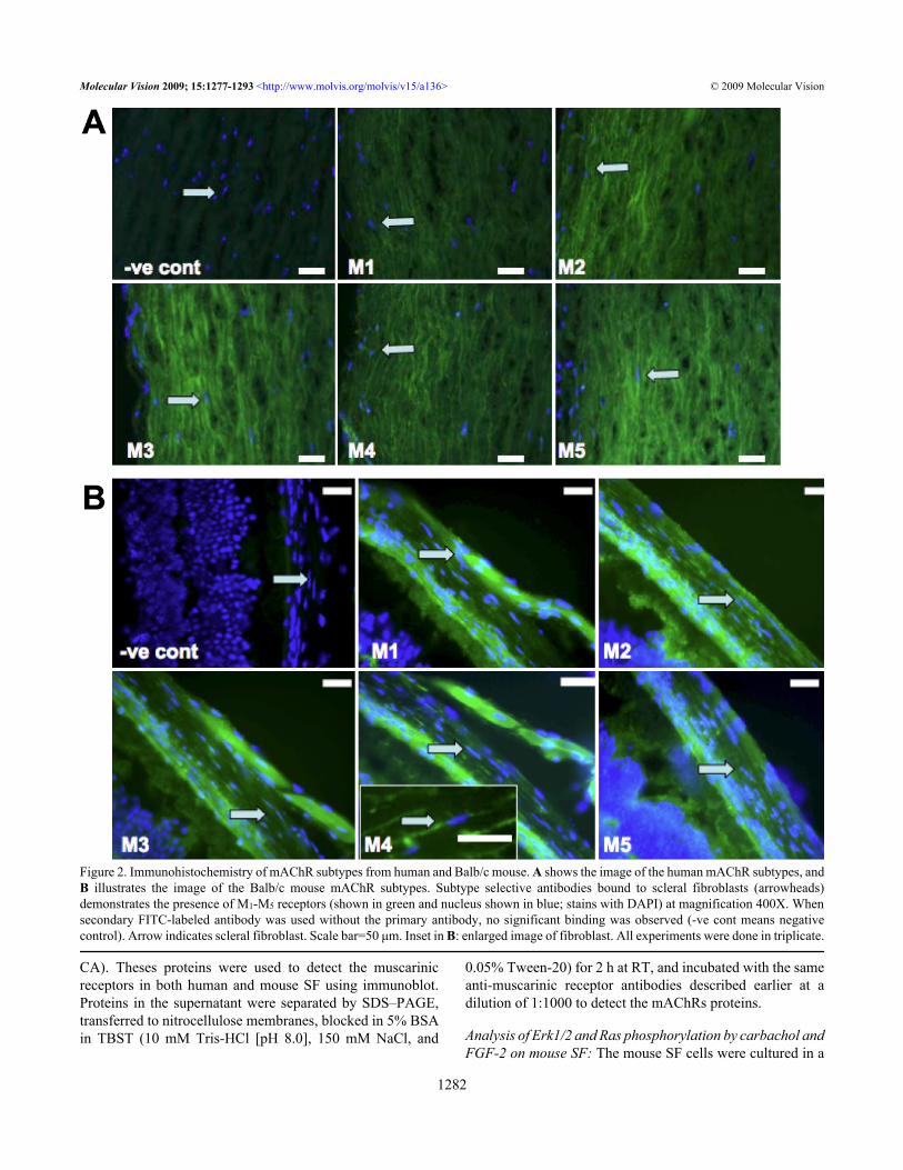
Figure 2. Immunohistochemistry of mAChR subtypes from human and Balb/c mouse. A shows the image of the human mAChR subtypes, andB illustrates the image of the Balb/c mouse mAChR subtypes. Subtype selective antibodies bound to scleral fibroblasts (arrowheads)demonstrates the presence of M1-M5 receptors (shown in green and nucleus shown in blue; stains with DAPI) at magnification 400X. Whensecondary FITC-labeled antibody was used without the primary antibody, no significant binding was observed (-ve cont means negativecontrol). Arrow indicates scleral fibroblast. Scale bar=50 μm. Inset in B: enlarged image of fibroblast. All experiments were done in triplicate.
Molecular Vision 2009; 15:1277-1293 <http://www.molvis.org/molvis/v15/a136> © 2009 Molecular Vision
1282
serum-containing medium in six wells (2×10⁵ cells/well).Cultured mouse SFs were starved for 24 hours and then wasincubated with 50 mg/ml carbachol or 50 ng/ml FGF-2 for 0.5,1, 2, 6 h and over night to detect the optimal time required forMAPK (ERK1/2) and Ras phosphorylation in these cells. Fordetection, western blot was performed as described below.Analysis of phosphorylation by muscarinic agents in mouseSF: The mouse SF cells were cultured in a serum-containingmedium in six wells (2×10⁵ cells/well). From the earlieranalysis, we found that 30 min was the optimal time to detectthe ERK1/2 phosphorylation. Hence, we have treated themouse SFs with freshly prepared atropine, pirenzepine, andcarbachol at a concentration of 50 µM and MT-1 and MT-7at a concentration of 0.5 µM for 30 min to determine thesignaling phosphorylation of EGF-R, PKC, Pyk2, B-Raf, Ras,JNK1/2 and ERK1/2 proteins with the effect of muscarinicagents. Protein extraction and electrophoresis were performedas described above.
For detection, the membrane was incubated with anti-Rasclone RAS10, phospho-EGF-R, PKC, Pyk2, B-Raf, Ras,JNK1/2, and ERK1/2 antibodies at the dilution of 1:1000
according to the instructions provided by the company(Millipore, Billerica, MA) for 2 h at RT, and β-tubulin wasused as a loading control and was incubated for 1 h at RT.
All three experiments’ membranes were washed threetimes in TBST and incubated with HRP-conjugated secondaryantibody (Chemicon International, Temecula, CA) at adilution of 1:2500 for 1 h at RT. Immunoreactive bands werevisualized using the enhanced chemiluminescence method(GE Healthcare, Buckinghamshire, UK). The membrane waswrapped in plastic and placed against X-ray film, exposed foran appropriate length of time (30 s to 5 min), and developed(Kodak, Rockford, IL).Growth factor receptors ELISA: Mouse SF cell lysate wasexposed to atropine and carbachol at concentrations of 0, 0.1,1, 10, and 100 µM for 24 h. Cellular EGF, fibroblast growthfactor-2 (FGF-2), and transforming growth factor (TGF)-β1receptors in the presence of muscarinic agents was quantifiedby an ELISA. Equal amounts of samples or standards wereadded to each well (anti-EGF-R, -FGF-2, and -TGF-β1antibody pre-coated) and incubated for 2 h at RT. The wellswere aspirated and washed with PBST. Anti-EGF, -FGF-2
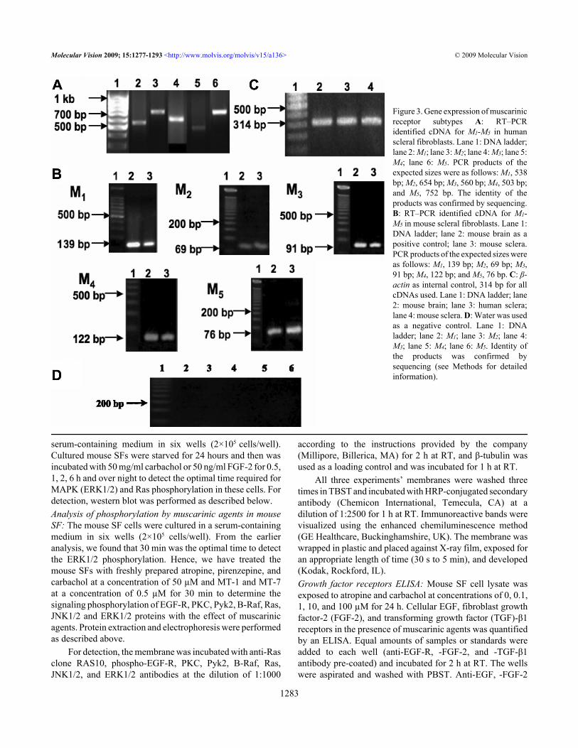
Figure 3. Gene expression of muscarinicreceptor subtypes A: RT–PCRidentified cDNA for M1-M5 in humanscleral fibroblasts. Lane 1: DNA ladder;lane 2: M1; lane 3: M2; lane 4: M3; lane 5:M4; lane 6: M5. PCR products of theexpected sizes were as follows: M1, 538bp; M2, 654 bp; M3, 560 bp; M4, 503 bp;and M5, 752 bp. The identity of theproducts was confirmed by sequencing.B: RT–PCR identified cDNA for M1-M5 in mouse scleral fibroblasts. Lane 1:DNA ladder; lane 2: mouse brain as apositive control; lane 3: mouse sclera.PCR products of the expected sizes wereas follows: M1, 139 bp; M2, 69 bp; M3,91 bp; M4, 122 bp; and M5, 76 bp. C: β-actin as internal control, 314 bp for allcDNAs used. Lane 1: DNA ladder; lane2: mouse brain; lane 3: human sclera;lane 4: mouse sclera. D: Water was usedas a negative control. Lane 1: DNAladder; lane 2: M1; lane 3: M2; lane 4:M3; lane 5: M4; lane 6: M5. Identity ofthe products was confirmed bysequencing (see Methods for detailedinformation).
Molecular Vision 2009; 15:1277-1293 <http://www.molvis.org/molvis/v15/a136> © 2009 Molecular Vision
1283

(Chemicon International Inc.), and -TGF-β1 (AmershamPharmacia Biotech, Buckinghamshire, UK) mouse IgGconjugated with horse-radish peroxidase was added to therespective wells and incubated for 2 h at RT. Substratesolution was added to each well, resulting in a color changeproportional to the amount of EGF-R, FGF-2, and TGF-β1present in the samples. Samples were allowed to develop colorfor 20 min at RT. Adding stop solution stopped the reaction,and the optical density was determined within 30 min usingSpectrafluor Plus microplate reader (TECAN) at dualwavelengths of 450 nm and 620 nm. Quantification wasachieved by the construction of a standard curve using theknown concentration of EGF, FGF-2, and TGF-β1 receptors.
Data analysis: Statistical comparisons between experimentalgroups were conducted using Student’s t-test or one-wayANOVA (Statistica 6.0; SPSS, Chicago, IL) followed byTukey post hoc test. A significance level of p<0.05 was used.Data are presented as means±SEM (standard error of themean).
RESULTSCellular expression of M1-M5: Immunohistochemicallocalization of muscarinic receptors M1-M5 was performed inpassage 2-cultured human and mouse scleral fibroblasts (SFs)as well as scleral tissues. Positive immunostaining for all fivemAChR subtypes was shown in the cultured human andmouse SFs (Figure 1A,B, respectively) as well as in humanand mouse scleral tissues (Figure 2A,B, respectively). TheM1-M5 receptors were localized to the cell membrane as wellas to the cytoplasm. No immunostaining was observed in thenegative controls. Thus, all five receptor types wererepresented in mouse and human scleral fibroblasts at passage2 as well as in the mouse scleral tissue.
Figure 4. Muscarinic receptor sub-types transcript levels in mousesclera. The bar graph compares the relative gene expression of mousescleral muscarinic receptor subtypes to the corresponding mousebrain mRNA level (range) after normalization with 18S rRNAinternal standard. The mRNA level of M1, M3, and M4 in mouse sclerawas less than in the mouse brain. However, the mRNA level of M2
and M5 was abundant in the mouse sclera.
Gene expression of muscarinic receptor subtypes: RNA(1 µg) sample from each collected tissue (n=10 sclera) wasseparated on a 1.2% agarose gel to determine the RNA puritybefore further analysis. By conventional PCR, expression ofall five mAChRs was detected in human and mouse sclera(Figure 3A,B, respectively). The identity of each PCR productwas further confirmed by sequencing. Mouse primersequences showed a high sequence identity to the NCBI BlastmAChR subtypes of mouse sequences (M1 99%, M2 100%,M3 100%, M4 100%, M5 100%). As none of the five mAChRgenes has introns, the PCR product from genomic DNA wouldyield the same-sized amplicons. To rule out possiblecontamination by genomic DNA, we chose a pair of β-actinprimers at two adjacent exons that spanned a 206 bp intron oncDNA derived from β-actin mRNA. The identification of asingle reverse transcription polymerase chain reaction (RT–PCR) product of predicted size for β-actin mRNA rules outcontamination by genomic DNA. The β-actin primers weredesigned to span a 206 bp intron, finding a single band of 314bp verifies that genomic DNA was not present.
Figure 5. Muscarinic receptor protein expression. A:Immunoreactive bands corresponding to each muscarinic receptorsubtype in mouse scleral fibroblasts and their estimated molecularweights are shown: ~58 kDa (M1), ~52 kDa (M2), ~52 kDa (M3),~66 kDa (M4), and ~65 kDa (M5). Blots are representative data fromat least three independent experiments. B: Immunoreactive bandscorresponding to each muscarinic receptor subtypes in human scleralfibroblasts and their estimated molecular weights are shown:~57 kDa (M1), ~52 kDa (M2), ~56 kDa (M3), ~58 kDa (M4), and~51 kDa (M5). Blots are representative data from at least threeindependent experiments. Molecular standards (50-75 kDa) were runon the same blot parallel with muscarinic receptor proteins. Thepositions are shown in the left hand side.
Molecular Vision 2009; 15:1277-1293 <http://www.molvis.org/molvis/v15/a136> © 2009 Molecular Vision
1284

Quantitative real-time PCR was used to compare therelative abundance of the transcript of each mAChR in thesclera. These levels were also compared to M1-M5 geneexpression in the mouse brain cerebellum. Normalizedexpression levels of M1, M3, and M4 were lower in the sclerawhen compared to the levels for the mouse cerebellum(p<0.001). However, the levels of M2 and M5 were greater inthe sclera when compared to the mouse cerebellum (p<0.05,n=10 sclera from five mice; Figure 4).Western blot analysis: Western blot analysis using SFs atpassage 3 was performed using human and mouse cells to
determine levels of protein expression of each muscarinicreceptor subtype. Figure 5 shows that major bandsrepresenting M1 (58 kDa), M2 (52 kDa), M3 (52 kDa), M4 (66kDa), and M5 (65 kDa) were detected. All five mAChRproteins were expressed in both mouse (Figure 5A) and human(Figure 5B) SFs.Muscarinic antagonists inhibited DNA synthesis: Asmuscarinic receptors are often linked to proliferation, we usedthis as an indicator of cell function of pharmacological controlexerted through mAChRs. SFs were exposed to atropine,pirenzepine, or MT-7 for 24 h using a range of concentrations.
Figure 6. Muscarinic agents and mousescleral fibroblast cell proliferation. A:The effect of muscarinic agents onmouse scleral fibroblast cellproliferation is illustrated on the graph.SFs were incubated with atropine,pirenzepine, carbachol, himbacine, and4-DAMP at 0.1–100 μM and withmuscarinic toxin-7 (MT-7), muscarinictoxin-1 (MT-1) at 0.1, 1, 2, 4 μM all for24 h, and BrdU incorporation wasmeasured by ELISA. Antagonistssignificantly inhibited DNA synthesis ina dose-dependent manner (p<0.05,ANOVA, n=4). In contrast, muscarinicreceptor agonists, carbachol and MT-1,increased cell proliferation in a dose-dependent manner (p<0.05, ANOVA,n=4). Data are represented as mean±SEM. The asterisk indicates p<0.05versus control (Post Hoc Analysis;Tukey Honest Significant Difference).B: Effects of muscarinic agents on cellproliferation of human scleralfibroblasts are shown. Scleralfibroblasts were incubated withatropine, pirenzepine, carbachol, andatropine/carbachol at 0.1–100 μM for 24h, and BrdU incorporation wasmeasured by ELISA. Antagonistssignificantly inhibited DNA synthesis ina dose-dependent manner (p<0.05,ANOVA, n=4). In contrast, muscarinicreceptor agonist, carbachol, increasedcell proliferation in a dose-dependentmanner (p<0.05, ANOVA, n=4).Atropine was more effective at 10 and100 μM than pirenzepine at all of theconcentrations. Data are represented asmean±SEM. The asterisk indicatesp<0.05 versus control (Post HocAnalysis; Tukey Honest SignificantDifference).
Molecular Vision 2009; 15:1277-1293 <http://www.molvis.org/molvis/v15/a136> © 2009 Molecular Vision
1285
By microscopic observation, cell morphology showed littledifferences between treatment groups and control groups, andviability as determined by the trypan blue exclusion assayshowed that there was no toxicity after 24 h incubation of thecells with up to 100 μM of atropine (EC50=501.1 μm or0.5011 mM) and pirenzepine (EC50=7040 μm or 7.040 mM)or 1 μM of MT-7. DNA synthesis, measured after 24 h usinga BrdU ELISA assay, was inhibited after atropine,
pirenzepine, and MT-7 exposure when compared withuntreated control SFs (p<0.05, n=4; Figure 6A,B). In addition,when examining the competition between the agonists andantagonists, atropine, pirenzepine, and MT-7 blocked theeffect of the action of carbachol (EC50=177.827 mM) andMT-1 (muscarinic agonist) when SFs were pre-treated (3 h)with an equimolar amount of antagonists (Figure 7). MT-7,which is selective for the M1, was almost as effective as
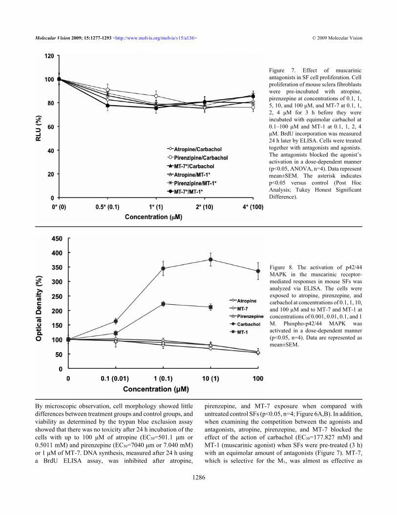
Figure 7. Effect of muscarinicantagonists in SF cell proliferation. Cellproliferation of mouse sclera fibroblastswere pre-incubated with atropine,pirenzepine at concentrations of 0.1, 1,5, 10, and 100 μM, and MT-7 at 0.1, 1,2, 4 μM for 3 h before they wereincubated with equimolar carbachol at0.1–100 μM and MT-1 at 0.1, 1, 2, 4μM. BrdU incorporation was measured24 h later by ELISA. Cells were treatedtogether with antagonists and agonists.The antagonists blocked the agonist’sactivation in a dose-dependent manner(p<0.05, ANOVA, n=4). Data representmean±SEM. The asterisk indicatesp<0.05 versus control (Post HocAnalysis; Tukey Honest SignificantDifference).
Figure 8. The activation of p42/44MAPK in the muscarinic receptor-mediated responses in mouse SFs wasanalyzed via ELISA. The cells wereexposed to atropine, pirenzepine, andcarbachol at concentrations of 0.1, 1, 10,and 100 µM and to MT-7 and MT-1 atconcentrations of 0.001, 0.01, 0.1, and 1M. Phospho-p42/44 MAPK wasactivated in a dose-dependent manner(p<0.05, n=4). Data are represented asmean±SEM.
Molecular Vision 2009; 15:1277-1293 <http://www.molvis.org/molvis/v15/a136> © 2009 Molecular Vision
1286

atropine, a pan-muscarinic blocker, in decreasing BrdUuptake.Effect of muscarinic agents on MAPK activity: From thepreceding data, it would be suggested that the MAPKpathways should be activated. We examined phosphorylationof p42/44 MAPK in mouse SFs following exposure toatropine, pirenzepine, and carbachol at concentrations of 0.1,1, 10, and 100 µM and to MT-7 and MT-1 at concentrationsof 0.001, 0.01, 0.1, and 1 µM using ELISA. Phospho-p42/44MAPK was formed in a dose-dependent manner (Figure 8).Phospho-p42/44 MAPK decreased with the muscarinicantagonists and increased in response to the agonists (p<0.05,n=4).
Immunoblot analysis: The muscarinic receptors have beeninvolved in trans-activation with tyrosine kinase receptors.We examined several specific points in the pathway to providefurther information about the MAPK pathway. Phospho-EGF-R, -PKC, -Pyk2, -B-Raf, -Ras, -JNK1/2, and total/phospho-Erk1/2 were expressed at protein levels in mousescleral fibroblasts following 30 min exposure to muscarinicagents, and β-tubulin was used as the loading control (Figure9). The antagonists (atropine, pirenzepine, and MT-7)inhibited the formation of phospho-EGF-R, -PKC, -Pyk2, -B-Raf, -Ras, -JNK1/2, and -ERK1/2. In the absence of treatment,there was no effect on total Erk1/2, β-tubulin, or control. In
Figure 9. Effect of muscarinic agents on MAPK signaling proteins.Phospho-EGF-R, PKC, Pyk2, B-Raf, Ras, JNK1/2 andERK1/2activity was detected by immunoblot in the cultured mouseSF cells after being treated with atropine, pirenzepine, and carbacholat a concentration of 50 µM and with MT-7 and MT-1 at aconcentration of 0.5 µM all for 30 min. β-tubulin was used as aloading control for all the treatment. Antagonists increased theiractivation, and agonists reversed this.
contrast, in SFs that were treated with the agonists (carbacholand MT-1), the activation of phospho-EGF-R, -PKC, -Pyk2,-B-Raf, -Ras, -JNK1/2, and -ERK1/2 increased.Carbachol-induced Ras and ERK1/2 phosphorylation: Wefurther demonstrated the carbachol-Induced Ras and ERK1/2phosphorylation in mouse SFs to show the muscarinic agentsalso had parallel effects on the appropriate signal transductionpathways. As seen below, carbachol produced a time-anddose-dependent increase in Ras and MAPK/ERKphosphorylation in SFs.
We examined the activation of Ras and p42/44 MAPK inthe muscarinic receptor-mediated responses in SF cells.Stimulation with 50 mg/ml of carbachol resulted in increasedactivity in Ras and p42/44 MAPK in a time-dependent manner(Figure 10). Ras and p42/44 MAPK activation was initiallyobserved at 30 min with 50 mg/ml of carbachol stimulation(p<0.05, n=4). As a comparison to the activation of p42/44kinase elicited by carbachol, we tested fibroblast growthfactor (FGF-2) at 50 ng/ml, which was also found to activateRas and p42/44 MAPK (Figure 11) with peak phosphorylationat 30 min (p<0.05, n=4). At 2 h, the level of Ras and phospho-
Figure 10. Activation of ERK1/2 and Ras by 50 mg/ml carbachol incultured mouse scleral fibroblast cells. Cultured mouse SF cells wereserum starved for 24 h in DMEM and then incubated with 50 mg/mlof carbachol for 0.5, 1, 2, and 6 h as well as overnight. Time-dependent change of phospho-p42/44 MAPK and Ras in the presenceof 50 mg/ml of carbachol was detected by immunoblot analysis.
Figure 11. Activation of p42/44 MAPK and Ras proteins with FGF-2treatment. Activation of p42/44 MAPK and Ras by 50 ng/ml ofFGF-2 in cultured mouse scleral fibroblast cells was detected byimmunoblot analysis. Cultured mouse SF cells were serum starvedfor 24 h in DMEM and then incubated with 50 ng/ml of FGF-2 for0.5, 1, 2, and 6 h as well as overnight. Time-dependent change ofp42/44 MAPK in the presence of 50 ng/ml of FGF-2 was detected.
Molecular Vision 2009; 15:1277-1293 <http://www.molvis.org/molvis/v15/a136> © 2009 Molecular Vision
1287
p42/44 MAPK returned to basal levels (p<0.05, n=4). Thep42/44 MAPK activation patterns for carbachol and FGF-2were similar (Figure 10 and Figure 11).Carbachol stimulates EGF-R and TGF-β1 activity: Past studyillustrated that scleral cells are directly responsible for DNAsynthesis and extracellular matrix synthesis that producesaxial elongation [33]. Muscarinic agents may act directly onsclera fibroblast through the G protein coupled muscarinicreceptor to modulate postnatal eye development. We assumethat the effect of muscarinic antagonists may also be mediateddirectly or indirectly by growth factors through receptortyrosine kinases, which would then control sclera fibroblast
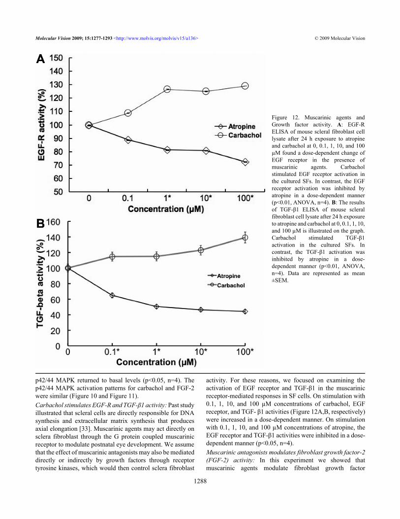
activity. For these reasons, we focused on examining theactivation of EGF receptor and TGF-β1 in the muscarinicreceptor-mediated responses in SF cells. On stimulation with0.1, 1, 10, and 100 µM concentrations of carbachol, EGFreceptor, and TGF- β1 activities (Figure 12A,B, respectively)were increased in a dose-dependent manner. On stimulationwith 0.1, 1, 10, and 100 µM concentrations of atropine, theEGF receptor and TGF-β1 activities were inhibited in a dose-dependent manner (p<0.05, n=4).Muscarinic antagonists modulates fibroblast growth factor-2(FGF-2) activity: In this experiment we showed thatmuscarinic agents modulate fibroblast growth factor
Figure 12. Muscarinic agents andGrowth factor activity. A: EGF-RELISA of mouse scleral fibroblast celllysate after 24 h exposure to atropineand carbachol at 0, 0.1, 1, 10, and 100µM found a dose-dependent change ofEGF receptor in the presence ofmuscarinic agents. Carbacholstimulated EGF receptor activation inthe cultured SFs. In contrast, the EGFreceptor activation was inhibited byatropine in a dose-dependent manner(p<0.01, ANOVA, n=4). B: The resultsof TGF-β1 ELISA of mouse scleralfibroblast cell lysate after 24 h exposureto atropine and carbachol at 0, 0.1, 1, 10,and 100 µM is illustrated on the graph.Carbachol stimulated TGF-β1activation in the cultured SFs. Incontrast, the TGF-β1 activation wasinhibited by atropine in a dose-dependent manner (p<0.01, ANOVA,n=4). Data are represented as mean±SEM.
Molecular Vision 2009; 15:1277-1293 <http://www.molvis.org/molvis/v15/a136> © 2009 Molecular Vision
1288
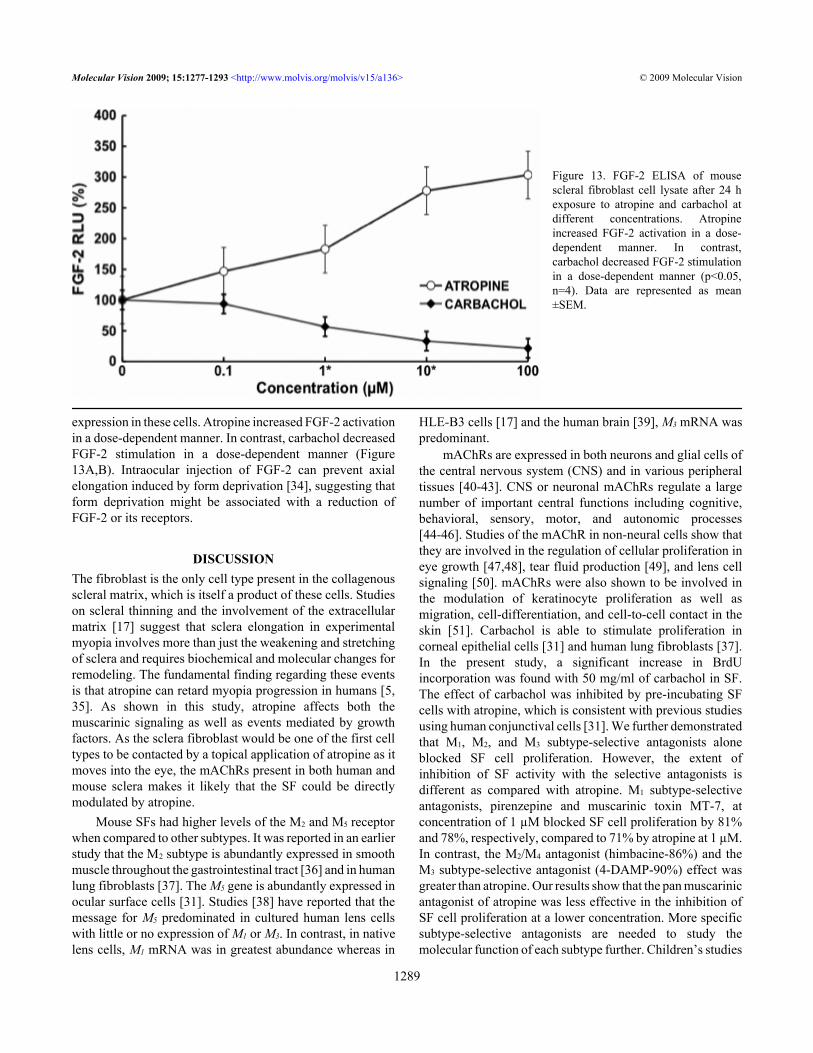
expression in these cells. Atropine increased FGF-2 activationin a dose-dependent manner. In contrast, carbachol decreasedFGF-2 stimulation in a dose-dependent manner (Figure13A,B). Intraocular injection of FGF-2 can prevent axialelongation induced by form deprivation [34], suggesting thatform deprivation might be associated with a reduction ofFGF-2 or its receptors.
DISCUSSIONThe fibroblast is the only cell type present in the collagenousscleral matrix, which is itself a product of these cells. Studieson scleral thinning and the involvement of the extracellularmatrix [17] suggest that sclera elongation in experimentalmyopia involves more than just the weakening and stretchingof sclera and requires biochemical and molecular changes forremodeling. The fundamental finding regarding these eventsis that atropine can retard myopia progression in humans [5,35]. As shown in this study, atropine affects both themuscarinic signaling as well as events mediated by growthfactors. As the sclera fibroblast would be one of the first celltypes to be contacted by a topical application of atropine as itmoves into the eye, the mAChRs present in both human andmouse sclera makes it likely that the SF could be directlymodulated by atropine.
Mouse SFs had higher levels of the M2 and M5 receptorwhen compared to other subtypes. It was reported in an earlierstudy that the M2 subtype is abundantly expressed in smoothmuscle throughout the gastrointestinal tract [36] and in humanlung fibroblasts [37]. The M5 gene is abundantly expressed inocular surface cells [31]. Studies [38] have reported that themessage for M5 predominated in cultured human lens cellswith little or no expression of M1 or M3. In contrast, in nativelens cells, M1 mRNA was in greatest abundance whereas in
HLE-B3 cells [17] and the human brain [39], M3 mRNA waspredominant.
mAChRs are expressed in both neurons and glial cells ofthe central nervous system (CNS) and in various peripheraltissues [40-43]. CNS or neuronal mAChRs regulate a largenumber of important central functions including cognitive,behavioral, sensory, motor, and autonomic processes[44-46]. Studies of the mAChR in non-neural cells show thatthey are involved in the regulation of cellular proliferation ineye growth [47,48], tear fluid production [49], and lens cellsignaling [50]. mAChRs were also shown to be involved inthe modulation of keratinocyte proliferation as well asmigration, cell-differentiation, and cell-to-cell contact in theskin [51]. Carbachol is able to stimulate proliferation incorneal epithelial cells [31] and human lung fibroblasts [37].In the present study, a significant increase in BrdUincorporation was found with 50 mg/ml of carbachol in SF.The effect of carbachol was inhibited by pre-incubating SFcells with atropine, which is consistent with previous studiesusing human conjunctival cells [31]. We further demonstratedthat M1, M2, and M3 subtype-selective antagonists aloneblocked SF cell proliferation. However, the extent ofinhibition of SF activity with the selective antagonists isdifferent as compared with atropine. M1 subtype-selectiveantagonists, pirenzepine and muscarinic toxin MT-7, atconcentration of 1 µM blocked SF cell proliferation by 81%and 78%, respectively, compared to 71% by atropine at 1 µM.In contrast, the M2/M4 antagonist (himbacine-86%) and theM3 subtype-selective antagonist (4-DAMP-90%) effect wasgreater than atropine. Our results show that the pan muscarinicantagonist of atropine was less effective in the inhibition ofSF cell proliferation at a lower concentration. More specificsubtype-selective antagonists are needed to study themolecular function of each subtype further. Children’s studies
Figure 13. FGF-2 ELISA of mousescleral fibroblast cell lysate after 24 hexposure to atropine and carbachol atdifferent concentrations. Atropineincreased FGF-2 activation in a dose-dependent manner. In contrast,carbachol decreased FGF-2 stimulationin a dose-dependent manner (p<0.05,n=4). Data are represented as mean±SEM.
Molecular Vision 2009; 15:1277-1293 <http://www.molvis.org/molvis/v15/a136> © 2009 Molecular Vision
1289
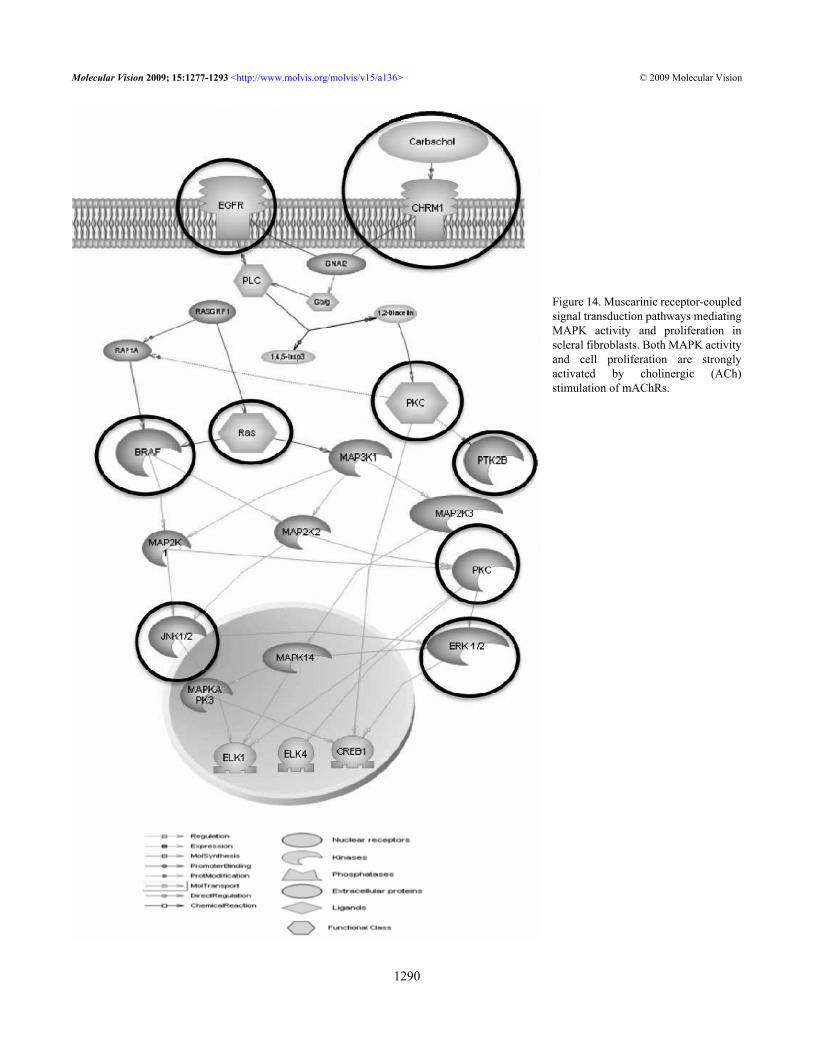
Figure 14. Muscarinic receptor-coupledsignal transduction pathways mediatingMAPK activity and proliferation inscleral fibroblasts. Both MAPK activityand cell proliferation are stronglyactivated by cholinergic (ACh)stimulation of mAChRs.
Molecular Vision 2009; 15:1277-1293 <http://www.molvis.org/molvis/v15/a136> © 2009 Molecular Vision
1290
with 2% pirenzepine [6] showed that 2% pirenzepine was notas effective as 1% atropine [35] against myopia progression,which shows that the M1 receptor is not as critical in myopiaas are some of the other types.
Epidermal growth factor (EGF) and transforming growthfactor-β1 (TGF-β1) have a vital role in cell repair, stimulationof cell proliferation, cellular adhesion regulation,differentiation, hematopoiesis, apoptosis, tumorogenesis,migration, and extracellular matrix (ECM) production [52].EGF functions via the epidermal growth factor receptor (EFG-R), a member of the ErbB family of receptor tyrosine kinases[53]. Tyrosine phosphorylation of EGF-R leads to theactivation of the ERK/MAPK pathways [54]. EGF-Rfunctions to transmit intracellular signals leading to regulationof cell growth. Alterations in the regulation of EGF-R functionand overexpression of receptor or ligand results in cellproliferation. Anti-myopic effects of atropine may bemediated directly by muscarinic receptor or indirectly throughgrowth factors such as FGF-2 and TGF-β, which then controlSF cell proliferation. Growth factors modulate cellproliferation and composition of the extracellular matrix.
Carbachol-stimulated conjunctival epithelial cellproliferation correlates with the activation of p42/44 MAPK[27]. Our study showed that 50 mg/ml of carbachol activatedp42/44 MAPK with kinetics similar to EGF-R. This wassimilar to the earlier study reported in corneal epithelial cells[31]. Carbachol-stimulated activation of p42/44 MAPK hasbeen observed in goblet cells through increased intracellularCa2+ concentration or transactivation of EGF pathways viaphosphorylation of Pyk2 and Src [55,56]. Carbachol-inducedcontractions in the urinary bladder are reported to be mediatedby G-protein coupled muscarinic M3 receptors [57]. M3
muscarinic receptors also mediate contraction in the guineapig taenia cecum [58], and regulation of G protein levels couldbe playing a role in controlling muscarinic receptor activity invivo [59].
Selected entities from our studies were analyzed withinteracting muscarinic pathways using pathway studio 6.0(Ariadne Genomics, Rockville, MD; Figure 14) to explore theunderlying intracellular signaling pathways involved in SFstreated with muscarinic agents. EGF-R is in the upstream, andall mAChRs are mediated via activation of the Ras-Raf-MAPK cascade. Both MAPK activity and cell proliferationare strongly activated by cholinergic (ACh) stimulation ofmAChRs. Our data also support a role for G-protein coupledreceptor-signaling pathway, which could be important for themuscarinic receptor mediated scleral cell proliferation [60].
In summary, our study showed the quantitativedistribution of the five mAChRs in mouse SFs for the firsttime and also confirmed the presence of all five mAChRs inhuman and mouse SFs. Activation of mAChRs by carbacholled to increased SF cell proliferation. The mitogenic effect ofcarbachol correlated with the activation of p42/44 MAPK.
The SF treated together with antagonists and agonists, themuscarinic antagonists blocked the agonist’s activation in adose-dependent manner. Muscarinic agents modulatefibroblast growth factor expression in these cells. Atropineincreased FGF-2 activation in a dose-dependent manner. Incontrast, carbachol decreased FGF-2 stimulation in a dose-dependent manner. M2 and M5 genes are abundantly expressedin SFs and should be considered in the future design andselection of muscarinic receptor agonists or antagonists foruse in topical eye drops.
It is shown that mAChRs mediate proliferation of humanand mouse SF, a mechanism possibly involved in scleralremodeling. Blockade of these receptors might contribute tolong-term beneficial effects of an anti-cholinergic drug inmyopia.
ACKNOWLEDGMENTSThis study was supported by a Research Grant from theSingapore National Medical Research Council(R284/28/2002-PG) and IBG-core. Part of this study waspresented at the 2nd SERI-ARVO Meeting on Research inVision and Ophthalmology (16–19 February 2005) at SuntecCity, Singapore. The authors also wish to thank HowardCajucom-Uy from the Singapore Eye Bank for assisting withthe human scleral tissues.
REFERENCES1. Wu HM, Seet B, Yap EP, Saw SM, Lim TH, Chia KS. Does
education explain ethnic differences in myopia prevalence?A population-based study of young adult males in Singapore.Optom Vis Sci 2001; 78:234-9. [PMID: 11349931]
2. Saw SM. A synopsis of the prevalence rates and environmentalrisk factors for myopia. Clin Exp Optom 2003; 86:289-94.[PMID: 14558850]
3. Saw SM. How blinding is pathological myopia? Br JOphthalmol 2006; 90:525-6. [PMID: 16622078]
4. Shih YF, Hsiao CK, Chen CJ, Chang CW, Hung PT, Lin LL.An intervention trial on efficacy of atropine and multi-focalglasses in controlling myopic progression. Acta OphthalmolScand 2001; 79:233-6. [PMID: 11401629]
5. Saw SM, Gazzard G, Au Eong KG, Tan DT. Myopia: attemptsto arrest progression. Br J Ophthalmol 2002; 86:1306-11.[PMID: 12386095]
6. Tan DT, Lam DS, Chua WH, Shu-Ping DF, Crockett RS, AsianPirenzepine Study Group. One-year multicenter, double-masked, placebo-controlled, parallel safety and efficacy studyof 2% pirenzepine ophthalmic gel in children with myopia.Ophthalmology 2005; 112:84-91. [PMID: 15629825]
7. Leech EM, Cottriall CL, McBrien NA. Pirenzepine preventsform deprivation myopia in a dose dependent manner.Ophthalmic Physiol Opt 1995; 15:351-6. [PMID: 8524553]
8. Tigges M, Iuvone PM, Fernandes A, Sugrue MF, Mallorga PJ,Laties AM, Stone RA. Effects of muscarinic cholinergicreceptor antagonists on postnatal eye growth of rhesusmonkeys. Optom Vis Sci 1999; 76:397-407. [PMID:10416935]
Molecular Vision 2009; 15:1277-1293 <http://www.molvis.org/molvis/v15/a136> © 2009 Molecular Vision
1291

9. Duncan G, Collison DJ. Role of the non-neuronal cholinergicsystem in the eye: a review. Life Sci 2003; 72:2013-9. [PMID:12628451]
10. Barathi VA, Boopathi VG, Yap EP, Beuerman RW. Twomodels of experimental myopia in the mouse. Vision Res2008; 48:904-16. [PMID: 18289630]
11. Caulfield MP. Muscarinic receptors-characterization, couplingand function. Pharmacol Ther 1993; 58:319-79. [PMID:7504306]
12. Wessler I, Kirkpatrick CJ, Racke K. Non-neuronalacetylcholine, a locally acting molecule, widely distributed inbiological systems: expression and function in humans.Pharmacol Ther 1998; 77:59-79. [PMID: 9500159]
13. Wessler I, Kilbinger H, Bittinger F, Unger R, Kirkpatrick CJ.The non-neuronal cholinergic system in humans: expression,function and pathophysiology. Life Sci 2003; 72:2055-61.[PMID: 12628456]
14. McBrien NA, Cottriall CL, Annies R. Retinal acetylcholinecontent in normal and myopic eyes: a role in ocular growthcontrol? Vis Neurosci 2001; 18:571-80. [PMID: 11829303]
15. Weon SR, Barathi VA, Beuerman RW. Muscarinic toxins fromgreen mamba venom modulate the proliferative actions ofmuscarinic receptor subtypes (mAChRs) expressed in mouseand human scleral fibroblasts. Proceedings of the 2nd SERI-ARVO Meeting on Research in Vision and Ophthalmology;2005 February 16-20; Singapore.
16. Fischer AJ, McKinnon LA, Nathanson NM, Stell WK.Identification and localization of muscarinic acetylcholinereceptors in the ocular tissues of the chick. J Comp Neurol1998; 392:273-84. [PMID: 9511918]
17. Lind GJ, Chew SJ, Marzani D, Wallman J. Muscarinicacetylcholine receptor antagonists inhibit chick scleralchondrocytes. Invest Ophthalmol Vis Sci 1998; 39:2217-31.[PMID: 9804129]
18. Kanno H, Horikawa Y, Hodges RR, Zoukhri D, Shatos MA,Rios JD, Dartt DA. Cholinergic agonists transactivate EGFRand stimulate MAPK to induce goblet cell secretion. Am JPhysiol Cell Physiol 2003; 284:C988-98. [PMID: 12620895]
19. Zachary I, Rozengurt E. Focal adhesion kinase (p125fak); apoint of convergence in the action of neuropeptides, integrinsand oncogenes. Cell 1992; 71:891-4. [PMID: 1458538]
20. Aoki Y, Isselbacher KJ, Pillai S. Bruton tyrosine kinase istyrosine phosphorylated and activated in pre B lymphocytesand receptor ligated B cells. Proc Natl Acad Sci USA 1994;91:10606-9. [PMID: 7524098]
21. August A, Gibson S, Kawakami Y, Kawakami T, Mills GB,Dupont B. CD28 is associated with an induces the immediatetyrosine phosphorylation and activation of the Tec familykinase ITK/EMT in the human Jurkat leukemic T cell line.Proc Natl Acad Sci USA 1994; 91:9347-51. [PMID:7524075]
22. Chen JM, Aimes RT, Ward GR, Youngleib GL, Quigley JP.Isolation and characterisation of a 70kd metalloproteinase(gelatinase) that is elevated in Rous sarcoma virustransformed embryo fibroblasts. J Biol Chem 1991;266:5113-21. [PMID: 1848240]
23. Collison DJ, Coleman RA, James RS, Carey J, Duncan G.Characterization of muscarinic receptors in human lens cells
by pharmacologic and molecular techniques. InvestOphthalmol Vis Sci 2000; 41:2633-41. [PMID: 10937576]
24. Gil DW, Krauss HA, Bogardus AM, WoldeMussie E.Muscarinic receptor subtypes in human iris-ciliary bodymeasured by immunoprecipitation. Invest Ophthalmol VisSci 1997; 38:1434-42. [PMID: 9191607]
25. Matthiesen S, Bahulayan A, Kempkens S, Haag S, FuhrmannM, Stichnote C, Juergens UR, Racke K. Muscarinic receptorsmediate stimulation of human lung fibroblast proliferation.Am J Respir Cell Mol Biol 2006; 35:621-7. [PMID:16902194]
26. Ryberg AT, Warfvinge G, Axelsson L, Soukup O, Götrick B,Tobin G. Expression of muscarinic receptor subtypes insalivary glands of rats, sheep and man. Arch Oral Biol 2008;53:66-74. [PMID: 17825245]
27. Qu J, Zhou X, Xie R, Zhang L, Hu D, Li H, Lu F. The presenceof m1 to m5 receptors in human sclera: evidence of the scleraas a potential site of action for muscarinic receptorantagonists. Curr Eye Res 2006; 31:587-97. [PMID:16877267]
28. Truong HT, Cottriall CL, McBrien NA. Expression ofmuscarinic receptors in tree shrew ocular tissues. Mol Vis2009; 15:464-75. [PMID: 19262686]
29. Liu Q, Wu J, Wang X, Zeng J. Changes in muscarinicacetylcholine receptor expression in form deprivation myopiain guinea pigs. Mol Vis 2007; 13:1234-44. [PMID: 17679952]
30. Barathi VA, Weon SR, Kam JH, Wess J, Beuerman RW. (2007)Experimental myopia in muscarinic receptor knockout mice:role of specific muscarinic receptor subtypes. ARVO AnnualMeeting; 2007 May 6-10; Fort Lauderdale (FL).
31. Liu S, Li J, Tan DT, Beuerman RW. The eyelid margin: atransitional zone for 2 epithelial phenotypes. ArchOphthalmol 2007; 125:523-32. [PMID: 17420373]
32. Brink N, Szamel M, Young AR, Wittern KP, Bergemann J.Comparative quantification of IL-1β, IL-10, IL-10γ, TNFαand IL-7 mRNA levels in UV-irradiated human skin in vivo.Inflamm Res 2000; 49:290-6. [PMID: 10939619]
33. Wu YR. DNA, collagen, and uronic acid in form deprivationmyopia. Invest Ophthalmol Vis Sci 1990; 31:254.
34. Rohrer B, Stell WK. Basic fibroblast growth factor (bFGF) andtransforming growth factor beta (TGF-beta) act as stop andgo signals to modulate postnatal ocular growth in the chick.Exp Eye Res 1994; 58:553-61. [PMID: 7925692]
35. Tong L, Huang XL, Koh AL, Zhang X, Tan DT, Chua WH.Atropine for the treatment of childhood myopia: effect onmyopia progression after cessation of atropine.Ophthalmology 2009; 116:572-9. [PMID: 19167081]
36. Ehlert FJ, Ostrom RS, Sawyer GW. Subtypes of the muscarinicreceptor in smooth muscle. Life Sci 1997; 61:1729-40.[PMID: 9365220]
37. Matthiesen S, Bahulayan A, Holz O, Racke K. MAPK pathwaymediates muscarinic receptor-induced human lung fibroblastproliferation. Life Sci 2007; 80:2259-62. [PMID: 17383686]
38. Shepard AR, Rae JL. Ion transporters and receptors in cDNAlibraries from lens and cornea epithelia. Curr Eye Res 1998;17:708-19. [PMID: 9678416]
39. Levey AI, Kitt CA, Simonds WF, Price DL, Brann MR.Identification and localization of muscarinic acetylcholine-receptor proteins in brain with subtype specific antibodies. JNeurosci 1991; 11:3218-26. [PMID: 1941081]
Molecular Vision 2009; 15:1277-1293 <http://www.molvis.org/molvis/v15/a136> © 2009 Molecular Vision
1292
40. Wess J. Molecular biology of muscarinic acetylcholinereceptors. Crit Rev Neurobiol 1996; 10:69-99. [PMID:8853955]
41. Caulfield MP, Birdsall NJM. International Union ofPharmacology. XVII. Classification of muscarinicacetylcholine receptors. Pharmacol Rev 1998; 50:279-90.[PMID: 9647869]
42. Volpicelli LA, Levey AI. Muscarinic acetylcholine receptorsubtypes in cerebral cortex and hippocampus. Prog Brain Res2004; 145:59-66. [PMID: 14650906]
43. Abrams P, Andersson KE, Buccafusco JJ, Chapple C, de GroatWC, Fryer AD, Kay G, Laties A, Nathanson NM, PasrichaPJ, Wein AJ. Muscarinic receptors: their distribution andfunction in body systems, and the implications for treatingoveractive bladder. Br J Pharmacol 2006; 148:565-78.[PMID: 16751797]
44. Felder CC, Bymaster FP, Ward J, DeLapp N. Therapeuticopportunities for muscarinic receptors in the central nervoussystem. J Med Chem 2000; 43:4333-53. [PMID: 11087557]
45. Eglen RM, Choppin A, Watson N. Therapeutic opportunitiesfrom muscarinic receptor research. Trends Pharmacol Sci2001; 22:409-14. [PMID: 11479003]
46. Wess J. Muscarinic acetylcholine receptor knockout mice:novel phenotypes and clinical implications. Annu RevPharmacol Toxicol 2004; 44:423-50. [PMID: 14744253]
47. Gimbel HV. The control of myopia with atropine. Can JOphthalmol 1973; 8:527-32. [PMID: 4751898]
48. Lind GJ, Chew SJ, Marzani D, Wallman J. Muscarinicacetylcholine receptor antagonists inhibit chick scleralchondrocytes. Invest Ophthalmol Vis Sci 1998; 39:2217-31.[PMID: 9804129]
49. Dartt DA. Regulation of lacrimal gland secretion byneurotransmitters and the EGF family of growth factors. ExpEye Res 2001; 73:741-52. [PMID: 11846506]
50. Collison DJ, Coleman RA, James RS, Carey J, Duncan G.Characterization of muscarinic receptors in human lens cellsby pharmacologic and molecular techniques. InvestOphthalmol Vis Sci 2000; 41:2633-41. [PMID: 10937576]
51. Wessler I, Kirkpatrick CJ, Racke K. Non-neuronalacetylcholine, a locally acting molecule, widely distributed in
biological systems: expression and function in humans.Pharmacol Ther 1998; 77:59-79. [PMID: 9500159]
52. Yue J, Mulder KM. Transforming growth factor-beta signaltransduction in epithelial cells. Pharmacol Ther 2001;91:1-34. [PMID: 11707292]
53. Hackel PO, Zwick E, Prenzel N, Ullrich A. Epidermal growthfactor receptors: critical mediators of multiple receptorpathways. Curr Opin Cell Biol 1999; 11:184-9. [PMID:10209149]
54. Voldborg BR, Damstrup L, Spang-Thomsen M, Poulsen HS.Epidermal Growth Factor Receptor (EGFR) and EGFRMutations, Function and Possible Role in Clinical Trials. AnnOncol 1997; 8:1197-206. [PMID: 9496384]
55. Dartt DA. Regulation of mucin and fluid secretion byconjunctival epithelial cells. Prog Retin Eye Res 2002;21:555-76. [PMID: 12433377]
56. Kanno H, Horikawa Y, Hodges RR, Zoukhri D, Shatos MA,Rios JD, Dartt DA. Cholinergic agonists transactivate EGFRand stimulate MAPK to induce goblet cell secretion. Am JPhysiol Cell Physiol 2003; 284:C988-98. [PMID: 12620895]
57. Uchiyama T, Chess-Williams R. Muscarinic receptor subtypesof the bladder and gastrointestinal tract. J Smooth Muscle Res2004; 40:237-47. [PMID: 15725706]
58. Elnatan A, Mitchelson F. The interaction of McN-A- 343 withmuscarine receptors in cardiac and smooth muscle. BiochemPharmacol 1993; 46:993-1003. [PMID: 7692853]
59. Burstein ES, Spalding TA, Brann MR. Pharmacology ofmuscarinic receptor subtypes constitutively activated by Gproteins. Mol Pharmacol 1997; 51:312-9. [PMID: 9203637]
60. Yuryev A, Mulyukov Z, Kotelnikova E, Maslov S, Egorov S,Nikitin A, Daraselia N, Mazo I. Automatic pathway buildingin biological association networks. BMC Bioinformatics2006; 7:171. [PMID: 16563163]
61. Diebold Y, Rios JD, Hodges RR, Rawe I, Dartt DA. Presenceof nerves and their receptors in mouse and humanconjunctival goblet cells. Invest Ophthalmol Vis Sci 2001;42:2270-82. [PMID: 11527940]
Molecular Vision 2009; 15:1277-1293 <http://www.molvis.org/molvis/v15/a136> © 2009 Molecular Vision
The print version of this article was created on 27 June 2009. This reflects all typographical corrections and errata to the articlethrough that date. Details of any changes may be found in the online version of the article.
1293