expression of large tenascin-c splice variants by hepatic stellate cells in chronic hepatitis c...
TRANSCRIPT
Expression of Large Tenascin-C Splice Expression of Large Tenascin-C Splice Variants by Hepatic Stellate Cells in Variants by Hepatic Stellate Cells in
Chronic Hepatitis C Chronic Hepatitis C
Dr.Amro El-karefDr.Amro El-karefAssistant Professor of PathologyAssistant Professor of Pathology
Department of PathologyDepartment of PathologyMansoura UniversityMansoura University
IntroductionIntroduction* Chronic Hepatitis C is a worldwide liver * Chronic Hepatitis C is a worldwide liver disease that causes liver fibrosis, cirrhosis disease that causes liver fibrosis, cirrhosis & hepatocellular carcinoma. & hepatocellular carcinoma. (Lauer 2001)(Lauer 2001)
* Tenascin-C (TN-C), a hexameric ECM glyco-* Tenascin-C (TN-C), a hexameric ECM glyco- protein, is a part of provisional matrix that protein, is a part of provisional matrix that modulates cellular functions during tissue modulates cellular functions during tissue remodleing & cancer invasion. remodleing & cancer invasion. (Chiquet-Ehrismann 1993)(Chiquet-Ehrismann 1993)
* TN-C has different isoforms due to alternative * TN-C has different isoforms due to alternative splicing of its mRNA. splicing of its mRNA. (Jones 2001)(Jones 2001)
* Serum levels of large TN-C was found to * Serum levels of large TN-C was found to correlate well with the grades of piecmeal correlate well with the grades of piecmeal necrosis. necrosis. (Tanaka 2005 – in press)(Tanaka 2005 – in press)
The structure of human TN-C molecule and the recognition sites of different anti-TN-C antibodies
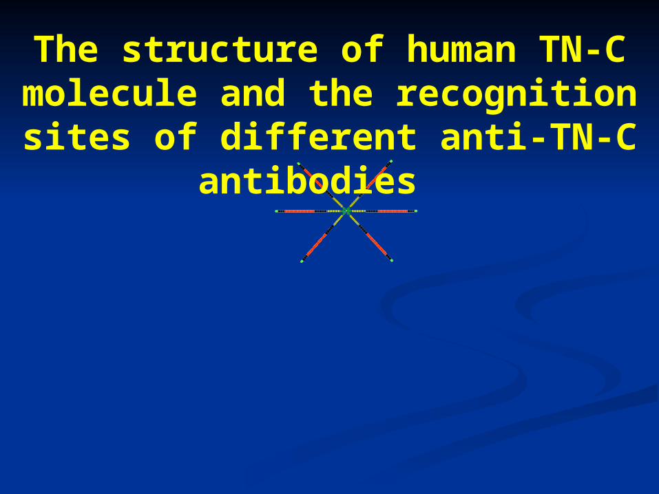
The structure of human TN-C molecule and the recognition sites of different anti-TN-C antibodies
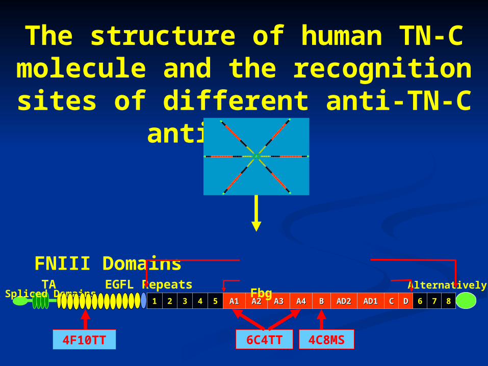
11 22 33 44 55 A1A1 A2A2 A3A3 A4A4 BB AD2AD2 AD1AD1 CC DD 66 77 88
FNIII Domains TA EGFL Repeats Alternatively Spliced Domains Fbg
4F10TT 6C4TT 4C8MS
The structure of human TN-C molecule and the recognition sites of different anti-TN-C antibodies
Materials and methods
Materials and methods
A) Monoclonal antibodies preparation (4F10TT, 6C4TT & 4C8MS)
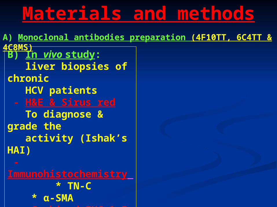
B) In vivo study: liver biopsies of chronic HCV patients - H&E & Sirus red To diagnose & grade the activity (Ishak’s HAI) - Immunohistochemistry * TN-C * α-SMA - Combined IHC & In situ hybridization α-SMA & human TN-C mRNA probes
Materials and methods
A) Monoclonal antibodies preparation (4F10TT, 6C4TT & 4C8MS)
C) In vitro study: - LI90 human HSC line culture stimulated with PDGF-BB and TGF-B. - Immunoblotting of culture medium & cell lysate to detect TN-C & its splice variants. - RNA isolation, RT-PCR & Real time PCR to quantify TN-C mRNA. - Sequencing of transcripts of TN-C to conform the different splice variants
4F10TT 6C4TT 4C8MS
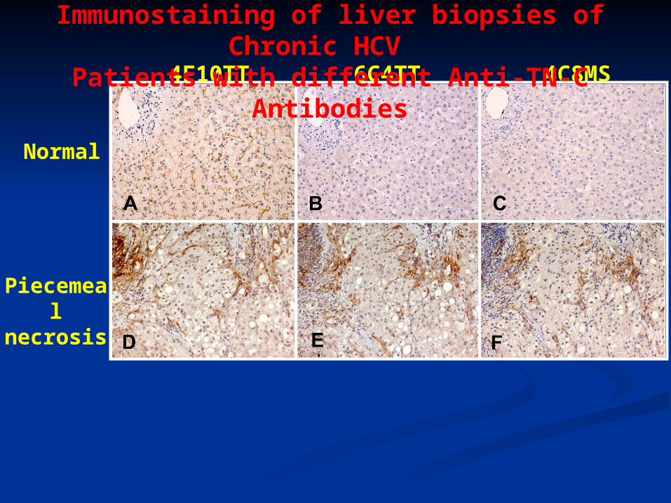
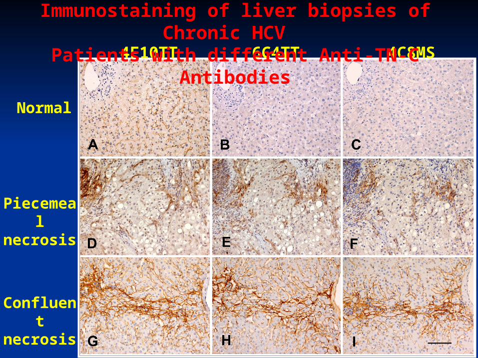
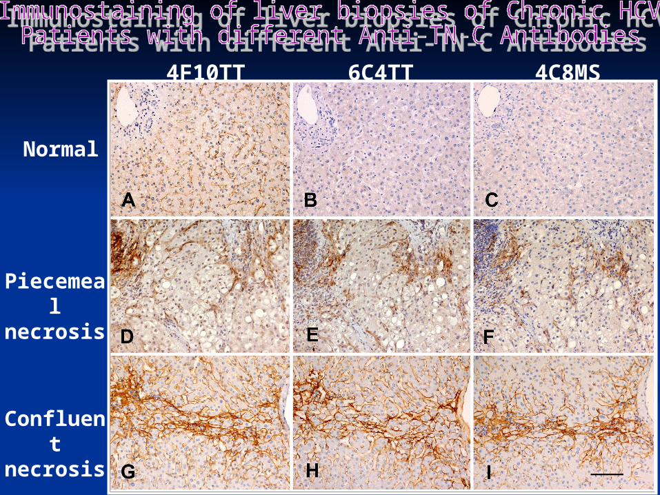
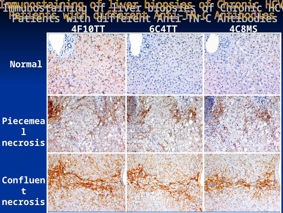
Immunostaining of liver biopsies of Chronic HCV Patients with different Anti-TN-C Antibodies
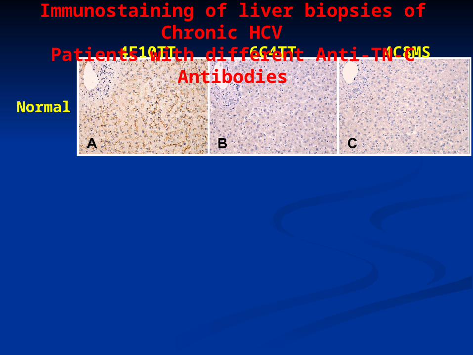
Normal
4F10TT 6C4TT 4C8MS
Immunostaining of liver biopsies of Chronic HCV Patients with different Anti-TN-C Antibodies
Normal
Piecemeal necrosis
4F10TT 6C4TT 4C8MS
Immunostaining of liver biopsies of Chronic HCV Patients with different Anti-TN-C Antibodies

Normal
Piecemeal necrosis
Confluent necrosis
4F10TT 6C4TT 4C8MS
Immunostaining of liver biopsies of Chronic HCV Patients with different Anti-TN-C Antibodies
Normal
Piecemeal necrosis
Confluent necrosis
4F10TT 6C4TT 4C8MS
Immunostaining of liver biopsies of Chronic HCV Patients with different Anti-TN-C Antibodies
Normal
Piecemeal necrosis
Confluent necrosis
4F10TT 6C4TT 4C8MS
Normal
Piecemeal necrosis
Confluent necrosis
4F10TT 6C4TT 4C8MS
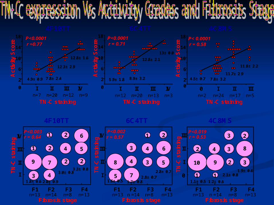
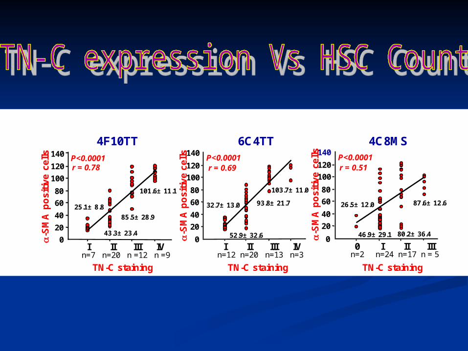
TN-C stainingn=7 n=20 n=12 n=9
Ac
tivi
ty S
co
re
P<0.0001r =0.77
4.9± 0.9
I II III IV
7.8± 2.4
12.3±2.9
12.8± 1.6
Ac
tivi
ty S
co
ren=12 n=20 n=13 n=3
5.9± 1.8 8.9±3.2
12.8± 2.1
13± 0.0
I II III IV
P<0.0001r = 0.71
2
6
10
14
18
n=2 n=24 n=17 n=5 0 I II III
4.5± 0.7 7.8± 3.211.7± 2.9
11.8±2.2
P< 0.0001r = 0.58
Ac
tivi
ty S
co
re
2
6
10
14
18
4F10TT
TN-C staining
6C4TT 4C8MS
TN-C staining
2
6
10
14
18
00 0
Fibrosis stage
F1 F2 F3 F4n=13 n=14 n=8 n=13
TN
-C s
tain
ing IV
III
II
I 3.0± 0.83.3± 0.8
P<0.003r = 0.64
3
9
1
4
7
2
2
4
2
2
6
51
4F10TT 6C4TT 4C8MS
Fibrosis stage
F1 F2 F3 F4n=13 n =14 n=8 n=13
2.8± 0.7
2.8± 0.7
P<0.002r = 0.57
3
1
4
7
4
5
Fibrosis stage
F1 F2 F3 F4n=13 n =14 n=8 n=13
P<0.019r = 0.53
3
1
42
2
2
1
3
5
8 10 9
3
3
8
TN
-C s
tain
ing IV
III
II
IT
N-C
sta
inin
g0
III
II
I
1.8± 0.6 2.0± 0.9 1.7± 0.81.6± 0.5 1.1± 0.5 1.2± 0.6
2.1± 0.81.9± 0.6
6
2
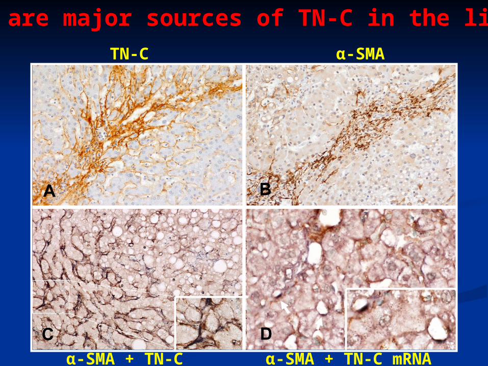
HSCs are major sources of TN-C in the liver
TN-C α-SMA
α-SMA + TN-C α-SMA + TN-C mRNA
TN-C staining
-S
MA
po
siti
ve
cel
ls
TN-C staining
4C8MS
n=2 n=24 n=17 n = 5
P<0.0001r = 0.51
0
20
40
60
80
100
120
140
0 I II IIII II III IVn=12 n=20 n=13 n=3
P<0.0001r = 0.69
0
20
40
60
80
100
120
140
TN-C staining
6C4TT4F10TT
0
20
40
60
80
100
120
140 P<0.0001r = 0.78
n=7 n=20 n =12 n =9 I II III IV
-S
MA
po
siti
ve
cel
ls
-S
MA
po
siti
ve
cells
25.1± 8.8
43.3± 23.4
85.5± 28.9
101.6± 11.1
32.7± 13.0
52.9± 32.6
93.8± 21.7
103.7± 11.0
26.5± 12.0
46.9± 29.1 80.2± 36.4
87.6± 12.6
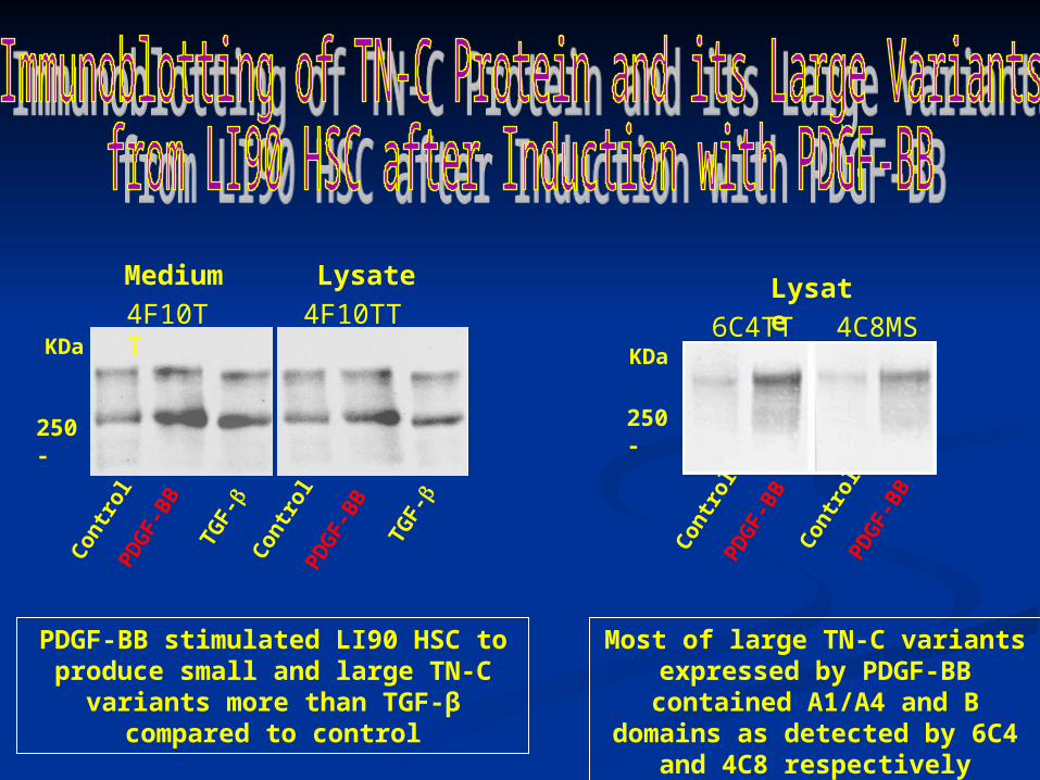
Medium
250 -
KDa
Lysate
4F10TT 4F10TT
Con
trol
PDG
F-B
B
TGF-
C
ontr
olPD
GF-
BB
TGF-
KDa6C4TT 4C8MS
250 -
Lysate
Con
trol
PDG
F-B
B C
ontr
olPD
GF-
BB
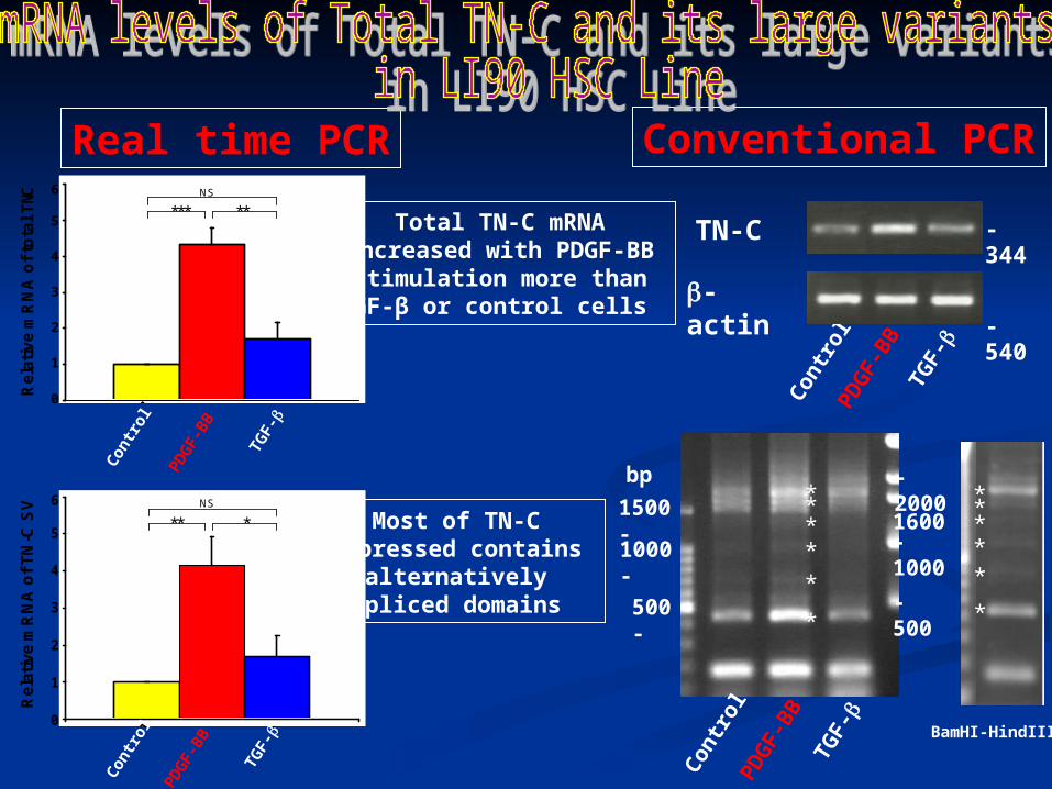
PDGF-BB stimulated LI90 HSC to produce small and large TN-C variants more than TGF-β compared to control
Most of large TN-C variants expressed by PDGF-BB contained A1/A4 and B domains as detected
by 6C4 and 4C8 respectively
-actin
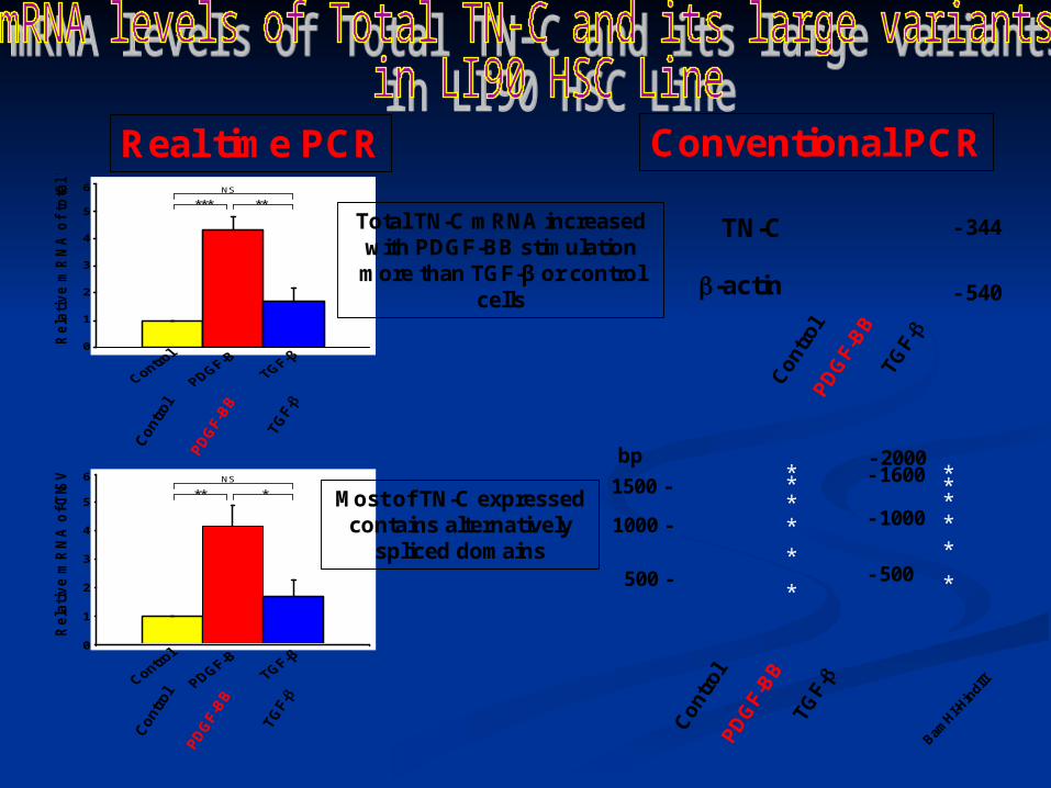
TN-C - 344
- 540
Con
trol
PDG
F-B
BTG
F-
500 -
1000 -
1500 -
- 500
- 1000
- 1600- 2000
Con
trol
PDG
F-B
BTG
F-
*
*****
bp
*
*****
BamHI-H
indI
II
**NS
*
6
5
4
3
2
1
0
Rela
tive m
RN
A o
f T
N-C
SV
6
5
4
3
2
1
0Rela
tive m
RN
A o
f to
tal
TN
-C
***NS
**
Total TN-C mRNA increased with PDGF-BB stimulation more than TGF-β or control
cells
Most of TN-C expressed contains alternatively
spliced domains
Conventional PCRReal time PCRC
ontr
ol
PDG
F-B
B
TGF-
Con
trol
PDG
F-B
B
TGF-
-actin
TN-C - 344
- 540
Con
trol
PDG
F-B
BTG
F-
500 -
1000 -
1500 -
- 500
- 1000
- 1600- 2000
Con
trol
PDG
F-B
BTG
F-
*
*****
bp
*
*****
BamHI-HindIII
Control
**NS
PDGF-B
TGF-β
*
6
5
4
3
2
1
0
Rel
ativ
e m
RN
A o
f T
N-C
SV
6
5
4
3
2
1
0Rela
tive m
RN
A o
f to
tal
TN-C
***NS
**Total TN-C mRNA increased with PDGF-BB stimulation
more than TGF-β or control cells
Most of TN-C expressed contains alternatively
spliced domains
Conventional PCRReal time PCR C
ontr
ol
PDG
F-B
B
TGF-
Con
trol
PDG
F-B
B
TGF-

54321 54321 876 876
DD
DD
DD
DD
DD
DD
DD
DD
DD
DD
DD
DD
DD
DD
DD
DD
DD
CC
CC
CC
CC
CC
BB
BB
BB
BB
BB
BB
BB
BB
BB
BB
BBA4A4
A4A4
A4A4
A4A4
A4A4
A4A4
A4A4
A4A4
A4A4
A4A4
A3A3
A3A3
A3A3
A3A3
A3A3
A3A3
A2A2
A2A2
A2A2
A2A2
A2A2
A2A2
A2A2
A2A2
A2A2
A2A2
A2A2
A2A2A1A1
A1A1
A1A1
A1A1
A1A1
A1A1
A1A1
A1A1
A1A1
A1A1
A1A1
A2A2
A1A1
A1A1
A1A1
A1A1
DDA2A2A1A1
BB
No. of FNIII repeats1
2
3
4
5
6
% Clones screened
100 60
40
30
23 43
4
3
44
27
15 41
12
2
55
14
14 22
9
4
4
68
15
9 22
4
4
100 50
TN-C variants
produced by
LI90 HSC
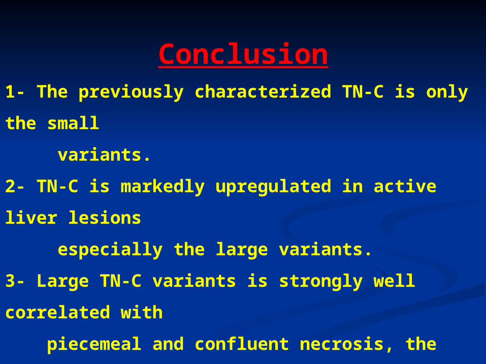
Conclusion1- The previously characterized TN-C is only the small
variants.
2- TN-C is markedly upregulated in active liver lesions
especially the large variants.
3- Large TN-C variants is strongly well correlated with
piecemeal and confluent necrosis, the most reliable
prognostic lesions of chronic hepatitis.
4- Hepatic stellate cells are major sources of large TN-C
variants after their activation by growth factors.