expression of disease-causing lamin a mutants impairs the
TRANSCRIPT
2704 Research Article
IntroductionLamins are nuclear proteins that are components of a fibrousnetwork underlying the inner nuclear membrane termed thenuclear lamina, and are also distributed throughout the interiorof the nucleus (reviewed by Goldman et al., 2002). Laminshave been classified into two types based on biochemicalproperties and expression patterns. B-type lamins areexpressed in most cells and are encoded by two separate genes,B1 and B2, whereas the A-type lamins have been detectedprimarily in differentiated cell types. The lamin A gene(LMNA) encodes lamin A and C transcripts, as well as germ-cell-specific lamin C2. Lamins belong to the intermediatefilament family of proteins and have a short N-terminal headdomain followed by an �-helical rod domain and a globulartail domain. The C-termini of lamins A, B1 and B2 bear aCAAX motif that is post-translationally modified and, in thecase of lamin A, is subjected to a further maturation step bywhich prelamin A is proteolytically cleaved to give maturelamin A (reviewed by Stuurman et al., 1998).
Mutations in human LMNA cause several debilitatingdiseases, collectively termed laminopathies, that affect skeletaland cardiac muscle, adipose, bone and neuronal tissues, andalso cause premature ageing syndromes. The majority ofmutations cause autosomal dominant Emery-Dreifussmuscular dystrophy (EDMD) (Bonne et al., 1999), whereasother mutations cause dilated cardiomyopathy with conductionsystem disease (DCM) (Fatkin et al., 1999), limb girdlemuscular dystrophy (Muchir et al., 2000), or familial partiallipodystrophy (FPLD) (Shackleton et al., 2000; Cao andHegele, 2000). Mutations in LMNA have also been linked to
the relatively rare autosomal recessive disorders Charcot-Marie-Tooth disorder type 2 (De Sandre-Giovannoli et al.,2002) and mandibuloacral dysplasia (Novelli et al., 2002), andmore recently to restrictive dermopathy (Navarro et al., 2004).Many of the above mutations are missense mutations that occurthroughout the gene, although the FPLD mutations areclustered near the C-terminus. An interesting finding has beenthe linkage of mutations in LMNA to Hutchinson-Gilfordprogeria syndrome (HGPS) (Eriksson et al., 2003; De Sandre-Giovannoli et al., 2003) and to atypical Werner’s syndrome(Chen et al., 2003). The most frequent mutation in HGPS is anucleotide substitution (GGC to GGT) that does not cause anamino acid change (G608G) but activates a cryptic splice sitethat leads to a deletion of 50 amino acids near the C-terminus(residues 607-656), giving rise to a truncated protein termedlamin A del50 or progerin, which retains the C-terminus ofprelamin A and is not proteolytically processed. A fewmissense mutations such as R471C and R527C that do notaffect processing of the C-terminus of lamin A have also beenlinked to HGPS and atypical progerias (Eriksson et al., 2003;Cao and Hegele, 2003). Cells from patients expressing mutantlamins often exhibit a range of dominant-negative effects suchas abnormal nuclear morphology, aberrant lamin assembly andaltered gene regulation (reviewed by Wilson, 2000; Wormanand Courvalin, 2002; Hutchison and Worman, 2004). HGPScells in particular show serious defects in nuclear structure aswell as early senescence (Eriksson et al., 2003; De Sandre-Giovannoli et al., 2003; Mounkes et al., 2003, Goldman et al.,2004), and recent reports indicate that HGPS cells and cellslacking functional Zmpste24, a metalloproteinase required for
A-type lamins are components of the nuclear lamina.Mutations in the gene encoding lamin A are associatedwith a range of highly degenerative diseases termedlaminopathies. To evaluate sensitivity to DNA damage,GFP-tagged lamin A cDNAs with disease-causingmutations were expressed in HeLa cells. The inner nuclearmembrane protein emerin was mislocalised uponexpression of the muscular dystrophy mutants G232E,Q294P or R386K, which aberrantly assembled into nuclearaggregates, or upon expression of mutants causing progeriasyndromes in vivo (lamin A del50, R471C, R527C andL530P). The ability of cells expressing these mutants toform DNA repair foci comprising phosphorylated H2AX inresponse to mild doses of cisplatin or UV irradiation was
markedly diminished, unlike the nearly normal response ofcells expressing wild-type GFP-lamin A or disease-causingH222P and R482L mutants. Interestingly, mutants thatimpaired the formation of DNA repair foci mislocalisedATR (for ‘ataxia telangiectasia-mutated and Rad3-related’) kinase, which is a key sensor in the response toDNA damage. Our results suggest that a subset of lamin Amutants might hinder the response of components of theDNA repair machinery to DNA damage by alteringinteractions with chromatin.
Key words: Nuclear lamina, Laminopathies, DNA repair,Phosphorylated H2AX, Emerin, ATR kinase
Summary
Expression of disease-causing lamin A mutantsimpairs the formation of DNA repair fociKaliyaperumal Manju, Bhattiprolu Muralikrishna and Veena K Parnaik*Centre for Cellular and Molecular Biology, Uppal Road, Hyderabad 500 007, India*Author for correspondence (e-mail: [email protected])
Accepted 5 April 2006Journal of Cell Science 119, 2704-2714 Published by The Company of Biologists 2006doi:10.1242/jcs.03009
Jour
nal o
f Cel
l Sci
ence
JCS ePress online publication date 13 June 2006

2705Lamin A mutants hinder DNA repair
prelamin A processing, are defective in DNA repair (Liu et al.,2005; Varela et al., 2005). Mice that lack intact LMNA developsevere muscle wasting similar to human EDMD by 3-4 weeksand die by 8 weeks after birth (Sullivan et al., 1999). Lamin-deficient cells have defective nuclear mechanics (Lammerdinget al., 2004) and altered gene expression (Nikolova et al.,2004).
In addition to providing structure and shape to the nucleus,the lamins are involved in organising several nuclear functionssuch as DNA replication (Moir et al., 2000a), RNA polymeraseII (pol II) transcription (Spann et al., 2002; Kumaran et al.,2002) and mitotic assembly and disassembly. Lamin-chromatin interactions have also been proposed to have animportant role in nuclear organisation (reviewed by Goldmanet al., 2002). During the late stages of apoptosis, the laminsare proteolytically cleaved by caspases, which helps todisassemble the lamina and allows nuclear condensation andfragmentation to proceed (reviewed by Cohen et al., 2001).Ectopic expression of various lamin A mutants upontransfection in cultured cells has been shown to affect normallamin assembly and alter the localisation of the inner nuclearmembrane protein emerin, which is involved in lamin-chromatin interactions (Östlund et al., 2001; Vigoroux et al.,2001; Raharjo et al., 2001; Holt et al., 2003). Certain mutantsalso hinder muscle differentiation and promote cell death uponserum withdrawal (Favreau et al., 2004; Mariappan andParnaik, 2005). Expression of lamin A del50 leads to stronglydominant-negative effects on nuclear architecture, and blocksprogression of cells through S phase (Goldman et al., 2004).Although certain lamin A mutants cause cell division defects,it is not clear whether cells expressing lamin mutants areaffected in their response to DNA-damaging agents.
The initial response to DNA damage involves the chromatin-dependent activation of complex checkpoint signallingpathways that delay the cell cycle and repair the defects. Themajor kinases that serve as damage sensors and regulate cell-cycle checkpoints and DNA repair by phosphorylation of keysubstrates are ATM (for ‘ataxia telangiectasia mutated’) andATR (for ‘ATM and Rad3 related’) kinases that belong to thephosphoinositide 3-kinase-related family of serine/threonineprotein kinases (reviewed by Zhou and Elledge, 2000;Abraham, 2001; Bartek et al., 2004). We have examined theearly response of cells expressing disease-causing lamin Amutants to DNA damage induced by cisplatin or UV, which
primarily cause replicational stress as a result of stalledreplication forks in S phase. A wide range of disease-causingmutants have been analysed and have been linked to progeroidsyndromes and autosomal dominant forms of EDMD, DCMand FPLD. We observe that several mutants impair theformation of phosphorylated H2AX at DNA repair foci andhinder the recruitment of 53BP1 to repair sites. These mutantsdisrupt emerin localisation and, importantly, also mislocaliseATR kinase in untreated cells. Hence, lamin mutants are likelyto affect the response of the DNA repair machinery to DNAdamage by altered interactions with chromatin.
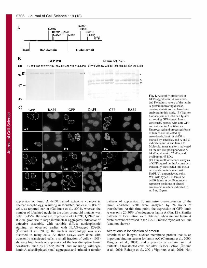
ResultsLocalisation of lamin A mutantsThe intracellular localisation of mutant lamin A proteins wasdetermined by transient transfection of fusion constructsbearing the GFP tag fused to the N-termini of the constructsinto HeLa cells. The assembly of wild-type GFP-lamin A intoa typical nuclear rim pattern has been well documented (Broerset al., 1999; Moir et al., 2000b). The mutations studied wereE203G, H222P, G232E, Q294P, R386K, R471C, R482L,R527C, L530P and lamin A del50 (see Fig. 1A for positionsof mutations). In addition to the lamin A del50, R471C andR527C mutations that cause human progeroid syndromes, micehomozygous for the L530P mutation show strong progeroidsymptoms (Mounkes et al., 2003), although in humans thismutation leads to AD-EDMD (Bonne et al., 1999). H222P,G232E, Q294P and R386K have been identified in AD-EDMD(Bonne et al., 2000). E203G is mutated in DCM (Fatkin et al.,1999), and the R482L mutation is found in patients with FPLD(Shackleton et al., 2000).
Appropriate expression of the mutant proteins wasconfirmed by western blot analysis of transfected cell lysateswith antibodies to GFP. None of the constructs yieldedabnormally sized proteins. Lamin A del50 gave a product thatwas ~5 kDa smaller than the other constructs, as expected (seeFig. 1B). Each mutant protein migrated as a doublet and,except for lamin A del50, the faster migrating bandrepresenting processed lamin A was more prominent.Microscopic analysis of transfected cells indicated that theprogeroid constructs (R471C, R527C, L530P and lamin Adel50) as well as E203G, H222P and R482L werepredominantly localised to the nuclear periphery (Fig. 1C anda summary of the data is given in Table 1). However,
Table 1. Summary of effects of ectopically expressed lamin A mutantsMutant Disease Nuclear location/morphology Emerin ATR �-H2AX§
E203G DCM Peripheral*/normal Disrupted Mislocalised LowH222P AD-EDMD Peripheral/normal Normal Normal NormalG232E AD-EDMD Aggregates/distorted Disrupted Mislocalised LowQ294P AD-EDMD Aggregates/distorted Disrupted Mislocalised LowR386K AD-EDMD Aggregates/distorted Disrupted Mislocalised LowR471C HGPS Peripheral/few lobulated† Disrupted Mislocalised LowR482L FPLD Peripheral/normal Normal Normal NormalR527C HGPS Peripheral/few lobulated† Disrupted Mislocalised LowL530P AD-EDMD/HGPS Peripheral/few lobulated† Disrupted Mislocalised LowDel50 HGPS Peripheral/lobulated‡ Disrupted Mislocalised Low
*With small aggregates at the periphery in ~40% of transfected cells.†10-15% of transfected nuclei, rest normal.‡60% of transfected nuclei.§After treatment with cisplatin for 4 hours or UV for 30 minutes.
Jour
nal o
f Cel
l Sci
ence
2706
expression of lamin A del50 caused extensive changes innuclear morphology, resulting in lobulated nuclei in ~60% ofcells, as reported earlier (Goldman et al., 2004), whereas thenumber of lobulated nuclei in the other progeroid mutants wasonly 10-15%. By contrast, expression of G232E, Q294P andR386K gave rise to large intranuclear aggregates indicative ofdefective assembly, with variable diffuse nucleoplasmicstaining, as observed earlier with FLAG-tagged R386K(Östlund et al., 2001); the nuclear morphology was alsodistorted in many cells. As these assays were done withtransiently transfected cells, a small fraction of cells (~10%)showing high levels of expression of the less disruptive laminconstructs, such as H222P, R482L and including wild-typelamin A, also displayed small aggregates and striated or tubular
Journal of Cell Science 119 (13)
patterns of expression. To minimise overexpression of thelamin construct, cells were analysed by 24 hours oftransfection. At this time point, the expression of GFP laminA was only 20-30% of endogenous lamin A (Fig. 1B). Similarpatterns of localisation were obtained when mutant lamin Aproteins were expressed in the C2C12 mouse myoblast cell line(data not shown).
Alterations in localisation of emerinEmerin is an integral nuclear membrane protein that is animportant binding partner for lamin A/C (Clements et al., 2000;Vaughan et al., 2001), and expression of certain lamin Amutants in transfected cells can alter its localisation (Östlundet al., 2001; Raharjo et al., 2001; Vigoroux et al., 2001; Holt
Fig. 1. Assembly properties ofGFP-tagged lamin A constructs.(A) Domain structure of the laminA protein indicating disease-causing mutations that have beenanalysed in this study. (B) Westernblot analysis of HeLa cell lysatesexpressing GFP-tagged laminconstructs, probed with anti-GFPand anti-lamin A antibodies.Unprocessed and processed formsof lamins are indicated byarrowheads, lamin A del50 ismarked by asterisks, and A and Cindicate lamin A and lamin C.Molecular mass markers indicatedon the left are: phosphorylase b,94 kDa; albumin, 67 kDa; andovalbumin, 43 kDa.(C) Immunofluorescence analysisof GFP-tagged lamin A constructstransiently transfected into HeLacells and counterstained withDAPI. Ut, untransfected cells;WT, wild-type GFP-lamin A;del50, lamin A del50; numbersrepresent positions of alteredamino acid residues indicated inA. Bar, 10 �m.
Jour
nal o
f Cel
l Sci
ence
2707Lamin A mutants hinder DNA repair
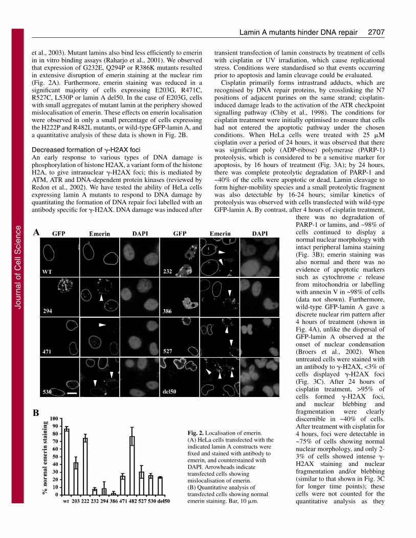
et al., 2003). Mutant lamins also bind less efficiently to emerinin in vitro binding assays (Raharjo et al., 2001). We observedthat expression of G232E, Q294P or R386K mutants resultedin extensive disruption of emerin staining at the nuclear rim(Fig. 2A). Furthermore, emerin staining was reduced in asignificant majority of cells expressing E203G, R471C,R527C, L530P or lamin A del50. In the case of E203G, cellswith small aggregates of mutant lamin at the periphery showedmislocalisation of emerin. These effects on emerin localisationwere observed in only a small percentage of cells expressingthe H222P and R482L mutants, or wild-type GFP-lamin A, anda quantitative analysis of these data is shown in Fig. 2B.
Decreased formation of �-H2AX fociAn early response to various types of DNA damage isphosphorylation of histone H2AX, a variant form of the histoneH2A, to give intranuclear �-H2AX foci; this is mediated byATM, ATR and DNA-dependent protein kinases (reviewed byRedon et al., 2002). We have tested the ability of HeLa cellsexpressing lamin A mutants to respond to DNA damage byquantitating the formation of DNA repair foci labelled with anantibody specific for �-H2AX. DNA damage was induced after
transient transfection of lamin constructs by treatment of cellswith cisplatin or UV irradiation, which cause replicationalstress. Conditions were standardised so that events occurringprior to apoptosis and lamin cleavage could be evaluated.
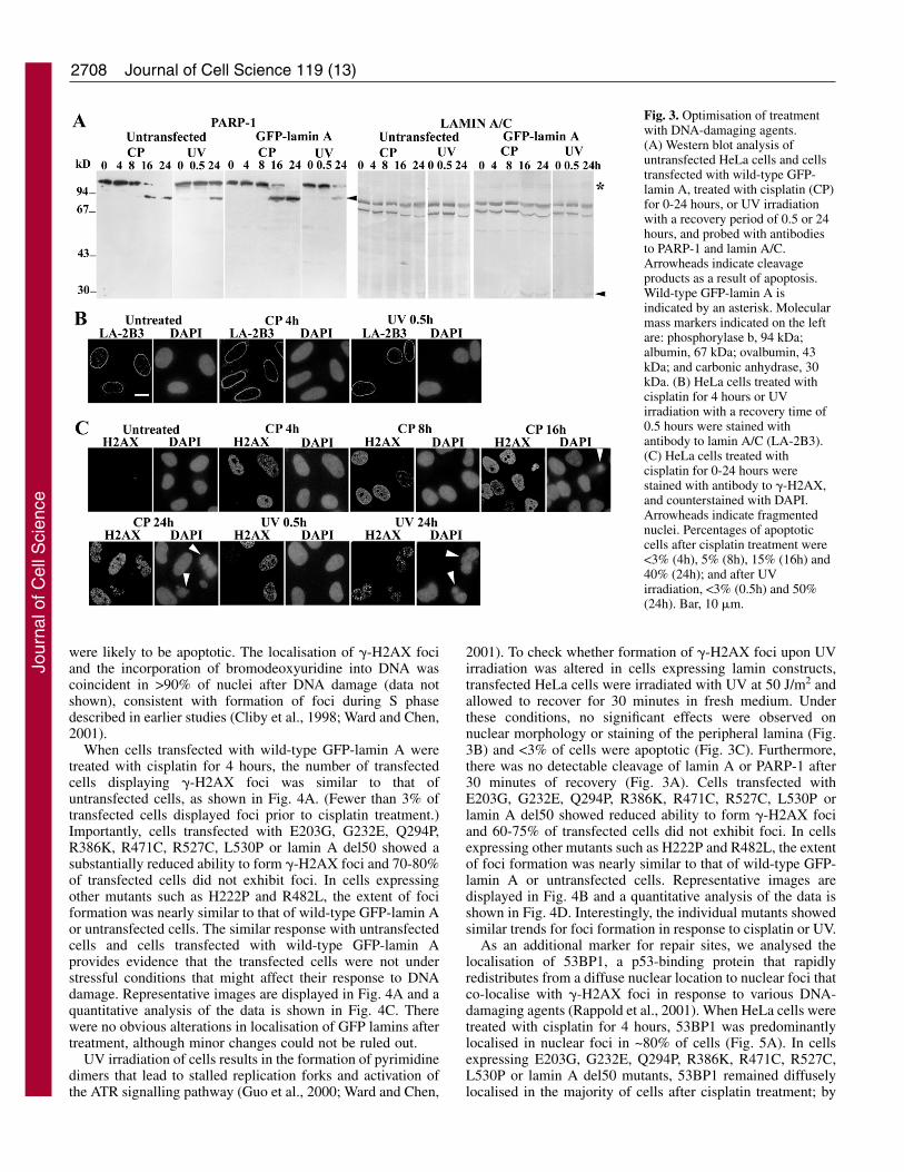
Cisplatin primarily forms intrastrand adducts, which arerecognised by DNA repair proteins, by crosslinking the N7positions of adjacent purines on the same strand; cisplatin-induced damage leads to the activation of the ATR checkpointsignalling pathway (Cliby et al., 1998). The conditions forcisplatin treatment were initially optimised to ensure that cellshad not entered the apoptotic pathway under the chosenconditions. When HeLa cells were treated with 25 �Mcisplatin over a period of 24 hours, it was observed that therewas significant poly (ADP-ribose) polymerase (PARP-1)proteolysis, which is considered to be a sensitive marker forapoptosis, by 16 hours of treatment (Fig. 3A); by 24 hours,there was complete proteolytic degradation of PARP-1 and~40% of the cells were apoptotic or dead. Lamin cleavage toform higher-mobility species and a small proteolytic fragmentwas also detectable by 16-24 hours; similar kinetics ofproteolysis was observed with cells transfected with wild-typeGFP-lamin A. By contrast, after 4 hours of cisplatin treatment,
there was no degradation ofPARP-1 or lamins, and ~98% ofcells continued to display anormal nuclear morphology withintact peripheral lamina staining(Fig. 3B); emerin staining wasalso normal and there was noevidence of apoptotic markerssuch as cytochrome c releasefrom mitochondria or labellingwith annexin V in ~98% of cells(data not shown). Furthermore,wild-type GFP-lamin A gave adiscrete nuclear rim pattern after4 hours of treatment (shown inFig. 4A), unlike the dispersal ofGFP-lamin A observed at theonset of nuclear condensation(Broers et al., 2002). Whenuntreated cells were stained withan antibody to �-H2AX, <3% ofcells displayed �-H2AX foci(Fig. 3C). After 24 hours ofcisplatin treatment, >95% ofcells formed �-H2AX foci,and nuclear blebbing andfragmentation were clearlydiscernible in ~40% of cells.After treatment with cisplatin for4 hours, foci were detectable in~75% of cells showing normalnuclear morphology, and only 2-3% of cells showed intense �-H2AX staining and nuclearfragmentation and/or blebbing(similar to that shown in Fig. 3Cfor longer time points); thesecells were not counted for thequantitative analysis as they
Fig. 2. Localisation of emerin.(A) HeLa cells transfected with theindicated lamin A constructs werefixed and stained with antibody toemerin, and counterstained withDAPI. Arrowheads indicatetransfected cells showingmislocalisation of emerin.(B) Quantitative analysis oftransfected cells showing normalemerin staining. Bar, 10 �m.
Jour
nal o
f Cel
l Sci
ence
2708
were likely to be apoptotic. The localisation of �-H2AX fociand the incorporation of bromodeoxyuridine into DNA wascoincident in >90% of nuclei after DNA damage (data notshown), consistent with formation of foci during S phasedescribed in earlier studies (Cliby et al., 1998; Ward and Chen,2001).
When cells transfected with wild-type GFP-lamin A weretreated with cisplatin for 4 hours, the number of transfectedcells displaying �-H2AX foci was similar to that ofuntransfected cells, as shown in Fig. 4A. (Fewer than 3% oftransfected cells displayed foci prior to cisplatin treatment.)Importantly, cells transfected with E203G, G232E, Q294P,R386K, R471C, R527C, L530P or lamin A del50 showed asubstantially reduced ability to form �-H2AX foci and 70-80%of transfected cells did not exhibit foci. In cells expressingother mutants such as H222P and R482L, the extent of fociformation was nearly similar to that of wild-type GFP-lamin Aor untransfected cells. The similar response with untransfectedcells and cells transfected with wild-type GFP-lamin Aprovides evidence that the transfected cells were not understressful conditions that might affect their response to DNAdamage. Representative images are displayed in Fig. 4A and aquantitative analysis of the data is shown in Fig. 4C. Therewere no obvious alterations in localisation of GFP lamins aftertreatment, although minor changes could not be ruled out.
UV irradiation of cells results in the formation of pyrimidinedimers that lead to stalled replication forks and activation ofthe ATR signalling pathway (Guo et al., 2000; Ward and Chen,
Journal of Cell Science 119 (13)
2001). To check whether formation of �-H2AX foci upon UVirradiation was altered in cells expressing lamin constructs,transfected HeLa cells were irradiated with UV at 50 J/m2 andallowed to recover for 30 minutes in fresh medium. Underthese conditions, no significant effects were observed onnuclear morphology or staining of the peripheral lamina (Fig.3B) and <3% of cells were apoptotic (Fig. 3C). Furthermore,there was no detectable cleavage of lamin A or PARP-1 after30 minutes of recovery (Fig. 3A). Cells transfected withE203G, G232E, Q294P, R386K, R471C, R527C, L530P orlamin A del50 showed reduced ability to form �-H2AX fociand 60-75% of transfected cells did not exhibit foci. In cellsexpressing other mutants such as H222P and R482L, the extentof foci formation was nearly similar to that of wild-type GFP-lamin A or untransfected cells. Representative images aredisplayed in Fig. 4B and a quantitative analysis of the data isshown in Fig. 4D. Interestingly, the individual mutants showedsimilar trends for foci formation in response to cisplatin or UV.
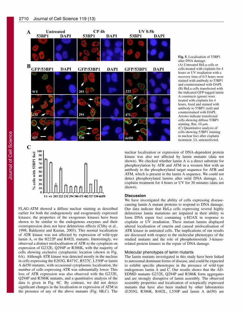
As an additional marker for repair sites, we analysed thelocalisation of 53BP1, a p53-binding protein that rapidlyredistributes from a diffuse nuclear location to nuclear foci thatco-localise with �-H2AX foci in response to various DNA-damaging agents (Rappold et al., 2001). When HeLa cells weretreated with cisplatin for 4 hours, 53BP1 was predominantlylocalised in nuclear foci in ~80% of cells (Fig. 5A). In cellsexpressing E203G, G232E, Q294P, R386K, R471C, R527C,L530P or lamin A del50 mutants, 53BP1 remained diffuselylocalised in the majority of cells after cisplatin treatment; by
Fig. 3. Optimisation of treatmentwith DNA-damaging agents.(A) Western blot analysis ofuntransfected HeLa cells and cellstransfected with wild-type GFP-lamin A, treated with cisplatin (CP)for 0-24 hours, or UV irradiationwith a recovery period of 0.5 or 24hours, and probed with antibodiesto PARP-1 and lamin A/C.Arrowheads indicate cleavageproducts as a result of apoptosis.Wild-type GFP-lamin A isindicated by an asterisk. Molecularmass markers indicated on the leftare: phosphorylase b, 94 kDa;albumin, 67 kDa; ovalbumin, 43kDa; and carbonic anhydrase, 30kDa. (B) HeLa cells treated withcisplatin for 4 hours or UVirradiation with a recovery time of0.5 hours were stained withantibody to lamin A/C (LA-2B3).(C) HeLa cells treated withcisplatin for 0-24 hours werestained with antibody to �-H2AX,and counterstained with DAPI.Arrowheads indicate fragmentednuclei. Percentages of apoptoticcells after cisplatin treatment were<3% (4h), 5% (8h), 15% (16h) and40% (24h); and after UVirradiation, <3% (0.5h) and 50%(24h). Bar, 10 �m.
Jour
nal o
f Cel
l Sci
ence

2709Lamin A mutants hinder DNA repair
contrast, with H222P and R482L mutants or wild-type GFP-lamin A, recruitment to nuclear foci was observed in most ofthe transfected cells (Fig. 5B). A quantitative analysis of thedata is shown in Fig. 5C. Expression of lamin mutants did notaffect the diffuse nuclear staining of 53BP1 in untreated cells(data not shown). Upon 30 minutes of recovery after UVirradiation, 53BP1 was localised in very small 53BP1 foci witha diffuse background (Fig. 5A), so we did not quantitate theeffects of lamin mutants after UV treatment. Importantly, thelamin mutants that affect 53BP1 redistribution correspond tothose that are impaired in �-H2AX production, which isconsistent with the reported requirement of �-H2AX forbinding to and recruitment of 53BP1 to repair sites (Ward etal., 2003).
Aberrant localisation of ATRTo determine whether lamin mutants could affect localisationof specific signalling components and thus cause an impairedresponse to DNA damage, we checked the localisation of ATRand ATM kinases in cells expressing lamin mutants incotransfection studies performed in cells not subjected toDNA-damaging conditions. Cells expressing FLAG-ATR or
Fig. 4. Formation of �-H2AXfoci after DNA damage. HeLacells transfected with theindicated GFP-tagged lamin Aconstructs (green) were (A)treated with cisplatin for 4 hoursor (B) irradiated with UV andallowed to recover for 0.5 hours,fixed and stained with antibodyto �-H2AX (red) andcounterstained with DAPI. Bar,10 �m. (C,D) Quantitativeanalysis of �-H2AX stainingwith cisplatin-treated and UV-treated HeLa cells, respectively.Percentage of untransfected (Ut)or transfected cells positive(solid bars) or negative (openbars) for �-H2AX foci areplotted.
Jour
nal o
f Cel
l Sci
ence
2710
FLAG-ATM showed a diffuse nuclear staining as describedearlier for both the endogenously and exogenously expressedkinases; the properties of the exogenous kinases have beenshown to be similar to the endogenous enzymes and theiroverexpression does not have deleterious effects (Cliby et al.,1998; Bakkenist and Kastan, 2003). This normal localisationof ATR kinase was not affected by expression of wild-typelamin A, or the H222P and R482L mutants. Interestingly, weobserved a distinct mislocalisation of ATR to the cytoplasm onexpression of G232E, Q294P or R386K, with the majority ofcells showing exclusive cytoplasmic location (shown in Fig.6A). Although ATR kinase was detected mostly in the nucleusin cells expressing the E203G, R471C, R527C, L530P or laminA del50 mutants, with occasional cytoplasmic localisation, thenumber of cells expressing ATR was substantially lower. Thisloss of ATR expression was also observed with the G232E,Q294P and R386K mutants, and a quantitative analysis of thedata is given in Fig. 6C. By contrast, we did not detectsignificant changes in the localisation or expression of ATM inthe presence of any of the above mutants (Fig. 6B,C). The
Journal of Cell Science 119 (13)
nuclear localisation or expression of DNA-dependent proteinkinase was also not affected by lamin mutants (data notshown). We checked whether lamin A is a direct substrate forphosphorylation by ATR and ATM in a western blot with anantibody to the phosphorylated target sequence for ATR andATM, which is present in the lamin A sequence. We could notdetect phosphorylated lamins after mild DNA damage, i.e.cisplatin treatment for 4 hours or UV for 30 minutes (data notshown).
DiscussionWe have investigated the ability of cells expressing disease-causing lamin A mutant proteins to respond to DNA damage.Our data indicate that HeLa cells expressing several highlydeleterious lamin mutations are impaired in their ability toform DNA repair foci containing �-H2AX in response tocisplatin or UV irradiation. These mutant lamins displayedaltered localisation of emerin and caused mislocalisation ofATR kinase in untreated cells. The implications of our resultsare discussed with respect to the molecular phenotypes of thestudied mutants and the role of phosphoinositide 3-kinase-related protein kinases in the repair of DNA damage.
Molecular phenotypes of lamin mutantsThe lamin mutants investigated in this study have been linkedto autosomal dominant forms of disease, and could be expectedto exhibit specific phenotypes in the presence of wild-typeendogenous lamin A and C. Our results shows that the AD-EDMD mutants G232E, Q294P and R386K form aggregatesand are strongly disruptive of lamin assembly. The observedassembly properties and localisation of ectopically expressedmutants that have also been studied by other laboratories(E203G, R386K, R482L, L530P and lamin A del50) are
Fig. 5. Localisation of 53BP1after DNA damage.(A) Untreated HeLa cells orcells treated with cisplatin for 4hours or UV irradiation with arecovery time of 0.5 hours werestained with antibody to 53BP1and counterstained with DAPI.(B) HeLa cells transfected withthe indicated GFP-tagged laminA constructs (green) weretreated with cisplatin for 4hours, fixed and stained withantibody to 53BP1 (red) andcounterstained with DAPI.Arrows indicate transfectedcells showing diffuse 53BP1staining. Bar, 10 �m.(C) Quantitative analysis ofcells showing 53BP1 stainingin nuclear foci after cisplatintreatment. Ut, untransfected.
Jour
nal o
f Cel
l Sci
ence

2711Lamin A mutants hinder DNA repair
consistent with previous reports (Östlund et al., 2001; Bechertet al., 2003; Goldman et al., 2004; Scaffidi and Misteli, 2005).Lamin A del50 has been shown to have strongly deleteriouseffects on nuclear morphology and chromatin organisation(Goldman et al., 2004), which have been attributed to retentionof the farnesylated C-terminus. In our study, the expression oflamin A del50 also gave rise to highly convoluted nuclei withabnormal morphology, whereas the other progeroid constructs– R471C, R527C and L530P – were predominantly localisedwith smooth staining at the nuclear rim and displayed lowlevels of lobulated nuclei. Although L530P has been linked toAD-EDMD in humans, it gives rise to HGPS-like symptomsin mice in a knockin experiment; this might be a result ofaberrantly spliced forms of lamin A observed in the mousemodel (Mounkes et al., 2003). However, we did not detect anyproducts from differentially spliced forms of L530P in westernblots of transfected cell lysates. Constructs such as H222Ppredominantly showed a typical nuclear rim localisation andnormal localisation of emerin, consistent with the milderproperties observed in a recently reported knockin mouse
model of AD-EDMD with the H222P mutation (Arimura et al.,2005). Similar localisation patterns of the mutants wereobserved in HeLa and C2C12 cells, although variations inexpression and localisation of lamin mutants in other cell typescannot be ruled out. There is considerable evidence for theinvolvement of lamins in the spatial organisation of RNA polII transcription (Spann et al., 2002; Kumaran et al., 2002), andspecific genes are misregulated in HGPS, FPLD and DCMcells (Csoka et al., 2004; Capanni et al., 2005; Mounkes et al.,2005). When we analysed RNA pol II transcription in cellsexpressing lamin mutants, we did not observe significantalterations in staining of active pol II (data not shown),although we cannot rule out effects on transcription of specificgenes.
We have observed that emerin is distinctly mislocalised incells expressing mutants like R386K, Q294P and G232E thatform aggregates and are defective in assembly at the nuclearperiphery. Furthermore, emerin levels at the nuclear envelopewere depleted on expression of constructs with mutations at theC-terminus (R471C, R527C, L530P and lamin A del50), whichmight be unable to bind to emerin and localise it to the nuclearenvelope, since emerin has been shown to associate with theC-terminus of lamin A (Vaughan et al., 2001). Expression ofthe DCM mutant E203G, which forms small aggregates at theperiphery in ~40% of cells, also resulted in mislocalisation ofemerin. By contrast, expression of the FPLD mutant R482Ldid not alter the nuclear rim localisation of emerin in themajority of cells. However, co-localisation of lamin and emerinmight not always imply correct intermolecular interactions. Ina previous study with fibroblasts from FPLD cells expressingthis mutant, about 80% of cells showed normal rim localisationof emerin, whereas 15-20% cells displaying lamin A/Caggregates showed reduced staining of emerin; binding ofemerin to lamin A but not to lamin C was affected in the FPLD
Fig. 6. Localisation of ATRand ATM kinases in untreatedcells. (A) HeLa cellstransfected with a FLAG-ATRconstruct (ATR) orcotransfected with theindicated GFP-tagged lamin Aconstructs (green) were fixedand stained with anti-FLAGantibody (red), andcounterstained with DAPI.Arrows indicate cells showingexclusive cytoplasmic stainingof ATR. (B) As in A, exceptthat a FLAG-ATM construct(ATM) was used. Bar, 10 �m.(C) Quantitative analysis oftransfected cells expressingATR, including nuclear and/orcytoplasmic staining (openbars), or ATM (solid bars),both normalised to wild type as100%.
Jour
nal o
f Cel
l Sci
ence

2712
fibroblasts (Capanni et al., 2003). Emerin is an importantbinding partner for lamin A, and also binds to barrier-to-autointegration factor (BAF), a DNA-binding chromatinprotein (Lee et al., 2001). Emerin and BAF, together with otherlamin-binding proteins such as lamina-associated polypeptide2� (Dechat et al., 2000), have been proposed to cooperate withlamins in the spatial organisation of chromatin. An importantcorrelation we have observed is that the lamin mutants thatshow altered localisation of emerin are hindered in theirresponse to DNA damage and show mislocalisation of ATRkinase, as discussed in the next section.
Aberrant chromatin organisation has been observed invarious conditions of lamin mis-expression. Expression oflamin A del50 leads to the depletion of heterochromatin andabnormal nuclear morphology, which have been attributed tothe accumulation of prelamin A (Goldman et al., 2004). Also,cardiomyocytes from lamin A/C-deficient mice displayrelocalisation of heterochromatin from the periphery to theinterior of the nucleus (Nikolova et al., 2004). Analysis of thedynamics of various GFP-tagged lamin mutants in live cellshas given further insights into the disparate effects of differentmutations in lamin A and C on lamin organisation andchromatin structure. Wild-type lamin A and the R482Wmutant, which causes FPLD, showed slow kinetics ofmovement at the nuclear periphery, consistent with theirincorporation into a stable polymer (Gilchrist et al., 2004). Bycontrast, AD-EDMD mutants in the rod domain as well asL530P displayed increased dynamics; the rod domain mutantsare likely to be impaired in filament assembly whereas L530P,being buried in the C-terminal core domain, might destabiliseprotein folding. The R482W mutant is surface exposed at theC-terminus and thus might not affect the structure of theprotein (Dhe-Paganon et al., 2002). In another study,significant decrease in integration into intranuclear sites wasobserved for the R386K mutant, in addition to increasedmobility at the nuclear rim, which was proposed to be a resultof loss of interaction with chromatin (Broers et al., 2005). Withreference to the other mutants we have studied, the structureof the globular tail domain predicts that substitutions at R527are likely to perturb structure as a result of disruption of a saltbridge at the domain surface, whereas those at R471, which isburied in the domain core, are likely to disrupt protein foldingand stability (Dhe-Paganon et al., 2002). Of the rod domainmutations we have analysed, G232E and Q294P markedlyaffect filament assembly and the mutants form large nuclearaggregates. E203G also affects filament assembly as smallaggregates are formed in 40% of transfected cells. However,H222P assembles normally into filaments at the periphery.
Implications for repair of DNA damageWhen DNA is damaged by agents such as UV irradiation orcrosslinking agents, this leads to stalled replication forks in Sphase and activation of the ATR pathway (Zhou and Elledge,2000; Abraham, 2001; Bartek et al., 2004). Treatment withionising radiation or certain radiomimetic drugs results inrandom double-strand breaks that lead primarily to theactivation of the ATM pathway by autophosphorylation ofATM, with a requirement for ATR kinase also in this response(Shiloh, 2003; Bakkenist and Kastan, 2003). Furthermore,ATR appears to carry out genome surveillance functions innormal cell cycles (Ward and Chen, 2001). Defects in ATR
Journal of Cell Science 119 (13)
or ATM signalling pathways are highly deleterious. ATR-knockout mice are embryonic lethal (Brown and Baltimore,2003) and a hypomorphic mutation in ATR has been linked tothe human disease Seckel syndrome in which there is severegrowth retardation as well as skeletal and brain abnormalities(O’Driscoll et al., 2003). In humans, lack of ATM results inataxia telangiectasia, which is characterised by neuronaldegeneration, immunodeficiency, premature ageing and apredisposition to cancer; similar abnormalities are seen inATM-null mice (Shiloh, 2003). In the absence of the geneencoding H2AX in mice, cells show increased genomicinstability and more sensitivity to DNA damage, and areimpaired in the formation of DNA repair foci. Prevention ofH2AX phosphorylation by specific kinase inhibitors alsoblocks the formation of DNA repair foci, and recruitment ofother repair proteins such as BRCA1, Nbs1, 53BP1, RAD50and RAD51 to sites of DNA damage (Redon et al., 2002). DNArepair pathways are also defective in cells accumulatingprelamin A as a result of lack of functional Zmpste24 or inHGPS cells subjected to DNA damage (Liu et al., 2005; Varelaet al., 2005).
Our present findings provide a mechanistic basis for thedefects in DNA repair caused by mutations in lamin A. Weshow that ATR kinase is mislocalised and/or mis-expressed incells expressing lamin mutants that are impaired in the DNAdamage response to UV and cisplatin. ATR normally residesin the nucleus bound to chromatin and is directly complexedwith ATR-interacting protein, but details of its mechanism ofnuclear transport have not been reported. We suggest that themislocalisation of ATR might be attributed to loss of specificbinding sites on chromatin, leading to altered nuclear transportproperties, retention in the cytoplasm and subsequentdegradation. In response to DNA damage, replication proteinA (a single-stranded-DNA-binding protein) bound at the DNAlesion is able to recruit the complex of ATR-interacting proteinand ATR, thus facilitating the phosphorylation of crucialsubstrates such as �-H2AX by ATR; this might involveadditional proteins such as claspin, minichromosomemaintenance proteins and RAD proteins (Bartek et al., 2004).Mislocalisation of ATR by lamin mutants may thus lead toimpaired formation of �-H2AX foci, which in turn reducesrecruitment of 53BP1, as seen with the same set of mutants.Although lamin mutants do not directly affect ATMlocalisation, we suggest that effects on ATR might be sufficientto impair DNA repair pathways requiring both ATM and ATRfunctions. As the mutants showing mislocalisation of ATR alsodisplay altered localisation of emerin, we propose that normallamin-chromatin interactions are required for the correctnuclear localisation of ATR and subsequent activation of theATR signalling pathway.
Our study has demonstrated that several lamin mutantscausing progerias and muscle-specific disorders inducemislocalisation of emerin and aberrant localisation of ATRprior to damage, resulting in defects in ATR signallingpathways such as reduced phosphorylation of H2AX andinadequate recruitment of 53BP1 to repair sites in responseto DNA damage in cell culture. With regard to diseasemechanisms, ectopic expression studies in an immortal cellline such as HeLa do have potential limitations in interpretationfor the disease in the whole animal. However, in the context ofour findings, it may be noted that wastage of tissues is a
Jour
nal o
f Cel
l Sci
ence
2713Lamin A mutants hinder DNA repair
hallmark of several laminopathies. Hence, impairment of DNArepair processes ultimately leading to cell death might be amore general mechanism that could contribute to pathogenesisin some laminopathies, in addition to the well-documentedeffects on tissue differentiation.
Materials and MethodsPlasmid constructsLamin A constructs were made as fusions with green fluorescent protein (GFP). Thewild-type GFP-lamin A construct has been described earlier (Mariappan andParnaik, 2005). Point mutations were introduced into lamin A cDNA by PCR-basedmutagenesis of the 1.4 kb HindIII fragment (502-1886 bp) in a two-step PCR usingthe appropriate mutant primers and flanking normal primers. The mutated segmentswere ligated to lamin A cDNA fragments to give full-length constructs. Allconstructs were verified by automated DNA sequence analysis. To enable ectopicexpression of lamin A del50, appropriate fragments of lamin A cDNA wereamplified by PCR and religated so as to delete the 1819-1968 bp segment (aminoacids 607-656). All lamin constructs were expressed as GFP fusions from thecytomegalovirus (CMV) promoter in the pEGFP-C vector. Mammalian expressionvectors for FLAG-tagged ATR and ATM kinases were generous gifts from P.Ngheim (Massachusetts General Hospital, Charlestown, MA) and M. Kastan (StJude’s Children’s Research Hospital, Memphis, TN), respectively.
Cell culture, DNA transfection and treatment with DNA-damaging agentsHeLa cells were grown in DMEM supplemented with 10% fetal bovine serum at37°C in a humidified atmosphere containing 5% CO2. DNA transfections werecarried out with Lipofectamine PLUS (Invitrogen) according to the manufacturer’sinstructions. Transfection efficiencies were ~25%. At 24 hours after transfection,cells were treated with 25 �M cisplatin for 4 hours (or longer as indicated). Fortreatment with UV irradiation, transfected cells were subjected to UV irradiation at50 J/m2 and then allowed to recover in fresh medium for 30 minutes.
Immunofluorescence microscopyHeLa cells were washed with phosphate-buffered saline (PBS) and then routinelyfixed by treatment with 3.5% formaldehyde for 10 minutes followed by 0.5% (v/v)Triton X-100 for 6 minutes at room temperature, or with methanol at –20°C for 10minutes for LA-2B3 antibody. Cells were then incubated with 0.5% gelatin in PBSfor 1 hour followed by incubation with first antibody for 1 hour and then biotinylatedsecond antibody and avidin-Cy3 for 1 hour each at room temperature. Samples weremounted in Vectashield (Vector Laboratories) containing 1 �g/ml DAPI. The primaryantibodies used were mouse monoclonal antibodies (mAbs) to lamin A/C (LA-2B3),which stains the nuclear periphery (Jagatheesan et al., 1999), �-H2AX from UpstateBiotechnology, emerin (4G5) from Novocastra Laboratories, 53BP1 (H-300) fromSanta Cruz Biotechnology, and FLAG (M2) from Sigma-Aldrich. Secondaryantibody conjugates were from Jackson ImmunoResearch Laboratories, MolecularProbes or Vector Laboratories. There was no crossreactivity of the fluorescent secondantibodies in control experiments in which primary antibodies were omitted.Confocal laser-scanning immunofluorescence microscopy (CLSM) was carried outwith a Zeiss LSM510 META confocal microscope. Image analysis was done usingLSM510 META Software (Carl Zeiss), and images were assembled using AdobePhotoshop 6.0. Quantitative analysis was carried out by inspection of n=100 cellsper sample in three separate experiments and values expressed as mean ± s.d.
Western blot analysisHeLa cells were harvested, lysed in Laemmli’s sample buffer, boiled andelectrophoresed through SDS-10% polyacrylamide gels. Gels were electroblottedonto PVDF membrane filters and blocked overnight in 5% BLOTTO in Tris-buffered saline containing 0.1% Tween-20. Filters were incubated with primaryantibody for 2 hours, followed by secondary antibody for 1 hour. The primaryantibodies used were polyclonal antibodies to lamin A from Santa CruzBiotechnology, poly ADP-ribose polymerase-1 (PARP-1) from Roche AppliedScience and GFP from Clontech. Bound antibody was visualised using achemiluminescence kit (Roche Applied Science) for peroxidase-conjugatedsecondary antibody (for PARP-1) or by colour reaction using nitroblue tetrozoliumand 5-bromo-4-chloro-indolyl phosphate for alkaline phosphatase-conjugatedsecondary antibody (for lamin A and GFP).
We are grateful to M. Kastan and P. Ngheim for gifts of plasmids.We thank N. Rangaraj for expert assistance with confocal microscopy;and N. Nagesh and M. Sultana for DNA sequencing and synthesisof PCR primers. Financial support from the Department ofBiotechnology, India, for this work is gratefully acknowledged. K.M.was supported by a predoctoral research fellowship from the Councilof Scientific and Industrial Research, India.
ReferencesAbraham, R. T. (2001). Cell cycle checkpoint signalling through the ATM and ATR
kinases. Genes Dev. 15, 2177-2196.Arimura, T., Helbling-Leclerc, A., Massart, C., Varnous, S., Niel, F., Lacène, E.,
Fromes, Y., Toussaint, M., Mura, A., Keller, D. I. et al. (2005). Mouse modelcarrying H222P-Lmna mutation develops muscular dystrophy and dilatedcardiomyopathy similar to human striated muscle laminopathies. Hum. Mol. Genet. 14,155-169.
Bakkenist, C. J. and Kastan, M. B. (2003). DNA damage activates ATM throughintermolecular autophosphorylation and dimer dissociation. Nature 422, 499-506.
Bartek, J., Lukas, C. and Lukas, J. (2004). Checking on DNA damage in S phase.Nature 5, 792-804.
Bechert, K., Lagos-Quintana, M., Harborth, J., Weber, K. and Osborn, M. (2003).Effects of expressing lamin A mutant protein causing Emery-Dreifuss musculardystrophy and familial partial lipodystrophy in HeLa cells. Exp. Cell Res. 286, 75-86.
Bonne, G., Di Barletta, M. R., Varnous, S., Becane, H. M., Hammonda, E. H.,Merlini, L., Muntoni, F., Greenberg, C. R., Gary, F., Urtizberea, J. A. et al. (1999).Mutations in the gene encoding lamin A/C cause autosomal dominant Emery-Dreifussmuscular dystrophy. Nat. Genet. 21, 285-288.
Bonne, G., Mercuri, E., Muchir, A., Urtiziberea, A., Becane, H. M., Reca, D., Merlini,L., Wehnert, M., Boor, R., Reuner, U. et al. (2000). Clinical and molecular geneticspectrum of autosomal dominant Emery Dreifuss muscular dystrophy due to mutationsof the lamin A/C gene. Ann. Neurol. 48, 170-180.
Broers, J. L., Machiels, B. M., van Eys, G. J., Kuijpers, H. J., Manders, E. M., vanDriel, R. and Ramaekers, F. C. (1999). Dynamics of the nuclear lamina as monitoredby GFP-tagged A-type lamins. J. Cell Sci. 112, 3463-3475.
Broers, J. L. V., Bronnenberg, N. M. H. J., Kuijpers, H. J. H., Schutte, B., Hutchison,C. J. and Ramaekers, F. C. S. (2002). Partial cleavage of A-type lamins concurs withtheir total disintegration from the nuclear lamina during apoptosis. Eur. J. Cell Biol.81, 677-691.
Broers, J. L. V., Kuijpers, H. J. H., Östlund, C., Worman, H. J., Endert, J. andRamaekers, F. C. S. (2005). Both lamin A and lamin C mutations cause laminainstability as well as loss of internal nuclear lamin organization. Exp. Cell Res. 304,582-592.
Brown, E. J. and Baltimore, D. (2003). Essential and dispensable roles of ATR in cellcycle arrest and genome maintenance. Genes Dev. 17, 615-628.
Cao, H. and Hegele, R. A. (2000). Nuclear lamin A/C R482Q mutation in Canadiankindreds with Dunnigan-type familial partial lipodystrophy. Hum. Mol. Genet. 9, 109-112.
Cao, H. and Hegele, R. A. (2003). LMNA is mutated in Hutchinson-Gilford progeria(MIM 176670) but not in Wiedemann-Rautenstrauch progeroid syndrome (MIM264090). J. Hum. Genet. 48, 271-274.
Capanni, C., Cenni, V., Mattioli, E., Sabatelli, P., Ognibene, A., Columbaro, M.,Parnaik, V. K., Wehnert, M., Maraldi, N. M., Squarzoni, S. et al. (2003). Failureof lamin A/C to functionally assemble in R482L mutated familial partial lipodystrophyfibroblasts: Altered intermolecular interaction with emerin and implications for genetranscription. Exp. Cell Res. 291, 122-134.
Capanni, C., Mattioli, E., Columbaro, M., Lucarelli, E., Parnaik, V. K., Novelli, G.,Wehnert, M., Cenni, V., Maraldi, N. M., Squarzoni, S. et al. (2005). Altered pre-lamin A processing is a common mechanism leading to lipodystrophy. Hum. Mol.Genet. 14, 1489-1502.
Chen, I., Lee, I., Kudlow, B. A., Dos Santos, H. G., Stervoid, O., Shafeghati, Y., Botha,E. G., Garg, A., Hanson, N. B., Martin, G. M. et al. (2003). LMNA mutations inatypical Werner’s syndrome. Lancet 362, 440-445.
Clements, L., Manilal, S., Love, D. R. and Morris, G. E. (2000). Direct interactionbetween emerin and lamin A. Biochem Biophys. Res. Commun. 267, 709-714.
Cliby, W. A., Roberts, C. J., Cimprich, K. A., Stringer, C. M., Lamb, J. R., Schreiber,S. L. and Friend, S. H. (1998). Overexpression of a kinase-inactive ATR proteincauses sensitivity to DNA-damaging agents and defects in cell cycle checkpoints.EMBO J. 17, 159-169.
Cohen, M., Lee, K. K., Wilson, K. L. and Gruenbaum, Y. (2001). Transcriptionalrepression, apoptosis, human disease and the functional evolution of the nuclear lamina.Trends Biochem. Sci. 26, 41-47.
Csoka, A. B., English, S. B., Simkevich, C. P., Ginzinger, D. G., Butte, A. J., Schatten,G. P., Rothman, F. G. and Sedivy, J. M. (2004). Genome-scale expression profilingof Hutchinson-Gilford progeria syndrome reveals widespread transcriptionalmisregulation leading tomesodermal/mesenchymal defects and acceleratedatherosclerosis. Aging Cell 3, 235-243.
De Sandre-Giovannoli, A., Chaouch, M., Kozlov, S., Vallat, J. M., Tazir, M., Kassouri,N., Szepetowski, P., Hammadouche, T., Vandenberghe, A., Stewart, C. L. et al.(2002). Homozygous defects in LMNA, encoding lamin A/C nuclear-envelope proteins,cause autosomal recessive axonal neuropathy in human (Charcot-Marie-Tooth disordertype 2) and mouse. Am. J. Hum. Genet. 70, 726-736.
De Sandre-Giovannoli, A., Bernard, R., Cau, P., Navarro, C., Amiel, J., Boccaccio,I., Lyonnet, S., Stewart, C. L., Munnich, A., Le Merrer, M. et al. (2003). Lamin Atruncation in Hutchinson-Gilford progeria. Science 300, 2055.
Dechat, T., Korbei, B., Vaughan, O. A., Vlcek, S., Hutchison, C. J. and Foisner, R.(2000). Lamina-associated polypeptide 2� binds intranuclear A-type lamins. J. CellSci. 113, 3473-3484.
Dhe-Paganon, S., Werner, E. D., Chi, Y. and Shoelson, S. E. (2002). Structure of theglobular tail of nuclear lamin. J. Biol. Chem. 277, 17381-17384.
Eriksson, M., Brown, W. T., Gordon, L. B., Glynn, M. W., Singer, J., Scott, L., Erdos,M. R., Robbins, C. M., Moses, T. Y., Berglund, P. et al. (2003). Recurrent de novo
Jour
nal o
f Cel
l Sci
ence

point mutations in lamin A cause Hutchinson-Gilford progeria syndrome. Nature 423,293-298.
Fatkin, D., MacRae, C., Sasaki, T., Wolff, M. R., Porcu, M., Frenneaux, M., Atherton,J., Vidaillet, H. J., Jr, Spudich, S., De Girolami, U. et al. (1999). Missense mutationsin the rod domain of the lamin A/C gene as causes of dilated cardiomyopathy andconduction-system disease. New Engl. J. Med. 341, 1715-1724.
Favreau, C., Higuet, D., Courvalin, J.-C. and Buendia, B. (2004). Expression of amutant lamin A that causes Emery-Dreifuss muscular dystrophy inhibits in vitrodifferentiation of C2C12 myoblasts. Mol. Cell. Biol. 24, 1481-1492.
Gilchrist, S., Gilbert, N., Perry, P., Östlund, C., Worman, H. J. and Bickmore, W. A.(2004). Altered protein dynamics of disease-associated lamin A mutants. BMC CellBiol. 5, 46.
Goldman, R. D., Gruenbaum, Y., Moir, R. D., Shumaker, D. K. and Spann, T. P.(2002). Nuclear lamins: building blocks of nuclear architecture. Genes Dev. 16, 533-547.
Goldman, R. D., Shumaker, D. K., Erdos, M. R., Eriksson, M., Goldman, A. E.,Gordon, L. B., Gruenbaum, Y., Khuon, S., Mendez, M., Varga, R. et al. (2004).Accumulation of mutant lamin A causes progressive changes in nuclear architecturein Hutchinson-Gilford progeria syndrome. Proc. Natl. Acad. Sci. USA 101, 8963-8968.
Guo, Z., Kumagai, A., Wang, S. X. and Dunphy, W. G. (2000). Requirement of ATRin phosphorylation of Chk1 and cell cycle regulation in response to DNA replicationblocks and UV-damaged DNA in Xenopus egg extracts. Genes Dev. 14, 2745-2756.
Holt, I., Östlund, C., Stewart, C. L., Man, N. T., Worman, H. J. and Morris, G. E.(2003). Effect of pathogenic missense mutations in lamin A on its interaction withemerin in vivo. J. Cell Sci. 116, 3027-3035.
Hutchison, C. J. and Worman, H. J. (2004). A-type lamins: guardians of the soma? Nat.Cell Biol. 6, 1062-1067.
Jagatheesan, G., Thanumalayan, S., Muralikrishna, B., Rangaraj, N., Karande, A.A. and Parnaik, V. K. (1999). Colocalization of intranuclear lamin foci with RNAsplicing factors. J. Cell Sci. 112, 4651-4661.
Kumaran, R. I., Muralikrishna, B. and Parnaik, V. K. (2002). Lamin A/C specklesmediate spatial organisation of splicing factor compartments and RNA polymerase IItranscription. J. Cell Biol. 159, 783-793.
Lammerding, J., Schulze, P. C., Takahashi, T., Kozlov, S., Sullivan, T., Kamm, R. D.,Stewart, C. L. and Lee, R. T. (2004). Lamin A/C deficiency causes defective nuclearmechanics and mechanotransduction. J. Clin. Invest. 113, 370-378.
Lee, K. K., Haraguchi, T., Lee, R. S., Koujin, T., Hiraoka, Y. and Wilson, K. L. (2001).Distinct functional domains in emerin bind lamin A and DNA-bridging protein BAF.J. Cell Sci. 114, 4567-4573.
Liu, B., Wang, J., Chan, K. M., Tjia, W. M., Deng, W., Guan, X., Huang, J. D., Li,K. M., Chau, P. Y., Chen, D. J. et al. (2005). Genomic instability in laminopathy-based premature aging. Nat. Med. 11, 780-785.
Mariappan, I. and Parnaik, V. K. (2005). Sequestration of pRb by cyclin D3 causesintranuclear reorganization of lamin A/C during muscle cell differentiation. Mol. Biol.Cell 16, 1948-1960.
Moir, R. D., Spann, T. P., Herrmann, H. and Goldman, R. D. (2000a). Disruption ofnuclear lamin organization blocks the elongation phase of DNA replication. J. CellBiol. 149, 1179-1192.
Moir, R. D., Yoon, M., Khuon, S. and Goldman, R. D. (2000b). Nuclear lamins A andB1: different pathways of assembly during nuclear envelope formation in living cells.J. Cell Biol. 151, 1155-1168.
Mounkes, I. C., Kozlov, S., Hernandez, I., Sullivan, T. and Stewart, C. L. (2003). Aprogeroid syndrome in mice is caused by defects in A-type lamins. Nature 423, 298-301.
Mounkes, L. C., Kozlov, S. V., Rottman, J. N. and Stewart, C. L. (2005). Expressionof a LMNA-N195K variant of A-type lamins results in cardiac conduction defects anddeath in mice. Hum. Mol. Genet. 14, 2167-2180.
Muchir, A., Bonne, G., van der Kooi, A. J., van Meegan, M., Baas, F., Bolhuis, P. A.,de Visser, M. and Schwartz, K. (2000). Identification of mutations in the geneencoding lamins A/C in autosomal dominant limb girdle muscular dystrophy withatrioventricular conduction disturbances. Hum. Mol. Genet. 9, 1453-1459.
Navarro, C., De Sandre-Giovannoli, A., Bernard, R., Boccaccio, I., Boyer, A.,Genevieve, D., Hadj-Rabia, S., Gaudy-Marqueste, C., Smith, H. S., Vabres, P. et
al. (2004). Lamin A and ZMPSTE24 (FACE-1) defects cause nuclear disorganizationand identify restrictive dermopathy as a lethal neonatal laminopathy. Hum. Mol. Genet.13, 2493-2503.
Nikolova, V., Leimena, C., McMahon, A. C., Tan, J. C., Chandar, S., Jogia, D.,Kesteven, S. H., Michalicek, J., Otway, R., Verheyen, F. et al. (2004). Defects innuclear structure and function promote dilated cardiomyopathy in lamin A/C-deficientmice. J. Clin. Invest. 113, 357-369.
Novelli, G., Muchir, A., Sangiuolo, F., Helbling-Leclerc, A., D’Apice, M. R., Massart,C., Capon, F., Sbraccia, P., Federici, M., Lauro, R. et al. (2002). Mandibuloacraldysplasia is caused by a mutation in LMNA encoding lamins A/C. Am. J. Hum. Genet.71, 426-431.
O’Driscoll, M., Ruiz-Perez, V. L., Woods, C. G., Jeggo, P. A. and Goodship, J. A.(2003). A splicing mutation affecting expression of Ataxia-telangiectasia-and-Rad3-related protein (ATR) results in Seckel syndrome. Nat. Genet. 33, 497-501.
Östlund, C., Bonne, G., Schwartz, K. and Worman, H. J. (2001). Properties of laminA mutants found in Emery-Dreifuss muscular dystrophy, cardiomyopathy andDunnigan-type partial lipodystrophy. J. Cell Sci. 114, 4435-4445.
Raharjo, W. H., Enarson, P., Sullivan, T., Stewart, C. L. and Burke, B. (2001). Nuclearenvelope defects associated with LMNA mutations cause dilated cardiomyopathy andEmery-Dreifuss muscular dystrophy. J. Cell Sci. 114, 4447-4457.
Rappold, I., Iwabuchi, K., Date, T. and Chen, J. (2001). Tumor suppressor p53 bindingprotein 1 (53BP1) is involved in DNA damage signalling pathways. J. Cell Biol. 153,613-620.
Redon, C., Pilch, D., Rogakou, E., Sedelnikova, O., Newrock, K. and Bonner, W.(2002). Histone H2A variants H2AX and H2AZ. Curr. Opin. Genet. Dev. 12, 162-169.
Scaffidi, P. and Misteli, T. (2005). Reversal of the cellular phenotype in the prematureaging disease Hutchinson-Gilford progeria syndrome. Nat. Med. 11, 440-445.
Shackleton, S., Lloyd, D. J., Jackson, S. N. J., Evans, R., Niermeijer, M. F., Singh, B.M., Schmidt, H., Brabant, G., Kumar, S., Durrington, P. N. et al. (2000). LMNA,encoding lamin A/C, is mutated in partial lipodystrophy. Nat. Genet. 24, 153-156.
Shiloh, Y. (2003). ATM and related protein kinases: safeguarding genome integrity. Nat.Rev. Cancer 3, 155-168.
Spann, T. P., Goldman, A. E., Wang, C., Huang, S. and Goldman, R. D. (2002).Alteration of nuclear lamin organisation inhibits RNA polymerase II-dependenttranscription. J. Cell Biol. 156, 603-608.
Stuurman, N., Heins, S. and Aebi, U. (1998). Nuclear lamins: their structure, assemblyand interactions. J. Struct. Biol. 122, 42-66.
Sullivan, T., Escalante-Alcade, D., Bhatt, H., Anver, M., Bhat, N., Nagashima, K.,Stewart, C. L. and Burke, B. (1999). Loss of A-type lamin expression compromisesnuclear envelope integrity leading to muscular dystrophy. J. Cell Biol. 147, 913-920.
Varela, I., Cadinanos, J., Pendas, A. M., Gutierrez-Fernandez, A., Folgueras, A. R.,Sanchez, L. M., Zhou, Z., Rodriguez, F. J., Stewart, C. L., Vega, J. A. et al. (2005).Accelerated ageing in mice deficient in Zmpste24 protease is linked to p53 signallingactivation, Nature 437, 564-568.
Vaughan, A., Alvarez-Reyes, M., Bridger, J. M., Broers, J. L., Ramaekers, F. C.,Wehnert, M., Morris, G. E., Whitfield, W. G. F. and Hutchison, C. J. (2001). Bothemerin and lamin C depend on lamin A for localization at the nuclear envelope. J. CellSci. 114, 2577-2590.
Vigouroux, C., Auclair, M., Dubosclard, E., Pouchelet, M., Capeau, J., Courvalin, J.C. and Buendia, B. (2001). Nuclear envelope disorganisation in fibroblasts fromlipodystrophic patients with heterozygous R482Q/W mutations in the lamin A/C gene.J. Cell Sci. 114, 4459-4468.
Ward, I. M. and Chen, J. (2001). Histone H2AX is phosphorylated in an ATR-dependentmanner in response to replicational stress. J. Biol. Chem. 276, 47759-47762.
Ward, I. M., Minn, K., Jorda, K. G. and Chen, J. (2003). Accumulation of checkpointprotein 53BP1 at DNA breaks involves its binding to phosphorylated histone H2AX.J. Biol. Chem. 278, 19579-19582.
Wilson, K. (2000). The nuclear envelope, muscular dystrophy and gene expression.Trends Cell Biol. 10, 125-129.
Worman, H. J. and Courvalin, J. C. (2002). The nuclear lamina and inherited disease.Trends Cell Biol. 12, 591-598.
Zhou, B. B. and Elledge, S. J. (2000). The DNA damage response, putting checkpointsin perspective. Nature 408, 433-439.
Journal of Cell Science 119 (13)2714
Jour
nal o
f Cel
l Sci
ence