expression in pulmonary tissues of rats arterial blood
TRANSCRIPT

Page 1/12
Effects of Elemental Mercury Vapor Inhalation onArterial Blood Gases, Lung Histology, and Interleukine-1Expression in Pulmonary Tissues of RatsLiqaa A. Raffee ( [email protected] )
Jordan University of Science and Technology Faculty of MedicineKhaled Z. Alawneh
Jordan University of Science and TechnologyRuba A. Alassaf
Jordan University of Science and TechnologyAbdallah Alzoubi
Jordan University of Science and TechnologyMusa A. Alshehabat
Jordan University of Science and TechnologyNadeem Alabdallah
Jordan University of Science and TechnologyAbdel-Hameed Al-Mistarehi
Jordan University of Science and Technology
Research Article
Keywords: Heavy metal toxicity, Mercury vapor, Mercury inhalation, Respiratory acidosis, Arterial blood gases, ABG,Pulmonary pathology, Lung histology, Interleukine-1, IL-1, Rats
Posted Date: April 26th, 2021
DOI: https://doi.org/10.21203/rs.3.rs-448977/v1
License: This work is licensed under a Creative Commons Attribution 4.0 International License. Read FullLicense
Page 2/12
AbstractWe investigated the effects of elemental mercury vapor inhalation on arterial blood gases (ABG's), lung histology,and interleukin-1 (IL-1) expression in pulmonary tissues in rats. A total of 42 Sprague-Dowley rats were dividedrandomly into three groups. Rats in the �rst group were used as control (CG). A Short-Term Group (STG) and Long-Term Group (LTG) were exposed to 7.3 µl of elemental mercury vapor for 21 days and 65 days, respectively. Afterexposure periods were completed, arterial blood samples were obtained, and ABG’s were measured. Lung tissuesections were prepared for histology evaluation and immune-stained to detect IL-1 expression. There was asigni�cant decrease in body weight in both STG (15%) and LTG (22%) compared to the CG. In the LTG, six ratsdied (43%), while none of the rats in the STG died during the experiment. In both STG and LTG, a signi�cant acid-base imbalance was characterized by a signi�cant decrease in blood PH values and a signi�cant increase inPCO2 values. Both PO2 and SpO2 blood values were signi�cantly decreased in the STG and LTG, while nochanges were observed in HCO3 values in all groups. Histological evaluation of lung tissues revealed severelesions characterized by pulmonary emphysema and in�ammatory cellular in�ltrate. IL-1 expression in lung tissuewas not signi�cantly different between exposed rats and control subjects. These results indicate signi�cantalterations in blood acid-base status characterized by severe respiratory acidosis with hypoxemia and no evidenceof compensatory alkalosis in rats after short and long-term elementary mercury vapor exposure.
IntroductionMercury is a highly toxic heavy metal with signi�cant public health and safety implications worldwide (Ishitob etal. 2010; Azevedo et al. 2012; Pizzorno 2011; Bernhoft 2012; Chakraborty 2017). Mercury is commonly found innature in many different forms (Zahir et al. 2005; Bernhoft 2012; Zhu et al. 2020). Inorganic mercury includesmetallic mercury, mercury vapor (Hg0), and mercurous (Hg+ 2) or mercuric salts, while the organic form of mercuryincludes compounds containing carbon atoms such as methyl, ethyl, or phenyl groups (Costa et al. 2020). Allforms of mercury compounds mentioned earlier can be found, and chemically interchangeable, in the environment(Graeme and Pollack 1998).
According to the World Health Organization (WHO), most human exposure to mercury occurs through theinhalation of elemental mercury vapor via occupational or dental amalgam exposure (WHO 1991; Zhu et al. 2020).The ingestion of seafood contaminated with organic mercury has been reported as another major route via whichpeople are exposed to mercury (Zhu et al. 2020). Although mercury is present naturally in the environment, recenthuman industrial activities have resulted in the dangerous accumulation of more mercury in the land, water, andfood supplies (Clarkson 2008; Zhu et al. 2020). This accumulation of mercury in the environment carries gravehealth risks and consequences. The most infamous case of mercury poisoning of the 20th century is the“Minamata disease” incident, with the �rst cases noted in 1956 (Harada 1995). Industrial waste containingmethylmercury was being released into Minamata Bay in Japan by a Japanese chemical factory, which thenreached the locals through contaminated �sh as food. Thousands of locals have been affected since then, andeven babies born in the 1960s-1970s to mothers that have been exposed to the contaminated �sh were noted tohave brain damage, mental retardation, and a variety of other diseases (Graeme and Pollack 1998). Industrialregulations have been put into action ever since recognizing the toxic nature of mercury and its ability to cross theblood-brain barrier. However, industrial activities such as coal combustion still produce high mercury levelsdisposed of in the atmosphere, land, and water (Streets et al. 2018).
Page 3/12
The pathogenesis of mercury poisoning is often multifaceted as it manifests in many forms and can impact allbody systems depending on the underlying pathways and enzymes affected. This vast pathogenic potential isdue to the tendency of mercury to bind to sulfur groups (Graeme and Pollack 1998), which are an essentialcomponent of the chemical structure of cellular proteins, enzymes, channels, and pumps, thereby disrupting theirphysiological function and inducing pathological change. One molecular effect of mercury is the inhibition ofvascular endothelial enzymes such as Na/K-ATPase and Ca2-ATPase, leading to disrupted vascular reactivity(Vassallo et al. 2011). Another effect that mercury has on the body vasculature is mercury-induced nitric oxide(NO) inhibition, as a result of endothelial nitric oxide synthase (NOS) pathway inhibition (Omanwar et al. 2013).This leads to the disruption of normal vasodilation and vasoconstriction of the vasculature. Mercury has alsobeen shown to increase the production of reactive oxygen species (ROS), which in turn led to the inactivation ofnumerous enzymes such as Paraoxonase, Glutathione peroxidase, Phospholipase D, and Mitogen-activatedprotein kinases (MAPKs) (Azevedo et al. 2012; Vassallo et al. 2011; Haase et al. 2010). MAPKs are extremelyimportant to the functioning of the immune system, as they play an essential role in T-cell activation. Haase et al.(2010) demonstrated that mercury binding to MAPKs did not signi�cantly result in dysfunction of MAPKs, but theoverproduction of ROS triggered by the mercury was the cause of the MAPKs dysfunction. Mercury has also beenassociated with developing clinical manifestations of metabolic syndrome such as obesity, insulin resistance, andhypertension (Tinkov et al. 2015). Tinkov et al. (2015) proposed that mercury affects the renin-angiotensin-aldosterone system (RAAS), leading to hypertension and altering β-cell functionality leading to insulin resistance.
The IL-1 cytokines act to regulate pro-in�ammatory mediators in tissue injury (Weber et al. 2010). The effect ofmercury on IL-1 expression has been demonstrated in some studies, but the results have been inconclusive(Zdolsek et al., 1994; Batista-Duharte et al., 2018). IL-1 production has been shown to increase due to the presenceof mercury in the tissue (Zdolsek et al., 1994), while in another study, mercury was shown to have the oppositeeffect and reduce IL-1 expression (Batista-Duharte et al., 2018).
Elemental mercury vapor inhalation has been shown to lead to direct lung tissue injury, capillary destruction,pulmonary edema, and eventually �brosis (Asano et al. 2000). Acute severe exposure to elemental mercury vaporhas been reported to lead to fatality as a result of pulmonary insu�ciency and acute renal failure (Asano et al.2000). Generally, mercury exposure is chronic at a low dosage resulting in subtle toxic manifestationscharacterized by loss of appetite, weakness, malaise, loss of weight, and gastrointestinal upset (Bernhoft 2012;Zhu et al. 2020). However, in the acute form, more severe manifestations related to the immune system,gastrointestinal tract, renal, cardiopulmonary, and nervous systems have been reported (Houston 2011; Bernhoft2012). Rapid recognition of mercury poisoning and its complications is critical in avoiding poor patient outcomesand lifesaving. The clinical management of mercury vapor poisoning revolves around maintaining ventilation,decontamination, chelation, and treating complications (Rafati-Rahimzadeh et al. 2014). In severe acute caseswith high plasma concentrations of mercury, plasma exchange can be used as well (Russi and Marson 2011).
To our knowledge, no recent scienti�c reports are documenting the effects of exposure to elementary mercuryvapor on various blood gas parameters and underlying pulmonary lesions that might explain possible acid-basealterations associated with inhalation of mercury vapor. Therefore, this study was designed to investigate thetoxic effects of elemental mercury vapor on various arterial blood gas parameters and to determine the possibleunderlying pulmonary pathology that might lead to acid-base alterations using Sprague-Dawley rats.
Materials And Methods

Page 4/12
Animals:All experimental procedures performed in this study were reviewed and approved by the Institutional Animal Careand Use Committee (IACUC) of Jordan University of Science and Technology (JUST). A total of 42 adult Sprague-Dawley rats weighing between 150–200 grams were used in the study. Rats were randomly divided into threeequal groups; 14 rats for each group with seven males and seven females. The �rst group received no mercuryvapor exposure and was de�ned as a control group (CG). In the second group (short-term group, STG) and thethird group (long-term group, LTG), rats were exposed to elementary mercury vapor for 2 hours daily for 21 daysand 65 days, respectively. During the experiment, rats were housed individually in cages and offered feed andfresh drinking water ad libitum. The room temperature was maintained at 22–25 degrees Celsius and 12/12day/night cycles.
Experimental design:In STG and LTG, rats were exposed to 7.3 µl elemental mercury (Shijiazhuang Shuliang Commerce Trade CO.,China) using stainless steel exposure chamber (50 cm x 50 cm x 80 cm) and with plastic top cover and rubbersealing as described previously (Ishitob et al. 2010). The chamber on its top was connected by a tube to anoxygen cylinder to provide pure oxygen at a rate of 10 L/min during the mercury vapor exposure. The chamberwas also connected to a vacuum pump on its lower part to suck out the air saturated with mercury vapor at theend of exposure and provide the exposure chamber with fresh air. Mercury was injected into the chamber at 7.3 µl(equal to 500 mg/ m3) by micropipette for 2 hours per day for 21 days (STG) and 65 days (LTG) using smallheated glass bottles. A whole-body exposure technique was used to expose the rats to mercury vapor. Allpersonnel and researchers are responsible for strictly performing the experiments followed the safety protocolsand procedures, and adhered to university laboratory policies to ensure their safety. The research team worepersonal protective equipment (PPE) during the experiment, including gloves, head cap, shoe cover, gown, and N95face mask. Micropipette cover, mercury residuals, and PPE used in the experiment were kept in plastic zipper bagsinside a biohazard box and then handed over to the safety, occupational, and environmental health department atJUST.
Clinical monitoring:During the exposure and afterward, rats were closely monitored for any abnormal signs such as respiratorydistress, weakness, or diarrhea.
Arterial blood sample collection:After completing the exposure periods (21 days for the STG and 65 days for the LTG), arterial blood was collectedfrom each rat under light sedation using ether in a glass chamber. Arterial blood was collected via cardiacpuncture of the left ventricle using heparinized syringes attached to 22 gauge needles (Becton, Dickinson, andCompany; USA). Arterial blood gases (ABG's) were measured immediately using a blood gas analyzer (Cobas;Roche Diagnostics, Switzerland).
Necropsy and histopathology:After arterial blood was collected, rats were humanely euthanized using ether overdose in a glass chamber. Athorough necropsy was performed on all rats, and any abnormal gross �ndings were recorded. Tissue samplesfrom both lungs were collected and placed immediately in 10% neutral buffered formalin. A portion of the tissue
Page 5/12
samples was processed and stained with H&E for routine histology examination as previously described(Alturkistani et al. 2016).
Immunohistochemistry:Another portion of the tissue samples was subjected to immunohistochemistry staining to evaluate IL-1expression (Suker et al. 2017). Brie�y, 4µm thick para�n-embedded sections were dewaxed twice using xyleneand then hydrated in descending grades of ethyl alcohol. Antigen retrieval was carried out using a microwaveinstrument to heat the slides in citrate buffer for 3 minutes. Sections were left to cool down and then treated with2.5% hydrogen peroxide to block endogenous peroxidase activity. Nonspeci�c binding was prevented byincubating slides with nonspeci�c serum for 15 minutes. Slides were covered by the IL-1 antibody (diluted 1:100)for 60 minutes, then washed twice with phosphate-buffered saline (PBS). Slides were then covered with thesecondary antibody for 30 minutes. Again, slides were washed twice with PBS, and the signal was detected bycolor development using a DAB chromogen kit (Biocare Medical, USA). Finally, slides were counterstained withMayers hematoxylin and mounted with DPX. The primary antibody (IL-1) was omitted in the control slides.Sections were viewed under light microscopy. Analysis of the immunohistochemistry images was performedusing Image J software (https://imagej.nih.gov/ij/download.html) according to previously published methods(Jensen 2013).
Statistical analysis:The ABG's results were expressed as mean ± standard deviation. Data were analyzed using one-way ANOVAfollowed by Post Hock Test (Bonferroni). Independent sample T-test was used to compare subjects within eachgroup by sex. A p-value of less than or equal to 0.05 was considered statistically signi�cant. Statistical analysiswas performed using SPSS version 23 statistical software package (IBM Statistics, USA).
ResultsResults were obtained for ABG's, a histological study of rats’ lung tissues, an immunohistochemical study of rats'lung tissues, and an animal wellbeing assessment based on animal weight and mortality.
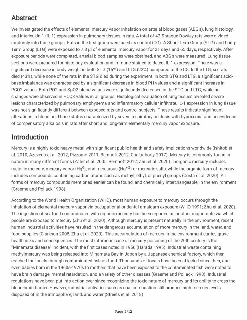
Animal Well-Being:The exposure of the STG and LTG to 7.3 µl of elemental mercury vapor led to a statistically signi�cant reduction inweight compared to the CG, which in contrast, gained weight during the experiment. Results are reported in Fig. 1.The percentages of body weight loss were 15% and 22% in the STG and LTG, respectively. The CG and STGsubjects all survived the course of the study. The LTG had 6 out of the 14 (42.8%) subjects die during the study.
Arterial Blood Gas Parameters:Five parameters in the ABG's were compared between groups, and those parameters were: PH, PCO2, PO2, SPO2,and HCO3. The results for ABG’s �ndings in all groups after short- and long-term exposure to elemental mercuryvapor inhalation, as well as, CG are reported in Table 1. The ABG’s testing showed a signi�cant decrease in PHvalues for STG and LTG (p < 0.05) throughout the study, with no signi�cant change in the control PH values beingobserved. PH values within the exposed groups were lower than the CG (p < 0.05). PCO2 values were signi�cantlyhigher in the exposed groups compared to the CG (p < 0.001). PO2 values were signi�cantly lower in the STG thanthe CG (p = 0.03), while no signi�cant difference was found between the LTG and the CG PO2 values (p = 0.21).
Page 6/12
SPO2 values in the STG and LTG were signi�cantly lower than the SpO2 in the CG (p = 0.001). No signi�cantdifference was found in the HCO3 values between the exposed groups and the CG (p = 0.506). No signi�cantdifference in the ABG’s parameters examined was found between male and female subjects within the samegroup in the STG or LTG (p > 0.05).
Table 1The arterial blood gas parameters in Sprague-Dowley rats after short- and long-term elemental mercury vapor
exposure and in those with no mercury vapor exposure.ArterialBlood GasParameters
Groups
Short-term exposure Long-term exposure Control
Allanimals
Males Females Allanimals
Males Females Allanimals
Males Females
PH 7.23 ± 0.05*
7.22 ± 0.03
7.24 ± 0.07
7.18 ± 0.02*
7.18 ± 0.02
7.19 ± 0.0
7.36 ± 0.09
7.32 ± 0.02
7.4 ± 0.06
PCO2 62 ± 10*
65 ± 6 60 ± 13 68 ± 4* 69 ± 4 66 ± 4 44 ± 8 50 ± 10
37 ± 4
PO2 49 ± 21*
42 ± 10
55 ± 18 52 ± 14*
55 ± 15
44 ± 5 67 ± 16 57 ± 13
77 ± 11
SpO2 66 ± 19*
63 ± 17
69 ± 20 71 ± 16*
73 ± 18
65 ± 7 89 ± 9 84 ± 10
94 ± 3
HCO3 25 ± 3 26 ± 2 24 ± 3 25 ± 2 25 ± 15
25 ± 5 24 ± 3 25 ± 2 23 ± 2
* statistically signi�cant in comparison to the control group (p ≤ 0.05)
Lung Histopathology:Histological study under light microscopy was performed for dissected and prepared lung tissues for all groups.Lung tissue sections obtained from rats after short-term and long-term exposure are presented under lightmicroscopy in Fig. 2. The histological study of lung tissue from the CG revealed mild in�ammation. Theexamination of lung tissue sections from the STG and the LTG showed marked in�ammatory cellular in�ltration,emphysema, and dilatation of the alveoli with destruction and obstruction of intra-alveolar septae. The LTG tissuesections showed a higher degree of tissue injury and more severe in�ammation than the STG.
Immunohistochemical Study of Rats' Lung Tissues:Immunohistochemistry staining to detect IL-1 expression in lung tissues was performed and is presented in Fig. 3.No signs of signi�cant IL-1 expression were noted in any of the groups.
DiscussionThis study is one of the �rst scienti�c studies that evaluated the toxic effects of elementary mercury vaporinhalation on various arterial blood gas parameters in rats and attempted to evaluate possible underlying lungpathology as a direct causal effect of signi�cant alterations in blood acid-base balance. Several side effectsrelated to various body organs and systems have been reported after acute or chronic exposure to elementalmercury (Clarkson and Magos 2006; Thomas and Clarkson 2008; Houston 2011; Park and Zheng 2012;
Page 7/12
Chakraborty 2017). In human beings, exposure to mercury inhalation was reported to cause �u-like symptoms,including fever, cough, dyspnea, and chest pain (Cortes et al. 2018). In this study, rats in both exposure groups lostweight signi�cantly (p ≤ 0.05) during the experiment, while rats in the CG gained weight. A total of 6 rats out of 14died (42.8%), all belonging to the LTG, while none of the rats in the short-term or control groups died during theexperiment. The high mortality rates in the LTG can be explained by severe lung injury, hypoxia, acidemia, andweight loss, observed in such a group, due to the long duration of exposure to elemental mercury vapor.
In both the STG and LTG, there was a signi�cant decrease in blood PH values compared to the CG. This state ofsevere acidemia was accompanied by a signi�cant increase in blood concentrations of CO2 and a signi�cantdecrease in blood concentration and saturation of O2 regardless of sex. Simultaneously, no signi�cant changeswere observed in blood concentrations of HCO3, indicating a lack of renal compensation. However, the PO2 andSPO2 levels were found to be higher at the end of the study in the LTG than in the STG, indicating some form ofadaptive response to the long-term exposure to elemental mercury vapor, leading to improved oxygen saturationof the blood. The ABG’s results indicate that rats exposed to elemental vapor inhalation suffered a signi�cantdegree of respiratory acidosis, in contrast to previously established evidence in the literature leaningpredominantly towards metabolic acidosis with mercury poisoning in both animals (Pathak and Bhowmik 1998)and humans, predominantly children (Husband and McKellar 1970; Counter and Buchanan 2004). The higherincidence of acidosis in the pediatric age group than in adults exposed to elemental mercury in the literature canbe explained by the difference in body weight and physiological maturity, which results in a much lower degree ofexposure being necessary to bring mercury to highly toxic and potentially fatal concentrations in the blood andtissues. This carries clinically signi�cant implications in managing pediatric mercury poisoning cases, wherehealthcare providers should critically consider both metabolic and respiratory acidosis.
Histopathological examination of lung tissues from rats exposed to elementary mercury vapor inhalation revealedsubstantial pulmonary in�ammation characterized by in�ammatory cellular in�ltration, emphysema, anddilatation of the alveoli with thickening of the intra-alveolar septae. These in�ammatory lesions appeared morepronounced in the rats in the LTG than those in the STG. Interestingly, pulmonary lesions were found to be mostsevere in female rats compared to males. The severe in�ammatory lesions of the pulmonary tissues arepresumably the underlying cause of the changes noted in the acid-base status of exposed rats. This also isevident by the observed clinical signs of hyperventilation, hypoxia, and weakness. These �ndings are congruentwith previously reported pulmonary lesions in human beings after acute exposure to mercury vapor (Smiechowiczet al. 2017). Diffuse in�ammatory cellular in�ltrates, acute pulmonary edema and emphysema, chemicalpneumonitis, bronchiolitis, and pneumothorax, and death have been reported in humans after acute mercury vaporinhalation (Smiechowicz et al. 2017). These �ndings emphasize the importance of early treatment of lung tissueinjury with medications, supplemental oxygen, endotracheal intubation, and mechanical ventilation. These earlyinterventions are crucial to maintaining adequate gas exchange in elemental mercury vapor poisoning to preventwell-known respiratory complications such as hypoxemia and lesser-known signi�cant complications that mayarise, such as respiratory acidemia.
In this study, immunohistochemistry results to detect IL-1 expression in lung tissues revealed no signi�cantdifferences between all groups. These results disagree with previous �ndings where mercury vapor inhalationinduced increased secretion of IL-1 (Zdolsek et al. 1994). The effects of mercury on the IL-1 have beendemonstrated in several laboratory animal models (Gardner et al. 2009). It has been suggested that exposure ofmice to methylmercury induced expression of IL-1 in the brain tissue, causing central nervous cytotoxicity
Page 8/12
(Takahashi et al. 2015). In contrast, one study found that macrophages from mercury-treated mice exhibited areduced capacity to produce IL-1 (Batista-Duharte et al. 2018). Therefore, the exact effects of mercury moleculeson the production and expression of IL-1 in different body tissues remain controversial, and further clinical trialsare warranted to determine the exact pathophysiological effects of mercury on pro-in�ammatory cytokinesproduction and function.
ConclusionsThe �ndings of this study indicate that exposure to elementary mercury vapor induces signi�cant pulmonaryinjury resulting in severe clinical manifestations, severe respiratory acidosis with hypoxemia, and no evidence ofcompensatory alkalosis in Sprague-Dowley rats. These manifestations are correlated with the duration of mercuryvapor exposure regardless of gender. Health care professionals should consider respiratory acidosis in theirtreatment plans for patients affected with mercury exposure.
Declarations
AcknowledgmentThe authors would like to thank the Deanship of Research for the �nancial support (Grant # 2017/287)
FundingA grant from the deanship of Research at Jordan University of Science and Technology (Grant # 2017/287).
Con�icts of InterestThere is no con�ict of interest.
Availability of Data and MaterialThe datasets generated and analyzed during the current study are available with the corresponding authors onreasonable request.
Code AvailabilityNot Applicable.
Authors contributionsAll authors have seen and approved the content, ful�lled the authorship criteria, and have contributed signi�cantlyto this work. All authors presented substantial contributions to the conception and design of the study and/or to
Page 9/12
the acquisition, analysis, and interpretation of data, and they drafted the manuscript and revised it critically forcontent. All authors read and approved the �nal manuscript version submitted for publication.
Ethics ApprovalAll experimental procedures performed in this study were reviewed and approved by the Institutional Animal Careand Use Committee (IACUC) of Jordan University of Science and Technology (JUST).
Consent to ParticipateNot applicable.
Consent for PublicationThis study was approved for publication by the Institutional Animal Care and Use Committee (IACUC) of JordanUniversity of Science and Technology (JUST).
References1. Alturkistani, H.A., Tashkandi, F.M. and Saleh, Z.M. (2016). Histological stains: A literature review and case
study. Global Journal of Health Science 8(3):72-9.
2. Asano, S., Eto, K., Kurisaki, E., Gunji, H., Hiraiwa, K., Sato, M., ... & Wakasa, H. (2000). Acute inorganic mercuryvapor inhalation poisoning. Pathology International, 50(3), 169-174.
3. Azevedo, B.F., Furieri, L.B., Pecanha, F.M., Wiggers, G.A., Vassallo, P.F., Simoes, M.R., Fiorim, J., de Batista, P.R.,Fioresi, M., Rossoni, L., Stefanon, I., Alonso, M.J., Salaices, M. and Vassallo, D.V. (2012). Toxic effects ofmercury on the cardiovascular and central nervous systems. Journal of Biomedicine and Biotechnology.Volume 2012, Article ID 949048, 11 pages.
4. Batista-Duharte, A., Téllez-Martínez, D., Jellmayer, J.A., Fuentes, D.L.P., Polesi, M.C., Baviera, A.M. and Carlos,I.Z. (2018). Repeatede exposition to mercury (II) chloride enhances susceptibility to S. schenckii sensu strictoinfection in mice. Journal of Fungi 4(64): 1-12.
5. Bernhoft, R. (2012). Mercury toxicity and treatment: a review of the literature. Journal of Environmental andPublic Health. Volume 2012, Article ID 460508, 10 pages.
�. Chakraborty, P. (2017). Mercury exposure and Alzheimer’s disease in India-an imminent threat? Science ofthe Total Environment 589: 232-235.
7. Clarkson, T.W. (2008). The toxicology of mercury. Critical Reviews In Clinical Laboratory Sciences 34:4, 369-403.
�. Clarkson, T. and Magos, L. (2006). The toxicology of mercury and its chemical compounds. Critical ReviewsIn Toxicology 36:609-62.
9. Cortes, J., Peralta, J. and Díaz-Navarro, R. (2018). Acute respiratory syndrome following accidental inhalationof mercury vapor. Clinical Case Reports 6:1535–1537.
Page 10/12
10. Costa, F., Coelho, J.P., Baptista, J., Martinho, F., Pereira, M.E. and Parda, M.A. (2020). Mercury accumulation in�sh species along the Portuguese coast: Are there potential risks to human health? Marine Pollution Bulletin150: 110740.
11. Counter, S. A., & Buchanan, L. H. (2004). Mercury exposure in children: a review. Toxicology and appliedpharmacology, 198(2), 209-230.
12. Gardner, R.M., Nyland, J.F., Evans, S.L., Wang, S.B., Doyle, K.M., Crainiceanu, C.M. and Silbergeld, E.K. (2009).Mercury induces an unopposed in�ammatory response in human peripheral blood mononuclear cells in vitro.Environmental Health Perspectives 117(12): 1932-1938.
13. Graeme, K. A., & Pollack Jr, C. V. (1998). Heavy metal toxicity, part I: arsenic and mercury. The Journal ofemergency medicine, 16(1), 45-56.
14. Haase, H., Engelhardt, G., Hebel, S., & Rink, L. (2011). Mercuric ions inhibit mitogen-activated protein kinasedephosphorylation by inducing reactive oxygen species. Toxicology and applied pharmacology, 250(1), 78-86.
15. Harada, M. (1995). Minamata disease: methylmercury poisoning in Japan caused by environmental pollution.Critical reviews in toxicology, 25(1), 1-24.
1�. Houston, M.C. (2001). Role of mercury toxicity in hypertension, cardiovascular disease, and stroke. TheJournal of Clinical Hypertension 13(8):621-7.
17. Husband, P., & McKellar, W. J. D. (1970). Infantile renal tubular acidosis due to mercury poisoning. Archives ofdisease in childhood, 45(240), 264-268.
1�. Ishitob, H., Stern, S., Thurston, S.W., Zareba, G., Langdon, M. and Gelein, R. (2010). Organic and inorganicmercury in neonatal rat brain after prenatal exposure to methylmercury and mercury vapor. EnvironmentalHealth Perspectives 118(2):242-8.
19. Jensen, E.C. (2013). Quantitative analysis of histological staining and �uorescence using ImageJ. TheAnatomical Record 296(3): 378-381.
20. Omanwar, S., Saidullah, B., Ravi, K., & Fahim, M. (2014). Vasorelaxant effects of mercury on rat thoracic aorta:the nitric oxide signaling mechanism. Human & experimental toxicology, 33(9), 904-910.
21. Pizzorno, J. (2011). Is mercury toxicity an epidemic? Integrative Medicine 10(5):52-3.
22. Park, J.D. and Zheng, W. (2012). Human exposure and health effects of inorganic and elemental mercury.Journal of Preventive Medicine and Public Health 45(6):344-52.
23. Pathak, S. K., & Bhowmik, M. K. (1998). The chronic toxicity of inorganic mercury in goats: clinical signs,toxicopathological changes and residual concentrations. Veterinary research communications, 22(2), 131-138.
24. Smiechowicz, J., Skoczynska, A., Nieckula-Szwarc, A., Kulpa, k and Kübler, A. (2017). Occupational mercuryvapor poisoning with a respiratory failure, pneumomediastinum and severe quadriparesis. SAGE OpenMedical Case Reports 5: 1–4.
25. Rafati-Rahimzadeh, M., Rafati-Rahimzadeh, M., Kazemi, S., & Moghadamnia, A. A. (2014). Current approachesof the management of mercury poisoning: need of the hour. DARU Journal of Pharmaceutical Sciences, 22(1),1-10.
2�. Russi, G., & Marson, P. (2011). Urgent plasma exchange: how, where and when. Blood Transfusion, 9(4), 356.
27. Streets, D. G., Lu, Z., Levin, L., Ter Schure, A. F., & Sunderland, E. M. (2018). Historical releases of mercury toair, land, and water from coal combustion. Science of the Total Environment, 615, 131-140.

Page 11/12
2�. Suker, D.K., AL- Badran, A.I., Abbas, E.K. and Abdullah, S.A. (2017). Immunohistochemistry analysis forInterleukin-6 expression from the tumor tissue. International Journal of Sciences 6(3): 14-24.
29. Takahashi, T., Iwai-Shimada, M., Syakushi, Y., Kim, M., Hwang, G., Miura, N. and Naganuma, A. (2015).Methylmercury induces expression of interleukin-1β and interleukin-19 in mice brains. FundamentalToxicological Sciences 2(6): 239-243.
30. Tinkov, A. A., Ajsuvakova, O. P., Skalnaya, M. G., Popova, E. V., Sinitskii, A. I., Nemereshina, O. N., ... & Skalny, A.V. (2015). Mercury and metabolic syndrome: a review of experimental and clinical observations. Biometals,28(2), 231-254.
31. Vassallo, D. V., Simões, M. R., Furieri, L. B., Fioresi, M., Fiorim, J., Almeida, E. A. S., ... & Salaices, M. (2011).Toxic effects of mercury, lead and gadolinium on vascular reactivity. Brazilian Journal of Medical andBiological Research, 44(9), 939-946.
32. Weber, A., Wasiliew, P., & Kracht, M. (2010). Interleukin-1 (IL-1) pathway. Science signaling, 3(105), cm1-cm1.
33. World Health Organization. (1991). International Program on Chemical Safety. Inorganic mercury:environmental health criteria 118. Geneva.
34. Zahir, F., Rizwi, S.J., Haq, S.K. and Khan, R.H. (2005). Low dose mercury toxicity and human health.Environmental Toxicology and Pharmacology 20: 351–360.
35. Zdolsek, J.M., Soder, O. and Hultman, P. (1994). Mercury induces in vivo and in vitro secretion of interleukin-1in mice. Journal of Immunopharmacology 28:201-8.
3�. Zhu, Y., Peng, S., Lu, P., Chen, T. and Yang, Y. (2020). Mercury removal from aqueous solutions using modi�edpyrite: A column experiment. Minerals 10, 43.
Figures

Page 12/12
Figure 1
The effects of elemental mercury vapor exposure for short-term (21 days) and long-term (65 days) on body weightin rats (N=14).
Figure 2
H&E stained lung tissue sections obtained from rats after short-term (left) and long-term (right) exposure toelemental mercury vapor inhalation. Severe in�ammatory cellular in�ltration, emphysema, intra-alveolar edema,and intra-alveolar septal thickening, especially in rats with long-term exposure.
Figure 3
Immunohistochemistry staining to detect IL-1 expression in lung tissues of rats after short-term (left) and long-term (right) exposure to elemental mercury vapor inhalation. No signi�cant differences were observed in IL-1expression in any of the groups.