expression and functional analysis of the propamocarb
TRANSCRIPT
RESEARCH ARTICLE Open Access
Expression and functional analysis of thePropamocarb-related gene CsDIR16 incucumbersChunhong Liu1,2, Zhiwei Qin1*, Xiuyan Zhou1, Ming Xin1, Chunhua Wang1, Dong Liu1 and Shengnan Li1
Abstract
Background: Cucumber downy mildew is among the most important diseases that can disrupt cucumber production.Propamocarb, also known as propyl-[3-(dimethylamino)propyl]carbamate (PM), is a systemic carbamate fungicidepesticide that is widely applied in agricultural production because of its high efficiency of pathogens control, especiallycucumber downy mildew. However, residual PM can remain in cucumbers after the disease has been controlled. Toexplore the molecular mechanisms of PM retention, cucumber cultivars ‘D9320’ (with the highest residual PM content)and ‘D0351’ (lowest residual PM content) were studied. High-throughput tag-sequencing (Tag-Seq) results showed thatthe CsDIR16 gene was related to PM residue, which was verified using transgenic technology.
Results: We investigated the activity of a dirigent cucumber protein encoded by the CsDIR16 in gene response to stressinduced by PM treatment. Gene-expression levels of CsDIR16 were up-regulated in the fruits, leaves, and stems of ‘D0351’plants in response to PM treatment. However, in cultivar ‘D9320’, CsDIR16 levels were down-regulated in the leaves andstems after PM treatment, with no statistically significant differences observed in the fruits. Induction by jasmonicacid, abscisic acid, polyethylene glycol 4000, NaCl, and Corynespora cassiicola Wei (Cor) resulted in CsDIR16 up-regulationin ‘D0351’ and ‘D9320’. Expression after salicylic acid treatment was up-regulated in ‘D0351’, but was down-regulated in‘D9320’. CsDIR16 overexpression lowered PM residues, and these were more rapidly reduced in CsDIR16(+)transgenic ‘D9320’ plants than in wild-type ‘D9320’ and CsDIR16(−) transgenic plants.
Conclusions: Analyses of the CsDIR16-expression patterns in the cucumber cultivars with the highest and lowest levelsof PM residue, and transgenic validation indicated that CsDIR16 plays a positive role in reducing PM residues. The findingsof this study help understand the regulatory mechanisms occurring in response to PM stress in cucumbers and inestablishing the genetic basis for developing low-pesticide residue cucumber cultivars.
Keywords: Cucumber, Cucumber downy mildew, Propamocarb, Dirigent protein, CsDIR16
BackgroundPesticides are among the most widely used chemicals inthe world. With their application in modern agriculture,up to 80% of crop yield were protected from pest andweeds [1]. However, contamination of products andenvironment was triggered by the extensive use of pesti-cides in most regions [2]. Food consumption is one of themost common routes of pesticide exposure in consumers[3]. Pesticide residues represent a major food safety issue
since some of them are suspected to mutagenic, carcino-genic, and teratogenic activities. For example, many pesti-cides cause acute toxicity, as well as sublethal effects bycausing the endocrine disorders, sperm quality declineand reproductive development abnormalities [4–7]. Con-siderable efforts have been made to reduce the pesticideresidues of agricultural products through both tradition-ally breeding programs and contemporarily genetic trans-formation. Identifying biotransformation mechanisms ofpesticide residues at the molecular level has emerged as anew approach for studying pesticide residues [8, 9].Downy mildew is a devastating disease affecting cucurbits
that is caused by infection by zoospores. The associatedlesions can induce chlorosis or yellowing, and further
* Correspondence: [email protected] of Horticulture and Landscape Architecture, Key Laboratory ofBiology and Genetic Improvement of Horticultural Crops (Northeast Region),Northeast Agricultural University, Harbin 150030, ChinaFull list of author information is available at the end of the article
© The Author(s). 2018 Open Access This article is distributed under the terms of the Creative Commons Attribution 4.0International License (http://creativecommons.org/licenses/by/4.0/), which permits unrestricted use, distribution, andreproduction in any medium, provided you give appropriate credit to the original author(s) and the source, provide a link tothe Creative Commons license, and indicate if changes were made. The Creative Commons Public Domain Dedication waiver(http://creativecommons.org/publicdomain/zero/1.0/) applies to the data made available in this article, unless otherwise stated.
Liu et al. BMC Plant Biology (2018) 18:16 DOI 10.1186/s12870-018-1236-2
destroy the entire leaf in a few days [10]. Propamocarb,also known as propyl-[3-(dimethylamino) propyl] carba-mate (PM), is a low-toxicity fungicide with systemic activityafter absorption through leaves, stems, and roots, and bytransportation throughout treated plants via the vascularsystem [11]. The fungicide PM has good protective andcurative activities against downy mildew with no phyto-toxicity effects in fruits and vegetables, such as tomatoes,potatoes and cucumbers [12–16]. PM causes slight cyto-toxicity to cortical neurons and moderately effects on theintracellular membrane potential, glucose consumption,ATP levels, and the cytoskeleton [17]. Bone-marrow mi-cronucleus and chromosome aberration test results withSwiss albino mice suggested that PM was not genotoxic inmouse bone marrow in vivo, but had cytotoxic effects[18]. These data indicate that PM residues in fruits andvegetables pose potential health risks in humans.Cucumber downy mildew, caused by Pseudoperonospora
cubensis (Berk. & M.A.Curtis, Rostovzev), is an importantleaf disease that can spread quickly and reduce cucumberyields [19, 20]. Both production and quality of cucumberhave thus been affected, and then led to economic losses.PM is an effective control for downy mildew; however,PM residues can remain in cucumber plants after thedisease has been controlled. PM residues may accumulateat levels higher than the international maximum residuelimits (MRLs) [21]. Improving the yield and quality of cu-cumber products, ensuring food safety for consumers, andimproving the international competitiveness of Chinesevegetable products are important objectives.Despite mounting concerns regarding pesticide residues
on vegetables, numerous scientific advances have beenmade in detection technologies and physiological mecha-nisms influencing pesticide-residue levels. Methods forassessing PM-residue levels have been established [22].Twenty-eight cucumber germplasm resources have beencollected and used to compare PM residues among culti-vars. Among them, cultivar ‘D0351’ was found to have thelowest PM-residue levels, as well as ‘D9320’ the highest[21, 23]. To investigate the molecular mechanisms thatdetermine PM-residue levels in cucumbers, high-throughputtag-sequencing (Tag-Seq) has been performed using Illuminaanalysis (based on the Solexa Genome Analyzer plat-form), in order to study gene-expression profiles in con-trol and PM-treated fruit from ‘D0351’ plants [21]. Datafrom several studies have shown that pesticide residuelevels are controlled by multiple genes [22]. Transcriptomicanalysis revealed that CsABC19 and CsWRKY30 are 2positive regulators of plant tolerance to PM stress.Overexpression of CsABC19 and CsWRKY30 in Arabidop-sis enhanced tolerance to PM stresse and decreased PMresidues [24, 25]. CsDIR16 is another key gene involvedin responses to PM stress, as revealed by transcriptomicanalysis.
Dirigent proteins (DIRs) were first discovered by Davinet al. [26] in Forsythia intermedia. Subsequently, DIRand DIR-like family proteins have been reported in manyplant species, including gymnosperms and angiosperms.The DIR gene family encodes several proteins involvedin secondary metabolism, lignan and lignin formationbiosynthesis [27–29], or responses to pathogen infectionand abiotic stress [30–34]. DIRs lack a catalytically active(oxidative) center and function only as guiding proteins[35]. The mechanism of action is thought to involve cap-ture of free radicals produced by the oxidation of coniferylalcohol, and the FiDIR protein catalyze 8–8′ coupling toproduce (+)-pinoresinol, the AtDIR5 and AtDIR6 proteinscatalyze 8–8′ coupling to produce (−)-pinoresinol [36–38].This is followed by intramolecular cyclization to increasetotal lignin accumulation and improve resistance to stress[39]. DIRs affect the acidity of lignin by changing the com-position and connection of the lignin monomer, therebyreducing cellular damage caused by drought or water stressto help plants increase stress resistance [40]. The DIRgenes serve a universal function in terms of stress resist-ance, but their roles in response to PM treatment have notbeen determined. The CsDIR16 gene was upregulated in‘D0351’ plants exposed to PM stress, which suggests thatCsDIR16 plays a vital role in plant responses to PM stressand may be of value for future production of transgeniccucumbers with enhanced PM stress responses.
ResultsCloning and bioinformatics analysis of CsDIR16In this study, we investigated a cucumber PM-responsivegene (Csa4M280630.1), designated here as CsDIR16.The full-length cDNA sequences of CsDIR16 were clonedfrom cucumber fruits of ‘D0351’ and ‘D9320’ by reversetranscription-PCR (RT-PCR) (Fig. 1a). Then the sequenceof CsDIR16 was confirmed by repeated sequencing.Comparison with the cucumber genome database (http://www.icugi.org/), the sequences of CsDIR16 cloned fromcucumber fruits ‘D9320’ showed no difference. However, aA-to-G mutation at position 556 was found in the se-quences of CsDIR16 cloned from cucumber fruits ‘D0351’,resulting in an I-to-V amino acid change at position 186.CsDIR16 has a 588-base pair (bp) open-reading frame(ORF) encoding 195 amino acids, and the calculated mo-lecular weight is 21.55 kDa (Fig. 1b).The CsDIR16 protein contains a dirigent protein domain
(PF03018) (Fig. 1c) and 5 conserved motifs (Additional file 1:Figure S1) [40]. An N-terminal signal peptide and a cleav-age site between amino acids 28 and 29 were predicted inthe protein by SignalP 4.0 (Additional file 1: Figure S2). Atransmembrane helix between residues 10 and 27 waspredicted by TMHMM 2.0 (Additional file 1: Figure S3).CsDIR16 has 2 N-glycosylation sites at amino acids 77(Asn) and 135 (Asn). Such N-glycosylation sites have been
Liu et al. BMC Plant Biology (2018) 18:16 Page 2 of 12
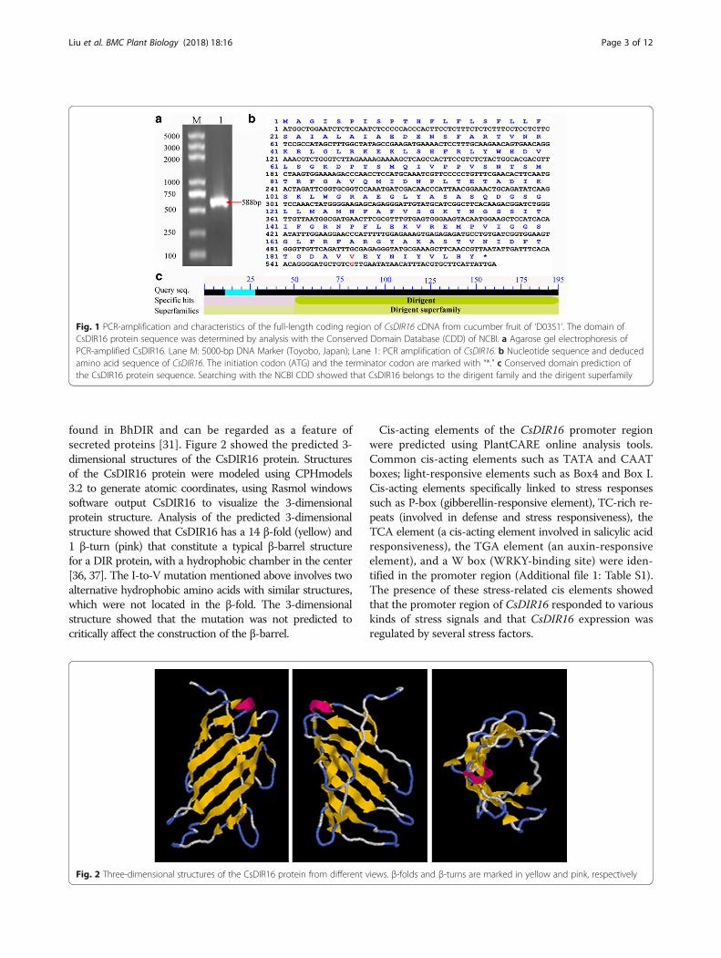
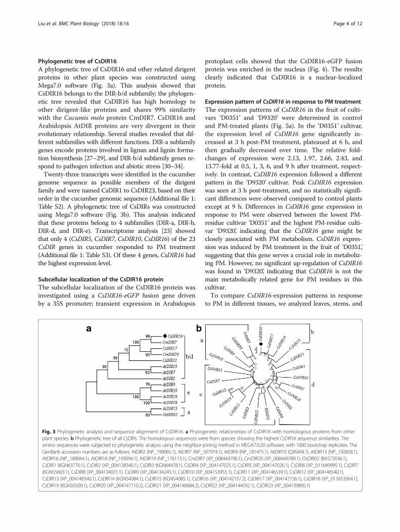
found in BhDIR and can be regarded as a feature ofsecreted proteins [31]. Figure 2 showed the predicted 3-dimensional structures of the CsDIR16 protein. Structuresof the CsDIR16 protein were modeled using CPHmodels3.2 to generate atomic coordinates, using Rasmol windowssoftware output CsDIR16 to visualize the 3-dimensionalprotein structure. Analysis of the predicted 3-dimensionalstructure showed that CsDIR16 has a 14 β-fold (yellow) and1 β-turn (pink) that constitute a typical β-barrel structurefor a DIR protein, with a hydrophobic chamber in the center[36, 37]. The I-to-V mutation mentioned above involves twoalternative hydrophobic amino acids with similar structures,which were not located in the β-fold. The 3-dimensionalstructure showed that the mutation was not predicted tocritically affect the construction of the β-barrel.
Cis-acting elements of the CsDIR16 promoter regionwere predicted using PlantCARE online analysis tools.Common cis-acting elements such as TATA and CAATboxes; light-responsive elements such as Box4 and Box I.Cis-acting elements specifically linked to stress responsessuch as P-box (gibberellin-responsive element), TC-rich re-peats (involved in defense and stress responsiveness), theTCA element (a cis-acting element involved in salicylic acidresponsiveness), the TGA element (an auxin-responsiveelement), and a W box (WRKY-binding site) were iden-tified in the promoter region (Additional file 1: Table S1).The presence of these stress-related cis elements showedthat the promoter region of CsDIR16 responded to variouskinds of stress signals and that CsDIR16 expression wasregulated by several stress factors.
Fig. 1 PCR-amplification and characteristics of the full-length coding region of CsDIR16 cDNA from cucumber fruit of ‘D0351’. The domain ofCsDIR16 protein sequence was determined by analysis with the Conserved Domain Database (CDD) of NCBI. a Agarose gel electrophoresis ofPCR-amplified CsDIR16. Lane M: 5000-bp DNA Marker (Toyobo, Japan); Lane 1: PCR amplification of CsDIR16. b Nucleotide sequence and deducedamino acid sequence of CsDIR16. The initiation codon (ATG) and the terminator codon are marked with “*.” c Conserved domain prediction ofthe CsDIR16 protein sequence. Searching with the NCBI CDD showed that CsDIR16 belongs to the dirigent family and the dirigent superfamily
Fig. 2 Three-dimensional structures of the CsDIR16 protein from different views. β-folds and β-turns are marked in yellow and pink, respectively
Liu et al. BMC Plant Biology (2018) 18:16 Page 3 of 12
Phylogenetic tree of CsDIR16A phylogenetic tree of CsDIR16 and other related dirigentproteins in other plant species was constructed usingMega7.0 software (Fig. 3a). This analysis showed thatCsDIR16 belongs to the DIR-b/d subfamily; the phylogen-etic tree revealed that CsDIR16 has high homology toother dirigent-like proteins and shares 99% similaritywith the Cucumis melo protein CmDIR7. CsDIR16 andArabidopsis AtDIR proteins are very divergent in theirevolutionary relationship. Several studies revealed that dif-ferent subfamilies with different functions. DIR-a subfamilygenes encode proteins involved in lignan and lignin forma-tion biosynthesis [27–29], and DIR-b/d subfamily genes re-spond to pathogen infection and abiotic stress [30–34].Twenty-three transcripts were identified in the cucumber
genome sequence as possible members of the dirigentfamily and were named CsDIR1 to CsDIR23, based on theirorder in the cucumber genomic sequence (Additional file 1:Table S2). A phylogenetic tree of CsDIRs was constructedusing Mega7.0 software (Fig. 3b). This analysis indicatedthat these proteins belong to 4 subfamilies (DIR-a, DIR-b,DIR-d, and DIR-e). Transcriptome analysis [23] showedthat only 4 (CsDIR5, CsDIR7, CsDIR10, CsDIR16) of the 23CsDIR genes in cucumber responded to PM treatment(Additional file 1: Table S3). Of these 4 genes, CsDIR16 hadthe highest expression level.
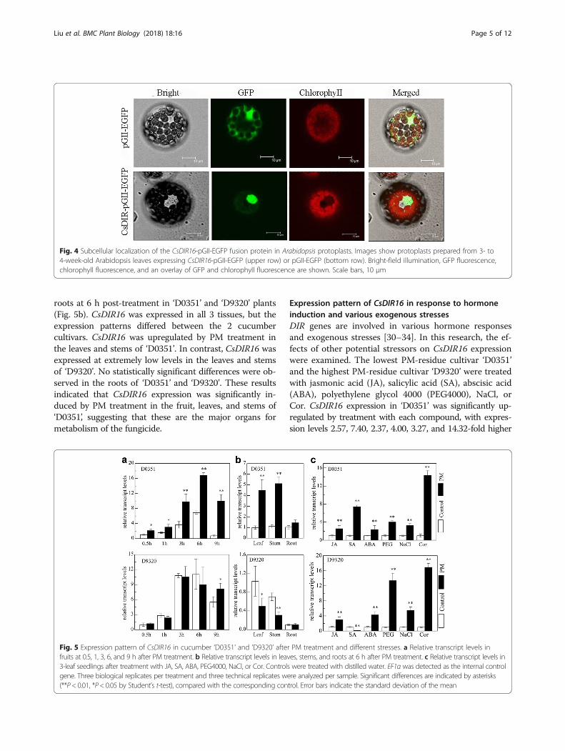
Subcellular localization of the CsDIR16 proteinThe subcellular localization of the CsDIR16 protein wasinvestigated using a CsDIR16-eGFP fusion gene drivenby a 35S promoter; transient expression in Arabidopsis
protoplast cells showed that the CsDIR16-eGFP fusionprotein was enriched in the nucleus (Fig. 4). The resultsclearly indicated that CsDIR16 is a nuclear-localizedprotein.
Expression pattern of CsDIR16 in response to PM treatmentThe expression patterns of CsDIR16 in the fruit of culti-vars ‘D0351’ and ‘D9320’ were determined in controland PM-treated plants (Fig. 5a). In the ‘D0351’ cultivar,the expression level of CsDIR16 gene significantly in-creased at 3 h post-PM treatment, plateaued at 6 h, andthen gradually decreased over time. The relative fold-changes of expression were 2.13, 1.97, 2.66, 2.43, and13.77-fold at 0.5, 1, 3, 6, and 9 h after treatment, respect-ively. In contrast, CsDIR16 expression followed a differentpattern in the ‘D9320’ cultivar. Peak CsDIR16 expressionwas seen at 3 h post-treatment, and no statistically signifi-cant differences were observed compared to control plantsexcept at 9 h. Differences in CsDIR16 gene expression inresponse to PM were observed between the lowest PM-residue cultivar ‘D0351’ and the highest PM-residue culti-var ‘D9320’, indicating that the CsDIR16 gene might beclosely associated with PM metabolism. CsDIR16 expres-sion was induced by PM treatment in the fruit of ‘D0351’,suggesting that this gene serves a crucial role in metaboliz-ing PM. However, no significant up-regulation of CsDIR16was found in ‘D9320’, indicating that CsDIR16 is not themain metabolically related gene for PM residues in thiscultivar.To compare CsDIR16-expression patterns in response
to PM in different tissues, we analyzed leaves, stems, and
Fig. 3 Phylogenetic analysis and sequence alignment of CsDIR16. a Phylogenetic relationships of CsDIR16 with homologous proteins from otherplant species. b Phylogenetic tree of all CsDIRs. The homologous sequences were from species showing the highest CsDIR16 sequence similarities. Theamino sequences were subjected to phylogenetic analysis using the neighbor-joining method in MEGA7.0.20 software, with 1000 bootstrap replicates. TheGenBank accession numbers are as follows: AtDIR2 (NP_199065.1), AtDIR7 (NP_187974.1), AtDIR9 (NP_181475.1), AtDIR10 (Q9SIA8.1), AtDIR13 (NP_192858.1),AtDIR16 (NP_189044.1), AtDIR18 (NP_193094.1), AtDIR19 (NP_176113.1), CmDIR7 (XP_008449790.1), CmDIR20 (XP_008449789.1), OsDIR02 (BAS72036.1),CsDIR1 (KGN63770.1), CsDIR2 (XP_004138340.1), CsDIR3 (KGN64478.1), CsDIR4 (XP_004147025.1), CsDIR5 (XP_004147026.1), CsDIR6 (XP_011649995.1), CsDIR7(KGN55603.1), CsDIR8 (XP_004134021.1), CsDIR9 (XP_004134245.1), CsDIR10 (XP_004153955.1), CsDIR11 (XP_004146539.1), CsDIR12 (XP_004146540.1),CsDIR13 (XP_004146540.1), CsDIR14 (KGN54084.1), CsDIR15 (KGN54085.1), CsDIR16 (XP_004142157.2), CsDIR17 (XP_004142156.1), CsDIR18 (XP_011653564.1),CsDIR19 (KGN50509.1), CsDIR20 (XP_004147710.2), CsDIR21 (XP_004140684.2), CsDIR22 (XP_004144592.1), CsDIR23 (XP_004139895.1)
Liu et al. BMC Plant Biology (2018) 18:16 Page 4 of 12
roots at 6 h post-treatment in ‘D0351’ and ‘D9320’ plants(Fig. 5b). CsDIR16 was expressed in all 3 tissues, but theexpression patterns differed between the 2 cucumbercultivars. CsDIR16 was upregulated by PM treatment inthe leaves and stems of ‘D0351’. In contrast, CsDIR16 wasexpressed at extremely low levels in the leaves and stemsof ‘D9320’. No statistically significant differences were ob-served in the roots of ‘D0351’ and ‘D9320’. These resultsindicated that CsDIR16 expression was significantly in-duced by PM treatment in the fruit, leaves, and stems of‘D0351’, suggesting that these are the major organs formetabolism of the fungicide.
Expression pattern of CsDIR16 in response to hormoneinduction and various exogenous stressesDIR genes are involved in various hormone responsesand exogenous stresses [30–34]. In this research, the ef-fects of other potential stressors on CsDIR16 expressionwere examined. The lowest PM-residue cultivar ‘D0351’and the highest PM-residue cultivar ‘D9320’ were treatedwith jasmonic acid (JA), salicylic acid (SA), abscisic acid(ABA), polyethylene glycol 4000 (PEG4000), NaCl, orCor. CsDIR16 expression in ‘D0351’ was significantly up-regulated by treatment with each compound, with expres-sion levels 2.57, 7.40, 2.37, 4.00, 3.27, and 14.32-fold higher
Fig. 4 Subcellular localization of the CsDIR16-pGII-EGFP fusion protein in Arabidopsis protoplasts. Images show protoplasts prepared from 3- to4-week-old Arabidopsis leaves expressing CsDIR16-pGII-EGFP (upper row) or pGII-EGFP (bottom row). Bright-field illumination, GFP fluorescence,chlorophyll fluorescence, and an overlay of GFP and chlorophyll fluorescence are shown. Scale bars, 10 μm
Fig. 5 Expression pattern of CsDIR16 in cucumber ‘D0351’ and ‘D9320’ after PM treatment and different stresses. a Relative transcript levels infruits at 0.5, 1, 3, 6, and 9 h after PM treatment. b Relative transcript levels in leaves, stems, and roots at 6 h after PM treatment. c Relative transcript levels in3-leaf seedlings after treatment with JA, SA, ABA, PEG4000, NaCl, or Cor. Controls were treated with distilled water. EF1α was detected as the internal controlgene. Three biological replicates per treatment and three technical replicates were analyzed per sample. Significant differences are indicated by asterisks(**P< 0.01, *P< 0.05 by Student’s t-test), compared with the corresponding control. Error bars indicate the standard deviation of the mean
Liu et al. BMC Plant Biology (2018) 18:16 Page 5 of 12
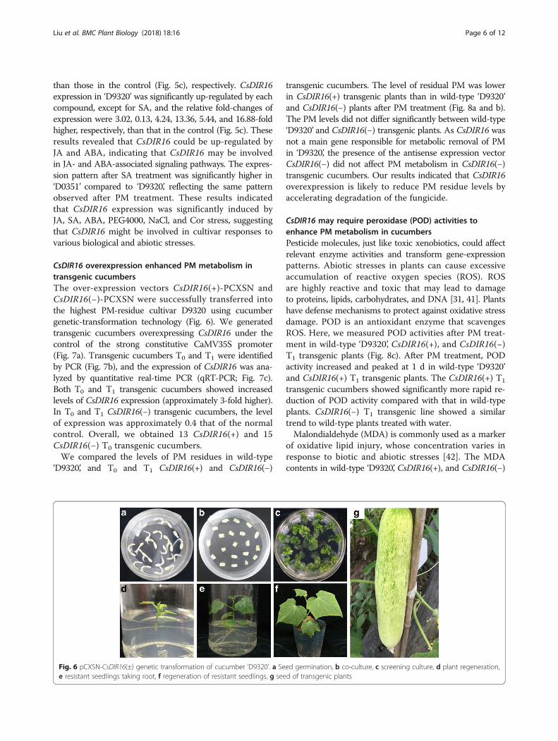
than those in the control (Fig. 5c), respectively. CsDIR16expression in ‘D9320’ was significantly up-regulated by eachcompound, except for SA, and the relative fold-changes ofexpression were 3.02, 0.13, 4.24, 13.36, 5.44, and 16.88-foldhigher, respectively, than that in the control (Fig. 5c). Theseresults revealed that CsDIR16 could be up-regulated byJA and ABA, indicating that CsDIR16 may be involvedin JA- and ABA-associated signaling pathways. The expres-sion pattern after SA treatment was significantly higher in‘D0351’ compared to ‘D9320’, reflecting the same patternobserved after PM treatment. These results indicatedthat CsDIR16 expression was significantly induced byJA, SA, ABA, PEG4000, NaCl, and Cor stress, suggestingthat CsDIR16 might be involved in cultivar responses tovarious biological and abiotic stresses.
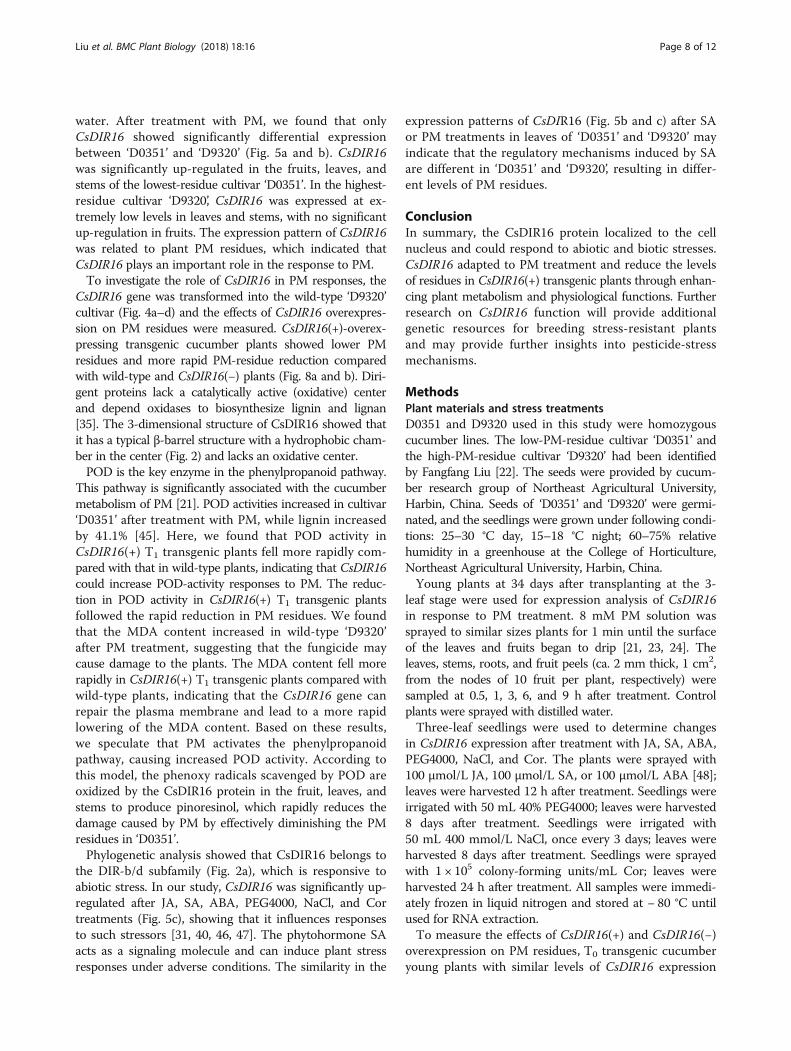
CsDIR16 overexpression enhanced PM metabolism intransgenic cucumbersThe over-expression vectors CsDIR16(+)-PCXSN andCsDIR16(−)-PCXSN were successfully transferred intothe highest PM-residue cultivar D9320 using cucumbergenetic-transformation technology (Fig. 6). We generatedtransgenic cucumbers overexpressing CsDIR16 under thecontrol of the strong constitutive CaMV35S promoter(Fig. 7a). Transgenic cucumbers T0 and T1 were identifiedby PCR (Fig. 7b), and the expression of CsDIR16 was ana-lyzed by quantitative real-time PCR (qRT-PCR; Fig. 7c).Both T0 and T1 transgenic cucumbers showed increasedlevels of CsDIR16 expression (approximately 3-fold higher).In T0 and T1 CsDIR16(−) transgenic cucumbers, the levelof expression was approximately 0.4 that of the normalcontrol. Overall, we obtained 13 CsDIR16(+) and 15CsDIR16(−) T0 transgenic cucumbers.We compared the levels of PM residues in wild-type
‘D9320’, and T0 and T1 CsDIR16(+) and CsDIR16(−)
transgenic cucumbers. The level of residual PM was lowerin CsDIR16(+) transgenic plants than in wild-type ‘D9320’and CsDIR16(−) plants after PM treatment (Fig. 8a and b).The PM levels did not differ significantly between wild-type‘D9320’ and CsDIR16(−) transgenic plants. As CsDIR16 wasnot a main gene responsible for metabolic removal of PMin ‘D9320’, the presence of the antisense expression vectorCsDIR16(−) did not affect PM metabolism in CsDIR16(−)transgenic cucumbers. Our results indicated that CsDIR16overexpression is likely to reduce PM residue levels byaccelerating degradation of the fungicide.
CsDIR16 may require peroxidase (POD) activities toenhance PM metabolism in cucumbersPesticide molecules, just like toxic xenobiotics, could affectrelevant enzyme activities and transform gene-expressionpatterns. Abiotic stresses in plants can cause excessiveaccumulation of reactive oxygen species (ROS). ROSare highly reactive and toxic that may lead to damageto proteins, lipids, carbohydrates, and DNA [31, 41]. Plantshave defense mechanisms to protect against oxidative stressdamage. POD is an antioxidant enzyme that scavengesROS. Here, we measured POD activities after PM treat-ment in wild-type ‘D9320’, CsDIR16(+), and CsDIR16(−)T1 transgenic plants (Fig. 8c). After PM treatment, PODactivity increased and peaked at 1 d in wild-type ‘D9320’and CsDIR16(+) T1 transgenic plants. The CsDIR16(+) T1
transgenic cucumbers showed significantly more rapid re-duction of POD activity compared with that in wild-typeplants. CsDIR16(−) T1 transgenic line showed a similartrend to wild-type plants treated with water.Malondialdehyde (MDA) is commonly used as a marker
of oxidative lipid injury, whose concentration varies inresponse to biotic and abiotic stresses [42]. The MDAcontents in wild-type ‘D9320’, CsDIR16(+), and CsDIR16(−)
Fig. 6 pCXSN-CsDIR16(±) genetic transformation of cucumber ‘D9320’. a Seed germination, b co-culture, c screening culture, d plant regeneration,e resistant seedlings taking root, f regeneration of resistant seedlings, g seed of transgenic plants
Liu et al. BMC Plant Biology (2018) 18:16 Page 6 of 12

T1 transgenic cucumbers after PM treatment were deter-mined (Fig. 8d). We found that MDA contents increasedsignificantly and reached a peak at 9 h in the CsDIR16(+)T1 transgenic line, but this peak was only reached at 3 dafter PM treatment in the wild-type ‘D9320’ cultivar andthe CsDIR16(−) T1 transgenic line. After 9 h, the MDAcontent in CsDIR16(+) T1 transgenic plants decreased morerapidly compared with wild-type plants and CsDIR16(−) T1
transgenic plants. These results suggested that CsDIR16overexpression reduced PM residues through acceleratingPM degradation.
DiscussionMany plant genes are induced by biological and abioticstresses, such as insects, fungi, drought, and high-salinity.Not only may these genes function in stress responses, butin stress tolerance as well [43, 44]. Gene-expression pat-terns are often associated with gene function [24]. In aprevious study, the transcriptome changes in cucumberfruit of cultivar ‘D0351’ in response to PM treatment wereanalyzed by our research group. [21]. The transcriptomedata indicated that CsDIR16 showed the highest differ-ential expression between plants treated with PM versus
Fig. 7 Characterization of CsDIR16-overexpression transgenic plants. a Schematic representation of the CsDIR16-overexpression plasmid. bTransgenic plants T0 and T1 were identified by PCR. c Relative CsDIR16 transcript levels in transgenic plants T0 and T1. Error bars indicate thestandard deviation of the mean. Superscripted letters represent significant differences at the 0.05 level, based on Tukey’s test
Fig. 8 Quantitative analysis of propamocarb (PM) residues, peroxidase (POD) activity, and the malondialdehyde (MDA) content in transgenicplants. a Quantitative analysis of PM residues in T0 transgenic plants using gas chromatography. b Quantitative analysis of PM residues in T1transgenic plants using gas chromatography. c Quantitative analysis of POD activity in T1 transgenic plants. d Quantitative analysis of the MDAcontent in the T1 transgenic plants. The means ± SD of 3 replicates are shown. Superscripted letters represent significant differences at the 0.05level, based on Tukey’s test
Liu et al. BMC Plant Biology (2018) 18:16 Page 7 of 12
water. After treatment with PM, we found that onlyCsDIR16 showed significantly differential expressionbetween ‘D0351’ and ‘D9320’ (Fig. 5a and b). CsDIR16was significantly up-regulated in the fruits, leaves, andstems of the lowest-residue cultivar ‘D0351’. In the highest-residue cultivar ‘D9320’, CsDIR16 was expressed at ex-tremely low levels in leaves and stems, with no significantup-regulation in fruits. The expression pattern of CsDIR16was related to plant PM residues, which indicated thatCsDIR16 plays an important role in the response to PM.To investigate the role of CsDIR16 in PM responses, the
CsDIR16 gene was transformed into the wild-type ‘D9320’cultivar (Fig. 4a–d) and the effects of CsDIR16 overexpres-sion on PM residues were measured. CsDIR16(+)-overex-pressing transgenic cucumber plants showed lower PMresidues and more rapid PM-residue reduction comparedwith wild-type and CsDIR16(−) plants (Fig. 8a and b). Diri-gent proteins lack a catalytically active (oxidative) centerand depend oxidases to biosynthesize lignin and lignan[35]. The 3-dimensional structure of CsDIR16 showed thatit has a typical β-barrel structure with a hydrophobic cham-ber in the center (Fig. 2) and lacks an oxidative center.POD is the key enzyme in the phenylpropanoid pathway.
This pathway is significantly associated with the cucumbermetabolism of PM [21]. POD activities increased in cultivar‘D0351’ after treatment with PM, while lignin increasedby 41.1% [45]. Here, we found that POD activity inCsDIR16(+) T1 transgenic plants fell more rapidly com-pared with that in wild-type plants, indicating that CsDIR16could increase POD-activity responses to PM. The reduc-tion in POD activity in CsDIR16(+) T1 transgenic plantsfollowed the rapid reduction in PM residues. We foundthat the MDA content increased in wild-type ‘D9320’after PM treatment, suggesting that the fungicide maycause damage to the plants. The MDA content fell morerapidly in CsDIR16(+) T1 transgenic plants compared withwild-type plants, indicating that the CsDIR16 gene canrepair the plasma membrane and lead to a more rapidlowering of the MDA content. Based on these results,we speculate that PM activates the phenylpropanoidpathway, causing increased POD activity. According tothis model, the phenoxy radicals scavenged by POD areoxidized by the CsDIR16 protein in the fruit, leaves, andstems to produce pinoresinol, which rapidly reduces thedamage caused by PM by effectively diminishing the PMresidues in ‘D0351’.Phylogenetic analysis showed that CsDIR16 belongs to
the DIR-b/d subfamily (Fig. 2a), which is responsive toabiotic stress. In our study, CsDIR16 was significantly up-regulated after JA, SA, ABA, PEG4000, NaCl, and Cortreatments (Fig. 5c), showing that it influences responsesto such stressors [31, 40, 46, 47]. The phytohormone SAacts as a signaling molecule and can induce plant stressresponses under adverse conditions. The similarity in the
expression patterns of CsDIR16 (Fig. 5b and c) after SAor PM treatments in leaves of ‘D0351’ and ‘D9320’ mayindicate that the regulatory mechanisms induced by SAare different in ‘D0351’ and ‘D9320’, resulting in differ-ent levels of PM residues.
ConclusionIn summary, the CsDIR16 protein localized to the cellnucleus and could respond to abiotic and biotic stresses.CsDIR16 adapted to PM treatment and reduce the levelsof residues in CsDIR16(+) transgenic plants through enhan-cing plant metabolism and physiological functions. Furtherresearch on CsDIR16 function will provide additionalgenetic resources for breeding stress-resistant plantsand may provide further insights into pesticide-stressmechanisms.
MethodsPlant materials and stress treatmentsD0351 and D9320 used in this study were homozygouscucumber lines. The low-PM-residue cultivar ‘D0351’ andthe high-PM-residue cultivar ‘D9320’ had been identifiedby Fangfang Liu [22]. The seeds were provided by cucum-ber research group of Northeast Agricultural University,Harbin, China. Seeds of ‘D0351’ and ‘D9320’ were germi-nated, and the seedlings were grown under following condi-tions: 25–30 °C day, 15–18 °C night; 60–75% relativehumidity in a greenhouse at the College of Horticulture,Northeast Agricultural University, Harbin, China.Young plants at 34 days after transplanting at the 3-
leaf stage were used for expression analysis of CsDIR16in response to PM treatment. 8 mM PM solution wassprayed to similar sizes plants for 1 min until the surfaceof the leaves and fruits began to drip [21, 23, 24]. Theleaves, stems, roots, and fruit peels (ca. 2 mm thick, 1 cm2,from the nodes of 10 fruit per plant, respectively) weresampled at 0.5, 1, 3, 6, and 9 h after treatment. Controlplants were sprayed with distilled water.Three-leaf seedlings were used to determine changes
in CsDIR16 expression after treatment with JA, SA, ABA,PEG4000, NaCl, and Cor. The plants were sprayed with100 μmol/L JA, 100 μmol/L SA, or 100 μmol/L ABA [48];leaves were harvested 12 h after treatment. Seedlings wereirrigated with 50 mL 40% PEG4000; leaves were harvested8 days after treatment. Seedlings were irrigated with50 mL 400 mmol/L NaCl, once every 3 days; leaves wereharvested 8 days after treatment. Seedlings were sprayedwith 1 × 10⁵ colony-forming units/mL Cor; leaves wereharvested 24 h after treatment. All samples were immedi-ately frozen in liquid nitrogen and stored at − 80 °C untilused for RNA extraction.To measure the effects of CsDIR16(+) and CsDIR16(−)
overexpression on PM residues, T0 transgenic cucumberyoung plants with similar levels of CsDIR16 expression
Liu et al. BMC Plant Biology (2018) 18:16 Page 8 of 12

were sprayed with 8 mM PM solution. Leaves wereharvested at 0.5, 1, 3, 6, and 9 h after treatment. Wild-type‘D9320’ plants were sprayed as controls. Seeds of the T1
generation were obtained via the self-cross of the T0 gen-eration lines. Three-leaf T1 transgenic cucumber seedlingswith similar levels of CsDIR16 expression were sprayedwith 1 mM PM solution. Leaves were harvested at 1 h,3 h, 6 h, 9 h, 1 d, 3 d, 5 d, and 7 d after treatment. Wild-type ‘D9320’ plants were sprayed as controls.
Gene cloning and bioinformatics analysis of CsDIR16Total RNAs (from leaves, stems, roots, and fruit peels)were extracted using the TRIzol reagent (Invitrogen).Total RNA (1 μg) was reverse transcribed with a ReverTraAce qPCR RT Kit (Toyobo, Japan) for cDNA synthesis.The cucumber genome database was applied for searchingthe full-length coding sequences (CDS) of CsDIR16gene (gene ID Csa4M280630.1), and Primer Premier5.0 (PREMIER Biosoft International, CA, USA) was usedto design the specific primers for cloning the full-lengthCDS. The full-length CsDIR16 ORF was amplified by PCRusing the primers CsDIR-F (5′-ATGGCTGGAATCTCTCCAAT-3′) and CsDIR-R (5′-TCAATAATGAAGCACGTAAATGTTA-3′). PCR reaction was performed using thefollowing thermocycling conditions: 94 °C for 5 min;followed by 31 cycles with 94 °C for 30s, 55 °C for 30s,72 °C for 30s; and then 72 °C for 10 min. The ampli-cons were cloned into the pEASY-T3 vector (TransGenBiotech) and sequenced by GENEWIZ.The deduced CsDIR16 protein sequence was analyzed
using the Conserved Domain Database (CDD) of NCBI(https://www.ncbi.nlm.nih.gov/Structure/cdd/wrpsb.cgi).DNAMAN software (http://www.lynnon.com/) was uti-lized to peform sequence alignments. A phylogenetic treewas constructed by MEGA7.0 software using neighbor-joining algorithm. All sequences data were obtained fromNCBI (Additional file 2: Table S4).
Promoter sequence analysisThe promoter sequence, which was located 1410 bp up-stream of the transcription start site, was obtained by aBLAST search of the cucumber genome database (http://www.icugi.org/). The online tool Plant CARE (http://bioinformatics.psb.ugent.be/webtools/plantcare/html/) was usedto analysis.
Subcellular-localization analysisA CsDIR16-GFP (green fluorescent protein) vector wasconstructed by cloning the CsDIR16 ORF into a pGII-eGFPvector using the primers 5′-AACGGATCCATGGCTGGAATCTCTCCAAT-3′ (HindIII site underlined) and 5′-TCCCCCGGGAATAATGAAGCACGTAAATGTTA-3′(SmaI site underlined). The plasmids pGII-eGFP andpGII:CsDIR16-eGFP were transformed into Arabidopsis
protoplast cells [24]. Subcellular localization in protoplastswas observed using a TCS SP2 confocal spectral micro-scope imaging system (Leica, Germany).
qRT-PCR analysisTotal RNA was extracted and subjected to reverse tran-scription as described above. qRT-PCR was performedusing SYBR® Green Realtime PCR Master Mix (Toyobo,Japan) in an iQ5 (Bio-Rad) thermocycler. The amplifica-tion conditions were as follows: denaturation at 95 °Cfor 10 min, followed by 40 cycles of 95 °C for 15 s and55 °C for 15 s. Relative quantitation of gene expressionwas performed using CsEF1α (GenBank Accession Num-ber: XM_004138916) as control [43]. Four replicates wereused for each treatment. Melting-curve analysis wasperformed after the amplification was complete. The2-ΔΔCT method was used for analyzing the real-timeqPCR results.The following gene-specific primers were used: CsDIR-qF
(5′-ATAGCCGAAGATGAAAACTCCT-3′) and CsDIR-qR(5′-TTGGACCGCACCGAATC-3′); EF1α-qF (5′-CCAAGGCAAGGTACGATGAAA-3′) and EF1α-qR (5′-AGAGATGGGAACGAAGGGGAT-3′).
Expression vector construction and transformation ofcucumbersThe plant expression vector pCXSN was used for TAcloning. The T-DNA region selection markers for hygro-mycin resistance were replaced by the herbicide-resistancegene bar [49, 50]. There were two XcmI restriction sitesdownstream of CaMV35S promoter in pCXSN. The over-expression vector was constructed by the RT-PCR prod-ucts of CsDIR16 ligating into the pCXSN vector, whichhad been digested with XcmI (Additional file 1: Figure S4).An overexpression vector, CsDIR16(+)-PCXSN, was con-structed through TA cloning, as well as an antisense ex-pression vector, CsDIR16(−)-PCXSN. The directionality ofthe target gene within the vector was confirmed by se-quencing using the primers pCXSN-F (5′-CGGCAACAGGATTCAATCTTA-3′) and pCXSN-R (5′-CAAGCATTCTACTTCTATTGCAGC-3′).The recombinant plasmids CsDIR16(+)-PCXSN and
CsDIR16(−)-PCXSN were separately introduced intoAgrobacterium tumefaciens strain LBA4404, then simul-taneously transferred into ‘D9320’ cucumber cotyledonsusing the cucumber genetic-transformation system [48, 51],and tested for resistance to glufosinate (1 mg/L). PCRand qRT-PCR analyses were performed on the trans-genic plants.
Measurements of PM residues, POD activity, and MDAcontentThe level of PM residue was measured as described byMeng et al. [24]. Briefly, approximately 5.0 g of cucumber
Liu et al. BMC Plant Biology (2018) 18:16 Page 9 of 12

tissue was added to 25 mL of acetonitrile and homoge-nized with a high-speed homogenizer (Heidolph SilentCrusher-M®) for 2–3 min at 14–15000×g, and stood atroom temperature for 1 h. 3 g NaCl was added into eachextraction, then vortexed vigorously for 1 min and centri-fuged for 10 min at 5000×g. 5 mL of each supernatant wasdried with Termovap sample concentrator. 1 mL methylalcohol was added to the residues, and then filteredthrough a 0.22-μm polypropylene filter. Agilent 7890Agas chromatography system (Agilent Technologies)equipped with a capillary column (HP-5, 30 m× 0.25 mm×0.25 μm) was applied to analyze the level of PM residue.The column temperature was sustained 40 °C for 2 min,and then raised to 200 °C at the speed of 25 °C·min− 1, andheld at that temperature for 8 min. Nitrogen was used asthe carrier gas, with a hydrogen flow rate of 60 ml·min− 1,an air flow rate of 400 ml·min− 1, and a tail wind flow rateof 60 ml N2·min− 1. The injection port temperature anddetection temperature were both set at 240 °C.To determine the activities of POD enzymes, fresh leaf
material (500 mg) was homogenized in 5 ml of 50 mmolphosphate buffer (pH = 7.0) containing 1% soluble poly-vinylpyrrolidone. The homogenates were centrifuged at15000×g for 10 min, and the POD activities in the super-natants were determined spectrophotometrically by meas-uring the absorbance at 470 nm, as described previously[52]. The reaction mixture contained 5 × 10− 3 M guaiacoland 5 × 10− 3 M H2O2 in 0.1 M phosphate buffer (pH =6.0). The reaction was initiated by adding 20 μL of proteinextract to 3 mL of reaction mixture. Changes in absorb-ance, due to the catalytic conversion of pyrogallol to pur-purogallin, were measured at 30-s intervals for 3 min at470 nm.MDA levels were measured as described by Wu et al.
[53]. The thiobarbituric acid (TBA) method was used todetect the amount of MDA. Briefly, approximately 0.5 gof sample was homogenized with 5 mL 10% trichloroaceticacid (TCA). The homogenate was centrifuged at 11,000×gfor 15 min. 1 mL supernatant was mixed with 2 mL 10%TCA containing 0.67% TBA. The mixture was heated inboiling water bath for 15 min, then cooled immediately inan ice bath, and centrifuged at 4000×g for 20 min. Theabsorbance values of the supernatant at 600, 532, and450 nm were determined with ultraviolet–visible spectro-photometer (Shimadzu, Japan). The quantity of MDA wascalculated using the following equations:
cMDAðμmol=LÞ ¼ 6:45� ðA532�A600Þ�0:56A450
MDA content μmol=gFWð Þ ¼ cMDA � V=W FWð Þ
Statistical analysisAll data measurements were replicated at least 3 times.The data were subjected to statistical analyses using
the Origin8.0, DPS7.05 (Data Processing System), andGraphPad Prism 6 programs. Data were expressed asthe mean ± SD. Significant differences between the treat-ment and control groups were confirmed by Student’st-tests. The data were analyzed by analysis of variance(ANOVA; p < 0.001), followed by Tukey’s test to com-pare differences between the groups at a significancelevel of p < 0.05.
Additional files
Additional file 1: Figure S1. Amino acid sequence alignment of theDIR family from cucumber, Arabidopsis thaliana (At) and Cucumis melo(Cm). Alignment generated using ClustalW (blosum matrix, gap open andgap extension penalties of 5 and 1.0, respectively) and Boxshade.Conserved similarity shading is based on 50% identity (black) and 50%similarity (gray). Figure S2. Signal peptide prediction of CsDIR16 codingprotein Figure S3. Transmembrane analysis of CsDIR16 coding protein,Figure S4. Construction of plant vector CsDIR16-pCXSN(±), Table S1.Locations and sequences of cis-elements in the promoter regions of theCsDIR16 genes, Table S2. Identified CsDIR genes in cucumber genome,Table S3. CsDIRs gene that responds to PM stress. (DOCX 433 kb)
Additional file 2: Table S4. All sequences data in Fig. 2. (DOCX 26 kb)
AbbreviationsABA: abscisic acid; cDNA: complementrary DNA; CDS: coding sequences;Cor: Corynespora cassiicola Wei; DIR: dirigent protein; GFP: green fluorescentprotein; JA: jasmonic acid; PCR: polymerase chain reaction; PEG4000: Polyethyleneglycol 4000; PM: propyl-[3-(dimethylamino) propyl]carbamate; qRT-PCR: real-timequantitative reverse transcription-polymerase chain reaction; RNA: ribonuncleicacid; RT-PCR: reverse transcription-polymerase chain reaction; SA: salicylic acid;Tag-Seq: high-throughput tag-sequencing
AcknowledgementsWe acknowledge associate Professor Yongguang Li (Key Laboratory ofNortheastern Soybean Biology and Genetic Breeding of the Ministry ofAgriculture, China) for providing the pCXSN vector. We thank ProfessorHuazhong Ren (College of Agronomy and Biotechnology, ChinaAgricultural University, Beijing) for providing the method for genetictransformation of cucumber.
FundingThe design of study and collection of samples were funded by The NationalNatural Science Foundation of China (31272158). The analysis and interpretationof data were financially supported by the National Natural Science Foundationof China (31401863). The regents and supplies financially supported by theYoung University Innovative Talent Training Program of Heilongjiang Province(UNPYSCT-2016007).
Availability of data and materialsThe data sets supporting the results of this article are included within thearticle and additional file. Materials are available by contacting the correspondingauthor. Bioinformatics data are provided in Fig. 1, Additional file 1: Figure S2 andFigure S3. Data on the three-dimensional structure of the CsDIR16 protein, fromdifferent views are in Fig. 2. Data on phylogenetic analysis and sequencealignment of CsDIR16 are in Fig. 3, Additional file 1: Figure S1, Tables S2and S3. Data on subcellular localization are in Fig. 4. Data on expressionpattern after PM treatment and different stresses are in Fig. 5. Data onpCXSN-CsDIR16(±) genetic transformation are in Fig. 6 and Additional file 1:Figure S4. Data on the characterization of CsDIR16-overexpression transgenicplants are in Fig. 7. Data on the content of PM residues, POD and MDA intransgenic plants are in Fig. 8. Data on the cis elements in the CsDIR16promoter are provided in Additional file 1: Table S1. All sequences data inFig. 3 are in Additional file 2: Table S4.
Liu et al. BMC Plant Biology (2018) 18:16 Page 10 of 12
Authors’ contributionsCHL, ZQ, XZ and MX designed experiments. CHL performed the followingexperiments: expression pattern analysis, transformation of cucumber,determination of PM residues, peroxidase activity and malondialdehydecontent. CW and SL prepared the plant materials, performed RNA extractionandand the subcellular localization. CHL wrote the manuscript and analyzed thedata. ZQ revised the manuscript critically for important intellectual content.DL did the analysis and interpretation of data and revised the manuscript.All authors have read and approved the final manuscript.
Ethics approval and consent to participateNot applicable.
Consent for publicationNot applicable.
Competing interestsThe authors declare that they have no competing interests.
Publisher’s NoteSpringer Nature remains neutral with regard to jurisdictional claims inpublished maps and institutional affiliations.
Author details1College of Horticulture and Landscape Architecture, Key Laboratory ofBiology and Genetic Improvement of Horticultural Crops (Northeast Region),Northeast Agricultural University, Harbin 150030, China. 2Department ofApplied Chemistry, College of Science, Northeast Agricultural University,Harbin 150030, China.
Received: 6 August 2017 Accepted: 14 January 2018
References1. Zhou Y, Xia X, Yu G, Wang J, Wu J, Wang M, et al. Brassinosteroids play a
critical role in the regulation of pesticide metabolism in crop plants. Sci Rep.2015;5:9018.
2. Saravi SS, Shokrzadeh M. Effects of washing, peeling, storage, andfermentation on residue contents of carbaryl and mancozeb in cucumbersgrown in greenhouses. Toxicol Ind Health. 2016;32:1135–42.
3. Leili M, Pirmoghani A, Samadi MT, Shokoohi R, Roshanaei G,Poormohammadi A. Determination of pesticides residues in cucumbersgrown in greenhouse and the effect of some procedures on their residues.Iran J Public Health. 2016;45:1481–90.
4. Pretali L, Bernardo L, Butterfield TS, Trevisan M, Lucini L. Botanical and biologicalpesticides elicit a similar induced systemic response in tomato (Solanumlycopersicum) secondary metabolism. Phytochemistry. 2016;130:56–63.
5. Mahboob S, Niazi F, Alghanim K, Sultana S, Almisned F, Ahmed Z. Healthrisks associated with pesticide residues in water, sediments and the muscletissues of Catla Catla at head Balloki on the river Ravi. Environ Monit Assess.2015;187:81.
6. Cremonese C, Piccoli C, Pasqualotto F, Clapauch R, Koifman RJ, Koifman S,et al. Occupational exposure to pesticides, reproductive hormone levels andsperm quality in young Brazilian men. Reprod Toxicol. 2017;67:174–85.
7. Clara V, Nieto R, Rosa M, Nadia B, Victoria LL, Horacio R, et al. Pesticidechlorpyrifos acts as an endocrine disruptor in adult rats causing changes inmammary gland and hormonal balance. J Steroid Biochem Mol Biol.2015;156:1–9.
8. Farivar TN, Peymani A, Najafipour R, Mehr MA, Alizadeh S, Johari P.Biodegradation of paraoxan as an organophosphate pesticide with Pseudomonasplecoglocissida transfected by opd gene. Biotech Health Sci. 2017;4:e13435.
9. Mehr MA, Farivar TN, Najafipour R, Peymani A, Alizadeh S, Johari P.Biodegradation of endosulfan as an organochlorine pesticide withPseudomonas plecoglocissida transfected by LinA gene. Biotech Health Sci.2017; https://doi.org/10.5812/bhs.45306.
10. Oerke EC, Steiner U, Dehne HW, Lindenthal M. Thermal imaging ofcucumber leaves affected by downy mildew and environmental conditions.J Exp Bot. 2006;57(9):2121–32.
11. Elliott M, Shamoun SF, Sumampong G. Effects of systemic and contactfungicides on life stages and symptom expression of PhytophthoraRamorum Invitro and in planta. Crop Prot. 2015:67136–44.
12. Taylor JC, Hird SJ, Sykes MD, Startin JR. Determination of residues ofPropamocarb in wine by liquid chromatography-electrospray massspectrometry with direct injection. Food Addit Contam. 2004;21(6):572–7.
13. Hiemstra M, Kok AD. Determination of Propamocarb in vegetables usingpolymer-based high-performance liquid chromatography coupled withelectrospray mass spectrometry. J Chromatogr A. 2002;972(2):231–9.
14. Sahoo SK, Battu RS, Singh B. Development and validation of Quechersmethod for estimation of Propamocarb residues in tomato (mill) and soil.Am J Anal Chem. 2011; https://doi.org/10.4236/ajac.2011.228120.
15. Abd-Alrahman SH, Almaz MM. Degradation of Propamocarb-hydrochloridein tomatoes, potatoes and cucumber using HPLC–DAD and QuEChERSmethodology. Bull Environ Contam Toxicol. 2012;89(2):302–5.
16. Mpina MH, Hosea F. Fenamidone + propamocarb hydrochloride: apromising package for the control of early and late blights of tomatoes innorthern tanzania. Int J res Agr Forestry. 2016;3(3):1–7.
17. Schmuck G, Mihail F. Effects of the carbamates Fenoxycarb, Propamocarband Propoxur on energy supply, glucose utilization and Sh-groups inneurons. Arch Toxicol. 2004;78(6):330–7.
18. Aydemir N, Bilaloğlu R. The investigation of the genotoxic effects of Fenarimoland Propamocarb in mouse bone marrow in vivo. Toxicol Lett. 2004;147(1):73–8.
19. Liu D, Xin M, Zhou X, Wang C, Zhang Y, Qin Z. Expression and functionalanalysis of the transcription factor-encoding gene CsERF004 in cucumberduring Pseudoperonospora cubensis and Corynespora cassiicola infection.BMC Plant Biol. 2017;17:96.
20. Holmes GJ, Ojiambo PS, Hausbeck MK, Quesada-Ocampo L, Keinath AP.Resurgence of cucurbit downy mildew in the United States: a watershedevent for research and extension. Plant Dis. 2015;99:428–41.
21. Wu P, Qin Z, Zhao W, Zhou X, Wu T, Xin M, et al. Transcriptome analysis revealsdifferentially expressed genes associated with propamocarb response incucumber (Cucumis sativus L.) fruit. Acta Physiol Plant. 2013;35:2393–406.
22. Liu F, Qin Z, Zhou X. Screening germplasm resources of cucumber plantwith low pesticide residue content. J Northeast Agr Univ. 2010;41:32–6.
23. Wu P. Studies on physiological and molecular basis of low propamocarbresidue in cucumber. Har bin: Northeast Agricultural University; 2013.
24. Meng J-J, Qin Z-W, Zhou X-Y, Xin M. An ATP-binding cassette transportergene from Cucumis Sativus L., Csabc19, is involved in propamocarb stress inArabidopsis thaliana. Plant Mol Biol Report. 2016;34:947–60.
25. Li S, Qin Z, Xin M, Zhou X. Expression and Functional analysis of Cswrky30in cucumber under propamocarb stress. Sci Agric Sin. 2016;49:1277–88.
26. Davin LB, Wang H-B, Crowell AL, Bedgar DL, Martin DM, Sarkanen S, et al.Stereoselective bimolecular phenoxy radical coupling by an auxiliary(dirigent) protein without an active center. Science. 1997;275:362–7.
27. Gang DR, Costa MA, Fujita M, Dinkova-Kostova AT, Wang H-B, Burlat V, et al.Regiochemical control of monolignol radical coupling: a new paradigm forlignin and lignan biosynthesis. Chem Biol. 1999;6:143–51.
28. Burlat V, Kwon M, Davin LB, Lewis NG. Dirigent proteins and dirigent sites inlignifying tissues. Phytochemistry. 2001;57:883–97.
29. Dalisay DS, Kim KW, Lee C, Yang H, Rübel O, Bowen BP, et al. Dirigentprotein-mediated lignan and cyanogenic glucoside formation in flax seed:integrated omics and MALDI mass spectrometry imaging. J Nat Prod.2015;78:1231–42.
30. Rang Z, Zhou Q. Bioinformatic analysis of the dirigent gene family in rice.J Hunan Agri Univ (Nat Sci). 2013;2:111–20.
31. Wu R, Wang L, Wang Z, Shang H, Liu X, Zhu Y. Cloning and expressionanalysis of a dirigent protein gene from the resurrection plant Boeahygrometrica. Prog Nat Sci. 2009;19:347–52.
32. Guo J. Quantitative RNA-Seq analysis of the sugarcane response to peg andthe identification of differentially expressed genes: Fujian Agriculture andForestry University; 2013.
33. Zhao F, Fang W, Yang X, Xie D, Li W, Tang Z. Cloning and analysis ofupland cotton (Gossypium hirsutum) dirigent-like gene (GhDIR). ActaAgriculturae Boreali-Sinica. 2011;5:29–33.
34. Jin-long G, Li-ping X, Jing-ping F, Ya-chun S, Hua-ying F, You-xiong Q, et al.A novel dirigent protein gene with highly stem-specific expression fromsugarcane, response to drought, salt and oxidative stresses. Plant Cell Rep.2012;31:1801–12.
35. Davin LB, Lewis NG. Lignin primary structures and dirigent sites. Curr OpinBiotechnol. 2005;16:407–15.
36. Pickel B, Pfannstiel J, Steudle A, Lehmann A, Gerken U, Pleiss J, et al. Amodel of dirigent proteins derived from structural and functional similaritieswith allene oxide cyclase and lipocalins. FEBS J. 2012;279:1980–93.
Liu et al. BMC Plant Biology (2018) 18:16 Page 11 of 12
37. Li Q, Chen J, Xiao Y, Di P, Zhang L, Chen W. The dirigent multigene familyin Isatis Indigotica: gene discovery and differential transcript abundance.BMC Genomics. 2014;15:388.
38. Ma Q-H, Liu Y-C. TaDIR13, a dirigent protein from wheat, promotes lignanbiosynthesis and enhances pathogen resistance. Plant Mol Biol Report.2015;33:143–52.
39. Halls SC, Davin LB, Kramer DM, Lewis NG. Kinetic study of coniferyl alcoholradical binding to the (+)-pinoresinol forming dirigent protein. Biochemistry.2004;43:2587–95.
40. Thamil Arasan SK, Park JI, Ahmed NU, Jung HJ, Hur Y, Kang KK, et al.Characterization and expression analysis of dirigent family genes related tostresses in Brassica. Plant Physiol Biochem. 2013;67:144–53.
41. Erinle KO, Zhao J, Ma B, Li J, Chen Y, Ur-Rehman K, et al. Exogenous calciuminduces tolerance to atrazine stress in Pennisetum seedlings and promotesphotosynthetic activity, antioxidant enzymes and psbA gene transcripts.Ecotoxicol Environ Safety. 2016;132:403–12.
42. Davey M, Stals E, Panis B, Keulemans J, Swennen R. High-throughputdetermination of malondialdehyde in plant tissues. Anal Biochem. 2005;347(2):201–7.
43. Zhai Y, Zhang L, Xia C, Fu S, Zhao G, Jia J, et al. The wheat transcriptionfactor, TabHLH39, improves tolerance to multiple abiotic stressors intransgenic plants. Biochem Biophys Res Commun. 2016;473:1321–7.
44. Liu Y, Zhang X, Zhu S, Zhang H, Li Y, Zhang T, et al. Overexpression ofGhSARP1 encoding a E3 ligase from cotton reduce the tolerance to salt intransgenic Arabidopsis. Biochem Biophys Res Commun. 2016;478:1491–6.
45. Wu P, Guo QQ, Qin ZW. The fungicide propamocarb increases lignin byactivating the phenylpropanoid pathway in Cucumis sativus L. HorticEnviron Biotechnol. 2016;57:511–8.
46. Behr M, Legay S, Hausman JF, Guerriero G. Analysis of cell wall-relatedgenes in organs of Medicago sativa L. under different abiotic stresses. Int JMol Sci. 2015;16:16104–24.
47. Liao Y, Liu S, Jiang Y, Hu C, Zhang X, Cao X, et al. Genome-wide analysisand environmental response profiling of dirigent family genes in rice(Oryza sativa). Genesn. 2017;39:47–62.
48. Li S. Functional analysis of CsWRKY30 gene in cucumber under propamcarbstress. Harbin: Northeast Agricultural University; 2016.
49. Chen S, Songkumarn P, Liu J, Wang GL. A versatile zero background T-vector system for gene cloning and functional genomics. Plant Physiol.2009;150(3):1111.
50. Li M, Tan W, Sun M, Li Y, Li W. Construction of TA cloning plant expressionvector with herbicide resistance. Genomics Appl Biol. 2015;11:2436–40.
51. Zhang Y, Zhang X, Liu B, Wang W, Liu X, Chen C, et al. A Gamybhomologue CsGAMYB1 regulates sex expression of cucumber via anethylene-independent pathway. J Exp Bot. 2014;65:3201–13.
52. Lepeduš H, Gaća V, Cesar V. Guaiacol peroxidases and photosyntheticpigments during maturation of spruce needles. Croatica Chemic Acta.2005;78(3):355–60.
53. Peng WU, Qin ZW, Tao WU, Zhou XY, Xin M, Guo QQ. Proteomic analysis ofcucumber defense responses induced by propamocarb. J Integr Agric.2013;12:2022–35.
• We accept pre-submission inquiries
• Our selector tool helps you to find the most relevant journal
• We provide round the clock customer support
• Convenient online submission
• Thorough peer review
• Inclusion in PubMed and all major indexing services
• Maximum visibility for your research
Submit your manuscript atwww.biomedcentral.com/submit
Submit your next manuscript to BioMed Central and we will help you at every step:
Liu et al. BMC Plant Biology (2018) 18:16 Page 12 of 12