exploring dengue virus entry through small molecule
TRANSCRIPT
Exploring Dengue Virus Entry throughSmall Molecule Inhibition and
Mutagenesis of the Envelope ProteinThe Harvard community has made this
article openly available. Please share howthis access benefits you. Your story matters
Citation Clark, Margaret Jean. 2012. Exploring Dengue Virus Entry throughSmall Molecule Inhibition and Mutagenesis of the Envelope Protein.Doctoral dissertation, Harvard University.
Citable link http://nrs.harvard.edu/urn-3:HUL.InstRepos:10416139
Terms of Use This article was downloaded from Harvard University’s DASHrepository, and is made available under the terms and conditionsapplicable to Other Posted Material, as set forth at http://nrs.harvard.edu/urn-3:HUL.InstRepos:dash.current.terms-of-use#LAA
© 2012 – Margaret Jean Clark
All rights reserved
iii
Dissertation Advisor: Dr. Priscilla L. Yang Margaret Jean Clark
Exploring Dengue Virus Entry Through Small Molecule Inhibition and Mutagenesis
of the Envelope Protein
Abstract
Over one-third of the world’s population is at risk for infection with dengue virus
(DENV), a mosquito-borne virus that can cause a severe febrile disease. There are no
specific treatments available for dengue infection, and much remains unknown about how
DENV interaction with the host cell leads to a successful infection. This dissertation
examines DENV entry using small molecule inhibitors and mutagenesis of the envelope
(E) protein, the major protein on the viral surface.
This work grew from our initial observation that small molecule GNF-2 is capable of
lowering DENV yield when present at two separate points during DENV infection.
Treatment of infected cells with GNF-2 post-entry significantly lowered DENV yield,
most likely due to GNF-2’s documented activity against Abl kinase. However, we also
observed that treatment of virus inocula with GNF-2 prior to cellular infection
significantly lowered DENV yield. We discovered that GNF-2 bound directly to the
dengue virion and co-localized with DENV envelope protein shortly after cellular
infection. Using GNF-2 as a scaffold, we performed a structure-activity relationship
study and identified twenty-one compounds that have similar or increased potency as
GNF-2 when pre-incubated with virus. Using a subset of compounds from this study, we
demonstrated that they block completion of DENV fusion in vitro, suggesting that the

iv
compounds inhibit DENV entry by preventing the completion of viral fusion inside
cellular endosomes.
In experiments complementing the mechanism of action studies, we selected for
inhibitor-resistant virus by passaging virus in the presence of small molecules. We
identified a single point mutation in the envelope protein located in the domain I/II
interface that enhanced viral entry and conferred resistance to virus particles against
select compounds in a single-cycle reporter virus system. Further examination of this E
protein “hinge region” found that mutations in this area may affect both release and entry
of reporter virus particles. The work presented in this dissertation may inform the design
of future small molecule inhibitors of DENV as well as increase our understanding of
how point mutations in the DENV E protein can influence viral entry and other steps of
the viral life cycle.

v
Table of Contents
Abstract
Table of Contents
List of Figures
List of Tables
Acknowledgments
Chapter 1: Introduction
Chapter 2: Characterization of GNF-2, a small molecule inhibitor of
cellular Abl kinases, as an inhibitor of dengue virus entry
Chapter 3: Exploring the mechanism of action of select GNF-2
analogs during dengue virus entry
Chapter 4: Exploring potential resistance mutations in DENV
structural proteins against small molecule entry inhibitors
Chapter 5: Discussion
Appendix A: Table of GNF-2 analogs active during DENV entry
iii
v
vi
ix
x
1
23
64
99
139
152

vi
List of Figures
Figure
1-1
1-2
1-3
2-1
2-2
2-3
2-4
2-5
2-6
2-7
2-8
2-9
2-10
2-11
2-12
Title
Organization of the dengue virion
Crystal structures of the DENV2 envelope protein
Schematic of DENV fusion
Structures of GNF-2 and imatinib
GNF-2 lowers DENV titer when pre-incubated with
virus inoculum
Structure of NITD6
GNF-2 has additive effects on DENV titer
c-GNF-2 structure and dose-response titration against
DENV2
GNF-2-CY5 structure and dose-response titration
against DENV2
GNF-2-CY5 co-localizes with DENV E protein
GNF-2-CY5 is not taken up in presence of VSV
GNF-2-biotin structure and dose-response titration
against DENV2
GNF-2-biotin interacts with purified DENV2
GNF-2-FITC interacts with recombinant soluble
DENV2 E protein
Dose-response titration of GNF-2 and c-GNF-2 against
DENV1, 3, and 4 strains
Page
6
8
11
28
31
32
34
36
38
39
40
42
43
45
47

vii
Figure
3-1
3-2
3-3
3-4
3-5
3-6
3-7
3-8
3-9
3-10
4-1
4-2
4-3
Title
Structures of small molecules 2-12-2 and 8-24-3
Small molecules lower DENV titer when pre-incubated
with virus
DENV titer is reduced when free compound is removed
prior to infection
Inhibition by compounds is lost with increasing initial
MOI
Pre-incubation at 4°C causes loss of inhibition by
compounds
Pre-incubation time with DENV2 affects magnitude of
inhibition
Inhibition by compounds may be reversible
Compounds do not block DENV2 attachment to cells
Compounds do not inhibit DENV association with target
membranes
Compounds prevent completion of viral fusion with
target membranes
Crystal structure of the DENV2 envelope protein
Passaging of DENV2 in the presence of selected
compounds
Structure of compound 7-148-6
Page
70
71
73
75
76
78
79
82
84
85
103
107
108
viii
Figure
4-4
4-5
4-6
4-7
4-8
4-9
4-10
4-11
4-12
Title
Compound 7-148-6 inhibits DENV titer when pre-
incubated with virus inoculum
Compound 7-148-6 does not affect virus attachment to
cells and prevents completion of DENV fusion
Location of residue 196 on DENV2 E protein
DENV structural protein mutations affect virus entry in
a single-cycle infection assay
RVPs with DENV structural protein changes show
decreased sensitivity to compound 7-148-6
Location of selected residues for mutagenesis on the
DENV2 envelope crystal structure
Changes in the DENV E protein DI/DII interface affect
production and entry of RVPs
DENV envelope protein mutations affect virus particle
production and entry in a single-cycle infection assay
E(Q52A) and E(Q131A) may confer resistance to small
molecule entry inhibitors
Page
109
110
112
115
117
119
121
123
124

ix
List of Tables
Table
1-1
2-1
Title
Previously published DENV entry
inhibitors
Small molecule protein kinase
inhibitors that inhibit DENV infection
Page
14
27
x
Acknowledgements
There are many people to whom I owe a debt of gratitude; I would not have made
it through my time in graduate school without both personal and professional support
from many corners. First and foremost, I want to thank my family. They are the most
amazing group of people, and my favorite times are when we are all gathered. My parents
always listened, supported, and encouraged me in every endeavor. My sisters, Katy and
Elizabeth, are amazing, strong women, and I am lucky to be related to them. Finally, my
devoted husband Bruno, who has been eternally patient at my ever-changing timeline,
supported me in every way possible despite the physical distance, and allowed me to
finally understand selfless love and commitment.
My second thanks goes to my advisor, Dr. Priscilla L. Yang. She has been
invested me in as a scientist and as an individual from the moment I began my rotation,
and I cannot thank her enough on either level. With her guidance and support, I have
grown more than I thought possible as a scientist and experimentalist. I am also in debt to
other members of the Yang lab, past and present: Mary Rodgers, Valerie Villareal, Mike
Vetter, Margot Carocci, Christie Hershey, and Melissanne de Wispelaere.
As a whole, the graduate student community in the Virology program is
exceptional, and I have greatly enjoyed it in my time here. Thanks to several of my peers
have greatly influenced or helped in my research (or in preserving my sanity, which is
related): Jennifer Spangle, Anna Bruchez, Silvia Piccinotti, Melissa Laird, and Shaila
Rahman. A special thanks to Aaron Schmidt, for his valuable support both personally and
experimentally.

1
Chapter 1: Introduction

2
SIGNIFICANCE
There are over 50 million cases of dengue fever worldwide each year, and almost
forty percent of the world’s population is at risk for infection by the causative agent,
dengue virus (DENV) (WHO, 2012). DENV is transmitted by the bite of infected
mosquitoes, predominantly Aedes aegypti, and there are currently no specific treatments
or therapies available. Despite its global presence, much remains unknown about basic
biological processes of the virus and its life cycle, as historically most research has
focused on patient care. One possible avenue to blocking dengue virus infection may be
to inhibit viral entry, as has been done for HIV using T-20, a peptide that binds and
inhibits viral glycoprotein-mediated membrane fusion (Kilby et al., 1998).
In broader efforts to use chemical biology to interrogate cellular factors and processes
required for successful DENV infection, we conducted a screen of known kinase
inhibitors and identified several small molecules that had potent activity against DENV in
cell culture (Chu and Yang, 2007). Subsequently, while exploring the function of GNF-2,
a small molecule of intracellular Abl kinases, we found that GNF-2 inhibited DENV
infectivity when incubated with virus inoculum prior to cellular infection. The work
presented in this study explores the activity of GNF-2 and its derivatives as DENV entry
inhibitors. Small molecule inhibitors have traditionally been useful as molecular probes
in studying the function of their respective targets. Here, we have approached the
mechanistic study of GNF-2 and related compounds blocking viral entry as a means by
which to discover more about the DENV entry process and the structure and function of
the DENV envelope protein, which is responsible for mediating all steps of viral entry.
This knowledge may inform the development of future antivirals.

3
INTRODUCTION
Dengue virus as an emerging global human pathogen
In the past fifty years, dengue virus (DENV) has become a major global human
pathogen; the World Health Organization (WHO) estimates that there are currently
between 50 and 100 million dengue fever cases worldwide each year, and over 2.5 billion
people are at risk for contracting the disease. Cases of dengue fever have increased
alarmingly over the past forty to fifty years in both frequency and geographic spread.
Prior to 1970, only nine countries had reported dengue outbreaks, while by 2010 over 100
countries were considered to have endemic dengue (WHO, 2012). While the recent
increase may be due to increased reporting and surveillance, evidence suggests that the
number of dengue cases worldwide is actually under-reported (Amarasinghe et al., 2011;
WHO, 2012). The explosion of DENV cases has been attributed to increased urbanization
and the accompanying spread of the primary carrier of DENV, the mosquito species
Aedes aegypti. Mosquitoes are infected with DENV when they bite an infected human
with a high viral load, and, after a short incubation period during which the virus works
its way to the salivary glands, the insects transmit the virus for the rest of their lives
(Gubler, 1976). It has also been shown that DENV can be transmitted vertically from
mosquitoes to their progeny (Rosen, 1987).
Dengue pathogenesis
Dengue fever is the most common form of disease caused by DENV. It is
characterized by a high fever, joint pain, rash, and flu-like symptoms that can last from
two to seven days and typically appear four to seven days after contracting the virus

4
(Lindenbach and Rice, 2001). Although rarely fatal, particularly if patients are given
supplemental fluids, it can be a painful and debilitating disease, so much that it is
commonly referred to as “breakbone fever.” A more serious presentation of dengue virus
infection is severe dengue, a form of the disease in which a patient can experience
bleeding, fluid buildup, and plasma leakage. This form of dengue, which has a 20-30%
mortality rate if left untreated, is the leading cause of hospitalization and death among
children in many endemic areas (WHO, 2012).
There are four serotypes of DENV (DENV1, DENV2, DENV3, and DENV4) that are
defined by their antigenic differences (Calisher et al., 1989). It is not clear if certain
DENV serotypes cause more severe illness (Balmaseda et al., 2006; Vaughn et al., 2000),
but it is known that surviving a specific serotype provides life-long immunity to that
serotype while providing only short-term protection against other serotypes (Lindenbach
and Rice, 2001). In fact, surviving one serotype increases the risk of developing severe
dengue if a person subsequently is infected with a different serotype (Alvarez et al.,
2006). This increased risk of severe disease is likely due to a phenomenon known as
antibody-dependent enhancement (ADE), in which antibodies developed against one
serotype bind to but do not neutralize virions of a different DENV serotype. These
antibody-coated virus particles experience enhanced binding and infection of cells
expressing Fc receptors, such as macrophages, which then leads to higher viremia in
patients and more severe manifestations of the disease (Peiris and Porterfield, 1979).
ADE is of increasing concern to the public health community as DENV spreads around
the globe; as of 2004, many regions of the world reported all four serotypes circulating in
the population (Mackenzie et al., 2004). Due to the risk of ADE, vaccine development for

5
DENV is a major challenge, as vaccines must elicit a protective antibody response
against all four serotypes.
Despite the rapid global spread of dengue virus and large risk of infection to the
world’s population, there is a dearth of knowledge about many basic DENV processes.
Historically, most research has focused on patient health, symptoms, outbreaks,
genotyping, or epidemiology. Thus, while the need for specific treatments and
therapeutics is urgent as the virus continues to spread to new areas of the world, the
design of such antivirals is severely hampered by lack of knowledge about how DENV
successfully carries out infection and replication.
Dengue virus life cycle
Dengue virus is a member of the Flaviviridae family, which includes other human
pathogens such as yellow fever virus (YFV), Hepatitis C virus (HCV), and the closely
related West Nile virus (WNV). Dengue virions contain a positive-sense, single-stranded
RNA genome, approximately 11kb in length, that associates with the small viral capsid
(C) protein (Lindenbach and Rice, 2001). The genome contains short non-coding regions
at both the 3’ and 5’ end that have been shown to be important for both translation and
replication (Holden et al., 2006; Kinney et al., 2005). The genome is translated as one
polyprotein that is co- and post-translationally cleaved into three structural (C, prM, and
E) and seven nonstructural proteins (NS1, NS2A/B, NS3, NS4A/B, and NS5).
DENV has a lipid envelope and mature virions are approximately 50 nm in diameter.
There are 180 copies of the envelope (E) protein on the virion surface that form 90
homodimers arranged in a tight herringbone structure [Figure 1-1, (Kuhn et al., 2002)].
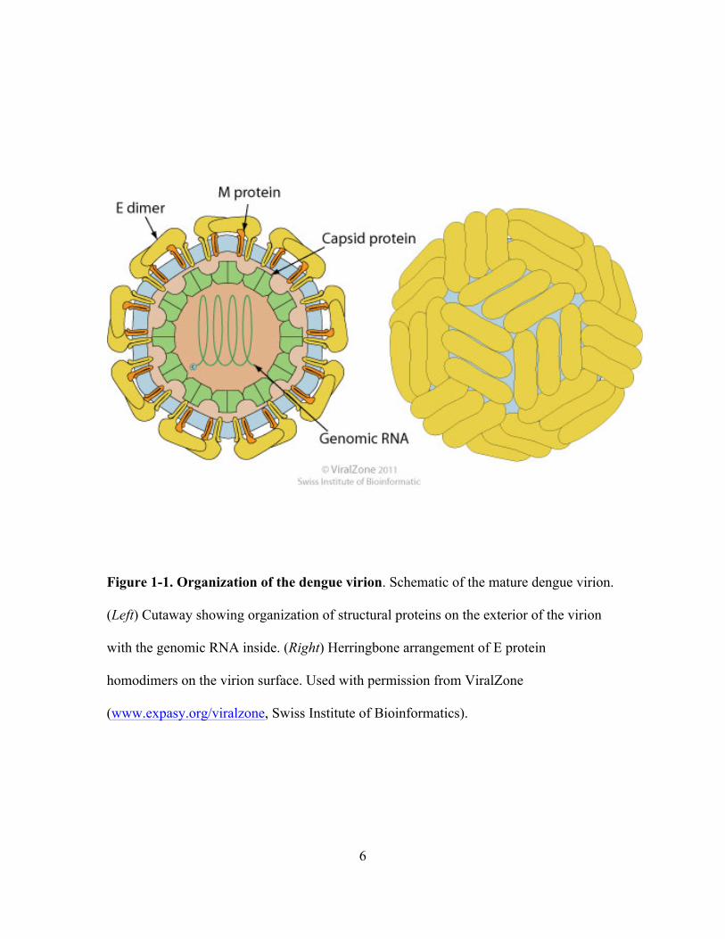
6
Figure 1-1. Organization of the dengue virion. Schematic of the mature dengue virion.
(Left) Cutaway showing organization of structural proteins on the exterior of the virion
with the genomic RNA inside. (Right) Herringbone arrangement of E protein
homodimers on the virion surface. Used with permission from ViralZone
(www.expasy.org/viralzone, Swiss Institute of Bioinformatics).

7
These E protein dimers mediate binding to cells, after which the virus is taken up by
clathrin-mediated endocytosis into endosomes (Acosta et al., 2008; van der Schaar et al.,
2008), where the low pH triggers viral fusion (Gollins and Porterfield, 1986). The viral
genome is released into the cytoplasm, where it undergoes translation and replication in
membrane-associated complexes in the perinuclear region (Westaway et al., 1997). The
polyprotein is cleaved by viral and cellular proteases and nascent virions are assembled
by budding into the endoplasmic reticulum (Mackenzie and Westaway, 2001). New
virions are exocytosed through the cellular secretory pathway, during which prM, a
chaperone protein that protects premature triggering of the E protein, is cleaved by furin
(Stadler et al., 1997) and the E protein rearranges into homodimers (Stiasny et al., 1996).
DENV envelope protein
The DENV envelope (E) protein is the major glycoprotein present on the exterior of
the mature virion and mediates all aspects of viral entry. E consists of three domains, a
stem region, and a transmembrane domain. The pre-fusion and post-fusion crystal
structures of DENV2 soluble envelope protein, containing domains I-III, have been
solved and are similar in appearance to other flavivirus E proteins [Figure 1-2; (Modis et
al., 2003, 2004)]. Domain III contains an immunoglobulin-like region that is believed to
be the receptor-binding domain, while finger-like domain II contains the fusion loop at its
distal end (Mukhopadhyay et al., 2005). The stem region, although not solved as part of
the crystal structures, is thought to consist of two highly-conserved amphipathic helices
and a loop that lie close to the viral membrane in the pre-fusion conformation and play an

8
A
90°
B
C
Figure 1-2. Crystal structures of the DENV2 envelope (E) protein. (A) Pre-fusion E
protein homodimer as viewed from above virion surface. Domains I, II, and III of a single
monomer are shown in in red, yellow, and blue, respectively. Stem region of E is not
shown. (B) Side view of the E homodimer. The virion membrane would be located
underneath the structure in this orientation. (C) Post-fusion E trimer. Note the large
movement of domain III in a monomer relative to the pre-fusion dimer. PDB IDs: 1OAN
[dimer; (Modis et al., 2003)], 1OK8 [trimer; (Modis et al., 2004)].

9
important role in creation and stabilization of the post-fusion E trimer (Zhang et al.,
2003a).
Throughout the viral life cycle, the E protein undergoes a series of conformational
rearrangements and protein associations. Initially expressed as part of the polyprotein, E
is translocated into the ER lumen and quickly forms a heterodimer with prM, which acts
as a chaperone protein and is necessary for proper folding of E (Konishi and Mason,
1993). These E/prM heterodimers appear as trimer spikes on the surface of newly
assembled, immature virions (Zhang et al., 2003b). As the virus passes through the low
pH of the trans-Golgi network, the E protein rearranges into dimers and the pr peptide is
cleaved by the cellular protease furin but remains associated (Yu et al., 2008), protecting
the fusion loop to prevent premature triggering of the post-fusion trimer rearrangement
(Zhang et al., 2004). The remnant of prM, M, is a small (~8kD) protein that sits
underneath the E homodimer on the virion surface and consists of an alpha helix that
remain membrane associated (Zhang et al., 2003a). As the dengue virion is exocytosed
from the cell, the pr peptide is released, and the E protein is found in its pre-fusion,
homodimer, herringbone arrangement on the virion surface (Figure 1-1) (Zhang et al.,
2003a).
Mature virions bind to cells via interactions with unknown receptor(s), although
several classes of surface proteins, including DC-SIGN (Tassaneetrithep et al., 2003) and
glycosaminoglycans (Chen et al., 1997), have been suggested to play supporting roles.
Once bound to cells, the virus is taken up into clathrin-coated pits and transported to
endosomal compartments where viral fusion occurs (van der Schaar et al., 2008). The
exact trigger for DENV fusion remains unclear, as a study using flavivirus tick-borne

10
encephalitis virus (TBEV) showed that protonation of a key histidine residue was
responsible for triggering fusion (Fritz et al., 2008), while another study in WNV showed
that no single histidine residue on E was necessary for fusion initiation (Nelson et al.,
2009). The optimal pH for triggering DENV fusion is also unknown; WNV fusion has
been reported at pH levels as high as 6.9 (Moesker et al., 2010), although the threshold
for DENV is typically agreed to be around 6.3 (Lee et al., 1997; Nelson et al., 2009).
DENV fusion
The DENV E protein is a class II viral fusion protein. The crystal structures of the
pre-fusion dimer and post-fusion trimer have been solved, revealing several large
conformational changes (Figure 1-2) (Modis et al., 2003, 2004). Fusion is hypothesized
to be a multi-step process and have several distinct structural intermediates (Figure 1-3).
Upon first encountering low pH, the E homodimer is thought to dissociate into
monomers, allowing exposure of the fusion loop and extension of domain II away from
the viral membrane. The fusion loop, located at the tip of domain II distal from the viral
membrane, inserts into the target membrane and domain III folds back, allowing for the
two membranes to be brought in proximity. The alpha helices of the stem region of E
begin to “zip up” along the E protein domains. As the stem region extends and increases
its contact with the E protein, hemifusion occurs, in which the outer lipids of each
membrane mix. This is followed by the final pore formation, which allows for the viral
genome to be released into the cytoplasm. It is unclear how many E proteins are
necessary to mediate successful fusion pore formation, and it is possible that the fusion
pore may “flicker” before opening completely (Harrison, 2008). Supportive mechanistic

11
Figure 1-3. Current model of DENV fusion. (A) DENV envelope protein homodimer in
its pre-fusion state, resting parallel to the virus membrane. Domains I, II, and III are
shown in red, yellow, and blue, respectively. Stem region shown in pink. (B) Triggered
by low pH, the E protein dimer dissociates and the fusion loop, located at the tip of
domain II, inserts into target membrane. E protein rearranges into trimers. (C) Domain III
folds back and stem regions begin to zip up the outside of the trimer, bringing membranes
in proximity. (D) Hemifusion occurs, in which the outer lipid membranes of the virus and
target membrane mix. (E) Stem region completes zipping up, fusion pore is formed, and
content mixing occurs between virus and target membrane. Adapted from (Harrison,
2005)
A B
C D
E
12
evidence for the extended structural intermediate of Class II fusion proteins has been
shown in alphaviruses (Sánchez-San Martín et al., 2008), while a study showing that stem
region peptides interfere with late stages of fusion provide support for interactions of the
stem region alpha helices with the post-fusion trimer (Schmidt et al., 2010).
Despite the obvious importance of the DENV E protein in entry, much remains
unknown about how changes in E structure or sequence influence virus entry or other
aspects of the viral life cycle. Two studies have suggested that changes in residues around
the domain I/II interface may alter the optimal pH of viral fusion (Beasley and Aaskov,
2001; Lee et al., 1997), and this region has been suggested to act as a “hinge” during the
movement of domain II that occurs during viral fusion (Modis et al., 2003). One recent
study performed mutagenesis in this region and found that many amino acid changes
altered either growth kinetics or fusion ability in both mammalian and insect cells
(Butrapet et al., 2011). Recent work from our lab and others has shown that residues in
the E DI/DIII linker region (de Wispelaere and Yang, 2012) or stem region (Hsieh et al.,
2010; Lin et al., 2011) can impact virus assembly. These studies suggest that the E
protein may play a role in steps of the viral life beyond mediating entry.
Inhibition of DENV entry
Since the publication of T-20 as a peptide inhibitor of HIV entry (Kilby et al.,
1998), viral entry has been considered a viable target for antivirals. For DENV, the first
reported instances of inhibitors that acted at entry were peptides loosely derived from the
stem region of E (Hrobowski et al., 2005). These peptides had an EC50 value of 10-20
13
µM when pre-incubated with virus inocula and present during viral infection, but no
further exploration into the mechanism of action was explored.
More recent studies have focused on small molecule, rather than peptide, inhibitors of
DENV entry (Table 1-1). Many of the studies used high-throughput computational
screening techniques to identify molecules predicted to bind in the domain I/II interface.
When the pre-fusion dimer of the DENV2 E protein was crystallized, a single molecule
of beta-octo-glucoside (BOG), a detergent used in the crystallization buffer, was
discovered tucked in this region. This area of the E protein was subsequently dubbed the
“BOG pocket,” and it was hypothesized that a small molecule occupying this same space
may prevent DENV fusion (Modis et al., 2003).
The first antiviral discovered based on high-throughput screening against the
BOG pocket was published in 2008 (Zhou et al., 2008). The lead compound, P02,
interacted with the recombinant dengue E protein as detected by NMR and also competed
with BOG for binding. However, in cellular assays, the compound was tested against a
yellow fever reporter virus, which, while a flavivirus, has only 44% envelope protein
sequence similarity to DENV. In addition, P02 was found to be active against a YFV
replicon that lacks E and other viral structural proteins, suggesting that the compound has
additional targets downstream of entry that may be responsible for the anti-DENV
activity observed in cell culture. In fact, the IC50 value against the virus (13 µM) was
similar to the IC50 against the replicon (17 µM), complicating interpretation of P02’s
antiviral mechanism(s) of action.
A later study explored other potential small molecule inhibitory sites on E and
identified a compound hypothesized to make contacts with domains II and III that

14
Table 1-1. Previously published inhibitors of DENV entry Inhibitor name
Source Structure Activity
(DENV2) Test fusion of DENV?
Activity ag. other flaviviruses?
DN59 peptide Hrobowski et
al, 2005
N/A
EC50 10 µM
No WNV (EC50 10 µM)
P02 Zhou et al, 2008
Not tested No YFV (EC50 13 µM)
R1 Yennamalli et
al, 2009
EC50 4 µM
No Not tested
NITD6 Wang et al,
2009
EC50 119 nM
No DENV1 (108 nM), DENV3 (496 nM), DENV4 (334 nM),
YFV (470 nM), JEV (1.42 µM), WNV (564 nM)
A5 Kampmann et
al, 2009
IC50 1.2 µM
Yes (Fusion
from within)
Kunjin (3.8 µM), YFV (1.6 µM)
H2NNH
HN NNH
ONH
NHN
NH
NH2
Cl
O
O
NH
NN
N
N
N
HN N
S
Cl
Cl
O
N
Cl
S
NCH3
CH3
N NH

15
Table 1-1, continued. Previously published inhibitors of DENV entry
Inhibitor name Source
Structure Activity (DENV2)
Test fusion of DENV?
Activity ag. other flaviviruses?
NITD448 Poh et al, 2009
IC50 9.8 µM
Yes In vitro
Not tested
DV2419-447 peptide
Schmidt et al, 2010
N/A
IC90 0.3 µM
Yes In vitro
(hemifusion and pore
formation)
DENV1 (0.1 µM), DENV3 (2 µM), DENV4 (0.7 µM)
3-110-22 Schmidt et al,
2012
IC90 740 nM
Yes In vitro
DENV1 (not calc.), DENV3
(not calc.), DENV4 (2.3 µM), Kunjin (not active)
ClCl
OS
OH
F
FF
O
O
O OH
O
O
CN
NHN CF3
16
lowered DENV yield (IC50 4 µM), although the mechanism of action was not
investigated (Yennamalli et al., 2009). A highly potent compound, NITD6, was identified
as a potential ligand of the BOG pocket and shown to associate with dengue virions
(Wang et al., 2009). NITD6 exhibited an EC50 value of 119 nM in a cell-based
immunodetection assay and caused the arrest of dengue virions inside endosomes,
suggesting that inhibition occurred at a late step in viral entry. Other studies have used a
fusion-from-within assay that measured syncytia formation in insect cells to test whether
compounds inhibit viral fusion mediated by the E protein (Kampmann et al., 2009). More
recently, studies have used an in vitro approach to measure DENV fusion in the presence
of inhibitors using purified virus particles and liposomes as target membranes (Poh et al.,
2009; Schmidt et al., 2012). One study exploring the activity of stem region peptides
measured both DENV pore formation and hemifusion to determine that the peptides
inhibited a late step in viral fusion (Schmidt et al., 2010).
Despite the recent number of publications of DENV entry and fusion inhibitors,
questions remain. In several cases, the biochemical mechanism(s) by which entry
inhibition occurs remains to be addressed. In addition, many of the inhibitors have been
modeled to fit in the BOG pocket of the E protein and make contacts with specific E
protein amino acids, but high resolution structural data unequivocally establishing the
BOG pocket as the binding site are lacking. Likewise, no resistance mutations that could
yield clues to a binding location have been published. Thus, the binding location for all
molecules remains unclear.

17
Current work
The identification of a spectrum of small molecule and peptide inhibitors of
DENV entry suggest that entry inhibition may be a viable approach to creating antivirals.
While currently described DENV entry inhibitors have not yet advanced to preclinical
development, they have been useful as molecular tools to study mechanisms of DENV
entry. This study describes our surprising discovery that GNF-2, a small molecule
inhibitor of intracellular Abl kinases, inhibited DENV when GNF-2 was pre-incubated
with virus inoculum. Using GNF-2 as a scaffold, we performed an SAR study and
explored mechanism of action for the identified small molecules. We found that the small
molecules act post-attachment of virus to cells and block a late step of viral fusion in
vitro. Through viral passaging against entry inhibitors, we found a single point mutation
in the DI/II region of the E protein, E(M196V), supporting previous results showing this
region is important for DENV fusion and consistent with our observation that the
compounds inhibit this step of DENV entry. The E(M196V) mutation enhanced both
viral particle production and entry in a single-cycle reporter assay as well as rendered the
virus particles insensitive to small molecule entry inhibitors. Through a focused alanine
mutagenesis screen of this region, we identified two other mutations in this region that
may confer resistance against our small molecules, possibly through enhanced entry as
well. Overall, this work adds to the growing story of small molecule inhibitors of DENV
entry and gives support to the earlier hypothesis that the DI/DII interface in the E protein
is a critical area for viral entry.

18
REFERENCES:
Acosta, E.G., Castilla, V., and Damonte, E.B. (2008). Functional entry of dengue virus into Aedes albopictus mosquito cells is dependent on clathrin-mediated endocytosis. Journal of General Virology 89, 474-484. Alvarez, M., Rodriguez-Roche, R., Bernardo, L., Vazquez, S., Morier, L., Gonzalez, D., Castro, O., Kouri, G., Halstead, S.B., and Guzman, M.G. (2006). Dengue Hemorrhagic Fever Caused by Sequential Dengue 1–3 Virus Infections over a Long Time Interval: Havana Epidemic, 2001–2002. The American Journal of Tropical Medicine and Hygiene 75, 1113-1117. Amarasinghe, A., Kuritsky, J.N., Letson, G.W., and Margolis, H.S. (2011). Dengue virus infectio in Africa. In Emerging Infectious Diseases [serial on the Internet] (Centers for Disease Control and Prevention). Balmaseda, A., Hammond, S.N., Perez, L., Tellez, Y., Saborio, S.I., Mercado, J.C., Cuadra, R., Rocha, J., Perez, M.A., Silva, S., et al. (2006). Serotype-Specific Differences in Clinical Manifestations of Dengue. The American Journal of Tropical Medicine and Hygiene 74, 449-456. Beasley, D.W.C., and Aaskov, J.G. (2001). Epitopes on the Dengue 1 Virus Envelope Protein Recognized by Neutralizing IgM Monoclonal Antibodies. Virology 279, 447-458. Butrapet, S., Childers, T., Moss, K.J., Erb, S.M., Luy, B.E., Calvert, A.E., Blair, C.D., Roehrig, J.T., and Huang, C.Y.H. (2011). Amino acid changes within the E protein hinge region that affect dengue virus type 2 infectivity and fusion. Virology 413, 118-127. Calisher, C.H., Karabatsos, N., Dalrymple, J.M., Shope, R.E., Porterfield, J.S., Westaway, E.G., and Brandt, W.E. (1989). Antigenic Relationships between Flaviviruses as Determined by Cross-neutralization Tests with Polyclonal Antisera. Journal of General Virology 70, 37-43. Chen, Y., Maguire, T., Hileman, R.E., Fromm, J.R., Esko, J.D., Linhardt, R.J., and Marks, R.M. (1997). Dengue virus infectivity depends on envelope protein binding to target cell heparan sulfate. Nat Med 3, 866-871. Chu, J.J.H., and Yang, P.L. (2007). c-Src protein kinase inhibitors block assembly and maturation of dengue virus. Proceedings of the National Academy of Sciences 104, 3520-3525. de Wispelaere, M., and Yang, P.L. (2012). Mutagenesis of the DI/DIII Linker in Dengue Virus Envelope Protein Impairs Viral Particle Assembly. Journal of Virology 86, 7072-7083.
19
Fritz, R., Stiasny, K., and Heinz, F.X. (2008). Identification of specific histidines as pH sensors in flavivirus membrane fusion. The Journal of Cell Biology 183, 353-361. Gollins, S.W., and Porterfield, J.S. (1986). The Uncoating and Infectivity of the Flavivirus West Nile on Interaction with Cells: Effects of pH and Ammonium Chloride. Journal of General Virology 67, 1941-1950. Gubler, D.J. (1976). A simple technique for demonstrating transmission of dengue virus my mosquitoes without the use of vertebrate hosts. American Journal of Tropical Medicine and Hygiene 25, 5. Harrison, S.C. (2005). Mechanism of Membrane Fusion by Viral Envelope Proteins. In Advances in Virus Research, R. Polly, ed. (Academic Press), pp. 231-261. Harrison, S.C. (2008). Viral membrane fusion. Nature Structural & Molecular Biology 15, 9. Holden, K.L., Stein, D.A., Pierson, T.C., Ahmed, A.A., Clyde, K., Iversen, P.L., and Harris, E. (2006). Inhibition of dengue virus translation and RNA synthesis by a morpholino oligomer targeted to the top of the terminal 3′ stem–loop structure. Virology 344, 439-452. Hrobowski, Y.M., Garry, R.F., and Michael, S.F. (2005). Peptide inhibitors of dengue virus and West Nile virus infectivity. Virology Journal 2. Hsieh, S.-C., Tsai, W.-Y., and Wang, W.-K. (2010). The Length of and Nonhydrophobic Residues in the Transmembrane Domain of Dengue Virus Envelope Protein Are Critical for Its Retention and Assembly in the Endoplasmic Reticulum. Journal of Virology 84, 4782-4797. Kampmann, T., Yennamalli, R., Campbell, P., Stoermer, M.J., Fairlie, D.P., Kobe, B., and Young, P.R. (2009). In silico screening of small molecule libraries using the dengue virus envelope E protein has identified compounds with antiviral activity against multiple flaviviruses. Antiviral Research 84, 234-241. Kilby, J.M., Hopkins, S., Venetta, T.M., DiMassimo, B., Cloud, G.A., Lee, J.Y., Alldredge, L., Hunter, E., Lambert, D., Bolognesi, D., et al. (1998). Potent suppression of HIV-1 replication in humans by T-20, a peptide inhibitor of gp41-mediated virus entry. Nature Medicine 4, 6. Kinney, R.M., Huang, C.Y.-H., Rose, B.C., Kroeker, A.D., Dreher, T.W., Iversen, P.L., and Stein, D.A. (2005). Inhibition of Dengue Virus Serotypes 1 to 4 in Vero Cell Cultures with Morpholino Oligomers. Journal of Virology 79, 5116-5128. Konishi, E., and Mason, P.W. (1993). Proper maturation of the Japanese encephalitis virus envelope glycoprotein requires cosynthesis with the premembrane protein. Journal of Virology 67, 1672-1675.

20
Kuhn, R.J., Zhang, W., Rossmann, M.G., Pletnev, S.V., Corver, J., Lenches, E., Jones, C.T., Mukhopadhyay, S., Chipman, P.R., Strauss, E.G., et al. (2002). Structure of Dengue Virus: Implications for Flavivirus Organization, Maturation, and Fusion. Cell 108, 717-725. Lee, E., Weir, R.C., and Dalgarno, L. (1997). Changes in the Dengue Virus Major Envelope Protein on Passaging and Their Localization on the Three-Dimensional Structure of the Protein. Virology 232, 281-290. Lin, S.-R., Zou, G., Hsieh, S.-C., Qing, M., Tsai, W.-Y., Shi, P.-Y., and Wang, W.-K. (2011). The Helical Domains of the Stem Region of Dengue Virus Envelope Protein Are Involved in both Virus Assembly and Entry. Journal of Virology 85, 5159-5171. Lindenbach, B.D., and Rice, C. (2001). Flaviviridae: The Viruses and Their Replication. Fundamentals of Virology, eds Knipe and Howley 1, 589-639. Mackenzie, J.M., and Westaway, E.G. (2001). Assembly and Maturation of the Flavivirus Kunjin Virus Appear To Occur in the Rough Endoplasmic Reticulum and along the Secretory Pathway, Respectively. Journal of Virology 75, 10787-10799. Mackenzie, J.S., Gubler, D.J., and Petersen, L.R. (2004). Emerging flaviviruses: the spread and resurgence of Japanese encephalitis, West Nile and dengue viruses. Nature Medicine 10, 12. Modis, Y., Ogata, S., Clements, D., and Harrison, S.C. (2003). A ligand-binding pocket in the dengue virus envelope glycoprotein. Proceedings of the National Academy of Sciences of the United States of America 100, 6986-6991. Modis, Y., Ogata, S., Clements, D., and Harrison, S.C. (2004). Structure of the dengue virus envelope protein after membrane fusion. Nature 427, 313-319. Moesker, B., Rodenhuis-Zybert, I.A., Meijerhof, T., Wilschut, J., and Smit, J.M. (2010). Characterization of the functional requirements of West Nile virus membrane fusion. Journal of General Virology 91, 389-393. Mukhopadhyay, S., Kuhn, R.J., and Rossmann, M.G. (2005). A structural perspective of the flavivirus life cycle. Nat Rev Micro 3, 13-22. Nelson, S., Poddar, S., Lin, T.-Y., and Pierson, T.C. (2009). Protonation of Individual Histidine Residues Is Not Required for the pH-Dependent Entry of West Nile Virus: Evaluation of the “Histidine Switch” Hypothesis. Journal of Virology 83, 12631-12635. Peiris, J., and Porterfield, J. (1979). Antibody-mediated enhancement of Flavivirus replication in macrophage-like cell lines. Nature 282, 3.

21
Poh, M.K., Yip, A., Zhang, S., Priestle, J.P., Ma, N.L., Smit, J.M., Wilschut, J., Shi, P.-Y., Wenk, M.R., and Schul, W. (2009). A small molecule fusion inhibitor of dengue virus. Antiviral Research 84, 260-266. Rosen, L. (1987). Sexual transmission of dengue vruses by Aedes albopictus. American Journal of Tropical Medicine and Hygiene 37, 5. Sánchez-San Martín, C., Sosa, H., and Kielian, M. (2008). A Stable Prefusion Intermediate of the Alphavirus Fusion Protein Reveals Critical Features of Class II Membrane Fusion. Cell Host & Microbe 4, 600-608. Schmidt, A.G., Lee, K., Yang, P.L., and Harrison, S.C. (2012). Small-Molecule Inhibitors of Dengue-Virus Entry. PLoS Pathog 8, e1002627. Schmidt, A.G., Yang, P.L., and Harrison, S.C. (2010). Peptide Inhibitors of Dengue-Virus Entry Target a Late-Stage Fusion Intermediate. PLoS Pathog 6, e1000851. Stadler, K., Allison, S.L., Schalich, J., and Heinz, F.X. (1997). Proteolytic activation of tick-borne encephalitis virus by furin. Journal of Virology 71, 8475-8481. Stiasny, K., Allison, S.L., Marchler-Bauer, A., Kunz, C., and Heinz, F.X. (1996). Structural requirements for low-pH-induced rearrangements in the envelope glycoprotein of tick-borne encephalitis virus. Journal of Virology 70, 8142-8147. Tassaneetrithep, B., Burgess, T.H., Granelli-Piperno, A., Trumpfheller, C., Finke, J., Sun, W., Eller, M.A., Pattanapanyasat, K., Sarasombath, S., Birx, D.L., et al. (2003). DC-SIGN (CD209) Mediates Dengue Virus Infection of Human Dendritic Cells. The Journal of Experimental Medicine 197, 7. van der Schaar, H.M., Rust, M.J., Chen, C., van der Ende-Metselaar, H., Wilschut, J., Zhuang, X., and Smit, J.M. (2008). Dissecting the Cell Entry Pathway of Dengue Virus by Single-Particle Tracking in Living Cells. PLoS Pathog 4, e1000244. Vaughn, D.W., Green, S., Kalayanarooj, S., Innis, B.L., Nimmannitya, S., Suntayakorn, S., Endy, T.P., Raengsakulrach, B., Rothman, A.L., Ennis, F.A., et al. (2000). Dengue Viremia Titer, Antibody Response Pattern, and Virus Serotype Correlate with Disease Severity. Journal of Infectious Diseases 181, 2-9. Wang, Q.-Y., Patel, S.J., Vangrevelinghe, E., Xu, H.Y., Rao, R., Jaber, D., Schul, W., Gu, F., Heudi, O., Ma, N.L., et al. (2009). A Small Molecule Dengue Virus Entry Inhibitor. Antimicrob Agents Chemother, AAC.01148-01108. Westaway, E.G., Mackenzie, J.M., Kenney, M.T., Jones, M.K., and Khromykh, A.A. (1997). Ultrastructure of Kunjin virus-infected cells: colocalization of NS1 and NS3 with double-stranded RNA, and of NS2B with NS3, in virus-induced membrane structures. Journal of Virology 71, 6650-6661.

22
WHO (2012). Dengue and severe dengue. Yennamalli, R., Subbarao, N., Kampmann, T., McGeary, R., Young, P., and Kobe, B. (2009). Identification of novel target sites and an inhibitor of the dengue virus E protein. Journal of Computer-Aided Molecular Design. Yu, I.M., Zhang, W., Holdaway, H.A., Li, L., Kostyuchenko, V.A., Chipman, P.R., Kuhn, R.J., Rossmann, M.G., and Chen, J. (2008). Structure of the Immature Dengue Virus at Low pH Primes Proteolytic Maturation. Science 319, 1834-1837. Zhang, W., Chipman, P.R., Corver, J., Johnson, P.R., Zhang, Y., Mukhopadhyay, S., Baker, T.S., Strauss, J.H., Rossmann, M.G., and Kuhn, R.J. (2003a). Visualization of membrane protein domains by cryo-electron microscopy of dengue virus. Nature Structural & Molecular Biology 10, 6. Zhang, Y., Corver, J., Chipman, P.R., Zhang, W., Pletnev, S.V., Sedlak, D., Baker, T.S., Strauss, J.H., Kuhn, R.J., and Rossmann, M.G. (2003b). Structures of immature flavivirus particles. EMBO 22, 10. Zhang, Y., Zhang, W., Ogata, S., Clements, D., Strauss, J.H., Baker, T.S., Kuhn, R.J., and Rossmann, M.G. (2004). Conformational Changes of the Flavivirus E Glycoprotein. Structure 12, 1607-1618. Zhou, Z., Khaliq, M., Suk, J.-E., Patkar, C., Li, L., Kuhn, R.J., and Post, C.B. (2008). Antiviral Compounds Discovered by Virtual Screening of Small Molecule Libraries against Dengue Virus E Protein. ACS Chemical Biology 3, 765-775.

23
Chapter 2: Characterization of GNF-2, a small molecule inhibitor of cellular Abl
kinases, as an inhibitor of dengue virus entry

24
Acknowledgments:
The initial cell-based screen of small molecule kinase inhibitors that led to this project
was performed by Dr. Justin Chu during his time in the Yang lab. The fluorescence
polarization assay with recombinant dengue envelope protein was performed by Aaron
Schmidt in Dr. Stephen Harrison’s lab. All GNF-2 analogs, both conjugated and those
tested as part of the structure-activity relationship study, were synthesized by Drs.
Chandra Miduturu, Jinhua Wang, and Xianming Deng, members of Dr. Nathanael Gray’s
lab. The activity assay for GNF-2 analogs in Bcr-Abl transformed Ba/F3 cells was
performed by Dr. Jianming Zhang in the Gray lab. I would also like to thank members of
the Yang lab, past and present, for helpful discussions of my project as it unfolded.
25
SUMMARY
In this chapter, we found that GNF-2, a previously characterized small molecule
inhibitor of cellular Bcr-Abl kinase, lowers dengue virus (DENV) titer when present at
two separate times during viral infection. We focused on characterizing the inhibition of
DENV2 when GNF-2 is pre-incubated with virus inoculum and present only during the
first hour of target cell infection, an effect that could not be recapitulated with another
small molecule Abl kinase inhibitor. We found that fluorophore-conjugated GNF-2 co-
localized with DENV2 envelope (E) protein inside cells shortly after infection, and that a
biotinylated analog of GNF-2 bound directly to purified dengue virions in vitro. Using
fluorescent polarization assays and recombinant soluble envelope (E) protein, we found
that a FITC-conjugated GNF-2 derivative interacts with the pre-fusion dimer
conformation of E. Initially, we used a cell-impermeable analog of GNF-2, c-GNF-2, to
confirm that inhibition of DENV2 infectivity is independent of intracellular Abl kinases.
Since c-GNF-2 was over three times less effective at reducing DENV2 infectivity, we
performed a focused medicinal chemistry study to identify other disubstituted
pyrimidines with increased potency compared to GNF-2’s activity in the cellular
infectivity assay. We tested these molecules against other DENV serotypes and found
that they had wide ranges of activities. Based on the interaction of GNF-2 analogs with
recombinant E protein and purified virions as well as the range of activities against
DENV serotypes, we hypothesize that this class of compounds inhibits DENV entry via a
direct interaction with the DENV envelope protein on the surface of the virion.
26
INTRODUCTION
Dengue virus (DENV) is a mosquito-borne virus that causes an estimated annual
50 million cases of dengue fever worldwide (WHO, 2012). Despite its status as an
international health concern, much remains unknown about the DENV life cycle. The
DENV genome encodes only ten viral proteins, making the virus highly dependent on a
variety of cellular proteins and processes for a successful, productive infection. In an
attempt to identify cellular kinases that were necessary for successful DENV infection,
our lab performed a cell-based microscopy screen of small molecule inhibitors that were
previously published as cellular kinase inhibitors (Chu and Yang, 2007).
Several small molecules were identified that significantly reduced DENV
infection. When the kinase selectivities of these compounds were examined, we found six
of the molecules were known to inhibit the Abl kinase family (Table 2-1). Four of these
six molecules also have documented activity against Src family kinases (Hennequin et al.,
2006; Holen et al., 1995; Okram et al., 2006; Shah et al., 2004), while one, imatinib, is
known to inhibit c-Kit, PDGFR, and DDR1/2 (Buchdunger et al., 1996). However, the
last compound, GNF-2, is an allosteric kinase inhibitor and known to be highly selective,
exhibiting measurable activity against only Abl family kinases when screened against a
panel of 450 kinases (Adrian et al., 2006; Zhang et al., 2010). In order to explore the role
of Abl kinases in DENV infection, we performed additional experiments with imatinib
and GNF-2, which have similar measured activity against Abl kinases in biochemical and
cellular assays (Figure 2-1), but work via different molecular mechanisms. Imatinib is an
active site inhibitor that traps Abl in the ‘DFG-out’ conformation, while GNF-2 is an

27
Table 2-1. Small molecule protein kinase inhibitors that inhibit DENV infection, as
monitored by an immunofluorescence image-based screen. Adapted from (Chu and Yang,
2007)
Inhibitor Src Abl c-Kit, PDGFR, VEGFR Other kinase targets
K002 CDKs
K014 (imatinib) X X
K039 c-Raf
K040 JAK1, -2, -3
K003 X X X
K013 (GNF-2) X
K030 X Kdr
K032 CK II
K117 X
K045 (AZD0530) X X
K005 (Dasatinib) X X X
K025 (SU11652) X FGFR
K028 (Lavendustin A) X EGFR
K026 (SU5271) EGFR
K144 (Kenpaullone) X CDKs, GSK3-b
K115 (Lavendustin C) X CaMK II
K116 (MC7) MLCK
K118 (Tyrphostin46) X X X Multi-targeted

28
GNF-2, IC50 (kinase) 138 ± 5 nM
Imatinib, IC50 (kinase) 190 ± 6 nM
Figure 2-1. Structures of (top) GNF-2 and (bottom) imatinib. IC50 values shown are in
vitro against Abl family kinases. (Adrian et al., 2006; Buchdunger et al., 1996)
NH2
O
N
N
HN O
F
F
F
N
N
HN
HN
O
N N
N

29
allosteric inhibitor of Abl kinase that binds in a hydrophobic myristoyl-binding pocket
located near the C-terminus of the kinase domain.
In order to determine when in the viral infectious cycle these compounds exhibit
anti-DENV activity, we performed order of addition experiments and discovered that
GNF-2, but not imatinib, inhibited DENV infection when present during two separate
times during viral infection. While both small molecules inhibited DENV when added to
cells after initial infection, only GNF-2 lowered DENV yield when pre-incubated with
virus inoculum. In this chapter, we describe our characterization of GNF-2’s inhibition of
DENV infectivity when present prior to and during initial cellular infection. We applied
both cellular and biochemical approaches to explore how GNF-2 inhibits DENV
infectivity, and collectively our findings led to the hypothesis that GNF-2 interacts with
DENV envelope protein on the surface of the virion and affects entry. This hypothesis
was corroborated by our discovery, made in collaboration with the Gray laboratory, of
GNF-2 analogs that lack Abl kinase inhibitory activity yet have greater potency than
GNF-2 when present during initial DENV infection. Collectively, these results identified
GNF-2 and its analogs as inhibitors of DENV infectivity via interactions with the DENV
E protein and provide chemical tools to investigate mechanism(s) of action in future
experiments.
RESULTS
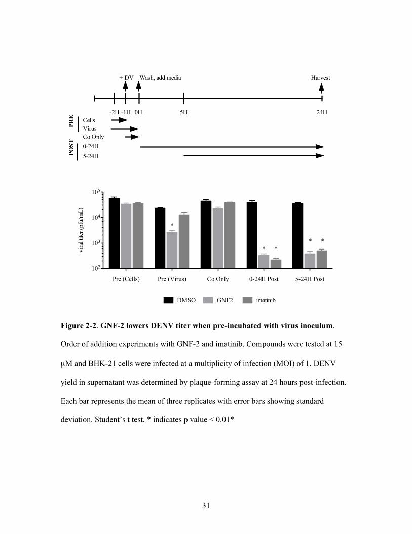
GNF-2 acts at two separate parts of the DENV life cycle
We initially performed order of addition experiments with GNF-2 and imatinib to
determine the point(s) in the DENV life cycle during which the small molecules exert

30
their effect(s). With compounds present at various times during infection (Figure 2-2), we
infected cells at an MOI of 1 and determined DENV yield by plaque-forming assay
(PFA) at twenty-four hours post infection, corresponding to approximately one complete
life cycle of DENV (Lambeth et al., 2005). We found that GNF-2 and imatinib both led
to a significant reduction in DENV yield when added directly after viral infection, and
addition of either inhibitor five hours post-infection caused a similar magnitude of
inhibition (Figure 2-2). The average time between surface binding of virions and
endosomal fusion for DENV has been shown to be 12.5 minutes (van der Schaar et al.,
2007), strongly suggesting that the small molecules are affecting events downstream of
viral entry in the DENV life cycle. Our current hypothesis is that this post-entry
inhibition is due to inhibition of host Abl kinases, since it is observed at a similar
magnitude with imatinib and GNF-2. Surprisingly, we also observed a significant
decrease in DENV yield when GNF-2 was pre-incubated with the viral inoculum prior to
infection. This anti-DENV activity was not observed when the inoculum was pre-
incubated with imatinib, suggesting that it may be due to an Abl-independent mechanism.
In addition, no reduction in DENV yield was observed when GNF-2 was pre-incubated
with cells or only present during the one hour initial infection, demonstrating that pre-
incubation of the compound with virus was a critical step for inhibiting DENV. This led
us to hypothesize that the observed DENV inhibition is due to an interaction of GNF-2
with a target present in the virus inoculum.
To further illustrate that GNF-2 inhibits DENV at two separate steps in the viral
life cycle, one mediated by Abl kinases and one mediated by an independent target, we
performed additivity experiments with GNF-2, imatinib, and NITD6 (Figure 2-3), a
31
Figure 2-2. GNF-2 lowers DENV titer when pre-incubated with virus inoculum.
Order of addition experiments with GNF-2 and imatinib. Compounds were tested at 15
µM and BHK-21 cells were infected at a multiplicity of infection (MOI) of 1. DENV
yield in supernatant was determined by plaque-forming assay at 24 hours post-infection.
Each bar represents the mean of three replicates with error bars showing standard
deviation. Student’s t test, * indicates p value < 0.01*
Pre (Cells) Pre (Virus) Co Only 0-24H Post 5-24H Post
102
103
104
105
DMSO GNF2 imatinib
vira
l tite
r (pf
u/m
L)
-2H -1H 0H 5H 24HCells
+ DV Wash, add media Harvest
VirusCo Only0-24H5-24H
PRE
POST
*
* ** *

32
NITD6, IC50 198 nM in A549 cells against DENV2
Figure 2-3. Structure of NITD6, a previously published DENV small molecule entry
inhibitor (Wang et al., 2009)
N
N
HN N
S
Cl

33
previously validated inhibitor of DENV entry shown to interact with the DENV envelope
protein (Wang et al., 2009). EC90 values for each compound were determined in dose-
response titration experiments. GNF-2 and imatinib were both found to have EC90 values
of 8 µM when added to cells post-infection. When compounds were incubated with virus
inocula at 37°C for 45 minutes prior to addition to cells, NITD6 had a calculated EC90
value when present during entry of 200 nM, while GNF-2’s EC90 during entry was 18
µM.
Pre-treatment of the virus inoculum with GNF-2 or NITD6 significantly reduced
DENV titer measured at 24 hours post-infection (Figure 2-4), while post-treatment of
infected cells with equal molar concentrations of GNF-2 or imatinib also showed
significant inhibition of DENV yield. Combining pre-treatment of the inoculum with
NITD6 and post-treatment of cells with imatinib had an additive effect on DENV yield,
consistent with inhibition of DENV via two separate mechanisms. Importantly, an
additive effect was also observed when GNF-2 pre-treatment of the inoculum was
combined with GNF-2 post-infection treatment of DENV-infected cells, showing that
GNF-2 alone, when present at two separate times during infection, can recapitulate the
combined effects of NITD6 and imatinib.
Together, these data suggest that GNF-2 acts at two separate stages of the DENV
life cycle: first, blocking DENV entry into cells via a target present in the virus inoculum,
and second, inhibiting DENV at a post-entry step mediated by a target shared by imatinib
and GNF-2, presumably Abl kinases. GNF-2’s activity as an intracellular Abl kinase
inhibitor has been described previously (Adrian et al., 2006), and the role of Abl kinases
in DENV infection post-entry is currently under investigation by others in our laboratory.

34
Figure 2-4. GNF-2 has additive effects on DENV titer. All virus and cell treatments
were done at EC90 values determined empirically. Virus treatment was carried out for 45
minutes at 37°C prior to initial cell infection (MOI 1). Treatment of BHK-21 cells was
begun immediately after the initial one-hour infection. Each bar represents the mean of
three replicates with error bars showing standard deviation. * indicates p value < 0.01
DMSO
Imati
nib/D
MSO
Quinazo
line/D
MSO
GNF2/DMSO
DMSO/GNF-2
DMSO/Imati
nib
GNF-2/GNF-2
Quinazo
line/I
matinib
102
103
104
DMSODMSO
imatinibDMSO
NITD6DMSO
GNF2DMSO
DMSOimatinib
DMSOGNF2
GNF2GNF2
NITD6imatinib
cellsvirus
treatment
vira
l tite
r (p
fu/m
L) **
35
In this project, we focused on characterizing the previously unknown Abl-independent
activity of GNF-2 as an inhibitor of DENV infectivity.
A cell impermeable analog of GNF2 retains anti-DENV activity
In previous studies, GNF-2 has been screened for activity against more than 450
kinases and found to be a highly selective inhibitor of Abl family kinases (Adrian et al.,
2006). This fact, combined with our result that GNF-2 must be pre-incubated with the
virus inoculum to cause a significant decrease in DENV yield, led us to the hypothesis
that GNF-2 acts on an extracellular target present in the virus inoculum. We chose to test
this hypothesis by using a cell impermeable analog in which the carboxamide
functionality of GNF-2 was replaced with a carboxylate (c-GNF-2; Figure 2-5). The
activity of c-GNF-2 against c-Abl kinase in a biochemical kinase assay is comparable to
that observed with GNF-2 (Choi et al., 2009); however, c-GNF2 was unable to inhibit the
proliferation of Bcr-Abl transformed Ba/F3 cells (IC50 > 10 !M versus IC50 140 nM for
GNF-2), confirming its inability to penetrate the plasma membrane and access the
intracellular kinase target. In order to assess c-GNF-2’s ability to inhibit DENV infection,
we pre-incubated the viral inoculum with c-GNF-2 (20-100 !M, 45 minutes, 37 ºC) and
added the mixture to cells for the initial hour infection, the same conditions used to
determine the EC90 value during entry of GNF-2. Supernatants were collected at 24 hours
post-infection and yield of infectious virus present was measured by PFA. Under these
conditions, we observed a one-log drop in DENV at approximately 60 !M (Figure 2-5),
showing that c-GNF-2 inhibits DENV infectivity, despite its inability to penetrate the

36
c-GNF-2
Figure 2-5. c-GNF-2 structure and dose-response titration against DENV2. The
change to a carboxyl from GNF-2 is circled on the structure. c-GNF-2 was pre-incubated
with virus inocula for 45 minutes at 37°C before infection of BHK-21 cells (MOI 1).
Each point represents the mean of three replicates with error bars showing standard
deviation. EC90 during entry of c-GNF-2 ~60 µM.
HO
O
N
N
HN O
F
F
F
0 20 40 60 80 100101
102
103
104
105
[µM]
vira
l tite
r (pf
u/m
l)
37
cellular membrane. This result supports our initial hypothesis that GNF-2 has an
extracellular target present in the virus inoculum.
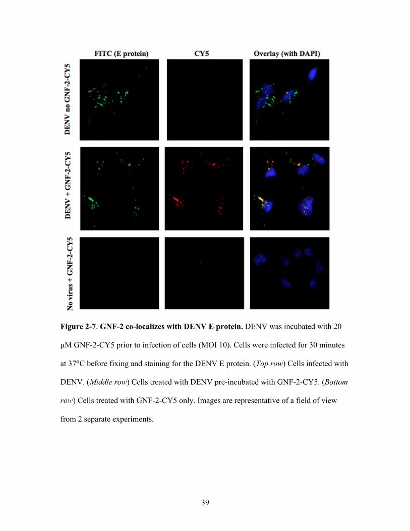
CY5-GNF-2 co-localizes with dengue virions after virion uptake
We hypothesized that GNF-2 may target the virion itself as a way of inhibiting
DENV infectivity. To examine the location of GNF-2 during viral entry into cells, we
synthesized a derivative of GNF-2 conjugated to the fluorescent dye CY5. The resulting
compound, CY5-GNF-2 (Figure 2-6), inhibited DENV in cellular assays with an EC90
value during entry (~20 !M) similar to that of GNF-2. DENV was incubated with 25 !M
GNF-2-CY5 before infecting cells (MOI 10) for 30 minutes at 37°C, conditions that were
empirically determined to permit binding and internalization of virions. Unbound
compound and virus were removed by extensive washing and cells were fixed to permit
detection of the DENV envelope (E) protein by immunofluorescence. DENV was visible
inside cells as punctate staining (Figure 2-7, top row). GNF-2-CY5 was observed inside
cells when the compound was pre-incubated with DENV inoculum, and the majority of
this signal co-localized with the E protein (Figure 2-7; middle row). In the absence of
DENV, little GNF-2-CY5 was observed inside cells (Figure 2-7, bottom row),
demonstrating the inability of this compound to penetrate the plasma membrane and the
efficient removal of extracellular compound during wash steps. The uptake of GNF-2-
CY5, moreover, appears to be DENV-specific since it was not observed with a control
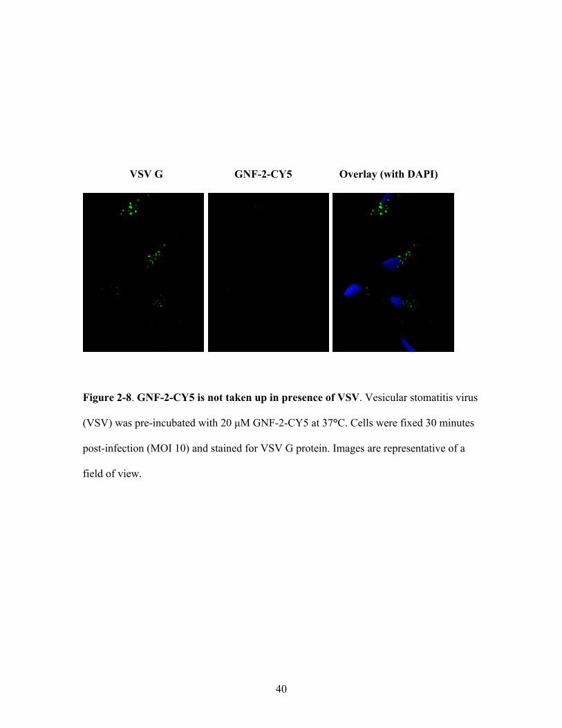
virus, vesicular stomatitis virus (VSV) (Figure 2-8), whose yield was unaffected by pre-
incubation with GNF-2 (data not shown).

38
GNF-2-CY5
Figure 2-6. GNF-2-CY5 structure and dose-response titration against DENV2. GNF-
2-CY5 was pre-incubated with virus inocula for 45 minutes at 37°C before infection of
BHK-21 cells (MOI 1). EC90 value during entry ~20 µM. Each point represents the mean
of three replicates with error bars showing standard deviation.
N N
HN
OCF3
NSO3
N
O3S
NH
OO5
NH
O
0 10 20 30102
103
104
105
[µM]
vira
l tite
r (pf
u/m
l)
39
Figure 2-7. GNF-2 co-localizes with DENV E protein. DENV was incubated with 20
µM GNF-2-CY5 prior to infection of cells (MOI 10). Cells were infected for 30 minutes
at 37°C before fixing and staining for the DENV E protein. (Top row) Cells infected with
DENV. (Middle row) Cells treated with DENV pre-incubated with GNF-2-CY5. (Bottom
row) Cells treated with GNF-2-CY5 only. Images are representative of a field of view
from 2 separate experiments.
40
VSV G GNF-2-CY5 Overlay (with DAPI)
Figure 2-8. GNF-2-CY5 is not taken up in presence of VSV. Vesicular stomatitis virus
(VSV) was pre-incubated with 20 µM GNF-2-CY5 at 37°C. Cells were fixed 30 minutes
post-infection (MOI 10) and stained for VSV G protein. Images are representative of a
field of view.

41
GNF-2 interacts directly with the dengue virus envelope protein
The fact that the anti-DENV effects of both GNF-2 and c-GNF-2 require pre-
incubation with virus inocula prior to infection coupled with the co-localization of GNF-
2-CY5 with DENV E protein inside cells led us to hypothesize that GNF-2 targets the
dengue virion. To explore this hypothesis, we tested whether a biotin-conjugated
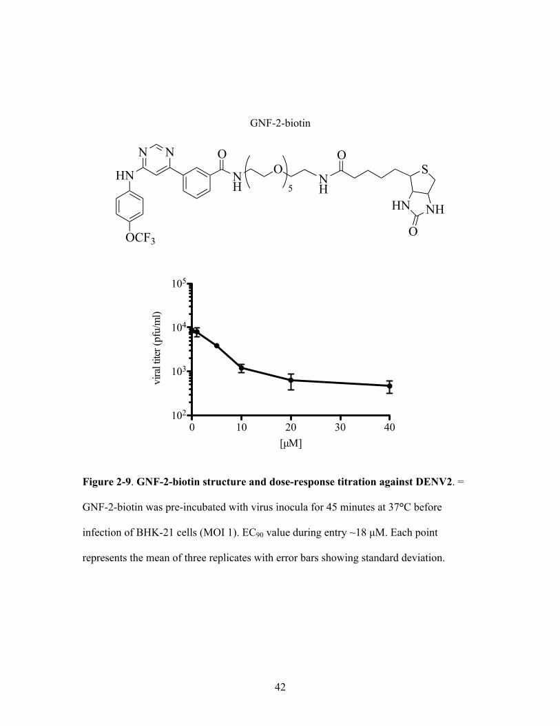
derivative of GNF-2 (GNF-2-biotin, Figure 2-9) could bind and capture purified DENV2
virions. GNF-2-biotin was determined to have an EC90 value during entry in cellular
assays (~18 µM) similar to that of the parental GNF-2.
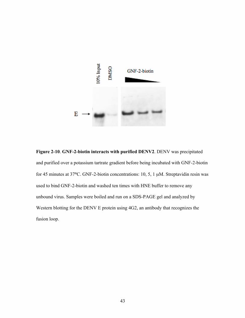
Dengue virions were harvested from cell culture supernatant, concentrated by
PEG precipitation, purified over a potassium tartrate gradient, and dialyzed in TAN
buffer prior to quantification and incubation with GNF-2-biotin at 37° for 45 minutes.
Streptavidin beads were used to bind GNF-2-biotin and washed ten times to remove any
unbound DENV. The beads were boiled in SDS buffer and the released proteins were
analyzed by Western blot for the DENV E protein. We found that GNF-2-biotin captures
purified DENV2 virions in a dose-dependent manner (Figure 2-10).
Knowing that GNF-2 directly interacts with the dengue virion, we next wanted to
investigate how this interaction between small molecule and virion occurs. The main
protein component on the dengue virion surface is the envelope (E) protein, which on the
mature virion associates as a homodimer and tightly packs in a herringbone arrangement
(Kuhn et al., 2002). It is possible that GNF-2 and conjugated derivatives could bind to the
virion via non-specific interactions with the lipid bilayer, but we hypothesized that the
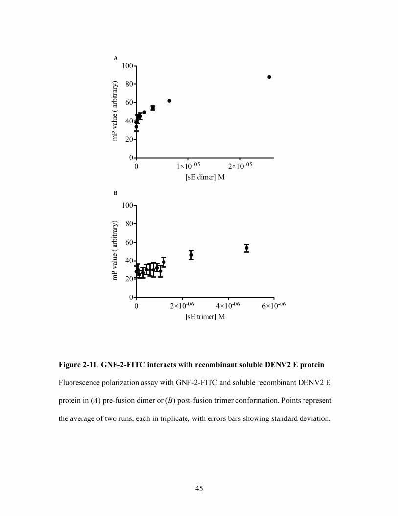
small molecules have a direct interaction with the E protein. To test this, our collaborator
in Dr. Stephen Harrison’s lab used a fluorescence polarization assay to monitor whether a
42
GNF-2-biotin
Figure 2-9. GNF-2-biotin structure and dose-response titration against DENV2. =
GNF-2-biotin was pre-incubated with virus inocula for 45 minutes at 37°C before
infection of BHK-21 cells (MOI 1). EC90 value during entry ~18 µM. Each point
represents the mean of three replicates with error bars showing standard deviation.
N N
HN
OCF3
OO5
NH N
HS
NHHN
O
O
0 10 20 30 40102
103
104
105
vira
l tite
r (pf
u/m
l)
[µM]
43
Figure 2-10. GNF-2-biotin interacts with purified DENV2. DENV was precipitated
and purified over a potassium tartrate gradient before being incubated with GNF-2-biotin
for 45 minutes at 37°C. GNF-2-biotin concentrations: 10, 5, 1 µM. Streptavidin resin was
used to bind GNF-2-biotin and washed ten times with HNE buffer to remove any
unbound virus. Samples were boiled and run on a SDS-PAGE gel and analyzed by
Western blotting for the DENV E protein using 4G2, an antibody that recognizes the
fusion loop.

44
FITC-conjugated GNF2 analog (GNF-2-FITC) interacts with a recombinant, soluble form
of the DENV2 E protein comprised of domains I, II, and III without the stem region,
which exists as a pre-fusion dimer in aqueous solution (sE dimer). Incubation of GNF-2-
FITC with increasing concentrations of DENV2 sE dimer led to a decrease in
fluorescence anisotropy (Figure 2-11A), suggesting a physical interaction between GNF-
2-FITC and the pre-fusion DENV2 sE dimer. We also tested whether GNF-2-FITC
interacted with the DENV2 post-fusion trimer conformation of sE, and found no change
in fluorescence polarization (Figure 2-11B). These data suggest that the conformation of
sE affects the affinity of GNF-2-FITC for the protein.
Taken together, the fluorescence microscopy and in vitro interaction data suggest
that GNF-2 affects DENV infectivity by binding to the dengue virion, more specifically
the envelope protein dimer on the virion surface. The envelope protein is responsible for
all major events of virus entry (binding/attachment, uptake, membrane fusion), making it
possible that GNF-2 binding could affect any of these events.
GNF-2 has variable inhibitory activity against the DENV serotypes
Our work up to this point exclusively utilized the DENV2 New Guinea C (NGC)
strain, so we next asked if GNF-2 could inhibit entry of other DENV serotypes. DENV
serotypes are defined by differences in the amino acid sequences of the E protein
(Lindenbach and Rice, 2001), and since we hypothesized that E is the target of GNF-2,
we reasoned that we might observe variable activity of GNF-2 against other DENV
serotypes. We performed dose-response titration experiments to determine the effect of
GNF-2 on the infectivity of strains representative of the other three DENV serotypes.
45
Figure 2-11. GNF-2-FITC interacts with recombinant soluble DENV2 E protein
Fluorescence polarization assay with GNF-2-FITC and soluble recombinant DENV2 E
protein in (A) pre-fusion dimer or (B) post-fusion trimer conformation. Points represent
the average of two runs, each in triplicate, with errors bars showing standard deviation.
0 1!10-05 2!10-050
20
40
60
80
100
[sE dimer] M
mP
valu
e ( ar
bitra
ry)
0 2!10-06 4!10-06 6!10-060
20
40
60
80
100
[sE trimer] M
mP
valu
e ( ar
bitra
ry)
A
B
46
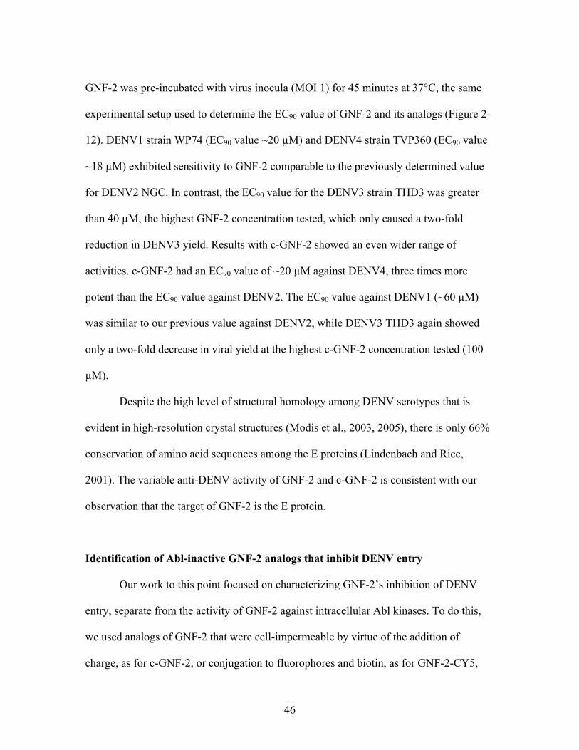
GNF-2 was pre-incubated with virus inocula (MOI 1) for 45 minutes at 37°C, the same
experimental setup used to determine the EC90 value of GNF-2 and its analogs (Figure 2-
12). DENV1 strain WP74 (EC90 value ~20 !M) and DENV4 strain TVP360 (EC90 value
~18 !M) exhibited sensitivity to GNF-2 comparable to the previously determined value
for DENV2 NGC. In contrast, the EC90 value for the DENV3 strain THD3 was greater
than 40 !M, the highest GNF-2 concentration tested, which only caused a two-fold
reduction in DENV3 yield. Results with c-GNF-2 showed an even wider range of
activities. c-GNF-2 had an EC90 value of ~20 !M against DENV4, three times more
potent than the EC90 value against DENV2. The EC90 value against DENV1 (~60 !M)
was similar to our previous value against DENV2, while DENV3 THD3 again showed
only a two-fold decrease in viral yield at the highest c-GNF-2 concentration tested (100
!M).
Despite the high level of structural homology among DENV serotypes that is
evident in high-resolution crystal structures (Modis et al., 2003, 2005), there is only 66%
conservation of amino acid sequences among the E proteins (Lindenbach and Rice,
2001). The variable anti-DENV activity of GNF-2 and c-GNF-2 is consistent with our
observation that the target of GNF-2 is the E protein.
Identification of Abl-inactive GNF-2 analogs that inhibit DENV entry
Our work to this point focused on characterizing GNF-2’s inhibition of DENV
entry, separate from the activity of GNF-2 against intracellular Abl kinases. To do this,
we used analogs of GNF-2 that were cell-impermeable by virtue of the addition of
charge, as for c-GNF-2, or conjugation to fluorophores and biotin, as for GNF-2-CY5,
47
Figure 2-12. Dose-response titration of GNF-2 and c-GNF-2 against DENV1, 3, and
4 strains. All DENV strains were diluted to infect BHK-21 cells at an MOI of 1 and pre-
treated with (A) GNF-2 or (B) c-GNF-2 for 45 minutes at 37°C at noted concentrations.
Each point represents the mean of three replicates with error bars showing standard
deviation. GNF-2 EC90 values: DENV1 ~20 !M; DENV3 N/A; DENV4 ~18 !M. c-
GNF-2 EC90 values: DENV1 ~60 !M; DENV3 N/A; DENV4 ~20 !M
0 10 20 30 40102
103
104
vira
l tite
r (pf
u/m
l)
[µM]
DENV1DENV3DENV4
[µM]
vira
l tite
r (pf
u/m
l)
0 20 40 60 80 100100
101
102
103
104
105DENV1DENV3DENV4
A
B

48
GNF-2-FITC, and GNF-2-biotin. Despite being cell-impermeable and thus unable to
access intracellular Abl kinases, these latter molecules still inhibited DENV2 infectivity
with potencies comparable to that of the parental compound, GNF-2, in cellular assays.
We sought to use medicinal chemistry to more explicitly determine the structure-activity
relationships governing GNF-2’s effects on Abl kinases versus its effects on DENV
infectivity. Specifically, we wanted to identify compounds that inhibit DENV entry but
are devoid of activity against cellular Abl kinases.
Towards this end, a focused structure-activity relationship study was performed to
vary the substitution pattern and the substituents on the central pyrimidine core. All
compounds were designed and synthesized by members of Dr. Nathanael Gray’s lab. A
total of 112 compounds were initially tested in our pre-incubation assay (37°C, 45
minutes) at 75 !M; compounds that showed a minimum of a one-log drop in DENV2
yield were re-tested at 25 !M. Compounds that maintained a one-log drop at this
concentration were then tested at six concentrations in the pre-incubation assay against all
four DENV serotypes to obtain an approximate EC90 value (Appendix A). We also tested
the cytotoxicity of compounds using an ATP proliferation assay (Niles et al., 2007),
allowing us to calculate an LD90 value. The Gray lab confirmed that all active compounds
were not able to inhibit the proliferation of Bcr-Abl transformed Ba/F3 cells (IC50 >10
µM), showing that the compounds are either cell-impermeable or inactive against cellular
Abl kinases.
We identified a total of twenty-three disubstituted pyrimidines that inhibited
DENV2 infectivity with EC90 values equal to or lower than that of GNF-2. Testing these
compounds against other DENV serotype strains revealed a wide range of activities,

49
consistent with our hypothesis that E is target of these compounds and mediates their
effect on DENV infectivity (Appendix A). The DENV3 strain THD3 was insensitive to
many of the compounds, with no calculable EC90 value for 15 out of the 23 compounds,
mirroring its lack of sensitivity to GNF-2 and c-GNF-2. On the other end of the spectrum,
the DENV1 strain had a high level of sensitivity to the presence of many compounds,
with EC90 values in the single-digit micromolar against 18 out of 23 compounds, and
EC90 values against the other four compounds were all under 20 µM. The DENV1 results
are particularly surprising since DENV2 was the focus of our initial SAR study, and we
only identified nine out of 21 compounds that had single-digit micromolar EC90 values
against DENV2. Notably, one compound (CVM-7-128-B) was capable of inhibiting all
four serotypes with EC90 values below 15 µM, suggesting that it may be possible to
develop pan-serotype inhibitors.
DISCUSSION
In this chapter, we have identified an unexpected, previously unknown viral target
for GNF-2, known to be a small molecule inhibitor of intracellular Abl kinases. We found
that GNF-2 reduces DENV infectivity as measured in yield reduction assays when the
compound is pre-incubated with virus inoculum. In addition, a combination of
biochemical, cellular, and chemical experiments collectively indicate that the target of
GNF-2 is the DENV envelope protein in its pre-fusion form on the virion surface. These
data do not address how binding of GNF-2 to the DENV E protein leads to a block in
viral entry; the potential mechanisms of action of GNF-2 and related anti-DENV
50
compounds generated in our medicinal chemistry study are the focus of the following
chapter.
Over the last decade, there has been an increased focus on small molecule and
peptide inhibitors of viral entry as potential antiviral agents. Entry is considered an
attractive target for therapeutic intervention since it is an essential step in the viral life
cycle and occurs early in the viral life cycle before genome replication, thus minimizing
the chance that the virus may be able to actively evolve an escape mutation. In addition,
viral entry occurs in distinct, specific steps often involving discrete cellular receptors or
pathways, thus making it a more tractable target than some downstream steps that often
use a variety of cellular proteins, not all of them known. However, as demonstrated by
small molecule HIV entry inhibitors (Araújo et al., 2012; Lu et al., 2012; Westby et al.,
2007) as well as work discussed in Chapter 4 of this dissertation, viruses are still able to
develop resistance to small molecule or peptide entry inhibitors.
Virus entry is a multistep process, including initial attachment to cells, uptake into
cells, and fusion, and there are inhibitors that have been shown to act during all of these
steps (Baldick et al., 2010; Chen et al., 1997; Kilby et al., 1998; Schmidt et al., 2012). For
DENV, the E protein on the surface of the virion is responsible for mediating all steps of
viral entry, and, consistent with our results in the fluorescence polarization assay with
GNF-2-FITC, has been shown to bind several small molecule or peptide DENV entry
inhibitors (Schmidt et al., 2012; Schmidt et al., 2010a; Zhou et al., 2008) and
hypothesized to be the target of several others (Hrobowski et al., 2005; Kampmann et al.,
2009; Poh et al., 2009; Wang et al., 2009; Yennamalli et al., 2009). No experimental
evidence unequivocally demonstrating the binding site on the E protein binding of GNF-2

51
or previous DENV entry inhibitors has been reported, although many inhibitors have
been hypothesized to bind in the interface between domains I and II of E. This region is a
ligand-binding “pocket” that has been shown to contain a single molecule of beta-octo-
glucoside (BOG) in pre-fusion dimer crystal structures and is hypothesized to be critical
for conformational rearrangements of E that occur during its mediation of viral fusion
(Modis et al., 2003). Many DENV entry inhibitors were identified via virtual screening
against this pocket (Kampmann et al., 2009; Poh et al., 2009; Wang et al., 2009),
although other studies have identified entry inhibitors by focusing on other regions of the
E protein (Schmidt et al., 2012; Schmidt et al., 2010a; Yennamalli et al., 2009). Thus, the
location of binding on E for all DENV entry inhibitors remains an open question.
Consistent with targeting of the DENV2 E protein by GNF-2, we found that GNF-
2 and its analogs identified in the SAR study had a wide range of activities against other
DENV serotypes. These results echo previously published DENV entry inhibitors that
were tested either against other serotypes or flaviviruses. One group showed DENV2-
derived peptides (Schmidt et al., 2010b) as well as small molecule entry inhibitors
(Schmidt et al., 2012) had a range of activity against other DENV serotypes, but no
activity against WNV (Kunjin strain). Another study found that a DENV2 entry inhibitor
(IC50 value 1.2 µM) had activity against WNV (Kunjin strain) as well as YFV
(Kampmann et al., 2009). The most broadly tested compound published to date, NITD6,
had measurable activity against all DENV serotypes as well as YFV, WNV, and JEV
(Wang et al., 2009). However, there were wide variations in potencies; EC50 values
ranged from 68 nM against DENV2 to 1.42 µM against JEV. It is interesting that several
studies made note that DENV3 strains were insensitive to both small molecule and

52
peptide inhibitors of DENV entry; for NITD6, DENV3 had an EC50 value over four times
higher than against DENV1 or DENV2 (Wang et al., 2009). One group (Schmidt et al.,
2012; Schmidt et al., 2010b) found that DENV3 was less sensitive than other serotypes to
several peptide and small molecule inhibitors, while modeling done by another group
(Yennamalli et al., 2009) suggested that the DENV3 envelope protein had lower affinity
for their small molecule entry inhibitor than even the WNV envelope was predicted to
have. This mirrors our results with GNF-2 and its analogs, in which we observed a one-
log drop in DENV3 yield with only seven of the twenty-five total molecules tested.
Further exploration of this pattern of DENV3 insensitivity to entry inhibitors could yield
information that may help to inform the design of future antivirals targeting the entry step
or the E protein.
Although DENV3 was not inhibited by many of our GNF-2 analogs, it was
sensitive to some. In particular, compound 7-128-B was able to reduce DENV yield by
one log in all four serotypes at concentrations of 15 µM or less. This result shows that it
is possible to have pan-serotype small molecule inhibitors of DENV entry. Closer
examination of the results of our SAR study may provide useful information for the
design of future entry inhibitors that use GNF-2 as a scaffold and inhibit all DENV
serotypes with greater potency.
GNF-2’s anti-DENV activity during entry is not as potent as most other published
DENV entry inhibitors, but it is unique among these entry inhibitors due to the fact that it
also inhibits DENV at a later stage of the viral life cycle, most likely via its inhibition of
cellular Abl kinases. The unexpected observation that GNF-2 acts at two points in the
viral life cycle leads to the possible concept of dual-action viral inhibitors. This concept
53
has been tentatively explored previously in other viruses. One HIV study identified a
peptide that inhibits glycoprotein gp120 interactions with both of its cell protein ligands
(Gopi et al., 2006), while another study discovered a small molecule that inhibits two
separate steps of the HIV integration process (Tsiang et al., 2012). For rhinovirus, an
inhibitor was identified that prevented activity of two separate viral proteases (Wang et
al., 1998). However, our results for GNF-2 reveal a molecule that inhibits not at two
points during one step of the viral life cycle, but rather acts at two separate points in the
viral life cycle and is likely doing so via two separate targets. This concept is not without
precedent; a dendrimer was identified with the capacity to inhibit both herpes simplex
virus (HSV) entry as well as late stages of viral replication (Gong et al., 2002), although
potential targets were not explored. We propose that it may be possible to use rational
design to identify molecules that can inhibit viruses via two separate targets at separate
points in the viral life cycle. The targets could be viral, cellular, or both, as we believe is
the case for GNF-2. The challenging part of this approach would be to balance
optimization of inhibitor activity between the two targets. While such dual-action
inhibitors would be attractive due to their potentially higher barriers to viral resistance,
this proposition remains to be experimentally explored.
In summary, this chapter describes the previously unknown ability of GNF-2 to
inhibit dengue virus entry in an Abl-independent manner. We used cellular, biochemical,
and chemical approaches to characterize this activity and determined that GNF-2 most
likely inhibits DENV entry via interactions with the pre-fusion envelope dimer on the
virion surface. Our follow-up work in the next two chapters focuses on GNF-2 analogs
that were identified through a structure-activity relationship study and explores both
54
potential mechanism of action of the small molecules as well as the question of virus
resistance to the inhibition of entry.

55
METHODS AND MATERIALS
Cells and virus:
BHK-21 cells were maintained in MEM-alpha containing 5% FBS with 5% CO2. C6/36
were maintained in L-15 media with 10% FBS and no gas exchange. All virus stocks
were grown in C6/36 cells in L-15 media and 2% FBS, and viral titer was determined by
plaque- or focus-forming assay in the appropriate cell line (see below). C6/36 cells were
infected for 1 hour with 3 mL of DENV diluted in EBSS, after which time media was
added (for a total volume of 20 mL) and the infection proceeded for 4-5 days, until lifting
of the cellular monolayer was observed. Supernatant was harvested and spun at 1200 rpm
for 5 minutes to remove cell debris, after which glycerol was added (final concentration
20%) and samples were aliquoted and frozen at -80°C.
Order of addition experiments:
Experiments were carried out in BHK-21 cells using DENV2 NGC in the presence of 15
!M of GNF-2 or imatinib at times noted in schematic. All virus was diluted in EBSS to
achieve a multiplicity of infection (MOI) of 1. Cells or virus were pre-incubated in 100
!L medium containing compounds for one hour at 37°C. For co-infection, 15 !M of
compound was added to the virus inoculum directly before addition to cells. After an
initial one hour infection, cells were washed twice with 1X PBS to remove unbound
compound and virus and 1 mL cell medium (MEM-alpha with 2% FBS) was added.
Medium was replaced with compound-containing medium at the times noted in
schematic. Twenty-four hours after initial infection, culture supernatant was collected and
viral titer was determined by plaque-forming assay.

56
Plaque- and focus-forming assays:
To determine viral titer, 105 cells/well (BHK-21 cells for DENV1, 2, and 4; Vero for
DENV3) were plated in a 24-well plate and left overnight at 37°C to form a confluent
monolayer. Ten-fold serial dilutions of the supernatant were made in Earle’s balanced
salt solution (EBSS) and 100 µL of the dilutions was used to infect each well for one
hour at 37°C, after which cells were washed once with 1X PBS. Overlay medium was
added (1.05% CMC with MEM!, 2% FBS, Pen/Strep for BHK-21; 1.05% CMC with
DMEM, 2% FBS, Pen/Strep for Vero) and plates were incubated for 5 (BHK-21) or 6
days (Vero). Assays were washed twice with 1X PBS to remove overlay medium and
cells were fixed. For DENV2, plaque-forming units (pfu/mL) were measured by staining
the cells with crystal violet and counting the number of plaques. For DENV1, DENV3,
and DENV4, cells were fixed with 3.7% formaldehyde for 15 minutes at room
temperature, followed by a 15 minute incubation at RT with 20mM NH4Cl. Cold 60/40
(vol/vol) MeOH/Acetone was added and plates were kept at -20°C until staining (30
minutes – overnight). To visualize foci, monolayers were blocked with 1% FBS and 0.1%
Tween-20 in 1X PBS for 10 minutes at RT and incubated with 1:1000 dilution of mouse
monoclonal antibody 4G2 for 30 minutes at 37°C. Plates were incubated with horseradish
peroxidase goat-anti-mouse antibody (1:500) for 30 minutes at 37°C and foci were
visualized using the Vector VIP substrate kit (Vector Labs, Cat#SK4600).
EC90 values:
Compound EC90 values were determined by performing a 6 point titration curve with
compounds either added immediately after infection and present for the 24 hours of
infection (imatinib and GNF-2), or pre-incubated with the virus inocula for 45 minutes at
57
37°C and added to cells for a one hour infection (GNF-2 and all analogs). Cells were
infected at MOI 1; after 24 hours, supernatants were harvested and PFAs or FFAs were
performed to determine viral titer. “EC90 of entry” describes compounds that were tested
by incubating with DENV inocula for 45 minutes at 37°C prior to a one-hour infection of
cells before being removed along with unbound virus by washing. Viral titers were
graphed, and the EC90 value was determined to be the point at which a one-log drop in
titer was observed compared to a DMSO control. EC90 values of entry: NITD6 – 200 nM;
GNF-2 – 18 !M. EC90 values for cellular treatment: GNF-2 – 8 !M; imatinib – 8 !M.
Additivity of compounds:
All compounds were used at EC90 values that were empirically determined (see
previous). Virus inocula were incubated with compounds in 100 !L for 45 minutes at
37°C prior to a one-hour infection of BHK-21 cells. After infection, cells were washed
twice with 1X PBS and 1 mL of fresh medium was added. Twenty-four hours later,
supernatants were harvested and viral titer was determined by PFA.
Pull down of DENV:
Dengue virus was purified from infected C6/36 cell supernatant by incubation overnight
with PEG8000 to precipitate virus. The virus was pelleted (30 minutes, 9000g),
resuspended in TAN buffer, placed over a two-step gradient (40% w/v potassium tartrate
with 30% glycerol, 10% w/v potassium tartrate with 7.5% glycerol), and spun for 2.5
hours at 35,000 rpm at 4°C. Gradient fractions containing virions (as determined by
Western blot against the E and C protein) were pooled and potassium tartrate was
removed by washing with TAN buffer (15 mM TEA,100 mM NaCl, pH 8.0) and
concentrating the virus in an Amicon filter (Millipore, Cat. # UFC810024). Purified

58
dengue virions (between 300-400 ng, as measured by Bradford assay) were incubated
with varying concentrations of GNF-2-biotin for 45 minutes at 37°C in 200 µL HNE
buffer. Meanwhile, streptavidin resin (GE Biosciences, Cat. #17-5113-01) was rinsed
twice with HNE buffer, blocked with 5% FBS for one hour at 4°C, and resuspended in
HNE buffer. Twenty !L of resin was added to the compound and virus and incubated for
twenty minutes at 4°C. Each sample was washed ten times with 1 mL HNE buffer before
being resuspended in 20 µL Laemmli sample buffer without beta-mercaptoethanol
(BioRad, Cat. #161-0737). Samples were boiled (>95°C) for twenty minutes, spun down,
and run on a 10% SDS-PAGE gel for separation. Proteins were transferred to PVDF
membrane by semi-dry transfer (10V, 1.5 hrs) and membranes were blotted with 4G2
antibody for detection of the DENV E protein. Immunoblots were visualized by
chemiluminescence (Pierce ECL reagent cat#32106).
Fluorescence microscopy of DENV and CY5-GNF2:
Vero cells (3x10⁴) were plated on coverslips for infection with purified dengue virions
(0.5 !g) that had been incubated with 25 !M GNF2-Cy5 at 37°C for 1.5 hours. Cells
were infected for 30 minutes at 37°C, then washed three times with 1X PBS to remove
any unbound virus or compound and fixed with methanol. Coverslips were stained for the
DENV E protein (US Biological, Cat. #D2810-05, 1:200) and visualized using FITC-
conjugated goat anti-mouse secondary (Jackson Laboratories Cat. #115-095-146, 1:400).
Images are at 600X magnification and typical of a broader field of view.
Fluorescence polarization assay:
Binding experiments were carried out in Corning, low-volume 384 well microplates and
analyzed in a PerkinElmer EnVision instrument (excitation wavelength, 485nm; emission
59
wavelength, 535 nm). Stock concentrations of GNF-2-FITC were made in DMSO and
dissolved in assay buffer immediately before use. Increasing concentrations of protein
were used with a constant concentration of compound (25 nM). Each well contained a
final volume of 20uL in TAN buffer.

60
REFERENCES:
Adrian, F.J., Ding, Q., Sim, T., Velentza, A., Sloan, C., Liu, Y., Zhang, G., Hur, W., Ding, S., Manley, P., et al. (2006). Allosteric inhibitors of Bcr-abl-dependent cell proliferation. Nat Chem Biol 2, 95-102. Araújo, L.A.L., Junqueira, D.M., de Medeiros, R.M., Matte, M.C.C., and Almeida, S.E.d.M. (2012). Naturally occurring resistance mutations to HIV-1 entry inhibitors in subtypes B, C, and CRF31_BC. Journal of Clinical Virology 54, 6-10. Baldick, C.J., Wichroski, M.J., Pendri, A., Walsh, A.W., Fang, J., Mazzucco, C.E., Pokornowski, K.A., Rose, R.E., Eggers, B.J., Hsu, M., et al. (2010). A Novel Small Molecule Inhibitor of Hepatitis C Virus Entry. PLoS Pathog 6, e1001086. Buchdunger, E., Zimmermann, J., Mett, H., Meyer, T., Müller, M., Druker, B.J., and Lydon, N.B. (1996). Inhibition of the Abl Protein-Tyrosine Kinase in Vitro and in Vivo by a 2-Phenylaminopyrimidine Derivative. Cancer Research 56, 100-104. Chen, Y., Maguire, T., Hileman, R.E., Fromm, J.R., Esko, J.D., Linhardt, R.J., and Marks, R.M. (1997). Dengue virus infectivity depends on envelope protein binding to target cell heparan sulfate. Nat Med 3, 866-871. Choi, Y., Seeliger, M.A., Panjarian, S.B., Kim, H., Deng, X., Sim, T., Couch, B., Koleske, A.J., Smithgall, T.E., and Gray, N.S. (2009). N-myristoylated c-Abl tyrosine kinase localizes to the endoplasmic reticulum upon binding to an allosteric inhibitor. J Biol Chem 284, 29005-29014. Chu, J.J.H., and Yang, P.L. (2007). c-Src protein kinase inhibitors block assembly and maturation of dengue virus. Proceedings of the National Academy of Sciences 104, 3520-3525. Gong, Y., Matthews, B., Cheung, D., Tam, T., Gadawski, I., Leung, D., Holan, G., Raff, J., and Sacks, S. (2002). Evidence of dual sites of action of dendrimers: SPL-2999 inhibits both virus entry and late stages of herpes simplex virus replication. Antiviral Research 55, 319-329. Gopi, H.N., Tirupula, K.C., Baxter, S., Ajith, S., and Chaiken, I.M. (2006). Click Chemistry on Azidoproline: High-Affinity Dual Antagonist for HIV-1 Envelope Glycoprotein gp120. ChemMedChem 1, 54-57. Hennequin, L.F., Allen, J., Breed, J., Curwen, J., Fennell, M., Green, T.P., Lambert-van der Brempt, C., Morgentin, R., Norman, R.A., Olivier, A., et al. (2006). N-(5-Chloro-1,3-benzodioxol-4-yl)-7-[2-(4-methylpiperazin-1-yl)ethoxy]-5- (tetrahydro-2H-pyran-4-yloxy)quinazolin-4-amine, a Novel, Highly Selective, Orally Available, Dual-Specific c-Src/Abl Kinase Inhibitor†. Journal of Medicinal Chemistry 49, 6465-6488.
61
Holen, I., Str, P.E., Gordon, P.B., Fengsrud, M., Berg, T.O., and Seglen, P.O. (1995). Inhibition of Autophagy and Multiple Steps in Asialoglycoprotein Endocytosis by Inhibitors of Tyrosine Protein Kinases (Tyrphostins). Journal of Biological Chemistry 270, 12823-12831. Hrobowski, Y.M., Garry, R.F., and Michael, S.F. (2005). Peptide inhibitors of dengue virus and West Nile virus infectivity. Virology Journal 2. Kampmann, T., Yennamalli, R., Campbell, P., Stoermer, M.J., Fairlie, D.P., Kobe, B., and Young, P.R. (2009). In silico screening of small molecule libraries using the dengue virus envelope E protein has identified compounds with antiviral activity against multiple flaviviruses. Antiviral Research 84, 234-241. Kilby, J.M., Hopkins, S., Venetta, T.M., DiMassimo, B., Cloud, G.A., Lee, J.Y., Alldredge, L., Hunter, E., Lambert, D., Bolognesi, D., et al. (1998). Potent suppression of HIV-1 replication in humans by T-20, a peptide inhibitor of gp41-mediated virus entry. Nature Medicine 4, 6. Kuhn, R.J., Zhang, W., Rossmann, M.G., Pletnev, S.V., Corver, J., Lenches, E., Jones, C.T., Mukhopadhyay, S., Chipman, P.R., Strauss, E.G., et al. (2002). Structure of Dengue Virus: Implications for Flavivirus Organization, Maturation, and Fusion. Cell 108, 717-725. Lambeth, C.R., White, L.J., Johnston, R.E., and de Silva, A.M. (2005). Flow Cytometry-Based Assay for Titrating Dengue Virus. Journal of Clinical Microbiology 43, 3267-3272. Lindenbach, B.D., and Rice, C. (2001). Flaviviridae: The Viruses and Their Replication. Fundamentals of Virology, eds Knipe and Howley 1, 589-639. Lu, L., Tong, P., Yu, X., Pan, C., Zou, P., Chen, Y.-H., and Jiang, S. (2012). HIV-1 variants with a single-point mutation in the gp41 pocket region exhibiting different susceptibility to HIV fusion inhibitors with pocket- or membrane-binding domain. Biochimica et Biophysica Acta (BBA) - Biomembranes 1818, 2950-2957. Modis, Y., Ogata, S., Clements, D., and Harrison, S.C. (2003). A ligand-binding pocket in the dengue virus envelope glycoprotein. Proceedings of the National Academy of Sciences of the United States of America 100, 6986-6991. Modis, Y., Ogata, S., Clements, D., and Harrison, S.C. (2005). Variable Surface Epitopes in the Crystal Structure of Dengue Virus Type 3 Envelope Glycoprotein. Journal of Virology 79, 1223-1231. Niles, A.L., Moravec, R.A., Eric Hesselberth, P., Scurria, M.A., Daily, W.J., and Riss, T.L. (2007). A homogeneous assay to measure live and dead cells in the same sample by detecting different protease markers. Analytical Biochemistry 366, 197-206.

62
Okram, B., Nagle, A., Adrián, F.J., Lee, C., Ren, P., Wang, X., Sim, T., Xie, Y., Wang, X., Xia, G., et al. (2006). A General Strategy for Creating “Inactive-Conformation” Abl Inhibitors. Chemistry & Biology 13, 779-786. Poh, M.K., Yip, A., Zhang, S., Priestle, J.P., Ma, N.L., Smit, J.M., Wilschut, J., Shi, P.-Y., Wenk, M.R., and Schul, W. (2009). A small molecule fusion inhibitor of dengue virus. Antiviral Research 84, 260-266. Schmidt, A.G., Lee, K., Yang, P.L., and Harrison, S.C. (2012). Small-Molecule Inhibitors of Dengue-Virus Entry. PLoS Pathog 8, e1002627. Schmidt, A.G., Yang, P.L., and Harrison, S.C. (2010a). Peptide Inhibitors of Dengue-Virus Entry Target a Late-Stage Fusion Intermediate. PLoS Pathog 6, e1000851. Schmidt, A.G., Yang, P.L., and Harrison, S.C. (2010b). Peptide Inhibitors of Flavivirus Entry Derived from the E Protein Stem. Journal of Virology 84, 6. Shah, N.P., Tran, C., Lee, F.Y., Chen, P., Norris, D., and Sawyers, C.L. (2004). Overriding Imatinib Resistance with a Novel ABL Kinase Inhibitor. Science 305, 399-401. Tsiang, M., Jones, G.S., Niedziela-Majka, A., Kan, E., Lansdon, E.B., Huang, W., Hung, M., Samuel, D., Novikov, N., Xu, Y., et al. (2012). New Class of HIV-1 Integrase (IN) Inhibitors with a Dual Mode of Action. Journal of Biological Chemistry 287, 21189-21203. van der Schaar, H.M., Rust, M.J., Waarts, B.-L., van der Ende-Metselaar, H., Kuhn, R.J., Wilschut, J., Zhuang, X., and Smit, J.M. (2007). Characterization of the Early Events in Dengue Virus Cell Entry by Biochemical Assays and Single-Virus Tracking. Journal of Virology 81, 12019-12028. Wang, Q.-Y., Patel, S.J., Vangrevelinghe, E., Xu, H.Y., Rao, R., Jaber, D., Schul, W., Gu, F., Heudi, O., Ma, N.L., et al. (2009). A Small Molecule Dengue Virus Entry Inhibitor. Antimicrob Agents Chemother, AAC.01148-01108. Wang, Q.M., Johnson, R.B., Jungheim, L.N., Cohen, J.D., and Villarreal, E.C. (1998). Dual Inhibition of Human Rhinovirus 2A and 3C Proteases by Homophthalimides. Antimicrobial Agents and Chemotherapy 42, 916-920. Westby, M., Smith-Burchnell, C., Mori, J., Lewis, M., Mosley, M., Stockdale, M., Dorr, P., Ciaramella, G., and Perros, M. (2007). Reduced Maximal Inhibition in Phenotypic Susceptibility Assays Indicates that Viral Strains Resistant to the CCR5 Antagonist Maraviroc Utilize Inhibitor-Bound Receptor for Entry. Journal of Virology 81, 2359-2371. WHO (2012). Dengue and severe dengue.

63
Yennamalli, R., Subbarao, N., Kampmann, T., McGeary, R., Young, P., and Kobe, B. (2009). Identification of novel target sites and an inhibitor of the dengue virus E protein. Journal of Computer-Aided Molecular Design. Zhang, J., Adrian, F.J., Jahnke, W., Cowan-Jacob, S.W., Li, A.G., Iacob, R.E., Sim, T., Powers, J., Dierks, C., Sun, F., et al. (2010). Targeting Bcr-Abl by combining allosteric with ATP-binding-site inhibitors. Nature 463, 501-506. Zhou, Z., Khaliq, M., Suk, J.-E., Patkar, C., Li, L., Kuhn, R.J., and Post, C.B. (2008). Antiviral Compounds Discovered by Virtual Screening of Small Molecule Libraries against Dengue Virus E Protein. ACS Chemical Biology 3, 765-775.

64
Chapter 3: Exploring the mechanism of action of select GNF-2 analogs during dengue
virus entry

65
Acknowledgments:
I would like to thank Dr. Aaron Schmidt in Dr. Stephen Harrison’s lab for helpful
discussions and teaching me several of the in vitro assays featured in this chapter. I would
also like to express my appreciation to Drs. Chandra Miduturu and Jinhua Wang in the
Gray lab who synthesized all the compounds used in this chapter and kept me steadily
supplied. Thanks to members of the Yang lab, past and present, for helpful discussions
and suggestions along the way.
66
SUMMARY
In earlier work, we discovered that GNF-2, a small molecule inhibitor of cellular
Abl kinases, inhibited dengue virus (DENV) infectivity via interactions with the envelope
protein on the virion surface. We performed a focused structure-activity relationship
study and identified twenty-three small molecule analogs of GNF-2 that had no cellular
activity against Abl kinases yet inhibited DENV infectivity more potently than GNF-2.
We chose two of the most potent compounds, 2-12-2 and 8-24-3 (DENV2 EC90 values
during entry ~5 µM), for further characterization and use in mechanism of action studies.
We found that the inhibition by these compounds was temperature-dependent, could be
overcome with higher initial MOIs, and may be reversible. In mechanistic studies we
found that neither 2-12-2 nor 8-24-3 inhibited attachment of DENV to cells, the first step
in viral entry. When we measured DENV fusion in vitro, we found that, after treatment
with the compounds, DENV did not complete the fusion process, although virions still
stably associated with target membranes in the presence of the compounds. We
hypothesize that our compounds inhibit DENV entry by interacting with the E protein on
the surface of the virion in a way that prevents viral fusion with the cellular membrane
inside endosomes, thus blocking release of the viral genome into the cytoplasm for
replication.

67
INTRODUCTION
Dengue virus (DENV) is a mosquito-borne virus that can cause severe febrile
disease in humans. Over forty percent of the world’s population is at risk to contract this
virus, and there are currently no specific treatments or vaccines available (WHO, 2012).
Dengue is a small, enveloped virion that in its mature, infectious form has one major
surface protein, the envelope (E) protein, which mediates all steps of viral entry,
including attachment to cells and fusion between the viral and cellular membranes inside
endosomes (Harrison, 2005). There are 180 copies of the E protein on the surface of the
virus, which on the mature virion form 90 homodimers arranged in a tight herringbone
pattern (Kuhn et al., 2002). E consists of three distinct domains as well as a stem region
and transmembrane domain; domain I has a !-barrel shape and is centrally located,
domain II is finger-like in structure and contains the fusion loop at the end distal to the
viral membrane, and domain III contains the purported receptor binding domain
(Mukhopadhyay et al., 2005). The stem region consists of two alpha helices that, on the
mature virion surface, sit underneath the rest of the E protein, close to the viral
membrane.
E undergoes a series of conformational changes as it mediates fusion between the
viral and cellular membranes (Figure 1-3). The pre- and post-fusion structures of E have
been solved, allowing deduction of the conformational rearrangements that likely occur
during fusion (Modis et al., 2003, 2004). The initial trigger for these rearrangements is
the low pH of the endosome, which is thought to initially cause dissociation of the E
homodimer, allowing extension of domain II and insertion of the fusion loop into the
cellular membrane. It is thought that domain III then folds back, moving away from

68
domain II, which forms new contacts with the stem region as the helices move away from
the viral membrane. Collectively, these structural rearrangements bring the viral and
cellular membranes in proximity, thereby catalyzing mixing of the outer lipid layers of
virus and cell to yield a hemifusion intermediate, followed by mixing of the inner lipid
layer and creation of a fusion pore, allowing release of the viral genome into the cellular
cytoplasm.
In earlier work, we discovered that GNF-2, a molecule inhibitor of cellular Abl
kinases, inhibited DENV infectivity and bound to both purified dengue virions as well as
recombinant E protein in its pre-fusion, dimer conformation (Chapter 2). We
hypothesized that GNF-2 inhibits DENV infectivity by interacting with the E protein on
the virion surface and inhibiting E’s functions in viral entry. As part of this work, we
conducted a structure-activity relationship study to identify analogs of GNF-2 with
increased inhibition of DENV infectivity. We identified twenty-three small molecules
that inhibited DENV2 during entry more potently than GNF-2 yet had no measurable
inhibition when tested against intracellular Abl kinases (Appendix A).
This chapter describes efforts to learn more about how select GNF-2 analogs
inhibit DENV infectivity. To this end, we chose two of the most potent compounds, 2-12-
2 and 8-24-3, from the SAR study (EC90 values ~5 µM) for use in cellular and
biochemical assays. We examined several parameters, including temperature and time of
pre-incubation with virus inocula, to gain a better understanding of how the compounds
interact with dengue virions. In addition, to determine the mechanism by which
compounds inhibit entry, we examined early and late steps of DENV entry in the
presence of compounds.

69
Compounds 2-12-2 and 8-24-3 did not significantly decrease DENV attachment
to cells, suggesting that they act at a later step of the viral entry process. Using in vitro
assays, we found that both compounds inhibit a late step in the viral fusion cascade. We
hypothesize that these compounds interact with the DENV E protein and inhibit DENV
infectivity by preventing conformational rearrangements necessary for the E protein to
mediate pore formation, thus preventing release of the genome into the cytoplasm for
replication.
RESULTS
Confirmation of GNF-2 analogs as DENV entry inhibitors
Our structure-activity relationship (SAR) study using GNF-2 as a scaffold
identified twenty-three small molecules that significantly lowered DENV2 infectivity at
concentrations < 25 µM (Appendix A). All compounds were individually tested for
inhibition of Ba/F3 cell proliferation as a measure of Abl kinase activity and found to be
inactive (IC50 > 10 µM). We selected two compounds, 2-12-2 and 8-24-3 for further
experiments to characterize inhibition of DENV infectivity (Figure 3-1). Compound 2-
12-2 had an EC90 value when present during DENV2 entry of ~5 µM and was one of only
two compounds to inhibit DENV3 yield with a single-digit EC90 value (~5 µM), although
it was not potent against DENV4 (EC90 value ~40 µM). Compound 8-24-3 was the most
potent compound against all serotypes other than DENV3 (EC90 values: DENV1, 3 µM;
DENV2, 5 µM; DENV4, 10 µM).
In order to confirm that 2-12-2 and 8-24-3 exerted their anti-DENV activity
during viral entry, we performed order of addition experiments (Figure 3-2). As expected,

70
A 2-12-2
B 8-24-3
Figure 3-1. Structures of small molecules 2-12-2 and 8-24-3 Small molecules were
identified in the structure-activity relationship study using GNF-2 as a scaffold. EC90
values during DENV entry: (A) 2-12-2 – DENV1 4 µM, DENV2 5 µM, DENV3 5 µM,
DENV4 40 µM; (B) 8-24-3 – DENV1 3 µM, DENV2 5 µM, DENV3 >40 µM, DENV4
10 µM
N
N
NHHN
OCF3
OCF3
N
N
NHHN
CF3OCF3

71
Figure 3-2. Small molecules lower DENV titer when pre-incubated with virus.
(A) Cells or (B) virus inocula were pre-incubated with DMSO or 5 µM of compounds for
45 minutes at 37°C prior to one-hour initial infection (MOI 1). (C) Compounds were
added to cells at the same time as virus inocula for a one hour infection. (D) Compounds
were added for one hour after initial infection. Supernatants were harvested 24 hours
post-infection and DENV yields were determined by plaque-forming assay. Each bar
represents the mean of three replicates with error bars showing standard deviation.
* indicates p value < 0.01
Pre-incubation with cells
DMSO2-1
2-28-2
4-3102
103
104
105vi
ral t
iter
(pfu
/mL)
Co-infection
DMSO2-1
2-28-2
4-3101
102
103
104
vira
l tite
r (p
fu/m
L)
Pre-incubation with virus
vira
l tite
r (p
fu/m
L)
DMSO2-1
2-28-2
4-3100
101
102
103
104
* *
Post-infection of cells
vira
l tite
r (p
fu/m
L)
DMSO2-1
2-28-2
4-3101
102
103
104
A B
DC
72
the compounds did not significantly inhibit DENV when added to cells before, during, or
after viral infection. A significant drop in DENV yield was observed when compounds
were pre-incubated with virus inocula, consistent with a viral target. In order to ensure
that the compounds did not target the cell during the initial hour infection, we also
performed pre-incubation of the virus with compounds followed by removal of unbound
compound via gel filtration column. Using this pre-incubated virus inocula to infect cells,
we observed that DENV infectivity was still significantly reduced, as evidenced by a
large decrease in the yield of infectious virus produced by 24 hours post-infection;
moreover, this inhibition was of similar magnitude to the inhibition observed when
compound was pre-incubated with virus inoculum and then present during the initial
infection (Figure 3-3).
As another way to show that the compounds act via a viral target, we performed
an MOI titration with the compounds. If the target of our compounds is the dengue
virion, as we hypothesized, we would expect that increasing the amount of virus in the
initial inocula would overcome the inhibitory effects of the compounds. The EC90 value
of 5 µM for 2-12-2 and 8-24-3 had been calculated when cells were infected at a
multiplicity of infection (MOI) of 1, so we retained this compound concentration while
varying the initial MOI, as measured by plaque-forming units (pfu). Virus and compound
were pre-incubated for 45 minutes at 37°C and this mixture was added to cells for one
hour. Cells were washed to remove unbound virus and compound, then medium was
added and supernatant was harvested 24 hours later to determine DENV titer. We found
that doubling the amount of infectious virus in the inoculum (MOI of 2) led to a decrease
in inhibition of DENV infectivity by 2-12-2 and 8-24-3, while by an MOI of 4, no
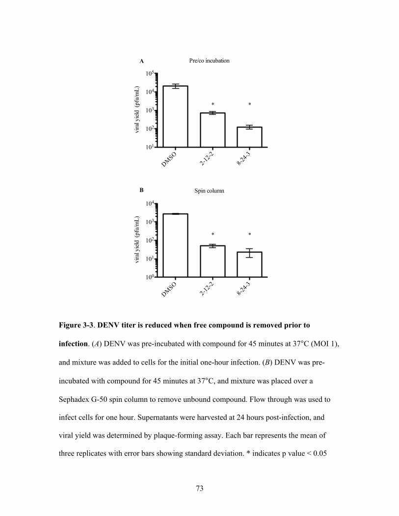
73
Figure 3-3. DENV titer is reduced when free compound is removed prior to
infection. (A) DENV was pre-incubated with compound for 45 minutes at 37°C (MOI 1),
and mixture was added to cells for the initial one-hour infection. (B) DENV was pre-
incubated with compound for 45 minutes at 37°C, and mixture was placed over a
Sephadex G-50 spin column to remove unbound compound. Flow through was used to
infect cells for one hour. Supernatants were harvested at 24 hours post-infection, and
viral yield was determined by plaque-forming assay. Each bar represents the mean of
three replicates with error bars showing standard deviation. * indicates p value < 0.05
Pre/co incubation
DMSO2-1
2-28-2
4-3101
102
103
104
105
vira
l yiel
d (p
fu/m
L)
* *
Spin column
DMSO2-1
2-28-2
4-3100
101
102
103
104
vira
l yiel
d (p
fu/m
L)
* *
A
B
74
difference in DENV yield could be detected between control and compound treated virus
(Figure 3-4). This result is consistent with our hypothesis that the compounds act via a
viral target.
Investigating the characteristics of inhibition of DENV entry by compounds
After confirming that 2-12-2 and 8-24-3 inhibit DENV when pre-incubated with
virus inocula and appear to act via a viral target, we wanted to further investigate how the
compounds inhibited DENV infectivity. We asked if the anti-DENV activity of 2-12-2
and 8-24-3 was retained when pre-incubation with virus was not done at 37°C. Instead,
we pre-incubated compound and virus at 4°C, then added the mixture to cells for an
initial one hour infection at 37°C. We observed no inhibition of DENV infectivity with
compound treatment under these conditions (Figure 3-5), showing that pre-incubation at
37°C is necessary for the compounds to exert their anti-DENV activity. We hypothesize
that thermal motion of the dengue envelope is important in allowing the compounds to
bind, possibly through transient exposure of binding site(s).
In all experiments conducted up to this point, compounds 2-12-2 and 8-24-3 had
been pre-incubated with virus inocula for 45 minutes. Our previous results showed that
addition of compounds and virus to cells simultaneously did not significantly affect
DENV infectivity (Figure 3-2), suggesting that some period of pre-incubation is
necessary for the compound’s anti-DENV activity. We performed a pre-incubation time
course with 8-24-3 and 2-12-2 in which cells were infected with compound-treated
DENV after increasing pre-incubation times at 37°C. Unbound compound and virus were
removed by washing after one hour on cells, and the infection was allowed to proceed for

75
Figure 3-4. Inhibition by compounds is lost with increasing initial MOI. Virus
inocula (various multiplicities of infection) were pre-incubated with 5 µM 2-12-2 or 8-
24-3 (EC90 values for MOI 1) for 45 minutes at 37°C, and mixture was added to cells for
the initial one-hour infection. Supernatants were harvested at 24 hours post-infection, and
viral yield was determined by plaque-forming assay. Each point represents the mean of
three replicates with error bars showing standard deviation.
0 2 4 6 8 10101
102
103
104
105
initial MOI
vira
l tite
r (p
fu/m
L)DMSO2-12-28-24-3
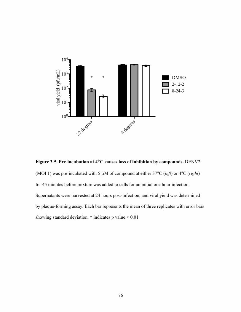
76
Figure 3-5. Pre-incubation at 4°C causes loss of inhibition by compounds. DENV2
(MOI 1) was pre-incubated with 5 µM of compound at either 37°C (left) or 4°C (right)
for 45 minutes before mixture was added to cells for an initial one hour infection.
Supernatants were harvested at 24 hours post-infection, and viral yield was determined
by plaque-forming assay. Each bar represents the mean of three replicates with error bars
showing standard deviation. * indicates p value < 0.01
37 de
grees
4 deg
rees
100
101
102
103
104vi
ral y
ield
(pfu
/mL)
DMSO2-12-28-24-3
**

77
24 hours, at which point supernatants were harvested and DENV titer was measured.
With 5 µM 8-24-3, we observed an 80% reduction in DENV infectivity compared to a
DMSO control after only five minutes pre-incubation, although ten minutes or longer was
necessary to observe the full one-log drop in DENV titer observed previously with a 45
minute pre-incubation (Figure 3-6). The anti-DENV activity of 2-12-2 did not occur as
quickly; a five minute pre-incubation only led to a 45% reduction compared to the
DMSO control, although this dropped to 80% by 10 minutes, and 45 minutes pre-
incubation was necessary to achieve the full one-log drop in DENV titer. These results
suggest that binding by 2-12-2 and 8-24-3 to virus may not be instantaneous, but a ten
minute exposure to virus prior to cellular infection is sufficient to observe a significant
decrease in DENV infectivity.
After learning that our compounds significantly inhibited DENV infectivity
within ten minutes of pre-incubation, we next asked if we could observe reversion of
inhibition by 2-12-2 or 8-24-3 when compounds are diluted below active concentrations.
To test this, we pre-incubated DENV with 20 µM of the compounds in 5 µL for 40
minutes at 37°C, then diluted the mixture 100-fold to bring compound concentrations to
200 nM, well below the value at which we can detect anti-viral activity. We used this
diluted mixture either to immediately infect cells or returned the mixture to 37°C for an
additional 40 minutes (final MOI 0.4). We found that pre-treatment with 2-12-2 or 8-24-3
significantly inhibited DENV infectivity when mixture was added to cells immediately
after the dilution (Figure 3-7). After an additional 40 minute incubation at a higher
volume, we found no inhibition of DENV infectivity in 2-12-2 treated virus compared to
our DMSO control, suggesting that the virus inocula recovered from treatment with

78
Figure 3-6. Pre-incubation time with DENV2 affects magnitude of inhibition.
DENV2 (MOI 1) was incubated with DMSO or 5 µM of compound at 37°C for various
lengths of time before addition to cells for a one-hour initial infection. Supernatants were
harvested at 24 hours post-infection, and viral yield was determined by plaque-forming
assay. Values are expressed as percent of the DMSO control for each time point. Each
point represents the mean of three replicates with error bars showing standard deviation.
10 20 30 40 500
20
40
60
80
100
Time of pre-incubation (minutes)
vira
l yiel
d (%
of D
MSO
cont
rol)
2-12-28-24-3

79
Figure 3-7. Inhibition by compounds may be reversible. Five µL DENV was
incubated with 20 µM compounds for 45 minutes at 37°C, after which it was diluted
100X fold and either used (A) to immediately infect cells or (B) incubated an additional
45 minutes at 37°C before being added to cells (final MOI 0.4). Supernatants were
harvested 24 hours post-infection, and yield was determined by PFA. Each bar represents
the mean of three replicates with error bars showing standard deviation. * indicates p
value < 0.05
DMSO2-1
2-28-2
4-3102
103
104
105
vira
l tite
r (p
fu/m
L)* *
DMSO2-1
2-28-2
4-3102
103
104
vira
l tite
r (p
fu/m
L)
*
A
B

80
2-12-2. Interestingly, we observed that 8-24-3 treated virus was still significantly
inhibited relative to the DMSO control even when incubated for 40 minutes at the
increased volume (Figure 3-7B), although some reversal of inhibition was clearly
observed when compared to the sample infected with 8-24-3 treated virus immediately
following dilution (Figure 3-7A). These data, along with the results from the pre-
incubation time course, suggest that the inhibition of DENV infectivity with 2-12-2 and
8-24-3 is not due to nonspecific inactivation of DENV. The results with 8-24-3 may be
due to a slower off-rate than that of 2-12-2 from the virus, and a longer post-dilution
incubation may allow full recovery of DENV infectivity.
Compounds do not block DENV attachment to cells
Since DENV entry is a multi-step process, we wanted to explore the mechanism
by which 2-12-2 and 8-24-3 exert their anti-DENV activity. We examined whether the
first step of viral entry, DENV attachment to cells, was altered in the presence of 2-12-2
or 8-24-3. Previously, it has been shown that at 4°C, DENV attaches to but is not taken
up by cells (van der Schaar et al., 2007). Thus, after incubating DENV inocula with
compounds at 37°C, all subsequent steps were performed at 4°C and washed with chilled
buffer to prevent cellular uptake of virions. DENV (MOI 1) was incubated for 45 minutes
at 37°C, moved to 4°C briefly to cool, and then added to chilled cells and rocked gently
for 2 hours. Cells were washed multiple times to remove unbound compound and virus,
and either total RNA was extracted from cells immediately or infection was allowed to
proceed for 24 hours and supernatants were collected. The number of DENV genomes
bound was determined by qRT-PCR, and we found that treating virus inocula with 2-12-2

81
or 8-24-3 did not significantly lower the number of viral genomes bound to cells (Figure
3-8A). When the infection was allowed to proceed, DENV titer was still significantly
reduced in the compound-treated conditions compared to the DMSO control (Figure 3-
8B). As a positive control, heparin was added to virus inoculum; previous studies show
that this inhibits DENV entry by preventing binding of virions to cells (Chen et al.,
1997). Taken together, these data show that 2-12-2 and 8-24-3 do not affect attachment of
DENV to cells and the inhibition by these compounds occurs downstream of initial
DENV attachment to cells.
Compounds inhibit a late step of DENV fusion in vitro
We next measured the effect of 2-12-2 and 8-24-3 on the last step of DENV entry,
the fusion of viral and cellular membranes. DENV fusion with the cellular membrane
occurs inside endosomes and is hypothesized to be a multi-step process (Figure 1-3),
making it challenging to dissect specific steps of this biochemical pathway in live cells.
In order to examine individual steps of DENV fusion, we performed in vitro fusion
assays in which liposomes are used as a model membrane for fusion with DENV.
Initially, we examined whether dengue virions formed stable associations with
target membranes after incubation with 2-12-2 or 8-24-3. This association is a surrogate
for the fusion loop insertion step in viral fusion (Figure 1-3B) and can be detected in a
co-flotation assay by determining in which fraction of a sucrose gradient DENV
equilibrates. At pH 8.0, DENV fusion loops are not exposed and thus virions will not
form stable association with liposomes; DENV equilibrates near the bottom of the
gradient. When exposed to pH 5.5, however, fusion loops are exposed and inserted into

82
Figure 3-8. Compounds do not block DENV2 attachment to cells. DENV2 was pre-
incubated with DMSO or small molecules at 37°C for 45 minutes before addition to
chilled cells. Cells were rocked continuously for one hour at 4°C before being washed to
remove any unbound virus. (A) The number of DENV2 genomes bound to cells
immediately post-washing, as determined by collecting total RNA and performing qRT-
PCR with DENV2 primers. (B) Infection was allowed to proceed for 24 hours, and
DENV yield was determined by plaque-forming assay. Each bar represents the mean of
three replicates with error bars showing standard deviation. * indicates p value < 0.01
DMSO2-1
2-28-2
4-3
Hepari
n
Uninfec
ted10
100
1000
10000
DEN
V g
enom
e cop
ies
*
DMSO2-1
2-28-2
4-3
Hepari
n101
102
103
104
105
vira
l tite
r (p
fu/m
L)
* * *
A
B
83
target membranes, so DENV equilibrates at a lower density, near the top of the gradient.
Dengue virions are detected by fractionation of the sucrose gradient, precipitation of
proteins, and Western blot analysis for the DENV E protein. After control treatment,
purified DENV equilibrates near the bottom of the gradient at pH 8.0, while at pH 5.5,
the banding pattern shows a shift in DENV sedimentation (Figure 3-9). Treatment of
virus with 4G2, an antibody that targets the fusion loops of DENV, caused DENV to
equilibrate near the bottom of the gradient even at pH 5.5, confirming that DENV
association with liposomes is mediated by the viral fusion loops. Treatment of DENV
with 2-12-2 or 8-24-3 revealed the same banding pattern of DENV E in sucrose gradients
as the DMSO-treated virus, showing that compound treatment of dengue virions does not
inhibit stable association of virus with target membranes.
Fusion loop exposure and insertion are early steps in DENV fusion, so we next
asked if DENV completes fusion in the presence of 2-12-2 and 8-24-3. Formation of a
fusion pore, the last step in DENV fusion (Figure 1-3E), can be measured via a capsid-
protection assay (Poh et al., 2009; Schmidt et al., 2010). Successful formation of the
fusion pore between dengue virions and trypsin-loaded liposomes permits trypsin access
to the contents of the virion, resulting in the degradation of the core (C) protein, an event
that can be monitored by immunoblotting. The capsid signal is maintained when virus
and liposomes are incubated at pH 8.0, whereas exposure to pH 5.5 causes a loss of
capsid signal due to fusion of the virion with the liposome and subsequent digestion of
the nucleocapsid by trypsin. DENV2 treated with 2-12-2 and 8-24-3 showed retention of
capsid signal under fusion-promoting conditions (Figure 3-10A and B), while treatment
of DENV with an inactive control compound, 111-2-B, showed a complete loss of capsid

84
Figure 3-9. Compounds do not inhibit DENV association with target membranes.
Western blot of coflotation assays with purified DENV2 and liposomes. T=top of
gradient (lowest density); B=bottom of gradient (highest density) (Top row) At pH 8.0,
DMSO-treated DENV, as detected by immunoblotting against the E protein, equilibrates
near the bottom of a sucrose gradient, while exposure to pH 5.5 causes association with
liposomes and equilibration near the top of the gradient. (Second row) DENV treated
with 4G2, a mouse antibody targeted at the DENV fusion loop, does not associate with
liposomes regardless of pH. Antibody against the capsid (C) protein was used to detect
location of virus in fractions. (Bottom two rows) DENV pre-treated with 2-12-2 or 8-24-3
shows a pH-dependent sedimentation pattern similar to DMSO-treated virus.
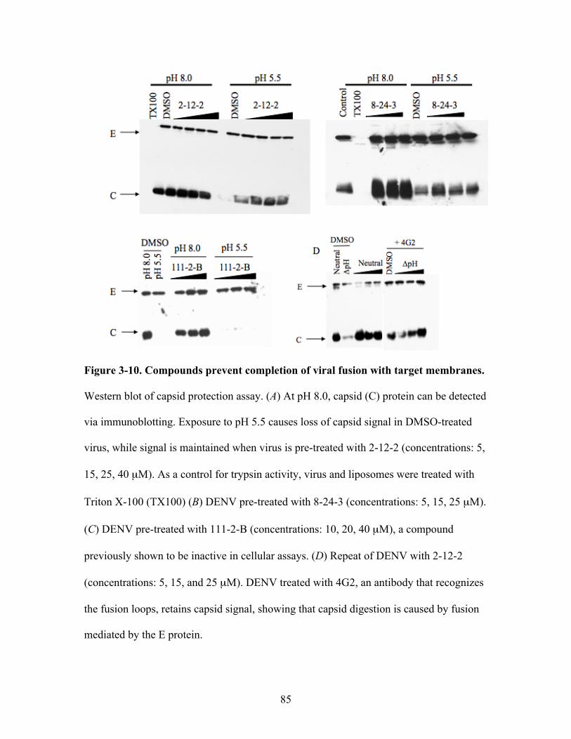
85
Figure 3-10. Compounds prevent completion of viral fusion with target membranes.
Western blot of capsid protection assay. (A) At pH 8.0, capsid (C) protein can be detected
via immunoblotting. Exposure to pH 5.5 causes loss of capsid signal in DMSO-treated
virus, while signal is maintained when virus is pre-treated with 2-12-2 (concentrations: 5,
15, 25, 40 µM). As a control for trypsin activity, virus and liposomes were treated with
Triton X-100 (TX100) (B) DENV pre-treated with 8-24-3 (concentrations: 5, 15, 25 µM).
(C) DENV pre-treated with 111-2-B (concentrations: 10, 20, 40 µM), a compound
previously shown to be inactive in cellular assays. (D) Repeat of DENV with 2-12-2
(concentrations: 5, 15, and 25 µM). DENV treated with 4G2, an antibody that recognizes
the fusion loops, retains capsid signal, showing that capsid digestion is caused by fusion
mediated by the E protein.

86
signal (Figure 3-10C). This strongly suggests that DENV2 does not complete fusion in
the presence of 2-12-2 or 8-24-3. To confirm that degradation of the capsid protein is due
to fusion mediated by the DENV E protein in this assay, we treated DENV with 4G2, an
antibody that recognizes the viral fusion loops, and found retention of the capsid protein
at pH 5.5 (Figure 3-10D).
These data suggest a model in which 2-12-2 and 8-24-3 prevent DENV entry by
blocking a late step in the viral fusion cascade, thus preventing escape of the DENV
capsid from the endosome and the delivery of the DENV genome to the cytoplasm for
replication and productive infection of the host cell.
DISCUSSION
In this chapter, we examine the mechanism by which two small molecules, 2-12-2
and 8-24-3, identified in an SAR study using GNF-2 as a scaffold, inhibit DENV entry.
In addition to exploring the parameters of compound pre-incubation with virus inocula to
learn more about how the compounds interact with the virus, we also investigated the
biochemical mechanism of action of the compounds to determine what step of DENV
entry they inhibit.
Pre-incubation of virus inocula with both 2-12-2 and 8-24-3 had to be carried out
at 37°C in order to observe DENV inhibition (Figure 3-5), suggesting that the
compounds’ anti-DENV activity may be dependent on dynamic properties of the DENV
envelope for transient exposure to binding site(s), either on the E protein itself or the
virion lipid membrane. Significant inhibition of DENV infectivity by 2-12-2 and 8-24-3
required a short pre-incubation time with virus inocula (Figure 3-6), as no significant
87
reduction was observed when virus and compounds were added to cells simultaneously
(Figure 3-2).
We hypothesize that 2-12-2 and 8-24-3 prevent a late step in the viral fusion
cascade, since compound-treated DENV formed stable associations with liposomes
(Figure 3-9) but could not complete fusion as measured by formation of the final fusion
pore (Figure 3-10). The E protein is hypothesized to go through several intermediate,
transient conformational forms between the pre-fusion dimer and the post-fusion trimer
that involve movement or rotation of separate domains (Figure 1-3). Our results with 2-
12-2 and 8-24-3 suggest that they block a step after insertion of the fusion loops into the
target membrane, possibly the fold-back of domain III or association of the stem region
with the trimer. One way to differentiate between these possibilities would be to examine
whether hemifusion occurs when DENV is treated with the compounds. An in vitro assay
measuring hemifusion of pyrene-labeled DENV with liposomes has previously been
performed to test the activity of inhibitory peptides derived from the stem region of E
(Schmidt et al., 2010). However, our attempts with this assay did not yield sufficient
dynamic range to confidently detect hemifusion of DENV. Further optimization of this
assay may be possible and would allow us to more exactly pinpoint the fusion step
blocked by 2-12-2 and 8-24-3. Another possible approach to explore DENV fusion more
precisely would be to test whether recombinant soluble E (sE) protein forms stable post-
fusion trimers after incubation with compounds.
Further exploration of the mechanism of inhibition by 2-12-2 and 8-24-3 may
reveal valuable experimental support for the hypothesized structural fusion intermediates,
which currently is scarce for flaviviruses. Studies in alphaviruses, which also encode a
88
class II fusion protein, have confirmed an extended intermediate structure, in which the
fusion loops are inserted into target membranes, and also revealed that this conformation
is reversible (Roman-Sosa and Kielian, 2011). However, it is unclear if this is the case in
DENV, since other studies have shown that domain I/II constructs of the glycoprotein in
alphaviruses form stable trimers independent of domain III (Sánchez-San Martín et al.,
2008), while DENV domain I/II trimers are not stable until exogenous domain III is
added (Liao et al., 2010).
For this study, we chose two of the most potent compounds from the twenty-three
compounds identified from a SAR study using small molecule GNF-2 as a scaffold. Our
previous work showed that GNF-2 interacts directly with dengue virions and the DENV
recombinant sE pre-fusion dimer (Chapter 2). We hypothesize that 2-12-2 and 8-24-3
also target the E protein but do not have direct experimental evidence to support this. A
biotinylated derivative of 2-12-2 synthesized by the Gray lab exhibited to a significant
loss of activity when tested in cellular assays. The order of addition, MOI titration
experiments, and the fact that the compounds inhibit fusion, a process mediated by E,
strongly suggest that 2-12-2 and 8-24-3 target the virion or dengue E protein, although it
is possible, though unlikely, that the compounds interact non-specifically with the lipid
envelope of the virus. In addition, the location of binding on the E protein remains
unknown for GNF-2, 2-12-2, and 8-24-3; even if the latter compounds interact with E,
the binding sites may differ. Another open question is whether GNF-2, as well as other,
untested compounds from the SAR study, block the same step of DENV entry as 2-12-2
and 8-24-3. Testing GNF-2 and the other analogs in the cellular and biochemical assays
presented in this chapter would address this question.
89
One potential binding location on the E protein for GNF-2, 2-12-2, and 8-24-3 is
the “hinge region” located between domains I and II. This site has previously been shown
to be a ligand binding site, as crystallization of recombinant E revealed a single molecule
of beta-octo-glucoside (BOG) in the pocket formed by domains I and II (Modis et al.,
2003). Several other small molecule inhibitors of DENV entry have also been
hypothesized to bind in this area (Poh et al., 2009; Schmidt et al., 2012; Wang et al.,
2009). However, binding in this region was originally predicted to inhibit the extension
of domain II of E away from the viral membrane and hence block the insertion of the
fusion loops into a target membrane (Modis et al., 2003). Our experiments show that
DENV treated with 2-12-2 or 8-24-3 makes stable associations with lipid bilayers,
suggesting the block by these compounds occurs later in the fusion cascade. It is possible
that binding in the domain I/II interface does not prevent fusion loop exposure, or the
compounds may bind in a different location on the E protein. A previous study identified
two other regions of the E protein that were predicted to be potential small molecule
binding locations that prevent conformational changes of E that occur after insertion of
the fusion (Yennamalli et al., 2009), so it is possible that our compounds could bind in
either of these additional locations.
Overall, this chapter characterizes inhibition of DENV infectivity by two Abl-
inactive small molecules that were identified in our SAR study. Using both cellular and
biochemical assays, we have discovered that inhibition by 2-12-2 and 8-24-3 is
temperature-dependent and lost when initial DENV MOI is increased. Neither compound
blocked attachment of the virus to cells or prevented viral association with membranes,
although they did prevent viral fusion pore formation with target membranes. Further

90
exploration of how these compounds inhibit DENV fusion may reveal valuable
information about conformational intermediates of the E protein as it mediates viral
fusion.
91
METHODS AND MATERIALS
Order of addition experiments
DENV2 (NGC strain) was used to infect BHK-21 cells in 24-well plates at an MOI of 1
for all experiments. Compounds were used at 5 µM. Cells or virus were pre-incubated in
100 µL medium containing compounds or DMSO at 37°C (cells: 1 hour; virus: 45
minutes) or 4°C (pre-incubation temperature; Figure 3-5). For co-infection, 5 !M of
either compound or DMSO was added to virus inoculum directly before addition to cells.
After an initial one hour infection, cells were washed twice with 1X PBS to remove
unbound compound and virus and 1 mL cell medium (MEM-alpha with 2% FBS) was
added. For post-treatment of cells, compound was added to medium for 1 hour post-
infection, then removed and cells were washed twice and medium was replaced. Twenty-
four hours after initial infection, supernatant was collected and viral titer was determined
by plaque-forming assay.
Removal of unbound compounds by gel filtration column
DENV2 (NGC strain) inocula (MOI 2) was pre-incubated in 100 !L with 10 !M of
compound or DMSO for 45 minutes at 37°C, then placed over a Sephadex G50 spin
column and spun for 2 minutes at 2000 rpm at room temperature. Eluant was used to
infect cells for 1 hour, after which cells were washed twice with 1X PBS to remove
unbound compound and virus, and 1 mL cell medium (MEM-alpha with 2% FBS) was
added. Twenty-four hours after initial infection, supernatant was collected, and viral titer
was determined by plaque-forming assay.

92
Multiplicity of infection (MOI) titration
DENV2 (NGC strain) inocula (various MOI) were pre-incubated with 5 !M of
compound or DMSO for 45 minutes at 37°C. After an initial one hour infection, cells
were washed twice with 1X PBS to remove unbound compound and virus, and 1 mL cell
medium (MEM-alpha with 2% FBS) was added. Twenty-four hours after initial infection,
supernatant was collected, and viral titer was determined by plaque-forming assay.
Pre-incubation timecourse
DENV2 (NGC strain) inocula (MOI 1) was pre-incubated with 5 !M of either compound
or DMSO for noted time at 37°C. Mixture was added to cells and left at 37°C for exactly
one hour with gentle rocking. After the one hour infection, cells were washed twice with
1X PBS to remove unbound compound and virus, and 1 mL cell medium (MEM-alpha
with 2% FBS) was added. Twenty-four hours after initial infection, supernatant was
collected, and viral titer was determined by plaque-forming assay.
Reversibility experiments
Due to the initial small volume of the virus, experiments were performed to determine the
correct concentration of compounds to use in order to observe a consistent one-log drop
in DENV titer. Five DENV !L (107 pfu/ml) was incubated with 20 !M of 2-12-2 or 8-
24-3 or DMSO for 40 minutes at 37°C. Virus was diluted 100X and 200 !L was used to
infect BHK cells (effective MOI: 0.4) immediately or after an additional 40 minute
incubation at 37°C. One hour after virus mixture was added to cells, cells were washed
twice with 1X PBS to remove unbound compound and virus, and 1 mL cell medium
(MEM-alpha with 2% FBS) was added. Twenty-four hours after initial infection,

93
supernatant was collected, and viral titer was determined by plaque-forming assay.
Plaque-forming assays
To determine viral titer, 105 BHK-21 cells/well were plated in a 24-well plate to form a
confluent monolayer. Ten-fold serial dilutions of culture supernatant were made in
Earle’s balanced salt solution (EBSS), and 100 µL of the dilutions were used to infect
cells for one hour at 37°C with gentle rocking, after which cells were washed once with
1X PBS. Overlay medium was added (1.05% CMC with MEM", 2% FBS, Pen/Strep)
and plates were incubated for 5 days (BHK-21). Assays were washed twice with 1X PBS
to remove overlay medium, and cells were fixed with a mixture of 0.5% crystal violet and
3.7% formaldehyde. After a minimum of four hours, plates were washed, and plaques
were counted.
DENV binding to cells
DENV2 (NGC strain) was incubated with either 5 !M of 2-12-2 or 8-24-3, 20 !g/ml of
heparin, or DMSO for 45 minutes at 37°C, then added to BHK cells (MOI 1) at 4°C for
one hour with constant rocking. Cells were washed three times with cold 1X PBS to
remove unbound virus or compound. For quantitation of viral genomes bound, total RNA
was extracted using Trizol according to manufacturer’s instructions. For viral yield
determination, one mL of medium was added (MEM-alpha with 2% FBS), and culture
supernatant was harvested 24 hours later. RT-qPCR was performed to determine number
of DENV genomes present using primers against the C protein region of the genome. To
determine viral titer in supernatant, plaque-forming assays were performed.
94
Quantification of DENV genomes
Quantification of DENV genomes was done on a MyIQ Cycler (Biorad). After RNA
extraction and quantification, 5 !L of RNA was used to make cDNA with the iScript kit
(Biorad) according to manufacturer’s instructions. The resulting cDNA was digested by
RNaseH for 1 hour at 37°C. Quantification by rt-PCR was done on each sample in
triplicate (25 !L each) using SybrGreen Supermix (Biorad) according to manufacturer’s
instructions. Primers used targeted the DENV C gene.
DV2Cfor 5’-AATATGCTGAAACGCGAGAGA-3’
DV2Crev 5’-GGGATTGTTAGGAAACGAAGG-3’
Liposomes
Liposomes were made with 1-palmitoyl-2-oleoyl-sn-glycero-3-phosphocholine (POPC),
1-palmitoyl-2-oleoyl-sn-glycero-3-phosphoethanolamine (POPE) (Avanti Polar Lipids
cat # 850457 and 850757, respectively), and cholesterol (Sigma-Aldrich cat#C3045) at
1:1:2 molar ratio in TAN buffer (20mM triethanolamine, 100mM NaCl, pH 8.0). Lipids
were dried down and resuspended thoroughly by vortexing and sonication. Liposomes
were prepared by extrusion through a 0.2 micron filter after five freeze/thaw cycles. For
trypsin-containing liposomes, 10 mg of trypsin was added to 1 mL of lipids (3 mg each)
after the third freeze/thaw cycle, prior to extrusion. Liposomes were separated from
unincorporated trypsin by size-exclusion chromatography using a Superdex 200 column
on an Akta fast performance liquid chromatography (FPLC) system. Trypsin-containing
liposomes were used within 24 hours; liposomes for coflotation were used within three
days.
95
Coflotation assay
Purified dengue 2 virus (600 ng as quantified by Bradford assay) was incubated with 40
!M of compounds or DMSO for 45 minutes at 37°C in 200 !L TAN buffer (pH 8.0).
Liposomes (20 !L) were added for ten minutes, followed by lowering of the pH by
addition of 15 !L 1M sodium acetate (pH 5.0). Following a ten minute incubation,
samples were back-neutralized with 30 !L 1M TEA (unbuffered). Samples were then
mixed with 25% sucrose in TAN buffer (pH 8.0) and placed into a four step gradient
(60%, 25%, 15%, 5% sucrose in TAN buffer) and spun for 3 hours at 44,000 rpm.
Gradients were fractionated and 20% TCA (total volume) was added to precipitate
proteins. Samples were washed twice with 1 mL acetone, and sample buffer was added.
Fractions were boiled for 20 minutes and run on a 10% SDS-PAGE gel followed by
semi-dry transfer to a nitrocellulose membrane. 4G2 antibody, grown in house from
ATCC cell line HB-112, was used for detection of DENV envelope (dilution varies by
lot), and the neat supernatant of hybridoma cell line 6F3.1, kindly provided by Dr. John
Aaskov, was used to detect capsid protein.
Capsid protection assay
Purified dengue 2 virus (300 ng) was incubated with various concentrations of
compounds (5-40 !M) for 45 minutes at 37°C in TAN buffer (pH 8.0) prior to addition of
liposomes (7.5 !L of peak fraction from column) for ten minutes. After incubation with
liposomes, 7.5 !L 1M sodium acetate (pH 5.0) was added to drop pH to 5.5 for ten
minutes. Samples were back-neutralized with 15 !L 1M TEA (unbuffered) and incubated
for forty-five minutes at 37°C to allow trypsin digestion (total volume: 100 !L). SDS
sample buffer (20 !L) was added, and samples were boiled for 20 minutes before being

96
run on a 12% SDS-PAGE gel to separate proteins. Proteins were transferred to a
nitrocellulose membrane using a semi-dry transfer. 4G2 antibody, grown in house from
ATCC cell line HB-112, was used for detection of DENV envelope (dilution varies by
lot), and the neat supernatant of hybridoma cell line 6F3.1, kindly provided by Dr. John
Aaskov, was used to detect capsid protein.
97
REFERENCES:
Chen, Y., Maguire, T., Hileman, R.E., Fromm, J.R., Esko, J.D., Linhardt, R.J., and Marks, R.M. (1997). Dengue virus infectivity depends on envelope protein binding to target cell heparan sulfate. Nat Med 3, 866-871. Harrison, S.C. (2005). Mechanism of Membrane Fusion by Viral Envelope Proteins. In Advances in Virus Research, R. Polly, ed. (Academic Press), pp. 231-261. Kuhn, R.J., Zhang, W., Rossmann, M.G., Pletnev, S.V., Corver, J., Lenches, E., Jones, C.T., Mukhopadhyay, S., Chipman, P.R., Strauss, E.G., et al. (2002). Structure of Dengue Virus: Implications for Flavivirus Organization, Maturation, and Fusion. Cell 108, 717-725. Liao, M., Martín, C.S.-S., Zheng, A., and Kielian, M. (2010). In Vitro Reconstitution Reveals Key Intermediate States of Trimer Formation by the Dengue Virus Membrane Fusion Protein. Journal of Virology 84, 5730-5740. Modis, Y., Ogata, S., Clements, D., and Harrison, S.C. (2003). A ligand-binding pocket in the dengue virus envelope glycoprotein. Proceedings of the National Academy of Sciences of the United States of America 100, 6986-6991. Modis, Y., Ogata, S., Clements, D., and Harrison, S.C. (2004). Structure of the dengue virus envelope protein after membrane fusion. Nature 427, 313-319. Mukhopadhyay, S., Kuhn, R.J., and Rossmann, M.G. (2005). A structural perspective of the flavivirus life cycle. Nat Rev Micro 3, 13-22. Poh, M.K., Yip, A., Zhang, S., Priestle, J.P., Ma, N.L., Smit, J.M., Wilschut, J., Shi, P.-Y., Wenk, M.R., and Schul, W. (2009). A small molecule fusion inhibitor of dengue virus. Antiviral Research 84, 260-266. Roman-Sosa, G., and Kielian, M. (2011). The Interaction of Alphavirus E1 Protein with Exogenous Domain III Defines Stages in Virus-Membrane Fusion. Journal of Virology 85, 12271-12279. Sánchez-San Martín, C., Sosa, H., and Kielian, M. (2008). A Stable Prefusion Intermediate of the Alphavirus Fusion Protein Reveals Critical Features of Class II Membrane Fusion. Cell Host & Microbe 4, 600-608. Schmidt, A.G., Lee, K., Yang, P.L., and Harrison, S.C. (2012). Small-Molecule Inhibitors of Dengue-Virus Entry. PLoS Pathog 8, e1002627. Schmidt, A.G., Yang, P.L., and Harrison, S.C. (2010). Peptide Inhibitors of Dengue-Virus Entry Target a Late-Stage Fusion Intermediate. PLoS Pathog 6, e1000851.

98
van der Schaar, H.M., Rust, M.J., Waarts, B.-L., van der Ende-Metselaar, H., Kuhn, R.J., Wilschut, J., Zhuang, X., and Smit, J.M. (2007). Characterization of the Early Events in Dengue Virus Cell Entry by Biochemical Assays and Single-Virus Tracking. Journal of Virology 81, 12019-12028. Wang, Q.-Y., Patel, S.J., Vangrevelinghe, E., Xu, H.Y., Rao, R., Jaber, D., Schul, W., Gu, F., Heudi, O., Ma, N.L., et al. (2009). A Small Molecule Dengue Virus Entry Inhibitor. Antimicrob Agents Chemother, AAC.01148-01108. WHO (2012). Dengue and severe dengue. Yennamalli, R., Subbarao, N., Kampmann, T., McGeary, R., Young, P., and Kobe, B. (2009). Identification of novel target sites and an inhibitor of the dengue virus E protein. Journal of Computer-Aided Molecular Design.

99
Chapter 4: Exploring potential resistance mutations in DENV structural proteins against
small molecule entry inhibitors
100
Acknowledgments: I would like to thank Dr. Ted C. Pierson of the NIH for generously
supplying reagents and protocols for the recombinant viral particle system. I would also
like to thank members of the Gray lab for synthesizing the small molecule entry
inhibitors, and members of the Yang lab for discussion and advice.

101
SUMMARY
In this chapter, we examined the effect(s) of amino acid changes in the DENV
structural proteins on susceptibility of the virus to small molecule entry inhibitors. We
serially passaged DENV2 in the presence of select compounds and tracked viral titer to
monitor the emergence of inhibitor-resistant DENV. This approach yielded a rise in titer
against one out of ten tested compounds, 7-148-6. Sequencing of the structural proteins
from this virus revealed two changes: one each in the E protein, E(M196V) and M
protein, M(H28Y). Testing of these individual residues in a single-cycle infection assay
using recombinant viral particles (RVPs) (Wang et al., 2009a) revealed that E(M196V)
enhanced entry of virus particles, while M(H28Y) decreased the efficiency of entry.
RVPs containing either change showed no significant decrease in reporter signal when
pretreated with 7-148-6. E(M196V) is located in a “hinge” region between domains I and
II of the E protein that has been hypothesized to be critical for conformational
rearrangements necessary to mediate membrane fusion (Modis et al., 2003). Using the
RVP system, we performed site-directed mutagenesis of eleven selected residues in this
region and found that eight mutations decreased production or impaired entry of RVPs.
We focused on two mutations, E(Q52A) and E(Q131A), and found that they conferred
resistance against three small molecule entry inhibitors. Overall, our data suggest changes
in the E protein domain I/II interface may confer resistance to small molecule entry
inhibitors and affect production or release of viral particles as well as their entry into
target cells.
102
INTRODUCTION
Dengue virus (DENV) is one of the most common vector-borne viruses in the
world, causing an estimated 50 to 100 million illnesses per year (WHO, 2012). There are
no specific therapeutics or vaccines available against DENV, making antivirals to treat
the disease highly desirable. Virus entry is considered an attractive antiviral target in part
because blocking entry can prevent primary infection or viral spread. For DENV, virus
entry is mediated by the envelope (E) protein, the major glycoprotein on the virion
surface.
DENV E consists of three domains (Figure 4-1) and mediates binding to cells as
well as fusion with the cellular membrane, which is triggered by low pH inside
endosomes after DENV is endocytosed (Gollins and Porterfield, 1986). Domain I is
centrally located, while finger-like domain III extends along the virion surface and
contains the fusion loop at its distal end, and domain III has an immunoglobulin-like fold
that contains the purported receptor-binding region (Mukhopadhyay et al., 2005). When
the E protein is first synthesized into the ER lumen, it associates with prM, a protein that
acts as a chaperone for E, and forms a heterodimer. This E/prM heterodimer is found on
the surface of newly-assembled virions in the endoplasmic reticulum as trimeric spikes
(Zhang et al., 2003b). As virions pass through the trans-Golgi network, E reassociates
into homodimers, and the pr peptide is cleaved by furin, leaving the small M protein
resting underneath the E dimers on the now-mature virion surface (Zhang et al., 2003a).
It is hypothesized that the domain I/II interface of E acts as a hinge region during
viral fusion, allowing extension of the fusion loop towards the cellular lipid membrane
(Modis et al., 2004). The region between domains I and II of the E protein is often

103
Figure 4-1. Crystal structure of the DENV2 envelope protein. DENV2 envelope (E)
protein as a homodimer, the pre-fusion form found on the surface of mature virions.
Domains I, II, and III of one E monomer are shown in red, yellow and blue, respectively.
PDB ID: 1OAN (Modis et al., 2003)

104
referred to as the “BOG” pocket, as it was discovered to contain a single molecule of
beta-octo-glucoside (BOG) under certain crystallization conditions (Modis et al., 2003).
In high-resolution crystal structures, the pocket adopts a closed conformation with the k-l
loop covering an opening between the domains when BOG is present but exhibits an
open pocket due to shifting of the k-l loop when BOG is absent. Based on this
observation and previous studies indicating that residues lining this region may alter the
pH threshold of fusion (Beasley and Aaskov, 2001; Lee et al., 1997), it was proposed that
the presence of a small molecule ligand in the BOG pocket may interfere with DENV
entry by preventing the structural rearrangements involved in DENV fusion. An attractive
feature of this proposal is that any mutations in this region that could confer resistance
would be expected to decrease the fitness of the virus due to their potential effect(s) on
fusion.
Since this potential strategy for inhibiting DENV was proposed, several inhibitors
of DENV entry hypothesized to bind in the BOG pocket have been reported (Kampmann
et al., 2009; Poh et al., 2009; Schmidt et al., 2012; Wang et al., 2009b); however,
structural data directly demonstrating this has yet to be published. Likewise, genetic data
mapping resistance mutations to this or any other region of the E protein have not been
reported, although whether this is due to the effect of such mutations on E’s function and
the fitness of the corresponding virus remains unclear.
Previously, we identified several small molecules that inhibited DENV when pre-
incubated with virus inocula prior to initial infection of cells. We hypothesized that these
compounds act on a viral target, specifically the envelope protein on the surface of
virions, and showed that select compounds inhibited DENV fusion in vitro. In this study,

105
we report efforts to learn more about the molecular interactions of these inhibitors with
the dengue virion by using mutational analysis to explore the molecular target of the
inhibitors and potential mechanisms of resistance to these inhibitors. We serially
passaged DENV2 in the presence of ten select compounds and monitored for outgrowth
of DENV that would suggest viral resistance. We observed outgrowth of DENV in only
one out of ten compound treatments, with small molecule 7-148-6. Consensus sequencing
of the structural proteins of this resistant quasispecies led us to the identification of two
point mutations, one in the domain I/II of the E protein, E(M196V), and one in the
ectodomain of M, M(H28Y). We found that in a single-cycle DENV reporter assay, the
presence of either mutation was sufficient to confer insensitivity to treatment of virus
particles with 7-148-6. In parallel, we performed a rational, site-directed mutagenesis
study of eleven residues around the domain I/II interface of E. We found that eight of
these changes drastically reduced virus particle production or entry. We tested two amino
acid mutations that were still permissive for efficient RVP entry, Q52A and Q131A,
against select small molecule entry inhibitors GNF-2, 2-12-2, and 7-148-6, and found
both the presence of either mutation reduced sensitivity to treatment relative to wild-type
RVPs. Taken together, our data suggest that the DI/DII interface is sensitive to many
amino acid changes; however, changes that are tolerated may decrease sensitivity to the
small molecule entry inhibitors examined in these experiments. The results presented
here aid in understanding the interaction of our small molecule inhibitors with the DENV
E protein and also suggest that point mutations at the interface of domains I and II of E
affect viral particle production and entry.

106
RESULTS
Potential resistance of DENV2 to presence of small molecule 7-148-6
To examine the potential development of resistance to a subset of our small
molecule entry inhibitors, we serially passaged virus in the presence of compounds using
two approaches. We first tried passaging DENV2 in the presence of 20 µM of four small
molecules (7-128-B, 7-148-1, 7-148-6, and 7-148-8) previously shown to inhibit DENV
infection yet inactive against intracellular Abl kinases (Appendix A). Over the course of
eight passages, we saw a rise in viral titer suggestive of a resistant virus strain against
only one of the compounds, 7-148-6 (Figure 4-2). Concerned that the high initial
concentration of compounds had prevented outgrowth of resistant DENV, we selected
another six compounds, 1-100-1, 2-12-2, 2-21-2, 8-24-3, 8-24-5, and 2-119-2-B, for a
second serial passage. We gradually increased compound concentrations from 2.5 to 20
µM over the course of nine passages, but found that viral titers crashed as the EC90 values
of compounds were reached and did not recover (data not shown). Thus, out of ten
compounds tested, we identified viral outgrowth against only one, suggesting that DENV
may not easily develop resistance to the small molecule entry inhibitors.
We turned our attention to compound 7-148-6 (Figure 4-3) and confirmed that it
mirrored the activity of our previously tested compounds, 2-12-2 and 8-24-3, in the order
of addition experiments and that it exhibited anti-DENV activity even after size-
exclusion chromatography was used to remove unbound compound from the inoculum
prior to infection of cells (Figure 4-4). We tested 7-148-6 in biochemical mechanism
assays and found that it does not inhibit attachment of DENV to cells but does block
completion of DENV fusion in vitro (Figure 4-5). We hypothesize that 7-148-6 inhibits

107
Figure 4-2. Passaging of DENV2 in the presence of selected compounds. DENV2 was
passaged every four days in Vero cells in the presence of DMSO or 20 µM of each
compound. DENV titer was determined by plaque-forming assay. Each point represents
the mean of three replicates with error bars showing standard deviation.
0 2 4 6 8100
101
102
103
104
105
106
107
Passage number
vira
l tite
r (pf
u/m
l)
DMSO7-128-B7-148-17-148-67-148-8

108
Figure 4-3. Structure of compound 7-148-6. EC90 values against DENV serotypes in
infectivity assay: DENV1 – 8 µM; DENV2 – 10 µM; DENV3 >40 µM; DENV4 >40 µM.
N
N
NHHNOCF3

109
Figure 4-4. Compound 7-148-6 inhibits DENV titer when pre-incubated with virus
inoculum. (A) Order of addition experiments show a significant reduction in viral yield
only when compound 7-148-6 (10 µM) is pre-incubated with DENV2 inoculum at 37°C.
(B) Significant inhibition of DENV infectivity by 7-148-6 is observed when free
compound was removed via a gel filtration spin column prior to cellular infection. Each
bar represents the mean of three replicates with error bars showing standard deviation.
* indicates p value < 0.01
37°C
pre/c
o
4°C pr
e/co
Co only Post
101
102
103
104
vira
l tite
r (pf
u/m
l)
DMSO7-148-6*
Spin column
DMSO
7-148
-6101
102
103
104
vira
l tite
r (pf
u/m
l)
*
A
B
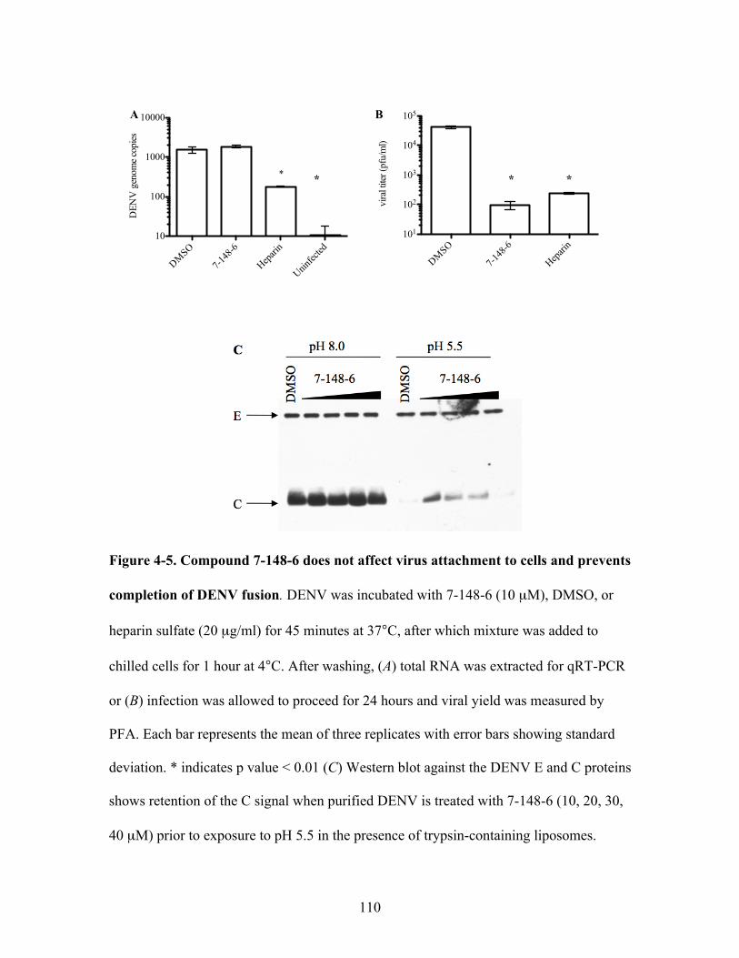
110
Figure 4-5. Compound 7-148-6 does not affect virus attachment to cells and prevents
completion of DENV fusion. DENV was incubated with 7-148-6 (10 µM), DMSO, or
heparin sulfate (20 µg/ml) for 45 minutes at 37°C, after which mixture was added to
chilled cells for 1 hour at 4°C. After washing, (A) total RNA was extracted for qRT-PCR
or (B) infection was allowed to proceed for 24 hours and viral yield was measured by
PFA. Each bar represents the mean of three replicates with error bars showing standard
deviation. * indicates p value < 0.01 (C) Western blot against the DENV E and C proteins
shows retention of the C signal when purified DENV is treated with 7-148-6 (10, 20, 30,
40 µM) prior to exposure to pH 5.5 in the presence of trypsin-containing liposomes.
DMSO
7-148
-6
Hepari
n
Uninfec
ted10
100
1000
10000D
ENV
gen
ome c
opies
*
vira
l tite
r (pf
u/m
l)
DMSO
7-148
-6
Hepari
n101
102
103
104
105
* **
A B

111
DENV entry by blocking a late step in DENV fusion via interactions with the E protein
on the virion surface. These results confirm that 7-148-6 behaves similarly to our other
tested compounds, 2-12-2 and 8-24-3 (Chapter 3), and can be used as a tool to explore
DENV resistance against the small molecule entry inhibitors.
Sequencing of resistant virus reveals two changes in the DENV structural proteins
In order to identify what sequence change(s) caused the observed rise in DENV2
titer, we extracted viral RNA from passage 6 of the 7-148-6 passaged virus (Figure 4-2).
Consensus sequencing of the viral structural proteins C, prM, and E revealed a single
amino acid change in the DENV2 envelope protein at position 196, from methionine to
valine (Figure 4-6). This E(M196V) change was not observed in DENV2 that had been
passaged in the presence of DMSO. Residue 196 is located at the base of the BOG pocket
located between domains I and II, a region of the protein that has been hypothesized to
act as a hinge during the conformational changes that occur during viral fusion (Modis et
al., 2003), consistent with our hypothesis that the anti-DENV activity of 7-148-6 is due to
inhibition of DENV fusion. The methionine at this position is conserved across all four
DENV serotypes although it is not present in more distantly related flaviviruses such as
West Nile or Japanese encephalitis virus.
Our consensus sequencing, while identifying E(M196V), also revealed that this
change was incomplete. This raised the possibility that consensus sequencing may have
obscured other changes in the structural genes, so we performed a single round of virus
plaque isolation, expansion, and sequencing. This viral sequencing led to the
identification of an additional amino acid change, present in 6/7 plaques of the 7-148-6

112
Figure 4-6. Location of residue 196 on DENV2 E protein. Pre-fusion envelope dimer
crystal structure (Modis et al., 2003). Magnification is of the domain I (red) – domain II
(yellow) interface. (Inset) The methionine at position 196 is highlighted in green. In
DENV2 serially passaged in the presence of 20 µM of 7-148-6, consensus sequencing
revealed a valine at this position.

113
passaged virus and 0/3 of virus plaques passaged in the presence of DMSO. This change
was a histidine to tyrosine mutation located at residue 28 in the ectodomain of the M
protein. The M protein is the post-cleavage remnant of the chaperone protein prM, and no
functional role has yet been described for M in the viral entry process. A study that used
electron density to map the location of M on the surface of the mature dengue virion
suggests that the ectodomain takes the form of two alpha helices, one of which is
amphipathic and makes contact with the lipid bilayer of the virion and possibly the
underside of the E protein as well (Kuhn et al., 2002); M(H28Y) is hypothesized to be
located in this helix.
Effect of structural protein mutations on production and entry of single-cycle
reporter virus
To investigate the potential of each identified amino acid change to confer
resistance, we used a reporter virus particle (RVP) system (Ansarah-Sobrinho et al.,
2008). The RVP system uses two plasmids to produce single-cycle infectious viral
particles, with luciferase signal as the readout of a successful infection. One plasmid
encodes for DENV structural proteins C, prM, and E, while the second encodes a West
Nile virus (WNV) genome in which structural proteins have been replaced with the
Renilla luciferase gene. Transfection of cells with both plasmids allows for the
production of recombinant virus particles (RVPs) comprised of the DENV structural
proteins and containing a WNV RNA genome. Entry of RVPs is mediated by the DENV
E protein on the particle surface and is thought to occur via the same mechanisms as
authentic DENV. Following entry, the WNV RNA is translated and replicated, and

114
quantification of the resulting luciferase activity provides a measure of the steady-state
abundance of WNV RNA. Accordingly, to examine the effects of the E(M196V) and
M(H28Y) mutations on RVP production and entry, we engineered each mutation into the
plasmid encoding the DENV2 CprME genes and used these plasmids to produce RVPs.
We initially determined the effect of E(M196V) or M(H28Y) on RVP production
by quantifying the number of WNV replicons present in producer cell supernatant by
qRT-PCR (Figure 4-7A). No significant difference in genome copy number was detected
for either mutation when compared to wild-type RVP production. It is important to note
that quantification of WNV genomes measures the release of recombinant viral particles
that encapsidate RNA, but does not measure any potential differences in empty particle
release. Unfortunately, due to the low number of RVPs in the supernatant, our attempts to
detect any changes in the release of E protein or empty particles by transfected cells were
unsuccessful.
We next asked if E(M196V) or M(H28Y) affected entry of RVPs into target cells.
Wild-type, E(M196V), or M(H28Y) RVPs were diluted to normalize for WNV genome
copy number and added to target cells for a one hour initial infection. Cells were washed
to remove unbound RVPs and fresh medium was added. Sixteen hours after infection,
cells were lysed, and luciferase activity was determined. We observed that E(M196V)
RVPs had a significant increase in luciferase signal compared to wild-type RVP signal,
while M(H28Y) RVPs had a significant decrease (Figure 4-7B). These data show that
both mutations impact entry of viral particles, although in different directions.

115
Figure 4-7. DENV structural protein mutations affect virus entry in a single-cycle
infection assay. Recombinant viral particles (RVPs) containing either E(M196V) or
H(28Y) were produced by transfection of 293T cells with two plasmids: one encoding the
WNV luciferase reporter replicon and one encoding the DENV structural cassette.
(A) RVPs in producer cell supernatant were quantified by performing qRT-PCR against
the WNV replicon. Each bar represents the mean of three replicates with error bars
showing standard deviation. (B) After quantification, RVPs were diluted to normalize for
WNV copy number and BHK-21 cells were infected. Sixteen hours post-infection, cells
were lysed and luciferase signal was measured. Each bar represents the mean of four
replicates with error bars showing standard deviation. **p value <0.001, *p value <0.05
Wild
-type
E(M19
6V)
M(H28
Y)0.0
5.0!1005
1.0!1006
1.5!1006
2.0!1006
2.5!1006
WN
V re
plic
on c
opie
slu
cife
rase
sign
al (c
ps)
wild-ty
pe
E(M19
6V)
M(H28
Y)0
2!1005
4!1005
6!1005
8!1005
**
*
A
B
116
Resistance of DENV structural protein mutations to 7-148-6
To examine the effect of E(M196V) and M(H28Y) mutations on sensitivity to
compound 7-148-6, we normalized inocula by WNV genome copy number and treated
RVPs with 7-148-6 prior to cell infection. Wild-type RVPs treated with 7-148-6 showed
a significant decrease in luciferase signal when cells were lysed 16 hours post-infection
and compared to DMSO-treated RVP signal (Figure 4-8). The luciferase signal produced
by cells infected with E(M196V) RVPs did not decrease when RVPs were pre-treated
with 7-148-6, indicating that E(M196V) confers resistance to 7-148-6. M(H28Y) RVPs
did not have a significant decrease in luciferase signal after 7-148-6 treatment either,
although the low overall luciferase activity of this mutant makes this result difficult to
interpret.
These results suggest that the E(M196V) change may be responsible for the
insensitivity of our passaged virus to the presence of 7-148-6. It is possible that M(H28Y)
also contributes to resistance, and in the authentic virion, both mutations may contribute
to resistance to 7-148-6. These data show that a single point mutation in the DENV E
protein, even a conservative one from methionine to valine, enhances DENV entry and
may alter sensitivity to a small molecule entry inhibitor.
Probing DENV envelope protein changes with single-cycle infection reporter virus
Serially passaging DENV in the presence of ten compounds revealed outgrowth
of only one resistant virus. This suggests that it may be difficult for DENV to overcome
the presence of small molecule entry inhibitors, possibly because any mutations that
confer resistance may come at a high fitness cost for the virus. Since E(M196V) is

117
Figure 4-8. RVPs with DENV structural protein changes show decreased sensitivity
to compound 7-148-6. Recombinant viral particles (RVPs) encoding wildtype structural
proteins, E(M196V), or M(H28Y) mutant proteins were diluted to normalize for genome
copy and subsequently used to infect BHK-21 cells after pre-treatment with 10 µM
compound 7-148-6. Cells were lysed 16 hours post-infection, and luciferase signal was
measured. Each bar represents the mean of four replicates with error bars showing
standard deviation. *p value < 0.05
lucif
eras
e sig
nal (
cps)
DMSO
7-148
-60
1!1005
2!1005
3!1005*
Wild-type RVPs
DMSO
7-148
-60
2!1005
4!1005
6!1005
8!1005
lucif
eras
e sig
nal (
cps)
E(M196V) RVPs
lucif
eras
e sig
nal (
cps)
DMSO
7-148
-60.0
5.0!1004
1.0!1005
1.5!1005
M(H28Y) RVPs

118
located at the base of the BOG pocket, an area of the protein thought to act as a “hinge
region” for the large conformational change of E as it mediates membrane fusion (Modis
et al., 2003), we wanted to probe the tolerance of substitutions in this region more
directly. Using site-directed mutagenesis of the plasmid encoding the DENV structural
genes (the “CprME plasmid”), we mutated each of eleven selected amino acids to alanine
and tested production and entry in the RVP system (Figure 4-9). The chosen residues
were selected due to either conservation among DENV serotypes or previous implication
in altering the optimal pH of fusion, and mapped to three regions of the BOG pocket.
Three residues, Q52, V130, and Q131 are clustered around the bottom outer edge of the
pocket. V130 is conserved across DENV serotypes, while both Q52 and Q131 have been
previously implicated to alter the optimal pH of fusion (Modis et al., 2003). Another four
residues, D192, F193, N194, and M196 are found in a sequence, 192DFNEM196, which is
conserved across DENV serotypes; these residues form the bottom of the BOG pocket
and sit close to the lipid membrane of the virion. This region forms a pseudo-alpha helix
that may make inter-trimer contacts in the post-fusion trimer structure (Modis et al.,
2004) and, outside of the fusion loop, is one of the largest stretches of conserved amino
acids in the DENV envelope protein. M196 is also the residue altered to valine in the
passaging of DENV in the presence of 7-148-6, and we were curious to determine the
effect of an alanine at this position. The final set of amino acids chosen, G275, L277,
L278, and F279 are not part of the pocket itself, but rather located on the k-l loop that
covers the opening of the pocket. G275, L277, and F279 are conserved across DENV
serotypes, while F279 has also been suggested to alter optimal pH of fusion (Lee et al.,
1997).

119
Figure 4-9. Location of selected residues for mutagenesis on the DENV2 envelope
crystal structure. Crystal structure of DENV envelope protein (Modis et al., 2003).
(Top) Pre-fusion DENV E dimer. Each inset focuses on a group of amino acids selected
for mutagenesis. (Middle left) Q52, V130, and Q131, all located near the front and
bottom of the BOG pocket. (Middle right) D192, F193, N194, and M196, part of the
chain that forms the bottom of the BOG pocket (Bottom) G275, L277, L278, F279, all
located on the k-l loop.

120
We mutated each residue to an alanine in the CprME plasmid and used these
plasmids to transfect cells to produce RVPs. RVP preparations were titered by
quantifying the number of WNV genomes/mL present in serial dilutions of producer cell
supernatant. We graphed this value against luciferase activity detected in target cells 16
hours post-infection, allowing us to draw a picture of the effect of mutations on both RVP
production and entry (Figure 4-10). We found that three mutations, V130A, N194A, and
L277A, had detectable levels of WNV genomes in producer cell supernatant but low
luciferase activity in target cells. This suggests that these changes may have a detrimental
effect on RVP entry. In contrast, mutation to an alanine at positions 193 or 279, originally
phenylalanines, caused low luciferase activity in RVP target cells but also near
undetectable levels of WNV genomes in producer cell supernatant, suggesting these
change may impact production of RVPs.
The methionine to valine and methionine to alanine mutations at position 196
differed in their qualitative effects on RVP entry, which may suggest that loss of
hydrophobic bulk at this site has a deleterious effect on this viral process. While more
extensive mutagenesis of this region is necessary to validate this hypothesis, our current
results demonstrate that mutations in the E protein “hinge region” can affect both
production and entry of virus particles.
Changes in the DI/DII region may affect sensitivity to small molecule entry
inhibitors
We selected two mutations, Q52A and Q131A, from our RVP screen to test
against small molecule entry inhibitors to determine if these changes conferred resistance.

121
Figure 4-10. Changes in the DENV E protein DI/DII interface affect production and
entry of RVPs. RVPs with alanines introduced at select locations were produced and
qRT-PCR was performed on serial dilutions to determine RVP production (x axis).
Serially diluted supernatants were used to infect BHK-21 cells, and lysate was collected
16 hours post-infection to detect luciferase activity as a measure of RVP entry (y axis).
Each point represents the mean of four replicates with error bars showing standard
deviation.
0 50000 100000 150000102
103
104
105
106
WNV replicon copies
luci
fera
se c
ount
(cps
) Wild-type
F193AF279A
Q52AM196A
N194A
0 50000 100000102
103
104
105
106
WNV replicon copies
luci
fera
se c
ount
(cps
)
D192AWild-type
G275AL278AQ131AV130AL277A

122
Examination of our RVP screen suggested that these two mutants may enter cells more
efficiently than the wild-type, a phenotype similar to that of E(M196V). Thus, we
hypothesized that the increased infectivity of Q52A and Q131A might also be associated
with resistance to our compounds.
As we had done for E(M196V) and M(H28Y), we first measured E(Q52A) and
E(Q131A) RVP production by quantifying WNV genome in producer cell supernatant
(Figure 4-11A). We found that production of E(Q52A) RVPs was significantly reduced
compared to wild-type RVPs. However, when we measured luciferase activity after
normalizing to genome number, we found that E(Q52A) RVPs actually had a significant
increase in luciferase signal (Figure 4-11B). These results suggest that the E(Q52A)
mutation may have a negative impact on viral particle production but enhance viral entry.
We next normalized to WNV genome copy number in the supernatant and treated
RVPs with 7-148-6, 2-12-2, or GNF-2 prior to infecting target cells. Treatment of wild-
type RVPs by each compound led to significant decreases of luciferase activity in target
cells compared to a DMSO-treated control (Figure 4-12). In contrast, we found no
significant decrease in either E(Q52A) or E(Q131A) RVPs when treated with any of
these compounds. This suggests that these individual mutations may decrease sensitivity
of the RVPs to compound inhibition.
DISCUSSION
This chapter describes our investigation of effects of point mutations in the
DENV E protein on viral entry and sensitivity to a subset of our small molecule entry
inhibitors. Passaging of DENV2 in the presence of small molecules led to outgrowth of

123
Figure 4-11. DENV envelope protein mutations affect virus particle production and
entry in a single-cycle infection assay. (A) RVPs in producer cell supernatant were
quantified by performing qRT-PCR against the WNV replicon. Each bar represents the
mean of three replicates with error bars showing standard deviation. (B) After
quantification, RVPs were diluted to normalize for WNV copy number and BHK-21 cells
were infected. Sixteen hours post-infection, cells were lysed and luciferase signal was
measured. Each bar represents the mean of four replicates with error bars showing
standard deviation. *p value <.0.05
WN
V re
plic
on c
opie
swild
-type
E(Q52
A)
E(Q13
1A)
0
1!1006
2!1006
3!1006
4!1006
5!1006
*
luci
fera
se s
igna
l (cp
s)
wild-ty
pe
E(Q52
A)
E(Q13
1A)
0
2!1005
4!1005
6!1005
8!1005
1!1006
*
A
B
124
Figure 4-12. E(Q52A) and E(Q131A) may confer resistance to small molecule entry
inhibitors. RVPs containing wild-type E, E(Q52A), or E(Q131A) were diluted to
normalize for genome copy and pre-treated with the indicated compounds at their
previously calculated EC90 values against DENV2 (MOI of 1) before addition to cells.
Cells were lysed 16 hours post-infection and luciferase signal was measured. Each bar
represents the mean of four replicates with error bars showing standard deviation.
*p value < 0.01
Wild-type RVPs
DMSO
7-148
-62-1
2-2
GNF-20
1!1005
2!1005
3!1005
4!1005
5!1005lu
cifer
ase c
ount
(cps
)
RVP treatment
* * *
Q131A RVPs
RVP treatment
lucif
eras
e cou
nt (c
ps)
DMSO
7-148
-62-1
2-2
GNF-20
2!1005
4!1005
6!1005
8!1005
Q52A RVPs
DMSO
7-148
-62-1
2-2
GNF-20
2!1005
4!1005
6!1005
8!1005
lucif
eras
e cou
nt (c
ps)
RVP treatment

125
virus against only one out of ten small molecule, 7-148-6, suggesting that DENV does
not easily escape the presence of the compounds. Consistent with this hypothesis are the
results of our small alanine mutagenesis screen, in which eight of eleven mutations in the
domain I/II interface of E drastically reduced viral particle production or entry.
Sequencing of DENV2 passaged in the presence of 7-148-6 identified two
mutations that caused changes in the E and M proteins, the structural proteins on the
virion surface. Characterization of the E(M196V) and M(H28Y) mutations in a single-
cycle reporter virus assay suggests that these changes impact viral particle entry.
E(M196V) enhanced entry of reporter virus particles and caused insensitivity to the
compound 7-148-6, against which it was selected. M(H28Y) may also confer some
degree of resistance to 7-148-6, although the marked decrease in entry of these virus
particles makes this difficult to interpret. Despite the full recovery of entry signal in
E(M196V) RVPs after treatment with 7-148-6, it is probable that this single E protein
residue does not solely dictate compound sensitivity. This is due to the fact that the
methionine at position 196 is conserved across all DENV serotypes, yet 7-148-6 did not
inhibit DENV3 or DENV4, suggesting that other amino acids play a factor in the antiviral
activity of 7-148-6.
We showed that compound 7-148-6 likely has a viral target, is active during
DENV entry, and blocks DENV2 fusion in vitro. The results with E(M196V) RVPs are
consistent with the hypothesis that 7-148-6’s target is the E protein on the surface of the
virion. At this point, it is unclear how the E(M196V) change causes resistance to 7-148-6.
While escape mutations on proteins have traditionally been useful in inferring the binding
sites of antivirals on their respective targets, it is unclear if the M196V change inhibits

126
binding of the compound directly or if it allows for escape by altering how the E protein
mediates fusion or undergoes the conformational rearrangements that occur during fusion.
The E(M196V) change could impact fusion in a number of ways. Residue 196 is
located at the base of the BOG pocket, in a position that may make contact with the lipid
membrane of the virion; it is possible that the shorter side chain of valine at this position
allows for easier movement of domain II away from the lipid membrane. Another
possible hypothesis to explain enhanced virus entry or compound resistance is a mutation
at this position could affect the formation or stability of the E trimer, as the pseudo-alpha
helix (residues 192-196) is predicted to make intra-trimer contacts in the final post-fusion
E structure. This hypothesis could also explain why the M196A change resulted in
reduced entry of RVPs, as the smaller side chain of alanine could have a detrimental
effect on trimer formation or stability. Alternatively, it is possible that the M196V change
directly affects the ability of 7-148-6 to bind to the E protein. Our laboratory is currently
performing studies with recombinant dengue envelope protein to test this possibility.
The other identified change in the structural proteins of DENV was a histidine to
tyrosine change located in the ectodomain of the M protein, a mutation that significantly
reduced RVP entry. On its own, it is unclear how this change affects entry or could
confer resistance to 7-148-6. Little is known about what role, if any, the M protein plays
in DENV infectivity after the pr peptide is cleaved (Mukhopadhyay et al, 2005). One
study showed that, post-cleavage, the ectodomain of M rests partially buried in the virus
lipid envelope underneath the E protein homodimer (Kuhn et al., 2002). It is possible that
M makes contacts with E and could influence movement of domain II away from the
viral membrane. It is also possible that M(H28Y) appeared in passaged DENV2 as a

127
compensatory mutation for E(M196V). One way to test this hypothesis would be to
examine entry and compound sensitivity of RVPs that contain both E(M196V) and
M(H28Y), experiments that we are currently performing.
The alanine mutagenesis screen of the BOG pocket on the E protein was
performed to examine the effects of mutations in this region on viral entry. The majority
of mutations we introduced appeared to affect viral particle production and/or entry,
suggesting that changes in this region may have a fitness cost for the virus. Unfortunately,
due to our inability to monitor the presence of E protein release into producer cell
supernatant, we cannot rule out the possibility that some point mutations that appear to
affect viral particle production or release may, in fact, have interfered with earlier steps of
the DENV life cycle such as production of E or coating of immature viral particles
The contrasting results of E(M196V) and E(M196A) in the RVP system suggest
that specific substitutions may dictate the effect of mutations in E on viral entry, making
it difficult to draw conclusions from our limited mutagenesis screen about the
contribution of specific residues. In order to better understand potential mechanisms of
resistance and the impact of resistance mutations on fitness, additional mutagenesis
experiments should be performed targeting residues in this region that are not conserved
or that have not been implicated in altering the pH of viral fusion.
A recently published study reported site-directed mutagenesis of several residues
around the BOG pocket in authentic DENV and, similar to our results, found that many
of their mutations had effects on DENV entry or viability (Butrapet et al., 2011). Two
mutations in their study, F193A and F279A, were identical to mutations that we
examined in the RVP system. The E(F193A) mutant virus was found to have lower

128
replication efficiency while the E(F279A) mutant virus exhibited no change in the pH
threshold of fusion or in viral replication, although this mutant virus was found after
several passages to have a mixed population of alanine and leucine at position 279. In the
RVP system, we found that both of these mutations inhibited RVP production and would
be expected to cause reduced viral replication in the context of the authentic virus.
However, since the RVP system permits only single-cycle infection, it does not allow for
compensatory or reversion mutants, and this may provide a possible explanation for
apparent discrepancies between our study and the previous results using authentic virus.
Interestingly, Butrapet et al. (2011) also mutated Q52, although to an arginine
instead of alanine. In RVPs, we found that Q52A caused a significant inhibition of viral
particle production, but when normalized for WNV genome copy, significantly increased
RVP entry (Figure 4-11). In authentic virus, Butrapet et al found that Q52R caused
increased cytopathic effect upon passaging in cells and possibly a higher pH threshold for
fusion. An explanation consistent with both sets of results is that mutations at residue 52
lead to enhancement of DENV fusion.
We tested E(Q52A) and E(Q131A) against a subset of our small molecule entry
inhibitors and found that both changes conferred resistance to all three compounds
compared to wild-type RVPs. As with E(M196V), we do not currently know how these
changes confer resistance. Residues 52 and 131 are located close to the mouth of the
BOG pocket, while 196 sits at the very base (Figure 4-9), making it unlikely that all three
changes inhibit binding of small molecules. A more likely explanation is that one or more
of these changes alter DENV fusion as a means of escaping small molecule inhibition.
129
In the work presented here, we have begun to explore how changes to the DENV
E protein influence both the entry of virus particles as well as susceptibility to small
molecule entry inhibitors. We identified a point mutation in the envelope protein,
E(M196V) that rendered a single-cycle reporter virus insensitive to the presence of the
compound it was raised against. This is the first recorded instance of a potential
resistance mutation to a DENV small molecule entry inhibitor. However, further work
should be done to characterize this resistance mutation as well as explore the mechanism
of resistance. The mutation should be introduced back into authentic dengue virions,
which would allow for biochemical studies to examine DENV fusion in the presence or
absence of 7-148-6. In addition, we show that specific substitutions in the hinge region of
E can impact viral particle production or entry. These results provide promising leads to
learn more about the molecular interaction of our DENV small molecule entry inhibitors
with the E protein as well as to discover more about the functional contribution of
individual amino acids in the E protein to DENV fusion.

130
METHODS AND MATERIALS
DENV passaging
Initially, DENV2 (MOI 0.1) was incubated with selected compounds (see text) for 45
minutes at 37°C before addition to 106 Vero cells in a T25 flask. After a one hour
infection, 5 ml medium was added (2% FBS in DMEM) containing compound or DMSO.
Infections proceeded for four days, at which point supernatant was harvested and spun
briefly to remove cell debris. Five hundred µL of supernatant was used to infect a fresh
T25 of Vero for one hour, at which point 4.5 ml of medium containing compound was
added. Viral titer of each passage was determined by plaque-forming assay. The first
passaging with compounds maintained 20 µM of each compound over eight passages.
The second passaging started at 2.5 µM and increased by 2.5 µM each passage until 20
µM was reached, which was kept constant for two passages.
Order of addition experiments
DENV2 (NGC strain) was used to infect BHK-21 cells at an MOI of 1 for all
experiments. Compound was used at 10 µM. Virus was pre-incubated in 100 !L of
medium containing compound at 37°C. For co-infection, 10 !M of compound or DMSO
was added to virus inoculum directly before addition to cells. After an initial one hour
infection, cells were washed twice with 1X PBS to remove unbound compound and virus,
and 1 mL cell medium (MEM-alpha with 2% FBS) was added. For post-treatment of
cells, compound was added to medium for 1 hour post-infection at 10 µM, then removed
and cells were washed twice and medium was replaced. Twenty-four hours after initial
infection, supernatant was collected and viral titer was determined by plaque-forming
assay.

131
Removal of unbound compounds by gel filtration column
DENV2 (NGC strain) inocula (MOI 2) was pre-incubated in 100 !L with 10 !M of
compound or DMSO for 45 minutes at 37°C, then placed over a Sephadex G50 spin
column and spun for 2 minutes at 2000 rpm at room temperature. Eluant was used to
infect cells for 1 hour, after which cells were washed twice with 1X PBS to remove
unbound compound and virus, and 1 mL cell medium (MEM-alpha with 2% FBS) was
added. Twenty-four hours after initial infection, supernatant was collected, and viral titer
was determined by plaque-forming assay.
DENV attachment to cells
DENV2 (NGC strain) was incubated with either 5 !M of 2-12-2 or 8-24-3, 20 !g/ml of
heparin, or DMSO for 45 minutes at 37°C, then added to BHK cells (MOI 1) at 4°C for
one hour with constant rocking. Cells were washed three times with cold 1X PBS to
remove unbound virus or compound. For quantitation of viral genomes bound, total RNA
was extracted using Trizol according to manufacturer’s instructions and qRT-PCR was
performed. For viral yield determination, one mL of medium was added (MEM-alpha
with 2% FBS), and culture supernatant was harvested 24 hours later. To determine viral
titer in supernatant, plaque-forming assays were performed.
Quantification of DENV genomes
Quantification of DENV genomes was done on a MyIQ Cycler (Biorad). After RNA
extraction and quantification, 5 !L of RNA was used to make cDNA with the iScript kit
(Biorad) according to manufacturer’s instructions. The resulting cDNA was digested by
RNaseH for 1 hour at 37°C. Quantification by rt-PCR was done on each sample in

132
triplicate (25 !L each) using SybrGreen Supermix (Biorad) according to manufacturer’s
instructions. Primers used targeted the DENV C gene.
DV2Cfor 5’-AATATGCTGAAACGCGAGAGA-3’
DV2Crev 5’-GGGATTGTTAGGAAACGAAGG-3’
Liposomes
Liposomes were made with 1-palmitoyl-2-oleoyl-sn-glycero-3-phosphocholine (POPC),
1-palmitoyl-2-oleoyl-sn-glycero-3-phosphoethanolamine (POPE) (Avanti Polar Lipids
cat # 850457 and 850757, respectively), and cholesterol (Sigma-Aldrich cat#C3045) at
1:1:2 molar ratio in TAN buffer (20mM triethanolamine, 100mM NaCl, pH 8.0). Lipids
were dried down and resuspended thoroughly by vortexing and sonication. Liposomes
were prepared by extrusion through a 0.2 micron filter after five freeze/thaw cycles. For
trypsin-containing liposomes, 10 mg of trypsin was added to 1 mL of lipids (3 mg each)
after the third freeze/thaw cycle, prior to extrusion. Liposomes were separated from
unincorporated trypsin by size-exclusion chromatography using a Superdex 200 column
on an Akta fast performance liquid chromatography (FPLC) system. Trypsin-containing
liposomes were used within 24 hours.
Capsid protection assay
Purified dengue 2 virus (300 ng) was incubated with various concentrations of
compounds (5-40 !M) for 45 minutes at 37°C in TAN buffer (pH 8.0) prior to addition of
liposomes (7.5 !L of peak fraction from column) for ten minutes. After incubation with
liposomes, 7.5 !L 1M sodium acetate (pH 5.0) was added to drop pH to 5.5 for ten
minutes. Samples were back-neutralized with 15 !L 1M TEA (unbuffered) and incubated
for forty-five minutes at 37°C to allow trypsin digestion (total volume: 100 !L). SDS

133
sample buffer (20 !L) was added, and samples were boiled for 20 minutes before being
separated by SDS-PAGE on a 12% gel. Proteins were transferred to a nitrocellulose
membrane using a semi-dry transfer. 4G2 antibody, grown in house from ATCC cell line
HB-112, was used for detection of DENV envelope (dilution varies by lot), and the neat
supernatant of hybridoma cell line 6F3.1, kindly provided by Dr. John Aaskov, was used
to detect capsid protein.
Production of recombinant virus particles
Plasmids for WNV replicon and DENV structural proteins were the kind gift of Dr. Ted
Pierson. RVP production was performed according to protocols received from his lab
(Ansarah-Sobrinho et al, 2008). 293T-LX cells were transfected with plasmids using
Lipofectamine 2000 according to manufacturer’s instructions at a ratio of 3:1 (DENV
CprME:WNV replicon). Medium was changed 4 hours post-transfection and supernatant
was collected at 25 hours post-transfection. Supernatant was spun for 5 minutes at 1200
rpm to remove cell debris and was put through a 0.2 micron filter before freezing. Viral
RNA was extracted using the QIAamp Viral RNA kit according to manufacturer’s
instructions. RNA was transcribed to cDNA using iScript cDNA kit (Biorad) according
to manufacturer’s instructions and qPCR was performed using primers against the WNV
replicon to quantify genome copies.
Quantification of WNV replicon
Quantification of WNV replicons in RVP supernatant was done on a MyIQ Cycler
(Biorad). RVP RNA was extracted using the QIAamp Viral RNA Mini kit from Qiagen
(Cat#52906) and suspended in a final volume of 60 !L, 2 !L of which was used to make
cDNA with the iScript kit (Biorad) according to manufacturer’s instructions. The

134
resulting cDNA was digested by RNaseH for 1 hour at 37°C. Quantification by rt-PCR
was done on each sample in triplicate (25 !L/well) using SybrGreen Supermix (Biorad)
according to manufacturer’s instructions. Sequences of primers against the WNV 3’ UTR
replicon are listed below.
WNVfwd: 5’-AGAGTGCAGTCTGCGATAGTGC-3’
WNVrev: 5’-CCCTTCTCCCTGGTTAACA-3’
Site-directed mutagenesis of DENV CprME plasmid
Site-directed mutagenesis of the DENV CprME plasmid was performed using the
Stratagene Quikchange II kit according to manufacturer’s instructions (Cat#200518).
Primers to introduce all mutations were designed using the Quikchange Primer Design
website (sequences listed below). XL10 Gold competent cells were transformed with
mutated plasmids after digestion and grown at 30°C for 2 days. Selected colonies were
grown in superbroth at 30°C for 24 hours, then plasmids were isolated and sequenced to
confirm the presence of mutations.
M(H28Y): c1652t 5'-cagaaggggcctggaaatatgtccagagaattga-3'; c1652t_antisense 5'-
tcaattctctggacatatttccaggccccttctg-3'
E(M196V): a2381g 5'-gcctcgacttcaatgaggtggtgttgttgcagatg-3'; a2381g_antisense 5'-
catctgcaacaacaccacctcattgaagtcgaggc-3'
E(Q52A): c1949g_a1950c 5'-tgaactgataaaaacagaagccaaagcgcctgccacccta-3';
c1949g_a1950c_antisense 5'-tagggtggcaggcgctttggcttctgtttttatcagttca-3'
E(V130A): t2184c 5'-gaacatggaaggaaaagttgcgcaaccagaaaacttggaat-3'; t2184c_antisense
5'-attccaagttttctggttgcgcaacttttccttccatgttc-3'

135
E(Q131A): c2186g_a2187c 5'-gaacatggaaggaaaagttgtggcaccagaaaacttggaatacacc-3';
c2186g_a2187c_antisense 5'-ggtgtattccaagttttctggtgccacaacttttccttccatgttc-3'
E(D192A): a2370c 5'-ccaagaacgggcctcgccttcaatgagatggtg-3'; a2370c_antisense 5'-
caccatctcattgaaggcgaggcccgttcttgg-3'
E(F193A): t2372g_t2373c 5'-ccaagaacgggcctcgacgccaatgagatggtgttgtt-3';
t2372g_t2373c_antisense 5'-aacaacaccatctcattggcgtcgaggcccgttcttgg-3'
E(N194A): a2375g_a2376c 5'-gaacgggcctcgacttcgctgagatggtgttgttgc-3';
a2375g_a2376c_antisense 5'-gcaacaacaccatctcagcgaagtcgaggcccgttc-3'
E(M196A): a2381g_t2382c 5'-ggcctcgacttcaatgaggcggtgttgttgcagatgga-3';
a2381g_t2382c_antisense 5'-tccatctgcaacaacaccgcctcattgaagtcgaggcc-3'
E(G275A): g2619c 5'-agaaatccaaatgtcatcagcaaacttactcttcacaggac-3'; g2619c_antisense 5'-
gtcctgtgaagagtaagtttgctgatgacatttggatttct-3'
E(L277A): t2624g_t2625c 5'-aatccaaatgtcatcaggaaacgcactcttcacaggacatctcaag-3';
t2624g_t2625c_antisense 5'-cttgagatgtcctgtgaagagtgcgtttcctgatgacatttggatt-3'
E(L278A): c2627g_t2628c 5'-cagaaatccaaatgtcatcaggaaacttagccttcacaggacatctc-3';
c2627g_t2628c_antisense 5'-gagatgtcctgtgaaggctaagtttcctgatgacatttggatttctg-3'
E(F279A): t2630g_t2631c 5'-gtcatcaggaaacttactcgccacaggacatctcaagtgc-3';
t2630g_t2631c_antisense 5'-gcacttgagatgtcctgtggcgagtaagtttcctgatgac-3'
RVP infection of cells
RVPs were used to infect BHK-21 cells in 48-well plates. RVPs were diluted to 105
genomes/mL and incubated with small molecules at their respective EC90 values (as
determined in cellular entry assays) for 45 minutes at 37°C. This mixture was added to
cells for a one hour initial infection (37°C), after which cells were washed twice with 1X

136
PBS. All infections were done in quadruplicate. Medium was added (2% FBS in MEM-
alpha) and cells were incubated at 37°C for 16 hours. At 16 hours post-infection, cells
were washed once before lysis and lysate was moved to -20°C until use. Luciferase signal
was determined with a Renilla luciferase activity kit (Promega Cat#E2820).
137
REFERENCES:
Ansarah-Sobrinho, C., Nelson, S., Jost, C.A., Whitehead, S.S., and Pierson, T.C. (2008). Temperature-dependent production of pseudoinfectious dengue reporter virus particles by complementation. Virology 381, 67-74. Beasley, D.W.C., and Aaskov, J.G. (2001). Epitopes on the Dengue 1 Virus Envelope Protein Recognized by Neutralizing IgM Monoclonal Antibodies. Virology 279, 447-458. Butrapet, S., Childers, T., Moss, K.J., Erb, S.M., Luy, B.E., Calvert, A.E., Blair, C.D., Roehrig, J.T., and Huang, C.Y.H. (2011). Amino acid changes within the E protein hinge region that affect dengue virus type 2 infectivity and fusion. Virology 413, 118-127. Gollins, S.W., and Porterfield, J.S. (1986). The Uncoating and Infectivity of the Flavivirus West Nile on Interaction with Cells: Effects of pH and Ammonium Chloride. Journal of General Virology 67, 1941-1950. Kampmann, T., Yennamalli, R., Campbell, P., Stoermer, M.J., Fairlie, D.P., Kobe, B., and Young, P.R. (2009). In silico screening of small molecule libraries using the dengue virus envelope E protein has identified compounds with antiviral activity against multiple flaviviruses. Antiviral Research 84, 234-241. Kuhn, R.J., Zhang, W., Rossmann, M.G., Pletnev, S.V., Corver, J., Lenches, E., Jones, C.T., Mukhopadhyay, S., Chipman, P.R., Strauss, E.G., et al. (2002). Structure of Dengue Virus: Implications for Flavivirus Organization, Maturation, and Fusion. Cell 108, 717-725. Lee, E., Weir, R.C., and Dalgarno, L. (1997). Changes in the Dengue Virus Major Envelope Protein on Passaging and Their Localization on the Three-Dimensional Structure of the Protein. Virology 232, 281-290. Modis, Y., Ogata, S., Clements, D., and Harrison, S.C. (2003). A ligand-binding pocket in the dengue virus envelope glycoprotein. Proceedings of the National Academy of Sciences of the United States of America 100, 6986-6991. Modis, Y., Ogata, S., Clements, D., and Harrison, S.C. (2004). Structure of the dengue virus envelope protein after membrane fusion. Nature 427, 313-319. Mukhopadhyay, S., Kuhn, R.J., and Rossmann, M.G. (2005). A structural perspective of the flavivirus life cycle. Nat Rev Micro 3, 13-22. Poh, M.K., Yip, A., Zhang, S., Priestle, J.P., Ma, N.L., Smit, J.M., Wilschut, J., Shi, P.-Y., Wenk, M.R., and Schul, W. (2009). A small molecule fusion inhibitor of dengue virus. Antiviral Research 84, 260-266.

138
Schmidt, A.G., Lee, K., Yang, P.L., and Harrison, S.C. (2012). Small-Molecule Inhibitors of Dengue-Virus Entry. PLoS Pathog 8, e1002627. Wang, P.-G., Kudelko, M., Lo, J., Siu, L.Y.L., Kwok, K.T.H., Sachse, M., Nicholls, J.M., Bruzzone, R., Altmeyer, R.M., and Nal, B.a. (2009a). Efficient Assembly and Secretion of Recombinant Subviral Particles of the Four Dengue Serotypes Using Native prM and E Proteins. PLoS ONE 4, e8325. Wang, Q.-Y., Patel, S.J., Vangrevelinghe, E., Xu, H.Y., Rao, R., Jaber, D., Schul, W., Gu, F., Heudi, O., Ma, N.L., et al. (2009b). A Small Molecule Dengue Virus Entry Inhibitor. Antimicrob Agents Chemother, AAC.01148-01108. WHO (2012). Dengue and severe dengue. Zhang, W., Chipman, P.R., Corver, J., Johnson, P.R., Zhang, Y., Mukhopadhyay, S., Baker, T.S., Strauss, J.H., Rossmann, M.G., and Kuhn, R.J. (2003a). Visualization of membrane protein domains by cryo-electron microscopy of dengue virus. Nature Structural & Molecular Biology 10, 6. Zhang, Y., Corver, J., Chipman, P.R., Zhang, W., Pletnev, S.V., Sedlak, D., Baker, T.S., Strauss, J.H., Kuhn, R.J., and Rossmann, M.G. (2003b). Structures of immature flavivirus particles. EMBO 22, 10.

139
Chapter 5: Discussion
140
SUMMARY
The project covered in this dissertation stems from an unexpected observation:
namely, that DENV yield was lowered when initial virus inoculum was pre-incubated
with GNF-2, a previously identified small molecule inhibitor of intracellular Abl kinases.
This discovery serves as a reminder of the importance of exploring the possibility of off-
target effects for many scientific approaches, from RNAi to small molecules to peptides.
Chapter 2 discusses our initial observation of an unexpected drop in DENV yield
when virus inoculum is pre-incubated with a small molecule inhibitor of cellular Abl
kinases, GNF-2. Further experiments revealed that GNF-2 interacts with purified DENV
virions as well as recombinant envelope protein dimer, the pre-fusion form of the
envelope found on the surface of mature visions. Other small molecule inhibitors have
previously been reported to interact with dengue, but these were discovered through high-
throughput and in silico screens designed to identify compounds that bind directly to the
envelope protein of the virion (Kampmann et al., 2009; Poh et al., 2009; Schmidt et al.,
2012; Wang et al., 2009; Zhou et al., 2008). In contrast, GNF-2 is well established as an
inhibitor of Abl kinases that derives its high degree of selectivity by binding in the
myristate-binding pocket of the kinase and is inactive against over 450 other cellular
kinases (Adrian et al., 2006; Zhang et al., 2010). Our discovery that GNF-2 inhibits
DENV at two separate points in the viral life cycle raises the intriguing possibility of
designing small molecule inhibitors that block viruses via two targets at separate points
during the viral life cycle.
As a way to separate GNF-2’s two anti-DENV activities – one mediated by
intracellular Abl kinase and one mediated by interactions with the dengue virion – we
141
performed a structure-activity relationship study in collaboration with Dr. Nathanael
Gray’s laboratory. This led to the identification of twenty-three small molecules that
inhibited DENV infectivity with equivalent or improved activity relative to GNF-2. All
twenty-three molecules showed no inhibition of Abl kinases in cellular assays. In Chapter
3, we selected two of these compounds for further characterization. We found that DENV
yield was not inhibited if virus is pre-incubated with compound at 4°C, suggesting
dynamic motion of the virion surface may be necessary to allow binding of compounds.
When we examined specific steps of the DENV entry process, we found that the
compounds did not inhibit attachment of virions to cells, but did prevent completion of
viral fusion in vitro. These results are consistent with our current hypothesis that the
small molecule inhibitors interfere with the significant structural changes that the DENV
E protein must undergo during viral entry, although additional experiments are needed to
provide support for and refine this hypothesis. The SAR study and mechanistic work that
I have presented add to the growing body of identified DENV entry inhibitors that
prevent viral fusion.
In Chapter 4, we utilized a classical virology approach to learn more about the
mechanism of action of our DENV entry inhibitors. We passaged virus in the presence of
ten compounds, and observed outgrowth of an insensitive virus against only one, 7-148-
6. Sequencing of the structural genes of this virus quasispecies revealed two point
mutations; one each in the E and M proteins. Using a single-cycle reporter virus assay,
we found that treatment of wild-type virus particles with 7-148-6 reduced viral entry, but
no drop was observed with virus particles containing the E(M19V) or M(H28Y)
mutation, suggesting that both changes may play a role in the observed resistance of the
142
authentic virus. We then performed an alanine scanning study focused on the region
surrounding the E(M196V) mutation and, using the single-cycle reporter virus, found that
the majority of point mutations in this area were not well-tolerated, decreasing either viral
particle production or entry. Two mutations, E(Q52A) and E(Q131A), showed efficient
virus entry and were subsequently tested against a subset of our small molecule entry
inhibitors. Both mutations exhibited reduced sensitivity to all three small molecules
tested when compared to the compounds’ effect on wild-type virus particles.
Interestingly, the E(M196V), E(Q52A), and E(Q131A) reporter viruses all had higher
infectivity than wild type viruses, as reflected by luciferase signal per genome copy. This
possible enhancement of viral entry may explain the reduced sensitivity to compounds.
Alternatively, reduced sensitivity to the entry inhibitors may indicate that the E protein
mutations interfere with the interaction of compounds with E. Additional experiments
that elucidate not only the interactions mediating binding of the inhibitors to wild-type
and mutant E proteins but also the mechanism(s) of action of our entry inhibitors are
needed to distinguish between these two potential mechanisms of resistance, which are
not mutually exclusive. Our current results suggest that point mutations in the E domain
I/II interface may affect sensitivity to entry inhibitors and supports previous work that
individual residues in this region can impact DENV entry.
DISCUSSION AND FUTURE DIRECTIONS
The need for DENV therapeutics is undeniable; cases of DENV fever and severe
fever have increased exponentially over the past four decades and there are still no
specific vaccines or treatments available (WHO, 2012). However, the search for effective
143
antivirals is hampered by a lack of understanding of many of the basic viral processes
that DENV undergoes as well as what cellular proteins are critical for viral infection.
Until recently, the virus was endemic largely in countries that did not have resources to
devote to basic research of the virus (WHO, 2012). The reason for the initial screen that
led to this dissertation and identified GNF-2 as an inhibitor of DENV was to identify
cellular kinases critical for DENV infection (Chu and Yang, 2007). While a long-term
goal of our work is to contribute to the search for effective antivirals, a more immediate
goal is to investigate the DENV life cycle to identify molecules and processes that may
be pharmacologically targeted to inhibit the virus.
DENV entry as an antiviral target
Dengue virus encodes only a small number of viral proteins, thus making it highly
dependent upon host cells to successfully complete a life cycle. In order to take
advantage of the host cell machinery, DENV must first enter cells to gain access to the
host cytoplasm, where viral transcription and replication occur. DENV is taken up into
cells via clathrin-mediated endocytosis (Acosta et al., 2008; van der Schaar et al., 2008).
It is then trafficked to cellular endosomes, where exposure to the low pH triggers viral
fusion with the cellular membrane (Stiasny and Heinz, 2006; van der Schaar et al., 2008).
Following viral protein production and genome replication, nascent virions are
assembled, packaged, and undergo maturation during exocytosis prior to the start of a
new round of infection.
Each step in the viral life cycle presents a potential point for therapeutic
intervention, but viral entry may be considered particularly appealing for several reasons.
DENV entry is a discrete process and, relative to other viral processes such as replication

144
or assembly, may be of limited scope in terms of viral and cellular proteins involved. In
addition, the initial interaction between virus and cell occurs on the cell exterior, allowing
the virion to be targeted by non-cell permeable inhibitors or even antibodies. Finally,
viral entry occurs prior to genome replication, thereby decreasing the opportunity for
viral resistance to emerge.
Limitations of GNF-2 and current inhibitors as potential antivirals
Importantly, our discovery that GNF-2 and the derivatives identified in the
structure-activity relationship (SAR) study (Appendix A) inhibit DENV entry does not
automatically qualify them as promising DENV therapeutics. One limitation of the
compounds we identified is that none of them potently inhibited all four DENV
serotypes. During a DENV outbreak, it can initially be unclear which serotype or
serotypes are circulating, and thus it would be desirable for therapeutics to be active
against all four serotypes. It is important to note that our efforts in the SAR study were
directed at optimizing inhibition of DENV2 and only tested against other serotypes after
potency of DENV2 inhibition was determined. Even with the focus on DENV2, we were
able to identify five compounds that had measurable EC90 values against all four
serotypes, suggesting that identification of pan-serotype inhibitors may be possible. A
second limitation to the direct development of inhibitors such as 2-12-2, 7-148-6, and 8-
24-3 as antivirals is that their potency may be too low (EC90 values in the low single-digit
micromolar) to be useful antivirals in vivo. While this could be unequivocally established
via testing in an available animal model of DENV infection (Zompi and Harris, 2012), it
is likely that additional optimization of inhibitor potency would be necessary to achieve
therapeutically beneficial inhibition of DENV in vivo.

145
We are not the first group to identify small molecule inhibitors of DENV entry.
GNF-2 and the other twenty-one small molecules identified in this study add to the
growing body of DENV entry inhibitors that purportedly act by inhibiting viral fusion
(Table 1-1). Our compounds are not as potent as some other small molecules such as
NITD6 (reported EC50 value 119 nM) (Wang et al., 2009), 3-110-22 (reported IC90 value
790 nM) (Schmidt et al., 2012), or peptide inhibitors based on the stem region of E
(reported EC90 value 1 µM (Schmidt et al., 2010), but they do exhibit potencies
comparable to or exceeding leading compounds from several other studies, including a
peptide inhibitor with a reported EC50 value of 10 µM (Hrobowski et al., 2005), small
molecule A5 (reported EC50 value 4 µM) (Yennamalli et al., 2009), small molecule P02
(reported EC50 13 µM) (Zhou et al., 2008) and small molecule NITD448 (reported EC50
value 9.8 µM) (Poh et al., 2009). It is worth noting, however, that we calculated the EC90
values of our compounds, while most other studies calculated EC50 values of compounds.
Consequently, direct comparisons of anti-DENV potency must await side-by-side
comparisons of our compounds with reference compounds from prior studies, as we have
done with NITD6 (Chapter 2).
In addition, we note that in our SAR optimization, we utilized a relatively low-
throughput assay measuring differences in DENV yield. In the future, utilization of a
biochemical assay based on fluorescence polarization measurements to monitor
interaction of compounds with the soluble pre-fusion E dimer or cellular assays utilizing
the single-cycle reporter virus system may facilitate broader screening efforts and enable
more sensitive and quantitative detection of the antiviral activities of the GNF-2-inspired
entry inhibitors described in this study.

146
Probing mechanism(s) of action using biochemical assays and mutagenesis
As with most scientific research, the experiments performed in this dissertation
raise a number of further questions. While our biochemical in vitro experiments reveal
that the two tested compounds, 2-12-2 and 8-24-3, prevent the completion of DENV
fusion, we do not know exactly at what step this block occurs. The compounds could
prevent lipid mixing between the viral and cellular membranes or they may block at a
another step, such as the fold-back of the E domain III or final formation of the fusion
pore (Figure 1-3). It is possible to test whether lipid mixing has occurred in vitro using
pyrene-labeled virus and liposomes as a target membrane (Schmidt et al., 2010), although
our attempts at this delicate assay with DENV did not yield sufficient signal to
confidently detect fusion with untreated wild-type virus. However, if this assay were
optimized and had increased sensitivity, it may be a viable approach to differentiate
among these possible inhibitory mechanisms.
Another, broader, question about mechanism is whether GNF-2 and other
compounds that were not tested in Chapters 3 or 4 inhibit the same step of DENV
entry/fusion as 2-12-2, 8-24-3, and 7-148-6. It remains to be determined whether all of
the compounds identified in our SAR study bind to the same site on E or inhibit DENV
entry via the same molecule mechanism It is therefore of interest to use our in vitro
biochemical fusion assays to test a broader panel of compounds as well as test the above
compounds against other DENV serotypes. Likewise, efforts to identify the binding
site(s) of these compounds on the E structure via high-resolution structure determination
are of considerable interest.

147
In the meantime, the results from our mutagenesis studies constitute an important
step towards examining the potential mechanism of action of our DENV entry inhibitors
while also serving as a broader study of the fusion of this region of the E protein in
DENV entry. The E protein DI/DII interface has been of great interest to the flavivirus
field since 2003, when the possibility of inhibiting DENV entry by targeting this region
with a small molecule was first proposed (Modis et al., 2003). However, little
experimental work has been done to explore this region outside of using it to model or
test small molecule inhibitors. One recent study (Butrapet et al., 2011) performed a
mutational analysis in this region of the protein using reverse genetics and found that in
Vero cells, eight out of 15 selected residues were either lethal or unstable (i.e., did not
retain the engineered mutation or contained purported compensatory mutations in other
locations). These data, along with the RVP results in our studies demonstrating that point
mutations in the domain I/II interface potentially affect expression, release, and/or entry
of virus particles, suggest that changes in this area may have wide-ranging effects in the
DENV life cycle. Further cellular and biochemical experiments are needed to
characterize the phenotypic block in the DENV life cycle caused by mutations in this
region of E. Elucidation of the relationship between structure and function of this region
of the E protein should aid in rational design of either entry inhibitors or antivirals
targeted to other steps of DENV infection.
While our RVP data suggest that the E(M196V) mutation potentially enhances
viral entry, there are still many unanswered questions about this mutation, both within
and outside the context of our small molecule inhibitors. Our group is currently working
to determine whether the E(M196V) mutation alters the ability of recombinant envelope
148
protein to associate or interact with GNF-2 and other small molecules. This work will
help clarify how this mutation confers resistance to the small molecule inhibitors; if the
compound has reduced affinity for the E(M196V) protein, this suggests that resistance is
conferred by prevention of compound binding. Otherwise, E(M196V) may confer
resistance by causing E to become hyperfusogenic (Biering et al., 2012; Higgs and
Gould, 1991) thus allowing the virus to complete fusion despite the presence of the
compounds. E(M196V) could also alter the optimal pH of fusion, an event that has
previously been suggested to occur when changes are made in the domain I/II interface
(Beasley and Aaskov, 2001; Lee et al., 1997), and which could be tested by using virus
containing this change in the in vitro capsid protection assay, where the pH drop can be
tightly controlled. These two models are both consistent with our existing data and are
not mutually exclusive.
In addition, we are studying the E(M196V) and M(H28Y) mutations in the
context of full infectious virus by isolating plaque-purified DENV containing either or
both mutations. Efforts are underway to measure the effect of these mutations on viral
infectivity in the presence and absence of small molecule inhibitors and the behavior of
these mutant viruses in biochemical fusion assays. We hope these results will validate the
conclusions drawn from our single-cycle reporter virus studies. Likewise, independent
introduction of these mutations to a molecular clone of DENV via reverse genetics will
ensure that the phenotypes observed are the consequence of the introduced mutations and
not due to previously undetected mutations in the DENV genome.

149
The work presented in this dissertation provides functional confirmation of the
importance of the DI/DII interface of the E protein in DENV entry and adds to the
growing body of knowledge of small molecule inhibitors of DENV entry. Although not
the focus of my dissertation, GNF-2’s inhibition of DENV via two separate targets via
two independent mechanisms of action is also worthy of note. The concept of using
multi-targeted compounds to achieve a maximal therapeutic index is gaining traction in
other areas of biomedicine, notably oncology (Dar et al., 2012). Our early work with
GNF-2 provides conceptual support for the development of dual-action, dual-target small
molecule inhibitors of DENV that may have higher barriers to viral resistance. We found
that the compounds most likely target the E protein on the surface of the virion and block
a late stage in the viral fusion cascade. In pursuing further details about how the
compounds interact with the E protein, we found that single point mutation(s) in the
envelope protein can confer resistance to the compounds. While we do not yet know the
mechanism of resistance, our mutational screen of the domain I/II interface of E revealed
that changes in this area may have drastic effects on not only entry of the virus, but also
egress or release of virions. Somewhat ironically, while much of the work described in
this dissertation has been focused towards development of small molecules that act via a
single target in order to aid our efforts to establish E as a target mediating GNF-2’s
inhibition of DENV entry, the development of GNF-2 or GNF-2-inspired compounds as
potential anti-DENV agents might instead aim at optimizing the activity against Abl and
E simultaneously.

150
REFERENCES:
Acosta, E.G., Castilla, V., and Damonte, E.B. (2008). Functional entry of dengue virus into Aedes albopictus mosquito cells is dependent on clathrin-mediated endocytosis. Journal of General Virology 89, 474-484. Adrian, F.J., Ding, Q., Sim, T., Velentza, A., Sloan, C., Liu, Y., Zhang, G., Hur, W., Ding, S., Manley, P., et al. (2006). Allosteric inhibitors of Bcr-abl-dependent cell proliferation. Nat Chem Biol 2, 95-102. Beasley, D.W.C., and Aaskov, J.G. (2001). Epitopes on the Dengue 1 Virus Envelope Protein Recognized by Neutralizing IgM Monoclonal Antibodies. Virology 279, 447-458. Biering, S.B., Huang, A., Vu, A.T., Robinson, L.R., Bradel-Tretheway, B., Choi, E., Lee, B., and Aguilar, H.C. (2012). N-Glycans on the Nipah Virus Attachment Glycoprotein Modulate Fusion and Viral Entry as They Protect against Antibody Neutralization. Journal of Virology 86, 11991-12002. Butrapet, S., Childers, T., Moss, K.J., Erb, S.M., Luy, B.E., Calvert, A.E., Blair, C.D., Roehrig, J.T., and Huang, C.Y.H. (2011). Amino acid changes within the E protein hinge region that affect dengue virus type 2 infectivity and fusion. Virology 413, 118-127. Chu, J.J.H., and Yang, P.L. (2007). c-Src protein kinase inhibitors block assembly and maturation of dengue virus. Proceedings of the National Academy of Sciences 104, 3520-3525. Dar, A.C., Das, T.K., Shokat, K.M., and Cagan, R.L. (2012). Chemical genetic discovery of targets and anti-targets for cancer polypharmacology. Nature 486, 5. Higgs, S., and Gould, E. (1991). Difference in fusogenicity and mouse neurovirulence of Japanese encephalitis virus. Archives of Virology 119, 14. Hrobowski, Y.M., Garry, R.F., and Michael, S.F. (2005). Peptide inhibitors of dengue virus and West Nile virus infectivity. Virology Journal 2. Kampmann, T., Yennamalli, R., Campbell, P., Stoermer, M.J., Fairlie, D.P., Kobe, B., and Young, P.R. (2009). In silico screening of small molecule libraries using the dengue virus envelope E protein has identified compounds with antiviral activity against multiple flaviviruses. Antiviral Research 84, 234-241. Lee, E., Weir, R.C., and Dalgarno, L. (1997). Changes in the Dengue Virus Major Envelope Protein on Passaging and Their Localization on the Three-Dimensional Structure of the Protein. Virology 232, 281-290.
151
Modis, Y., Ogata, S., Clements, D., and Harrison, S.C. (2003). A ligand-binding pocket in the dengue virus envelope glycoprotein. Proceedings of the National Academy of Sciences of the United States of America 100, 6986-6991. Poh, M.K., Yip, A., Zhang, S., Priestle, J.P., Ma, N.L., Smit, J.M., Wilschut, J., Shi, P.-Y., Wenk, M.R., and Schul, W. (2009). A small molecule fusion inhibitor of dengue virus. Antiviral Research 84, 260-266. Schmidt, A.G., Lee, K., Yang, P.L., and Harrison, S.C. (2012). Small-Molecule Inhibitors of Dengue-Virus Entry. PLoS Pathog 8, e1002627. Schmidt, A.G., Yang, P.L., and Harrison, S.C. (2010). Peptide Inhibitors of Dengue-Virus Entry Target a Late-Stage Fusion Intermediate. PLoS Pathog 6, e1000851. Stiasny, K., and Heinz, F.X. (2006). Flavivirus membrane fusion. Journal of General Virology 87, 2755-2766. van der Schaar, H.M., Rust, M.J., Chen, C., van der Ende-Metselaar, H., Wilschut, J., Zhuang, X., and Smit, J.M. (2008). Dissecting the Cell Entry Pathway of Dengue Virus by Single-Particle Tracking in Living Cells. PLoS Pathog 4, e1000244. Wang, Q.-Y., Patel, S.J., Vangrevelinghe, E., Xu, H.Y., Rao, R., Jaber, D., Schul, W., Gu, F., Heudi, O., Ma, N.L., et al. (2009). A Small Molecule Dengue Virus Entry Inhibitor. Antimicrob Agents Chemother, AAC.01148-01108. WHO (2012). Dengue and severe dengue. Yennamalli, R., Subbarao, N., Kampmann, T., McGeary, R., Young, P., and Kobe, B. (2009). Identification of novel target sites and an inhibitor of the dengue virus E protein. Journal of Computer-Aided Molecular Design. Zhang, J., Adrian, F.J., Jahnke, W., Cowan-Jacob, S.W., Li, A.G., Iacob, R.E., Sim, T., Powers, J., Dierks, C., Sun, F., et al. (2010). Targeting Bcr-Abl by combining allosteric with ATP-binding-site inhibitors. Nature 463, 501-506. Zhou, Z., Khaliq, M., Suk, J.-E., Patkar, C., Li, L., Kuhn, R.J., and Post, C.B. (2008). Antiviral Compounds Discovered by Virtual Screening of Small Molecule Libraries against Dengue Virus E Protein. ACS Chemical Biology 3, 765-775. Zompi, S., and Harris, E. (2012). Animal Models of Dengue Virus Infection. Viruses 4, 62-82.
152
APPENDIX A
GNF-2 analogs identified in structure-activity relationship study
Structures, activities, and cytotoxicities of small molecules identified as active against
DENV2 in cellular assays. EC90 values were determined by a six point titration curve in
which molecules were pre-incubated with virus inocula; titers were determined by
plaque- (DENV2) or focus-forming (DENV1, 3, 4) assay. LD90 values were measured by
ATP quantification with Promega CellTiterGlo kit.
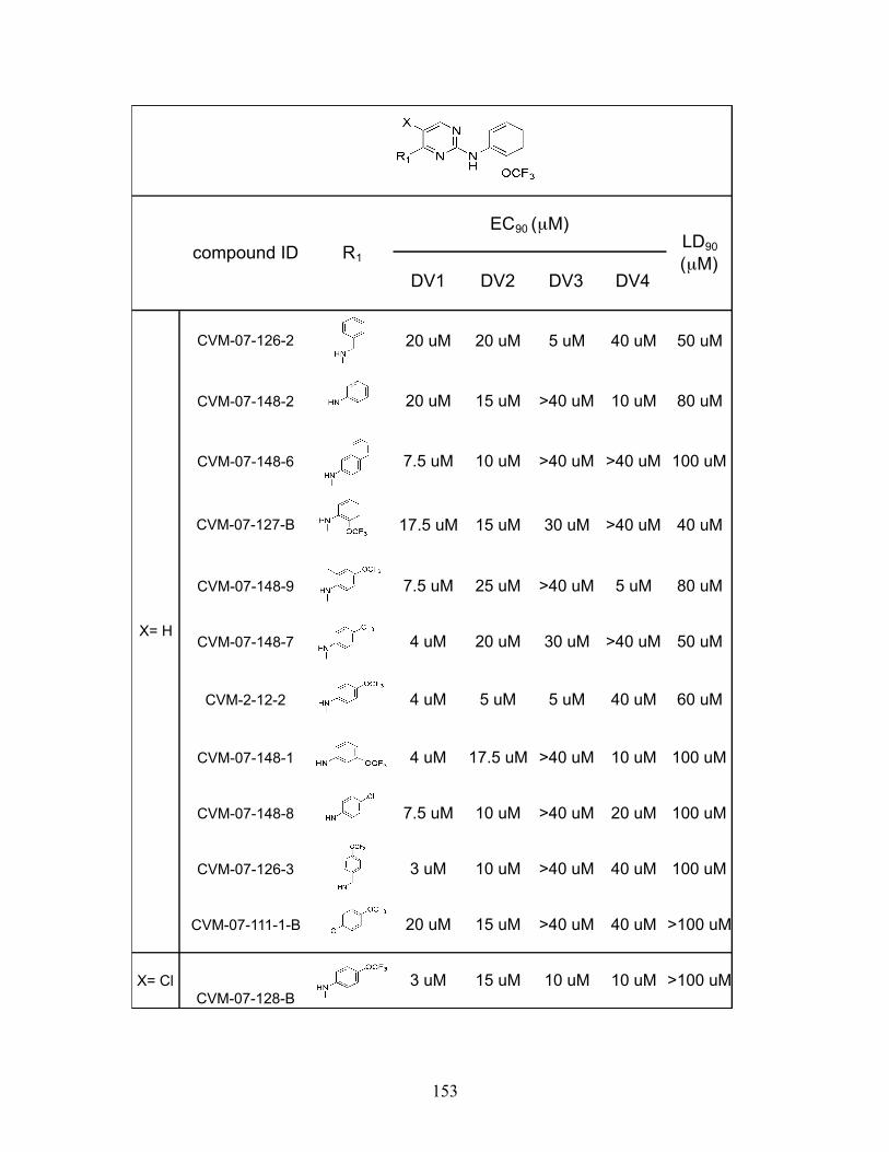
153
DV1 DV2 DV3 DV4
CVM-07-126-2 20 uM 20 uM 5 uM 40 uM 50 uM
CVM-07-148-2 20 uM 15 uM >40 uM 10 uM 80 uM
CVM-07-148-6 7.5 uM 10 uM >40 uM >40 uM 100 uM
CVM-07-127-B 17.5 uM 15 uM 30 uM >40 uM 40 uM
CVM-07-148-9 7.5 uM 25 uM >40 uM 5 uM 80 uM
CVM-07-148-7 4 uM 20 uM 30 uM >40 uM 50 uM
CVM-2-12-2 4 uM 5 uM 5 uM 40 uM 60 uM
CVM-07-148-1 4 uM 17.5 uM >40 uM 10 uM 100 uM
CVM-07-148-8 7.5 uM 10 uM >40 uM 20 uM 100 uM
CVM-07-126-3 3 uM 10 uM >40 uM 40 uM 100 uM
CVM-07-111-1-B 20 uM 15 uM >40 uM 40 uM >100 uM
X= ClCVM-07-128-B
3 uM 15 uM 10 uM 10 uM >100 uM
EC90 (µM)compound ID R1
X= H
LD90
(µM)
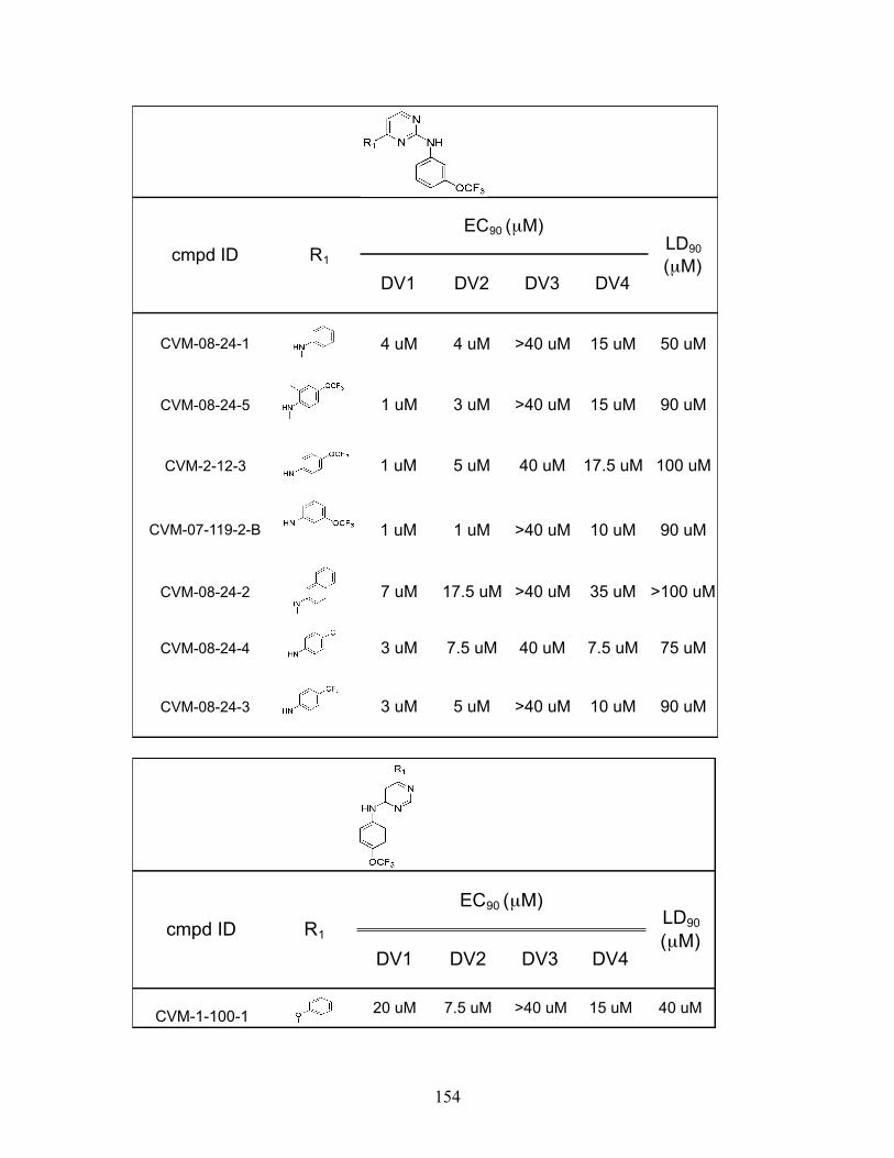
154
DV1 DV2 DV3 DV4
CVM-08-24-1 4 uM 4 uM >40 uM 15 uM 50 uM
CVM-08-24-5 1 uM 3 uM >40 uM 15 uM 90 uM
CVM-2-12-3 1 uM 5 uM 40 uM 17.5 uM 100 uM
CVM-07-119-2-B 1 uM 1 uM >40 uM 10 uM 90 uM
CVM-08-24-2 7 uM 17.5 uM >40 uM 35 uM >100 uM
CVM-08-24-4 3 uM 7.5 uM 40 uM 7.5 uM 75 uM
CVM-08-24-3 3 uM 5 uM >40 uM 10 uM 90 uM
EC90 (µM)LD90
(µM)cmpd ID R1
DV1 DV2 DV3 DV4
CVM-1-100-1 20 uM 7.5 uM >40 uM 15 uM 40 uM
EC90 (µM)cmpd ID R1
LD90
(µM)

155
DV1 DV2 DV3 DV4
CVM-2-12-4 1 uM 20 um 30 uM 5 uM >100 uM
CVM-07-120 7 uM 20 uM >40 uM 7.5 uM 75 uM
CVM-2-21-2 3 uM 3 uM >40 uM 30 uM >100 uM
EC90 (µM)LD90
(µM)cmpd ID R1