experimental models of hepatocellular carcinoma
TRANSCRIPT
www.elsevier.com/locate/jhep
Journal of Hepatology 48 (2008) 858–879
Review
Experimental models of hepatocellular carcinomaq
Philippa Newell1,2, Augusto Villanueva1, Scott L. Friedman1, Kazuhiko Koike3,Josep M. Llovet1,4,*
1Division of Liver Diseases, Mount Sinai School of Medicine, 1425 Madison Avenue, Box 1123, New York, NY 10029, USA2Department of Surgery, Mount Sinai School of Medicine, 1425 Madison Avenue, Box 1123, New York, NY 10029, USA
3Department of Infectious Diseases, Internal Medicine, Graduate School of Medicine, University of Tokyo, Japan4BCLC Group, Liver Unit, IDIBAPS, CIBERehd, Hospital Clı́nic, Barcelona, Spain
Hepatocellular carcinoma (HCC) is a common and deadly cancer whose pathogenesis is incompletely understood. Com-
parative genomic studies from human HCC samples have classified HCCs into different molecular subgroups; yet, the uni-
fying feature of this tumor is its propensity to arise upon a background of inflammation and fibrosis. This review seeks toanalyze the available experimental models in HCC research and to correlate data from human populations with them in
order to consolidate our efforts to date, as it is increasingly clear that different models will be required to mimic different
subclasses of the neoplasm. These models will be instrumental in the evaluation of compounds targeting specific molecular
pathways in future preclinical studies.
� 2008 European Association for the Study of the Liver. Published by Elsevier B.V. All rights reserved.
Keywords: Liver cancer; Hepatocellular carcinoma; Mouse models; Genetically engineered mice; Cirrhosis
0168-8278/$32.00 � 2008 European Association for the Study of the Liver.
doi:10.1016/j.jhep.2008.01.008
Associate Editor: M. Colomboq P. Newell is a recipient of an American Liver Foundation (ALF)
Postdoctoral Research Fellowship Award (2007). A. Villanueva issupported by grants from the Fundacı́on Caixa Galicia and theNational Cancer Center. S. Friedman is a Professor of Medicine andChief of the Division of Liver Diseases, supported by NIH Grantnumber DK37340. K. Koike is Chairman of Department of InfectiousDiseases, University of Tokyo, supported by grant from the Ministryof Health, Labor and Welfare, and Ministry of Education, Science,Sports and Culture of Japan. J.M. Llovet is Director of the HCCProgram in Mount Sinai and Professor of Research-ICREA in theHospital Clı́nic Barcelona, supported by National Institute of Health-NIDDK grant 1R01DK076986-01, National Institute of Health-I+DProgram (Spain) grant number SAF-2007-61898. The authors declarethat they do not have anything to disclose regarding funding fromindustries or conflict of interest with respect to this manuscript.
* Corresponding author. Address: Division of Liver Diseases, MountSinai School of Medicine, 1425 Madison Avenue, Box 1123, NewYork, NY 10029, USA. Tel.: +1 212 659 9503; fax: +1 212 849 2574.
E-mail address: [email protected] (J.M. Llovet).Abbreviations: HCC, hepatocellular carcinoma; HCV, hepatitis C
virus; TKR, tyrosine kinase receptor; HBV, hepatitis B virus; TSG,tumor suppressor gene; TSP, tissue specific promoter; Tg, transgene.
1. Introduction
Hepatocellular carcinoma is one of the world’s dead-liest cancers, ranking third among all cancer-relatedmortalities. Most cases occur in Asia and sub-SaharanAfrica, where viral hepatitis is endemic. The incidenceis rising in the West, likely due to the increase in patientsinfected with hepatitis C during the latter half of the lastcentury [1]. The liver, unique in its capacity for regener-ation following injury, also gives rise to this malignancycommonly associated with the inflammatory state ofadvanced fibrosis, or cirrhosis. Potentially curative ther-apies can be offered to approximately 30% of patients,but are complicated by a high rate of recurrence [2].
Encouraging progress has been made in understand-ing the molecular pathogenesis of cancer [1,2]. The dis-coveries of the signal transduction pathways, cascadesof protein–protein interactions transmitting informationfrom the cell surface to the nucleus, and of their link totumor biology, are particularly impressive.
Several key mouse models have been instrumental indefining the pathogenesis of HCC by introducing genetic
Published by Elsevier B.V. All rights reserved.

P. Newell et al. / Journal of Hepatology 48 (2008) 858–879 859
alterations into one or more aetiologic pathways thatcan be targeted exclusively to the liver. Moreover, theseprogrammed manipulations can be introduced systemat-ically, not only in this specific organ but also at definedtimes during development, growth and aging of theliver.
Nonetheless, substantial challenges persist in model-ing liver diseases whose natural history requires achronic inflammatory milieu. For example, infectious(hepatitis C virus), toxic (alcohol), metabolic (non-alco-holic steatohepatitis), or congenital (hemachromatosis)diseases share inflammation and fibrosis as precursorsto cancer, yet none is easily mimicked in animals. Thereare few rodent models of HCC arising spontaneouslywithin a background of regenerative nodules and cirrho-sis, and most depend on the administration of hepato-toxic and/or carcinogenic agents to recreate theinjury–fibrosis–malignancy cycle seen in chronic humanliver diseases.
Comparative genomic studies in human HCC sam-ples have begun to identify molecular subgroups withcharacteristic mutations, gene expression profiles andchromosomal gains and losses [3]. Moreover, since thereis no single dominant molecular pathology underlyingall HCCs, it is increasingly clear that different modelswill be required to mimic different subclasses of the neo-plasm. These models will be instrumental as pre-clinicaltools to evaluate compounds targeting specific molecu-lar pathways.
With these challenges in mind, the objective of thisreview is to assemble and evaluate the available modelsof both cirrhosis and HCC, to provide a blueprint forunderstanding the pathogenesis of HCC and for opti-mizing preclinical models for drug testing.
2. Experimental models in cancer research
Although many experiments focusing on liver physi-ology have been conducted in rats due to their propen-sity for the development of fibrosis, the laboratorymouse (Mus musculus) is considered among the bestmodel systems for cancer because of the availability ofgene targeting methods, as well as the animal’s sizeand breeding capacity, its lifespan of 3 years, and itsphysiologic and molecular similarities to human biology[4]. Significant advances have been made in modelingcancer genetics in mice, along a spectrum that rangesfrom simple xenograft models to more complex, geneti-cally modified mice. Examples of each of the followingare illustrated in Table 1.
2.1. Xenograft models
The demonstration that concentrated cancer cellsgrown in vitro could form tumors when implanted sub-
cutaneously into an immunocompromised mouse wasfirst established in 1969 [5]. This xenograft model hassince demonstrated several advantages that explain itspersistence as the mainstay of pre-clinical studies ofanti-neoplastic drugs in vivo: the tumors are rapidlyand easily induced, and their subcutaneous locationenables direct measurement of tumor growth. Morerecently, however, several critical differences betweenxenograft- and patient-derived specimens have becomeapparent, as discussed below. In addition, cancer isnow appreciated as a complex disease dependent uponthe interaction between transformed cells harboringoncogenic mutations, referred to as the ‘cell autono-mous compartment’, and their surrounding tumor envi-ronment, the ‘non-cell autonomous constituents’ madeup of normal cells, stromal cells, and immune cells [4],features that are not part of the xenograft approach.
Mouse models of cancer were first introduced over 60years ago. Shortly after its inception in 1955, the Devel-opmental Therapeutics Program at the National CancerInstitute (NCI) adopted the use of three transplantedrodent models of sarcoma, carcinoma, and leukemia,for the purposes of selecting agents for clinical use incancer patients. Thousands of molecules were tested inmice bearing murine leukemias during the first decadesof modern cancer drug development, circa 1945–1969[6]. This tumor panel was later expanded to includehuman tumor xenografts, with the intention to studydrug activity against solid tumors [7]. In 1990, theNCI focused on the development of in vitro assays in60 different cell lines in order to screen pharmaceuticalagents for their potency and their selective activityagainst either a particular disease category or specificcell line [8,9], the most promising of which were to besubsequently evaluated in the nude mouse xenograftmodel.
The validity of xenografts as a predictive indicatorof probable clinical activity is limited, with the mostsuccess seen in cytotoxic agents. A retrospective anal-ysis performed by the NCI for 39 compounds inwhich both xenograft testing and Phase II clinicaldata were available showing that less than 50% ofagents with activity in more than one-third of xeno-grafts showed clinical activity (p = 0.04) [6]. The samestudy demonstrated that activity in a particular histol-ogy in a tumor model did not closely correlate withactivity in the same human cancer histology [10], withthe exception of non-small cell lung and ovarian can-cer [11].
There are several variables inherent to the xenograftexperiments which may impact on the divergent out-comes compared to human disease, including growthproperties and size at initiation of treatment of xeno-graft tumor, ectopic versus orthotopic location oftumor, local versus metastatic disease [12], tolerancefor high doses of chemotherapeutic agents in mice [13],

Table 1
Available mouse models in cancer research
x
TSP Cre TgLoxP LoxP
Excised transgene onlyin special cell type; allother cells normal
TSP rTta
rTta
rTta
TetO Tg
TetO Tg
-Dox
+DoxTet On:
TissueSpecificPromoter
Oncogene
TissueSpecificPromoter
Dominant Negative Tumor Suppressor Gene
Technical Method Advanced Mouse Models of Cancer
Current Models in HCC Future Prospects: Wish List for HCC
Xen
og
raft
XenograftCOLON, BREAST, PROSTATE: Surgical orthotopic implantation: intact fragments of human cancer, including tumors taken directly from the patient, transplanted into the corresponding organ of immunodeficient rodents16
Orthotopic xenograft model in which hepatoma 129 cells originating from C3H mice are injected into fibrotic livers of mice pretreated with TAA and EtOH45
Mouse HCC cell line derived from GEM tumor with specific molecular pathway dysregulated, with immunofluorescent marker, injected into fibrotic liver of immune-competent mice
ConstitutiveTransgenic
PANCREATIC: KrasG12V and chronic pancreatitis169;Trp53R172H and KrasG12D double transgenic driven by insulin promoter170:two models of invasive and metastatic pancreatic cancer
Mouse C-myc/Human E2F-1 overexpression driven by albumin promoter171: HCC at 6-8 months
Double transgenic overexpressing profibrotic gene combined withliver-specific oncogene
Dominant Negative Transgenic
PITUITARY:Rb and p27Kip1 Cdk inhibitor tissue specific knockout mice develop pituitary tumors with different phenotypes172
Mdr-2 knockout mice are unable to secrete phospholipids into bile, and develop cholangitis and HCC at 6-12 months166
Double transgenic liver-specific E-cadherin knockout and β-catenin overexpression
Tra
nsg
enic
GE
M
InducibleTransgenic
MELANOMA:Double transgenic combining Tet-induced overexpression of mutated HrasV12G and Ink4a knockout173
Tet-inducible Met expression under albumin promoter: 60% HCC at 12 months; tumors regressed when transgene (Tg) was inactivated91
Tet-induced, liver-specific overexpression of known oncogene in fibrotic mice
En
do
gen
ou
s G
EM
Conditional Gene Targeting
PROSTATE:Double transgenic Cre-mediated PTEN-/-
homozygous loss and p19Arf +/-:cooperativity in cancer development174
Cre-mediated liver specific PTEN-/- knockout: 66% HCC at 8 months108
Cre-mediated, liver-specific knockout of known tumor suppressor gene in fibrotic mice
TSP tTa
tTa
tTa
TetO Tg
TetO Tg
-Dox
+DoxTet Off:
860 P. Newell et al. / Journal of Hepatology 48 (2008) 858–879
and variability in selected endpoints. These variables canbe minimized if given due consideration in the design ofpreclinical cancer drug experiments. However, the great-est discrepancies between success of cancer therapies inxenograft models and in human clinical trials are likelydue to critical differences in both the tumor cells andtheir microenvironment. Natural tumor progression isa micro-evolutionary process during which increasingly
aggressive clones, generated through genetic instability,emerge from an initially monoclonal lesion. Autochtho-nous tumors, those that evolve in situ from normal cells,tend to have a diminished genetic heterogeneity com-pared to tumor xenografts, although selective pressuresof cell culture or tissue explantation can cause a rapidexpansion of a certain clonal constituent of polyclonaltumors [14,15].

P. Newell et al. / Journal of Hepatology 48 (2008) 858–879 861
One solution to this disparity between cancer celllines and human tumors is surgical orthotopic implan-tation, in which intact fragments of human cancertaken directly from the patient are transplanted intothe corresponding organ of immunodeficient rodents,as reviewed by Hoffman [16]. This technique has beenapplied to breast, lung, and prostate cancer amongothers.
Additional advances have been made in the xenograftmodel through the addition of mesenchymal stem cellsto weakly metastatic cancer cell lines, which enhancesthe ability of the cell lines to form tumors and to metas-tasize [17]. Wu et al. were able to isolate a side popula-tion (SP) from 29 sarcomas which preferentially formedtumors when grafted into immunodeficient mice; onlycells from tumors that developed from the SP cells hadthe ability to initiate tumor formation upon serial trans-plantation [18].
Our deepening appreciation of the non-cell autono-mous constituents of the tumor microenvironment,including the stroma and immune cells relevant to liverpathology in particular, provides further evidence thatthe xenograft model is more appropriately termed ani-
mal culture, as suggested by Tuveson and Frese [4].
2.2. Genetically engineered mouse models (GEM)
The most sophisticated animal models of human can-cer are those that have been genetically engineered tomimic the pathophysiological and molecular featuresof human malignancies [4]. Such models enable theinvestigation of a range of discrete molecular stages thatoccur during tumor progression both within tumor cellsand within their microenvironment; additionally, miceharboring multiple mutations provide informationregarding pathway cooperativity and dependencyin vivo [19].
Despite these strengths, there are a number of impor-tant limitations in mouse models of cancer, such as var-iation in basic cellular processes, as well as in telomerelength and telomerase expression [20,21]. It is also welldocumented that identical genetic lesions can producedifferent pathologies in mice than in humans [22].GEM can be categorized as either transgenic or endog-enous models.
2.3. Transgenic models
Transgenic mice are those that are engineered toexpress either oncogenes or dominant-negative tumorsuppressor genes in a non-physiologic manner due toectopic promoter and enhancer elements [4,19]. Microin-jection of recombinant DNA directly into the pronu-cleus of a fertilized mouse egg is the classic method forgenerating transgenic mice [23], but transgenic micecan also be produced through gene targeting (‘‘knock-
in”) and lentiviral transduction in embryonic stemcells.
Constitutive expression of cellular and viral onco-genes and germline disruptions of tumor suppressorgenes were the first approaches used to create strainsof cancer-prone mice [19,24]. The cDNA constructscan contain promoter elements designed to restrict tissuetropism, so although the effect of the oncogenic gain willbe constitutive, its expression can be limited to specifictissues by the use of tissue-specific promoters [19], forexample the albumin promoter in liver transgenicmodels.
Germline tumor suppressor cell mutant mice were ini-tially developed to parallel human inherited cancer pre-disposition syndromes. However, although many ofthese heterozygous mice were tumor-prone and demon-strated loss of the wild-type allele in their tumors, few ofthem developed the clinical features of the cognatehuman syndrome. For example, loss of the retinoblas-toma gene product Rb in humans leads to retinoblasto-mas, osteosarcomas, and small cell lung cancer; whereasRb heterozygote mice develop thyroid and pituitarytumors but no retinoblastomas [25]. Rb heterozygotesare able to compensate for loss of Rb, a finding thathighlights the existence of shared and predictable cellu-lar process within both species [20,26]. So, althoughidentical genetic lesions may not perfectly recapitulatethe human disease in mice, there is no doubt that thesegenetically engineered mice are valuable tools for under-standing the underlying biological mechanisms oftumorigenesis [22]. Their ability to recapitulate thegenetic features of amplified proto-oncogenes, such asc-myc [27], has contributed greatly to our understandingof cancer biology.
There are, however, additional weaknesses of thesemodels that have spurred the development of moreadvanced methods. For example, because the genesaffected may be vital to normal development, over-expression or ablation may lead to embryonic lethalityor infertility [24]. Promoter fragments typically repre-sent the minimal sequence required for tissue-specificexpression and do not necessarily allow the same con-trol conferred by endogenous regulatory elements [28];for example, a typical transgene would not include alltranscription factor and microRNA binding sites[4,29]. And, although the DNA fragments are thoughtto associate by homologous recombination before inte-gration and in most cases insert at a single chromo-some site [23], there is little control over site ofintegration and copy number [22]. This can result inpronounced variability of expression patterns, as theexogenous gene can affect genes near its insertion siteor can be affected by endogenous control elements[22,30–32]. Also, although conventional mouse mutantsmay be useful for modeling familial forms of cancer,they do not mimic sporadic tumorigenesis because the

862 P. Newell et al. / Journal of Hepatology 48 (2008) 858–879
initiating mutation is present in all cells of the body,including those that constitute the tumor microenviron-ment [33].
2.4. Inducible systems of oncogene expression
Bujard and colleagues developed a strategy for tem-porally controlled and reversible transgene expression,using a tetracycline (tet-) inducible system [34]. Thesedrug- or ligand-inducible systems involve the use of achimeric transcriptional activator that reversibly acti-vates a target gene in response to the administration ofthe inducing agent.
The Escherichia coli tetracycline resistance operonhas been applied widely to generate cell lines and murinemodels with tightly regulated gene expression inresponse to tetracycline [35]. The tet transactivator func-tions either as a constitutive repressor that is induciblyinhibited by ligand to allow expression from the tet
operon (tTA), or it acts as an inducible activator ofthe tet operon upon ligand addition (rtTA) [19]. Thissystem has been particularly useful to study the conceptof oncogene addiction; nearly all oncogenes tested thusfar seem to be required not only for tumor initiation,but also for tumor maintenance [33].
2.5. Endogenous GEM: knock-out models
Endogenous GEM are those that lose the expressionof tumor suppressor genes (TSG) or that express domi-nant-negative tumor suppressor genes or oncogenesfrom their native promoters [4]. The original ‘knockout’mouse model entailed disruption of an allele in endoge-nous embryonic stem cells using a targeting vector.Biallelic disruption of TSG often results in embryoniclethality, but heterozygous mice can be used to deter-mine the tumorigenic potential of the genes, such asthe retinoblastoma tumor suppressor gene (Rb) [25].These germline mutations are present throughout themouse and are constitutively expressed, unlike the spo-radic mutations occurring in human tumors that are sur-rounded by normal tissue.
2.6. Endogenous GEM: conditional gene targeting
As reviewed by Maddison et al. [22], model systemshave now been developed which allow both spatialand temporal control of gene expression. These are pre-dominantly dependent on the creation of bi-transgenicmice: those carrying a tissue-specific, inducible transacti-vator gene are crossed to mice carrying the allele ofinterest which has been engineered to be controlled bythe transactivator. Offspring that carry both transgenicelements are treated with the inducer to express thetransactivator gene in a specific tissue, which then actson the desired allele. This system requires the exogenous
delivery of the cre gene (usually by an adeno- or retrovi-rus), and the induction is irreversible.
Conditional inactivation of tumor suppressor genesrelies on the ability of a viral or prokaryotic site-specificrecombinase to recognize a pair of target DNAsequences and catalyze recombination at these sites,which results in either deletion or inversion of the inter-vening DNA sequence [19]. A commonly used tool is theCre-Lox system, wherein Cre (Causes recombination)recombinase, isolated from bacteriophage P1, catalysessite-specific recombination between defined 34 bp LoxP sites (Locus v of crossover P1) [36,37]. If gene v isplaced between two Lox P sites and then exposed toCre, it will be excised, or ‘floxed out’. An alternative sys-tem to Cre-Lox uses the FLP recombinase, which recog-nizes the 48 bp Frt site [38]. Transgenic mice that expressrecombinase from a specific promoter are bred to micecarrying conditional tumor suppressor gene mutations,so that the TSG can be bi-allelically inactivated to allowthe generation of organ- and cell-lineage-specific tumorsmodels [19].
Conditional activation of oncogenes is created by theinsertion of a LoxP flanked transcriptional silencingelement between the promoter and the mutant onco-gene-encoding sequence. Conditional oncogenes areconstructed using classic transgenic technology, butexpression of the oncogene is only activated by therecombinase-mediated removal of the transcriptionalsilencer. This allows for tissue-specific oncogene expres-sion [39].
This second generation of GEM, which more faith-fully recapitulates sporadic tumor formation by theinduction of somatic mutations in a time- and tissue-specific fashion, has provided great insight into the con-tribution of genes in the initiation, progression, andtreatment of cancer. We will now discuss how each ofthese systems has been used to further our understand-ing of liver cancer.
3. Experimental models of hepatocellular cancer
Hepatocellular carcinoma universally arises upon abackground of inflammation and fibrosis. Creation ofanimal models of HCC presents a particular experimen-tal challenge because of the difficulty in modelingchronic inflammation without using carcinogens toinduce liver injury, and because of the heterogeneity ofmolecular pathways that are dysregulated during thistransition from cirrhosis to cancer.
HCC is preceded in both rodents and humans by thedevelopment of premalignant lesions including foci ofaltered hepatocytes and dysplastic nodules, which exhi-bit a higher risk of malignant evolution than normalcells [40,41]. Various genetic alterations and exposuresto chemical carcinogens have been studied in animals

P. Newell et al. / Journal of Hepatology 48 (2008) 858–879 863
in order to recapitulate the phenotypic, biological, andmolecular events that occur during this transformation.
3.1. Xenograft models of HCC
In a recent attempt to characterize primary humanxenografts in liver cancer, seven different primaryHCC cell lines were injected into SCID mice. The micewere then treated with common chemotherapeuticagents such as cisplatin and gefitinib. There were signif-icant differences in tumor growth inhibition betweenxenografts, which reinforced the concern for high inter-nal variability of this model in human cancer. Interest-ingly, the study concluded that most of thechemotherapeutic agents currently used in the treatmentof HCC have little or no anti-neoplastic activity in thesemodels [42].
Ma et al. have examined HCC cells expressing CD133[43], which exhibit stem cell properties and are chemore-sistant: purified CD133(+) HCC cells isolated fromhuman HCC cell lines and harvested from xenograftmouse models survived chemotherapy in increased pro-portions relative to most tumor cells which lack theCD133 phenotype [44]. The inclusion of stem cell-enriched HCC cell lines will likely enhance future pre-clinical studies in HCC therapeutics (see Table 1).
A group of investigators at the University HospitalBonn created an orthotopic xenograft model in whichhepatoma 129 cells originating from C3H mice wereinjected into fibrotic livers of mice pretreated with thi-oacetamide by intraperitoneal injection and alcoholper oral [45]. They found that tumors in fibrotic liversgrew significantly larger and more rapidly than thosein normal livers, and were able to metastasize and formsatellite nodules. Gene expression analysis revealedgreater intratumoral expression of vascular endothelialgrowth factor (VEGF) and its receptor (VEGFR), andof MMP-2 and MMP-9 in the fibrotic liver tumors. Thisuseful model provides a unique tool for testing drug effi-cacy in orthotopic hepatoma xenograft within the con-text of liver fibrosis.
3.2. Viral models of HCC
Infection causing latent or chronic viral hepatitis isthe most common aetiology of HCC, comprising 80%of cases worldwide. Hepatitis B virus (HBV) is endemicin China, Southeast Asia, and sub-Saharan Africa;there, vertical transmission of the virus results in highrates of HCC. Hepatitis C (HCV) viral infection is moreprevalent in the United States and Europe than eitherHBV or HIV [46]. The woodchuck hepatitis virus(WHV) induces a liver inflammation, injury and repairprocess in woodchucks similar to those of HBV-positivepatients and has therefore proven to be a useful modelof the disease.
3.2.1. Hepatitis B virus
HBV is a DNA virus that causes acute and chronichepatocyte injury, inflammation, and HCC. During pro-longed infection, viral DNA sequences integrate into thehost cell genome, where they and the flanking cellularsequences are commonly rearranged [47], a phenomenonthat can activate an adjacent cellular oncogene. In addi-tion, viral infection can induce hepatocyte injury medi-ated by the antiviral cellular immune response and, toa lesser extent, by direct injury to the cells. Althoughmost cases of HBV-associated HCC arise in a back-ground of inflammation and fibrosis, the virus is notori-ous for also causing HCC in the absence of cirrhosis,most likely by integrating into the host chromosomeand thereby promoting transcriptional transactivationof mitogenic factors.
The HBV virus is a circular DNA molecule contain-ing four open reading frames encoding four HB viralproteins: preS/S, preC/C, P and X protein (HBx). Themost common viral marker in HCC is the integrationof HBV genomic DNA encoding HBx. In 1994, Koikeet al. published their description of a transgenic mousemodel demonstrating that high levels of HBx expressionwere sufficient to generate HCC in 84% of male trans-genic mice at age 13–24 months [48] (see Table 2). Anal-ysis of proliferation and DNA content in these micesuggested that the continued expression of HBx gene ini-tiated tumor formation by inducing DNA synthesis andplacing large numbers of hepatocytes subjective to sec-ondary events for transformation [48]. Yu et al. alsoconfirmed the development of HCC in HBx transgenicmice [49]. Although another research group did notsee spontaneous HCC development, those HBx trans-genic mice were more susceptible to chemical carcino-genesis than control mice [50]. The reason for thisdiscrepancy is unclear, but the difference in genotypeof HBV should be noted: HCC tumors arose in geno-type C HBx transgenic mice but not in other genotypes[51].
Chisari et al. described a transgenic model thatoverproduces the hepatitis B virus large envelope poly-peptide and accumulates toxic quantities of hepatitis Bsurface antigen (HBsAg) [52]. This hepatocellularinjury initiates a programmed response within theliver, characterized by inflammation, regenerativehyperplasia, transcriptional deregulation, aneuploidyand eventually HCC. Inappropriate expression of asingle structural viral gene was thereby shown to besufficient to cause malignant transformation. The pro-cess of oncogenesis seen in this model also supportsthe theory that severe, prolonged cellular injury caninduce a proliferative response that fosters secondarygenetic events that lead to unrestrained growth [47].However, the level of viral protein expression in thismodel may well surpass the expression in humaninfection.
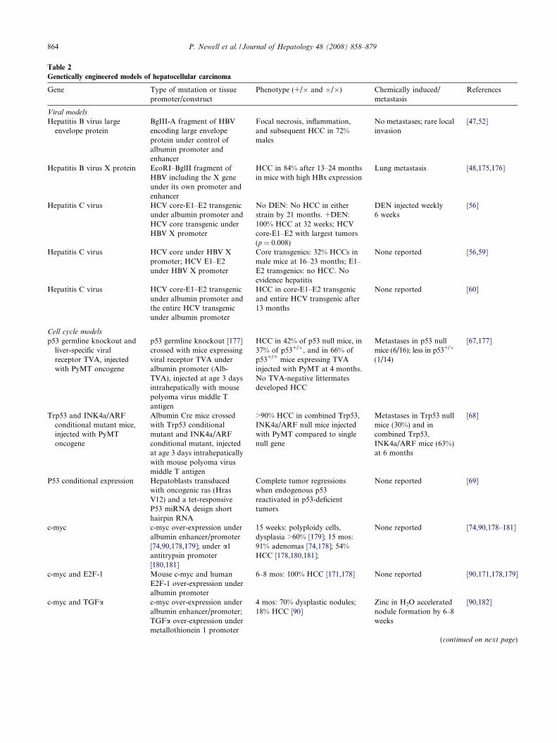
Table 2
Genetically engineered models of hepatocellular carcinoma
Gene Type of mutation or tissuepromoter/construct
Phenotype (+/� and �/�) Chemically induced/metastasis
References
Viral models
Hepatitis B virus largeenvelope protein
BgIII-A fragment of HBVencoding large envelopeprotein under control ofalbumin promoter andenhancer
Focal necrosis, inflammation,and subsequent HCC in 72%males
No metastases; rare localinvasion
[47,52]
Hepatitis B virus X protein EcoRI–BglII fragment ofHBV including the X geneunder its own promoter andenhancer
HCC in 84% after 13–24 monthsin mice with high HBx expression
Lung metastasis [48,175,176]
Hepatitis C virus HCV core-E1–E2 transgenicunder albumin promoter andHCV core transgenic underHBV X promoter
No DEN: No HCC in eitherstrain by 21 months. +DEN:100% HCC at 32 weeks; HCVcore-E1–E2 with largest tumors(p = 0.008)
DEN injected weekly �6 weeks
[56]
Hepatitis C virus HCV core under HBV Xpromoter; HCV E1–E2under HBV X promoter
Core transgenics: 32% HCCs inmale mice at 16–23 months; E1–E2 transgenics: no HCC. Noevidence hepatitis
None reported [56,59]
Hepatitis C virus HCV core-E1–E2 transgenicunder albumin promoter andthe entire HCV transgenicunder albumin promoter
HCC in core-E1–E2 transgenicand entire HCV transgenic after13 months
None reported [60]
Cell cycle models
p53 germline knockout andliver-specific viralreceptor TVA, injectedwith PyMT oncogene
p53 germline knockout [177]crossed with mice expressingviral receptor TVA underalbumin promoter (Alb-TVA), injected at age 3 daysintrahepatically with mousepolyoma virus middle Tantigen
HCC in 42% of p53 null mice, in37% of p53+/�, and in 66% ofp53+/+ mice expressing TVAinjected with PyMT at 4 months.No TVA-negative littermatesdeveloped HCC
Metastases in p53 nullmice (6/16); less in p53+/�
(1/14)
[67,177]
Trp53 and INK4a/ARFconditional mutant mice,injected with PyMToncogene
Albumin Cre mice crossedwith Trp53 conditionalmutant and INK4a/ARFconditional mutant, injectedat age 3 days intrahepaticallywith mouse polyoma virusmiddle T antigen
>90% HCC in combined Trp53,INK4a/ARF null mice injectedwith PyMT compared to singlenull gene
Metastases in Trp53 nullmice (30%) and incombined Trp53,INK4a/ARF mice (63%)at 6 months
[68]
P53 conditional expression Hepatoblasts transducedwith oncogenic ras (HrasV12) and a tet-responsiveP53 miRNA design shorthairpin RNA
Complete tumor regressionswhen endogenous p53reactivated in p53-deficienttumors
None reported [69]
c-myc c-myc over-expression underalbumin enhancer/promoter[74,90,178,179]; under a1antitrypsin promoter[180,181]
15 weeks: polyploidy cells,dysplasia >60% [179]; 15 mos:91% adenomas [74,178]; 54%HCC [178,180,181];
None reported [74,90,178–181]
c-myc and E2F-1 Mouse c-myc and humanE2F-1 over-expression underalbumin promoter
6–8 mos: 100% HCC [171,178] None reported [90,171,178,179]
c-myc and TGFa c-myc over-expression underalbumin enhancer/promoter;TGFa over-expression undermetallothionein 1 promoter
4 mos: 70% dysplastic nodules;18% HCC [90]
Zinc in H2O acceleratednodule formation by 6–8weeks
[90,182]
(continued on next page)
864 P. Newell et al. / Journal of Hepatology 48 (2008) 858–879

Table 2 (continued)
Gene Type of mutation or tissuepromoter/construct
Phenotype (+/� and �/�) Chemically induced/metastasis
References
SV40 T-antigen conditionaland inducible expression
SV40 T-antigen expressionunder albumin enhancer/promoter [74]; under majorurinary protein enhancer/promoter [183]; undermetallothionein 1 promoter[184]; under a1 antitrypsinpromoter [185]; underantithrombin III promoter[186]; tetracycline-inducibleexpression: mice expressingtTa under albumin promotercrossed with mice expressingT antigen under tTapromoter [75]
3–7 mos: adenomas and HCC[74]; 10–12 weeks: HCC[185];after 4–6 weeks: 100% HCC [186]
Lung metastases [186] [74,75,181,183–186]
E2F-1 E2F-1 over-expression undercontrol of albumin enhancer/promoter
10 mos: 100% adenomas anddysplastic nodules; 12 mos: 33%HCC
None reported [71,90,179]
Telomere dysfunction models
mTERT�/� and p53+/� orWT
Germline mTERT and p53knockout over severalgenerations and CCl4 liverinjury
50 weeks: 100% HCC in p53+/�
both generations (G0 and G3/G4); 44% in wild-type G0 versus9% HCC in wild-type G3/G4
CCl4 by IP injection 3�/week � 4 months
[66]
Pathway specific models
Wnt/b-cateninActivating mutation in
b-catenin: truncated NH2
terminal transgenic
EAB/9K/D N131 b-cateninconstruct under control ofliver-specific enhancer ofaldorase B gene (expressedthroughout embryonic andpost-natal development)
Death at 3 weeks fromhepatomegaly; no dysplastic fociin liver
N/A [127]
Activating mutation inb-catenin: exon 3conditional knockout
Catnblox(ex3) knockout andfatty acid binding proteinFabpl-cre transgenic
Death at 5 weeks from liverdamage/mitochondrial swelling.No dysplastic foci in liver;+intestinal polyps
N/A [128]
Activating mutation inb-catenin: exon 3conditional knockout
Catnblox(ex3) knockoutinjected with recombinantadenovirus expressing Crefrom human CMV promoter
High multiplicity injection (109
pfu/mouse): death at 3 weekswith hepatomegaly/mitochondrial swelling. Lowmultiplicity injection (107�8 pfu/mouse): No dysplastic foci inliver >6 mos
N/A [128]
b-catenin exon 3 knockoutand activated H-ras(H-rasG12V)
double-transgenicconditional
Catnblox(ex3) knockout andH-ras (Tglox(pA)H-ras*)double-transgenic withrecombinant adenovirusexpressing Cre from humanCMV promoter
Low multiplicity infection(108 pfu/mouse): 100% HCC at 6months
Intrahepatic invasion [131]
APC knockout liver-specific ApcDex14 knockout (�/�)injected in tails withrecombinant adenovirusexpressing Cre (injectionsinfected primarily andmassively the liver)
High multiplicity infection(109 pfu/mouse): Death within 2months and hepatomegaly.Lower multiplicity infection(0.5 � 109 pfu/mouse): 67% HCCat 9 months. Apc+/� had no liverabnormalities
None reported [130]
b-catenin wild-type b-catenin over-expressionunder control of albuminenhancer/promoter
Hepatomegaly (15% increasedliver/body weight ratio); nodysplastic nodules at 24 months
N/A [129]
P. Newell et al. / Journal of Hepatology 48 (2008) 858–879 865
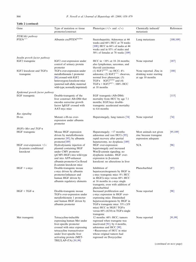
Table 2 (continued)
Gene Type of mutation or tissuepromoter/construct
Phenotype (+/� and �/�) Chemically induced/metastasis
References
PI3K/Akt pathway
PTEN�/� Albumin cre/PTENlox/lox Steatohepatitis; Adenomas at 44weeks and 66% HCC at 78 weeks[108]; HCC in 66% of males at 44weeks and in 83% of males and50% of females at 78 weeks [109]
Lung metastases [108,109]
Insulin growth factor pathway
IGF2 transgenic IGF2 over-expression undercontrol of urinary proteinpromoter
HCC in <10% at 18–24 months;also lymphomas, sarcomas, andthyroid carcinomas
None reported [187]
IGF2 knockout and TGFatransgenic
TGFa over-expression undermetallothionein 1 promoter[86] crossed with IGF2heterozygous knockout mice(paternal null allele; maternalwild-type, normally imprinted)
(1) IGF2wt/wt: no HCC; 4%adenoma; (2) IGF2+/�: dwarves,normal liver phenotype; (3)TGFa � IGF2wt/wt and (4)TGFa + IGF2wt/�: 100% HCCat 18 months
None reported. Zinc indrinking water startingat age 10 months
[188]
Epidermal growth factor pathway
EGF transgenic Double-transgenic of theliver construct Alb-DS4 thatencodes autocrine growthfactor IgEGF crossed withAAT-myc mice
EGF transgenic (Alb-DS4):mortality from HCC by age 7.1months; EGF/myc double-transgenic: accelerated mortalityto 4.4 months
[115]
Ras signaling
H-ras Mutant c-H-ras over-expression under albuminpromoter
Hepatomegaly, lung tumors [74] None reported [74]
HGF/c-Met and TGF-aHGF transgenic Mouse HGF expression
driven by metallothioneinpromoter [95]; by albuminpromoter[189]
Hepatomegaly; >17 months:adenomas and rare HCCs [95];rapid recovery after partialhepatectomy, no dysplasia [189]
Most animals not givenzinc because transgeneexpression adequate
[95,189]
HGF over-expression +/�b-catenin conditionalknockout
Hydrodynamic injection ofplasmid containing HGFunder CMV promoter(pCMV-HGF) into wild-typeand into AFP-enhanceralbumin promoter-Cre floxedb-catenin knockout mice
HGF over-expression:hepatomegaly and increasedWnt/b-catenin signaling; nodysplastic nodules. HGF over-expression in b-cateninknockout: no alterations in liver
N/A [96]
HGF + c-myc Double-transgenic mousec-myc driven by albuminpromoter/enhancer andhuman HGF driven byalbumin regulatory elements
Inhibition ofhepatocarcinogenesis by HGF inc-myc transgenic mice: 0% HCCin HGF/c-myc versus 60% HCCat 16 months in c-myc singletransgenic, even with addition ofphenobarbital
Phenobarbital [97]
HGF + TGF-a Double-transgenic mouseTGFa over-expression undermetallothionein 1 promoterand human HGF driven byalbumin promoter
Increased proliferation andc-myc expression in HGF over-expressing mice. Diminishedhepatocarcinogenesis by HGF inTGFa transgenic mice: 33% (3/9mice) HCC in HGF/ TGFaversus 60% (6/10) in TGFa singletransgenic
None reported [98]
Met transgenic Tetracycline-inducibleexpressing human Met underliver-specific promotercrossed with mice expressingtetracycline transactivatorunder liver-specific liveractivating protein (MET-TRE/LAP-tTA) [91,99]
12 months: 60% HCC; tumorsregressed when transgene wasinactivated [91]; by 4 months,adenomas and HCC [99].+Recurrence of HCC in micewhose original tumors hadregressed on Doxycycline
None reported [91,99]
(continued on next page)
866 P. Newell et al. / Journal of Hepatology 48 (2008) 858–879
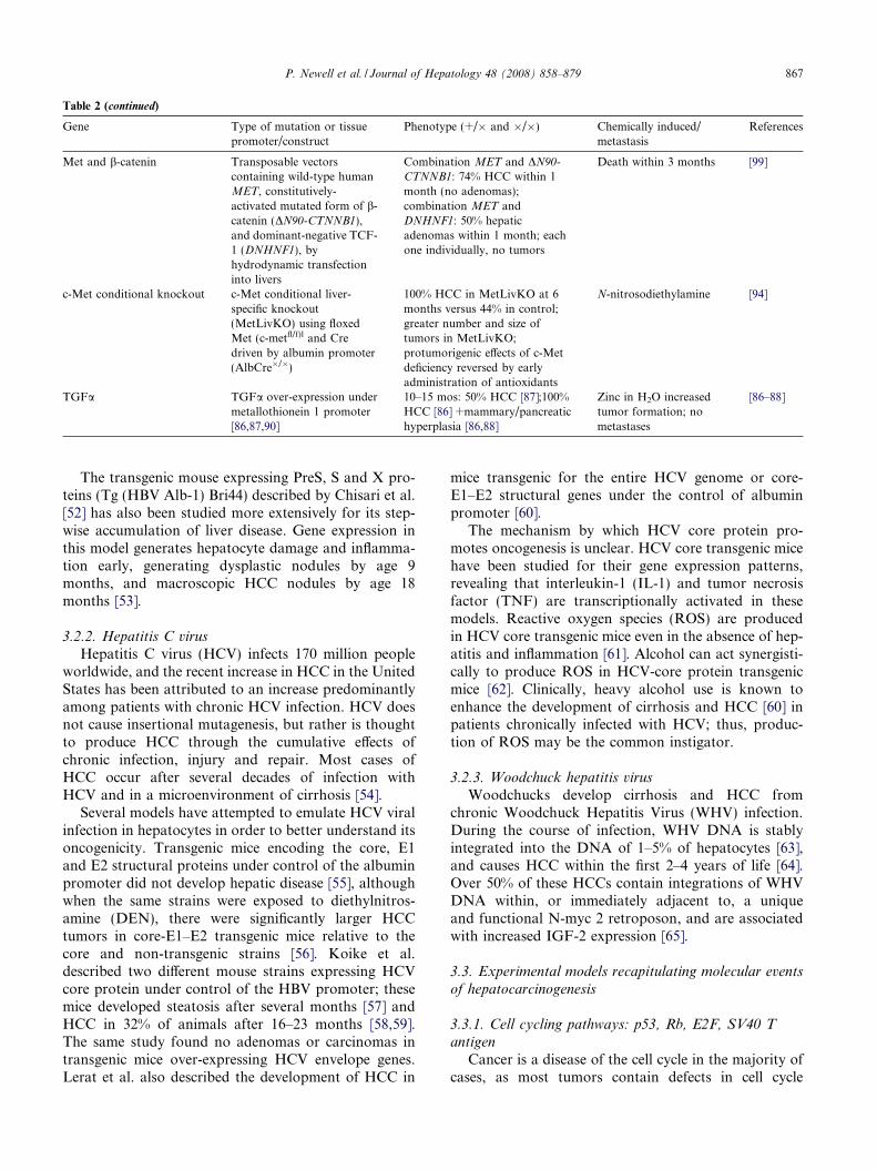
Table 2 (continued)
Gene Type of mutation or tissuepromoter/construct
Phenotype (+/� and �/�) Chemically induced/metastasis
References
Met and b-catenin Transposable vectorscontaining wild-type humanMET, constitutively-activated mutated form of b-catenin (DN90-CTNNB1),and dominant-negative TCF-1 (DNHNF1), byhydrodynamic transfectioninto livers
Combination MET and DN90-
CTNNB1: 74% HCC within 1month (no adenomas);combination MET andDNHNF1: 50% hepaticadenomas within 1 month; eachone individually, no tumors
Death within 3 months [99]
c-Met conditional knockout c-Met conditional liver-specific knockout(MetLivKO) using floxedMet (c-metfl/f)l and Credriven by albumin promoter(AlbCre�/�)
100% HCC in MetLivKO at 6months versus 44% in control;greater number and size oftumors in MetLivKO;protumorigenic effects of c-Metdeficiency reversed by earlyadministration of antioxidants
N-nitrosodiethylamine [94]
TGFa TGFa over-expression undermetallothionein 1 promoter[86,87,90]
10–15 mos: 50% HCC [87];100%HCC [86] +mammary/pancreatichyperplasia [86,88]
Zinc in H2O increasedtumor formation; nometastases
[86–88]
P. Newell et al. / Journal of Hepatology 48 (2008) 858–879 867
The transgenic mouse expressing PreS, S and X pro-teins (Tg (HBV Alb-1) Bri44) described by Chisari et al.[52] has also been studied more extensively for its step-wise accumulation of liver disease. Gene expression inthis model generates hepatocyte damage and inflamma-tion early, generating dysplastic nodules by age 9months, and macroscopic HCC nodules by age 18months [53].
3.2.2. Hepatitis C virus
Hepatitis C virus (HCV) infects 170 million peopleworldwide, and the recent increase in HCC in the UnitedStates has been attributed to an increase predominantlyamong patients with chronic HCV infection. HCV doesnot cause insertional mutagenesis, but rather is thoughtto produce HCC through the cumulative effects ofchronic infection, injury and repair. Most cases ofHCC occur after several decades of infection withHCV and in a microenvironment of cirrhosis [54].
Several models have attempted to emulate HCV viralinfection in hepatocytes in order to better understand itsoncogenicity. Transgenic mice encoding the core, E1and E2 structural proteins under control of the albuminpromoter did not develop hepatic disease [55], althoughwhen the same strains were exposed to diethylnitros-amine (DEN), there were significantly larger HCCtumors in core-E1–E2 transgenic mice relative to thecore and non-transgenic strains [56]. Koike et al.described two different mouse strains expressing HCVcore protein under control of the HBV promoter; thesemice developed steatosis after several months [57] andHCC in 32% of animals after 16–23 months [58,59].The same study found no adenomas or carcinomas intransgenic mice over-expressing HCV envelope genes.Lerat et al. also described the development of HCC in
mice transgenic for the entire HCV genome or core-E1–E2 structural genes under the control of albuminpromoter [60].
The mechanism by which HCV core protein pro-motes oncogenesis is unclear. HCV core transgenic micehave been studied for their gene expression patterns,revealing that interleukin-1 (IL-1) and tumor necrosisfactor (TNF) are transcriptionally activated in thesemodels. Reactive oxygen species (ROS) are producedin HCV core transgenic mice even in the absence of hep-atitis and inflammation [61]. Alcohol can act synergisti-cally to produce ROS in HCV-core protein transgenicmice [62]. Clinically, heavy alcohol use is known toenhance the development of cirrhosis and HCC [60] inpatients chronically infected with HCV; thus, produc-tion of ROS may be the common instigator.
3.2.3. Woodchuck hepatitis virus
Woodchucks develop cirrhosis and HCC fromchronic Woodchuck Hepatitis Virus (WHV) infection.During the course of infection, WHV DNA is stablyintegrated into the DNA of 1–5% of hepatocytes [63],and causes HCC within the first 2–4 years of life [64].Over 50% of these HCCs contain integrations of WHVDNA within, or immediately adjacent to, a uniqueand functional N-myc 2 retroposon, and are associatedwith increased IGF-2 expression [65].
3.3. Experimental models recapitulating molecular events
of hepatocarcinogenesis
3.3.1. Cell cycling pathways: p53, Rb, E2F, SV40 T
antigen
Cancer is a disease of the cell cycle in the majority ofcases, as most tumors contain defects in cell cycle

868 P. Newell et al. / Journal of Hepatology 48 (2008) 858–879
machinery. Fundamental to our understanding of can-cer biology have been models simulating loss of tumorsuppressors p53 and Retinoblastoma (Rb), key regula-tors of cell cycling and frequent targets of carcinogens.There is a large body of evidence indicating a pivotalrole for cell cycle deregulation during hepatocarcinogen-esis [41].
Tumor suppressor p53 acts to restrict proliferation inresponse to DNA damage or deregulation of mitogeniconcogenes, by leading to the induction of various cellcycle checkpoints, to apoptosis, or to cellular senes-cence. p53 heterozygous mutant mice appear to be sus-ceptible to HCC formation in the context of liverinjury, but only in the absence of intact telomerase [66].
Trp53 knockout mice develop larger, more invasivetumors than wild-type mice when mouse polyoma virusmiddle T antigen (PyMT) viral oncogene is introducedinto the liver under an albumin promoter [67]. Liver-spe-cific knockout of Trp53 when combined with liver-spe-cific PyMT expression also results in an invasive,metastatic phenotype. Concomitant loss of Ink4a/Arftumor suppressor locus accelerates this process [68].
Lowe and colleagues assessed the extent to which p53loss is required for maintaining established tumors [69].To do so, they first transduced hepatoblasts in vitro withoncogenic ras (HrasV12) and a tet-responsive p53shRNA (miR30 design short hairpin RNA), and theninjected the cells into the spleen of nude mice. Next, theyused RNA interference (RNAi) to conditionally regulatep53 expression in the nodules that had formed by trans-duced hepatoblasts seeding in the liver. The authorsconcluded that p53 loss can be required for the mainte-nance of aggressive carcinomas, and that the cellularsenescence program can act together with the innateimmune system to potently limit tumor growth.
The retinoblastoma (Rb) pathway plays its role in cellcycle regulation by guarding and triggering DNA repli-cation and cell cycle division in late G1. Rb binds mem-bers of the E2F family, and in doing so repressestranscription of E2F regulated genes, which mediateDNA synthesis and cell cycle regulation [70]. Afternoting upregulation of E2F in liver tumors from theirc-myc/TGF-a double-transgenic mice, Conner et al.generated E2F transgenic mice under control of thealbumin enhancer/promoter [71]. All of these miceformed adenomas after 10 months, and a minoritydeveloped HCC (2/6). When crossed with c-myc trans-genic mice, HCC development was accelerated, with100% tumor formation within 6–8 months. Furtherinvestigation of this model revealed activation of theWnt/b-catenin pathway in a majority of the tumors, asdemonstrated by accumulation of nuclear b-catenin; thisoccurred in the absence of mutations of b-catenin [72].
SV40 (Simian Vacuolating Virus 40) large T antigen(TAg) is an oncoprotein derived from the polyomavirusSV40 which is capable of transforming a variety of cell
types. The transforming activity of TAg is due mainlyto its perturbation of the retinoblastoma (pRB), p53and p105 tumor suppressor proteins. This causes thecells to leave G1 phase and enter into S phase, whichpermits DNA replication of both the cell and the viralgenome [73]. In addition, TAg binds to several other cel-lular factors, including the transcriptional co-activatorsp300 and CBP, which may contribute to its transform-ing capacity. SV40 T-antigen expression under the albu-min enhancer/promoters provoked the appearance ofadenomas and HCC within 3–7 months [74]. A tetracy-cline-inducible binary transgenic mouse model of SV40was found to develop hepatic neoplasia in 60% of cases(3/5); no neoplasia was observed in mice with suppres-sion of transgene expression by tetracycline administra-tion [75].
3.3.2. Telomere dysfunction
Telomeres are regions of DNA near the ends ofeukaryotic chromosomes that act to prevent loss ofgenetic information during chromosomal replication.They are synthesized and maintained by telomerase,part of a group of enzymes called TERT (telomerasereverse transcriptases). Because of cell division mecha-nisms and because telomerase expression is repressedin most human cells (with the exception of stem cellsand some leukocytes), telomere length decreases witheach cell division. Once telomeres reach a critically shortlength, they unfold; this uncapping is detected and thecell undergoes senescence (the ‘‘Hayflick limit”) [76].Neutralization of p53 or Rb function results in contin-ued telomere attrition, culminating in chromosomalinstability and cell death [77]. Low levels of telomeraseare associated with aging and tumorigenesis in sometumors such as colorectal cancer [78] but levels are typ-ically increased in HCC [79,80].
Telomere attrition has been documented in hepato-cytes from cirrhotic patients [81,82]. It is thought thatrepeated rounds of hepatocyte injury and regenerationmay promote telomere shortening, which would ulti-mately lead to chromosomal instability (CIN), a com-mon feature of HCC. Indeed a correlation betweenCIN, telomere shortening, and HCC was demonstratedin a series of 39 patients with HCC by analysis of liverbiopsies for ploidy and telomere length [83].
In mice, reduction in telomere length is not observed,probably due to long initial telomere length and active tel-omerase expression [84]. However, in p53-mutant mice,deficiency of telomerase promotes formation of non-reci-procal translocations and epithelial cancers [85]. Thecooperative roles of telomerase-induced chromosomalinstability and attenuated p53 function in the liver wasillustrated by a study which showed enhanced HCC for-mation in p53-mutated telomerase knockout mice(mTERT�/�). In the setting of intact telomeres, however,p53 mutation had no effect on tumor formation [66].

P. Newell et al. / Journal of Hepatology 48 (2008) 858–879 869
3.3.3. Growth factor signaling pathways
3.3.3.1. TGF-a and c-myc. Transforming Growth Factor(TGF)-a binds and activates EGFR and is mitogenictoward hepatocytes. In most organs, metallonein-drivenover-expression of TGF-a causes epithelial hyperplasia[41]. In liver and breast tissue, the phenotype extendsto neoplastic transformation. Tumor incidence is 100%in susceptible mice strains after a substantial latency[86–88]. Gefitinib, an EGFR inhibitor, significantlyreduces HCC development in rats with cirrhosis inducedby DEN administration [89].
Co-expression of TGF-a and c-myc can occur inHCC. Liver-specific c-myc over-expression induces per-sistent hepatocyte proliferation and eventual HCC.When c-myc and TGF-a are co-expressed, this processis accelerated [90].
3.3.3.2. Hepatocyte growth factor and c-Met pathway.
When stimulated by its ligand, hepatocyte growth factor(HGF) elicits multiple biological responses includingproliferation, migration, invasion, and morphogenesis[91]. Over-expression, amplification, and mutation ofthe MET proto-oncogene which encodes protein tyro-sine kinase receptor Met have been demonstrated inhuman HCC samples [92,93]. Nevertheless, experimen-tal mouse models of HCC have revealed that the netoutcome of HGF/c-Met activation could be either stim-ulation or inhibition of hepatocarcinogenesis [94].Transgenic mice over-expressing HGF driven by themetallothionein promoter developed HCC [95], butwhen HGF expression was driven by the CMV pro-moter, mice developed hepatomegaly but not dysplasia[96]. Inhibition of hepatocarcinogenesis by HGF in c-myc transgenic mice was demonstrated by Thorgeirssonet al. in 1996: none of the liver-specific HGF/c-mycover-expressing mice developed HCC and only 30%developed adenomas, versus HCC in 60% of the c-mycsingle transgenic, even with addition of phenobarbital[97]. Similarly, HGF co-expression inhibited tumor for-mation in TGF-a transgenic mice [98].
The paradoxical effects of HGF ligand expression aremirrored in Met receptor expression. Bishop and col-leagues demonstrated that over-expression of wild-typeMet in hepatocytes of transgenic mice leads to the devel-opment of HCC [91]. Interestingly, these mice werefound to have frequent activating mutations of b-cate-nin, and it was subsequently discovered that there wasa correlation between MET activation and b-cateninmutations in human HCCs. Spurred by these findings,vectors of human MET and b-catenin with activatingmutations were hydrodynamically cotransfected: thesemice developed larger HCCs with short latency periods,confirming a cooperative relationship between MET
over-expression and b-catenin mutations [99].Recently, however, Takami et al. reported that loss of
c-Met signaling enhanced rather than suppressed the
early stages of chemical hepatocarcinogenesis [94]: C-met conditional knockout (MetLivKO) mice treatedwith N-nitrosodiethylamine developed significantlymore and bigger tumors and with a shorter latency com-pared with control mice. These knockout mice hadincreased oxidative stress demonstrated signs of wasreversed by administration of antioxidant N-acetyl-L-cysteine. The authors concluded that intact HGF/c-Met signaling is essential for maintaining normal redoxhomeostasis in the liver. Further studies will be neededbefore definitive conclusions can be drawn regardingthe role of HGF/c-Met signaling in HCC.
3.3.3.3. PTEN/Akt/mTOR signaling pathway. The ser-ine/threonine kinase Akt (PKB) was first isolated asan oncogene transduced by the acute transforming ret-rovirus [100,101]. Its role in human cancer was estab-lished shortly thereafter by demonstration of itsfrequent amplification and over-expression in variouscancers, including breast and ovarian [102]. Akt actsas a cytoplasmic central regulator of numerous signalsrelated to cell cycling (Cyclin D1), cell survival(Mdm2/p53), cardiovascular homeostasis (eNOS), andcell growth (mTOR), among others [103]. PTEN is anegative regulator of the pathway and its loss activatesAkt.
Tissue-specific knockout models of PTEN in pan-creas develop tumors with high penetrance [104]. Trans-genic animals over-expressing Akt develop a hyperplasicbut not malignant phenotype, typically requiring a sec-ond hit to generate cancer [105,106]. Notably, mTORinhibition can reverse these phenotypes, suggesting thepresence of an mTOR-dependent survival signal down-stream of Akt [107]. Liver-specific deletion of PTENresults in hepatomegaly and steatohepatitis by 10 weeksand HCC in a majority of male mice by 20 months[108,109].
3.3.3.4. IGF and EGF signaling pathway. The insulingrowth factor (IGF1 and IGF2) signaling pathways reg-ulate cell growth, differentiation and survival, and play acentral role in embryogenesis and regulation of lifespan.IGF-2 possesses both mitogenic and metabolic proper-ties; 16–40% of human HCCs demonstrate over-expres-sion of IGF-2 [110].
The coordinated expression of IGF-2 and its receptorsuggests a role for IGF-2R in regulation of extracellularIGF-2 concentration; alterations in the expression ofIGF-2R in human tumors suggest it may act as a tumorsuppressor gene [111].
Transcriptional activation of IGF2 has been demon-strated in HCCs arising in HBV-associated human sam-ples [112] and in HBV transgenic mice [41]. Toinvestigate whether IGF-2 has a promoter role in aslowly developing HCC model, TGFa transgenic micewere crossed with IGF-2 hemizygous knockout mice

870 P. Newell et al. / Journal of Hepatology 48 (2008) 858–879
containing either only one maternal allele or two alleles.Imprinting usually blocks IGF-2 expression from thematernal allele in liver. However, IGF-2 re-expressionoccurred in all 4 of these models, and was chronologi-cally associated with late stages of progression towardHCC [113].
Epidermal growth factor (EGF) is a potent mitogento hepatocytes. Unlike in other malignancies, the EGFreceptor is rarely mutated in HCC, and several reportssuggest an EGF-mediated autocrine growth stimulationof hepatoma cells [114]. This was further supported bythe accelerated liver tumor formation after constitutiveover-expression of a secretable form of EGF (IgEGF).All double-transgenic mice with liver-specific IgEGFover-expression in cooperation with AAT-myc diedby 4.4 months from HCC, whereas only 44% ofATT-myc mice had developed HCC by age 14 months[115].
3.3.3.5. Wnt/b-catenin pathway. A key pathway impli-cated in hepatic tumorigenesis is the canonical Wntpathway, in which b-catenin acts as a co-activator ofthe TCF/LEF family of transcription factors and regu-lates the expression of several genes related to cell prolif-eration and apoptosis. The Wnt/b-catenin signalingpathway normally functions in cellular differentiation,proliferation, and apoptosis, and has a fundamental rolein embryogenesis. Liver development in xenopus, zebra-fish, and mouse embryogenesis has been shown to bedependent on functional Wnt signaling [116,117].
There is general agreement that Wnt signaling isupregulated in a subset of HCCs [118]. Mutations ofgenes encoding several components of the Wnt pathwayhave been described, including b-catenin (19–44%),AXIN1 and AXIN2 (5–14% and 3–10%) [119–123].The mutations of b-catenin identified in HCC arelocated in exon 3 of the CTNNB1 gene, the phosphory-lation site for GSK3a/b. In addition, immunohistologi-cal studies have demonstrated abnormal cytoplasmicand nuclear accumulation of b-catenin in 17–40% ofhuman HCCs [124,125]. In addition to accumulatedmutations, stimulation of proliferation in liver cancercell lines transfected with Hepatitis C core viral proteinis at least partially mediated by upregulation of Wnt-1protein expression [126]. This correlation betweenHCV and the Wnt pathway needs to be verified byin vivo studies.
Although mutations in b-catenin are thought to betumorigenic in human HCCs, transgenic mouse modelsover-expressing either a stable mutant form of b-catenin[127,128] or a constitutively activated, non-mutatedform of b-catenin exhibit hepatomegaly, but no HCC[127–129]. Surprisingly, although mutations in thetumor suppressor APC are very rarely seen in HCCand patients with germline APC mutations do not typi-cally develop HCC, it has been found that liver-targeted
loss of APC in mice can lead to HCC through activationof b-catenin signaling [130].
It seems that a second hit from an additional muta-tion is required to generate tumors in b-catenin trans-genic mice. Simultaneous co-expression of a Wnt-activating b-catenin mutation (Catnblox(ex3)) and muta-tion in H-ras introduced by adenovirus-mediated Creexpression resulted in HCC in 100% of the double-trans-genic progeny [131]. The interplay between the growthfactor signaling pathways and the Wnt/b-catenin path-way was amply illustrated in the simultaneous over-expression of HGF and b-catenin knockout mousemodel generated by Monga and colleagues [96]: the pro-liferative effects of HGF over-expression were mediatedby b-catenin stabilization, and were negated in b-cateninnull mice.
3.3.4. Other HCC models
3.3.4.1. Fibroblast growth factor in muscle. While mostmouse models of HCC express growth factors and onco-genes under liver-specific promoters, liver-specificexpression is not a requirement for development ofHCC. For example, a transgenic model over-expressingfibroblast growth factor 19 (FGF19) in skeletal muscledevelops HCC in 53% of mice by age 10–12 months[132]. Interestingly, unlike the vast majority of bothhuman tumors and murine models, these tumors aremore common in female progeny. Hepatocellular prolif-eration was significantly increased in these mice and innon-transgenic mice injected with FGF19 protein. Fur-thermore, immunostaining for b-catenin revealednuclear staining in 4/4 female mouse tumors, and subse-quent sequencing of the GSK3b phosphorylation site ofb-catenin revealed mutations in 16%, which implicatesactivation of the Wnt/b-catenin signaling pathway as apotential mechanism for hepatocellular transformationin this model.
3.3.4.2. Urokinase-type plasminogen activator. Not allgenetically modified models of HCC arise from pre-dicted oncogene over-expression, tumor suppressor loss,or liver injury. In a transgenic model over-expressingthe urokinase-type plasminogen activator (uPA) trans-gene under the albumin promoter, for example, mostmice died from liver hemorrhage within 4 days of birth;in the two transgenic lineages developed from survivingfounder mice, there was a surprising 100% incidence ofHCC at 8–20 months of age. Moreover, the survivingmice regained normal clotting function, and their liverswere repopulated by clonal, regenerative nodules that nolonger expressed the transgene. Tumor progenitor cellswere found to contain transgene-deleting chromosomalrearrangements which likely extended into flankingDNA. Therefore, the initiating event in this HCC modelwas likely extensive DNA rearrangements occurringduring rapid regeneration [133].

P. Newell et al. / Journal of Hepatology 48 (2008) 858–879 871
3.4. Chemically-induced fibrosis and hepatocarcinogenesis
Cirrhosis is a major cause of mortality as both a pre-cursor to malignancy and a cause for liver failure. As adisease with clear environmental non-hereditary compo-nents to its aetiology, liver fibrosis and cancer is wellsuited for modeling using chemical induction. Experi-mental models of liver disease can be categorized ascholestatic, nutritional, alcoholic, immunological, andtoxic, and have been reviewed elsewhere [134].
Briefly, several hepatotoxic agents have been usedboth in the induction of generalized liver disease andHCC (see Table 3). Chemical models of hepatocarcino-genesis often involve initiation by a carcinogen followedby a growth stimulus promoter to induce clonalexpansion of initiated cells, such as partial hepatectomy(Solt–Farber method [135]) or phenobarbital [136].Alternatively, rodents are subjected to repeated adminis-tration of carcinogens such as DEN, DMN, or CCl4over a prolonged period [136]. Most initiated cellsaccrue damage and ultimately undergo apoptosis, butthe small number that respond to promoters evolve intodysplastic foci and later to dysplastic nodules. These fociand nodules can disappear following the removal ofpromoters in a process termed remodeling, which typi-cally involves apoptosis of the preneoplastic cells [41].Nodules which have acquired the capacity for autono-mous growth progress to neoplastic nodules and HCC,an irreversible process involving the accumulation ofgenomic damage [137].
The most commonly employed model for liver diseaseis carbon tetrachloride (CCl4) administered in drinkingwater, in inhaled gases, or by intraperitoneal injection.The reactive metabolite trichloromethyl radical is pro-duced during the oxidative metabolism of CCl4 by cyto-chrome p450, and causes liver damage by elicitingproduction of reactive oxygen intermediates and by per-
Table 3
Toxic models of liver fibrosis and HCC
Diet or chemical Mechanism of action Phen
Choline-deficient and ethionine(CDE) diet
Oxidative DNA damage, DNAstrand breaks and chromosomalinstability [41]
30–35
Ciprofibrate Synthetic peroxisomeproliferators, non-genotoxiccarcinogen
60 we
Diethylnitrosamine (DENA) Genotoxic hepatocarcinogen 100%chrom
Thioacetamide (TAA) Metabolites induce oxidativestress
100%
2-Acetylaminoflouren (2-AAF) Genotoxic Usedproto
Phenobarbital Non-genotoxic Usedincreamice[197]
oxidative degradation of membrane phospholipids [138].Compounds like phenobarbital, ethanol, and acetoneinduce microsomal cytochrome p450 and thereforepotentiate the hepatotoxicity of CCl4, as does hypoxia;therefore, hepatocellular injury and necrosis are pre-dominantly seen in the centrolobular zone where theoxygen tension is low [134,138].
Dimethylnitrosamine (DMN) is a carcinogenic agentwhich causes liver injury by covalent binding and meth-ylation of nucleic acids and proteins in hepatocytes[139]. Animals administered DMN either per oral orby intraperitoneal injection develop cirrhosis within 3–4 weeks, and can continue to have stable or progressivedisease for several months after discontinuation of theagent [140].
Diethylnitrosamine (DEN) induces pericentral foci ofsmall dysplastic hepatocytes and acts by ethylatingnucleophilic sites in DNA [141,142], causing cirrhosisand multifocal HCC within 18 weeks [89,143]. Frequentb-catenin mutations have been found in HCCs inducedby DENA in mice [144], and when combined with amethyl-deficient diet, DEN administration generatesp53 mutation or rearrangement in rats [145].
Thioacetamide (TAA) in drinking water (0.03%) orby intraperitoneal injection induces fibrosis in rats andmice over a period of 2–3 months, which may be second-ary to the oxidant properties of TAA and the inductionof hepatic oxidative stress [134,146,147]. Acute liverinjury and subsequent fibrosis can be created by admin-istration of D-galactosamine (GalN), a hepatotoxin thatinduces liver damage by depleting uridine nucleotidesand therefore diminishing RNA and protein synthesis[148].
Cholestatic cirrhosis has been induced by extrahe-patic bile duct ligation (BDL) in rats, rabbits, dogs,and monkeys. Histologically, the BDL model is charac-terized by infiltration of connective tissue in the portal
otype References
weeks: 100% HCC [135,190–192]
eks: 100% HCC [193] [90,193,194]
HCC in males, 30% in females. Extensiveosomal damage
[90,168,194,195]
HCC [134,146]
primarily as promoter in initiation/promotioncols
[194,196]
as promoter in initiation/promotion protocols;ses HCC by 500%. Can inhibit tumor formation in
given DEN. Associated with b-catenin activation
[197,198]

872 P. Newell et al. / Journal of Hepatology 48 (2008) 858–879
zone and proliferation of bile duct epithelial cells andhepatocytes. This methods allows rapid four-weekinduction of cirrhosis, and the mortality is high [134].
Choline-, methionine-deficient diets administeredover 3–12 week periods induce cirrhosis and HCC inrats and mice, even when followed by an adequate diet[41]. Injury in these diets is most likely attributable todepletion of hepatic antioxidant mechanisms, such asreduced glutathione, which leads to oxidative DNAdamage, inflammation and fibrosis [41,149]. Histologicchanges seen in rodents fed this diet include periportalfatty liver, focal hepatocyte necrosis, oval cell prolifera-tion, infrequent cirrhosis [150] and HCC [151]. The var-iation in animal susceptibility to choline deficiency is adisadvantage to this model [134].
3.5. Models of liver fibrosis and HCC: creating a tumor
environment
The tumor microenvironment is emerging as a funda-mental determinant of oncogenesis and metastasis. Theliver presents an ideal organ in which to study the inter-action between tumors and their microenvironment, ashepatocellular carcinoma (HCC) develops in a back-ground of liver fibrosis in about 90% of cases. Whilethe notion that the tumor microenvironment may help
Table 4
Genetically modified models of liver fibrosis, inflammation, and HCC
Gene Type of mutation or tissuepromotor/construct
Phenotype
TGF-b Porcine TGF-b over-expressionunder albumin promoter
Early death dintestinal manmild fibrosis
TGF-b inducibletransgenic
Fusion CRP/TGF-b1 under CRPpromotor, induced by LPSinjection
Collagen depweeks
ELF+/� knockout ELF+/� knockout mice SteatosisPDGF-B PDGF-B over-expression using
Cre-LoxP under albuminpromoter; made Tamoxifene-inducible by breeding with miceexpressing Cre undertransthyretin receptor promoter
100% liver fibweeks
PDGF-C Human PDGF-C expressiondriven by albumin promoter
Fibrosis and
IL-6 knockout IL-6 knockout (IL-6�/�) Hepatocyte ncompensatorydecreased in I
MyD88 knockout MyD88�/� Diminished pMyD88�/� m
Alpha-1-antitrypsin(AAT)
Transgenic mice using AAT Zgenomic clones
High copy Zaccumulationreticulum; he
Mdr-2 Mdr-2 gene knockout Early: non-suinflammatory
Acox1�/� Fatty acyl-CoA oxidase null(AOX�/�) [167]
Steatohepatitregeneration
instigate tumor formation is gaining acceptance, themanner in which this occurs remains a mystery. In addi-tion to the traditional toxic method of inducing fibrosisin rodents, there are numerous transgenic models thathave been designed to recapitulate the phenotype ofchronic inflammation leading to fibrosis and HCC seenin humans (see Table 4).
Stellate cell transactivation is a hallmark of hepaticfibrogenesis. Many genetic models of liver fibrosis havefocused on the over-expression of TGF-b, a major fibr-ogenic factor that drives matrix deposition from acti-vated stellate cells [152]. Sanderson et al. generatedtransgenic mice containing a fusion gene (Alb/TGF-b1) under the control of the regulatory elements of themouse albumin gene; these mice developed mild fibrosisby 12 weeks, and rarely developed cirrhosis [153]. Simi-lar mild to moderately fibrotic phenotypes have beendemonstrated by other investigators [154,155]. Whenexposed to thioacetamide, TGF-b1-over-expressingtransgenic mice develop fibrosis at an accelerated rate[155], and develop HCC more frequently than wild-typemice (9/9 versus 4/10 mice at 9 months) [156].
Intracellular signaling from TGF-b occurs throughsignaling members TGF-b receptor type II (TBRII),SMAD2, SMAD4, and SMAD adaptor, which aretumor suppressors in gastrointestinal cancers. None of
Dysplasia or HCC References
ue to extra-ifestations [153];
[155,156]
100% HCC in transgenic micetreated with TAA [156]
[153,155,156]
osition at age 6 None reported [154]
40% HCC at >15 months [157,199]rosis at age 4–6 None reported [159]
steatosis 80% HCC at 12 months [160]
ecrosis andproliferation both
L-6�/� mice
<10% HCC in IL-6�/� micecompared to 100% HCC at 8months in male WT mice; 13%HCC in female WT mice
[162,163]
roduction of IL-6 inice
Suppression of DEN-inducedHCC: MyD88�/� mice had fewersmaller HCCs than WT mice
[162,163]
lineage: AATin endoplasmic
patitis and HCC
82% HCC at 16-18 months [164]
ppurativecholangitis
HCC at 6–12 months with +lungmetastasis [166]
[165,166]
is followed by 100% HCC at 15 months [167] [167]

P. Newell et al. / Journal of Hepatology 48 (2008) 858–879 873
the SMAD mutant models have developed HCC, how-ever. SMAD function is dependent upon adaptor pro-teins such as embryonic liver fodrin (ELF), a b-spectrinprotein. ELF associates with SMAD3, SMAD 4, andthe TGF-b receptor complex, and ultimately leads totheir translocation to the nucleus. Mishra et al. reportthat ELF+/� knockout mice develop steatosis and spon-taneous HCC. Loss of ELF in these mice results in cellcycle disruption with significant increases in Cdk4, cyclinD1 and pRb hyperphosphorylation [157].
In addition to TGF-b, activated stellate cells producea number of other profibrotic cytokines such as plateletderived growth factor (PDGF). Induction of PDGFreceptor mRNA is one of the earliest events in stellatecell activation, and its over-expression has been linkedto fibrosis [158]. Kanzler’s group developed a model inwhich the PDGF-B ligand is inducibly over-expressedin the liver. They found that PDGF-B expression causedhepatic stellate cell activation and collagen deposition[159]. Campbell et al. have described a PDGF-C trans-genic model expressing human PDGF-C driven by thealbumin promoter. These mice develop fibrosis and ste-atosis, and 80% develop HCC by 12 months of age [160].Interestingly, no cirrhosis or regenerating nodules wereobserved in either of these models.
Interleukin-6 (IL-6) is the cytokine largely responsiblefor hepatic response to infections and inflammation. IL-6serum concentrations are increased in patients with HBVand HCV infections and with HCC [161]. Naugler et al.induced liver disease with DEN in IL-6 knockout (IL-6�/�) mice to determine whether gender bias in IL-6 pro-duction accounts for the sex difference seen in HCCdevelopment in both humans and in rodent models[162]. The carcinogenic effects of DEN were suppressedin IL-6�/�male mice: <10% developed HCC by 8 monthsof age, compared to 100% in wild-type male mice. No dif-ference was seen in IL-6�/� versus WT female mice.Estrogens inhibit IL-6 promoter activity by decreasingactivity of the transcription factors NF-jB and C/EBPb,a process dependent on IKKb and toll-like receptor(TLR) adaptor Myd-88. In the same study, Myd-88was found to be required for IL-6 induction by necrotichepatocyte debris, and Myd-88 knockout (Myd-88�/�)male mice developed fewer and smaller HCCs in responseto injury by DEN than did WT male mice. The results ofthis experiment provide a potential explanation for thegender differences in the incidence of liver cancer, whichranges between 2:1 and 4:1 male to female ratio [163].
Alpha-1-antitrypsin (AAT)-deficient transgenic miceexpress the transport-impaired Z variant of the humandisease. These mice accumulate AAT and form foci ofhyperplasia surrounded by inflammatory infiltrates[41], developing hepatitis, adenomas after 12 months,and HCC after 16–20 months [164].
The Mdr-2 gene encodes a protein involved in trans-port of phosphatidylcholine into the bile. Mdr-2 knock-
out mice accumulate toxic bile salts in their intrahepaticbiliary system, which causes a non-suppurative inflam-matory cholangitis and ductular proliferation and even-tually nodules and HCC at 6–12 months [165,166]. Asimilar pathogenesis occurs in acyl-CoA oxidase(AOX) knockout mice, which develop steatohepatitisfollowed by a complete liver regeneration; this sequenceof inflammation followed by proliferation results in theformation of HCCs by the age of 15 months [167].
4. Integrating functional genomics in HCC: from mice to
humans
The progression from dysplastic foci to HCC involvesthe accumulation of genetic changes which can be mon-itored with cytogenetic studies that show karyotypicalterations in various chromosomes [137]. This type ofchromosomal gains and losses are particularly numer-ous in lesions from rodents subjected to the carcinogeninitiator–promoter protocol, or in SV40/T antigentransgenic mice. Various genes involved in hepatocarci-nogenesis such as c-H-ras, met, HGF, myc, and p53 arelocated on rat chromosomes exhibiting frequent aberra-tions [41].
Thorgeirrson et al. applied a genome-wide micro-array analysis to three transgenic mouse models ofHCC, and found that although gene expression profilesin tumors derived from the three transgenic lines werehighly similar, it was possible to identify oncogene-spe-cific gene expression signatures at an early dysplasticstage of hepatocarcinogenesis [168]. In a related study,gene expression patterns of HCC tumors from seven dif-ferent mouse models and 91 human HCCs from prede-fined subclasses were measured to compare themolecular features of mouse and human HCCs [90].The authors found that gene expression patterns intumors from Myc, E2f1 and Myc/E2f1 transgenic micewere similar to those of the better survival group ofhuman HCC, whereas the expression patterns in HCCsfrom Myc/Tgfa transgenic mice and from DEN-treatedmice were most similar to those of the poorer survivalgroup of human HCC. Gene expression patterns inHCC from Acox1�/� mice and in ciprofibrate-inducedHCCs were least similar to those observed in humanHCCs. This study supports the notion that comparisonof gene expression between the two species can be usedto identify the mouse models of HCC that most closelymimic the tumors in humans.
5. Conclusion
We have described both traditional models of carci-nogenesis in which the expression of oncogenes andtumor suppressor genes is genetically altered to produce

874 P. Newell et al. / Journal of Hepatology 48 (2008) 858–879
HCC, and other models in which tumor formation isdependent on inflammation. The natural history ofHCC development in humans, combined with the evi-dence that genetic mutations alone sometimes do notgenerate tumors unless initiated by a proinflammatoryagent, underscore the need to develop new models inwhich HCCs develop spontaneously in an environmentof fibrosis, in order to best recapitulate the human dis-ease process. In addition, integrative functional genomicstudies have suggested that human HCCs can be classi-fied into subgroups based on molecular pathway activa-tion. Comparison of gene expression between mousemodels and human HCC may allow us to create mousemodels in future which recapitulate the various sub-groups, which would make ideal models for preclinicalstudies.
Acknowledgement
We dedicate this work to our friend and colleagueEric Lemmer, M.D., Ph.D., whose presence at its incep-tion was highly inspirational, and whose absence todaywe still lament.
References
[1] El-Serag HB, Rudolph KL. Hepatocellular carcinoma: epidemi-ology and molecular carcinogenesis. Gastroenterology2007;132:2557–2576.
[2] Farazi PA, DePinho RA. Hepatocellular carcinoma pathogen-esis: from genes to environment. Nat Rev Cancer2006;6:674–687.
[3] Chiang D. Focal VEGFA gains and molecular classification ofhepatocellular carcinomas. Hepatology 2007;46:530A.
[4] Frese KK, Tuveson DA. Maximizing mouse cancer models. NatRev Cancer 2007;7:654–658.
[5] Rygaard J, Povlsen CO. Heterotransplantation of a humanmalignant tumour to ‘‘Nude” mice. Acta Pathol Microbiol Scand1969;77:758–760.
[6] Kelland LR. Of mice and men: values and liabilities of theathymic nude mouse model in anticancer drug development. EurJ Cancer 2004;40:827–836.
[7] Venditti JM. Preclinical drug development: rationale and meth-ods. Semin Oncol 1981;8:349–361.
[8] Alley MC, Scudiero DA, Monks A, Hursey ML, Czerwinski MJ,Fine DL, et al. Feasibility of drug screening with panels ofhuman tumor cell lines using a microculture tetrazolium assay.Cancer Res 1988;48:589–601.
[9] Monks A, Scudiero D, Skehan P, Shoemaker R, Paull K, VisticaD, et al. Feasibility of a high-flux anticancer drug screen using adiverse panel of cultured human tumor cell lines. J Natl CancerInst 1991;83:757–766.
[10] Johnson JI, Decker S, Zaharevitz D, Rubinstein LV, VendittiJM, Schepartz S, et al. Relationships between drug activity inNCI preclinical in vitro and in vivo models and early clinicaltrials. Br J Cancer 2001;84:1424–1431.
[11] Voskoglou-Nomikos T, Pater JL, Seymour L. Clinical predictivevalue of the in vitro cell line, human xenograft, and mouseallograft preclinical cancer models. Clin Cancer Res2003;9:4227–4239.
[12] Kerbel RS. Human tumor xenografts as predictive preclinicalmodels for anticancer drug activity in humans: better than
commonly perceived-but they can be improved. Cancer Biol Ther2003;2:S134–S139.
[13] Inaba M, Kobayashi T, Tashiro T, Sakurai Y. Pharmacokineticapproach to rational therapeutic doses for human tumor-bearingnude mice. Jpn J Cancer Res 1988;79:509–516.
[14] De Both NJ, Vermey M, Groen N, Dinjens WN, Bosman FT.Clonal growth of colorectal-carcinoma cell lines transplanted tonude mice. Int J Cancer 1997;72:1137–1141.
[15] Staroselsky AN, Radinsky R, Fidler IJ, Pathak S, ChernajovskyY, Frost P. The use of molecular genetic markers to demonstratethe effect of organ environment on clonal dominance in a humanrenal-cell carcinoma grown in nude mice. Int J Cancer1992;51:130–138.
[16] Hoffman RM. Orthotopic metastatic (MetaMouse) models fordiscovery and development of novel chemotherapy. MethodsMol Med 2005;111:297–322.
[17] Karnoub AE, Dash AB, Vo AP, Sullivan A, Brooks MW, BellGW, et al. Mesenchymal stem cells within tumour stromapromote breast cancer metastasis. Nature 2007;449:557–563.
[18] Wu C, Wei Q, Utomo V, Nadesan P, Whetstone H, Kandel R,et al. Side population cells isolated from mesenchymal neoplasmshave tumor initiating potential. Cancer Res 2007;67:8216–8222.
[19] Tuveson DA, Jacks T. Technologically advanced cancer model-ing in mice. Curr Opin Genet Dev 2002;12:105–110.
[20] Rangarajan A, Weinberg RA. Opinion: comparative biology ofmouse versus human cells: modelling human cancer in mice. NatRev Cancer 2003;3:952–959.
[21] Blasco MA, Lee HW, Hande MP, Samper E, Lansdorp PM,DePinho RA, et al. Telomere shortening and tumor formationby mouse cells lacking telomerase RNA. Cell 1997;91:25–34.
[22] Maddison K, Clarke AR. New approaches for modelling cancermechanisms in the mouse. J Pathol 2005;205:181–193.
[23] Jaenisch R. Transgenic animals. Science 1988;240:1468–1474.[24] Macleod KF, Jacks T. Insights into cancer from transgenic
mouse models. J Pathol 1999;187:43–60.[25] Jacks T, Fazeli A, Schmitt EM, Bronson RT, Goodell MA,
Weinberg RA. Effects of an Rb mutation in the mouse. Nature1992;359:295–300.
[26] Robanus-Maandag E, Dekker M, van der Valk M, CarrozzaML, Jeanny JC, Dannenberg JH, et al. p107 is a suppressor ofretinoblastoma development in pRb-deficient mice. Genes Dev1998;12:1599–1609.
[27] Adams JM, Harris AW, Pinkert CA, Corcoran LM, AlexanderWS, Cory S, et al. The c-myc oncogene driven by immunoglob-ulin enhancers induces lymphoid malignancy in transgenic mice.Nature 1985;318:533–538.
[28] Gannon M, Gamer LW, Wright CV. Regulatory regions drivingdevelopmental and tissue-specific expression of the essentialpancreatic gene pdx1. Dev Biol 2001;238:185–201.
[29] Mattick JS, Makunin IV. Non-coding RNA. Hum Mol Genet2006;15:R17–R29.
[30] Palmiter RD, Brinster RL. Germ-line transformation of mice.Annu Rev Genet 1986;20:465–499.
[31] Dorer DR. Do transgene arrays form heterochromatin invertebrates? Transgenic Res 1997;6:3–10.
[32] Politi K, Kljuic A, Szabolcs M, Fisher P, Ludwig T, EfstratiadisA. ‘Designer’ tumors in mice. Oncogene 2004;23:1558–1565.
[33] Jonkers J, Berns A. Conditional mouse models of sporadiccancer. Nat Rev Cancer 2002;2:251–265.
[34] Baron U, Bujard H. Tet repressor-based system for regulatedgene expression in eukaryotic cells: principles and advances.Methods Enzymol 2000;327:401–421.
[35] Gossen M, Freundlieb S, Bender G, Muller G, Hillen W, BujardH. Transcriptional activation by tetracyclines in mammaliancells. Science 1995;268:1766–1769.
[36] Le Y, Sauer B. Conditional gene knockout using Cre recombi-nase. Mol Biotechnol 2001;17:269–275.

P. Newell et al. / Journal of Hepatology 48 (2008) 858–879 875
[37] Branda CS, Dymecki SM. Talking about a revolution: Theimpact of site-specific recombinases on genetic analyses in mice.Dev Cell 2004;6:7–28.
[38] Sadowski PD. The Flp recombinase of the 2-microns plasmid ofSaccharomyces cerevisiae. Prog Nucleic Acid Res Mol Biol1995;51:53–91.
[39] Lakso M, Sauer B, Mosinger Jr B, Lee EJ, Manning RW, YuSH, et al. Targeted oncogene activation by site-specific recom-bination in transgenic mice. Proc Natl Acad Sci USA1992;89:6232–6236.
[40] Feo F, De Miglio MR, Simile MM, Muroni MR, Calvisi DF,Frau M, et al. Hepatocellular carcinoma as a complex polygenicdisease. Interpretive analysis of recent developments on geneticpredisposition. Biochim Biophys Acta 2006;1765:126–147.
[41] Feo F, Pascale R, Calvisi D. Models for liver cancer. In: AlisonM, editor. The cancer handbook. John Wiley & Sons; 2007, 1–16.
[42] Huynh H, Soo KC, Chow PK, Panasci L, Tran E. Xenografts ofhuman hepatocellular carcinoma: a useful model for testingdrugs. Clin Cancer Res 2006;12:4306–4314.
[43] Ma S, Chan KW, Hu L, Lee TK, Wo JY, Ng IO, et al.Identification and characterization of tumorigenic liver cancerstem/progenitor cells. Gastroenterology 2007;132:2542–2556.
[44] Ma S, Lee TK, Zheng BJ, Chan KW, Guan XY. CD133(+) HCCcancer stem cells confer chemoresistance by preferential expressionof the Akt/PKB survival pathway. Oncogene 2008;27:1749–1758.
[45] Kornek M, Raskopf E, Tolba R, Becker U, Klockner M,Sauerbruch T, et al. Accelerated orthotopic HCC growth is linkedto increased expression of pro-angiogenic and pro-metastaticfactors in murine liver fibrosis. Liver Int 2008;28:509–518.
[46] Rustgi VK. The epidemiology of hepatitis C infection in theUnited States. J Gastroenterol 2007;42:513–521.
[47] Chisari FV, Klopchin K, Moriyama T, Pasquinelli C, DunsfordHA, Sell S, et al. Molecular pathogenesis of hepatocellularcarcinoma in hepatitis B virus transgenic mice. Cell1989;59:1145–1156.
[48] Koike K, Moriya K, Iino S, Yotsuyanagi H, Endo Y, MiyamuraT, et al. High-level expression of hepatitis B virus HBx gene andhepatocarcinogenesis in transgenic mice. Hepatology1994;19:810–819.
[49] Yu DY, Moon HB, Son JK, Jeong S, Yu SL, Yoon H, et al.Incidence of hepatocellular carcinoma in transgenic mice express-ing the hepatitis B virus X-protein. J Hepatol 1999;31:123–132.
[50] Slagle BL, Lee TH, Medina D, Finegold MJ, Butel JS. Increasedsensitivity to the hepatocarcinogen diethylnitrosamine in trans-genic mice carrying the hepatitis B virus X gene. Mol Carcinog1996;15:261–269.
[51] Koike K. Transgenic mouse models of viral hepatitis: insight intoviral hepatocarcinogenesis. Viral Hepatitis Rev 1999;5:177–203.
[52] Chisari FV, Filippi P, Buras J, McLachlan A, Popper H, PinkertCA, et al. Structural and pathological effects of synthesis ofhepatitis B virus large envelope polypeptide in transgenic mice.Proc Natl Acad Sci USA 1987;84:6909–6913.
[53] Dunsford HA, Sell S, Chisari FV. Hepatocarcinogenesis due tochronic liver cell injury in hepatitis B virus transgenic mice.Cancer Res 1990;50:3400–3407.
[54] Liang TJ, Heller T. Pathogenesis of hepatitis C-associatedhepatocellular carcinoma. Gastroenterology 2004;127:S62–S71.
[55] Kawamura T, Furusaka A, Koziel MJ, Chung RT, Wang TC,Schmidt EV, et al. Transgenic expression of hepatitis C virusstructural proteins in the mouse. Hepatology 1997;25:1014–1021.
[56] Kamegaya Y, Hiasa Y, Zukerberg L, Fowler N, Blackard JT,Lin W, et al. Hepatitis C virus acts as a tumor accelerator byblocking apoptosis in a mouse model of hepatocarcinogenesis.Hepatology 2005;41:660–667.
[57] Moriya K, Yotsuyanagi H, Shintani Y, Fujie H, Ishibashi K,Matsuura Y, et al. Hepatitis C virus core protein induces hepaticsteatosis in transgenic mice. J Gen Virol 1997;78:1527–1531.
[58] Moriya K, Fujie H, Shintani Y, Yotsuyanagi H, Tsutsumi T,Ishibashi K, et al. The core protein of hepatitis C virus induceshepatocellular carcinoma in transgenic mice. Nat Med1998;4:1065–1067.
[59] Koike K, Moriya K, Kimura S. Role of hepatitis C virus in thedevelopment of hepatocellular carcinoma: transgenic approachto viral hepatocarcinogenesis. J Gastroenterol Hepatol2002;17:394–400.
[60] Lerat H, Honda M, Beard MR, Loesch K, Sun J, Yang Y, et al.Steatosis and liver cancer in transgenic mice expressing thestructural and nonstructural proteins of hepatitis C virus.Gastroenterology 2002;122:352–365.
[61] Moriya K, Todoroki T, Tsutsumi T, Fujie H, Shintani Y,Miyoshi H, et al. Increase in the concentration of carbon 18monounsaturated fatty acids in the liver with hepatitis C:analysis in transgenic mice and humans. Biochem Biophys ResCommun 2001;281:1207–1212.
[62] Moriya K, Nakagawa K, Santa T, Shintani Y, Fujie H, MiyoshiH, et al. Oxidative stress in the absence of inflammation in amouse model for hepatitis C virus-associated hepatocarcinogen-esis. Cancer Res 2001;61:4365–4370.
[63] Radaeva S, Li Y, Hacker HJ, Burger V, Kopp-Schneider A,Bannasch P. Hepadnaviral hepatocarcinogenesis: in situ visual-ization of viral antigens, cytoplasmic compartmentation, enzy-mic patterns, and cellular proliferation in preneoplastichepatocellular lineages in woodchucks. J Hepatol2000;33:580–600.
[64] Tennant BC, Toshkov IA, Peek SF, Jacob JR, Menne S,Hornbuckle WE, et al. Hepatocellular carcinoma in the wood-chuck model of hepatitis B virus infection. Gastroenterology2004;127:S283–S293.
[65] Yang D, Alt E, Rogler CE. Coordinate expression of N-myc 2and insulin-like growth factor II in precancerous altered hepaticfoci in woodchuck hepatitis virus carriers. Cancer Res1993;53:2020–2027.
[66] Farazi PA, Glickman J, Horner J, Depinho RA. Cooperativeinteractions of p53 mutation, telomere dysfunction, and chronicliver damage in hepatocellular carcinoma progression. CancerRes 2006;66:4766–4773.
[67] Lewis BC, Klimstra DS, Socci ND, Xu S, Koutcher JA, VarmusHE. The absence of p53 promotes metastasis in a novel somaticmouse model for hepatocellular carcinoma. Mol Cell Biol2005;25:1228–1237.
[68] Chen YW, Klimstra DS, Mongeau ME, Tatem JL, BoyartchukV, Lewis BC. Loss of p53 and Ink4a/Arf cooperate in a cellautonomous fashion to induce metastasis of hepatocellularcarcinoma cells. Cancer Res 2007;67:7589–7596.
[69] Xue W, Zender L, Miething C, Dickins RA, Hernando E,Krizhanovsky V, et al. Senescence and tumour clearance istriggered by p53 restoration in murine liver carcinomas. Nature2007;445:656–660.
[70] Yamasaki L. Balancing proliferation and apoptosis in vivo: theGoldilocks theory of E2F/DP action. Biochim Biophys Acta1999;1423:M9–M15.
[71] Conner EA, Lemmer ER, Omori M, Wirth PJ, Factor VM,Thorgeirsson SS. Dual functions of E2F-1 in a transgenic mousemodel of liver carcinogenesis. Oncogene 2000;19:5054–5062.
[72] Calvisi DF, Conner EA, Ladu S, Lemmer ER, Factor VM,Thorgeirsson SS. Activation of the canonical Wnt/beta-cateninpathway confers growth advantages in c-myc/E2F1 transgenicmouse model of liver cancer. J Hepatol 2005;42:842–849.
[73] Ali SH, DeCaprio JA. Cellular transformation by SV40 large Tantigen: interaction with host proteins. Semin Cancer Biol2001;11:15–23.
[74] Sandgren EP, Quaife CJ, Pinkert CA, Palmiter RD, Brinster RL.Oncogene-induced liver neoplasia in transgenic mice. Oncogene1989;4:715–724.

876 P. Newell et al. / Journal of Hepatology 48 (2008) 858–879
[75] Manickan E, Satoi J, Wang TC, Liang TJ. Conditional liver-specific expression of simian virus 40 T antigen leads toregulatable development of hepatic neoplasm in transgenic mice.J Biol Chem 2001;276:13989–13994.
[76] Hayflick L, Moorhead PS. The serial cultivation of humandiploid cell strains. Exp Cell Res 1961;25:585–621.
[77] Artandi SE, DePinho RA. Mice without telomerase: whatcan they teach us about human cancer? Nat Med 2000;6:852–855.
[78] Hastie ND, Dempster M, Dunlop MG, Thompson AM, GreenDK, Allshire RC. Telomere reduction in human colorectalcarcinoma and with ageing. Nature 1990;346:866–868.
[79] Llovet JM, Chen Y, Wurmbach E, Roayaie S, Fiel MI, SchwartzM, et al. A molecular signature to discriminate dysplasticnodules from early hepatocellular carcinoma in HCV cirrhosis.Gastroenterology 2006;131:1758–1767.
[80] Satra M, Gatselis N, Iliopoulos D, Zacharoulis D, Dalekos GN,Tsezou A. Real-time quantification of human telomerase reversetranscriptase mRNA in liver tissues from patients with hepatocel-lular cancer and chronic viral hepatitis. J Viral Hepat2007;14:41–47.
[81] Miura N, Horikawa I, Nishimoto A, Ohmura H, Ito H,Hirohashi S, et al. Progressive telomere shortening and telome-rase reactivation during hepatocellular carcinogenesis. CancerGenet Cytogenet 1997;93:56–62.
[82] Wiemann SU, Satyanarayana A, Tsahuridu M, Tillmann HL,Zender L, Klempnauer J, et al. Hepatocyte telomere shorteningand senescence are general markers of human liver cirrhosis.Faseb J 2002;16:935–942.
[83] Plentz RR, Caselitz M, Bleck JS, Gebel M, Flemming P,Kubicka S, et al. Hepatocellular telomere shortening correlateswith chromosomal instability and the development of humanhepatoma. Hepatology 2004;40:80–86.
[84] Kipling D, Cooke HJ. Hypervariable ultra-long telomeres inmice. Nature 1990;347:400–402.
[85] Artandi SE, Chang S, Lee SL, Alson S, Gottlieb GJ, Chin L,et al. Telomere dysfunction promotes non-reciprocal transloca-tions and epithelial cancers in mice. Nature 2000;406:641–645.
[86] Jhappan C, Stahle C, Harkins RN, Fausto N, Smith GH,Merlino GT. TGF alpha overexpression in transgenic miceinduces liver neoplasia and abnormal development of themammary gland and pancreas. Cell 1990;61:1137–1146.
[87] Lee GH, Merlino G, Fausto N. Development of liver tumors intransforming growth factor alpha transgenic mice. Cancer Res1992;52:5162–5170.
[88] Sandgren EP, Luetteke NC, Palmiter RD, Brinster RL, Lee DC.Overexpression of TGF alpha in transgenic mice: induction ofepithelial hyperplasia, pancreatic metaplasia, and carcinoma ofthe breast. Cell 1990;61:1121–1135.
[89] Schiffer E, Housset C, Cacheux W, Wendum D, Desbois-Mouthon C, Rey C, et al. Gefitinib, an EGFR inhibitor,prevents hepatocellular carcinoma development in the rat liverwith cirrhosis. Hepatology 2005;41:307–314.
[90] Lee JS, Chu IS, Mikaelyan A, Calvisi DF, Heo J, Reddy JK,et al. Application of comparative functional genomics to identifybest-fit mouse models to study human cancer. Nat Genet2004;36:1306–1311.
[91] Wang R, Ferrell LD, Faouzi S, Maher JJ, Bishop JM. Activationof the Met receptor by cell attachment induces and sustainshepatocellular carcinomas in transgenic mice. J Cell Biol2001;153:1023–1034.
[92] Zender L, Spector MS, Xue W, Flemming P, Cordon-Cardo C,Silke J, et al. Identification and validation of oncogenes in livercancer using an integrative oncogenomic approach. Cell2006;125:1253–1267.
[93] Park WS, Dong SM, Kim SY, Na EY, Shin MS, Pi JH, et al.Somatic mutations in the kinase domain of the Met/hepatocyte
growth factor receptor gene in childhood hepatocellular carci-nomas. Cancer Res 1999;59:307–310.
[94] Takami T, Kaposi-Novak P, Uchida K, Gomez-Quiroz LE,Conner EA, Factor VM, et al. Loss of hepatocyte growth factor/c-Met signaling pathway accelerates early stages of N-nitrosod-iethylamine induced hepatocarcinogenesis. Cancer Res2007;67:9844–9851.
[95] Sakata H, Takayama H, Sharp R, Rubin JS, Merlino G,LaRochelle WJ. Hepatocyte growth factor/scatter factor over-expression induces growth, abnormal development, and tumorformation in transgenic mouse livers. Cell Growth Differ1996;7:1513–1523.
[96] Apte U, Zeng G, Muller P, Tan X, Micsenyi A, Cieply B, et al.Activation of Wnt/beta-catenin pathway during hepatocytegrowth factor-induced hepatomegaly in mice. Hepatology2006;44:992–1002.
[97] Santoni-Rugiu E, Preisegger KH, Kiss A, Audolfsson T, ShiotaG, Schmidt EV, et al. Inhibition of neoplastic development in theliver by hepatocyte growth factor in a transgenic mouse model.Proc Natl Acad Sci USA 1996;93:9577–9582.
[98] Shiota G, Kawasaki H, Nakamura T, Schmidt EV. Character-ization of double transgenic mice expressing hepatocye growthfactor and transforming growth factor alpha. Res Commun MolPathol Pharmacol 1995;90:17–24.
[99] Tward AD, Jones KD, Yant S, Cheung ST, Fan ST, Chen X,et al. Distinct pathways of genomic progression to benign andmalignant tumors of the liver. Proc Natl Acad Sci USA2007;104:14771–14776.
[100] Bellacosa A, Testa JR, Staal SP, Tsichlis PN. A retroviraloncogene, akt, encoding a serine-threonine kinase containing anSH2-like region. Science 1991;254:274–277.
[101] Staal SP, Hartley JW, Rowe WP. Isolation of transformingmurine leukemia viruses from mice with a high incidence ofspontaneous lymphoma. Proc Natl Acad Sci USA1977;74:3065–3067.
[102] Bellacosa A, de Feo D, Godwin AK, Bell DW, Cheng JQ,Altomare DA, et al. Molecular alterations of the AKT2oncogene in ovarian and breast carcinomas. Int J Cancer1995;64:280–285.
[103] Vivanco I, Sawyers CL. The phosphatidylinositol 3-Kinase AKTpathway in human cancer. Nat Rev Cancer 2002;2:489–501.
[104] Stanger BZ, Stiles B, Lauwers GY, Bardeesy N, Mendoza M,Wang Y, et al. Pten constrains centroacinar cell expansion andmalignant transformation in the pancreas. Cancer Cell2005;8:185–195.
[105] Bernal-Mizrachi E, Wen W, Stahlhut S, Welling CM, PermuttMA. Islet beta cell expression of constitutively active Akt1/PKBalpha induces striking hypertrophy, hyperplasia, and hyperinsu-linemia. J Clin Invest 2001;108:1631–1638.
[106] Shioi T, McMullen JR, Kang PM, Douglas PS, Obata T, FrankeTF, et al. Akt/protein kinase B promotes organ growth intransgenic mice. Mol Cell Biol 2002;22:2799–2809.
[107] Majumder PK, Febbo PG, Bikoff R, Berger R, Xue Q,McMahon LM, et al. mTOR inhibition reverses Akt-dependentprostate intraepithelial neoplasia through regulation of apoptoticand HIF-1-dependent pathways. Nat Med 2004;10:594–601.
[108] Horie Y, Suzuki A, Kataoka E, Sasaki T, Hamada K, Sasaki J,et al. Hepatocyte-specific Pten deficiency results in steatohepatitisand hepatocellular carcinomas. J Clin Invest 2004;113:1774–1783.
[109] Watanabe S, Horie Y, Kataoka E, Sato W, Dohmen T, OhshimaS, et al. Non-alcoholic steatohepatitis and hepatocellular carci-noma: lessons from hepatocyte-specific phosphatase and tensinhomolog (PTEN)-deficient mice. J Gastroenterol Hepatol2007;22:S96–S100.
[110] Breuhahn K, Longerich T, Schirmacher P. Dysregulation ofgrowth factor signaling in human hepatocellular carcinoma.Oncogene 2006;25:3787–3800.

P. Newell et al. / Journal of Hepatology 48 (2008) 858–879 877
[111] Braulke T. Type-2 IGF receptor: a multi-ligand binding protein.Horm Metab Res 1999;31:242–246.
[112] Iizuka N, Oka M, Yamada-Okabe H, Mori N, Tamesa T, OkadaT, et al. Comparison of gene expression profiles betweenhepatitis B virus- and hepatitis C virus-infected hepatocellularcarcinoma by oligonucleotide microarray data on the basis of asupervised learning method. Cancer Res 2002;62:3939–3944.
[113] Schirmacher P, Held WA, Yang D, Chisari FV, Rustum Y,Rogler CE. Reactivation of insulin-like growth factor II duringhepatocarcinogenesis in transgenic mice suggests a role inmalignant growth. Cancer Res 1992;52:2549–2556.
[114] Yamaguchi K, Carr BI, Nalesnik MA. Concomitant and isolatedexpression of TGF-alpha and EGF-R in human hepatoma cellssupports the hypothesis of autocrine, paracrine, and endocrinegrowth of human hepatoma. J Surg Oncol 1995;58:240–245.
[115] Tonjes RR, Lohler J, O’Sullivan JF, Kay GF, Schmidt GH,Dalemans W, et al. Autocrine mitogen IgEGF cooperates with c-myc or with the Hcs locus during hepatocarcinogenesis intransgenic mice. Oncogene 1995;10:765–768.
[116] Tan X, Behari J, Cieply B, Michalopoulos GK, Monga SP.Conditional deletion of beta-catenin reveals its role in livergrowth and regeneration. Gastroenterology 2006;131:1561–1572.
[117] McLin VA, Zorn AM. Molecular control of liver development.Clin Liver Dis 2006;10:1–25.
[118] Thorgeirsson SS, Grisham JW. Molecular pathogenesis ofhuman hepatocellular carcinoma. Nat Genet 2002;31:339–346.
[119] Satoh S, Daigo Y, Furukawa Y, Kato T, Miwa N, Nishiwaki T,et al. AXIN1 mutations in hepatocellular carcinomas, andgrowth suppression in cancer cells by virus-mediated transferof AXIN1. Nat Genet 2000;24:245–250.
[120] Miyoshi Y, Iwao K, Nagasawa Y, Aihara T, Sasaki Y, ImaokaS, et al. Activation of the beta-catenin gene in primaryhepatocellular carcinomas by somatic alterations involving exon3. Cancer Res 1998;58:2524–2527.
[121] de La Coste A, Romagnolo B, Billuart P, Renard CA, BuendiaMA, Soubrane O, et al. Somatic mutations of the beta-cateningene are frequent in mouse and human hepatocellular carcino-mas. Proc Natl Acad Sci USA 1998;95:8847–8851.
[122] Taniguchi K, Roberts LR, Aderca IN, Dong X, Qian C, MurphyLM, et al. Mutational spectrum of beta-catenin, AXIN1, andAXIN2 in hepatocellular carcinomas and hepatoblastomas.Oncogene 2002;21:4863–4871.
[123] Laurent-Puig P, Legoix P, Bluteau O, Belghiti J, Franco D, BinotF, et al. Genetic alterations associated with hepatocellularcarcinomas define distinct pathways of hepatocarcinogenesis.Gastroenterology 2001;120:1763–1773.
[124] Wong CM, Fan ST, Ng IO. beta-Catenin mutation andoverexpression in hepatocellular carcinoma: clinicopathologicand prognostic significance. Cancer 2001;92:136–145.
[125] Wei Y, Van Nhieu JT, Prigent S, Srivatanakul P, Tiollais P,Buendia MA. Altered expression of E-cadherin in hepatocellularcarcinoma: correlations with genetic alterations, beta-cateninexpression, and clinical features. Hepatology 2002;36:692–701.
[126] Fukutomi T, Zhou Y, Kawai S, Eguchi H, Wands JR, Li J.Hepatitis C virus core protein stimulates hepatocyte growth:correlation with upregulation of wnt-1 expression. Hepatology2005;41:1096–1105.
[127] Cadoret A, Ovejero C, Saadi-Kheddouci S, Souil E, Fabre M,Romagnolo B, et al. Hepatomegaly in transgenic mice expressingan oncogenic form of beta-catenin. Cancer Res2001;61:3245–3249.
[128] Harada N, Miyoshi H, Murai N, Oshima H, Tamai Y, OshimaM, et al. Lack of tumorigenesis in the mouse liver afteradenovirus-mediated expression of a dominant stable mutantof beta-catenin. Cancer Res 2002;62:1971–1977.
[129] Tan X, Apte U, Micsenyi A, Kotsagrelos E, Luo JH, Ranga-nathan S, et al. Epidermal growth factor receptor: a novel target
of the Wnt/beta-catenin pathway in liver. Gastroenterology2005;129:285–302.
[130] Colnot S, Decaens T, Niwa-Kawakita M, Godard C, Hamard G,Kahn A, et al. Liver-targeted disruption of Apc in mice activatesbeta-catenin signaling and leads to hepatocellular carcinomas.Proc Natl Acad Sci USA 2004;101:17216–17221.
[131] Harada N, Oshima H, Katoh M, Tamai Y, Oshima M, TaketoMM. Hepatocarcinogenesis in mice with beta-catenin and Ha-rasgene mutations. Cancer Res 2004;64:48–54.
[132] Nicholes K, Guillet S, Tomlinson E, Hillan K, Wright B, FrantzGD, et al. A mouse model of hepatocellular carcinoma: ectopicexpression of fibroblast growth factor 19 in skeletal muscle oftransgenic mice. Am J Pathol 2002;160:2295–2307.
[133] Sandgren EP, Palmiter RD, Heckel JL, Brinster RL, Degen JL.DNA rearrangement causes hepatocarcinogenesis in albumin–plasminogen activator transgenic mice. Proc Natl Acad Sci USA1992;89:11523–11527.
[134] Claria J, Jimenez W. Experimental models of cirrhosis andascites. In: Gines PAV, Rodes J, Schrier RW, editors. Ascitesand Renal Dysfunction in Liver Disease: Pathogenesis, Diagno-sis, and Treatment. Malden, Massachusetts: Blackwell Publish-ing; 2005. p. 215–226.
[135] Solt DB, Medline A, Farber E. Rapid emergence of carcin-ogen-induced hyperplastic lesions in a new model for thesequential analysis of liver carcinogenesis. Am J Pathol1977;88:595–618.
[136] Farber E, Sarma DS. Hepatocarcinogenesis: a dynamic cellularperspective. Lab Invest 1987;56:4–22.
[137] Feo F, Pascale RM, Simile MM, De Miglio MR, Muroni MR,Calvisi D. Genetic alterations in liver carcinogenesis: implica-tions for new preventive and therapeutic strategies. Crit RevOncog 2000;11:19–62.
[138] McCay PB, Lai EK, Poyer JL, DuBose CM, Janzen EG.Oxygen- and carbon-centered free radical formation duringcarbon tetrachloride metabolism. Observation of lipid radicalsin vivo and in vitro. J Biol Chem 1984;259:2135–2143.
[139] Encell L, Foiles PG, Gold B. The relationship between N-nitrosodimethylamine metabolism and DNA methylation inisolated rat hepatocytes. Carcinogenesis 1996;17:1127–1134.
[140] Jenkins SA, Grandison A, Baxter JN, Day DW, Taylor I, ShieldsR. A dimethylnitrosamine-induced model of cirrhosis and portalhypertension in the rat. J Hepatol 1985;1:489–499.
[141] Goldfarb S, Pugh TD, Koen H, He YZ. Preneoplastic andneoplastic progression during hepatocarcinogenesis in miceinjected with diethylnitrosamine in infancy. Environ HealthPerspect 1983;50:149–161.
[142] Koen H, Pugh TD, Goldfarb S. Centrilobular distribution ofdiethylnitrosamine-induced hepatocellular foci in the mouse. LabInvest 1983;49:78–81.
[143] Pascale RM, Simile MM, Feo F. Genomic abnormalities inhepatocarcinogenesis. Implications for a chemopreventive strat-egy. Anticancer Res 1993;13:1341–1356.
[144] Tsujiuchi T, Tsutsumi M, Sasaki Y, Takahama M, Konishi Y.Different frequencies and patterns of beta-catenin mutations inhepatocellular carcinomas induced by N-nitrosodiethylamineand a choline-deficient L-amino acid-defined diet in rats. CancerRes 1999;59:3904–3907.
[145] Gomez-Angelats M, Teeguarden JG, Dragan YP, Pitot HC.Mutational analysis of three tumor suppressor genes in twomodels of rat hepatocarcinogenesis. Mol Carcinog1999;25:157–163.
[146] Li X, Benjamin IS, Alexander B. Reproducible production ofthioacetamide-induced macronodular cirrhosis in the rat with nomortality. J Hepatol 2002;36:488–493.
[147] Kang JS, Morimura K, Salim EI, Wanibuchi H, Yamaguchi S,Fukushima S. Persistence of liver cirrhosis in association withproliferation of nonparenchymal cells and altered location of

878 P. Newell et al. / Journal of Hepatology 48 (2008) 858–879
alpha-smooth muscle actin-positive cells. Toxicol Pathol2005;33:329–335.
[148] Keppler DO, Pausch J, Decker K. Selective uridine triphosphatedeficiency induced by D-galactosamine in liver and reversed bypyrimidine nucleotide precursors. Effect on ribonucleic acidsynthesis. J Biol Chem 1974;249:211–216.
[149] Ghoshal AK, Ahluwalia M, Farber E. The rapid induction ofliver cell death in rats fed a choline-deficient methionine-low diet.Am J Pathol 1983;113:309–314.
[150] Rushmore TH, Ghazarian DM, Subrahmanyan V, Farber E,Ghoshal AK. Probable free radical effects on rat liver nucleiduring early hepatocarcinogenesis with a choline-devoid lowmethionine diet. Cancer Res 1987;47:6731–6740.
[151] Chandar N, Lombardi B. Liver cell proliferation and incidenceof hepatocellular carcinomas in rats fed consecutively a choline-devoid and a choline-supplemented diet. Carcinogenesis1988;9:259–263.
[152] Bissell DM, Wang SS, Jarnagin WR, Roll FJ. Cell-specificexpression of transforming growth factor-beta in rat liver.Evidence for autocrine regulation of hepatocyte proliferation. JClin Invest 1995;96:447–455.
[153] Sanderson N, Factor V, Nagy P, Kopp J, Kondaiah P, WakefieldL, et al. Hepatic expression of mature transforming growthfactor beta 1 in transgenic mice results in multiple tissue lesions.Proc Natl Acad Sci USA 1995;92:2572–2576.
[154] Kanzler S, Lohse AW, Keil A, Henninger J, Dienes HP,Schirmacher P, et al. TGF-beta1 in liver fibrosis: an inducibletransgenic mouse model to study liver fibrogenesis. Am J Physiol1999;276:G1059–G1068.
[155] Schnur J, Olah J, Szepesi A, Nagy P, Thorgeirsson SS. Thioac-etamide-induced hepatic fibrosis in transforming growth factorbeta-1 transgenic mice. Eur J Gastroenterol Hepatol2004;16:127–133.
[156] Schnur J, Nagy P, Sebestyen A, Schaff Z, Thorgeirsson SS.Chemical hepatocarcinogenesis in transgenic mice overexpressingmature TGF beta-1 in liver. Eur J Cancer 1999;35:1842–1845.
[157] Kitisin K, Ganesan N, Tang Y, Jogunoori W, Volpe EA, KimSS, et al. Disruption of transforming growth factor-beta signal-ing through beta-spectrin ELF leads to hepatocellular cancerthrough cyclin D1 activation. Oncogene 2007;26:7103–7110.
[158] Wong L, Yamasaki G, Johnson RJ, Friedman SL. Induction ofbeta-platelet-derived growth factor receptor in rat hepaticlipocytes during cellular activation in vivo and in culture. J ClinInvest 1994;94:1563–1569.
[159] Czochra P, Klopcic B, Meyer E, Herkel J, Garcia-Lazaro JF,Thieringer F, et al. Liver fibrosis induced by hepatic overexpres-sion of PDGF-B in transgenic mice. J Hepatol 2006;45:419–428.
[160] Campbell JS, Hughes SD, Gilbertson DG, Palmer TE, HoldrenMS, Haran AC, et al. Platelet-derived growth factor C inducesliver fibrosis, steatosis, and hepatocellular carcinoma. Proc NatlAcad Sci USA 2005;102:3389–3394.
[161] Abiru S, Migita K, Maeda Y, Daikoku M, Ito M, Ohata K, et al.Serum cytokine and soluble cytokine receptor levels in patientswith non-alcoholic steatohepatitis. Liver Int 2006;26:39–45.
[162] Naugler WE, Sakurai T, Kim S, Maeda S, Kim K, ElsharkawyAM, et al. Gender disparity in liver cancer due to sex differences inMyD88-dependent IL-6 production. Science 2007;317:121–124.
[163] Wands J. Hepatocellular carcinoma and sex. N Engl J Med2007;357:1974–1976.
[164] Geller SA, Nichols WS, Kim S, Tolmachoff T, Lee S, DycaicoMJ, et al. Hepatocarcinogenesis is the sequel to hepatitis in Z#2alpha 1-antitrypsin transgenic mice: histopathological and DNAploidy studies. Hepatology 1994;19:389–397.
[165] Katzenellenbogen M, Pappo O, Barash H, Klopstock N, MizrahiL, Olam D, et al. Multiple adaptive mechanisms to chronic liverdisease revealed at early stages of liver carcinogenesis in theMdr2-knockout mice. Cancer Res 2006;66:4001–4010.
[166] Mauad TH, van Nieuwkerk CM, Dingemans KP, Smit JJ,Schinkel AH, Notenboom RG, et al. Mice with homozygousdisruption of the mdr2 P-glycoprotein gene. A novel animalmodel for studies of nonsuppurative inflammatory cholangitisand hepatocarcinogenesis. Am J Pathol 1994;145:1237–1245.
[167] Fan CY, Pan J, Usuda N, Yeldandi AV, Rao MS, Reddy JK.Steatohepatitis, spontaneous peroxisome proliferation and livertumors in mice lacking peroxisomal fatty acyl-CoA oxidase.Implications for peroxisome proliferator-activated receptoralpha natural ligand metabolism. J Biol Chem1998;273:15639–15645.
[168] Coulouaru C, Gomez-Quiroz LE, Lee JS, Kaposi-Novak P,Conner EA, Goldina TA, et al. Oncogene-specific gene expres-sion signatures at preneoplastic stages in mice define distinctmechanisms of hepatocarcinogenesis. Hepatology 2006;44:1003–1011.
[169] Guerra C, Schuhmacher AJ, Canamero M, Grippo PJ, Verda-guer L, Perez-Gallego L, et al. Chronic pancreatitis is essentialfor induction of pancreatic ductal adenocarcinoma by K-Rasoncogenes in adult mice. Cancer Cell 2007;11:291–302.
[170] Hingorani SR, Wang L, Multani AS, Combs C, Deramaudt TB,Hruban RH, et al. Trp53R172H and KrasG12D cooperate topromote chromosomal instability and widely metastatic pancre-atic ductal adenocarcinoma in mice. Cancer Cell 2005;7:469–483.
[171] Santoni-Rugiu E, Jensen MR, Thorgeirsson SS. Disruption ofthe pRb/E2F pathway and inhibition of apoptosis are majoroncogenic events in liver constitutively expressing c-myc andtransforming growth factor alpha. Cancer Res 1998;58:123–134.
[172] Chien WM, Garrison K, Caufield E, Orthel J, Dill J, Fero ML.Differential gene expression of p27Kip1 and Rb knockoutpituitary tumors associated with altered growth and angiogen-esis. Cell Cycle 2007;6:750–757.
[173] Wong AK, Chin L. An inducible melanoma model implicates arole for RAS in tumor maintenance and angiogenesis. CancerMetastasis Rev 2000;19:121–129.
[174] Wu X, Wu J, Huang J, Powell WC, Zhang J, Matusik RJ, et al.Generation of a prostate epithelial cell-specific Cre transgenicmouse model for tissue-specific gene ablation. Mech Dev2001;101:61–69.
[175] Chisari FV, Pinkert CA, Milich DR, Filippi P, McLachlan A,Palmiter RD, et al. A transgenic mouse model of the chronichepatitis B surface antigen carrier state. Science1985;230:1157–1160.
[176] Toshkov I, Chisari FV, Bannasch P. Hepatic preneoplasia inhepatitis B virus transgenic mice. Hepatology 1994;20:1162–1172.
[177] Jacks T, Remington L, Williams BO, Schmitt EM, Halachmi S,Bronson RT, et al. Tumor spectrum analysis in p53-mutant mice.Curr Biol 1994;4:1–7.
[178] Santoni-Rugiu E, Nagy P, Jensen MR, Factor VM, ThorgeirssonSS. Evolution of neoplastic development in the liver of transgenicmice co-expressing c-myc and transforming growth factor-alpha.Am J Pathol 1996;149:407–428.
[179] Conner EA, Lemmer ER, Sanchez A, Factor VM, ThorgeirssonSS. E2F1 blocks and c-myc accelerates hepatic ploidy intransgenic mouse models. Biochem Biophys Res Commun2003;302:114–120.
[180] Dalemans W, Perraud F, Le Meur M, Gerlinger P, Courtney M,Pavirani A. Heterologous protein expression by transimmortal-ized differentiated liver cell lines derived from transgenic mice(hepatomas/alpha 1 antitrypsin/ONC mouse). Biologicals1990;18:191–198.
[181] Perraud F, Dalemans W, Gendrault JL, Dreyer D, Ali-Hadji D,Faure T, et al. Characterization of trans-immortalized hepaticcell lines established from transgenic mice. Exp Cell Res1991;195:59–65.

P. Newell et al. / Journal of Hepatology 48 (2008) 858–879 879
[182] Murakami H, Sanderson ND, Nagy P, Marino PA, Merlino G,Thorgeirsson SS. Transgenic mouse model for synergistic effectsof nuclear oncogenes and growth factors in tumorigenesis:interaction of c-myc and transforming growth factor alpha inhepatic oncogenesis. Cancer Res 1993;53:1719–1723.
[183] Schirmacher P, Held WA, Yang D, Biempica L, Rogler CE.Selective amplification of periportal transitional cells precedesformation of hepatocellular carcinoma in SV40 large tagtransgenic mice. Am J Pathol 1991;139:231–241.
[184] Messing A, Chen HY, Palmiter RD, Brinster RL. Peripheralneuropathies, hepatocellular carcinomas and islet cell adenomasin transgenic mice. Nature 1985;316:461–463.
[185] Sepulveda AR, Finegold MJ, Smith B, Slagle BL, DeMayo JL,Shen RF, et al. Development of a transgenic mouse system forthe analysis of stages in liver carcinogenesis using tissue-specificexpression of SV40 large T-antigen controlled by regulatoryelements of the human alpha-1-antitrypsin gene. Cancer Res1989;49:6108–6117.
[186] Dubois N, Bennoun M, Allemand I, Molina T, Grimber G,Daudet-Monsac M, et al. Time-course development of differen-tiated hepatocarcinoma and lung metastasis in transgenic mice. JHepatol 1991;13:227–239.
[187] Rogler CE, Yang D, Rossetti L, Donohoe J, Alt E, Chang CJ,et al. Altered body composition and increased frequency ofdiverse malignancies in insulin-like growth factor-II transgenicmice. J Biol Chem 1994;269:13779–13784.
[188] Harris TM, Rogler LE, Rogler CE. Reactivation of thematernally imprinted IGF2 allele in TGFalpha induced hepato-cellular carcinomas in mice. Oncogene 1998;16:203–209.
[189] Shiota G, Wang TC, Nakamura T, Schmidt EV. Hepatocytegrowth factor in transgenic mice: effects on hepatocyte growth,liver regeneration and gene expression. Hepatology1994;19:962–972.
[190] Yaswen P, Goyette M, Shank PR, Fausto N. Expression of c-Ki-ras, c-Ha-ras, and c-myc in specific cell types during hepatocar-cinogenesis. Mol Cell Biol 1985;5:780–786.
[191] Chandar N, Lombardi B, Locker J. c-myc gene amplificationduring hepatocarcinogenesis by a choline-devoid diet. Proc NatlAcad Sci USA 1989;86:2703–2707.
[192] Nagy P, Evarts RP, Marsden E, Roach J, Thorgeirsson SS.Cellular distribution of c-myc transcripts during chemicalhepatocarcinogenesis in rats. Cancer Res 1988;48:5522–5527.
[193] Rao MS, Lalwani ND, Watanabe TK, Reddy JK. Inhibitoryeffect of antioxidants ethoxyquin and 2(3)-tert-butyl-4-hydroxy-anisole on hepatic tumorigenesis in rats fed ciprofibrate, aperoxisome proliferator. Cancer Res 1984;44:1072–1076.
[194] Groos J, Bannasch P, Schwarz M, Kopp-Schneider A. Compar-ison of mode of action of four hepatocarcinogens: a model-basedapproach. Toxicol Sci 2007;99:446–454.
[195] Poirier LA. Hepatocarcinogenesis by diethylnitrosamine in ratsfed high dietary levels of lipotropes. J Natl Cancer Inst1975;54:137–140.
[196] Williams GM, Iatropoulos MJ, Wang CX, Jeffrey AM, Thomp-son S, Pittman B, et al. Nonlinearities in 2-acetylaminofluoreneexposure responses for genotoxic and epigenetic effects leading toinitiation of carcinogenesis in rat liver. Toxicol Sci1998;45:152–161.
[197] Calvisi DF, Ladu S, Factor VM, Thorgeirsson SS. Activation ofbeta-catenin provides proliferative and invasive advantages in c-myc/TGF-alpha hepatocarcinogenesis promoted by phenobar-bital. Carcinogenesis 2004;25:901–908.
[198] Lee GH. Paradoxical effects of phenobarbital on mouse hepato-carcinogenesis. Toxicol Pathol 2000;28:215–225.
[199] Tang Y, Katuri V, Dillner A, Mishra B, Deng CX, Mishra L.Disruption of transforming growth factor-beta signaling in ELFbeta-spectrin-deficient mice. Science 2003;299:574–577.