experimental background and 21 years clinical experiences...
TRANSCRIPT

Experimental background and 21 years clinical experiences of autologous osteochondral
mosaicplasty
Prof. László Hangody MD, PhD, DScSemmelweis University Department of Traumatology
Uzsoki Hospital Department of OrthopedicsBudapest, Hungary
2. Jahreskongress der Deutschen Kniegesellschaft29-30. November 2013, Hamburg

drilling of the subchondral bone – K. H. Pridie
abrasion arthroplasty – L. L. Johnsonmicrofracture – R. J. Steadman
drilling of the subchondral bone – K. H. Pridie
abrasion arthroplasty – L. L. Johnsonmicrofracture – R. J. Steadman
BONE MARROW STIMULATION
„NEW WAYS”„NEW WAYS”periosteal flapping
– Rubak, O’Driscoll, Lorentzonperichondrial flapping
– Coutts, Homminga, Bruns chondrocyte implantation
– Green, Brittberg, Petersenallografts
– Lexer, Convery, Mankin, Gross, Reaganautografts
– Krompecher, Pap, Fabricciani, Yamashita, Wagner
periosteal flapping – Rubak, O’Driscoll, Lorentzon
perichondrial flapping – Coutts, Homminga, Bruns
chondrocyte implantation – Green, Brittberg, Petersen
allografts– Lexer, Convery, Mankin, Gross, Reagan
autografts– Krompecher, Pap, Fabricciani, Yamashita, Wagner
„From Hippocrates to the present age, it
is universally allowed that ulcerated cartilage is a
troublesome thing and that once
destroyed it is not repaired”
(Sir William Hunter, 1743)
„From Hippocrates to the present age, it
is universally allowed that ulcerated cartilage is a
troublesome thing and that once
destroyed it is not repaired”
(Sir William Hunter, 1743)

Pap, K. – Krompecher, I.: Arthroplasty of the knee – Experimental and clinical experiences. J. Bone Joint Surg. 43-A:523-537, 1961
Lane, J., M. – Brighton, C., T. – Ottens, H., T. et al.: Joint resurfacing in the rabbit using an autologous osteochondral graft. J Bone Joint Surg Am 59:218-222, 1977
Campanacci, M. – Cervellati, C. – Dontiti, U.: Autogenous patella as replacement for a resected femoral or tibial condyle. A report of 19 cases. J Bone Joint Surg 67-B:557-563, 1985
Yamashita, F. – Sakakida, K. – Suzu, F. – Takai, S.: The transplantation of an autogenic osteochondral fragment for osteochondritis dissecans of the knee. Clin Orthop 201:43-50, 1985
Outerbridge, H., K. – Outerbridge, A., R. – Outerbridge, R., E.: The use of a lateral patellar autogenous graft for the repair of a large osteochondral defect in the knee. J Bone Joint Surg 77-A:65-72, 1995
Osteochondral Autograft TransplantationOsteochondral Autograft Transplantation

Basic problems of autologous osteochondral grafting:
Basic problems of autologous osteochondral grafting:
lack of the appropriate donor area
congruency problems
technical difficulties
lack of the appropriate donor area
congruency problems
technical difficulties

AUTOLOGOUS OSTEOCHONDRAL
MOSAICPLASTY
AUTOLOGOUS OSTEOCHONDRAL
MOSAICPLASTY

abrader

15

25
1510


78.5 % 90 % ~ 100 %

1991 German Shepherd dogs
1995-1996 different types of dogs
1997 horses
1999-2000 German Shepherd dogs and horses
2004-2006 horses
1991 German Shepherd dogs
1995-1996 different types of dogs
1997 horses
1999-2000 German Shepherd dogs and horses
2004-2006 horses
Animal trials:Animal trials:


4 ws 6 ws 8 ws


1991 German Shepherd dogs 1995-1996 different types of dogs1997 horses1999-2000 German Shepherd dogs and horses2004-2005 horses
1991 German Shepherd dogs 1995-1996 different types of dogs1997 horses1999-2000 German Shepherd dogs and horses2004-2005 horses
Animal trials:Animal trials:
consistent survival of transplanted hyaline cartilage
deep matrix integration of transplanted tissue
cancellous bone filling and fibrocartilage coverage of donor tunnels

Hangody, L. - Kárpáti, Z. - Tóth, J. et al.: Autogenous osteochondral grafting in the knees of German Shepherd dogs: radiographic and histological analysis. Rev. Sportsmed. 35:177-123, 1994Hangody, L. - Kish, G. - Kárpáti, Z. et al.: Autogenous osteochondral graft technique for replacing knee cartilage defects in dogs. Orthopaedics 5:175-181, 1997Bodó, G. – Hangody, L. – Szabó, Zs. et al.: Arthroscopic autologous osteochondral mosaicplasty for the treatment of subchondral cystic lesion in the medial femoral condyle in a horse. Acta Vet. Hung. 48(3): 343-354, 2000Bodó, G. – Kaposi, A., D. – Hangody, L. et al.: The surgical technique and the age of the horse both influence the outcome of mosaicplasty in a cadaver equine stifle model. Acta Vet. Hung. 49:111-116, 2001Hangody, L. - Feczkó, P. – Kemény, D. – Bodó, G. – Kish, G.: Autologous osteochondral mosaicplasty for the treatment of full thickness cartilage defects of the knee and ankle. Clin.Orthop. 391: October, Suppl. 328-337, 2001Bodó, G. – Hangody, L. – Módis, L. – Hurtig, M.: Autologous osteochondral grafting (mosaic arthroplasty) for the treatment of subchondral cystic lesions in the equine stifle and fetlock. Veterinary Surgery 33: 588-596, 2004
Hangody, L. - Kárpáti, Z. - Tóth, J. et al.: Autogenous osteochondral grafting in the knees of German Shepherd dogs: radiographic and histological analysis. Rev. Sportsmed. 35:177-123, 1994Hangody, L. - Kish, G. - Kárpáti, Z. et al.: Autogenous osteochondral graft technique for replacing knee cartilage defects in dogs. Orthopaedics 5:175-181, 1997Bodó, G. – Hangody, L. – Szabó, Zs. et al.: Arthroscopic autologous osteochondral mosaicplasty for the treatment of subchondral cystic lesion in the medial femoral condyle in a horse. Acta Vet. Hung. 48(3): 343-354, 2000Bodó, G. – Kaposi, A., D. – Hangody, L. et al.: The surgical technique and the age of the horse both influence the outcome of mosaicplasty in a cadaver equine stifle model. Acta Vet. Hung. 49:111-116, 2001Hangody, L. - Feczkó, P. – Kemény, D. – Bodó, G. – Kish, G.: Autologous osteochondral mosaicplasty for the treatment of full thickness cartilage defects of the knee and ankle. Clin.Orthop. 391: October, Suppl. 328-337, 2001Bodó, G. – Hangody, L. – Módis, L. – Hurtig, M.: Autologous osteochondral grafting (mosaic arthroplasty) for the treatment of subchondral cystic lesions in the equine stifle and fetlock. Veterinary Surgery 33: 588-596, 2004


symptomatic, focal chondral or osteochondral defects
1.0 – 4.0 cm2
age: under 50
paralel treatment of the underlying cause
symptomatic, focal chondral or osteochondral defects
1.0 – 4.0 cm2
age: under 50
paralel treatment of the underlying cause
Requirements at the indication:Requirements at the indication:
no osteoarthritic changes
no kissing lesions
no tumor or synovial disease
no osteoarthritic changes
no kissing lesions
no tumor or synovial disease


treatment of the underlying cause
- if it is possible -
in one step !!!
treatment of the underlying cause
- if it is possible -
in one step !!!

immediately full range of motion
2-3 weeks non weight bearing
2 weeks partial loading (30-40 kg)
normal daily activity from 6-8. weeks
sports activity from 4 - 6. months !
(contained uncontained)
immediately full range of motion
2-3 weeks non weight bearing
2 weeks partial loading (30-40 kg)
normal daily activity from 6-8. weeks
sports activity from 4 - 6. months !
(contained uncontained)
Rehabilitation:Rehabilitation:

Christel, P. et al.: Les greffes osteo-chondrales selon la technique de la mosaicplasty.Maitrise Orthopedique, 76:1-13, 1998
Solheim, E.: Mosaikkplastikk ved leddbruskskader i kne. Tidsskr Nor Laegeforen, 27(119): 4022-4025, 1999
Marcacci, M. et al.: Use of autologous grafts for reconstruction of osteochondral defects of the knee. Orthopedics 22(6):595-600, 1999
Traub, S. et al.: Die Technik der osteochondralen autologen Knorpeltransplantation (OATS) zum Ersatz chondraler oder osteochondraler Defekte. Osteologie, 9: 46-55, 2000
Attmanspacher, W. et al.: Experiences with arthroscopic therapy of chondral and osteochondral defects of the knee joint with OATS. Zentralbl Chir.125(6):494-499, 2000
Barber, F., A. – Chow, J., C.: Arthroscopic osteochondral transplantation: Histologic results. Arthroscopy 17:832-835, 2001
Horas, U. et al.: Autologous chondrocyte implantation and osteochondral cylinder transplantation in cartilage repair of the knee joint. J Bone Joint Surg 85-A:185-192, 2003


Simonian, P., T. – Sussmann, P., S. – Wiczkiewicz, T., L. et al.: Contact pressures at osteochondral donor sites in the knee. Am J Sports Med, 26: 491-494, 1998Duchow, J. – Hess, T. – Kohn, D.: Primary stability of press fit-implanted osteochondral grafts: Influence of graft size, repeated insertion and harvesting technique. Am J Sports Med 28:24-27, 2000Ahmad, C., S. – Cohen, Z., A. – Levine, W., N. et al.: Biomechanical and topographical considerations for autologous osteochondral grafting in the knee. Am J Sports Med 29:201-206, 2001Pearce, S., P. – Hurtig, M., B. – Clarnette, R. et al.: An investigation of 2 techniques for optimizing joint surface congruency using multiple cylindrical osteochondral autografts. Arthroscopy 17:50-55, 2001Kordás, G. – Szabó, J. – Hangody, L.: Primary stability of osteochondral grafts used in mosaicplasty. Arthroscopy 22(4): 414-422, 2006Kordás, G. – Szabó, J., S. – Hangody, L.: The effect of drill-hole length on the primary stability of osteochondral grafts in mosaicplasty. Orthopedics 28: 401-404, 2005
Simonian, P., T. – Sussmann, P., S. – Wiczkiewicz, T., L. et al.: Contact pressures at osteochondral donor sites in the knee. Am J Sports Med, 26: 491-494, 1998Duchow, J. – Hess, T. – Kohn, D.: Primary stability of press fit-implanted osteochondral grafts: Influence of graft size, repeated insertion and harvesting technique. Am J Sports Med 28:24-27, 2000Ahmad, C., S. – Cohen, Z., A. – Levine, W., N. et al.: Biomechanical and topographical considerations for autologous osteochondral grafting in the knee. Am J Sports Med 29:201-206, 2001Pearce, S., P. – Hurtig, M., B. – Clarnette, R. et al.: An investigation of 2 techniques for optimizing joint surface congruency using multiple cylindrical osteochondral autografts. Arthroscopy 17:50-55, 2001Kordás, G. – Szabó, J. – Hangody, L.: Primary stability of osteochondral grafts used in mosaicplasty. Arthroscopy 22(4): 414-422, 2006Kordás, G. – Szabó, J., S. – Hangody, L.: The effect of drill-hole length on the primary stability of osteochondral grafts in mosaicplasty. Orthopedics 28: 401-404, 2005


Szerb, I. – Kárpáti, Z. – Hangody, L.:In vivo arthroscopic cartilage stiffness
measurement in the knee. Arthroscopy 22:682-683, 2006

between 6th February 1992 and 31st December 2011
between 6th February 1992 and 31st December 2011
1419 follow up cases in different joints
(knee, ankle, elbow, hip, shoulder)
1419 follow up cases in different joints
(knee, ankle, elbow, hip, shoulder)

modified HSS score 92modified Cincinatti score 89Lysholm score 95
(1002 cases, 13.1 /1-20/ years follow up)
modified HSS score 92modified Cincinatti score 89Lysholm score 95
(1002 cases, 13.1 /1-20/ years follow up)
Femoral condylar results:Femoral condylar results:

modified HSS score 87modified Cincinatti score 85Lysholm score 94
(45 cases, 10.2 /1-17/ years follow up)
modified HSS score 87modified Cincinatti score 85Lysholm score 94
(45 cases, 10.2 /1-17/ years follow up)
Tibial condylar results:Tibial condylar results:

Patellar and trochlear results:Patellar and trochlear results:
modified HSS score 79
modified Cincinatti score 72
Bandi score 76 %
(211 cases, 13.1 /1-20/ years follow up)
modified HSS score 79
modified Cincinatti score 72
Bandi score 76 %
(211 cases, 13.1 /1-20/ years follow up)
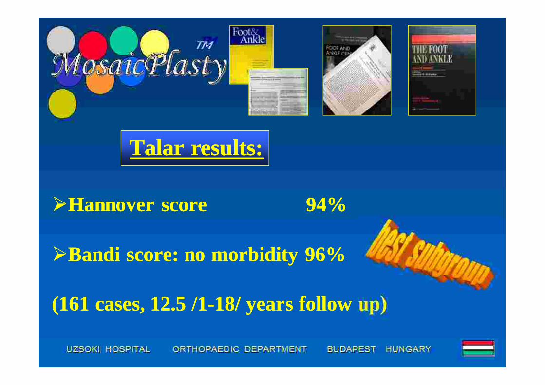
Talar results:Talar results:
Hannover score 94%
Bandi score: no morbidity 96%
(161 cases, 12.5 /1-18/ years follow up)
Hannover score 94%
Bandi score: no morbidity 96%
(161 cases, 12.5 /1-18/ years follow up)

Complications:Complications:
4 deep septic complications
8 deep venous thromboses
4 deep septic complications
8 deep venous thromboses

Donor site morbidity:Donor site morbidity:
long term morbidity: 3 % (Bandi score)
early morbidity - postop. bleeding 7 % !!!
long term morbidity: 3 % (Bandi score)
early morbidity - postop. bleeding 7 % !!!
Hangody, L.: Autologousosteochondral mosaicplasty.Actualités en biomatériaux, VolumeV.: 155-161,2000

Feczkó, P. – Hangody, L. – Varga, J. etal: Experimental results of donor sitefilling for autologous osteochondralmosaicplasty. Arthroscopy 19(7): 755-761, 2003
• polylactate, polygluconate-B, carbon rods, polycaprolactone, hydroxilapatite, compressed collagen• German Shepherd dogs, horses, 1 year follow up
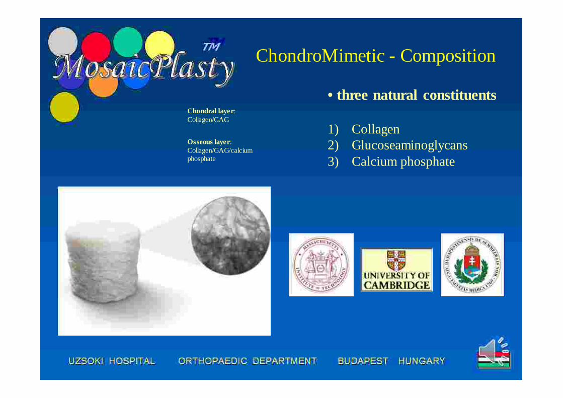
ChondroMimetic - Composition
Chondral layer: Collagen/GAG
Osseous layer: Collagen/GAG/calcium phosphate
• three natural constituents
1) Collagen2) Glucoseaminoglycans3) Calcium phosphate

Mode of implantation
47
Arthroscopic delivery device
Single step arthroscopic delivery
• Implant is available in 8mm, 10mm & 12mm diameter sizes
• All in one single use procedure pack comprising of site preparation instruments & a pre loaded delivery device
Procedure pack

Chondromimetic for Small Osteochondral Defects
Chondromimetic for Small Osteochondral Defects
Technique Technique

Histological evaluation – implant degradation, bone formation:
DMMB4x - bone
DMMB4x – bone/fibrocartilage

Histological evaluation – fibrocartilage formation:
PS10x – bone/fibrocartilage
DMMB4x - fibrocartilage

Mosaicplasty in 354 athletes (3 institutes):Mosaicplasty in 354 athletes (3 institutes):
soccerhandballwaterpolowrestlinggymnastothers

Defect location:Defect location:MFC
LFC
LTC
PF
talus
capitellumhumeri

Male : femaleMale : female
MaleFemale

MP results in 354 athletes:MP results in 354 athletes:* average defect size: 2.5 cm2 (1.0-5.5 cm2)* age: 24.3 (14-49) years* 185:169 male-female ratio* two third osteochondral defects, one third cartilage lesions* in 43% slight or moderate osteoarthritic changes praeoperatively !
* average defect size: 2.5 cm2 (1.0-5.5 cm2)* age: 24.3 (14-49) years* 185:169 male-female ratio* two third osteochondral defects, one third cartilage lesions* in 43% slight or moderate osteoarthritic changes praeoperatively !
Hangody, L. – Dobos, J. – Baló, E. – Pánics, G. – Hangody, L., R. – Berkes, I.: Clinical experiences with autologous osteochondral mosaicplasty in athletic population – a 17-years prospective multicenter study. American Journal of Sporstmedicine 38:1125-1133, 2010

MP results in 354 athletes:MP results in 354 athletes:* 9.6 ys. (2-17 ys.) follow up* no septic or thromboembolic complications * average 4.9 months rehabilitation period to return to the sports* 64 % return to same level sports activity* 19 % return to a lower level sports activity (incl. hobby sports)* 17 % no more sports activity (8% worse than praeop.)
* 9.6 ys. (2-17 ys.) follow up* no septic or thromboembolic complications * average 4.9 months rehabilitation period to return to the sports* 64 % return to same level sports activity* 19 % return to a lower level sports activity (incl. hobby sports)* 17 % no more sports activity (8% worse than praeop.)

?

Viewpoints at different cartilage repair techniques:
Viewpoints at different cartilage repair techniques:
optimal indication of the actual procedure (size, type and location of the defect; age; high or low demand patient; etc.)combination with other procedureslength of rehabilitation costs experience of the surgeon
optimal indication of the actual procedure (size, type and location of the defect; age; high or low demand patient; etc.)combination with other procedureslength of rehabilitation costs experience of the surgeon


26 ys. old gymnast2 sqcm cartilage defecton the MFC, torn ACLarthroscopic MP andBTB ACL MP 6 ys.beforefull recovery in 5ms.
26 ys. old gymnast2 sqcm cartilage defecton the MFC, torn ACLarthroscopic MP andBTB ACL MP 6 ys.beforefull recovery in 5ms.
Case report No. 1.Case report No. 1.

27 ys. old soccerplayer3.5 sqcm cartilagedefect on the LFCminiarthrotomy MP 2.5 ys. beforefull recovery
27 ys. old soccerplayer3.5 sqcm cartilagedefect on the LFCminiarthrotomy MP 2.5 ys. beforefull recovery
Case report No. 2.Case report No. 2.

Conclusions:
Autologous osteochondral mosaicplastymay be an alternative in the treatmentof small and medium sized focalchondral and osteochondral defects ofthe weight bearing surfaces.
- Hyaline like resurfacement- One step procedure- Combination with other procedures- Arthroscopic or minimal invasive
approach- Short rehabilitation